Embed Size (px)
Citation preview
Ultrasonics 81 (2017) 86–92
Contents lists available at ScienceDirect
Ultrasonics
journal homepage: www.elsevier .com/ locate/ul t ras
Noninvasive measurement of wave speed of porcine cornea in ex vivoporcine eyes for various intraocular pressures
http://dx.doi.org/10.1016/j.ultras.2017.06.0080041-624X/� 2017 Elsevier B.V. All rights reserved.
⇑ Corresponding author at: Department of Radiology, Mayo Clinic, 200 1st St SW,Rochester, MN 55905, USA.
E-mail address: [email protected] (X. Zhang).
Boran Zhou a, Arthur J. Sit b, Xiaoming Zhang a,c,⇑aDepartment of Radiology, Mayo Clinic, USAbDepartment of Ophthalmology, Mayo Clinic, USAcDepartment of Biomedical Engineering and Physiology, Mayo Clinic, USA
a r t i c l e i n f o
Article history:Received 8 March 2017Received in revised form 30 May 2017Accepted 5 June 2017Available online 7 June 2017
Keywords:Surface wave elastographyWave speedCorneaUltrasoundIntraocular pressure
a b s t r a c t
The objective of this study was to extend an ultrasound surface wave elastography (USWE) technique fornoninvasive measurement of ocular tissue elastic properties. In particular, we aim to establish the rela-tionship between the wave speed of cornea and the intraocular pressure (IOP). Normal ranges of IOP arebetween 12 and 22 mmHg. Ex vivo porcine eye balls were used in this research. The porcine eye ball wassupported by the gelatin phantom in a testing container. Some water was pour into the container for theultrasound measurement. A local harmonic vibration was generated on the side of the eye ball. An ultra-sound probe was used to measure the wave propagation in the cornea noninvasively. A 25 gauge butterflyneedle was inserted into the vitreous humor of the eye ball under the ultrasound imaging guidance. Theneedle was connected to a syringe. The IOP was obtained by the water height difference between thewater level in the syringe and the water level in the testing container. The IOP was adjusted between5 mmHg and 30 mmHg with a 5 mmHg interval. The wave speed was measured at each IOP for three fre-quencies of 100, 150 and 200 Hz. Finite element method (FEM) was used to simulate the wave propaga-tion in the corneal according to our experimental setup. A linear viscoelastic FEM model was used tocompare the experimental data. Both the experiments and the FEM analyses showed that the wave speedof cornea increased with IOP.
� 2017 Elsevier B.V. All rights reserved.
1. Introduction
Glaucoma is the second leading cause of blindness in the world.In the United States, more than 120,000 patients are blind fromglaucoma [1]. The IOP is strongly related with the prevalence andseverity of optic nerve axon damage in open angle glaucoma [2].The normal range of IOP is between 12 and 22 mmHg. Elevationin IOP is associated with increasing prevalence of glaucoma andreduction of IOP is able to slow the disease progression [3]. Cur-rently, reduction of IOP is the only available clinical treatment.However, a significant number of glaucoma patients continue todevelop vision loss and blindness despite this treatment [4]. In astudy, 738 eyes were categorized into three groups based on IOPdeterminations over the first three 6-month follow-up visits. Itwas found that eyes with early average IOP greater than17.5 mmHg had an estimated worsening during subsequentfollow-up that was 1 unit of visual field defect score greater than
eyes with average IOP less than 14 mmHg (p = 0.002). Contrari-wise, some patients with elevated IOP do not develop glaucoma[5]. These incongruities could arise due to alteration in stiffnessof the ocular tissues and their response to elevated IOP. These fac-tors are material determinants of IOP-derived lamina strain whichis related to glaucoma occurrence [6]. Elevated IOP induces disten-tion of the sclera and lamina cribrosa and impose direct mechani-cal strain on the axons of the optic nerve damaging nerve fibersand potentially impairing the nerve’s blood supply. Mechanicalstrain sustained by the optic nerve can also lead to thinning ofthe lamina cribrosa and increase in the translaminar pressure gra-dient, potentially impairing retrograde transport of neurotrophicfactors from the lateral geniculate nucleus to the retinal ganglioncells [7]. Abnormal biomechanical properties of ocular tissuesmay also give rise to greater IOP variability, which has beenincreasingly recognized as another risk factor for glaucoma [8–10].
We have developed an ultrasound surface wave elastography(USWE) technique to characterize the biomechanical propertiesof superficial and deeper tissues, such as skin [11], lung [12], andtendon [13]. Currently, there is no non-invasive technique clini-cally available for evaluating the in vivo biomechanical tissue prop-
B. Zhou et al. / Ultrasonics 81 (2017) 86–92 87
erties of the eye. The purpose of this work is to noninvasively mea-sure the wave speed of cornea in ex vivo porcine eyes and developthe relationship between the wave speed in cornea with IOP in thephysiology ranges.
Porcine ocular tissues are commonly used to evaluate themechanical behavior of various ocular tissues due to its similaritieswith human ocular tissues [14]. Moreover, with its availability, rel-evant animal models for systemic study of induced disease states,such as ocular hypertension, can be generated. Published investi-gations on various ocular tissues include optic nerve head[15,16], sclera [17], and laminar cribrosa [18].
Numerous elastography techniques have been developed tostudy soft tissues such as livers, however, only limited applicationsare published on ocular tissues [19–21]. The aim of this study wasto develop a noninvasive method for measuring the wave speed ofcornea for various IOP using porcine eye balls. In USWE, a shakerwas used to generate a small, local, short vibration on the surfaceof the eye, and the wave propagation in the ocular tissues wasmeasured by using ultrasonic imaging and analysis.
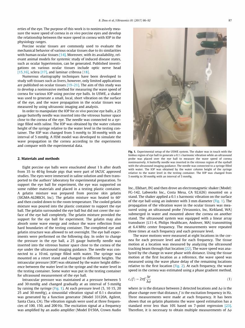
In order to manipulate the IOP for ex vivo porcine eye balls, a 25gauge butterfly needle was inserted into the vitreous humor spaceclose to the cornea of the eye. The needle was connected to a syr-inge filled with saline. The IOP was obtained by the water columnheight of the syringe relative to the water level in the testing con-tainer. The IOP was changed from 5 mmHg to 30 mmHg with aninterval of 5 mmHg. A FEM model was developed to simulate thewave propagation in the cornea according to the experimentsand compare with the experimental data.
Fig. 1. Experimental setup of the USWE system. The shaker was in touch with thelimbus region of eye ball to generate a 0.1 s harmonic vibration while an ultrasoundprobe was placed over the eye ball to measure the wave speed of corneanoninvasively. A butterfly needle was inserted in the vitreous region of the eyeballwith the ultrasound imaging guidance. The needle was connected to a syringe filledwith water. The IOP was obtained by the water column height of the syringerelative to the water level in the testing container. The IOP was changed from5 mmHg to 30 mmHg with an interval of 5 mmHg.
2. Materials and methods
Eight porcine eye balls were enucleated about 1 h after deathfrom 35 to 40 kg female pigs that were part of IACUC approvedstudies. The eyes were immersed in saline solution and then trans-ported to the authors’ laboratory for experimental preparation. Tosupport the eye ball for experiment, the eye was supported onsome rubber materials and placed in a testing plastic container.A gelatin mixture was prepared from porcine skin gelatin(SIGMA-ALDRICH, Inc). The gelatin mixture was heated to 60�and then cooled down to the room temperature. The cooled gelatinmixture was poured into the plastic container to support the eyeball. The gelatin surrounded the eye ball but did not cover the sur-face of the eye ball completely. The gelatin mixture provided thesupport for the eye ball for experiment. The gelatin may alsoabsorb some wave energy and reduce the wave reflection fromhard boundaries of the testing container. The completed eye andgelatin structure was allowed to set overnight. The eye ball exper-iments were performed on the following day. In order to changethe pressure in the eye ball, a 25 gauge butterfly needle wasinserted into the vitreous humor space close to the cornea of theeye under the ultrasound imaging guidance. The needle was con-nected to a 10 mL syringe filled with water. The syringe wasmounted on a retort stand and changed to different heights. Theintraocular pressure (IOP) was obtained by the water height differ-ence between the water level in the syringe and the water level inthe testing container. Some water was put in the testing containerfor ultrasound measurement of the eye ball.
Intraocular pressure was maintained at a pressure between 5and 30 mmHg and changed gradually at an interval of 5 mmHgby raising the syringe (Fig. 1). At each pressure level (5, 10 15, 2025 and 30 mmHg), a sinusoidal vibration signal of 0.1 s durationwas generated by a function generator (Model 33120A, Agilent,Santa Clara, CA). The vibration signals were used at three frequen-cies of 100, 150, and 200 Hz. The excitation signal at a frequencywas amplified by an audio amplifier (Model D150A, Crown Audio
Inc., Elkhart, IN) and then drove an electromagnetic shaker (Model:FG-142, Labworks Inc., Costa Mesa, CA 92,626) mounted on astand. The shaker applied a 0.1 s harmonic vibration on the surfaceof the eye ball using an indenter with 3 mm diameter (Fig. 1). Thepropagation of the vibration wave in the ocular tissues was mea-sured using an ultrasound probe (Verasonics, Inc, Kirkland, WA)submerged in water and mounted above the cornea on anotherstand. The ultrasound system was equipped with a linear arraytransducer (L11-4, Philips Healthcare, Andover, MA) transmittingat 6.4 MHz center frequency. The measurements were repeatedthree times at each frequency and each pressure level.
The wave motions were measured at eight locations in the cor-nea for each pressure level and for each frequency. The tissuemotion at a location was measured by analyzing the ultrasoundtracking beam through that location [22]. The wave speed was ana-lyzed by the change in wave phase with distance. Using the tissuemotion at the first location as a reference, the wave speed wasmeasured using the wave phase delay of the remaining locationsrelative to the first location (Fig. 2). At each frequency, the wavespeed in the cornea was estimated using a phase gradient method.
cs fð Þ ¼ 2pf DrD/
ð1Þ
where Dr is the distance between 2 detected locations and D/ is thephase change over that distance, f is the excitation frequency in Hz.Three measurements were made at each frequency. It has beenshown that on gelatin phantoms the wave speed estimation has astandard error less than 10% based on 7-point regression [23].Therefore, it is necessary to obtain multiple measurements of D/

cornea
gel
selected points
Fig. 2. Representative B-mode images of porcine ocular tissue. Eight locations inthe cornea were selected to measure the wave speed in cornea by using theultrasound tracking method. Blue dots indicate the points selected for measure-ment. (For interpretation of the references to colour in this figure legend, the readeris referred to the web version of this article.)
Fig. 3. FEM modeling of the porcine eye balls according to experimental setup.
88 B. Zhou et al. / Ultrasonics 81 (2017) 86–92
and a statistical regression to get accurate estimates of wave speed.Ideally, the phase of the surface wave / at a particular frequencyhas a linear relationship with the distance. The wave speed wasmeasured over eight locations in the central region of the corneaand analyzed with a linear regression curve obtained using aleast-squares fitting technique on multiple D/ measurements,
D/̂ ¼ �aDr þ b; ð2Þ
where D/̂denotes the linear regression value of multiple D/ mea-surements, a and b are regression parameters, and the wave speedis calculated as follows,
cs fð Þ ¼ 2pfa
ð3Þ
The quality of the measurement of wave speed was assured bythe sum of squares of linear regression residuals (R2) being �0.8[22]. R2 is the coefficient of determinant ranging from 0 to 1, a sta-tistical measure of how close the data fit the regression line. Ingeneral, the higher the R2, the better the model fits the data.
3. Numerical modeling
A FEMmodel was conducted as shown in Fig. 3 in ABAQUS (ver-sion 6.12-1, 3DS Inc, Waltham, MA). The eye and surrounding gela-tin was simulated by a 2D planar model of an infinite elasticmedium with the density of 1000 kg m�3. Representative geomet-ric model was reconstructed from image analysis of the surfaceprofile at a reference pressure of 0 mmHg: The white-to-white cor-neal diameter was 13.5 mm [24]. The central cornea thickness was0.6 mm based on the ultrasound measurements; its radius of cur-vature was 7.5 mm and eccentricity was 0.5. The sclera radius ofcurvature was 12.8 mm, the anterior thickness was 0.88 mm, theequatorial thickness was 1.02 mm, and the posterior thicknesswas 1.45 mm.
The cornea and sclera were divided by a radial line through thecenter of the sclera ellipse. Vitreous humor fills, composed largelyof hyaluronic acid, fill the posterior segment of the eye and weremodelled as an acoustic medium. The cornea and sclera were mod-
elled using a nearly incompressible, linear, viscoelastic generalizedMaxwell model. The constitutive relationship is defined as follows[25]:
rðtÞ ¼ Eequ � eðtÞ þXm
i¼1
Ei � _e � si � 1� e�tsi
� �ð4Þ
where Eequ is the equilibrium modulus, Ei is the relaxation modulusof the i-th branch. si is the time constant of the i-th branch, e is thestrain, and _e is the strain rate. In this study, a two branch model (i.e.,m = 2) was adopted [26]. The time constants (s1 and s2) are denotedas the short term time constant (ss) and the long time term constant(sl). The instantaneous (Einst) is defined as [27]:
Einst ¼ r�jt¼0 ð5Þ
The branch relaxation modulus were set to be equal (i.e., E1 ¼ E2).Thus, the branch relaxation moduli can be found as:
E1 ¼ E2 ¼ Einst � Eequ
2ð6Þ
It has been shown that sclera has stiffer response than cornea.So the instantaneous and equilibrium moduli of the sclera wereset to be 5 times of those of the cornea [28–30]. To the best ofour knowledge, viscoelastic properties of the porcine cornea andsclera have not been measured. The values of the material param-eters of ocular shell and vitreous humor were estimated based onapproximation of the model predictions with the results obtainedfrom the infusion experiments in porcine eyes and were summa-rized in Table 1 [28].
The model was excited using a line source in the sclera and thedisplacement was applied in the radial direction. Harmonic excita-tions were performed at 100, 150, and 200 Hz with a duration of0.1 s. A uniform pressure was applied on the inner surfaces ofthe cornea and sclera in the direction normal to the corneal andscleral surfaces at each point. The range of IOP was set between5 and 30 mmHg at an interval of 5 mmHg. The boundary of gelwas attached to an infinite region to minimize the wavereflections.
The mesh of the cornea and sclera, as well as the gel, were con-structed using linear quadrilateral elements (type CPS4R) with size0.25 mm � 0.25 mm for cornea and 0.5 mm � 0.5 mm for scleraand gel, enhanced with hourglass control and reduced integration,to minimize shear locking and hourglass effects. The vitreousmaterial was meshed using linear triangular acoustic elements(type AC2D3) with size 0.5 mm � 0.5 mm The infinite region was

Table 1Material parameters for the finite element model of the gelled porcine eye ball.
Material Einst [MPa] Eequ [MPa] ss [sec] sl [sec] Bulk modulus [GPa]
Cornea 1.1 0.265 0.35 68Sclera 5.5 1.325 0.35 68Vitreous 2.1404Gel
B. Zhou et al. / Ultrasonics 81 (2017) 86–92 89
meshed by infinite elements (type CINPE4) (Fig. 3). The dynamicresponses of the tissue model to the excitations were solved bythe ABAQUS explicit dynamic solver with automatic step size con-trol. Mesh convergence tests were performed so that further refin-ing the mesh did not change the solution significantly.
4. Results
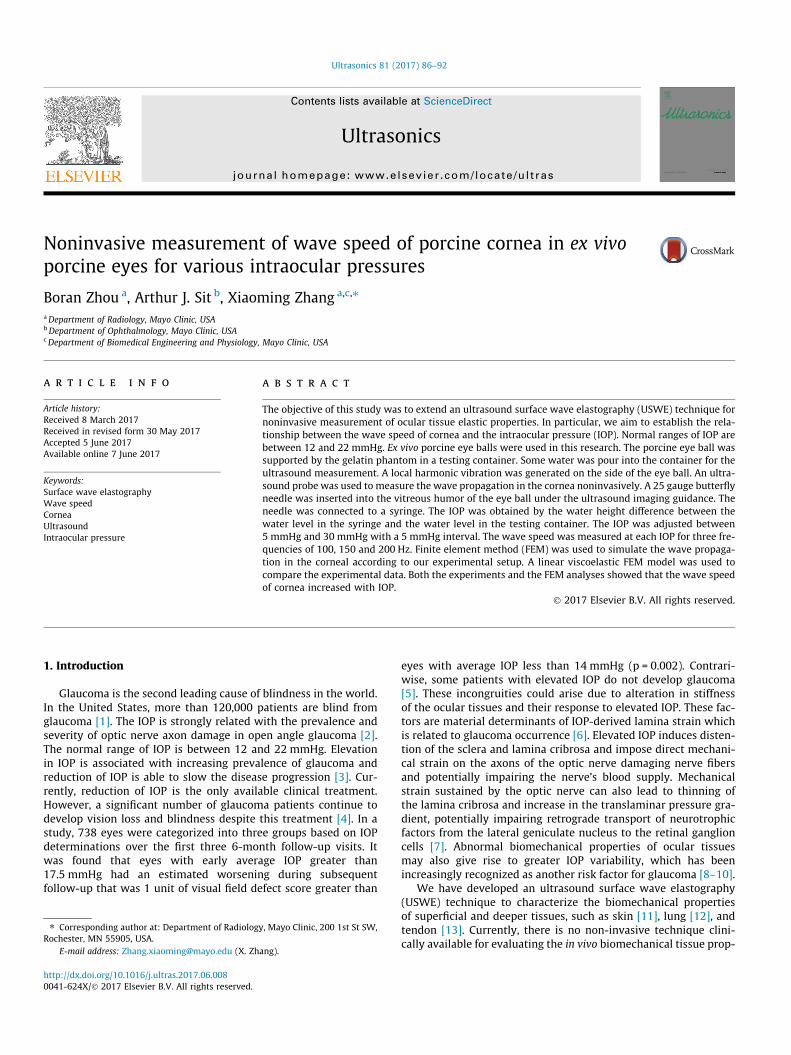
Eight porcine eyes were evaluated in this study. The tissuemotion in response to the vibration at different excitation frequen-cies (100, 150, and 200 Hz) was detected with a high frame rate of2000 frame/s. The wave speed is shown with 95% confidence inter-val, mean ± standard error (Fig. 4a–c). Fig. 7 shows the relationshipbetween wave speed and IOP in a representative experiment. Wavespeed in the cornea increased with frequency at each level of IOP,from 0.83 m/s at 100 Hz to 1.96 m/s at 200 Hz (Fig. 7). At the samefrequency, the wave speed increased with IOP, from 0.83 to 1.07 m/s at 100 Hz, from 0.86 to 1.29 m/s at 150 Hz and from 1.11 to1.96 m/s at 200 Hz (Fig. 7).
FEM analysis of gelled porcine eyes submerged in water wasused to investigate the effects of IOP on the wave speed in cornea.Harmonic excitation was used to propagate waves in the eye ball.The plane waves were excited by vibrating a segment of elementson the sclera perpendicular to the direction of wave propagation.Vitreous humor behaves as a fluid-like material. Acoustic mediumis used to model sound propagation problems in usually a fluid inwhich stress is purely hydrostatic (no shear stress). In this study,we think it is appropriate to model vitreous humor with acousticmedium. There is no elastic wave propagation in the vitreoushumor space as shown in Fig. 5. As the boundary of the gel wasassigned infinite elements, there is no wave reflection in theboundary. The temporal-spatial displacement field of a central seg-ment of cornea was extracted to minimize the influence of bound-ary effects. It showed that the wave pattern became denser withincreasing excitation frequency. 2D-FFT of the displacement versustime data was performed using
UyðK; FÞ ¼Xþ1
m¼�1
Xþ1
n¼�1uyðx; tÞe�j2pðKmxþFntÞ; ð7Þ
where uyðx; tÞ is the motion of the cornea perpendicular to the exci-tation as a function of distance from the excitation (x) and time (t).Here, K is the wave number and F is the temporal frequency of thewave. The coordinates of the k-space are the wave number (K) andthe frequency (F) [31]. The wave velocity is calculated,
�c ¼ FK; ð8Þ
(Fig 6). From the numerical simulation, it showed that the wavespeed in the cornea increased with IOP and excitation frequencyas well as fit well with the representative experimental measure-ments (Fig 7). Fig. 8 shows the relationship between wave speedand IOP from all experimental measurements. Wave speed in thecornea increased with frequency from 0.73 at 100 Hz to 1.93 m/sat 200 Hz (Fig. 8). At the same frequency, the wave speed increasedwith IOP from 0.78 to 1.02 m/s at 100 Hz, from 0.92 to 1.42 m/s at150 Hz and from 1.17 to 1.93 m/s at 200 Hz (Fig. 8).
5. Disuccsion
The aim of this study was to understand the relationshipbetween the wave speed in cornea with the IOP in ex vivo procineeye ball models. The level of IOP was adjusted by lifting the relativeheight between the eye and the water level in the syringe con-nected via a needle. At each pressure level, a shaker was used togenerate a harmonic mechanical vibration on the surface of theeye ball at three frequencies (100, 150, and 200 Hz). The resultingwave propagation in the corneal was noninvasively measuredusing an ultrasound technique. In this study, the wave propagationin the cornea was measured at three frequencies of 100 Hz, 150 Hzand 200 Hz. The magnitude of 100 Hz wave motion is strongerthan the 150 and 200 Hz wave motion. The wave length is inver-sely proportional to the wave frequency while attenuation riseswith the wave frequency. The frequency ranges chosen in thispaper consider the wave motion amplitude, spatial resolutionand wave attenuation. The wave speed in the cornea was deter-mined by analyzing ultrasound data directly from the cornea.Therefore, the wave speed measurement is local and independentof the location of excitation. A FEM model was built according tothe experiments to simulate the wave propagation in the cornea.We found that the wave speed increased with IOP at three differentexcitation frequencies both from experiments and FEM simulation.
The results from this study are within the range of publishedresults for other species. The wave speed in the porcine corneawas found to be from 0.72 to 1.93, which is similar to valuesreported for the rabbit cornea, indicating that the wave velocitieswere 1.14 ± 0.08 m/s and 1.30 ± 0.10 m/s in young and mature rab-bit corneas [32]. Shear wave speed of corneas in central regions onex vivo porcine model was approximately 2.3 m/s at 10 mmHg ofIOP [19]. In their study, they placed it in refrigerator prior to testingwhich may lead to stiffening of cornea and elevation of the magni-tude of corneal shear wave speed. The biomechanical properties ofhuman and porcine corneas were evaluated showing that humanand porcine corneas have almost the same biomechanical behaviorunder short and long-term loading, yet human corneas were stifferthan porcine corneas [33]. This research may be useful for us toanalyze the in vivo data of the wave speed of cornea betweenhealthy subjects and glaucoma patients. It is difficult to manipulatethe IOP in human subjects.
It has been shown that acoustic properties of material arealtered with deformation and pressurization, correspondinglychanging the wave propagation speed or reflected wave amplitudein the material [34,35]. Numerous studies have shown the relation-ship between the echo intensity and the stress or strain experi-enced by the isolated soft tissues under static or cyclic loadingscenarios [36,37]. Moreover, local speed of wave propagation isdirectly linked to local stiffness [38]. Corneal stiffness is a functionof intraocular pressure and increases with IOP [39]. In patientswith glaucoma, the cornea was in compression state due to theincrease of IOP. Our results in this ex vivo porcine eye modelshowed that the wave speed of cornea increased with the IOP.For the glaucoma diagnosis and management, the fine tuning cor-neal wave speedmeasurement could also correlate the applanationtonometry with the IOP.
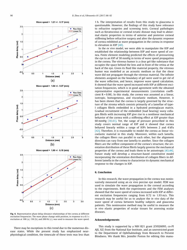
Fig. 4. Representative phase delay-distance relationships of the cornea at differentexcitation frequencies. The wave phase change with position, in response to a 0.1 sexcitation at 100 (a), 150 (b), and 200 Hz (c), was used to measure the wave speed.
90 B. Zhou et al. / Ultrasonics 81 (2017) 86–92
There may be exceptions to this trend due to the numerous dis-ease states. While the present study has emphasized near-physiological condition, the timescale of these tests was less than
1 h. The interpretation of results from this study to glaucoma isquestionable. However, the findings of this study have relevanceto refractive surgeries and screening tests. Corneal pathologiessuch as Keratoconus or corneal ectatic disease may lead to abnor-mal elastic properties in terms of anterior and posterior cornealstiffening before refractive surgery and alter the dynamic responseof cornea exhibited as wave propagation in the cornea in responseto elevation in IOP [40].
In the ex vivo model, we were able to manipulate the IOP andestablished the relationship between IOP and wave speed of cor-nea. Finite element modeling predicted the effects of pressurizingthe eye to an IOP of 30 mmHg in terms of wave speed propagationin the cornea. The vitreous humor is a clear gel-like substance thatoccupies the space behind the lens and in front of the retina at theback of the eye. Given its fluid like material property, the vitreoushumor was modelled as an acoustic medium so that the shearwave did not propagate through the vitreous material. The infiniteelements assigned on the boundary of gel were used to get rid ofthe wave reflection, and hence, improve wave speed calculation.It showed that the wave speed increased with IOP at different exci-tation frequencies, which is in good agreement with the obtainedrepresentative experimental measurements (correlation coeffi-cient R = 0.98). In this study, the cornea was assumed as a linear,isotropic, homogeneous, and viscoelastic medium. However, ithas been shown that the cornea is largely governed by the struc-ture of the stroma which consists primarily of a lamellae of type-I collagen fibrils embedded in a hydrated proteoglacan matrix.Gradual recruitment of the embedded (load bearing) wavy colla-gen fibrils with increasing IOP give rise to a nonlinear mechanicalbehavior of the cornea with a stiffening effect at IOP greater than60 mmHg [30,41]. Yet, the range of pressure prescribed in thisstudy covers normal range of IOP and it showed that corneabehaved linearly within a range of IOPs between 2 and 4 kPa[42]. Therefore, it is reasonable to model the cornea as linear vis-coelastic material in this study. Moreover, within each lamella,the collagen fibers run parallel to each other, but the alignmentdirection can vary from one lamella to another. Since the collagenfibers are the stiffest component of the cornea’s structure, the ori-entation distribution of these fibrils largely governs the mechanicalproperties of the cornea and leads them to be anisotropic [43,44].Future study will develop a structure-based constitutive modelincorporating the orientation distribution of collagen fibers in dif-ferent lamella in the cornea to characterize its dynamic mechanicalresponse to the changes in IOP.
6. Conclusion
In this research, the wave propagation in the cornea was nonin-vasively measured using an ex vivo porcine eye model. FEM wasused to simulate the wave propagation in the corneal accordingto the experiments. Both the experiments and the FEM analysesshowed that the wave speed of cornea increased with IOP at differ-ent excitation frequencies ranging from 0.72 to 1.93 m/s. Thisresearch may be useful for us to analyze the in vivo data of thewave speed of cornea between healthy subjects and glaucomapatients. This noninvasive method may be useful to measure thein vivo elastic properties of ocular tissues for assessing oculardiseases.
Acknowledgements
This study is supported by a NIH R21 grant (EY026095, co-PIAJS, XZ) from the National Eye Institute, and an unrestricted grantto the Department of Ophthalmology from Research to PreventBlindness. We thank Mrs. Jennifer Poston for editing this manu-
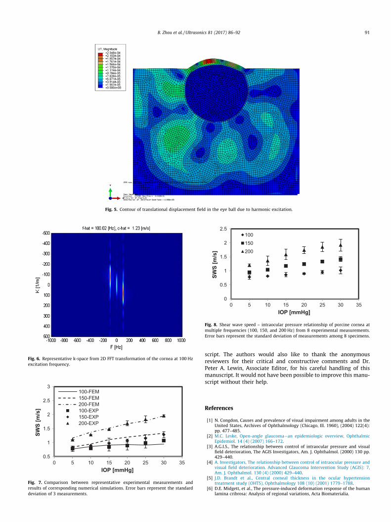
Fig. 5. Contour of translational displacement field in the eye ball due to harmonic excitation.
Fig. 6. Representative k-space from 2D FFT transformation of the cornea at 100 Hzexcitation frequency.
0.5
1
1.5
2
2.5
3
0 5 10 15 20 25 30 35
SWS
[m/s
]
IOP [mmHg]
100-FEM 150-FEM 200-FEM 100-EXP 150-EXP 200-EXP
Fig. 7. Comparison between representative experimental measurements andresults of corresponding numerical simulations. Error bars represent the standarddeviation of 3 measurements.
0
0.5
1
1.5
2
2.5
0 5 10 15 20 25 30 35
SWS
[m/s
]
IOP [mmHg]
100 150 200
Fig. 8. Shear wave speed – intraocular pressure relationship of porcine cornea atmultiple frequencies (100, 150, and 200 Hz) from 8 experimental measurements.Error bars represent the standard deviation of measurements among 8 specimens.
B. Zhou et al. / Ultrasonics 81 (2017) 86–92 91
script. The authors would also like to thank the anonymousreviewers for their critical and constructive comments and Dr.Peter A. Lewin, Associate Editor, for his careful handling of thismanuscript. It would not have been possible to improve this manu-script without their help.
References
[1] N. Congdon, Causes and prevalence of visual impairment among adults in theUnited States, Archives of Ophthalmology (Chicago, Ill. 1960), (2004) 122(4):pp. 477–485.
[2] M.C. Leske, Open-angle glaucoma—an epidemiologic overview, OphthalmicEpidemiol. 14 (4) (2007) 166–172.
[3] A.G.I.S., The relationship between control of intraocular pressure and visualfield deterioration, The AGIS Investigators, Am. J. Ophthalmol. (2000) 130 pp.429–440.
[4] A. Investigators, The relationship between control of intraocular pressure andvisual field deterioration. Advanced Glaucoma Intervention Study (AGIS): 7,Am. J. Ophthalmol. 130 (4) (2000) 429–440.
[5] J.D. Brandt et al., Central corneal thickness in the ocular hypertensiontreatment study (OHTS), Ophthalmology 108 (10) (2001) 1779–1788.
[6] D.E. Midgett, et al., The pressure-induced deformation response of the humanlamina cribrosa: Analysis of regional variations, Acta Biomaterialia.

92 B. Zhou et al. / Ultrasonics 81 (2017) 86–92
[7] M. Salinas-Navarro et al., Ocular hypertension impairs optic nerve axonaltransport leading to progressive retinal ganglion cell degeneration, Exp. EyeRes. 90 (1) (2010) 168–183.
[8] S. Asrani et al., Large diurnal fluctuations in intraocular pressure are anindependent risk factor in patients with glaucoma, J. Glaucoma 9 (2) (2000)134–142.
[9] J. Caprioli, A.L. Coleman, Intraocular pressure fluctuation: a risk factor forvisual field progression at low intraocular pressures in the Advanced GlaucomaIntervention Study, Ophthalmology. (2008) 115(7): p. 1123–1129. e3.
[10] D.C. Musch et al., Intraocular pressure control and long-term visual field loss inthe collaborative initial glaucoma treatment study, Ophthalmology 118 (9)(2011) 1766–1773.
[11] X. Zhang et al., Quantitative assessment of scleroderma by surface wavetechnique, Med. Eng. Phys. 33 (1) (2011) 31–37.
[12] X. Zhang et al., Noninvasive ultrasound image guided surface wave method formeasuring the wave speed and estimating the elasticity of lungs: a feasibilitystudy, Ultrasonics 51 (3) (2011) 289–295.
[13] Y. Wang et al., A non-invasive technique for estimating carpal tunnel pressureby measuring shear wave speed in tendon: a feasibility study, J. Biomech. 45(16) (2012) 2927–2930.
[14] Y. Zeng et al., A comparison of biomechanical properties between human andporcine cornea, J. Biomech. 34 (4) (2001) 533–537.
[15] I.A. Sigal et al., Finite element modeling of optic nerve head biomechanics,Invest. Ophthalmol. Vis. Sci. 45 (12) (2004) 4378–4387.
[16] A.J. Feola et al., Finite element modeling of factors influencing optic nerve headdeformation due to intracranial pressureICP affects ONH deformation, Invest.Ophthalmol. Vis. Sci. 57 (4) (2016) 1901–1911.
[17] R.E. Norman et al., Finite element modeling of the human sclera: influence onoptic nerve head biomechanics and connections with glaucoma, Exp. Eye Res.93 (1) (2011) 4–12.
[18] T. Newson, A. El-Sheikh, Mathematical modeling of the biomechanics of thelamina cribrosa under elevated intraocular pressures, J. Biomech. Eng. 128 (4)(2006) 496–504.
[19] T.-M. Nguyen et al., In Vivo Evidence of porcine cornea anisotropy usingsupersonic shear wave imaginganisotropy measured in porcine cornea usingSSI, Invest. Ophthalmol. Vis. Sci. 55 (11) (2014) 7545–7552.
[20] T.-M. Nguyen et al., Monitoring of cornea elastic properties changes duringUV-A/Riboflavin-Induced corneal collagen cross-linking using supersonicshear wave imaging: a pilot studymonitoring of cornea elastic propertychanges, Invest. Ophthalmol. Vis. Sci. 53 (9) (2012) 5948–5954.
[21] M. Tanter et al., High-resolution quantitative imaging of cornea elasticity usingsupersonic shear imaging, IEEE Trans. Med. Imaging 28 (12) (2009) 1881–1893.
[22] X. Zhang, T. Osborn, S. Kalra, A noninvasive ultrasound elastography techniquefor measuring surface waves on the lung, Ultrasonics 71 (2016) 183–188.
[23] X. Zhang, B. Qiang, J. Greenleaf, Comparison of the surface wave method andthe indentation method for measuring the elasticity of gelatin phantoms ofdifferent concentrations, Ultrasonics 51 (2) (2011) 157–164.
[24] B.C. Perez et al., Finite element modeling of the viscoelastic responses of theeye during microvolumetric changes, J. biomed. sci. eng. 6 (12A) (2013) 29.
[25] W.D. Callister, D.G. Rethwisch, Materials science and engineering. Vol. 5. 2011:John Wiley & Sons NY.
[26] A.J. Bellezza, C.F. Burgoyne, and R.T. Hart, Viscoelastic characterization ofperipapillary sclera: material properties by quadrant in rabbit and monkeyeyes. 2003.
[27] J.C. Downs et al., Viscoelastic material properties of the peripapillary sclera innormal and early-glaucoma monkey eyes, Invest. Ophthalmol. Vis. Sci. 46 (2)(2005) 540–546.
[28] H.J. Morris et al., Correlation between biomechanical responses of posteriorsclera and IOP elevations during micro intraocular volume change, Invest.Ophthalmol. Vis. Sci. 54 (12) (2013) 7215–7222.
[29] C.R. Ethier, M. Johnson, J. Ruberti, Ocular biomechanics and biotransport,Annu. Rev. Biomed. Eng. 6 (2004) 249–273.
[30] S.-Y. Woo et al., Nonlinear material properties of intact cornea and sclera, Exp.Eye Res. 14 (1) (1972) 29–39.
[31] M. Bernal et al., Material property estimation for tubes and arteries usingultrasound radiation force and analysis of propagating modes, J. Acoust. Soc.Am. 129 (3) (2011) 1344–1354.
[32] S. Wang, K.V. Larin, Shear wave imaging optical coherence tomography (SWI-OCT) for ocular tissue biomechanics, Opt. Lett. 39 (1) (2014) 41–44.
[33] A. Elsheikh, D. Alhasso, P. Rama, Biomechanical properties of human andporcine corneas, Exp. Eye Res. 86 (5) (2008) 783–790.
[34] H. Kobayashi, R. Vanderby, New strain energy function for acoustoelasticanalysis of dilatational waves in nearly incompressible, hyper-elasticmaterials, J. Appl. Mech. 72 (6) (2005) 843–851.
[35] H. Kobayashi, R. Vanderby, Acoustoelastic analysis of reflected waves in nearlyincompressible, hyper-elastic materials: forward and inverse problems, J.Acoust. Soc. Am. 121 (2) (2007) 879–887.
[36] L. Pan, L. Zan, F.S. Foster, Ultrasonic and viscoelastic properties of skin undertransverse mechanical stress in vitro, Ultrasound Med. Biol. 24 (7) (1998) 995–1007.
[37] S. Duenwald et al., Ultrasound echo is related to stress and strain in tendon, J.Biomech. 44 (3) (2011) 424–429.
[38] T. Deffieux et al., Assessment of the mechanical properties of themusculoskeletal system using 2-D and 3-D very high frame rate ultrasound,IEEE Trans. Ultrason. Ferroelectr. Freq. Control 55 (10) (2008) 2177–2190.
[39] D. Touboul et al., Supersonic shear wave elastography for the in vivoevaluation of transepithelial corneal collagen cross-linkingSSI evaluation ofTE-CXL, Invest. Ophthalmol. Vis. Sci. 55 (3) (2014) 1976–1984.
[40] E. Spoerl, T. Seiler, Techniques for stiffening the cornea, J. Refract. Surg. 15 (6)(1999) 711–713.
[41] D.A. Prim et al., A mechanical argument for the differential performance ofcoronary artery grafts, J. Mech. Behav. Biomed. Mater. 54 (2016) 93–105.
[42] K. Anderson, A. El-Sheikh, T. Newson, Application of structural analysis to themechanical behaviour of the cornea, J. R. Soc. Interface 1 (1) (2004) 3–15.
[43] T.M. Nejad, C. Foster, D. Gongal, Finite element modelling of corneamechanics: a review, Arquivos brasileiros de oftalmologia 77 (1) (2014) 60–65.
[44] T. Shazly et al., On the uniaxial ring test of tissue engineered constructs, Exp.Mech. 55 (1) (2015) 41–51.