Embed Size (px)
Citation preview
Remedy Publications LLC.
Journal of Heart and Stroke
2017 | Volume 2 | Issue 3 | Article 10261
IntroductionTrans thoracic echocardiography (TTE) study of the coronaries is a potentially important
diagnostic tool for coronary evaluation, both as a preliminary to an invasive procedure.TTE has become a relevant tool for coronary artery analysis, especially for their proximal course [1]. We reported the case of a non-STEMI event, in a patient with a hypoplastic left anterior descending coronary artery, earlier suspected after TTE.
Case PresentationA 60-year-old male, in generally good clinical conditions, non-smoker and carrier of a Gilbert
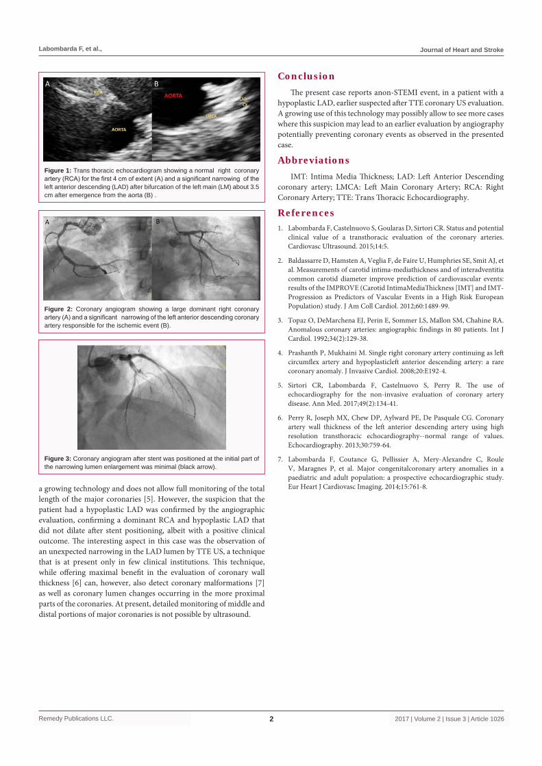
syndrome with modest hypercholesterolemia was followed by this Institution for over 10 years. His basal lipid levels were: total cholesterol 263, triglycerides 155, HDL 55 and Lp (a) 6 mg/dl. The patient was totally asymptomatic and was followed on a diet with phytosterols (1.6 g/day) keeping his LDL cholesterol around 100 mg/dl to 120 mg/dl. He had with no known allergies, while family history was negative for cardiovascular events and positive for dyslipidemia in his mother. He had recurrent prostatitis and had a prior episode of nephrolithiasis, with elimination of a kidney stone. In 2014, evaluation of the carotid intima media thickness (IMT) showed a mean IMT of 1.07 mm (60° to 70° percentile) and a IMT max 1.5 mm, (40° percentile). These are normal values in our institution and as well in the large IMPROVE study [2]. During this last carotid evaluation he underwent a non-invasive assessment of the coronaries by TTE. This evaluation noted a normal right coronary artery (RCA) for the first 4 cm of extent and, interestingly, a significant narrowing of the left anterior descending (LAD) after bifurcation of the left main coronary artery (LMCA) about 3.5 cm after emergence from the aorta (Figure 1). On January 2016, the patient reported acute chest pain and was treated for a non-STEMI with moderately elevated troponin levels. Upon coronary angiography, a large dominant RCA was noted (Figure 2A) and a significant narrowing of the LAD responsible for the ischemic event (Figure 3B). A stent was positioned at the initial part of the narrowing but interestingly, lumen enlargement was minimal (Figure 3B). He had an eventful dismissal on a combination of bisoprolol 1.25 mg/d, PY12 antagonist (prasugrel 10 mg/d) and, atorvastatin 20 mg/d.
DiscussionThis patient with normal carotid IMT, well controlled BP and lipid levels, underwent an
unexpected coronary event. The patient had a dominant RCA and a hypoplastic LAD that did not dilate after stent positioning. This rare type of coronary anomaly can cause angina-like symptoms and outright MI [3,4]. The patient, previously asymptomatic, presented with typical angina followed by a non-STEMI. Interestingly in the earlier TTE by ultrasound (US), there was evidence of a normal lumen LMCA, followed by narrowing of the LAD. Coronary evaluation with US is, at present, still
Non-STEMI from a Hypoplastic Left Anterior Descending Coronary: Earlier Assessment by Transthoracic
Coronaryultrasound Evaluation
OPEN ACCESS
*Correspondence:Labombarda F, Department of
Cardiology, CHU de Caen, Avenue Cote de Nacre, 14000 Caen, France, Tel:
(+33)-02-31-06-47-67;E-mail: [email protected]
Received Date: 09 Feb 2017Accepted Date: 04 May 2017Published Date: 12 May 2017
Citation: Sirtori CR, Castelnuovo S, Labombarda
F. Non-STEMI from a Hypoplastic Left Anterior Descending Coronary:
Earlier Assessment by Transthoracic Coronaryultrasound Evaluation. J Heart
Stroke. 2017; 2(3): 1026.ISSN: 2475-5702
Copyright © 2017 Labombarda F. This is an open access article distributed
under the Creative Commons Attribution License, which permits unrestricted
use, distribution, and reproduction in any medium, provided the original work
is properly cited.
Case ReportPublished: 12 May, 2017
AbstractTrans thoracic echocardiography study of the coronaries is a potentially important diagnostic tool for coronary evaluation, both as a preliminary to an invasive procedure. We reported the case of a non-STEMI event, in a patient with a hypoplastic left anterior descending coronary artery, earlier suspected after trans-thoracic echocardiogram.
Keywords: Coronary arteries; Echocardiography
Sirtori CR1, Castelnuovo S1 and Labombarda F2*1Department of Cardiology, University of Milano and Dyslipidemia Center, Italy
2Department of Cardiology, CHU de Caen, France
Labombarda F, et al., Journal of Heart and Stroke
Remedy Publications LLC. 2017 | Volume 2 | Issue 3 | Article 10262
a growing technology and does not allow full monitoring of the total length of the major coronaries [5]. However, the suspicion that the patient had a hypoplastic LAD was confirmed by the angiographic evaluation, confirming a dominant RCA and hypoplastic LAD that did not dilate after stent positioning, albeit with a positive clinical outcome. The interesting aspect in this case was the observation of an unexpected narrowing in the LAD lumen by TTE US, a technique that is at present only in few clinical institutions. This technique, while offering maximal benefit in the evaluation of coronary wall thickness [6] can, however, also detect coronary malformations [7] as well as coronary lumen changes occurring in the more proximal parts of the coronaries. At present, detailed monitoring of middle and distal portions of major coronaries is not possible by ultrasound.
Figure 1: Trans thoracic echocardiogram showing a normal right coronary artery (RCA) for the first 4 cm of extent (A) and a significant narrowing of the left anterior descending (LAD) after bifurcation of the left main (LM) about 3.5 cm after emergence from the aorta (B) .
Figure 2: Coronary angiogram showing a large dominant right coronary artery (A) and a significant narrowing of the left anterior descending coronary artery responsible for the ischemic event (B).
Figure 3: Coronary angiogram after stent was positioned at the initial part of the narrowing lumen enlargement was minimal (black arrow).
ConclusionThe present case reports anon-STEMI event, in a patient with a
hypoplastic LAD, earlier suspected after TTE coronary US evaluation. A growing use of this technology may possibly allow to see more cases where this suspicion may lead to an earlier evaluation by angiography potentially preventing coronary events as observed in the presented case.
AbbreviationsIMT: Intima Media Thickness; LAD: Left Anterior Descending
coronary artery; LMCA: Left Main Coronary Artery; RCA: Right Coronary Artery; TTE: Trans Thoracic Echocardiography.
References1. Labombarda F, Castelnuovo S, Goularas D, Sirtori CR. Status and potential
clinical value of a transthoracic evaluation of the coronary arteries. Cardiovasc Ultrasound. 2015;14:5.
2. Baldassarre D, Hamsten A, Veglia F, de Faire U, Humphries SE, Smit AJ, et al. Measurements of carotid intima-mediathickness and of interadventitia common carotid diameter improve prediction of cardiovascular events: results of the IMPROVE (Carotid IntimaMediaThickness [IMT] and IMT-Progression as Predictors of Vascular Events in a High Risk European Population) study. J Am Coll Cardiol. 2012;60:1489-99.
3. Topaz O, DeMarchena EJ, Perin E, Sommer LS, Mallon SM, Chahine RA. Anomalous coronary arteries: angiographic findings in 80 patients. Int J Cardiol. 1992;34(2):129-38.
4. Prashanth P, Mukhaini M. Single right coronary artery continuing as left circumflex artery and hypoplasticleft anterior descending artery: a rare coronary anomaly. J Invasive Cardiol. 2008;20:E192-4.
5. Sirtori CR, Labombarda F, Castelnuovo S, Perry R. The use of echocardiography for the non-invasive evaluation of coronary artery disease. Ann Med. 2017;49(2):134-41.
6. Perry R, Joseph MX, Chew DP, Aylward PE, De Pasquale CG. Coronary artery wall thickness of the left anterior descending artery using high resolution transthoracic echocardiography--normal range of values. Echocardiography. 2013;30:759-64.
7. Labombarda F, Coutance G, Pellissier A, Mery-Alexandre C, Roule V, Maragnes P, et al. Major congenitalcoronary artery anomalies in a paediatric and adult population: a prospective echocardiographic study. Eur Heart J Cardiovasc Imaging. 2014;15:761-8.