Embed Size (px)
Citation preview

Non Diabetic Endocrine Emergencies
Ping-Wei ChenDr. Stefan DaSilva
December 18th 2008
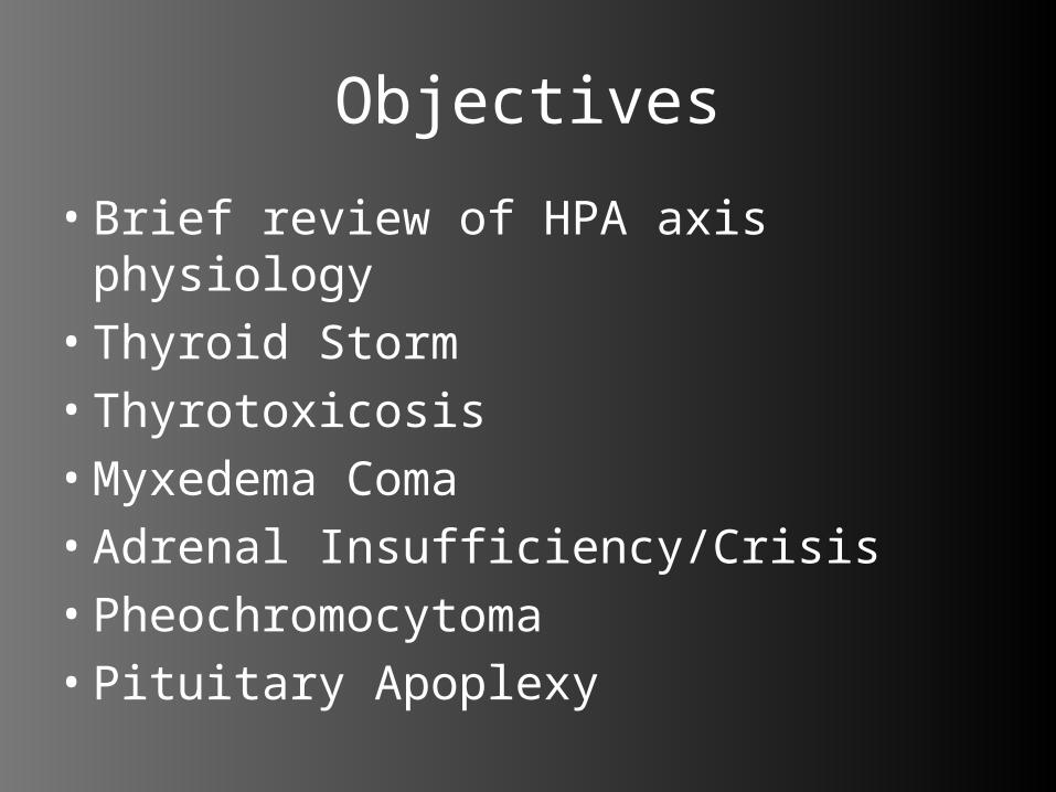
Objectives
• Brief review of HPA axis physiology• Thyroid Storm• Thyrotoxicosis• Myxedema Coma• Adrenal Insufficiency/Crisis• Pheochromocytoma• Pituitary Apoplexy
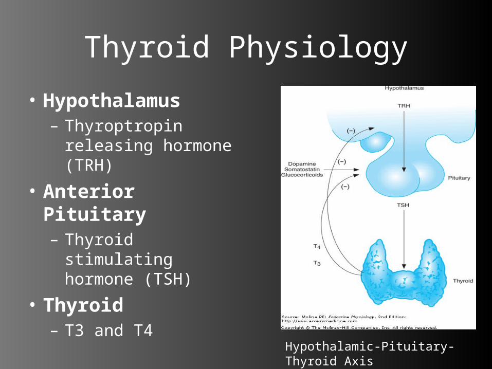
Thyroid Physiology
• Hypothalamus– Thyroptropin
releasing hormone (TRH)
• Anterior Pituitary– Thyroid stimulating
hormone (TSH)
• Thyroid – T3 and T4
Hypothalamic-Pituitary-Thyroid Axis
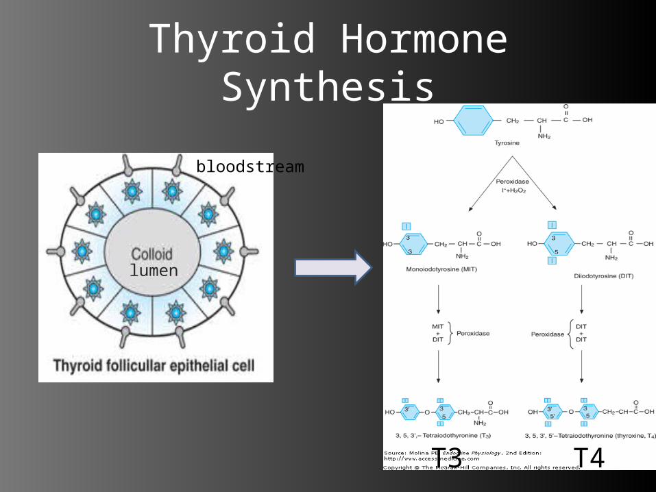
Thyroid Hormone Synthesis
T3 T4
bloodstream
lumen

5
Case 1: Cranked!!
• 60 yr old female presents to PLC ED concerned because she might have a “clot in the veins”.
• States feels heart beating fast and very sweaty.
• HR 140, BP 180/90, 98% RA, Temp 37.6, glucose 11.
5
6
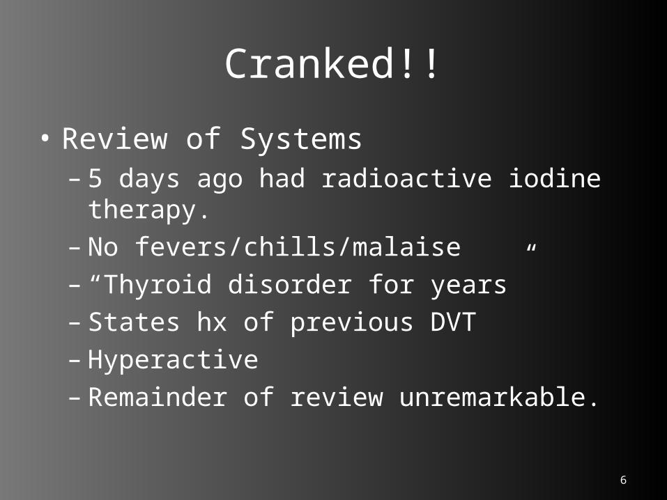
Cranked!!
• Review of Systems– 5 days ago had radioactive iodine
therapy.– No fevers/chills/malaise– “Thyroid disorder for years”– States hx of previous DVT– Hyperactive– Remainder of review unremarkable.
6
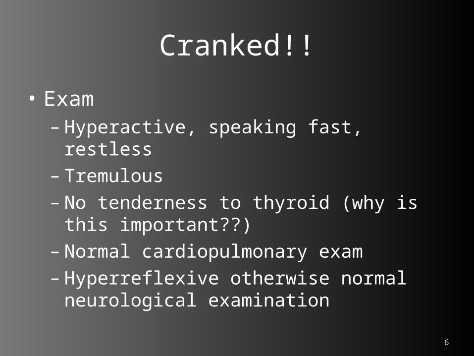
Cranked!!
• Exam– Hyperactive, speaking fast, restless– Tremulous– No tenderness to thyroid (why is this
important??)– Normal cardiopulmonary exam– Hyperreflexive otherwise normal
neurological examination
7
Cranked!!
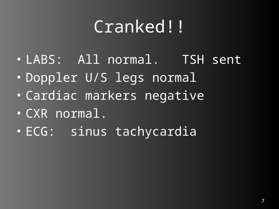
• LABS: All normal. TSH sent• Doppler U/S legs normal• Cardiac markers negative• CXR normal.• ECG: sinus tachycardia
7

9
Cranked!!
• Treatment– In ED gave Propranolol 2mg IV
q10minutes x 3 ---> heartrate decreased to 70 - 80
• During the day so discussed case with her primary endocrinologist.
• Wished her started back on Propanolol and Tapazole (methimazole).
• Agreed to see her the next day in clinic.

Hyperthyroidism/Thyrotoxicosis/Thyroid Storm
• Non-synonymous terms– But no consensus on definitions• Hyperthyroidism: the result of excessive
thyroid function• Thyrotoxicosis: a state of thyroid hormone
excess• Thyroid Storm: acute, life-threatening
exacerbation of thyrotoxicosis
• Rosen’s: “They refer to the continuum of disease that results from thyroid hyperfunction”.

Symptoms/Signs of Hyperthyroidism
Symptoms Signs
Hyperactivity/Irritable/Dysphoria Tachycardia/A. fib in elderly
Heat Intolerance/Sweating Tremor
Palpitations Goiter
Fatigue/Weakness Warm, moist skin
Weight loss/Hyperphagia Muscle Weakness/Proximal Myopathy
Diarrhea Lid retraction/Lag
Polyuria Gynecomastia
Oligomenorrhea/Dec. Libido
Harrison’s Principles of Internal Medicine 16th Ed. p2113
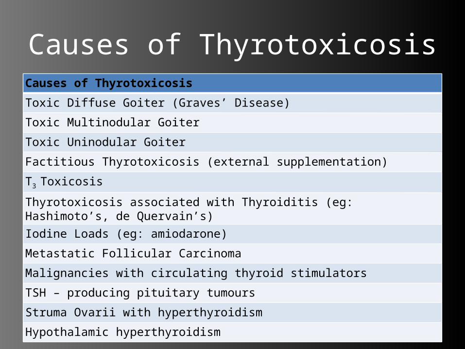
Causes of ThyrotoxicosisCauses of Thyrotoxicosis
Toxic Diffuse Goiter (Graves’ Disease)
Toxic Multinodular Goiter
Toxic Uninodular Goiter
Factitious Thyrotoxicosis (external supplementation)
T3 Toxicosis
Thyrotoxicosis associated with Thyroiditis (eg: Hashimoto’s, de Quervain’s)
Iodine Loads (eg: amiodarone)
Metastatic Follicular Carcinoma
Malignancies with circulating thyroid stimulators
TSH – producing pituitary tumours
Struma Ovarii with hyperthyroidism
Hypothalamic hyperthyroidism
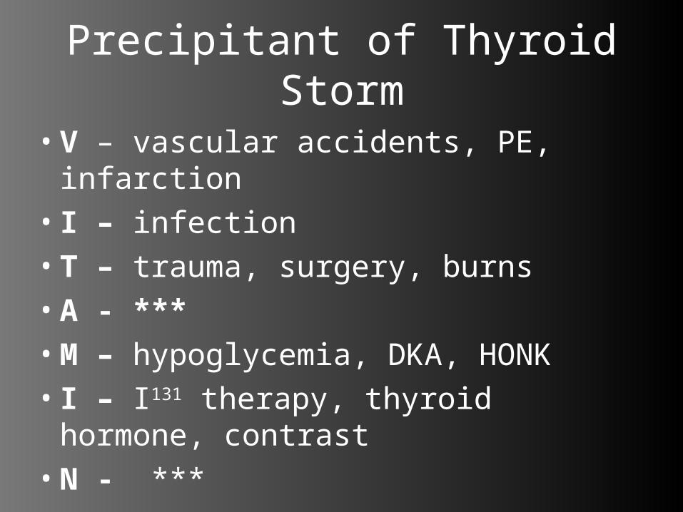
Precipitant of Thyroid Storm
• V – vascular accidents, PE, infarction• I – infection• T – trauma, surgery, burns• A - ***• M – hypoglycemia, DKA, HONK• I – I131 therapy, thyroid hormone,
contrast• N - ***

Thyroid Storm
• Exaggerated hyperthyroidism + Fever + Altered LOC
• Cardiovascular: hyperdynamic + excitable– Sinus tachycardia/Atrial tachycardia (A. fib)– CHF (±underlying heart disease)– Chest pain, Dyspnea, Palpitations, Inc. Pulse
Pressure, “Water Hammer” pulse
• Gastrointestinal: – Diarrhea, N/V, Abdominal pain– Liver dysfunction
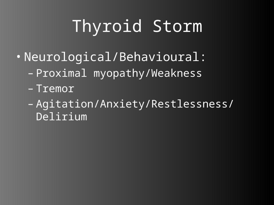
Thyroid Storm
• Neurological/Behavioural:– Proximal myopathy/Weakness– Tremor– Agitation/Anxiety/Restlessness/Delirium

16
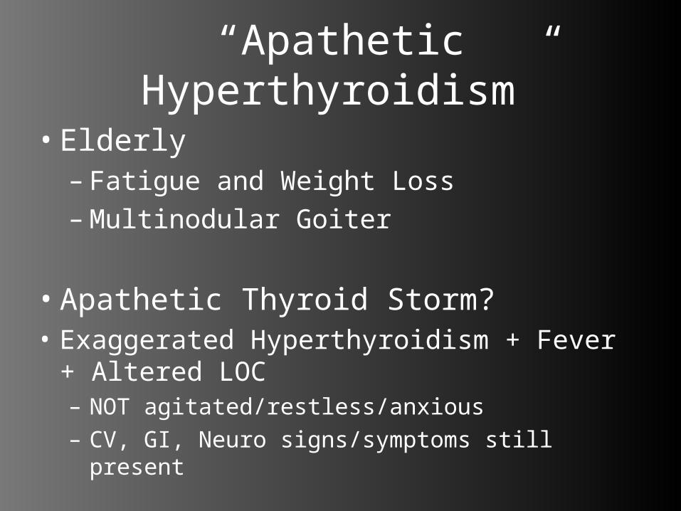
“Apathetic Hyperthyroidism”
• Elderly– Fatigue and Weight Loss–Multinodular Goiter
• Apathetic Thyroid Storm?• Exaggerated Hyperthyroidism + Fever +
Altered LOC– NOT agitated/restless/anxious– CV, GI, Neuro signs/symptoms still present
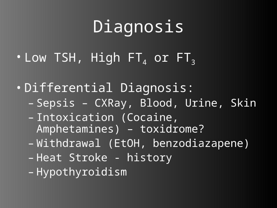
Diagnosis
• Low TSH, High FT4 or FT3
• Differential Diagnosis:– Sepsis – CXRay, Blood, Urine, Skin– Intoxication (Cocaine, Amphetamines) –
toxidrome?–Withdrawal (EtOH, benzodiazapene)– Heat Stroke - history– Hypothyroidism

Treatment of Thyroid Storm
• 5 Goals of Treatment:– 1) Inhibit Hormone Synthesis• Propylthiouracil (PTU) 600-1000mg PO/NG,
then 200-250mg q4-6h
– 2) Block Hormone Release (>1 hr post PTU)• Saturated Solution of KI (SSKI) 5 drops PO/NG
q6h• Iodine Anaphylaxis: Lithium Carbonate
300mg PO q6h• Iodine Overload Hyperthyroidism: Potassium
Perchlorate 500mg PO OD.

– 3) Prevent Peripheral Conversion of T4 to T3 • Propylthiouracil (PTU)• Dexamethasone 2mg IV q6h• Propranolol
– 4) Peripheral Adrenergic Blockade• Propranolol 1-2mg IV bolus q10-15mins until
effect
– 5) Supportive Care• Treat fever: Acetaminophen (Not ASA)• Treat CHF (digitalis, diuretics, oxygen)• Stress dose steroids (Hydrocortisone 100mg IV
q8h)• Treat Precipitating Factors
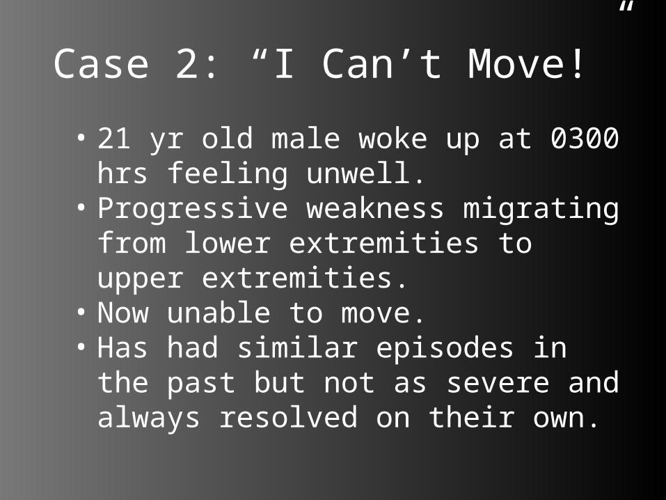
Case 2: “I Can’t Move!”
• 21 yr old male woke up at 0300 hrs feeling unwell.
• Progressive weakness migrating from lower extremities to upper extremities.
• Now unable to move.• Has had similar episodes in the
past but not as severe and always resolved on their own.

22
“I Can’t Move!!”
• Vitals: 130/75, 105HR, 96% RA, 18RR, glucose 7.6, Temp 36.4
• Recent URTI, no chest pain, shortness of breath, difficulty swallowing, back pain or bowel or bladder dysfunction.
• Recently immigrated from Mexico.
• Denies any medications or any medical history.
• Denies any drug or EtOH abuse.

“I Can’t Move!!”– HEENT: no palpable lymph nodes, normal
oropharynx– CVS: S1S2, no murmurs– RESP: Clear– ABDO: soft, non-tender, no organomegaly– NEURO: Cranial nerve exam normal,
complete paralysis both upper and lower extremities, markedly hyporeflexia bilaterally (upper and lower), sensation and proprioception remained intact, rectal tone normal
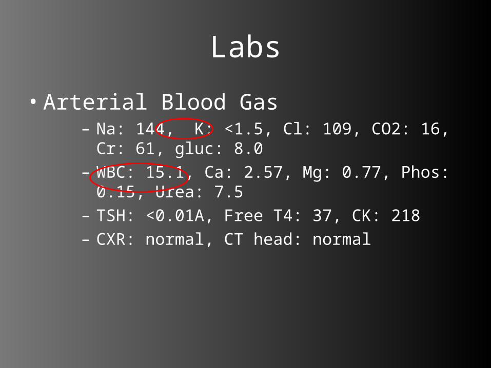
Labs
• Arterial Blood Gas– Na: 144, K: <1.5, Cl: 109, CO2: 16, Cr: 61,
gluc: 8.0– WBC: 15.1, Ca: 2.57, Mg: 0.77, Phos: 0.15,
Urea: 7.5– TSH: <0.01A, Free T4: 37, CK: 218– CXR: normal, CT head: normal

Thyrotoxic Periodic Paralysis• Asian Males most
common– Native Americans/African
Americans/South Americans
• Vigorous exercise/high carb meal
• Flaccid, ascending paralysis (proximal > distal)– Spares facial and
respiratory muscles
• Depressed/Absent DTR– Due to weakness
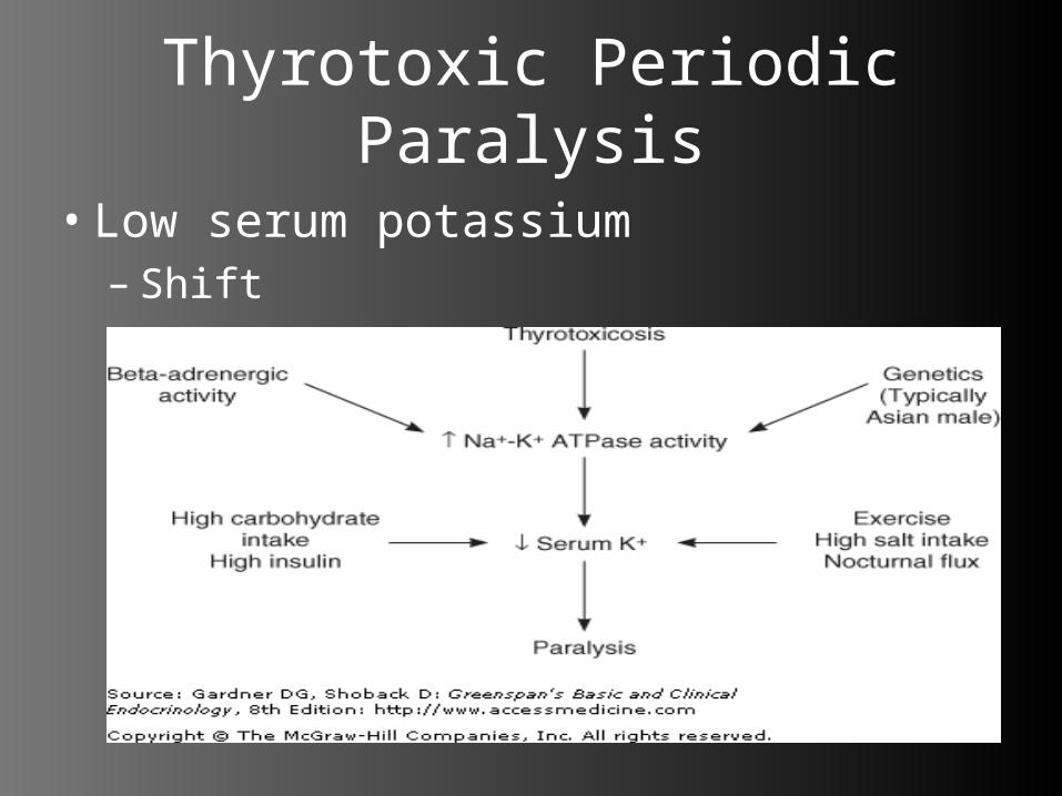
Thyrotoxic Periodic Paralysis
• Low serum potassium– Shift

Thyrotoxic Periodic Paralysis
• Management:– 1) Block β-adrenergic stimulation of Na/K
ATPase• Propranolol 60mg PO q6h
– 2) Replete Potassium• ORAL potassium (given not decreased total
stores)
– 3) Treat Hyperthyroidism
• AVOID: IV glucose, β-agonists
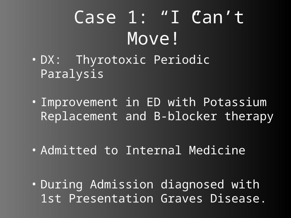
Case 1: “I Can’t Move!”
• DX: Thyrotoxic Periodic Paralysis
• Improvement in ED with Potassium Replacement and B-blocker therapy
• Admitted to Internal Medicine
• During Admission diagnosed with 1st Presentation Graves Disease.

Post Partum Thyroiditis• “Silent/Painless” thyroiditis• 5% postpartum cases• 3-4 months post-delivery
27

30
• Clinical Features:• Transient hyperthyroid followed by
transient hypothyroid• Triphasic course• Non-tender thyroid, Normal ESR (cf.
subacute thyroiditis)• No eye findings (cf. Graves’ Disease)

Post Partum Thyroiditis• Laboratory Findings– FT4 >> T3 – leakage of hormone from
gland
• Treatment (if needed)– Propranolol 20mg-40mg q6-8h
28

Case 2: “I Can’t Warm Up!”
• 70 yr old non-english speaking female brought by EMS because of decline in LOC and function of past few days.
• Multiple recent ED visits for hyponatremia.
• Complaints of malaise, fatigue, weakness and confusion.

Case 2: “I Can’t Warm Up!”
• Vitals 35.2, 45-55HR, 10RR, 150/74 (initial), glucose 5.7
• Past Medical History: HTN, RA, Shingles, Bilateral Hip Replacement
• Meds: BP med(water pill), acyclovir

Case 2: “I Can’t Warm Up!”
• Collateral History from son states multiple visits over past months for low salt, confusion and lethargy.
• Had been referred to Outpatient Internal Med Clinic.
• EXAM: puffy face, dry mm, tender epigastrium, tremelous, depressed reflexes, initial GCS 14/15, remainder of exam unremarkable.
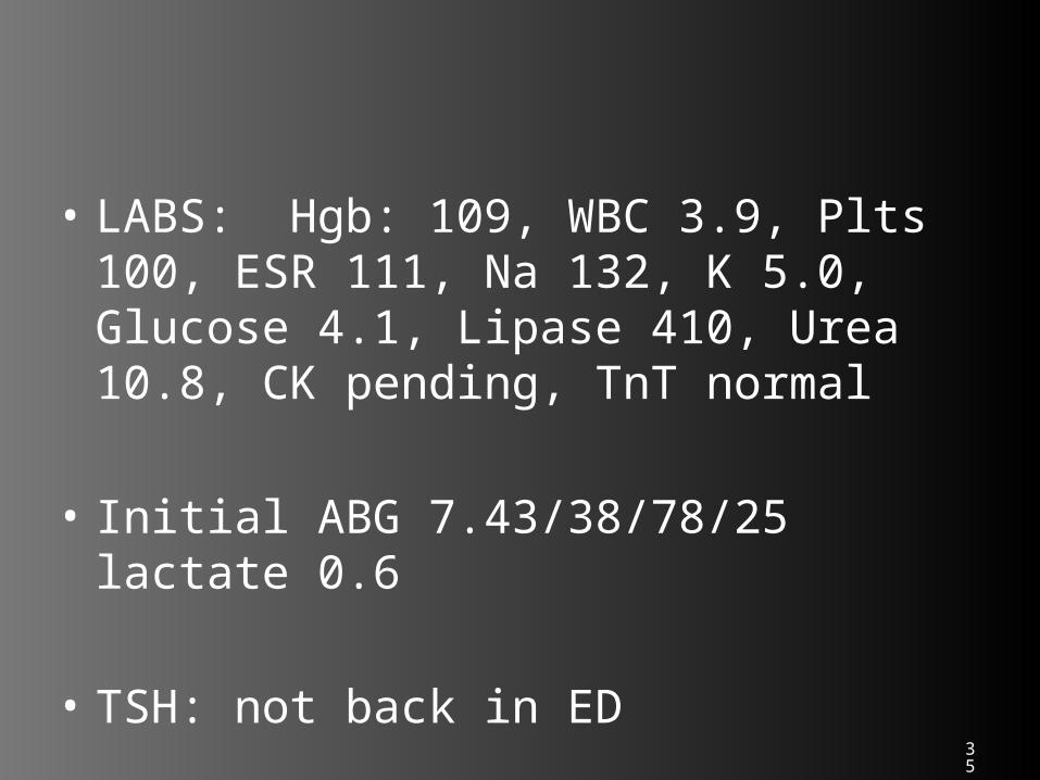
35
• LABS: Hgb: 109, WBC 3.9, Plts 100, ESR 111, Na 132, K 5.0, Glucose 4.1, Lipase 410, Urea 10.8, CK pending, TnT normal
• Initial ABG 7.43/38/78/25 lactate 0.6
• TSH: not back in ED

36
• CT head: normal
• CXR: normal
• Urine normal
• CT abdo/pelvis:probable ovarian mass, no diverticulitis or pancreatic abscess/pseudocyst, small bilat effusions seen.

Case 2: “I Can’t Warm Up!”• In ED declining GCS to 8/15• profoundly bradycardic, • borderline hypotensive, • hyponatremia and hypoglycemia • hypothermic (31.4C despite external re-
warming techniques)• decreased RR --> increasing CO2 on
ABG
• Intubated and lined in ED
• After induction agents and paralytics had worn off pt made no respiratory effort on own, nor response to painful stimuli

38
• DX: ?Myxedema Coma• Given steroids and thyroxine (also
given dose of abx after cultures drawn)
• Sent to ICU

Hypothyroidism
• Primary disease most common– Autoimmune– Iatrogenic
• Elderly Obese Females
Subclinical Disease Myxedema Coma

Signs/Symptoms of Hypothyroidism
Symptoms Signs
Fatigue/Weakness Dry /Cool Skin
Dry Skin Puffy face, hands, feet (myxedema)Cold intolerance Diffuse alopecia
Hair Loss Bradycardia
Difficulty Concentrating/Poor Memory
Peripheral Edema
Constipation Delayed DTRs
Weight Gain/Poor Appetite Carpal Tunnel Syndrome
Dyspnea Serous Cavity Effusion
Hoarse Voice
Menorrhagia
Paresthesia
Impaired Hearing
Harrison’s Principles of Internal Medicine 16th Ed. p2109
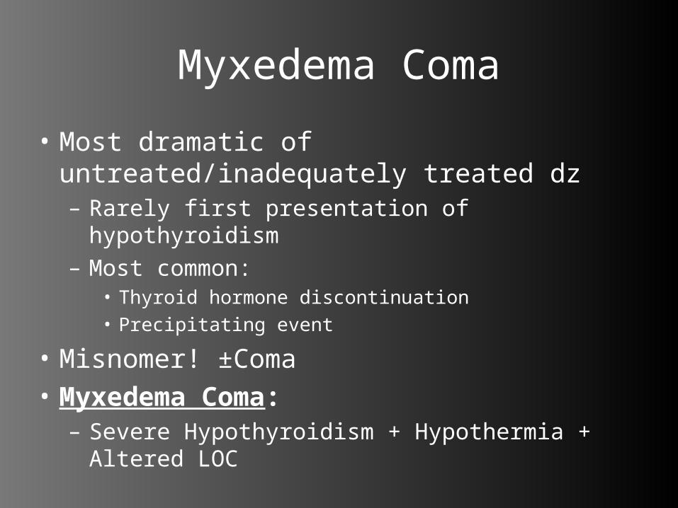
Myxedema Coma
• Most dramatic of untreated/inadequately treated dz– Rarely first presentation of hypothyroidism– Most common:
• Thyroid hormone discontinuation• Precipitating event
• Misnomer! ±Coma• Myxedema Coma:– Severe Hypothyroidism + Hypothermia + Altered
LOC
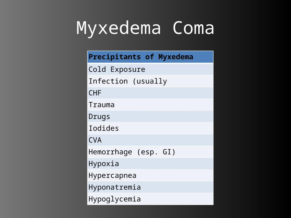
Myxedema ComaPrecipitants of Myxedema ComaCold Exposure
Infection (usually pulmonary)
CHF
Trauma
Drugs
Iodides
CVA
Hemorrhage (esp. GI)
Hypoxia
Hypercapnea
Hyponatremia
Hypoglycemia
Myxedema Coma
• Cardiovascular:– Sinus bradycardia– BP variable– Leaky capillaries• Effusions
• Respiratory:– Depressed respiratory drive (hypoxic +
hypercapneic)– Airway obstruction (from edema)
Myxedema Coma
• Gastrointestinal:– Decreased peristalsis• Abdominal pain, distension, constipation
• Neurological:– Paresthesias– Cerebellar-Like Symptoms • Due to increased muscle tone/prolonged
contraction
– Coma

Diagnosis
• High TSH and Low Free T4
– Note: Dopamine, Glucocorticoids, and Somatostatin suppress TSH at pharmacologic doses.
• Low/Normal TSH and Low Free T4?– Hypothalamic/Pituitary Disease

Differential Diagnosis
• Sepsis• Accidental Hypothermia• Nephrotic Syndrome/Renal Failure• Apathetic Hyperthyroidism• Hyperglycemia• Intoxication (sedatives)

Treatment of Myxedema Coma
• 4 Goals:– 1) Thyroid Hormone Replacement• Levothyroxine 500µg PO/IV, then 100µg/day
– 2) Correct Metabolic Abnormalities• Hypoventilation – Intubate + Ventilate• Hyponatremia – water restriction
• Hypoglycemia – D5W IV
– 3) Identify/Correct Precipitating Factors• Infection? CHF?

– 4) Supportive Care• Hypotension – Fluids, Pressors• Hypothermia – GENTLE Rewarming• Stress Dose Steroids – Hydrocortisone 300mg
IV, then 100mg q6-8h.

43
Some Pearls
• ***beware when giving IV thyroxine and pressors together as may result in VF/VT (should stop pressor when giving IV thyroxine)• ***try to avoid use of ASA in setting of storm
as may worsen disease.• ***can use CK as poor man’s TSH in setting
of presumed myxedema coma.• ***be diligent re: searching for precipitating
causes!!!
Case 3: “The Disappearing Tan Lines”
• 29 yr old male with fatigue, heart palpitations, vomiting and lightheadness for 1yr.
• Presented to ED because of frustration and multiple physician visits for similar.
• Vitals: 36.6, 67HR, 14RR, 112/65, 99% RA, gluc 8.0
Case 3: “The Disappearing Tan Lines”
• Review of Systems– Low BP (states at time as low as 85
systolic), wt loss of 20lbs over past year, Tingling and muscle weakness, shortness of breath on exertion, no chest pain, denies any drug or EtOH abuse
– Previously treated for depression– Family hx of hypothyroid and diabetes
Case 3: “The Disappearing Tan Lines”
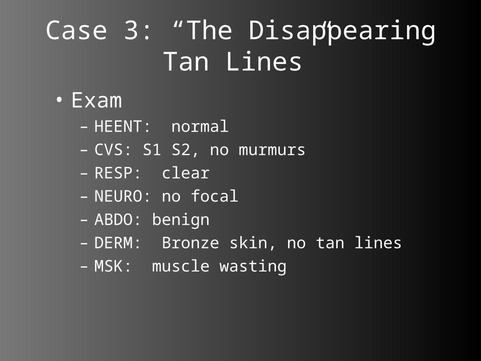
• Exam– HEENT: normal– CVS: S1 S2, no murmurs– RESP: clear– NEURO: no focal– ABDO: benign– DERM: Bronze skin, no tan lines– MSK: muscle wasting

Case 3: “The Disappearing Tan Lines”
• Labs: all normal in ED
• However, outpt lab work one month ago shows: – Na 131, K: 5.8, Cl: 99, CO2: 23, CK: 410,
Ferritin 364, Fe: 7, TSH 3.3

Adrenal Insufficiency
• An absolute or relative deficiency of adrenal hormones– Cortisol, Aldosterone, Androgen

Adrenal Physiology
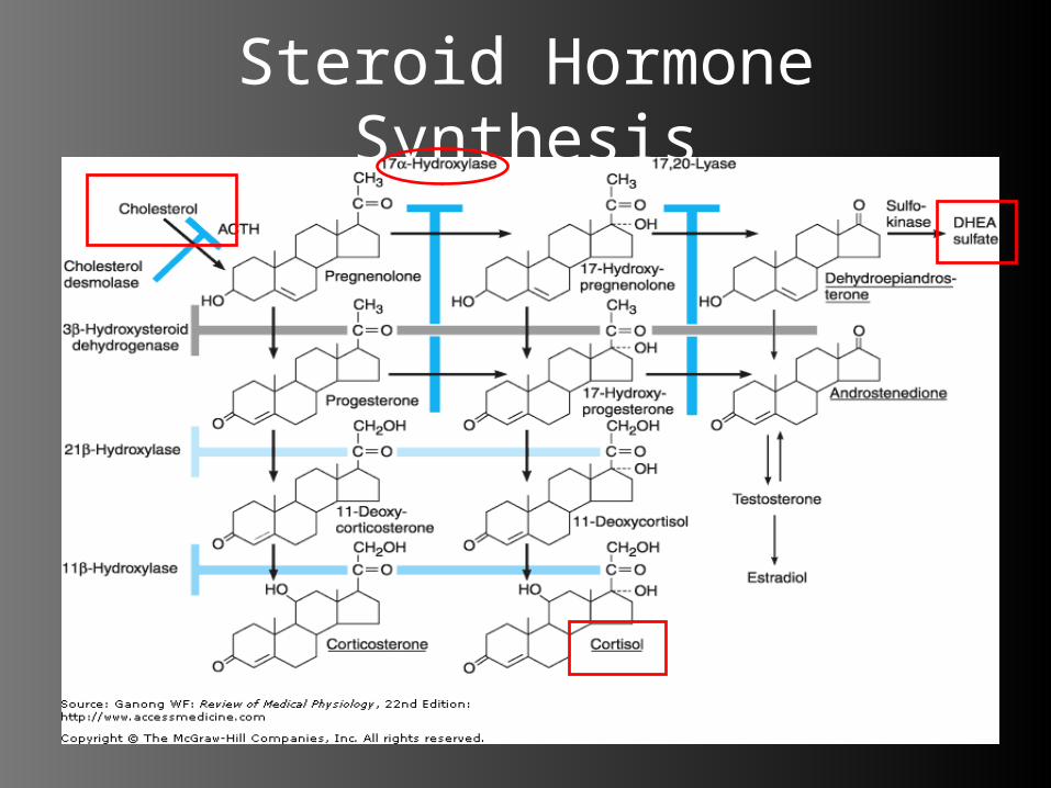
Steroid Hormone Synthesis

Steroid Hormone Synthesis
17α-Hydroxylase

Steroid Hormones
• Cortisol:– Intermediary metabolism
(carbs,protein,fat,NA)– Immune response (depressed)Hypothalam
usCRH
Anterior Pituitary
ACTH
Adrenal Cortex
(Cortisol)
Negative Feedback
Negative Feedback

Steroid Hormones
• Aldosterone– Blood Pressure– Vascular Volume– Electrolytes
• Regulation– Primarily by Renin-Angiotensin-
Aldosterone Axis• Small role by ACTH
Steroid Hormones
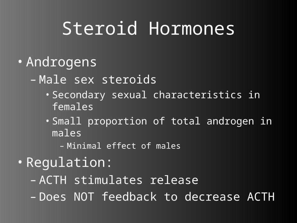
• Androgens–Male sex steroids• Secondary sexual characteristics in females• Small proportion of total androgen in males
– Minimal effect of males
• Regulation:– ACTH stimulates release– Does NOT feedback to decrease ACTH

Etiologies of Adrenal Insufficiency
• Primary– Idiopathic – autoimmune, idiopathic– Infectious – granulomatous, viral, fungal– Infiltrative – neoplasm, amyloidosis,
sarcoidosis– Iatrogenic – post-adrenalectomy– Hemorrhage– CAH – lack of 21β-Hydroxylase deficiency– Congenital Unresponsiveness to ACTH

Etiologies
• Secondary– Pituitary Insufficiency• Infarction, Hemorrhage, Tumour/Infiltration,
ACTH deficiency
– Hypothalamic Insufficiency– Head Trauma
• Functional Disease– Exogenous glucocorticoids
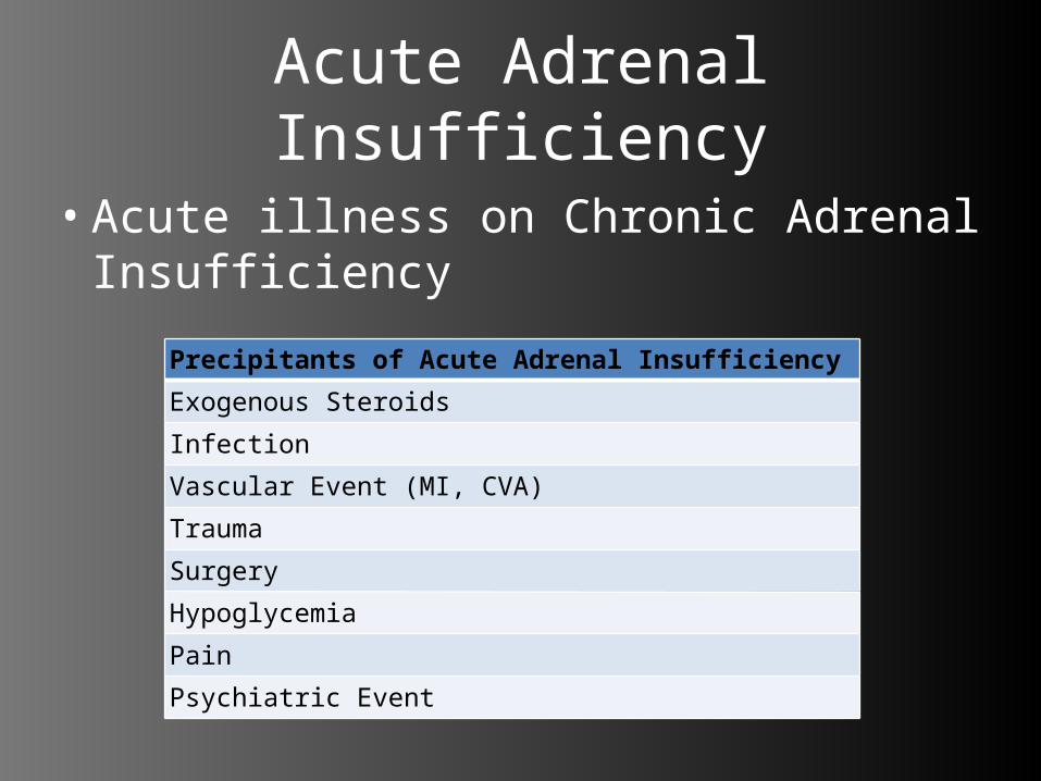
Acute Adrenal Insufficiency
• Acute illness on Chronic Adrenal Insufficiency
Precipitants of Acute Adrenal Insufficiency
Exogenous Steroids
Infection
Vascular Event (MI, CVA)
Trauma
Surgery
Hypoglycemia
Pain
Psychiatric Event
Special Cases
• Adrenal Hemorrhage– Waterhouse-Friedrickson Syndrome
• Sepsis from meningococcemia with associated adrenal hemorrhage (amongst hypotension,shock,DIC)
• Can also occur from Pseudomonas sepsis
– Acute, severe illness + anticoagulation/coagulopathy
• Pituitary Infarction– Sheehan Syndrome
• Delayed effect of intrapartum/post-partum hemorrhage leading to pituitary infarction

The Usual SuspectsSymptom/Sign Frequency (%)
Weakness 99
Pigmentation of Skin 98
Weight Loss 97
Anorexia/Nausea/Vomiting 90
Hypotension (<110/70) 87
Pigmentation of mucous membranes
82
Abdominal Pain 34
Salt Craving 22
Diarrhea 20
Constipation 19
Syncope 16
Vitiligo 9

Hyperpigmentation
Adrenal Crisis
• Hypotension– Decreased myocardial contractility– Decreased responsiveness to
catecholamines– Hypovolemia (Na wasting, N/V)
• Hypoglycemia– Decreased gluconeogenesis– Increased peripheral glucose use

Treatment
• Correct the greatest threats to life!– Hypotension: Fluid resuscitate ± pressors– Hypoglycemia: D5W or D50.9% saline
• Glucagon 1-2mg IM/SC– Correct hormone deficiency:
• Cortrosyn Stimulation Test– 0.25mg (25U) cosyntropin IV/IM– Serum cortisol at time: 0, 30 mins, 60 mins– Normal: cortisol >500nmol/L or >200nmol/L over
baseline• Dexamethasone 4mg IV q6-8h (during test)• Hydrocortisone 100mg IV/IM q6-8h
• Treat the Precipitating Factor!

Case 3: “The Disappearing Tan Lines”
• DX: Primary Adrenal Insufficiency/Addison’s Disease
• Referral made to Urgent Internal Medicine/Endo– Cosyntropin stim test performed– Started on Decadron–Marked improvement within 48hrs
Prevention
• Cortisol:– Acute Illness• Double dose of hydrocortisone
– Severe Illness• 75-150mg hydrocortisone/day
• Aldosterone:• Fludrocortisone 0.05-0.1mg• Increase salt in diet

Adrenal Medulla
NorepinephrineEpinephrine

Catecholamine Effects
• Norepinephrine/Epinephrine:– α and β effects• Increased CV contractility, excitability, heart
rate
– Increased gluconeogenesis/glycogenolysis
– Increased metabolic rate– Increased alertness/anxiety/fear

Pheochromocytoma
• Catecholamine secreting tumour– Adrenal or Extra-adrenal– Rare! – Young to Mid-Adult Life
• Clinical Presentation:– Hypertension – most common– Paroxysms • Hypertension, Headache, Sweating,
Palpitations, Apprehension, Sense of impending doom, Chest Pain, Abdo Pain, N/V, pallor/flushing

Differential Diagnosis
• Sympathomimetic Intoxication• MAOI Crisis• Withdrawal of Clonidine therapy• Seizures• Intracranial Lesions – posterior fossa
tumours• SAH
Pheochromocytoma
• Cardiovascular– Hypertension (DBP >120)– ECG• Sinus tachycardia, SVT, VT, V.Fib.• Non-specific ST changes, U-waves (hypoK)• Ventricular Strain• RBBB, LBBB• Prolonged QT
• Endocrine– Impaired glucose tolerance
Diagnosis
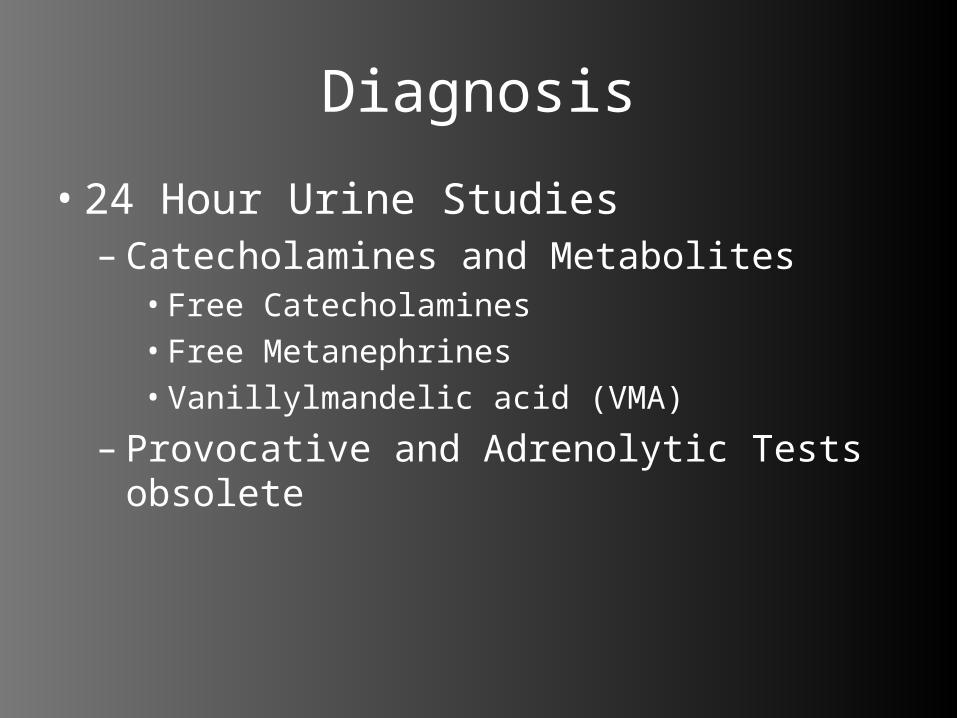
• 24 Hour Urine Studies– Catecholamines and Metabolites• Free Catecholamines • Free Metanephrines• Vanillylmandelic acid (VMA)
– Provocative and Adrenolytic Tests obsolete

Treatment
• α-adrenergic Blockade– Phentolamine 1-2mg IV q5mins– Phenoxybenzamine 10mg PO q12h (long
term)
• β-blockade– ONLY AFTER stable α-blockade achieved – usually reserved for tachydysrrhythmias– Propranolol 10mg PO q6-8h
• Nitroprusside, CCB, ACEi
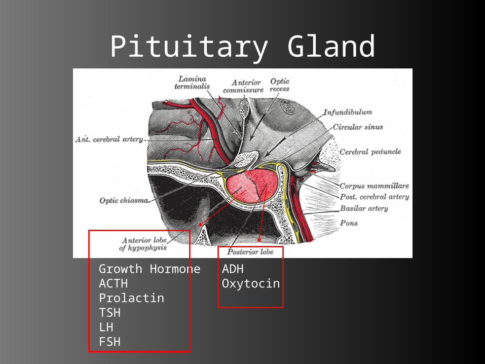
Pituitary Gland
Growth HormoneACTHProlactinTSHLHFSH
ADHOxytocin

Pituitary Apoplexy
• Infarction or Hemorrhage of Pituitary Gland– Pre-existing tumour– Head trauma– Pregnancy– Anti-coagulation– Hypertension– DKA– Irradiation– Estrogen– Diuretic use– Bromocriptine

Clinical Presentation
• Sudden onset headache• Visual abnormalities• Oculomotor abnormalities• Meningeal irritation • Altered mental status• Pituitary Insufficiency• Adrenal Insufficiency
Diagnosis
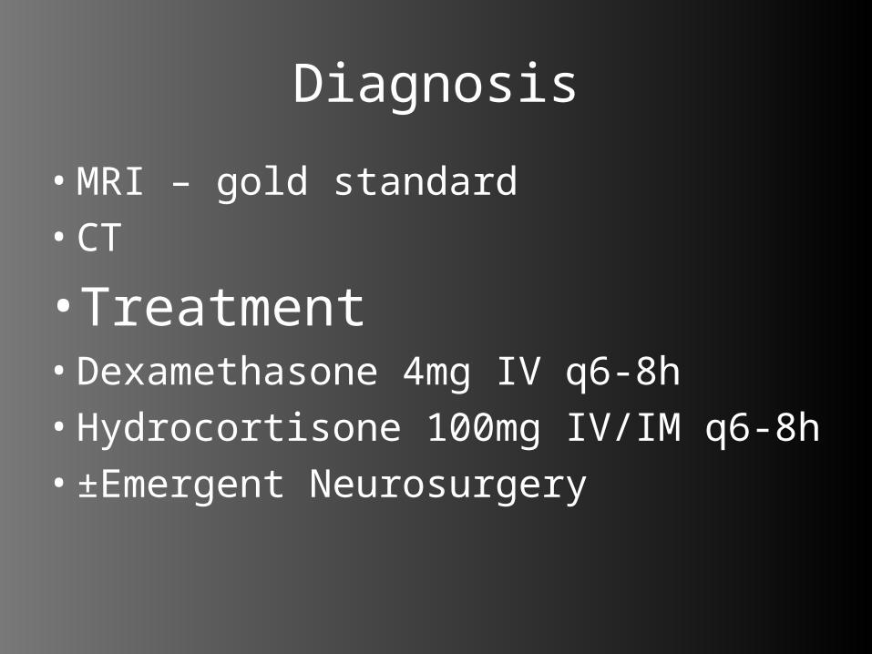
• MRI – gold standard• CT
• Treatment• Dexamethasone 4mg IV q6-8h• Hydrocortisone 100mg IV/IM q6-8h• ±Emergent Neurosurgery
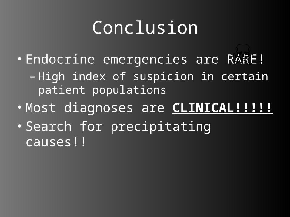
Conclusion
• Endocrine emergencies are RARE! – High index of suspicion in certain patient
populations
• Most diagnoses are CLINICAL!!!!!• Search for precipitating causes!!

Questions?