Embed Size (px)
Citation preview

NON-CLASSICAL ACTION OF THE MINERALOCORTICOID RECEPTOR IN MACROPHAGES: AT THE CROSSROADS OF INFLAMMATION AND
CARDIOVASCULAR DISEASE
by
Michael Goodwin Usher
A dissertation submitted in partial fulfillment of the requirements for the degree of
Doctor of Philosophy (Molecular and Integrative Physiology)
in The University of Michigan 2009
Doctoral Committee Professor Richard M. Mortensen, Chair Professor Roger J. Grekin
Professor Richard Keep Professor Ronald J. Koenig
Professor David Pinsky Professor Audrey F. Seasholtz
Associate Professor Jorge A. Iniguez-Lluhi

© Michael G. Usher All rights reserved
2009

ii
To my family

iii
ACKNOWLEDGEMENTS
Life as an MSTP student is often challenging especially given the multiple
transitions required for doctorates in science and medicine. Without the support of my
friends, family, colleagues, and mentors this journey would not have been as fun or as
fulfilling. First and foremost, I would like to thank my mentor Rick Mortensen for
providing an unparalleled education. He has imparted on me a sense of how to ask
meaningful questions; he has fostered my desire to draw parallels from a wide array of
different fields to broaden my appreciations; he has allowed me to participate directly in
intellectual direction of his lab in part through grant writing. Enthusiasm for science
often waxes and wanes as a graduate student. At the end of the journey I can confidently
say that when I finish my clinical training I will maintain interest in scientific pursuits.
This is a testament to Rick’s ability to sustain an entertaining and stimulating
environment even in the face of my own attention deficits.
I would also like to thank the physiology program and the MSTP program. In
each program I have found a home, good friends, and lasting memories. This would not
be possible without able administration from Ellen Elkin, Penny Morris, and Michele
Boggs. I would also like to thank the director of the MSTP program Ron Koenig, who
not only serves on my committee, convinced me to stay in science when I considered
leaving. That is one of the best pieces of advice I have ever received.

iv
I would like to thank my committee, whose critical advice has kept my focus in
the right place and not let my mind and my experiments wander aimlessly, or my scope
from becoming unmanageably large. I would like to also thank Stefan Berger and
Gunther Schutz, who provided the MR floxed mice which eventually became the center
of my thesis. I would like to thank Lou D’Lacey and Janet Hoff who provided technical
and surgical advice and provided expertise in blood pressure collection. Duan, a post doc
in Ricks lab also deserves special mention. Duan has been a constant stabilizing force in
the lab, whose natural good natured demeanor has made the lab a delight to work in.
Finally I would like to thank my family. My parents instilled a delight in learning
and fostered my own curiosity which I now carry constantly and is the main reason for
my success. I would also like to thank my wife who has made all of my experiences both
in the lab, and outside more meaningful.

v
TABLE OF CONTENTS
DEDICATION......................................................................................................... ii
ACKNOWLEDGMENTS….................................................................................. iii
LIST OF FIGURES ............................................................................................... x
LIST OF TABLES.................................................................................................. xiii
LIST OF ABREVIATIONS................................................................................... xiv
ABSTRACT…......................................................................................................... xx
CHAPTER
I. INTRODUCTION ................................................................................... 1
Overview …....................................................................................... 1
The mineralocorticoid receptor structure and function…................ 2
Tissue activation of MR ….... ........................................................... 4
Hypertension……….......................................................................... 7
Heart Failure…….............................................................................. 9
MR Blockade……............................................................................. 10
Inflammation…………...................................................................... 12
Macrophages in Inflammation……………………........................... 14
Macrophages in cardiovascular disease. … … … … ………........... 17
Nuclear Receptor control of macrophage activation… .................... 19
PPAR-γ in macrophages …………… ……………….......... 21
Glucocorticoid Receptor in Macrophages ................ ........... 22
LXR in macrophages............................................................. 26
MR Evolution.............. ..................................................................... 29

vi
Glucocorticoid occupied MR........................................... ............... 32
MR at the crossroads of inflammation and cardiovascular disease... 34
MR in macrophages.............................................. ............................ 37
Hypothesis.........................................................................................38
Specific Aims................................................. ................................... 38
II. MYELOID MINERALOCORTICOID RECEPTOR REGULATES MACROPHAGE POLARIZATION AND RESPONSE TO CARDIOVASCULAR DAMAGE......................................... ........................................................... 49 Abstract................................................................. ............................ 49
Introduction....................................................................................... 50
Results and Discussion......................................................................53
MR expression and regulation in macrophages..................... 53
MR directly regulates Macrophage activation...................... 58
MR knockout macrophages................................................... 68
MRKO protects against cardiovascular inflammation........... 73
MR inhibits alternative macrophage activation..................... 75
MR and PPAR-γ co-ordinately regulate macrophage polarization............................................................................ 82
MR regulates glucocorticoid signaling in the macrophage............................................................................ 84
Conclusions........................................................................................ 89
Methods............................................................................................. 90
III. NUCLEAR FACTOR BALANCE IN MACROPHAGE POLARIZATION......................................................... 94 Introduction....................................................................................... 94

vii
Results and Discussion...................................................................... 97
PPAR-γ does not solely enhance IL-4 responses.................. 97
PPAR-γ and IL-4 oppose GR in macrophage polarization.... 100
MR in nuclear factor balance................................................. 102
Conclusions........................................................................................ 106
IV. MACROPHAGE MR AND THE CONTROL OF INNATE AND ADAPTIVE IMMUNITY........................................... 108 Overview............................................................................................ 108
Introduction............................................................................ ........... 109
Mechanisms of control of gene transcription by MR............ 109
Macrophage/Monocyte differentiation.................................. 111
MR in macrophages and the control of adaptive immunity............................................................................... 113
Results................................................................................................ 115
MR controls macrophage transcription through multiple mechanisms............................................................. 115
Macrophage MR regulates antigen presentation and Lymphocyte proliferation...................................................... 123
MR in monocyte differentiation............................................. 123
Discussion and future directions....................................................... 126
MR mediated transcriptional control in macrophages........... 128
MR controls of monocyte/macrophage differentiation.......... 130
MR in macrophages and the control of adaptive immunity................................................................................ 131
MR in immune responses and inflammatory disease states...................................................................................... 132

viii
Summary............................................................................................ 136
V. MACROPHAGE MINERALOCORTICOID RECEPTOR IN CARDIOVASCULAR PHYSIOLOGY..................................................... 144
Overview........................................................................................... 144
Introduction........................................................................................ 144
Macrophage MR and cardiac hypertrophy............................ 144
Macrophage MR and cardiovascular circadian rhythms....... 146
Macrophage MR and ischemic stroke.................................... 149
Macrophage MR and diet induced obesity............................ 150
Results................................................................................................ 153
Macrophage MR is important in cardiac hypertrophy........... 153
Macrophage MR in hemodynamic circadian rhythms........... 155
Macrophage MRKO is protective in ischemic stroke............ 159
Macrophage MRKO does not protect against diet induced Obesity and insulin resistance............................................... 161
Discussion and future directions........................................................ 163
Macrophage MR and Cardiac Hypertrophy........................... 163
Macrophage MR and hemodynamic circadian rhythms........ 165
MΦMRKO protection in ischemic stroke.............................. 168
Macrophage MR in diet induced obesity............................... 169
Summary............................................................................................ 170
VI. SUMMARY AND CONCLUSIONS.................................................... 176
MR in macrophages is an important target of MR antagonists......................................................................................... 176
On Macrophage Polarization............................................................. 177

ix
Macrophages regulate hemodynamic responses................................ 178
On the clinical benefit of spironolactone and eplerenone.................. 179
MR and immune control.................................................................... 180
On the potential for aldosterone action on macrophages................... 182
Macrophage polarization as a paradigm for drug discovery................................ ........................................................... 183
MR in parenchymal tissues................................................................ 185
APPENDIX................................................................................................... 192

x
LIST OF FIGURES
Figure 1.1: Classical mineralocorticoid receptor action.................... ........... .......... 5 Figure 1.2: MR regulates two divergent physiologic systems................................... 6 Figure 1.3: Glucocorticoid Action on classical macrophage activation.................... 24 Figure 1.4: LXR, PPARγ, and GR coordinate to inhibit
classical macrophage activation......................................................... .......... 28
Figure 1.5: Evolution of the mineralocorticoid and glucocorticoid receptors ........ 31 Figure 1.6: MR and GR action in the brain .............................................................. 33 Figure 2.1: Tissue distribution of factors involved in local glucocorticoid concentration and responses............................................................. 54
Figure 2.2 MR in macrophages acts as a high affinity glucocorticoid
receptor.......................................................................................................... 56 Figure 2.3 Regulation of MR expression and activity
following macrophage activation.................................................................. 57
Figure 2.4 PPAR-γ represses MR. ............................................................................ 59 Figure 2.5 MR enhances pro-inflammatory cytokine
expression in macrophages........................................................................... 61
Figure 2.6 Aldosterone stimulates a pro-inflammatory, pro-fibrotic macrophage response ............................................................... 62
Figure 2.7 Low dose corticosterone does not mimic aldosterone’s MR dependant pro-inflammatory activity. ............................ 63
Figure 2.8: Antagonism of glucocorticoid occupied MR is anti-inflammatory.........65 Figure 2.9 Transient MR over-expression is pro-inflammatory in macrophages...... 66
xi
Figure 2.10. Glucocorticoid and Aldosterone occupied MR have different pro-inflammatory activities............................................ 68
Figure 2.11: Generation of macrophage specific MR knockout ............................... 70 Figure 2.12 MR deletion dramatically alters macrophage function ......................... 71 Figure 2.13 Divergent actions of glucocorticoid and aldosterone
occupied MR on macrophage function......................................................... 74
Figure 2.14 MΦMRKO is protective in against cardiac fibrosis and inflammation.............................................................................. 76
Figure 2.15 MΦMRKO protects from vascular remodeling..................................... 77 Figure 2.16: L-NAME/Ang-II results in an M1 polarized response
in cardiac tissue diminished by MΦMRKO.................................................. 88
Figure 2.17: MΦMRKO does not protect from glomerular injury induced by L-NAME/Ang-II ........................................................................ 79
Figure 2.18 MR controls macrophage polarization................................................... 81 Figure 2.19: MR and PPAR-γ play oppositional roles in
macrophage polarization............................................................................... 83
Figure 2.20: MR coordinates with glucocorticoid signaling in the macrophage.......................................................................................... 85
Figure 2.21: Control of macrophage activation by MR and GR.................... ........... 87 Figure 2.22: Overlap of MR and GR mediated effects
on macrophage activation............................................................................ 88
Figure 3.1: Macrophage polarization and nitrogen metabolism............................... 96 Figure 3.2: PPAR-γ controls alternative macrophage activation............................... 99 Figure 3.3 Glucocorticoids stimulate a unique AMΦ transcriptional profile............ 101 Figure 3.5 Nuclear receptor balance in macrophage polarization. .......................... 103 Figure 3.6: MR control of macrophage polarization ................................................ 105 Figure 4.1: Monocyte heterogeneity ......................................................................... 112
xii
Figure 4.2 MR regulates transcription through multiple mechanisms....................... 117 Figure 4.3 Bioinformatic prediction of the mineralocorticoid
receptor response element.............................................................................. 122
Figure 4.4: Follicular enlargement in MΦMRKO mice............................................ 124 Figure 4.5: Splenic Structure is disrupted by L-NAME/Ang-II ............................... 125 Figure 4.6: Circulating and bone marrow monocyte
populations are altered by L-NAME/Ang-II and MΦMRKO ...................... 127
Figure 4.7 Pleiotropic actions of myeloid MR on innate and adaptive immunity................................................................................... 138
Figure 5.1: Cardiovascular Circadian Rhythms......................................................... 147 Figure 5.2 Macrophage action and cardiac hypertrophy .......................................... 154 Figure 5.3: MΦMRKO results in diminished day time reductions
in heart rate, pulse pressure, and systolic pressure........................................ 156 Figure 5.4: MΦMRKO results in altered cardiovascular
response to high salt....................................................................................... 158
Figure 5.5: Infarct Volume following Middle Cerebral Artery (MCA) occlusion is reduced in MФMRKO mice .................................................... 160
Figure 5.6: MΦMRKO does not protect against diet induced obesity or insulin resistance .......................................................................... 162
Figure 6.1: MR occupancy and the positive feedback mechanisms which drive cardiovascular disease........................................... 189
xiii
LIST OF TABLES
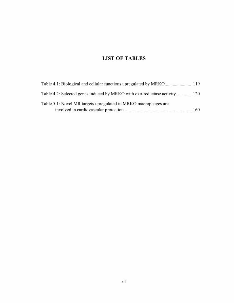
Table 4.1: Biological and cellular functions upregulated by MRKO....................... 119
Table 4.2: Selected genes induced by MRKO with oxo-reductase activity.............. 120
Table 5.1: Novel MR targets upregulated in MRKO macrophages are involved in cardiovascular protection ........................................................... 160
xiv
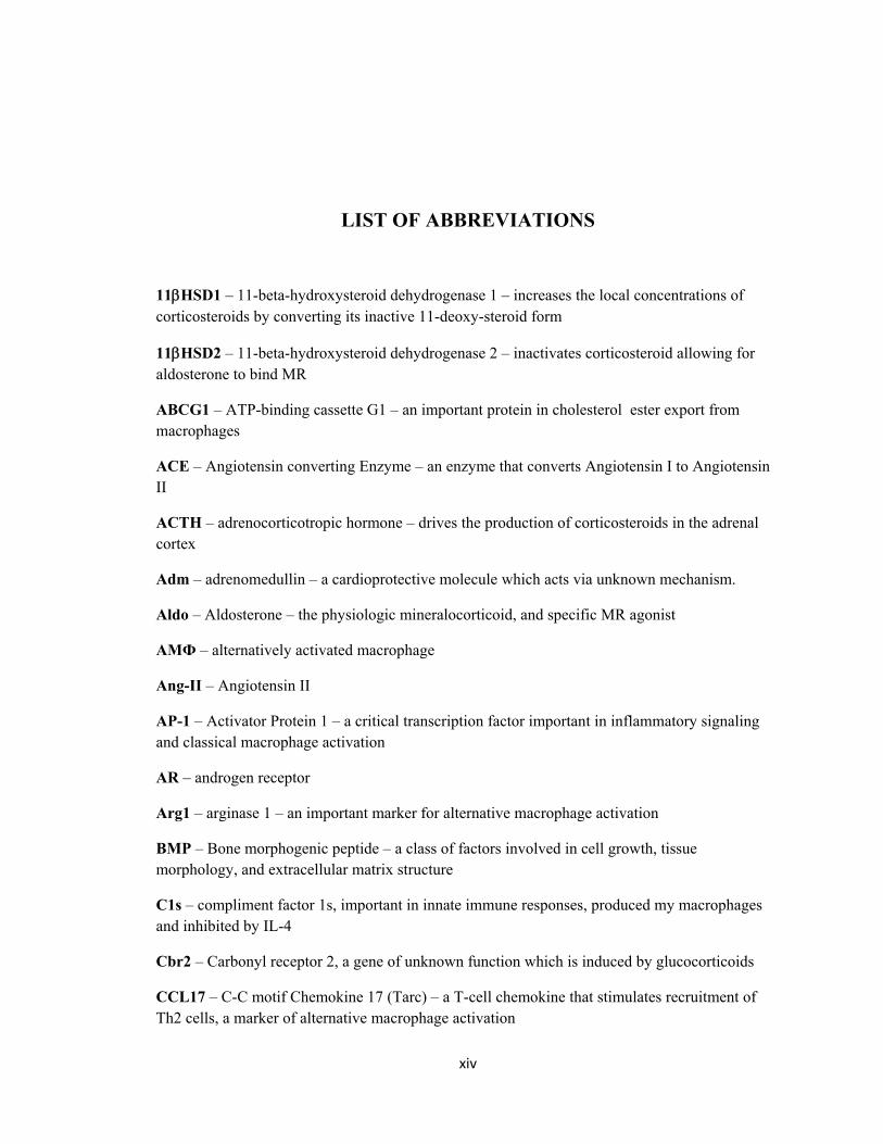
LIST OF ABBREVIATIONS
11βHSD1 – 11-beta-hydroxysteroid dehydrogenase 1 – increases the local concentrations of corticosteroids by converting its inactive 11-deoxy-steroid form
11βHSD2 – 11-beta-hydroxysteroid dehydrogenase 2 – inactivates corticosteroid allowing for aldosterone to bind MR
ABCG1 – ATP-binding cassette G1 – an important protein in cholesterol ester export from macrophages
ACE – Angiotensin converting Enzyme – an enzyme that converts Angiotensin I to Angiotensin II
ACTH – adrenocorticotropic hormone – drives the production of corticosteroids in the adrenal cortex
Adm – adrenomedullin – a cardioprotective molecule which acts via unknown mechanism.
Aldo – Aldosterone – the physiologic mineralocorticoid, and specific MR agonist
AMФ – alternatively activated macrophage
Ang-II – Angiotensin II
AP-1 – Activator Protein 1 – a critical transcription factor important in inflammatory signaling and classical macrophage activation
AR – androgen receptor
Arg1 – arginase 1 – an important marker for alternative macrophage activation
BMP – Bone morphogenic peptide – a class of factors involved in cell growth, tissue morphology, and extracellular matrix structure
C1s – compliment factor 1s, important in innate immune responses, produced my macrophages and inhibited by IL-4
Cbr2 – Carbonyl receptor 2, a gene of unknown function which is induced by glucocorticoids
CCL17 – C-C motif Chemokine 17 (Tarc) – a T-cell chemokine that stimulates recruitment of Th2 cells, a marker of alternative macrophage activation

xv
CCL24 – C-C motif Chemokine 24 – Eotaxin 2 important in recruitment of eosinophils in Th2 inflammation
CCL7 – C-C chemokine motif ligand 3, Monocyte chemoattractant protein 3, potently induced by IL-4 in macrophages
CCR2 – C-C chemokine receptor 2 – a receptor for MCP-1, important in the recruitment and differentiation of classically activated macrophages
CCR4 – C-C chemokine receptor 4, involved in the recruitment of classically activated macrophages
CD163 – Cell determinant 163 – a heme scavenger protein, activated by the glucocorticoid receptor
CD36 – Cell Determinant 36, a scavenger receptor involved in the response and uptake of LDL particles.
CD40L – Cell determinant 40 ligand – a co-activator molecule necessary for T-cell expansion
Cdh1- E-cadherin – an adhesion molecule and marker of alternative macrophage activation, involved in the formation of giant cells
Cdh2 – N cadherin – another adhesion molecule of unknown function in macrophages
CHF – Congestive heart failure
ChIP – Chromatin Immunoprecipitation – a technique which identifies specific sequences which are bound by nuclear factors
CLEC-2 – C-type lectin 2, a pro-inflammatory C-type lectin of unknown physiologic function
Clu – clusterin – an extracellular factor associated with fibrotic diseases
Col1a1 – Collagen I type a1 an important component of extracellular matrix and fibrotic processes, upregulated with left ventricular dysfunction
Col3a1 – collagen III type a1 – an important component of extracellular matrix and fibrotic processes, upregulated with left ventricular dysfunction
Col4a1 – collagen type 4 a1, an integral component to basement membranes, inhibited by IL-4
Cort – Corticosterone – the physiologic glucocorticoid in rodents
CTGF – connective tissue growth factor – an important growth factor in fibrotic processes
CVD – cardiovascular disease
CX3CR1 – C-X-3-C chemokine receptor 1 – a marker for the alternatively activated macrophage precursur

xvi
Cxcl12 – C-x-C motif chemokine ligand 12 – a chemokine induced by aldosterone which triggers the recruitment of fibrocytes and activates fibroblasts
Cyr61 – Cystine Rich Protein 61 – a BMP inhibitor that is associated with angiogenesis
DOCA – Deoxycorticosterone acetate – an MR agonist which is used pharmacologically
Epl – Eplerenone – a specific mineralocorticoid receptor antagonist
F13a1 – Clotting Factor 13 a1, a marker for wound healing macrophages and alternatively activated macrophages
F4/80 – Emr1 – a marker of fully differentiated tissue macrophages
FC – floxed littermate control – used as a genetic control for all experiments
Fcrls – Fc receptor lamda s – another marker of alternative macrophage activation sometimes referred to as Msr2, macrophage scavenger receptor 2
Fizz1, Rentl1a –Resistin like 1a– another marker of alternative macrophage activation of unknown function
Fgf9 – fetal growth factor 9 – a growth factor induced by aldosterone with unknown function
Fn1 – Fibronectin 1 – a marker of alternatively activatged macrophages, important in fibrotic processes
GM-CSF – granulocyte monocyte colony stimulating factor – necessary for the formation of macrophages and other granulocytes
GR – glucocorticoid receptor, nuclear steroid receptor that specifically binds corticosteroids
Hmga2 – High Mobility group A2 – a transcription factor of unknown cellular function strongly upregulated by glucocorticoid receptor
HPA – Hypothalamic – Pituitary – Adrenal Axis, neuro-hormone system involved in many physiologic functions including stress and circadian rhythms.
HRE – Hormone Response Element – a DNA element which binds to a nuclear hormone receptor (for example GRE binds the glucocorticoid receptor, MRE binds the mineralocorticoid receptor)
Htra1 – Serine Protease which is induced by glucocorticoids and is involved in extracellular matrix turnover
HW/BW – hear-weight/body weight ratio
IFNγ – Interferon gamma – a potenti stimulant of classical macrophage activation
IL-10 – interleukin 10 – a potent anti-inflammatory cytokine, marker of alternative macrophage activation

xvii
IL-12 – Interleukin 12, a cytokine produced by macrophages that stimulates Th1 proliferation
IL-13 – Interleukin 13, the other Th2 cell cytokine which acts similarly to IL-4 in the macrophages, but has distinct effects in the lung
IL-1β – Interleukin 1 beta – a marker of classical macrophage activation
IL-27ra – Interleukin 27 receptor a, a marker of alternative activated macrophages, strongly induced by IL-4
IL-33 – Interleukin 33, a cytokine that regulates lymphocyte proliferation
IL-4 – Interleukin 4, one of the primary Th2 cell cytokines which drives alternative macrophage activation
IL-6 – Interleukin 6 – a marker of classical macrophage activation
iNOS – inducible nitric oxide – an important marker for classical macrophage activation
IRF3 – Interferon Response Factor 3 – signaling factor involved in classical macrophage activation
L-NAME - L-NG-Nitroarginine methyl ester – a non-specific nitric oxide synthase inhibitor
LDL – low density lipoprotein – an important component in the pathogeneisis of atherosclerosis
LPS – lipopolysaccharide – Binds to TLR4 and stimulates classical macrophage activation
LXL – liver-x-receptor – nuclear receptor that binds oxysterols and lipids, and has anti-inflammatory activity
LysM-cre – lysozyme cre – an animal which allows for the generation of granulocyte specific deletions
M1 – Classically activated macrophage
MФRKO, MMRKO – macrophage specific MR knockout
MCP-1 – monocyte chemoattractant 1, a less specific marker of classical macrophage activation
Me1 – malic enzyme 1 – rate limiting enzyme in the NADPH, NADP shunt necessary for the maitenence of cytosolic NADPH stores
MHC – major histocompatability complex – involved in the stimulation of adaptive immunity
MMP-9 – matrix metaloprotease 9, a enzyme involved in degredation of extracellular matrix, involved in inflammatory cell recruitment, and is a marker for classical macrophage activation
xviii
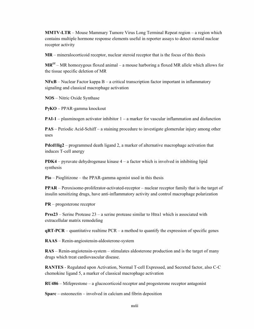
MMTV-LTR – Mouse Mammary Tumore Virus Long Terminal Repeat region – a region which contains multiple hormone response elements useful in reporter assays to detect steroid nuclear receptor activity
MR – mineralocorticoid receptor, nuclear steroid receptor that is the focus of this thesis
MRf/f – MR homozygous floxed animal – a mouse harboring a floxed MR allele which allows for the tissue specific deletion of MR
NFκB – Nuclear Factor kappa B – a critical transcription factor important in inflammatory signaling and classical macrophage activation
NOS – Nitric Oxide Synthase
PγKO – PPAR-gamma knockout
PAI-1 – plasminogen activator inhibitor 1 – a marker for vascular inflammation and disfunction
PAS – Periodic Acid-Schiff – a staining procedure to investigate glomerular injury among other uses
Pdcd1lig2 – programmed death ligand 2, a marker of alternative macrophage activation that induces T-cell anergy
PDK4 – pyruvate dehydrogenase kinase 4 – a factor which is involved in inhibiting lipid synthesis
Pio – Pioglitizone – the PPAR-gamma agonist used in this thesis
PPAR – Peroxisome-proliferator-activated-receptor – nuclear receptor family that is the target of insulin sensitizing drugs, have anti-inflammatory activity and control macrophage polarization
PR – progesterone receptor
Prss23 – Serine Protease 23 – a serine protease similar to Htra1 which is associated with extracellular matrix remodeling
qRT-PCR – quantitative realtime PCR – a method to quantify the expression of specific genes
RAAS – Renin-angiostensin-aldosterone-system
RAS – Renin-angiotensin-system – stimulates aldosterone production and is the target of many drugs which treat cardiovascular disease.
RANTES - Regulated upon Activation, Normal T-cell Expressed, and Secreted factor, also C-C chemokine ligand 5, a marker of classical macrophage activation
RU486 – Mifeprestone – a glucocorticoid receptor and progesterone receptor antagonist
Sparc – osteonectin – involved in calcium and fibrin deposition
xix
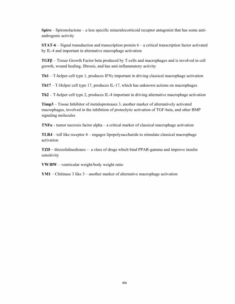
Spiro – Spironolactone – a less specific mineralocorticoid receptor antagonist that has some anti-androgenic activity
STAT-6 – Signal transduction and transcription protein 6 – a critical transcription factor activated by IL-4 and important in alternative macrophage activation
TGFβ – Tissue Growth Factor beta produced by T-cells and macrophages and is involved in cell growth, wound healing, fibrosis, and has anti-inflammatory activity
Th1 – T-helper cell type 1, produces IFNγ important in driving classical macrophage activation
Th17 – T-Helper cell type 17, produces IL-17, which has unknown actions on macrophages
Th2 – T-helper cell type 2, produces IL-4 important in driving alternative macrophage activation
Timp3 – Tissue Inhibitor of metaloproteases 3, another marker of alternatively activated macrophages, involved in the inhibition of proteolytic activation of TGF-beta, and other BMP signaling molecules
TNFα - tumor necrosis factor alpha – a critical marker of classical macrophage activation
TLR4 – toll like receptor 4 – engages lipopolysaccharide to stimulate classical macrophage activation
TZD – thiozolidinediones – a class of drugs which bind PPAR-gamma and improve insulin sensitivity
VW/BW – ventricular weight/body weight ratio
YM1 – Chitinase 3 like 3 – another marker of alternative macrophage activation

xx
ABSTRACT
The Mineralocorticoid Receptor (MR) is a multifunctional nuclear steroid
receptor which is responsible the actions of two classes of physiologic ligands:
mineralocorticoids (aldosterone) and glucocorticoids (corticosterone in rodents).
Mineralocorticoid receptor antagonists provide pleiotropic beneficial effects which
culminate in a marked reduction in mortality of patients with cardiovascular disease.
Since, inflammation is a common thread which connects the beneficial actions of MR
antagonists, we tested the hypothesis that they act as direct immunomodulatory agents.
To test this hypothesis we generated a macrophage specific MR knockout mouse
(MΦMRKO) to identify MR dependant macrophage actions, and illustrate the
importance macrophage MR in cardiovascular inflammation. Through broad
transcriptional analysis we show that glucocorticoid occupied MR is necessary for
efficient classical macrophage activation and represses alternative macrophage activation
programs. In vitro, macrophage MRKO synergizes with PPAR-γ and the glucocorticoid
receptor to enhance alternative activation. While ablation of glucocorticoid occupied MR
mimics the actions of MR antagonists, it did not overlap with the effect of aldosterone,
suggesting glucocorticoid and aldosterone occupied MR have markedly different
activities.
xxi
In vivo, MΦMRKO mimics MR antagonists and protects against cardiac
hypertrophy, fibrosis and vascular damage. This is despite a salt dependant day-time
increase in systolic pressure, heart rate, and pulse pressure. Cardiac injury results in the
recruitment of classically activated macrophages and a repression in alternative activation
markers both of which were mitigated in MΦMRKO mice. Together these data implicate
some macrophage actions as protective role in the inflammatory response to cardiac
stress.
These studies demonstrate that macrophage glucocorticoid•MR is an important
control point in macrophage polarization in innate immunity and likely illustrates a
conserved ancestral function of MR. We conclude that glucocorticoid•MR control of
macrophage polarization is a critical target for the beneficial cardiovascular action of MR
antagonists.

1
CHAPTER I:
INTRODUCTION
Overview
Cardiovascular disease is the leading cause of morbidity and mortality in the
world. With increasing prevalence of risk factors such as hyperlipidemia, obesity, and
hypertension, along with our aging population, CVD will present an even greater medical
and social burden in the future. The last five years have demonstrated that a combination
of public health initiatives and development of modern therapeutics can be effective in
combating this challenge. Despite worrying trends in cardiovascular risk factors in the
recent decade, from 2000 to 2006 mortality caused by cardiovascular diseases has
actually declined [3].
One of the defining features of cardiovascular disease is its clustering of risk
factors. Hemodynamic and metabolic derangements not only combine to increase risk of
a cardiac or vascular event, but are highly likely to coexist [5-7]. This implies the
existence of common underlying mechanisms that drive the development of these
pathologies. One approach to understanding the pathogenesis of cardiovascular disease is
to elucidate specific mechanisms underlying the success of effective therapeutics. The
focus of this thesis is on one particularly effective class of drugs, the mineralocorticoid
antagonists, which are used to treat many facets of cardiovascular disease.

2
The Mineralocorticoid receptor: structure and function
The target of MR antagonists is the mineralocorticoid receptor, a nuclear steroid receptor
mapped to human chromosome 4q31.1-q31.2 [8, 9]. Nuclear steroid receptors contain a
common domain structure and a highly conserved protein sequence across species. They
act by binding intracellular steroids which cause their dimerization and nuclear
translocation [11]. Steroid activation stimulates DNA binding to specific response
elements and regulation of transcription. The transcriptional effect of steroid hormone
receptors varies widely depending on the promoter context. Factors such as response
element sequence [12], chromatin structure [13], as well as the presence of co-activators
or co-repressors [14], nuclear protein-protein interactions and post-translational
modifications such as ubiquitination [15, 16] and SUMOylation [15, 17] of both the
receptors themselves and accessory factors all have dramatic effects of the transcriptional
activity of nuclear steroid receptors. It has been also demonstrated that ligand binding to
membrane bound nuclear steroid receptors, including MR, has acute cytosolic effects [18,
19]; however the physiologic significance of this activity remains unknown [20].
Transcription of the MR gene is driven by two independent promoters and the mature
RNAs generated have either 2 or 3 5’ untranslated exons and 10 translated exons[21].
The protein, which resembles other steroid nuclear receptors, contains four conserved
domains: the N-terminal domain, DNA binding domain, ligand binding domain, and C
terminal domain[8, 22-24]. The N-terminal domain of MR, is the largest among the
steroid receptor family consisting of 604 amino acids, and shares only 15% homology
with other steroid receptors[22]. Structure-function studies have demonstrated that the
N-terminal domain is important for MR’s ability to both activate and repress

3
transcription, interact with transcriptional co-activators. This region also plays a role in
intramolecular interactions with the ligand binding domain[25, 26].
The DNA binding domain is the most highly conserved region across the steroid receptor
family. Structurally, the DNA binding domain folds into two perpendicular alpha helices
and coordinates with two zinc molecules in the classic zinc-finger binding domain
fold[27, 28]. Type II steroid receptors such as MR, GR, and the androgen receptor and
progesterone receptor are known to bind the AGAACA half site, through this domain,
and thereby alter transcription[8, 29].
The ligand binding domain of MR is highly similar to that of the glucocorticoid receptor
(GR) and binds two physiologic ligands: aldosterone, which is the physiologic
mineralocorticoid, and glucocorticoids, such as cortisol in humans and corticosterone in
rodents[30]. MR binds physiological glucocorticoids and aldosterone with similar
affinities with a Kd of 0.87 nM for aldosterone and 1.36 nM for cortisol and
corticosterone[8, 29, 31]. While physiologic variations in circulating aldosterone occurs
primarily within the range of the mineralocorticoid receptor affinity serum
glucocorticoid concentrations are approximately three orders of magnitude higher in
concentration and sufficient to saturate MR[32]. This poses a paradox on how MR
activity is actually regulated by aldosterone and glucocorticoids and is a central question
of this thesis.
It is important to note that while the ligand binding domain of MR has been carefully
characterized; many important aspects of MR’s structure remain poorly understood.
There has been no full characterization of the response elements occupied by MR [22].
4
The impact of post-translational modifications is also not well understood. While many
MR cofactors have been identified, the relationship between those co-factors and MR’s
ability to regulate transcription in physiologically relevant targets and cell types has not
been fully addressed. Developing a novel system to study MR’s biochemistry in cell
types demonstrated to be physiologically important will be an important step in
understanding the structure-function relationships for MR.
Tissue activation of MR
MR is a nearly ubiquitously expressed protein. However high expression of MR has
been identified in tissues such as brown fat, colon, hippocampus, and renal epithelium
[33]. As was mentioned earlier, MR binds multiple physiologic ligands, aldosterone and
glucocorticoids. This is a poses an apparent paradox, as glucocorticoids and aldosterone
have different physiologic functions that are independently regulated. Glucocorticoid
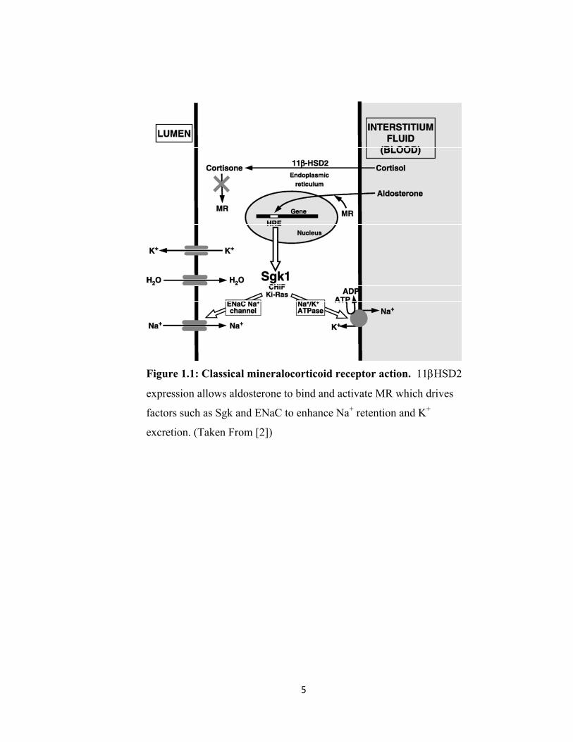
concentrations are in marked excess to aldosterone [34]. The enzyme 11βHSD2
alleviates this problem to a degree by converting corticosterone and cortisol to 11-
dehydrocortisone and cortisone respectively, which do not bind the mineralocorticoid
receptor (Figure 1.1). 11βHSD2 expression is limited to tissues involved in salt, and
water homeostasis, and contributes to hemodynamic stability such as the colon, vascular
endothelium, and renal epithelium [35].
Aldosterone is produced by the zona-glomerulosa of the adrenal cortex in response to
activation of the renin-angiotensin-system (RAS) and increases in serum potassium
5
Figure 1.1: Classical mineralocorticoid receptor action. 11βHSD2
expression allows aldosterone to bind and activate MR which drives
factors such as Sgk and ENaC to enhance Na+ retention and K+
excretion. (Taken From [2])

6
Figure 1.2: MR regulates two divergent physiologic systems. MR is the target of two
adrenal steroids: cortisol which is induced by HPA axis stimulation in response to stress,
and aldosterone induced by RAAS activation to regulate electrolyte homeostasis. The
interaction and differences in activity between cortisol and aldosterone occupied MR is a
fundamental unanswered question in the field.

7
(Figure 1.2). The Renin-Angiotensin system is stimulated by the initial production of
renin, which is secreted by juxtaglomerular cells in the kidney in response to sympathetic
drive, low salt delivery, and low perfusion pressure[36]. Renin acts as an endopeptidase
which cleaves the C-terminal two amino acids from angiotensinogen to generate
angiotensin I. Angiotensin I is subsequently cleaved by Angiotensin Converting Enzyme
(ACE) to generate Angiotensin II. Angiotensin II then synergizes with serum potassium
to drive aldosterone synthesis. Aldosterone then circulates, stimulating MR in renal and
colonic epithelium, which thereby increases the transcription of sodium hydrogen
exchanger (NHE), endothelial sodium channel (ENaC) and sodium potassium channel
which work in concert to increase the absorption of sodium and excretion of
potassium[37]. The actions of mineralocorticoid receptor in the kidney are necessary for
electrolyte homeostasis. Whole body deletion of MR in mice results in salt wasting
which results in death in the absence of a compensatory high salt diet [38, 39].
Pathological increases in MR activation such as in the setting of hyperaldosteronism or
11βHSD2 blockade, results in excessive salt and water absorption and hypertension with
hypokalemia [40]. Conversely, blockade of this action by MR antagonists such as
eplerenone and spironolactone results in blood pressure reduction and is utilized as a 4th
line treatment in hypertensive patients [41].
Hypertension
Hypertension is the single most common cause of prescription drug use in the
United States. It is currently estimated that between 58 to 65 million adults in the United
States alone suffer from primary hypertension [42]. Over half the 65 and older
demographic has some form of hypertension, suggesting that as the population ages,

8
hypertension is going to become an even greater problem [43]. Despite a relatively
simple diagnosis, and clear benefit of antihypertensive therapy in reducing stroke,
myocardial infarction and heart failure incidences, it is estimated that only 34% of
patients with hypertension are adequately controlled [44].
The etiology of primary hypertension is currently unknown and likely to be a
combination of a number of factors including increased sympathetic activity or
adrenergic response, insufficient nephron mass, as well as increased activity of or
sensitivity to the renin, angiotensin, aldosterone system (RAAS) [45]. The consequences
of uncontrolled hypertension are severe, including increased risk of left ventricular
hypertrophy leading to heart failure. Additionally, hypertensive patients are at an
increased risk for stroke, both ischemic and intracerebral hemorrhage, and chronic renal
insufficiency[46].
Treatment of hypertension primarily focuses on simultaneously addressing
cardiac preload, caused by relative volume excess, and afterload caused by elevated
peripheral vascular resistance. First line therapy generally involves diuretics such as
thiazides, which increase urinary excretion, and reduce plasma volume. In the case that
thiazides are not sufficiently effective, second line therapies include ACE inhibitors,
which block the conversion of angiotensin I to angiotensin II, angiotensin receptor
blockers, beta blockers and calcium channel blockers [47]. A recent clinical study
demonstrated that the mineralocorticoid receptor antagonist eplerenone was equally
effective to ACE inhibitors at reducing blood pressure, reducing left ventricular
hypertrophy, and reducing the incidence of renal disease and cardiovascular events [41].

9
The target of eplerenone and spironolactone, the mineralocorticoid receptor has been long
associated with the development of hypertension. In the mid 1950s two critical
discoveries began our understanding of steroid control of blood pressure. First, in 1952
and 1953 researchers isolated an adrenal cortical steroid which stimulated an increase in
blood pressure and later became known as aldosterone [48, 49]. Second, in 1955 the
first description of a patient harboring an adrenal cortical tumor who presented with
hypertension and hypokalemia, the hallmarks of what later became known as Conn’s
syndrome was published [50].
Primary hyperaldosteronism is a cause of hypertension, and is characterized by
variable hypokalemia, low renin, and evidence of aldosterone excess due to an adrenal
tumor or more rarely due to bi-lateral adrenal hyperplasia. Patients with primary
hyperaldosteronism have a marked increase in the relative risk of cardiac events
including stroke, myocardial infarction, and atrial fibrillation. While the prevalence of
primary hyperaldosteronism is unknown, it is suspected to be as high as 11.2% of patients
with essential hypertension [51, 52].
Heart Failure
One outcome of chronic, uncontrolled hypertension is congestive heart failure
(CHF). This disease is increasing in incidence, at least in part due to the aging population
and improved treatment of acute coronary disease. CHF is generally caused by either
reduced systolic function, leading to reduced ejection factor, or diastolic dysfunction
which prevents ventricular filling. The resulting reduction in cardiac output leads to
inadequate tissue perfusion. The physiologic response to the reduced perfusion enhances

10
vascular resistance and increases blood volume, thereby exacerbating the cardiac
dysfunction. Despite recent therapeutic advances, the mortality rate of patients with CHF
remains high [51, 52].
Aldosterone, which is typically found in serum concentrations of 0.1-0.5 nM on a
normal diet, and is significantly increased (as high as 5 nM) in patients with CHF [53].
This elevation played a key role in the initial discovery and isolation of aldosterone.
Serum from patients with CHF contained a substance which stimulated salt retention in
the kidney despite relatively elevated renal blood flow and glomerular filtration.
Additional sites such as the colon, salivary and sweat glands were also stimulated to
retain salt by the same substance which was later identified as the 18-aldehyde steroid,
aldosterone [49]. Salt retention stimulated by the RAAS leads to expanded intravascular
volume, an important pathophysiologic process in CHF. Therapeutic agents such as ACE
inhibitors, Angiotensin receptor (AT1) blockers, aldosterone synthesis blockers, and MR
antagonists target this response and are important tools in combating hypertension and
cardiovascular disease [54].
Hypertension, however, is one of the least predictive indicators of CHF risk suggesting
the underlying pathogenesis of heart failure is more complex. Other strong indicators
such as coronary heart disease, ischemic heart disease, cigarette smoking, diabetes, and
obesity all contribute to CHF risk [55]. At this stage, our understanding of the molecular
mechanisms which lead to reduced ventricular function are poorly understood. While
most treatments for CHF are geared towards reducing ventricular load and increasing
cardiac contractility, MR antagonists appear to improve CHF outcomes by a different and
as of yet, unknown mechanism.
11
MR Blockade
While mineralocorticoid receptor blockers have been long used as antihypertensives and
in the treatment of both primary hyperaldosteronism and apparent mineralocorticoid
excess, the RALES and EPHESUS studies demonstrated that there was a greater
involvement of MR in cardiovascular disease than merely acting as a regulator of
hemodynamics [56, 57]. Each of these multi-center randomized control studies focused
on high risk heart failure patients with post myocardial infarction and high end diastolic
volume. Each study showed that adding an MR antagonist, either eplerenone
(EPHESUS) or spironolactone (RALES), to current standard of care significantly
improved morbidity and mortality over an 18 month period [56, 57]. Of note, these
patients were already on optimal antihypertensive therapy, thus it is unlikely that the
hemodynamic changes afforded by MR blockade were the sole mechanism of improved
outcome. Subsequent studies have demonstrated that addition of an MR antagonist to
the regimen of patients with essential hypertension significantly improved blood pressure
in the absence of elevated aldosterone levels and to the same degree as ACE inhibitors,
and reduced left ventricular hypertrophy [41]. Subsequently, it was shown that the
efficacy of mineralocorticoid blockade could not be predicted by changes in potassium
excretion. This suggests that at least some of the beneficial aspects of eplerenone and
spironolactone may be independent of their ability to antagonize the actions of
aldosterone in renal epithelium [58].
An additional study investigated MR antagonists in the treatment of mild to moderate
heart failure patients with symptomatic idiopathic dilated cardiomyopathy. MR
antagonism was only shown to improve left ventricular diastolic function in a subgroup

12
of patients which demonstrated increased markers of cardiac fibrosis despite the fact that
both groups had similar aldosterone levels[59, 60]. These results strengthen the idea that
MR antagonists may provide additional benefits in the context of CHF pathogenesis
beyond blocking the actions of aldosterone.
Animal studies have strengthened the hypothesis that extrarenal actions of MR are
important contributors to cardiovascular disease. MR antagonists reverse cardiovascular
fibrosis even in the absence of mineralocorticoid excess[61]. MR antagonists also inhibit
models of diabetic nephropathy[62, 63], ischemic stroke, L-NAME induced renal
injury[64], and atherosclerosis[65, 66] in the absence of high aldosterone levels. A
common feature that connects the cardiovascular pathologies mitigated by MR
antagonists is inflammation.
Inflammation
Inflammation refers to the physiologic, cellular, and molecular changes that occur
following immune cell activation. Classically defined by Celsius in the 1st century AD,
inflammation has been associated with redness (rubor), heat (calor), swelling (tumor),
and pain (dolor) that commonly occurs with active hyperemia following an acute,
localized infection. In the last 20 years, great strides have been made in appreciating the
complexities of immune system interaction, and the remarkable impact that inflammatory
signaling has on normal physiology and pathogenesis.
Generally, the immune system is separated into two major categories, innate and
adaptive immunity. In innate immunity, pattern recognition receptors such as TLR4,
which bind lipopolysaccharide (LPS) from gram(-) bacteria, drive immediate

13
inflammatory responses. Specifically, TLR4 activation drives activation of STAT1, NF-
κB, and AP1 transcription factors, which enhances expression of pro-inflammatory
cytokines to induce protective actions in other cells. These mediators in turn recruit
additional immune cells, enhance phagocytic activity, as well as the production of
cytotoxic mediators to kill the invading pathogen [67]. More recent work has
demonstrated that innate immune responses are not limited to protecting against invading
pathogens. Circulating free fatty acids [68, 69] and minimally modified LDL [70], which
are both increased in cardiovascular disease, have been shown to bind TLR4 and activate
inflammatory processes.
Pattern recognition receptors on macrophages do not always produce equivalent
responses. For example CD163, a receptor expressed on glucocorticoid stimulated
macrophages is responsible for binding and stimulating the uptake of heme and
hemoglobin. It also is necessary for the processing of apoptotic cells and plays an
important anti-inflammatory role [71-74]. CD163 also illustrates how the definition of
innate immunity has expanded over recent years. Whereas it used to refer specifically to
acute recognition of pathogens, in this thesis we define it as any direct activation of the
immune system in response to invading pathogens, cell death, and cell stress through the
activation of low specificity pattern recognition receptors.
In contrast to the innate immune response, the adaptive immune system utilizes genetic
recombination to produce a variety of antigen receptors. The presentation of antigens
facilitates a rigorous positive and negative selection process to identify specific receptors
which respond to invading pathogens. Adaptive immunity provides highly specific and
long term immunity. Specific humoral immunity is provided by B-cells which produce
14
opsonizing or inactivating antibodies, and CD8(+) T cells (cytotoxic T cells), which bind
type I MHC complexes and stimulate cell mediated immunity.
Cross talk between innate and adaptive immunity is extensive and mediated primarily
through CD4(+) helper T cells. Helper T cells respond to presented antigen-type II MHC
complexes, and require co-stimulatory molecules expressed on APCs for proliferation.
Co-stimulatory molecules on macrophages require innate activation prior to their
expression. Without co-stimulatory molecules such as CD80 (B7.1), which is potently
upregulated by TLR4 on macrophages, T cells undergo anergy, leading to immune
tolerance [75]. Additionally, cytokines secreted by macrophages stimulate T-cell
differentiation into different populations. For example, IL-12 which is expressed by
activated macrophages, enhances Th1 cell proliferation which in turn stimulates IFNγ
production[76]. Conversely, macrophage derived cytokines CCL17, CCL24 [77, 78]
and IL-33[79] specifically stimulate Th2 proliferation and recruitment.
Innate responses are coordinately regulated throughout the evolution of the adaptive
response. First, helper T cells directly drive the activation of macrophages and other
innate immune effector cells. Second, antibody and antibody-antigen complexes, through
the engagement of Fc receptors, also stimulate innate immunity, and enhance the activity
of phagocytes such as macrophages.
Macrophages in Inflammation
One critical cell type in inflammation is the macrophage. Macrophages are
central to the development of every type and every phase of an inflammatory response.
Macrophages express chemokine and cytokine receptors which stimulate their

15
recruitment to a stressed tissue. Additionally macrophages express pattern recognition
receptors, which bind to specific structural motifs found on invading pathogens, cellular
particles and stressed or apoptotic cells.
Phagocytosis of foreign particles can cause activation of type 2 major
histocompatability complexes. In these cases, macrophages can migrate like dendritic
cells into lymphoid tissue, and stimulate T-cell and B-cell expansion and thus the
adaptive immune response. Interestingly, the type and degree of lymphoid response can
also be directly regulated by macrophage activity. Macrophages along with class 2
MHC, express CD40-ligand, which binds to CD40 on T-cells during MHC-TCR
engagement and is necessary for full T-cell activation and proliferation. CD40L
transcription is highly regulated in activated macrophages [80].
B-cell proliferation is similarly regulated by macrophages. B7, a protein which
has two splice isoforms in macrophages is important lymphocyte proliferation. The larger
isoform, upregulated following macrophage activation, is necessary for T-helper cell
mediated B-cell expansion. Conversely, the low molecular weight isoform found in
marginal zone macrophages of active B-cell follicles downregulates B-cell proliferation
[81]. Additionally, specific cytokines secreted by activated macrophages can skew
immune responses along certain pathways. Specifically, Th1 mediated responses are
driven primarily by IL-12, which in addition represses Th2 activation [82].
During resolution of an inflammatory response, stressed cells, and recruited
neutrophils and lymphocytes undergo apoptosis. Apoptosis stimulates the release of
TGFβ from cells and induces the expression of cell-surface markers that are recognized

16
by binding lectins on macrophages [83]. Engagement of apoptotic cell markers causes
their phagocytosis and subsequent lysis, and at the same time has potent anti-
inflammatory activity, by downregulating the expression of pro-inflammatory cytokines
[84-86].
Functional plasticity of macrophages is a hallmark of their ability to combat
widely diverse pathogens and insults. The normal evolution of an inflammatory response
requires carefully coordinated recruitment of functionally distinct subclasses of
macrophages, which fall within a spectrum between classically activated macrophages
(M1, expressing a high level of pro-inflammatory cytokines and reactive oxygen species)
and alternatively activated macrophages (AMΦ) involved in pathogen sequestration,
wound healing, and phagocytosis of apoptotic cells. Improper perturbation of this
dynamic balance has been associated with numerous diseases and thus is an important
consideration to the development of therapies to disorders with an inflammatory
component [87-91].
Macrophage polarization is guided by four components. First, recruitment of
different monocyte/macrophage sub-populations is driven by specific chemokines. For
example CCL17 and CCL24 are critical for the recruitment of classically activated
macrophages in the lung. CCR4 knockout or scavenging by the chemokine scavenger D6
can lead to an AMΦ polarized and more protective response in a model of pulmonary
fibrosis [92-94].
Secondly, macrophage activation is directly guided by the local cytokine milieu.
Specifically, Th2 cytokines IL-4 and IL-13 stimulate STAT6 activation, which activates
17
expression of AMФ markers and at the same time downregulates the expression of M1
markers. Conversely, IFN-gamma, produced by Th1 lymphocytes stimulates IRF3
mediated M1 polarized responses [87].
Thirdly, activation of pattern recognition receptors guides macrophages toward
either a pro-inflammatory M1 state, or an alternatively activated AMΦ state. TLR4
engagement by either lipopolysaccharide (LPS) found in the cell wall of bacteria, or by
free fatty acids, leads to upregulation of M1 markers. Conversely, activation of the
mannose receptor is associated with upregulation of PGC-1alpha, PPARγ, and arginase
[89, 90], which are markers of AMФ.
Thus, the macrophage samples the external environment through chemokine,
cytokine, and pattern recognition receptors, and integrates them with endocrine and
nutrient signals to guide a specific inflammatory response. As shown in this thesis, this
occurs in part through activation of nuclear receptors such as MR. Through the control of
a remarkably wide array of transcriptional networks, nuclear receptors not only play a
critical role the physiologic function of macrophages, but are also important therapeutic
targets. Understanding how nuclear receptors function in macrophages provides a model
of how MR may be guiding inflammation in cardiovascular disease.
Macrophages in cardiovascular disease:
Molecules which stimulate inflammation have been strongly associated with the
formation of atherosclerotic plaques and cardiovascular disease. Smoking, pro-
inflammatory adipokines, environmental stress, and chronic inflammatory states such as
rheumatoid arthritis all increase the risk of coronary artery disease. Conversely,
18
decreasing inflammation reduces the risk of cardiovascular disease and is an important
new approach to therapy.
Atherosclerosis is a disease that leads to destabilization of vessel walls, and
partial to full arterial occlusion. Initial endothelial dysfunction allows for leakage of
modified LDL which serves as substrates for macrophage activation. Cytokines
produced by activated macrophages in the arterial intima induce additional smooth
muscle and macrophage recruitment into the plaque[95-97]. Additionally, once activated,
macrophages produce reactive oxygen species which further enhance LDL modification.
Finally, cholesterol loading of macrophages leads to their differentiation into foam cells,
a major destabilizing force in the core of the plaque. All these mechanisms contribute to
the expansion and development of complex atherosclerotic fibrotic plaques which reduce
distal tissue perfusion, and increase the risk of embolization, two hallmarks of
cardiovascular disease [97].
Macrophages respond to a variety of pathogenic stimuli (e.g., oxidized LDL,
diabetes related glycosylation end products and angiotensin II), and produce a
programmed cytokine response. For example, modified LDL particles interact with
various cell surface receptors leading to NF-κB and AP-1 nuclear factor activation. The
subsequent cytokine release promotes intimal expansion and plaque development.
Inhibition of many cytokines including MCP-1, TNFα, and IL-8 has been shown to slow
the progression of atherosclerosis in mouse models [98].
Genetic studies have demonstrated that macrophages modulate other
cardiovascular risk factors as well. TNFα produced by macrophages has been shown to

19
directly induce insulin resistance in multiple cell types [99, 100]. Additionally, targeted
ablation of CCR2, a chemokine receptor in macrophages, resulted in resistance to diet
induced obesity and improved insulin sensitivity [101]. Obesity is associated with
enhanced macrophage infiltration into adipose tissue, though the consequences of this
phenomenon have not been thoroughly investigated [102].
Conversely, factors associated with cardiovascular disease also stimulate
inflammation. Obesity is associated with elevated levels of IL6 and TNFα, which
stimulate inflammation. Hyperglycemia as a result of insulin resistance results in
aberrant glycosylation products which can also induce inflammatory responses. Finally,
numerous pharmaceutical drugs including PPAR agonists [103], HMGCoA Reductase
inhibitors [104], and angiotensin receptor (AT1) blockers[105], which are known to
impact lipid transport, insulin signaling, and blood pressure, all inhibit inflammation.
Whether this occurs via a common pathway or through divergent mechanisms has not
been investigated thoroughly. However, it is clear that manipulation of macrophage
activation may be a critical component of cardioprotective drugs. A comprehensive
understanding of how macrophage activation is regulated may provide new targets in the
treatment of cardiovascular disease.
Nuclear Receptor control of macrophage activation
Classically, nuclear receptor activation involves binding of a small molecule ligand.
This allows dissociation from heat shock proteins and nuclear translocation. Upon
entering the nucleus nuclear receptors bind DNA response elements either as a
homodimer or heteromeric complexes and alter transcription. The relative simplicity of

20
the overall mechanism belies both the complexity of and breadth by which nuclear
receptors impact cellular function. Nuclear receptors traditionally are highly
promiscuous, binding multiple physiologic ligands which drive different transcriptional
responses. Second, post translational modifications, such as SUMOylation and
Ubiquitination and even phosphorylation can dramatically alter the transcription of target
genes [15, 17]. Allosteric regulation by interacting proteins, chromatin, methylated
bases, as well as the target DNA itself can alter both nuclear receptor affinity for ligands
and other transcription factors [106, 107]. These three factors combinatorially allow
nuclear receptors to create a wide array of context and promoter specific effects which
integrate nutrient and endocrine signals to create a nuanced response.
The mechanism of action of nuclear receptors along with the breadth of responses
they control makes them ideal targets for pharmacologic manipulation. Indeed, a number
of highly important therapies for numerous disorders act through modulating nuclear
receptor activity beyond merely hormone replacement. This is especially important for
the treatment of cardiovascular disease and its risk factors.
Recently, cell type specific deletions of nuclear receptors have demonstrated that
nuclear receptor action in immune cells, specifically in macrophages is critical for their
physiologic role. More importantly, deletion of therapeutic targets in macrophages
abolished the beneficial actions of the ligands, demonstrating that many treatments act
through direct immunomodulation. These results have highlighted the importance of
macrophages in coordinating many pathophysiologic changes which occur in
cardiovascular disease and metabolic syndrome.
21
Nuclear receptors temper specific patterns of macrophage activation which are
important to the metabolic, cellular, and physiologic response to cell stress and cell death.
Understanding the mechanisms by which they act, and in turn identifying transcriptional
programs which are important to cardiovascular disease may yield novel avenues for
therapy. Moreover, as nuclear receptor programs are identified, gene targeting can be
utilized as a window to identify novel nuclear receptor dependant macrophage roles in
physiologic adaptations and pathogenesis.
PPAR-γ in macrophages:
PPAR-γ has been shown to be a key regulator of M1/AMΦ polarization [108,
109]. PPAR-γ agonists have been shown to suppress the expression of M1 associated
pro-inflammatory cytokines TNF-α, IL-6, and IL-1β[1]. Coordinately, PPAR-γ
expression and activity is enhanced by AMΦ differentiation, and in turn is required for
the upregulation of numerous markers of AMΦ activity including arginase-1, mannose
receptor, and CD36. Moreover, PPAR-γ activation during macrophage maturation from
monocytes resulted in an enhanced AMΦ response. However, studies demonstrating that
PPAR-γ reverses cytotoxic T-lymphocyte suppression, a function of AMФ macrophages
[110], and the identification of M1 activated genes which are TZD resistant suggest that
PPAR-γ is involved in regulating a specific subset of genes, rather than globally tipping
the scales in the AMΦ direction.
At the molecular level, PPAR-γ alters macrophage function through a multitude
of mechanisms. PPAR-γ undergoes a ligand and SUMOylation dependent
conformational shift which allows direct binding to NFκB, recruitment of a co-repressor

22
complex, and subsequently suppresses transcription of NFκB target genes [111]. PPAR-γ
has also been shown to inhibit the activity of AP-1 and STAT-1. These transcription
factors are all involved in the induction of pro-inflammatory cytokines during M1
differentiation [1]. Concurrently, PPAR-γ activation enhances STAT-6 activity following
IL-4/13 stimulation, a signaling pathway which drives AMΦ differentiation [112]. More
work however needs to be done to determine the relative contribution of each mechanism
to PPAR-γ’s effects and context dependency [113].
Recent studies have demonstrated that direct effects of TZDs on macrophage
function are a central component of their physiologic effects. Deletion of PPAR-γ in
macrophages results in reduced glucose tolerance and impaired insulin sensitivity in
skeletal muscle and liver as well as enhanced weight gain and insulin resistance in high
fat fed mice [109, 114]. This was associated with an increase in pro-inflammatory
cytokine expression as well as a reduction ABCG1 expression, suggesting an impairment
of reverse cholesterol transport. These studies also demonstrated that macrophage
PPAR-γ is necessary for the full insulin sensitizing effects of TZDs. Finally, bone
marrow transplant of PPAR-γ-null cells into an LDLR -/- background resulted in a
significant increase in atherosclerotic plaque size [115]. This observation indicates that
PPAR-γ has direct, atheroprotective effects is consistent with the finding that PPAR-γ
activation enhances cholesterol efflux from macrophages and inhibits foam cell formation
[97]. However, if this is due to a direct alteration in macrophage polarization within the
plaque or specific recruitment of anti-atherogenic cells remains to be determined.
Glucocorticoid Receptor in Macrophages

23
Activation of the hypothalamic pituitary adrenal axis (HPA) by physiologic or
emotional stress leads to production of the glucocorticoid cortisol (corticosterone in
rodents). Cortisol, produced in the zona fasciculata of the adrenal cortex acts classically
through the glucocorticoid receptor (GR) to impart a number of important physiologic
adaptations. These include inducing insulin resistance, driving adipogenesis, inhibiting
osteogenesis, and down-regulating inflammatory responses.
Glucocorticoids have been utilized clinically for nearly 50 years, not only to
compensate for adrenal insufficiency, but to treat a number of inflammatory conditions
including asthma and autoimmune disorders, and are indicated as a preventive measure to
combat septic shock [116-118]. Glucocorticoids inhibit inflammatory responses through
a variety of cellular mechanisms, including decreasing adhesion molecule expression on
endothelial cells, reducing PMN cell viability, inhibiting Th1 cell proliferation, and
increasing lymphocyte apoptosis.
Recent genetic studies utilizing a macrophage specific knockout of GR suggest
that GR activation in macrophages is a necessary component of the mechanism by which
glucocorticoids inhibit inflammation. GR activation by cortisol and other pharmacologic
agents such as dexamethasone, or hydrocortisone, inhibits a multitude of activation
cascades within the macrophage [119]. First, GR has been shown to bind directly to the
inflammatory signaling molecules NFκB and AP1 and directly inhibit their nuclear
translocation, their affinity for DNA, and their ability to activate transcription.
Secondly, GR functions independently to alter gene transcription and to inhibit
inflammation. For example GR up-regulates IKβ, a molecule which inhibits NFκB

24
Figure 1.3: Glucocorticoid Action on classical macrophage activation.
Activation of GR causes pleiotropic anti-inflammatory actions on
macrophage. GR directly upregulates IKβ which sequesters cytosolic NFκB,
suppressor of cytokine signaling 3 (Socs3) and other anti-inflammatory
signaling molecules such as GILZ. Conversely, GR interferes with NFκB
transcriptional action through multiple biochemical mechanisms.

25
nuclear translocation [120, 121]. Other targets of GR such as GILZ and IL-10 are also
upregulated by glucocorticoid exposure and have direct anti-inflammatory activity
(Figure 1.3) [122]. Finally, GR can bind to atypical response elements such those found
in the IL-1β promoter to recruit co-repressors and directly inhibit the transcription of
inflammatory cytokines. At the level of macrophage polarization, the actions of GR are
primarily to inhibit Th1 and M1 responses[123]. Any direct role of GR in regulating
AMФ differentiation following IL-4 mediated STAT6 activation has not been
comprehensively assessed.
More recent studies investigating the effect of high dose glucocorticoids on
monocyte differentiation suggest that GR is involved in stimulating a third macrophage
subpopulation. This anti-inflammatory macrophage population mimics myeloid
suppressor cells which downregulate inflammation during cancer progression. This cell
type is characterized by high level expression of the anti-inflammatory IL-10 cytokine,
normal levels of MCP-1, possesses high chemotaxis activity, and high levels of the
adhesion molecule CD163 which is involved in the uptake of apoptotic cells[124].
Ultimately, glucocorticoids play an important role in the feedback regulation of
immune system activation. Pro-inflammatory cytokines such as TNFα and IL-1β
directly activate the paraventricular nucleus of the hypothalamus causing CRH release,
HPA axis activation, and increases of serum cortisol levels[125]. Cortisol levels in turn,
acutely inhibit inflammation, in part through downregulating classical macrophage
activation, and inducing the differentiation and recruitment of anti-inflammatory
monocytes and macrophages.

26
LXR in macrophages
Liver-X-Receptor (LXR) α and β are adopted orphan nuclear receptors similar to
PPAR-γ and bind to physiologic concentration of oxysterol metabolites of cholesterol.
LXRα is primarily expressed in the liver but also is expressed in inflammatory cells such
as macrophages and T-cells[126]. LXRβ is expressed ubiquitously[127]. LXRα is
primarily involved in regulating the cellular metabolism of cholesterol. In macrophages
this primarily consists of balancing intake with efflux to maintain homeostasis. Increases
in cholesterol intake by phagocytosis of modified LDL particles or apoptotic cells,
induces abc transporter proteins, primarily ABCA1 and ABCG1[128]. This allows the
macrophage to unload cholesterol into HDL particles and back to the liver for clearance
as bile acids or re-packaging.
Interestingly, like PPAR-γ, LXRs have potent anti-inflammatory activity.
Through binding to LXR response elements, agonist activated LXR directly inhibits
NFκB mediated induction of TNFα, IL-1β, iNOS, and others[128]. Unlike PPAR-γ and
GR, LXRα activation by oxysterols results in stimulation of SPα, an anti-apoptotic factor
important in macrophage survival during cholesterol loading. Remarkably, this pathway
is also necessary to combat intracellular pathogens such as Listeria monocytogenes[129,
130].
Conversely, inflammatory conditions, such as following TLR3 or TLR4
activation, result in IRF3 mediated intracellular signaling and downregulation of LXR
activity. This is important as it prevents unnecessary anti-inflammatory activity during
the course of an infection. An unfortunate side effect of this interaction is that TLR4
27
activation by free fatty acids or minimally modified LDL can also downregulate LXR
activity [128]. This creates an imbalance between cholesterol absorption and efflux
within the macrophage. The combination of dysregulated inflammatory signaling and
cholesterol loading leads to foam cell formation, a cardinal and destabilizing process in
atherosclerotic plaque development[97].
Due to their role in reverse cholesterol transport and their potent anti-
inflammatory activity, LXRs represent a promising target for therapeutics.
Unfortunately, first generation LXR agonists caused a primary effect of inducing
SREBP-1c in the liver and stimulating hepatic lipogenesis [131]. This effect raised
serum triglyceride levels and even overwhelmed PPAR-α mediated triglyceride synthesis
and export leading to hepatic steatosis. On a positive note, it has been demonstrated that
LXR regulates SRREBP-1c, cholesterol efflux proteins ABCA1 and ABCG1, and
possess anti-inflammatory activity by distinct mechanisms. This allows for the creation
of next generation LXR ligands which select for beneficial activities may be possible
[132].
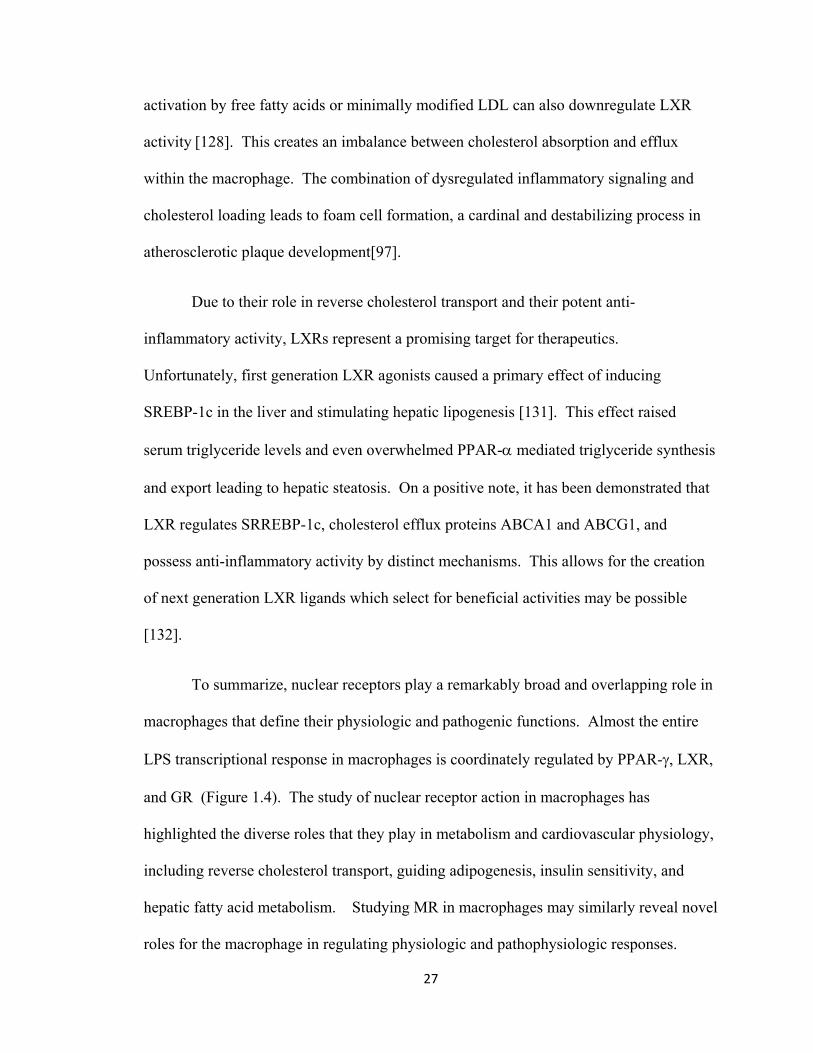
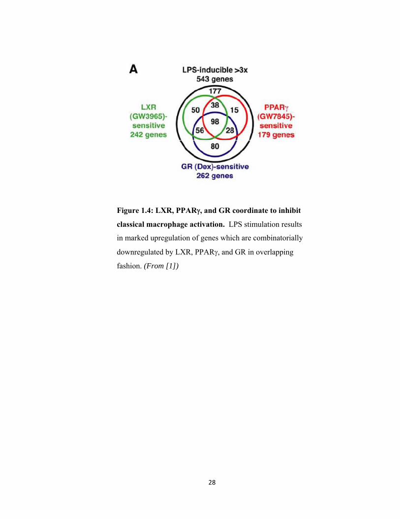
To summarize, nuclear receptors play a remarkably broad and overlapping role in
macrophages that define their physiologic and pathogenic functions. Almost the entire
LPS transcriptional response in macrophages is coordinately regulated by PPAR-γ, LXR,
and GR (Figure 1.4). The study of nuclear receptor action in macrophages has
highlighted the diverse roles that they play in metabolism and cardiovascular physiology,
including reverse cholesterol transport, guiding adipogenesis, insulin sensitivity, and
hepatic fatty acid metabolism. Studying MR in macrophages may similarly reveal novel
roles for the macrophage in regulating physiologic and pathophysiologic responses.
28
Figure 1.4: LXR, PPARγ, and GR coordinate to inhibit
classical macrophage activation. LPS stimulation results
in marked upregulation of genes which are combinatorially
downregulated by LXR, PPARγ, and GR in overlapping
fashion. (From [1])
29
MR Evolution:
The role of MR in controlling inflammation seems a far cry from its dogmatic role
in regulating electrolyte homeostasis. Understanding how the promiscuity and pleiotropy
of MR has evolved yields insights into how MR may be regulating inflammatory
processes. There are two competing models of the evolution of MR. The first model
involves the divergence of an ancestral corticoid receptor from other steroid receptors
which later split into the two functional glucocorticoid receptors MR and GR[4, 133,
134]. There are a number of lines of evidence that support this model. First, the DNA
binding domains of both MR and GR recognize remarkably similar sequence motifs, and
share stronger sequence homology when compared to the other steroid nuclear receptors.
Additionally, MR and GR share highly conserved ligand binding domains, allowing a
similar spectrum of agonists and antagonists, albeit with markedly different affinities.
Despite strong similarities between MR and GR in the DNA and ligand binding
domains, their N-terminal domains are starkly different. Sequence alignment of the
entire MR gene demonstrates weak, but greater homology with the progesterone receptor
(PR) and androgen receptor (AR) than with GR [133]. This suggests MR may be closer
to the ancestral steroid receptor as opposed to the more recently diverged corticoid
receptor. This hypothesis was strengthened by the discovery of the S810L point mutation
in MR which confers sensitivity to progesterone. Progesterone, which rises during
pregnancy, aberrantly activates MR in patents harboring this mutation, stimulating salt
retention and hypertension, in a dominant form of hereditary
pseudohyperaldosteronism[135]. Introduction of similar point mutations in the ligand
30
binding domain of MR can confer sensitivity to other physiologically important steroids
including androgens.
Together, these data present an interesting picture about the evolution of MR and
the evolution of steroid signaling in general. The binding promiscuity of MR early in
evolution was exploited through the increasing complexity of steroid biosynthesis.
Subsequent, gene duplications and mutations conferring binding specificity led to the
evolution of PR, GR, and AR. The ancestral position of MR early in evolution is
remarkable because the emergence of aldosterone synthase is a relatively recent event,
occurring when vertebrates first appeared on land [4]. Thus, it is likely that prior to the
evolution of aldosterone synthase, the primary mineralocorticoid receptor ligand was the
glucocorticoid cortisol.
In agnathans, and other early vertebrates, the physiologic role of cortisol is very
similar to what we observe in mammals today. Cortisol is induced by physiologic stress
and stimulates the glucocorticoid receptor which inhibits inflammatory processes and
promotes insulin resistance [136]. In contrast to tetrapods, cortisol also plays an
important role in electrolyte homeostasis in marine vertebrates including agnathans,
elasmobranchs and teleost fish. Corticosteroids activate MR, which subsequently
interacts with prolactin and growth hormone signaling to either stimulate NaCl
absorption, or excretion in the gills [137]. The physiologic consequence of this is
actually common knowledge among aquarium keepers, who recommend increasing the
salinity of water when manipulating fresh water fish, since stressed fish are less able to
regulate salt retention.

31
Figure 1.5: Evolution of the mineralocorticoid and glucocorticoid receptors. (A)
Early vertebrates expressed only one ancestral corticoid receptor which maintained
similar activity and binding affinity as the modern mineralocorticoid receptor (C). Prior
to the emergence of aldosterone synthase, the binding pocket of GR was altered by 2
mutations which conferred specificity to cortisol (blue), and relative insensitivity to DOC
(red) and aldosterone (green). MR’s activity has maintained its high affinity to both
glucocorticoid and mineralocorticoid since early evolution, a property which was not
altered following the emergence of aldosterone synthase and 11βHSD2 (From [4])

32
As vertebrates expanded to terrestrial environments a separate endocrine system evolved
to regulate salt homeostasis. The origin of aldosterone synthase is thought to be the result
from a duplication of 17-alpha-hydroxysteroid dehydrogenase [133]. Aldosterone acts as
the primary mineralocorticoid in terrestrial vertebrates as discussed previously.
Remarkably, unlike PR, GR, and AR, which evolved selectivity in their ligand binding
domains, MR conserved its ability to bind to both glucocorticoids and mineralocorticoids.
Instead, a secondary enzymatic system co-evolved with aldosterone synthase, 11βHSD2,
which inactivates cortisol and provides a mechanism for aldosterone to exert its effects in
a tissue specific manner [133]. The conservation of MR’s ability to bind corticosteroids,
however, suggests that glucocorticoid bound MR plays a necessary function, or that MR
is an important regulator of glucocorticoid signaling.
Glucocorticoid occupied MR
A majority of research into the biology of MR has been centered on the action of
aldosterone on renal epithelium, vascular endothelium, and cardiac tissue. Little attention
has been paid to glucocorticoid occupied MR in parenchymal tissues lacking 11βHSD2.
One extensively studied tissue which highly expresses MR and lacks 11βHSD2 is the
central nervous system. Understanding the role of MR in these tissues, especially relative
to glucocorticoid receptor activation and inflammatory signaling may hint at MR’s role in
similar tissues.
In the hippocampus, nuclear MR is thought to be mainly responsive to
glucocorticoids. It has been suggested that the cytosolic fraction of MR may be sensitive
to changes in glucocorticoid levels when they are at their lowest and induce rapid non

33
Figure 1.6: MR and GR action in the brain. Mineralocorticoid receptor coordinates
the neural response to stress through maintaining baseline function, as well as
instituting rapid non-genomic responses as glucocorticoid levels rise. MR may play a
similar role in macrophages: limiting and maintaining a threshold for the anti-
inflammatory actions of high dose glucocorticoids. (From [10])

34
genomic action such as triggering Src and Erk phosphorylation (Figure 1.6) [138].
However, the physiologic significance of these rapid actions is not known. In the
hippocampus, glucocorticoid activation of MR is necessary for the maintenance of neural
integrity and stable excitability, and sets the threshold of the stress response mediated by
the glucocorticoid receptor [10]. MR targets in the brain appear to counteract the actions
of GR through direct and indirect mechanisms [139]. Overexpression of MR in the
forebrain results in a reduction of anxiety responses associated with high stress [140].
Deletion of MR in the hippocampus results in structural changes associated with chronic
high dose glucocorticoid treatment associated with chronic stress [141]. Despite their
strong structural similarity, MR and GR drive opposing transcriptional programs with
only minor overlap [142].
MR at the crossroads of inflammation and cardiovascular disease:
Cardiovascular risk is determined by a number of highly co-morbid factors:
dyslipidemia, renal disease, insulin resistance, central obesity, atherosclerosis,
hypertension, cardiac hypertrophy and arrhythmia all contribute to increases in incidence
of cardiovascular events such as myocardial ischemia or ischemic stroke. The
mechanisms which lead to the high concordance and synergistic qualities between each
risk factor are incompletely understood.
One factor which is known to enhance each of these pathologies is inflammation.
Specific pro-inflammatory cytokines such as MCP-1, TNFα, and IL-1β are known to
exacerbate glomerular injury, trigger insulin resistance, increase vascular remodeling and
promote atherosclerosis, and are important in diet induced obesity [143]. Conversely,
35
cardiac arrhythmia, hypertrophy, obesity, glomerular disease, and dyslipidemia have all
been shown to trigger immune responses which appear detrimental to cardiovascular
health.
Remarkably, MR has also been linked to each cardiovascular risk factor. MR
antagonists have been shown to reduce proteinuria in non-diabetic renal disease[144] and
microalbuminurea in patients with mild to moderate diabetic nephropathy. MR
antagonism has been shown to be protective in rodent models of glomerular injury such
as L-NAME/Ang-II [145], salt fed spontaneous hypertensive rats [146], and diabetic
nephropathy [62]. While some models have demonstrated the importance of aldosterone
in the generation of glomerular disease, the fact that MR antagonism protects renal
disease even in the absence of hyperaldosteronism implies there is more to the
mechanism than aldosterone blockade.
The connection between obesity, dyslipidemia, and insulin resistance and MR is
less clear cut. In a few studies, plasma aldosterone levels have been shown to be
positively correlated with fasting plasma glucose, HOMA score (a measure of insulin
resistance), central adiposity, and incidence of metabolic syndrome [147]. Aldosterone
was also inversely correlated with plasma HDL levels [148]. However, a recent study
comparing patients with primary hyperaldosteronism and matched patients with essential
hypertension showed no differences in these metabolic parameters [149]. Unfortunately,
most studies investigating the correlation between MR activity and metabolic disease
ignore the fact that the tissues primarily associated with metabolic syndrome such as
adipocytes, liver, and skeletal muscle do not express 11βHSD2 and thus should be
largely insensitive to aldosterone. MR antagonism in high fat fed mice improved insulin

36
resistance, increased adiponectin levels, and reduced adipose tissue inflammation [150].
MR has also been associated with adipogenesis and differentiation of brown fat [151,
152], as well as direct downregulation of insulin receptor expression, all of which could
affect glucose disposal [153]. Unfortunately, there is insufficient clinical data to suggest
that MR antagonism may protect against the metabolic derangements which occur with
obesity.
Atherosclerosis is the result of a chronic inflammatory response in the intima of
large vessel walls in response to covalently modified lipoprotein particles such as LDL.
The inflammatory response results in endothelial dysfunction, smooth muscle hyperplasia
and migration into the vessel lumen, and macrophage recruitment, activation, and
differentiation into cholesterol laden foam cells [95, 97]. Atherosclerotic plaques
increase distal blood flow, and additionally can rupture causing clot and emboli
formation leading to ischemia of downstream tissues. As in other cases,
hyperaldosteronism in both humans and animal models is associated with the promotion
of atherosclerosis [154]. Aldosterone stimulates vascular endothelial adhesion
molecules, growth factors that contribute to smooth muscle hyperplasia, and enhances
oxidative stress through activation of NOS and NADPH oxidase which in turn can
stimulate further inflammation and promote LDL modification [155]. Conversely,
antagonism of MR even in the absence of elevated aldosterone levels protected against
atherosclerosis in ApoE -/- mice [65].
MR antagonists are potent anti-hypertensives, as has been discussed above.
However, they also have been demonstrated to reduce left ventricular hypertrophy even
in the absence of blood pressure lowering effects [53]. Similar results have been

37
demonstrated in animal models, where MR antagonists protect against hypertrophy in
spontaneous hypertensive rats and L-NAME/AngII mediated hypertensive mice even
without reducing blood pressure [156]. MR biology in cardiomyocytes is not fully
understood. Aldosterone binding activity and 11βHSD2 activity in cardiac tissue
remains controversial [32]. Hyperaldosteronism is strongly associated with increased
risk of arrhythmic death [157]. MR has been shown to stimulate delayed after
depolarizations which are a common underlying component of many arrhythmias and
was associated with upregulation of cardiac ryanodine receptor and downregulation of
FK506BP [158].
Interestingly, in a canine rapid pacing model of arrhythmia, which is not
associated with hyperaldosteronism, MR antagonist induced protective
electrophysiologic effects whereas ACE inhibitors had no significant effect, suggesting
again that some beneficial effect of MR antagonists is independent of aldosterone [159].
Finally, cardiac fibrosis which is a major contributing factor to both reduction of cardiac
contractility and progression to failure, as well as the duration and severity of cardiac
arrhythmia is linked to MR activity. Models of mineralocorticoid excess such as DOCA
salt and L-NAME/Ang-II stimulate peri-vascular and interstitial cardiac fibrosis that
mirrors those observed in cardiac failure patents [160]. Interestingly, removal of
mineralocorticoid stimulation did not result in reversal of the fibrosis; however,
administration of MR antagonists, even in the absence of aldosterone resulted in reversal
[161].
MR in macrophages.

38
As illustrated above, nuclear receptors play a remarkably broad role in
macrophages that define physiologic and pathogenic macrophage functions. The study of
nuclear receptor action in macrophages, highlights the diverse roles that they play for
example reverse cholesterol transport, guiding adipogenesis and insulin sensitivity,
hepatic steatosis. Interestingly, these responses fit into the same framework of how
macrophages respond to different invading pathogens.
For the most part, the described functions nuclear receptors primarily involve
anti-inflammatory effects such as down-regulating the production of pro-inflammatory
cytokines and upregulating other functions such as reverse cholesterol transport, or
driving beta oxidation in nearby tissues, or phagocytosing apoptotic cells. In this respect,
MR is unique among steroid nuclear receptors given its association with enhanced
inflammatory activity.
Hypothesis
Taken together the data presented in the previous sections argue for a direct role
for MR in enhancing inflammation and fibrotic processes in cardiovascular disease.
Since, macrophage play a prominent role in inflammatory models in which MR
antagonists are protective we hypothesize that macrophage MR is critically important in
detrimental innate immune response to cardiovascular injury. In this view, we propose
that MR in macrophages is required for the full protective effects of MR antagonists.
Specifically, we hypothesize that glucocorticoid occupied MR in macrophages promotes
classical macrophage activation, and that the anti-inflammatory action of MR antagonists

39
on macrophages is an important cardioprotective mechanism. Finally, we hypothesize
that one mechanism by which MR enhances inflammation is by antagonizing the actions
of glucocorticoids.
Specific Aims
To determine the role of MR in macrophage activation and polarization:
We utilized primary macrophage cultures and a transfectable macrophage cell line
to demonstrate that MR is expressed in macrophages and is activated by physiologic
concentrations of aldosterone and glucocorticoids. We then utilized careful combinations
of agonists and antagonists to distinguish the actions of MR on macrophage polarization
and appreciate the functional differences between glucocorticoid and mineralocorticoid
occupied MR. Finally, we generated a macrophage specific knockout of MR to test the
necessity of MR for the regulation of macrophage activation programs.
To determine that MR in macrophages drives cardiovascular inflammatory responses:
We applied the macrophage specific knockout of MR (MΦMRKO) to a model of
cardiac and vascular hypertension and fibrosis to determine whether MR in macrophages
was an important contributor to cardiovascular inflammation and subsequent pathology.
Similarities between MΦMRKO mice and MR antagonists would support the view that
MR antagonists act via a direct immunomodulatory mechanism.

40
References
1. Ogawa, S., et al., Molecular determinants of crosstalk between nuclear receptors and toll‐like receptors. Cell, 2005. 122(5): p. 707‐21.
2. Connell, J.M. and E. Davies, The new biology of aldosterone. J Endocrinol, 2005. 186(1): p. 1‐20.
3. Lloyd‐Jones, D., et al., Heart disease and stroke statistics‐‐2009 update: a report from the American Heart Association Statistics Committee and Stroke Statistics Subcommittee. Circulation, 2009. 119(3): p. 480‐6.
4. Bridgham, J.T., S.M. Carroll, and J.W. Thornton, Evolution of hormone‐receptor complexity by molecular exploitation. Science, 2006. 312(5770): p. 97‐101.
5. DeFronzo, R.A. and E. Ferrannini, Insulin resistance. A multifaceted syndrome responsible for NIDDM, obesity, hypertension, dyslipidemia, and atherosclerotic cardiovascular disease. Diabetes Care, 1991. 14(3): p. 173‐94.
6. Kaplan, N.M., The deadly quartet and the insulin resistance syndrome: an historical overview. Hypertens Res, 1996. 19 Suppl 1: p. S9‐11.
7. Shen, B.J., et al., Are metabolic risk factors one unified syndrome? Modeling the structure of the metabolic syndrome X. Am J Epidemiol, 2003. 157(8): p. 701‐11.
8. Arriza, J.L., et al., Cloning of human mineralocorticoid receptor complementary DNA: structural and functional kinship with the glucocorticoid receptor. Science, 1987. 237(4812): p. 268‐75.
9. Morrison, N., et al., Regional chromosomal assignment of the human mineralocorticoid receptor gene to 4q31.1. Hum Genet, 1990. 85(1): p. 130‐2.
10. Joels, M., et al., The coming out of the brain mineralocorticoid receptor. Trends Neurosci, 2008. 31(1): p. 1‐7.
11. Ribeiro, R.C., P.J. Kushner, and J.D. Baxter, The nuclear hormone receptor gene superfamily. Annu Rev Med, 1995. 46: p. 443‐53.
12. Meijsing, S.H., et al., DNA binding site sequence directs glucocorticoid receptor structure and activity. Science, 2009. 324(5925): p. 407‐10.
13. Oduro, A.K., M.K. Fritsch, and F.E. Murdoch, Chromatin context dominates estrogen regulation of pS2 gene expression. Exp Cell Res, 2008. 314(15): p. 2796‐810.
14. Wu, Y., et al., Nuclear compartmentalization of N‐CoR and its interactions with steroid receptors. Mol Cell Biol, 2006. 26(17): p. 6633‐55.
15. Tirard, M., et al., Sumoylation and proteasomal activity determine the transactivation properties of the mineralocorticoid receptor. Mol Cell Endocrinol, 2007. 268(1‐2): p. 20‐9.
16. Wallace, A.D. and J.A. Cidlowski, Proteasome‐mediated glucocorticoid receptor degradation restricts transcriptional signaling by glucocorticoids. J Biol Chem, 2001. 276(46): p. 42714‐21.
17. Chupreta, S., et al., Sumoylation‐dependent control of homotypic and heterotypic synergy by the Kruppel‐type zinc finger protein ZBP‐89. J Biol Chem, 2007. 282(50): p. 36155‐66.
18. Markos, F., V. Healy, and B.J. Harvey, Aldosterone rapidly activates Na+/H+ exchange in M‐1 cortical collecting duct cells via a PKC‐MAPK pathway. Nephron Physiol, 2005. 99(1): p. p1‐9.
19. Doolan, C.M., G.C. O'Sullivan, and B.J. Harvey, Rapid effects of corticosteroids on cytosolic protein kinase C and intracellular calcium concentration in human distal colon. Mol Cell Endocrinol, 1998. 138(1‐2): p. 71‐9.

41
20. Skott, O., et al., Rapid actions of aldosterone in vascular health and disease‐‐friend or foe? Pharmacol Ther, 2006. 111(2): p. 495‐507.
21. Zennaro, M.C., et al., Human mineralocorticoid receptor genomic structure and identification of expressed isoforms. J Biol Chem, 1995. 270(36): p. 21016‐20.
22. Pippal, J.B. and P.J. Fuller, Structure‐function relationships in the mineralocorticoid receptor. J Mol Endocrinol, 2008. 41(6): p. 405‐13.
23. Rogerson, F.M., F.E. Brennan, and P.J. Fuller, Dissecting mineralocorticoid receptor structure and function. J Steroid Biochem Mol Biol, 2003. 85(2‐5): p. 389‐96.
24. Rupprecht, R., et al., Transactivation and synergistic properties of the mineralocorticoid receptor: relationship to the glucocorticoid receptor. Mol Endocrinol, 1993. 7(4): p. 597‐603.
25. Obradovic, D., et al., DAXX, FLASH, and FAF‐1 modulate mineralocorticoid and glucocorticoid receptor‐mediated transcription in hippocampal cells‐‐toward a basis for the opposite actions elicited by two nuclear receptors? Mol Pharmacol, 2004. 65(3): p. 761‐9.
26. Rogerson, F.M. and P.J. Fuller, Interdomain interactions in the mineralocorticoid receptor. Mol Cell Endocrinol, 2003. 200(1‐2): p. 45‐55.
27. Luisi, B.F., et al., Crystallographic analysis of the interaction of the glucocorticoid receptor with DNA. Nature, 1991. 352(6335): p. 497‐505.
28. Remerowski, M.L., et al., 1H NMR studies of DNA recognition by the glucocorticoid receptor: complex of the DNA binding domain with a half‐site response element. Biochemistry, 1991. 30(50): p. 11620‐4.
29. Arriza, J.L., et al., The neuronal mineralocorticoid receptor as a mediator of glucocorticoid response. Neuron, 1988. 1(9): p. 887‐900.
30. Bledsoe, R.K., et al., A ligand‐mediated hydrogen bond network required for the activation of the mineralocorticoid receptor. J Biol Chem, 2005. 280(35): p. 31283‐93.
31. Alnemri, E.S., et al., Overexpression and characterization of the human mineralocorticoid receptor. J Biol Chem, 1991. 266(27): p. 18072‐81.
32. Funder, J.W., Mineralocorticoid receptors: distribution and activation. Heart Fail Rev, 2005. 10(1): p. 15‐22.
33. Bookout, A.L., et al., Anatomical profiling of nuclear receptor expression reveals a hierarchical transcriptional network. Cell, 2006. 126(4): p. 789‐99.
34. Rashid, S. and G.F. Lewis, The mechanisms of differential glucocorticoid and mineralocorticoid action in the brain and peripheral tissues. Clin Biochem, 2005. 38(5): p. 401‐9.
35. Naray‐Fejes‐Toth, A. and G. Fejes‐Toth, Novel mouse strain with Cre recombinase in 11beta‐hydroxysteroid dehydrogenase‐2‐expressing cells. Am J Physiol Renal Physiol, 2007. 292(1): p. F486‐94.
36. Sarzani, R., et al., Renin‐angiotensin system, natriuretic peptides, obesity, metabolic syndrome, and hypertension: an integrated view in humans. J Hypertens, 2008. 26(5): p. 831‐43.
37. Thomas, W., V. McEneaney, and B.J. Harvey, Aldosterone‐induced signalling and cation transport in the distal nephron. Steroids, 2008. 73(9‐10): p. 979‐84.
38. Bleich, M., et al., Rescue of the mineralocorticoid receptor knock‐out mouse. Pflugers Arch, 1999. 438(3): p. 245‐54.
39. Hubert, C., et al., Effects of mineralocorticoid receptor gene disruption on the components of the renin‐angiotensin system in 8‐day‐old mice. Mol Endocrinol, 1999. 13(2): p. 297‐306.

42
40. Armanini, D., et al., The pathogenesis of pseudohyperaldosteronism from carbenoxolone. J Endocrinol Invest, 1989. 12(5): p. 337‐41.
41. Pitt, B., et al., Effects of eplerenone, enalapril, and eplerenone/enalapril in patients with essential hypertension and left ventricular hypertrophy: the 4E‐left ventricular hypertrophy study. Circulation, 2003. 108(15): p. 1831‐8.
42. Fields, L.E., et al., The burden of adult hypertension in the United States 1999 to 2000: a rising tide. Hypertension, 2004. 44(4): p. 398‐404.
43. Hajjar, I., J.M. Kotchen, and T.A. Kotchen, Hypertension: trends in prevalence, incidence, and control. Annu Rev Public Health, 2006. 27: p. 465‐90.
44. Chobanian, A.V., et al., The Seventh Report of the Joint National Committee on Prevention, Detection, Evaluation, and Treatment of High Blood Pressure: the JNC 7 report. Jama, 2003. 289(19): p. 2560‐72.
45. Garbers, D.L. and S.K. Dubois, The molecular basis of hypertension. Annu Rev Biochem, 1999. 68: p. 127‐55.
46. Wilson, P.W., Established risk factors and coronary artery disease: the Framingham Study. Am J Hypertens, 1994. 7(7 Pt 2): p. 7S‐12S.
47. Kaplan, N.M., Treating prehypertension: a review of the evidence. Curr Hypertens Rep, 2008. 10(4): p. 326‐9.
48. Tait, J.F., S.A. Simpson, and H.M. Grundy, The effect of adrenal extract on mineral metabolism. Lancet, 1952. 1(6699): p. 122‐4.
49. Tait, S.A., J.F. Tait, and J.P. Coghlan, The discovery, isolation and identification of aldosterone: reflections on emerging regulation and function. Mol Cell Endocrinol, 2004. 217(1‐2): p. 1‐21.
50. Conn, J.W. and L.H. Louis, Primary aldosteronism: a new clinical entity. Trans Assoc Am Physicians, 1955. 68: p. 215‐31; discussion, 231‐3.
51. Rossi, G.P., T.M. Seccia, and A.C. Pessina, Primary aldosteronism: part II: subtype differentiation and treatment. J Nephrol, 2008. 21(4): p. 455‐62.
52. Rossi, G.P., T.M. Seccia, and A.C. Pessina, Primary aldosteronism ‐ part I: prevalence, screening, and selection of cases for adrenal vein sampling. J Nephrol, 2008. 21(4): p. 447‐54.
53. Funder, J.W. and A.S. Mihailidou, Aldosterone and mineralocorticoid receptors: Clinical studies and basic biology. Mol Cell Endocrinol, 2009. 301(1‐2): p. 2‐6.
54. Whaley‐Connell, A., et al., Renin‐angiotensin‐aldosterone system intervention in the cardiometabolic syndrome and cardio‐renal protection. Ther Adv Cardiovasc Dis, 2007. 1(1): p. 27‐35.
55. He, J., et al., Risk factors for congestive heart failure in US men and women: NHANES I epidemiologic follow‐up study. Arch Intern Med, 2001. 161(7): p. 996‐1002.
56. Effectiveness of spironolactone added to an angiotensin‐converting enzyme inhibitor and a loop diuretic for severe chronic congestive heart failure (the Randomized Aldactone Evaluation Study [RALES]). Am J Cardiol, 1996. 78(8): p. 902‐7.
57. Pitt, B., et al., The EPHESUS trial: eplerenone in patients with heart failure due to systolic dysfunction complicating acute myocardial infarction. Eplerenone Post‐AMI Heart Failure Efficacy and Survival Study. Cardiovasc Drugs Ther, 2001. 15(1): p. 79‐87.
58. Rocha, R. and J.W. Funder, The pathophysiology of aldosterone in the cardiovascular system. Ann N Y Acad Sci, 2002. 970: p. 89‐100.
59. Izawa, H., et al., Mineralocorticoid receptor antagonism ameliorates left ventricular diastolic dysfunction and myocardial fibrosis in mildly symptomatic patients with idiopathic dilated cardiomyopathy: a pilot study. Circulation, 2005. 112(19): p. 2940‐5.

43
60. Nagata, K., et al., Mineralocorticoid receptor antagonism attenuates cardiac hypertrophy and failure in low‐aldosterone hypertensive rats. Hypertension, 2006. 47(4): p. 656‐64.
61. Paulis, L., et al., Regression of left ventricular hypertrophy and aortic remodelling in NO‐deficient hypertensive rats: effect of L‐arginine and spironolactone. Acta Physiol (Oxf), 2008. 194(1): p. 45‐55.
62. Guo, C., et al., Mineralocorticoid receptor antagonist reduces renal injury in rodent models of types 1 and 2 diabetes mellitus. Endocrinology, 2006. 147(11): p. 5363‐73.
63. Ustundag, A., et al., The effects of spironolactone on nephron function in patients with diabetic nephropathy. Ren Fail, 2008. 30(10): p. 982‐91.
64. Ikeda, H., et al., Spironolactone suppresses inflammation and prevents L‐NAME‐induced renal injury in rats. Kidney Int, 2009. 75(2): p. 147‐55.
65. Keidar, S., et al., Effect of eplerenone, a selective aldosterone blocker, on blood pressure, serum and macrophage oxidative stress, and atherosclerosis in apolipoprotein E‐deficient mice. J Cardiovasc Pharmacol, 2003. 41(6): p. 955‐63.
66. Takai, S., et al., Eplerenone inhibits atherosclerosis in nonhuman primates. Hypertension, 2005. 46(5): p. 1135‐9.
67. Verstrepen, L., et al., TLR‐4, IL‐1R and TNF‐R signaling to NF‐kappaB: variations on a common theme. Cell Mol Life Sci, 2008. 65(19): p. 2964‐78.
68. Kim, F., et al., Toll‐like receptor‐4 mediates vascular inflammation and insulin resistance in diet‐induced obesity. Circ Res, 2007. 100(11): p. 1589‐96.
69. Nguyen, M.T., et al., A subpopulation of macrophages infiltrates hypertrophic adipose tissue and is activated by free fatty acids via Toll‐like receptors 2 and 4 and JNK‐dependent pathways. J Biol Chem, 2007. 282(48): p. 35279‐92.
70. Bae, Y.S., et al., Macrophages generate reactive oxygen species in response to minimally oxidized low‐density lipoprotein: toll‐like receptor 4‐ and spleen tyrosine kinase‐dependent activation of NADPH oxidase 2. Circ Res, 2009. 104(2): p. 210‐8, 21p following 218.
71. Fabriek, B.O., C.D. Dijkstra, and T.K. van den Berg, The macrophage scavenger receptor CD163. Immunobiology, 2005. 210(2‐4): p. 153‐60.
72. Fabriek, B.O., et al., The macrophage scavenger receptor CD163 functions as an innate immune sensor for bacteria. Blood, 2009. 113(4): p. 887‐92.
73. Philippidis, P., et al., Hemoglobin scavenger receptor CD163 mediates interleukin‐10 release and heme oxygenase‐1 synthesis: antiinflammatory monocyte‐macrophage responses in vitro, in resolving skin blisters in vivo, and after cardiopulmonary bypass surgery. Circ Res, 2004. 94(1): p. 119‐26.
74. Schaer, D.J., et al., CD163 is the macrophage scavenger receptor for native and chemically modified hemoglobins in the absence of haptoglobin. Blood, 2006. 107(1): p. 373‐80.
75. Goronzy, J.J. and C.M. Weyand, T‐cell co‐stimulatory pathways in autoimmunity. Arthritis Res Ther, 2008. 10 Suppl 1: p. S3.
76. Kang, B.Y. and T.S. Kim, Targeting cytokines of the interleukin‐12 family in autoimmunity. Curr Med Chem, 2006. 13(10): p. 1149‐56.
77. Medoff, B.D., et al., CD11b+ myeloid cells are the key mediators of Th2 cell homing into the airway in allergic inflammation. J Immunol, 2009. 182(1): p. 623‐35.
78. Mikhak, Z., et al., Contribution of CCR4 and CCR8 to antigen‐specific T(H)2 cell trafficking in allergic pulmonary inflammation. J Allergy Clin Immunol, 2009. 123(1): p. 67‐73 e3.

44
79. Smithgall, M.D., et al., IL‐33 amplifies both Th1‐ and Th2‐type responses through its activity on human basophils, allergen‐reactive Th2 cells, iNKT and NK cells. Int Immunol, 2008. 20(8): p. 1019‐30.
80. Elgueta, R., et al., Molecular mechanism and function of CD40/CD40L engagement in the immune system. Immunol Rev, 2009. 229(1): p. 152‐72.
81. Bhatia, S., et al., B7‐1 and B7‐2: similar costimulatory ligands with different biochemical, oligomeric and signaling properties. Immunol Lett, 2006. 104(1‐2): p. 70‐5.
82. Goriely, S., R. Cavoy, and M. Goldman, Interleukin‐12 family members and type I interferons in Th17‐mediated inflammatory disorders. Allergy, 2009. 64(5): p. 702‐9.
83. Chen, W., et al., TGF‐beta released by apoptotic T cells contributes to an immunosuppressive milieu. Immunity, 2001. 14(6): p. 715‐25.
84. de Almeida, C.J. and R. Linden, Phagocytosis of apoptotic cells: a matter of balance. Cell Mol Life Sci, 2005. 62(14): p. 1532‐46.
85. Hart, S.P., I. Dransfield, and A.G. Rossi, Phagocytosis of apoptotic cells. Methods, 2008. 44(3): p. 280‐5.
86. Kim, S., K.B. Elkon, and X. Ma, Transcriptional suppression of interleukin‐12 gene expression following phagocytosis of apoptotic cells. Immunity, 2004. 21(5): p. 643‐53.
87. Gordon, S., Alternative activation of macrophages. Nat Rev Immunol, 2003. 3(1): p. 23‐35.
88. Mantovani, A., et al., The chemokine system in diverse forms of macrophage activation and polarization. Trends Immunol, 2004. 25(12): p. 677‐86.
89. Martinez, F.O., L. Helming, and S. Gordon, Alternative activation of macrophages: an immunologic functional perspective. Annu Rev Immunol, 2009. 27: p. 451‐83.
90. Martinez, F.O., et al., Macrophage activation and polarization. Front Biosci, 2008. 13: p. 453‐61.
91. Varin, A. and S. Gordon, Alternative activation of macrophages: Immune function and cellular biology. Immunobiology, 2009.
92. Freeman, C.M., et al., CCR4 participation in Th type 1 (mycobacterial) and Th type 2 (schistosomal) anamnestic pulmonary granulomatous responses. J Immunol, 2006. 177(6): p. 4149‐58.
93. Martinez de la Torre, Y., et al., Increased inflammation in mice deficient for the chemokine decoy receptor D6. Eur J Immunol, 2005. 35(5): p. 1342‐6.
94. Trujillo, G., et al., A novel mechanism for CCR4 in the regulation of macrophage activation in bleomycin‐induced pulmonary fibrosis. Am J Pathol, 2008. 172(5): p. 1209‐21.
95. Libby, P., Inflammation in atherosclerosis. Nature, 2002. 420(6917): p. 868‐74. 96. Rader, D.J. and E. Pure, Lipoproteins, macrophage function, and atherosclerosis: beyond
the foam cell? Cell Metab, 2005. 1(4): p. 223‐30. 97. Shashkin, P., B. Dragulev, and K. Ley, Macrophage differentiation to foam cells. Curr
Pharm Des, 2005. 11(23): p. 3061‐72. 98. Charo, I.F. and M.B. Taubman, Chemokines in the pathogenesis of vascular disease. Circ
Res, 2004. 95(9): p. 858‐66. 99. Borst, S.E., The role of TNF‐alpha in insulin resistance. Endocrine, 2004. 23(2‐3): p. 177‐
82. 100. Lee, L.F., et al., The role of TNF‐alpha in the pathogenesis of type 1 diabetes in the
nonobese diabetic mouse: analysis of dendritic cell maturation. Proc Natl Acad Sci U S A, 2005. 102(44): p. 15995‐6000.

45
101. Weisberg, S.P., et al., CCR2 modulates inflammatory and metabolic effects of high‐fat feeding. J Clin Invest, 2006. 116(1): p. 115‐24.
102. Weisberg, S.P., et al., Obesity is associated with macrophage accumulation in adipose tissue. J Clin Invest, 2003. 112(12): p. 1796‐808.
103. Lehrke, M. and M.A. Lazar, The many faces of PPARgamma. Cell, 2005. 123(6): p. 993‐9. 104. Ky, B. and D.J. Rader, The effects of statin therapy on plasma markers of inflammation in
patients without vascular disease. Clin Cardiol, 2005. 28(2): p. 67‐70. 105. Dagenais, N.J. and F. Jamali, Protective effects of angiotensin II interruption: evidence for
antiinflammatory actions. Pharmacotherapy, 2005. 25(9): p. 1213‐29. 106. Brodie, J. and I.J. McEwan, Intra‐domain communication between the N‐terminal and
DNA‐binding domains of the androgen receptor: modulation of androgen response element DNA binding. J Mol Endocrinol, 2005. 34(3): p. 603‐15.
107. McEwan, I.J., Nuclear receptors: one big family. Methods Mol Biol, 2009. 505: p. 3‐18. 108. Bouhlel, M.A., et al., PPARgamma activation primes human monocytes into alternative
M2 macrophages with anti‐inflammatory properties. Cell Metab, 2007. 6(2): p. 137‐43. 109. Odegaard, J.I., et al., Macrophage‐specific PPARgamma controls alternative activation
and improves insulin resistance. Nature, 2007. 447(7148): p. 1116‐20. 110. Van Ginderachter, J.A., et al., Peroxisome proliferator‐activated receptor gamma
(PPARgamma) ligands reverse CTL suppression by alternatively activated (M2) macrophages in cancer. Blood, 2006. 108(2): p. 525‐35.
111. Pascual, G., et al., A SUMOylation‐dependent pathway mediates transrepression of inflammatory response genes by PPAR‐gamma. Nature, 2005. 437(7059): p. 759‐63.
112. Huang, J.T., et al., Interleukin‐4‐dependent production of PPAR‐gamma ligands in macrophages by 12/15‐lipoxygenase. Nature, 1999. 400(6742): p. 378‐82.
113. Straus, D.S. and C.K. Glass, Anti‐inflammatory actions of PPAR ligands: new insights on cellular and molecular mechanisms. Trends Immunol, 2007. 28(12): p. 551‐8.
114. Hevener, A.L., et al., Macrophage PPAR gamma is required for normal skeletal muscle and hepatic insulin sensitivity and full antidiabetic effects of thiazolidinediones. J Clin Invest, 2007. 117(6): p. 1658‐69.
115. Babaev, V.R., et al., Conditional knockout of macrophage PPARgamma increases atherosclerosis in C57BL/6 and low‐density lipoprotein receptor‐deficient mice. Arterioscler Thromb Vasc Biol, 2005. 25(8): p. 1647‐53.
116. Bomholt, S.F., et al., Involvement and role of the hypothalamo‐pituitary‐adrenal (HPA) stress axis in animal models of chronic pain and inflammation. Stress, 2004. 7(1): p. 1‐14.
117. Hadid, R., et al., Role of several mediators of inflammation on the mouse hypothalamo‐pituitary‐adrenal axis response during acute endotoxemia. Neuroimmunomodulation, 1999. 6(5): p. 336‐43.
118. Richards, L.J., et al., Protective effects of endotoxin in a rat model of chronic inflammation are accompanied by suppressed secretion of pro‐inflammatory cytokines and biphasic alteration in hypothalamo‐pituitary‐adrenal axis activity. J Neuroendocrinol, 2006. 18(11): p. 875‐82.
119. Valledor, A.F. and M. Ricote, Nuclear receptor signaling in macrophages. Biochem Pharmacol, 2004. 67(2): p. 201‐12.
120. De Bosscher, K., et al., Glucocorticoid‐mediated repression of nuclear factor‐kappaB‐dependent transcription involves direct interference with transactivation. Proc Natl Acad Sci U S A, 1997. 94(25): p. 13504‐9.

46
121. De Bosscher, K., W. Vanden Berghe, and G. Haegeman, Glucocorticoid repression of AP‐1 is not mediated by competition for nuclear coactivators. Mol Endocrinol, 2001. 15(2): p. 219‐27.
122. Elenkov, I.J. and G.P. Chrousos, Stress hormones, proinflammatory and antiinflammatory cytokines, and autoimmunity. Ann N Y Acad Sci, 2002. 966: p. 290‐303.
123. Elenkov, I.J., Glucocorticoids and the Th1/Th2 balance. Ann N Y Acad Sci, 2004. 1024: p. 138‐46.
124. Varga, G., et al., Glucocorticoids induce an activated, anti‐inflammatory monocyte subset in mice that resembles myeloid‐derived suppressor cells. J Leukoc Biol, 2008. 84(3): p. 644‐50.
125. Morand, E.F. and M. Leech, Hypothalamic‐pituitary‐adrenal axis regulation of inflammation in rheumatoid arthritis. Immunol Cell Biol, 2001. 79(4): p. 395‐9.
126. Baranowski, M., Biological role of liver X receptors. J Physiol Pharmacol, 2008. 59 Suppl 7: p. 31‐55.
127. Margolis, R.N., R.M. Evans, and B.W. O'Malley, The Nuclear Receptor Signaling Atlas: development of a functional atlas of nuclear receptors. Mol Endocrinol, 2005. 19(10): p. 2433‐6.
128. Hong, C. and P. Tontonoz, Coordination of inflammation and metabolism by PPAR and LXR nuclear receptors. Curr Opin Genet Dev, 2008. 18(5): p. 461‐7.
129. Joseph, S.B., et al., LXR‐dependent gene expression is important for macrophage survival and the innate immune response. Cell, 2004. 119(2): p. 299‐309.
130. Arai, S., et al., A role for the apoptosis inhibitory factor AIM/Spalpha/Api6 in atherosclerosis development. Cell Metab, 2005. 1(3): p. 201‐13.
131. Hegarty, B.D., et al., Distinct roles of insulin and liver X receptor in the induction and cleavage of sterol regulatory element‐binding protein‐1c. Proc Natl Acad Sci U S A, 2005. 102(3): p. 791‐6.
132. Bennett, D.J., A.J. Cooke, and A.S. Edwards, Non‐steroidal LXR agonists; an emerging therapeutic strategy for the treatment of atherosclerosis. Recent Pat Cardiovasc Drug Discov, 2006. 1(1): p. 21‐46.
133. Hu, X. and J.W. Funder, The evolution of mineralocorticoid receptors. Mol Endocrinol, 2006. 20(7): p. 1471‐8.
134. Baker, M.E., C. Chandsawangbhuwana, and N. Ollikainen, Structural analysis of the evolution of steroid specificity in the mineralocorticoid and glucocorticoid receptors. BMC Evol Biol, 2007. 7: p. 24.
135. Geller, D.S., et al., Activating mineralocorticoid receptor mutation in hypertension exacerbated by pregnancy. Science, 2000. 289(5476): p. 119‐23.
136. Bury, N.R. and A. Sturm, Evolution of the corticosteroid receptor signalling pathway in fish. Gen Comp Endocrinol, 2007. 153(1‐3): p. 47‐56.
137. Scott, G.R., et al., Physiological and molecular mechanisms of osmoregulatory plasticity in killifish after seawater transfer. J Exp Biol, 2008. 211(Pt 15): p. 2450‐9.
138. Olijslagers, J.E., et al., Rapid changes in hippocampal CA1 pyramidal cell function via pre‐ as well as postsynaptic membrane mineralocorticoid receptors. Eur J Neurosci, 2008. 27(10): p. 2542‐50.
139. Avital, A., M. Segal, and G. Richter‐Levin, Contrasting roles of corticosteroid receptors in hippocampal plasticity. J Neurosci, 2006. 26(36): p. 9130‐4.
140. Rozeboom, A.M., H. Akil, and A.F. Seasholtz, Mineralocorticoid receptor overexpression in forebrain decreases anxiety‐like behavior and alters the stress response in mice. Proc Natl Acad Sci U S A, 2007. 104(11): p. 4688‐93.

47
141. Berger, S., et al., Loss of the limbic mineralocorticoid receptor impairs behavioral plasticity. Proc Natl Acad Sci U S A, 2006. 103(1): p. 195‐200.
142. Datson, N.A., et al., Identification of corticosteroid‐responsive genes in rat hippocampus using serial analysis of gene expression. Eur J Neurosci, 2001. 14(4): p. 675‐89.
143. Libby, P., Inflammation and cardiovascular disease mechanisms. Am J Clin Nutr, 2006. 83(2): p. 456S‐460S.
144. Sato, A., K. Hayashi, and T. Saruta, Antiproteinuric effects of mineralocorticoid receptor blockade in patients with chronic renal disease. Am J Hypertens, 2005. 18(1): p. 44‐9.
145. Rocha, R., et al., Aldosterone: a mediator of myocardial necrosis and renal arteriopathy. Endocrinology, 2000. 141(10): p. 3871‐8.
146. Rocha, R., et al., Mineralocorticoid blockade reduces vascular injury in stroke‐prone hypertensive rats. Hypertension, 1998. 31(1 Pt 2): p. 451‐8.
147. Fallo, F., et al., Prevalence and characteristics of the metabolic syndrome in primary aldosteronism. J Clin Endocrinol Metab, 2006. 91(2): p. 454‐9.
148. Fallo, F., et al., The metabolic syndrome in primary aldosteronism. Curr Diab Rep, 2008. 8(1): p. 42‐7.
149. Matrozova, J., et al., Fasting plasma glucose and serum lipids in patients with primary aldosteronism: a controlled cross‐sectional study. Hypertension, 2009. 53(4): p. 605‐10.
150. Guo, C., et al., Mineralocorticoid receptor blockade reverses obesity‐related changes in expression of adiponectin, peroxisome proliferator‐activated receptor‐gamma, and proinflammatory adipokines. Circulation, 2008. 117(17): p. 2253‐61.
151. Le Menuet, D., et al., Targeted oncogenesis reveals a distinct tissue‐specific utilization of alternative promoters of the human mineralocorticoid receptor gene in transgenic mice. J Biol Chem, 2000. 275(11): p. 7878‐86.
152. Penfornis, P., et al., The mineralocorticoid receptor mediates aldosterone‐induced differentiation of T37i cells into brown adipocytes. Am J Physiol Endocrinol Metab, 2000. 279(2): p. E386‐94.
153. Calle, C., et al., Transcriptional inhibition of the human insulin receptor gene by aldosterone. J Steroid Biochem Mol Biol, 2003. 84(5): p. 543‐53.
154. Keidar, S., et al., Aldosterone administration to mice stimulates macrophage NADPH oxidase and increases atherosclerosis development: a possible role for angiotensin‐converting enzyme and the receptors for angiotensin II and aldosterone. Circulation, 2004. 109(18): p. 2213‐20.
155. Fiebeler, A., et al., Aldosterone, mineralocorticoid receptors, and vascular inflammation. Curr Opin Nephrol Hypertens, 2007. 16(2): p. 134‐42.
156. Oestreicher, E.M., et al., Aldosterone and not plasminogen activator inhibitor‐1 is a critical mediator of early angiotensin II/NG‐nitro‐L‐arginine methyl ester‐induced myocardial injury. Circulation, 2003. 108(20): p. 2517‐23.
157. Iravanian, S. and S.C. Dudley, Jr., The renin‐angiotensin‐aldosterone system (RAAS) and cardiac arrhythmias. Heart Rhythm, 2008. 5(6 Suppl): p. S12‐7.
158. Gomez, A.M., et al., Mineralocorticoid modulation of cardiac ryanodine receptor activity is associated with downregulation of FK506‐binding proteins. Circulation, 2009. 119(16): p. 2179‐87.
159. Shroff, S.C., et al., Selective aldosterone blockade suppresses atrial tachyarrhythmias in heart failure. J Cardiovasc Electrophysiol, 2006. 17(5): p. 534‐41.
160. Young, M., et al., Mineralocorticoids, hypertension, and cardiac fibrosis. J Clin Invest, 1994. 93(6): p. 2578‐83.
48
161. Lam, E.Y., et al., Mineralocorticoid receptor blockade but not steroid withdrawal reverses renal fibrosis in deoxycorticosterone/salt rats. Endocrinology, 2006. 147(7): p. 3623‐9.

49
CHAPTER II:
MYELOID MINERALOCORTICOID RECEPTOR REGULATES
MACROPHAGE POLARIZATION AND RESPONSE TO CARDIOVASCULAR
DAMAGE
Abstract
Clinical studies demonstrate that pharmacologic inhibition of the mineralocorticoid
receptor (MR) dramatically improves survival of heart failure patients[1, 2]. MR
antagonists are also effective in inhibiting fibrosis and inflammation in diverse animal
models of cardiovascular disease but the target cell type is unknown. We show that
monocyte/macrophage MR is critical in controlling macrophage polarization and innate
immunity, in vivo and in vitro, as well as the response to cardiac hypertrophy and
cardiovascular damage. MR activation stimulates a pro-inflammatory and pro-fibrotic
activation profile whereas macrophage MR knockout or pharmacologic inhibition of
glucocorticoid occupied MR is anti-inflammatory and induces an alternatively activated
macrophage (AMΦ) phenotype. This alternative activation overlaps with the phenotypes
induced by PPAR-γ activation, IL-4, and glucocorticoid treatment. MΦMRKO synergizes
with PPAR-γ agonists and corticosterone in macrophages to further enhance the
overlapping AMΦ phenotypes. In vivo, MΦMRKO mimics MR antagonists and protects

50
against cardiac hypertrophy, fibrosis and vascular damage. These studies demonstrate
that macrophage MR is an important control point in macrophage polarization in innate
immunity opposing the action of PPAR-γ and GR. MR resembles the ancestral corticoid
receptor [3, 4]and is occupied by glucocorticoids in macrophages [5] suggesting that
these functions reflect ancestral activities. We conclude that glucocorticoid occupied
macrophage MR control of macrophage polarization is an important target for the
beneficial clinical action of MR antagonists and suggests that targeting macrophage
polarization is a new paradigm for preventing cardiovascular disease
Introduction
Recent clinical studies have demonstrated that inhibition of MR by spironolactone
and eplerenone results in a significant protective effect in cardiac function. This cannot
be explained by their diuretic effect alone[1, 2]. The impact on MR antagonism on
cardiac risk has led to a plethora of studies which demonstrate that MR activity correlates
with oxidative stress, inflammatory response to ischemic damage [6], insulin
resistance[7], cardiac remodeling [6], and endothelial reactivity [8], all which could
contribute to cardiovascular disease.
There are a number of studies that implicate MR activity in the stimulation of
inflammation. First, elevated levels of aldosterone are associated with inflammatory
lesions. Aldosterone administration to ApoE -/- mice results in an increase in
atherosclerotic plaque size, enhancement in oxidative stress, and an increase in
macrophage directed LDL oxidation in vitro [9]. In another model of vascular
inflammation, treatment of mice with L-NAME, a NOS inhibitor, or a high salt diet along

51
with increasing levels of aldosterone result in significant peri-vascular and cardiac
macrophage infiltration, hypertension and fibrosis [10-12]. MR antagonists abrogated
these changes [13].
In other models of inflammation, MR antagonists appear to have protective
effects. In a model of diabetic nephropathy MR antagonist administration led to a
decrease in inflammation, and inhibited NFκB, a key component of inflammatory
signaling pathways [14, 15]. NFκB activation and nuclear translocation was also
inhibited in endothelial cells in response to myocardial ischemia-reperfusion by MR
blockade [16]. Macrophages have been shown to play a prominent role in the
development of vascular inflammation. Models of vascular fibrosis which are mitigated
by mineralocorticoid antagonists demonstrate marked macrophage recruitment to peri-
vascular spaces [17]. Disruption of macrophage inflammatory signaling molecules such
as TLR4, MCP-1, GM-CSF reduces cardiac remodeling and improves ventricular
function following injury [18-20].
More recently, it has been shown that functional variation in macrophage
subpopulations is an important contributor to cardiovascular disease. The normal
evolution of an inflammatory response requires carefully coordinated recruitment of
functionally distinct subclasses of macrophages, which fall within a spectrum between
classically activated macrophages (M1) expressing a high level of pro-inflammatory
cytokines and reactive oxygen species and alternatively activated macrophages (AMΦ)
involved in pathogen sequestration, wound healing, and phagocytosis of apoptotic cells
[21, 22].
52
A number of studies have demonstrated that altering macrophage polarization, a
concordant downregulation of classical activation markers (M1) and upregulation of
alternative markers (AMΦ) correlates with cardiovascular protection [23].
Administration of macrophages stimulated with IL-4 reduced renal damage in a model of
diabetic nephropathy[24].
Clearly macrophage polarization resembles more than a simple dichotomy since
AMΦ macrophages have different subtypes with different properties and functions
depending on the exact cytokine milieu [25]. The underlying mechanisms that drive
macrophage polarization, and the subtleties by which different macrophage subsets
exacerbate or mitigate disease has only recently begun to be investigated.
Characterization of macrophage subtypes involves identification of specific markers
which indicate underlying activation processes. The importance and biological relevance
of these factors in macrophages is generally poorly understood. In this thesis we are
mainly concerned with patterns of transcriptional regulation which underlies macrophage
activation and polarization as opposed to the specific roles of individual factors.
PPAR-γ, another nuclear hormone receptor and target of a class of insulin
sensitizing drugs TZDs, has been newly identified as a central regulator of AMΦ
activation. Deletion of PPAR-γ in macrophages coordinately reduced expression of
markers of alternative macrophage activation, increased the expression of M1 markers,
and reduced the protective benefit of TZDs in mice challenged by a high fat diet [26-29].
As TZDs and MR antagonists appear to have significant functional overlap, both
in improving glucose homeostasis in models of metabolic syndrome, as well as

53
mitigating vascular inflammation, it lead us to propose that they may have effects in
common on macrophages. By deduction, this leads to the hypothesis that MR may drive
M1 activation while suppressing AMΦ activation. To test this hypothesis we
investigated MR’s ability to regulate macrophage polarization, and through conditional
knockout the importance in macrophage MR in cardiovascular injury.
Results and discussion:
MR expression and regulation in macrophages:
MR expression has been demonstrated in a number of macrophage systems [30].
For experimental purposes we wished to confirm robust MR expression and activity in
the macrophage cell line RAW 264.7, and in primary macrophage cultures. We
confirmed macrophage MR expression by qRT-PCR and western blot. Expression of
MR as quantified by qRT-PCR was similar to most tissues, but lower in comparison to
colon, small intestine, and kidney (Figure 2.1).
MR’s activity was demonstrated through activation of an Murine Mammary
Tumor Virus (MMTV)-luciferase reporter construct containing the long terminal repeat
region (LTR) in RAW 264.7 cells. MMTV-LTR contains multiple steroid response
elements which lead to activation of the reporter, and quantification through detection of
luciferase levels. RAW 264.7 cells, cultured in charcoal stripped serum to remove
endogenous media steroids, were subsequently treated with increasing concentrations of
the MR agonist aldosterone. Aldosterone enhanced induction of luciferase at
concentrations consistent with MR binding properties. This response was blocked by
MR antagonists, and siRNA knockdown of MR, but not blocked by the glucocorticoid

54
Figure 2.1: Tissue distribution of factors involved in local
glucocorticoid concentration and responses. Heat map of hierarchical
cluster of qRT-PCR expression data of factors involved in maintaining
local glucocorticoid concentrations and their receptors. (Red is high,
black is absent)
55
receptor/progesterone receptor antagonist RU486 (Mifeprestone) demonstrating MR
specificity (Figure 2.2).
qRT-PCR also demonstrated that in macrophages, like other tissues such as liver,
adipose tissue, and skeletal muscle, MR was expressed in the absence of 11βHSD2 which
confers aldosterone sensitivity (Figure 2.1). When RAW 264.7 cells were cultured in
charcoal stripped serum and an excess of RU486 to block activation of GR,
corticosterone, the physiologic glucocorticoid in rodents, produced similar MMTV
activating effects as aldosterone, at similar concentrations (Figure 2.2). Conversely,
when RAW 264.7 were cultured in media containing steroids, antagonism of MR by
spironolactone or by siRNA knockdown resulted in inhibition of MMTV activation (data
not shown). MR over-expression under these conditions enhanced activation of MMTV,
where it did not in charcoal stripped serum containing media (Figure 2.9a).
Broad investigation in nuclear factor expression following macrophage activation
indicated MR was transcriptionally regulated. We confirmed that MR transcription was
downregulated following activation by Lipopolysaccharide (LPS) (Figure 2.3) and the
Th2 cytokine IL-4 (Figure 2.4). Activity assays which were measured by MMTV
activation following three hour stimulations by 10 nM aldosterone at different time points
following activation by LPS demonstrated that MR was only transiently downregulated
between 9 and 12 hours following macrophage activation. This activity quickly
recovered by 18 hours. Interestingly, this downregulation of activity, which was initially
calculated by fold increases caused by aldosterone over non-stimulated controls, did not
result in absolute dowregulation of MMTV-luciferase. In actuality, baseline expression

56
Figure 2.2 MR in macrophages acts as a high affinity glucocorticoid receptor. (A)
MMTV activation in RAW 264.7 macrophages by increasing corticosterone levels
demonstrates residual activation despite antagonism by the GR antagonist RU486. (B)
MMTV activation by aldosterone and corticosterone in RAW 264.7 cells cultured with
RU486 stimulate similar responses which are antagonized by MR antagonists (C) N=6, *
denotes P<.05 by a 2-tailed students T-test from untreated control. # denotes a significant
effect (P<.05) of adding the antagonist RU486, Eplerenone (Epl) or RU26752 to treated
controls

57
Figure 2.3 Regulation of MR expression and activity following macrophage
activation. (A) MR mRNA demonstrate a dramatic repression in MR following LPS
stimulation in RAW 264.7 cells. (B) Fold induction of MMTV by aldosterone was also
reduced following LPS stimulation; (C) however this was not due to an absolute
reduction in MMTV expression or protein reduction (D). N=6, * P<.05 by 2-tailed
students T-test relative to untreated controls. # denotes a significant effect of LPS
relative to untreated macrophage.

58
of MMTV was enhanced during this time period which masked the effects of MR. Three
hours after this event, MR activity was able to synergize to further enhance MMTV
activation. These results imply that the diminished MR activity observed through
MMTV luciferase was not due to a loss of protein, which was confirmed through western
blot, but due to changes in signaling at the promoter. These results indicate
transcriptional control around the MR/GRE changes at a specific period following LPS
stimulation. Further characterization of this effect may yield insight into how MR
interacts with inflammatory signaling.
As discussed in Chapter 1, the in vivo effects of TZDs and MR antagonists mimic
each other in many models of inflammation, implying counter-regulatory cellular roles.
We investigated the ability of PPAR-γ to regulate MR expression and vice versa. MR
activation or antagonism did not impact PPAR-γ expression. However, we found that
PPAR-γ activation by Pioglitizone inhibited MR expression significantly. Conversely,
primary PPAR-γ knockout (PγKO) macrophages demonstrated an increase in MR
expression levels, and an abolishment of MR’s repression by the AMΦ stimulant, IL-4
(Figure 2.4). These results implicate antagonistic roles of MR and PPAR-γ in the control
of macrophage polarization.
MR regulates Macrophage activation:
While MR expression in macrophages had been confirmed in a number of studies,
the ability of MR to regulate macrophage activation has never been clearly established.
Identifying MR’s role in macrophages is challenging for two major reasons. First, its
high affinity for glucocorticoids suggests it is occupied fully under native conditions.
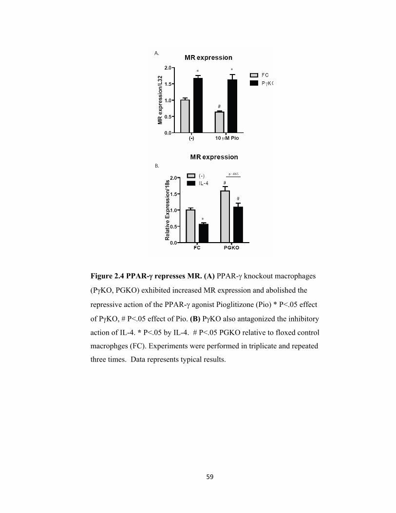
59
Figure 2.4 PPAR-γ represses MR. (A) PPAR-γ knockout macrophages
(PγKO, PGKO) exhibited increased MR expression and abolished the
repressive action of the PPAR-γ agonist Pioglitizone (Pio) * P<.05 effect
of PγKO, # P<.05 effect of Pio. (B) PγKO also antagonized the inhibitory
action of IL-4. * P<.05 by IL-4. # P<.05 PGKO relative to floxed control
macrophges (FC). Experiments were performed in triplicate and repeated
three times. Data represents typical results.

60
This is due to the high circulating corticosterone concentrations paired with the high
expression of 11βHSD1 in macrophages which increases local corticosterone
concentrations. This may indicate that MR does not primarily regulate gene transcription
through ligand activation. Second, MR has two physiologic ligands. Since, at the
physiologic level, glucocorticoids are an order of magnitude higher in concentration than
aldosterone, in all likelihood MR in macrophages is only responsive to aldosterone under
pathologically high concentrations. With these limitations in mind, we took a two
pronged approach to identifying MR action in macrophages. First, we cultured primary
macrophages in the absence of endogenous media steroids, adding back aldosterone and
glucocorticoids, to investigate
how specific activation of MR impacts macrophages transcriptional programs. In
parallel, we investigated the effect of MR antagonists.
In order to test the effect of MR activity on macrophage activation, primary bone
marrow derived and peritoneal macrophages as well as RAW 264.7 cells were cultured in
the absence of media steroids and stimulated for 21 hours with increasing concentrations
of aldosterone. Aldosterone stimulation resulted in significant upregulation of
inflammatory markers: TNFα, RANTES, IL-12, and MMP-9 (Figure 2.6) with a dose
response consistent with published binding properties of MR and our own transactivation
assay. The pro-inflammatory effect of aldosterone was mitigated by MR antagonists
eplerenone, spironolactone, and RU26752, and not blocked by GR antagonist RU486
(Figure 2.5, 2.7).

61
Figure 2.5 MR enhances pro-inflammatory cytokine expression in
macrophages. (A) TNFα expression is induced by 10 nM aldosterone and
blocked by 1 µM specific antagonists RU26752 (RU), Eplerenone (Epl), and
Spironolactone (Spiro) (B) TNFα induction by 10 nM aldosterone synergized
with LPS stimulation, and occurred at concentrations consistent with MR’s
binding properties (C) Graphs represent typical experimental results performed in
triplicate and repeated at least 3 times. Error bars=SEM, * =P<.05,

62
Figure 2.6 Aldosterone stimulates a pro-inflammatory,
pro-fibrotic macrophage response. 24 hours of 10 nM
aldosterone induced multiple pro-inflammatory cytokines
and factors involved in stimulation of fibrosis. Each effect
is statistically significant with a P <.05 by a 2-tailed
student T-test. Graphs represent typical experimental
results performed in triplicate and repeated at least 3
times.
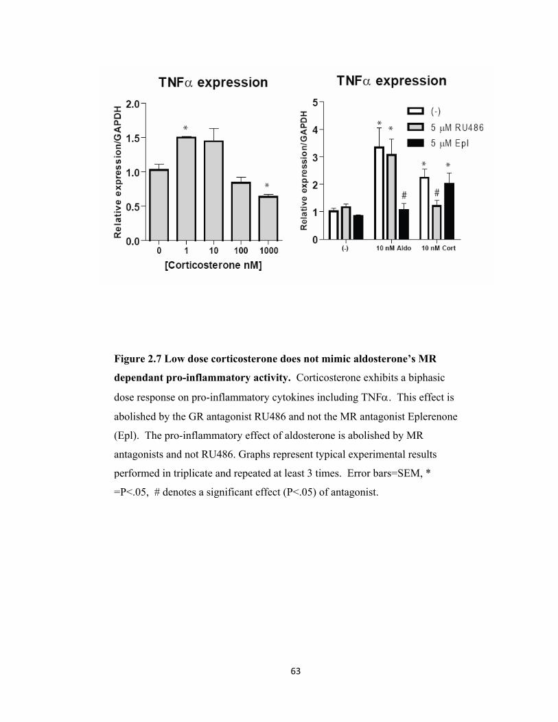
63
Figure 2.7 Low dose corticosterone does not mimic aldosterone’s MR
dependant pro-inflammatory activity. Corticosterone exhibits a biphasic
dose response on pro-inflammatory cytokines including TNFα. This effect is
abolished by the GR antagonist RU486 and not the MR antagonist Eplerenone
(Epl). The pro-inflammatory effect of aldosterone is abolished by MR
antagonists and not RU486. Graphs represent typical experimental results
performed in triplicate and repeated at least 3 times. Error bars=SEM, *
=P<.05, # denotes a significant effect (P<.05) of antagonist.
64
Since 11β-HSD2 activity could be not be detected by qRT-PCR consistent with previous
literature, we also investigated the ability of corticosterone to enhance pro-inflammatory
cytokine expression. Treatment of low concentrations of corticosterone resulted in a mild
pro-inflammatory effect which was not significantly reduced by MR antagonism, but was
blocked by RU486 (Figure 2.7). This demonstrates that at low occupancy GR can
enhance macrophage activation, consistent with previously published results.
Under normal culture conditions, due to the lack of 11β-HSD2 protection, MR is
likely to be significantly occupied by media glucocorticoids. We hypothesized this is
likely to be similar to the occupation state of MR in macrophages in vivo, and that MR
antagonists are most likely to be effecting this aspect of MR signaling. RAW 264.7 cells,
as well as primary macrophages were cultured in the presence of media steroids and
treated with increasing concentrations of MR antagonist. Following 3 hours of LPS
stimulation, MR antagonism significantly reduced pro-inflammatory cytokine expression
including TNFα, MCP-1, and MMP9 (Figure 2.8).
The ligand dependant, pro-inflammatory effect of MR was supported by transient
over-expression of MR. RAW 264.7 cells were transfected with a plasmid containing the
full length human MR cDNA clone (origene) driven by a CMV promoter (pCMV6-MR)
or with pCDNA 3.1(+) as a control. Functional overexpression was confirmed by co-
transfection with an MMTV reporter construct as described above demonstrating
approximately 3 fold increase in MR activity, and additionally by qRT-PCR and western
blot (Figure 2.9). Cells were maintained in media with serum containing endogenous
steroids or in charcoal/dextran treated medium with or without aldosterone administration

65
Figure 2.8: Antagonism of glucocorticoid occupied MR is anti-inflammatory. (A)
Increasing concentrations of Eplerenone (Epl) inhibited pro-inflammatory cytokine
production. (B) MR antagonists 5 µM Eplerenone, 1 µM RU26752, and 1 µM
spironolactone inhibited MMP9 expression induced by 3 hrs of 100 ng/ml LPS
stimulation, and a multitude of other pro-inflammatory cytokines all significantly
changed (P<.05) by 1 µM Spironolactone (Spiro) (C) Graphs represent typical
experimental results performed in triplicate and repeated at least 3 times. Error
bars=SEM, * =P<.05,

66
Figure 2.9 Transient MR over-expression is pro-inflammatory in macrophages.
Functional MR over-expression was confirmed by MMTV activity (A), western blot (D),
and (E) qRTPCR. MR over-expression in RAW 264.7 cells in charcoal stripped media
required ligand activation to induce a pro-inflammatory effect (B). Conversely, when
cultured in the presence of glucocorticoids MR overexpression induced a pro-
inflammatory effect (C,F) which was diminished by MR antagonism. N=6 Error
bars=SEM, * =P<.05 relative to the negative control. # denotes P<.05 relative to un-
treated, similarly transfected sample. @ P<.05 of both pairwise comparisons.

67
for 2 days, followed by 5 µM eplerenone treatment in some wells. At day three, half the
samples were treated with LPS. Cells were collected for qRT-PCR analysis of cytokine
expression. All expression levels were normalized to 18S and expression in untreated
controls. Results demonstrated that transient overexpression results in a marked increase
in MCP-1 and TNFα expression in LPS treated cells (Figure 2.9). This increase was
partially blocked by eplerenone suggesting this was primarily a ligand dependent effect,
an observation confirmed by the requirement of aldosterone for the induction of cytokine
expression when cells were cultured in the absence of media steroids. This experiment is
important as it demonstrates that even moderate alterations in MR abundance levels
produce dramatic effects on macrophage function. In addition, these data suggest that
MR is likely activated by glucocorticoids in macrophages cultured in normal serum and
canproduce a pro-inflammatory effect that is blocked by antagonists.
To determine the breadth of this effect we utilized affymetrix to identify novel
MR targets. Broad expression analysis of factors induced by aldosterone from primary
peritoneal macrophages cultured in charcoal stripped serum containing media,
demonstrated that MR controls a pro-inflammatory, pro-fibrotic transcriptional program
in macrophages. We identified additional pro-inflammatory cytokines induced by
aldosterone such as CXCL-12, which is important for the recruitment and activation of
fibrocytes and fibroblasts. Aldosterone induced pro-inflammatory chemokines such as
CXCL5, chemokine and cytokine receptors such as IL-1b receptor, and factors involved
in fibrotic processes such as Clusterin, Collagen I, Collagen III, Osteonectin (Sparc), and
CTGF (Figure 2.6).
68
Taken together, these data clearly demonstrate MR controls macrophage
activation. Specifically, MR enhances a unique pro-inflammatory pro-fibrotic
transcriptional profile. Conversely, MR antagonism has anti-inflammatory effects. We
observe clear differences in effect between MR occupied by glucocorticoids and
aldosterone, such that many pro-inflammatory effects of aldosterone were not mimicked
by similar concentrations of glucocorticoids in vitro. Moreover, addition of aldosterone
at supraphysiologic concentrations in normal serum produced additional pro-
inflammatory effects, blocked by MR antagonists and enhanced by GR antagonists
(Figure 2.10). However, a permissive effect could be observed indirectly via the anti-
inflammatory activity of MR antagonists, and pro-inflammatory impact of transient
overexpression of glucocorticoid occupied MR.
MR knockout macrophages:
To further investigate the role of MR in macrophages in vitro and in vivo, we
developed a macrophage specific knockout of MR. Having obtained a mouse strain
carrying the floxed MR allele backcrossed onto C57BL/6J for 12 generations, we crossed
this mouse with strain which specifically expressed cre in granulocytes and macrophages.
We show LysM cre, MRFl/Fl resulted in excision of MR’s exon 2 by PCR in isolated
macrophages, but not parenchymal tissues. It is important to note that we did not find
any excision in the liver, despite the high number of resident kupfer cells (Figure 2.11).
This is consistent with reports that LysM cre does not result in kupfer cell deletion.
Macrophages isolated from MΦMRKO mice demonstrated ablation of MR mRNA as
detected by qRT-PCR and western blot (Figure 2.11).

69
Figure 2.10. Glucocorticoid and Aldosterone occupied MR
have different pro-inflammatory activities. (A) RAW 264.7
macrophages cultured in the presence of media steroids and
stimulated with increasing concentrations of aldosterone
demonstrated additional pro-inflammatory effect independent of
GR. (B) In charcoal stripped serum, corticosterone was capable
of antagonizing the pro-inflammatory action of aldosterone in
the absence of GR activation. * P<.05 relative to untreated
control. # denotes a significant effect of RU486 when added to
treated macrophages.

70
Figure 2.11: Generation of macrophage specific MR knockout. In
mice harboring two conditional MR alleles and LysM cre
(MRflox/flox;LysMcre) the recombined allele (MRnull) is detected
by PCR only in isolated macrophages and not in parenchymal tissues
(A). Macrophage MRKO resulted in near complete abolishment of
detectable MR mRNA in isolated peritoneal macrophages by qRT-
PCR relative to macropahges isolated from floxed littermate control
animals (FC) (B) and protein by western blot (C).

71
Figure 2.12 MR deletion dramatically alters macrophage function. (A) Heatmap
of affymetrix data, of genes changed greater than 2 fold, demonstrates MRKO
results in significant alterations in ECM control, redox signaling, and BMP signaling
relative to untreated floxed control macrophages (FC) (B) A majority of changes
were confirmed by qRT-PCR. (C) Antagonism of glucocorticoid occupied MR
mimicked the effects of MRKO. (D) Culturing MRKO macrophages in charcoal
stripped serum abolished many differences indicating ligand dependency. * P<.05.
Experiments were performed in triplicate and repeated twice.

72
Isolated MRKO macrophages clearly confirmed that MR plays an important role in
macrophage activation. MRKO macrophages demonstrated reduced expression of pro-
inflammatory cytokines both at baseline and in response to LPS stimulation. Affymetrix
analysis of MRKO macrophages in comparison to floxed controls demonstrated
MR’sbroad impact on macrophage functions. Gene ontology analysis of transcripts
altered by MR, clearly demonstrated its role in control of extracellular matrix structure,
antigen presentation, redox signaling, and BMP signaling. All told, MR resulted in
upregulation of 87 factors greater than 2.5 fold and down-regulation of 31 factors.
Unfortunately RNase contamination of one control sample limited the N number to a
point that more robust statistics could not be performed. However, many factors
identified by affymetrix were significantly altered by MRKO by qRT-PCR (Figure 2.12).
Changes induced by MRKO relative to Floxed control macrophages, were likely
due to the abolishment of glucocorticoid occupied MR activity. Consistent with this,
culturing macrophages in the absence of media steroids abolished many of the effects of
MRKO. This is an indication that many of the changes observed in MRKO
macrophages are due to acute transcriptional regulation and not due to differentiation.
Changes induced by MRKO were also mimicked by antagonism of glucocorticoid
occupied MR by RU26752 and Eplerenone (Figure 2.12).
However, with few exceptions, transcripts altered by MRKO rarely overlapped
with genes impacted by aldosterone. MRKO had a much more dramatic and broad effect
on macrophage activation than aldosterone. A similar result was observed on the effects
of eplerenone and aldosterone on circulating leukocytes, where MR antagonism poorly
overlapped with aldosterone effects. This led to the authors concluding that many of
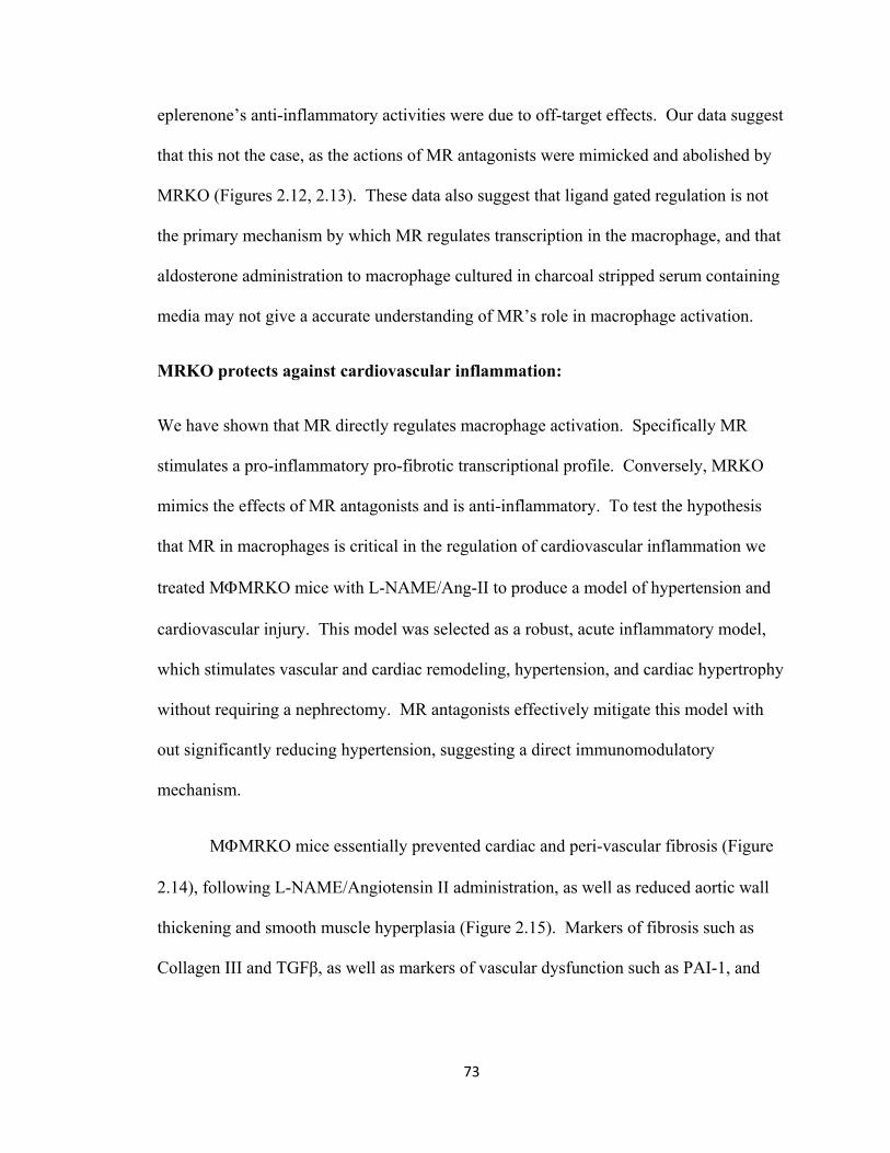
73
eplerenone’s anti-inflammatory activities were due to off-target effects. Our data suggest
that this not the case, as the actions of MR antagonists were mimicked and abolished by
MRKO (Figures 2.12, 2.13). These data also suggest that ligand gated regulation is not
the primary mechanism by which MR regulates transcription in the macrophage, and that
aldosterone administration to macrophage cultured in charcoal stripped serum containing
media may not give a accurate understanding of MR’s role in macrophage activation.
MRKO protects against cardiovascular inflammation:
We have shown that MR directly regulates macrophage activation. Specifically MR
stimulates a pro-inflammatory pro-fibrotic transcriptional profile. Conversely, MRKO
mimics the effects of MR antagonists and is anti-inflammatory. To test the hypothesis
that MR in macrophages is critical in the regulation of cardiovascular inflammation we
treated MΦMRKO mice with L-NAME/Ang-II to produce a model of hypertension and
cardiovascular injury. This model was selected as a robust, acute inflammatory model,
which stimulates vascular and cardiac remodeling, hypertension, and cardiac hypertrophy
without requiring a nephrectomy. MR antagonists effectively mitigate this model with
out significantly reducing hypertension, suggesting a direct immunomodulatory
mechanism.
MΦMRKO mice essentially prevented cardiac and peri-vascular fibrosis (Figure
2.14), following L-NAME/Angiotensin II administration, as well as reduced aortic wall
thickening and smooth muscle hyperplasia (Figure 2.15). Markers of fibrosis such as
Collagen III and TGFβ, as well as markers of vascular dysfunction such as PAI-1, and
74
Figure 2.13 Divergent actions of glucocorticoid and aldosterone occupied MR on
macrophage function. (A) Primary peritoneal macrophages from MΦMRKO mice and
floxed littermate controls (FC) were cultured in normal and charcoal stripped serum (B)
Genes important in basic cellular metabolism such as ME1 and PDK4 were induced by
MRKO and by antagonism in normal serum, whereas aldosterone produced no
significant effects. Conversely, aldosterone induced factors involved in fibrosis such as
Col3a1 and CTGF, where ablation of glucocorticoid occupied MR caused no effect. (C)
Comparison of affymetrix data from aldosterone stimulated and MRKO demonstrates
limited overlap between effects. * P<.05 by students T-test, all experiments were
performed in triplicate and repeated 3 times

75
macrophage recruitment, all strongly induced by L-NAME/AngII, were suppressed in the
hearts and aortas of MΦMRKO mice (Figure 2.14, 2.15).
While MΦMRKO mice exhibited a similar increase in blood pressure and heart
rate relative to controls, cardiac hypertrophy induced by L-NAME/AngII was
significantly reduced (Figure 2.14). We additionally observe a consistent reduction in
BNP, ANP, and a reversal of αMHC suppression (Figure 2.14). These results suggest
that MR dependant macrophage activation is an essential contributor to the cardiac
hypertrophic response to injury.
MR inhibits alternative macrophage activation:
MΦMRKO results in protection of cardiovascular injury, fibrosis, and
hypertrophy indicating that inflammatory signaling is important in these pathologies. As
discussed in chapter 1, macrophage responses are categorized into classically activated
(M1) and alternatively activated actions (AMΦ). In many models of cardiovascular
inflammation, M1 macrophages have been shown to be exacerbatory and AMΦ
macrophages or monocytes been shown to induce healing responses.
Consistent with this, we show L-NAME/ANG-II treatment resulted in recruitment
of M1 macrophages to the aorta and cardiac tissue as indicated by significant
upregulation of markers of classical macrophage activation including TNFα, RANTES,
IL-1β (Figure 2.16). Induction of these classically activated macrophage markers were
largely suppressed in MΦMRKO mice. L-Name/AngII treatment also resulted in
repression in some markers of alternative macrophage activation, which was reversed in
MΦMRKO mice (Figure 2.16). Although MΦMRKO showed an increase in AMΦ

76
Figu
re 2
.14
MΦ
MR
KO
is p
rote
ctiv
e in
aga
inst
car
diac
fibr
osis
and
infla
mm
atio
n. (A
) L-
NA
ME/
Ang
-II s
timul
ate
peri-
vasc
ular
dem
onst
rate
d he
re b
y H
&E
stai
n an
d in
ters
titia
l (B
) fib
rosi
s
dem
onst
rate
d by
Mas
on’s
Tric
hrom
e vi
ewed
und
er p
olar
ized
ligh
t is a
brog
ated
by
MΦ
MR
KO
. (C
) In
addi
tion
MPM
RK
O re
duce
d ca
rdia
c hy
pertr
ophy
as m
easu
red
by H
eart
Wei
ght,
Bod
y W
eigh
t Rat
io
(HW
/BW
)and
hyp
ertro
phy
mar
ker e
xpre
ssio
n, c
olla
gen
III e
xpre
ssio
n, a
nd m
acro
phag
e re
crui
tmen
t
(Mea
sure
d by
F4/
80) i
nduc
ed b
y L-
NA
ME/
Ang
II. N
=6, *
P<.
05 b
y st
uden
t’s T
-test
.

77
Figure 2.15 MΦMRKO protects from vascular remodeling. (A) Smooth muscle
hyperplasia and aortic wall thickening induced by L-NAME/Ang-II was abolished by
MΦMRKO as viewed by H&E staining of a similar cross section of the thoracic aorta.
(B) This coincided with reduction in PAI-1 expression, collagen III, TGF-β, and
macrophage recruitment (measured by F4/80 expression), detected by qRT-PCR. N=6,
*P<.05, **<P<.01 by students T-test.

78
Figure 2.16: L-NAME/Ang-II results in an M1 polarized
response in cardiac tissue diminished by MΦMRKO. qRT-PCR
of classical and alternative activation markers demonstrate
increases in M1 markers and repression of some AMΦ markers
following L-NAME/Ang-II stimulation. This skewed response was
mitigated by MΦMRKO, N=8

79
Figure 2.17: MΦMRKO does not protect from glomerular injury induced by L-
NAME/Ang-II. Similar to other tissues we observed a similar trend in an increase in
renal expression of AMΦ markers in MΦMRKO mice relative to floxed controls (FC)
(A). However, this did not result in protection against glomerular injury as measured by
Periodic acid-Schiff (PAS) staining which brightly labels damaged glomeruli (B).
80
marker expression in the kidney following L-NAME/Ang-II treatment this did not
translate into reduced glomerular injury, as indicated by proteinurea and PAS staining
(Figure 2.17). These results demonstrate a role for MR activation in controlling
macrophage polarization, specifically enhancing classical macrophage activation and
repressing alternative activation in response to cardiac injury. The AMΦ shift caused by
MΦMRKO was protective in cardiac and vascular inflammation, but not against
glomerular injuryThese results indicated a role of MR in regulating macrophage
polarization, specifically inhibiting AMΦ transcriptional programs and activating M1
programs. Isolated macrophages from MΦMRKO mice and floxed controls confirmed
this role. Many factors induced in MRKO macrophages were specific markers for
alternative macrophage activation, while genes downregulated in MRKO macrophages
were classified as M1 markers. MR deletion also reduced the induction of pro-
inflammatory factors such as IL-1β by LPS and increased induction of AMΦ markers
CCL7 and MSR2 by IL-4. These results demonstrate MR not only directly impacts the
baseline expression of M1 and AMΦ genes, but interacts with the signaling mechanisms
which induce macrophage polarization to further skew responses (Figure 2.18).
Specific activation of MR by aldosterone similarly enhanced M1 marker
expression, as mentioned above, and repressed the induction of AMΦ marker induction
Arg1 and YM1 by IL-4. Conversely, MR antagonism mimicked the effect of MФMRKO
and enhanced the expression of many AMΦ markers (Figure 2.18).
While MR activation and deletion altered the expression of M1 and AMΦ
markers, its effects were not global. MR did not enhance all M1 markers (such as IL-6

81
Figure 2.18 MR controls macrophage polarization. (A) MRKO macrophages exhibit a
robust AMΦ phenotype demonstrated by marked induction of AMФ markers, and
repression of M1 markers relative to macrophages isolated from floxed littermate
controls (FC). Induction of AMФ markers is mimicked by antagonism of MR in wildtype
macrophages cultured in normal serum (C). Addition of aldostone to floxed control
macrophages repressed Arg1 expression and induced M1 markers, and Pro-fibrotic
molecule, an effect which was abolished by MRKO. (B,D).

82
and iNOS, data not shown); conversely, MRKO did not enhance all AMΦ markers. In
some cases MФMRKO even inhibited some AMΦ markers such as E-cadherin and IL-27
receptor. This implies that MR does not regulate the upstream signaling components
such as STAT6 or IL-4 receptor which drive alternative macrophage activation.
MR and PPAR-γ coordinately regulate macrophage polarization:
MR antagonism and MR knockout have similarities to the effects of the
thiazolidinedione (TZD) PPAR-γ agonists in their ability to mitigate cardiovascular,
inflammation and fibrosis through their action on macrophages. They also enhance some
aspects of alternative activation, suggesting a potential common mechanism. We thus
attempted to identify the commonalities between MR inactivation with PPAR-γ
activation. Specifically, we compared expression profiles both at baseline, alternatively
activated, and classically activated states of MRKO with PPAR-γ knockout, and
Pioglitizone treatment.
MR deletion did not affect PPAR-γ expression, but enhanced the AMΦ inducing
effects of TZDs, both in suppressing TNFα and enhancing Arg1 and YM1 (Figure 2.19).
Hierarchical cluster analysis of genes altered in MR and PPAR-γ null macrophages
demonstrate significant overlap primarily in standard classical and AMΦ markers
indicating MR and PPAR-γ play opposing roles in the control of macrophage
polarization. Consistent with this hypothesis, PPAR-γ knockout which opposed IL-4
stimulation enhanced the M1 polarizing effects of aldosterone (Figure 2.19). While MR
and PPAR-γ play opposing roles on macrophage polarization, PPAR-γ deletion did not

83
Figure 2.19: MR and PPAR-γ play oppositional roles in macrophage polarization.
(A) Hierarchical clustering of M1 and AMΦ marker expression from primary peritoneal
macrophages demonstrates significant overlap in between PPAR-γ activation, and
MRKO. (B) PγKO in macrophages opposed the effects of IL-4, yielding a macrophage
with the opposite phenotype as MRKO. (C, E) Deletion of MR enhanced the AMΦ
polarizing effects of 10 μM Pioglitizone. (D) PγKO cultured in charcoal dextran stripped
media mimicked and enhanced the M1 polarizing effects of 10 nM Aldosterone. (F) MR
and PPAR-γ co-ordinately regulate IL-4 stimulation of the AMΦ marker Ccl7. Error bars
= SEM, * denotes P<.05 by 2 tailed student T-test.

84
effect the AMΦ inducing effects of MR antagonists, and MRKO actually enhanced
pioglitazone action (Figure 2.19) indicating they act via parallel, independent
mechanisms.
MR regulates glucocorticoid signaling in the macrophage
Macrophage polarization, however, is not a simple dichotomy, and other factors
such as FCγR engagement [31] and high dose glucocorticoids[32] stimulate alternative
macrophage activation with distinctly different profiles. MR has long been known to
coordinate the cellular response to glucocorticoids in tissues lacking 11-βHSD2 such as
the brain, where MR controls an overlapping counter-regulatory program against the
actions of GR [33, 34]. Since, a majority of MR’s evolution occurred prior to the
existence of physiologic aldosterone[3, 4], this cellular role is a likely conserved ancestral
function, observable in cell types such as neurons, cardiomyocytes, and macrophages.
We tested the necessity of MR in glucocorticoid mediated macrophage
polarization and show that GR activation by corticosterone induces an alternative state
that is distinct from PPAR-γ activation and IL-4 (Figure 2.20). Transcriptional changes
caused by MRKO overlapped with the glucocorticoid response (Figure 2.20-2.22). In
many genes identified as co-regulated by MR and GR including markers of alternative
macrophage activation such as YM1, Ccl7, and F13a1, MRKO synergized with
corticosterone to enhance or additionally repress transcription (Figure 2.20).
Additionally, we identified novel factors, such as the serine protease and TGFβ inhibitor
Htra1[35], and the protease inhibitor SerpinE2[36], where MR counteracts the effects of
corticosterone and are likely important in the control of extra cellular matrix remodelling.
85
Figure 2.20: MR coordinates with glucocorticoid signaling in the
macrophage. (A). Affymetrix analysis of peritoneal macrophages
treated with 1 μM corticosterone for 24 hrs yielded many genes where
MRKO mimicked or enhanced glucocorticoid responses. (B,C) MRKO
synergized with corticosterone to repress E-cadherin (Cdh1) and IL-27
receptor and induce genes important AMΦ macrophage polarization
(F13a1 and YM1). (D) MRKO abolished additional repression of IL-1β
and the pro-inflammatory C-type lectin CLEC-2. (E) MRKO and
corticosterone dramatically synergized to enhance the TGFβ inhibitor
Htra1 and Thrombin inhibitor SerpinE2 * P<.05 by students T-test
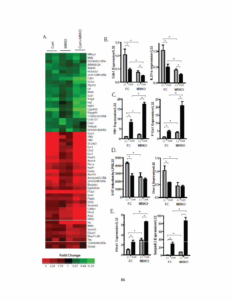
86

87
Figure 2.21: Control of macrophage activation by MR and GR. (A) Cluster analysis
and heatmap of affymetrix data illustrating genes coordinately regulated by MR and GR.
Three clusters identified show that MR can mimic the repression of pro-inflammatory
factors (C) and enhance the induction of AMΦ (B). We also identified a third cluster
where MRKO effects were masked by glucocorticoids, suggesting potentially redundant
functions (D).

88
Figure 2.22: Overlap of MR and GR mediated effects on macrophage activation.
Heatmap of affymetrix data of genes altered >2 fold by 24 hrs of 1 μM corticosterone
treatment to primary peritoneal macrophages from MΦMRKO mice or floxed littermate
controls. Genes significantly altered by corticosterone (A) and MRKO (B) demonstrate
overlap that only represents a minority of genes regulated by MR and GR activation (C)
89
Conversely, MRKO abolished additional suppression of pro-inflammatory factors
as IL-1β and CLEC-2 (Figure 2.20). These data show that, as in other tissues[37], MR
and GR coordinate counter-regulatory responses to changing glucocorticoid
concentrations and that for a few responses transcriptional repression caused by elevated
glucocorticoid levels requires MR.
In other cell types that lack 11β-HSD2 such as neurons, MR and GR control
opposing cellular roles through independent but overlapping mechanisms[34]. In
macrophages we observe a similar role, where MR and GR drive opposite polarizing
responses, often independently (Figure 2.20-2.22). These non-redundant functions allow
MR to repress multiple facets of AMΦ polarization that are stimulated by
glucocorticoids, PPAR-γ and IL-4. This is likely important in MR’s role in enhancing
cardiac injury and fibrosis, and the protective benefit conferred by MRKO or antagonism.
Conclusions:
To conclude, glucocorticoid occupied MR evolved an important role in
controlling classical and alternative macrophage activation that may represent a
conserved ancestral corticosteroid receptor function. We have shown that MR deletion or
inhibition co-ordinately removes a suppression of AMΦ sharing features of two subtypes,
and reduces classical macrophage activation. A loss of MR activity either by antagonism
or deletion provides a protective effect on experimentally induced cardiac and vascular
remodelling. Therefore, a critical mechanism by which MR antagonists are
cardioprotective is through blocking glucocorticoid occupied MR in macrophages, and
may explain the clinical benefit of MR antagonists in the absence of hyperaldosteronism.
90
MR in macrophages works in concert with other nuclear hormone receptors PPAR-γ and
GR, which allow endocrine signals to fine tune innate immune responses. Interestingly,
this pathway has been targeted by a number of drugs effective in mitigating
cardiovascular disease, and may represent a common mechanism of action suggesting
that alteration of macrophage polarization is a paradigm for drug discovery.
Methods
Generation of MMRKO mice MRfl/+ generated by the Schütz lab maintained on a
C57BL/6J background were crossed with LysMcre. Cohorts of FC (MRfl/fl) and
MΦMRKO (MRfl/fl; LysMcre) were generated by crossing MRfl/fl and MRfl/fl;
LysMcre animals. Genotyping was performed to detect the presence of both the flox and
deleted allele as previously described [38] . Littermate controls were used for all
experiments.
Macrophage isolation and treatment Primary peritoneal macrophages were isolated 4
days after an I.P. injection of aged 3% Brewer’s Thioglycolate as previously described.
Macrophages were then cultured in media containing 10%FBS or 10% Charcoal/Dextran
stripped FBS (Hyclone). Isolated macrophages were pre-treated with either MR agonist
or antagonist 18 hours prior to stimulation. Classical macrophage activation was
stimulated by 100ng/ml of LPS for 3 hours. Alternative macrophage activation was
achieved by 5 ng/ml of IL-4 for 24 hours.
Gene Expression Total RNA was isolated using an RNAeasy kit (Qiagen) following an
on column DNase digestion. First-strand cDNA synthesis was accomplished using
TaqMan Reverse Transcriptase kit (Roche). QRT-PCR was carried out on an iCycler

91
(Biorad). Relative expression was determined via the Ct method normalized to L32,
GAPDH, and 18s standards.
L-NAME/Ang-II treatment 8-10 week old mice were given a 30 mg/kg dose of N (G)-
nitro-L- arginine methyl ester (L-NAME) accompanied by .9% NaCl in the drinking
water. After 10 days of L-NAME treatment, 0.8 mg/kg/day Angiotensin II (Sigma) was
infused by a subcutaneous osmotic pump (Alzet). Blood pressures were recorded both by
telemetry and tail-cuff method as previously described [39].
Transient Transfections RAW264.7 cells (ATCC) were transiently transfected using
Superfect (Qiagen) with an expression construct driving mouse MR or pcDNA 3.1+
empty vector control. Expression was confirmed via qPCR, western blot, and through a
reporter plasmid. For MR activity, RAW264.7 cells were transfected with MMTV-
luciferase as previously described. Luciferase production following MR activation was
determined by Dual Luciferase assay (Promega) and normalized to a Renilla standard.
Statistics and cluster analysis Pairwise comparisons were compared via a 2 tailed
student’s T-test with statistical significance attributed to a P value of less than .05.
Multiple comparisons were also tested with a 2 tailed ANOVA, and bonferroni post test
where indicated. Cluster analysis was performed using centered complete linkage
clustering using Cluster 3.0. The arrayed expression data was then plotted using
TreeView. Venn diagrams were performed using GeneVenn
(http://www.bioinformatics.org/gvenn/) with a list of genes changed in each condition
greater than 2 fold.

92
References 1. Effectiveness of spironolactone added to an angiotensin‐converting enzyme inhibitor and
a loop diuretic for severe chronic congestive heart failure (the Randomized Aldactone Evaluation Study [RALES]). Am J Cardiol, 1996. 78(8): p. 902‐7.
2. Pitt, B., et al., The EPHESUS trial: eplerenone in patients with heart failure due to systolic dysfunction complicating acute myocardial infarction. Eplerenone Post‐AMI Heart Failure Efficacy and Survival Study. Cardiovasc Drugs Ther, 2001. 15(1): p. 79‐87.
3. Bridgham, J.T., S.M. Carroll, and J.W. Thornton, Evolution of hormone‐receptor complexity by molecular exploitation. Science, 2006. 312(5770): p. 97‐101.
4. Hu, X. and J.W. Funder, The evolution of mineralocorticoid receptors. Mol Endocrinol, 2006. 20(7): p. 1471‐8.
5. Funder, J.W., Mineralocorticoid receptors: distribution and activation. Heart Fail Rev, 2005. 10(1): p. 15‐22.
6. Kuster, G.M., et al., Mineralocorticoid receptor inhibition ameliorates the transition to myocardial failure and decreases oxidative stress and inflammation in mice with chronic pressure overload. Circulation, 2005. 111(4): p. 420‐7.
7. Fallo, F., et al., Prevalence and characteristics of the metabolic syndrome in primary aldosteronism. J Clin Endocrinol Metab, 2006. 91(2): p. 454‐9.
8. Gomez‐Sanchez, E.P., M. Zhou, and C.E. Gomez‐Sanchez, Mineralocorticoids, salt and high blood pressure. Steroids, 1996. 61(4): p. 184‐8.
9. Keidar, S., et al., Aldosterone administration to mice stimulates macrophage NADPH oxidase and increases atherosclerosis development: a possible role for angiotensin‐converting enzyme and the receptors for angiotensin II and aldosterone. Circulation, 2004. 109(18): p. 2213‐20.
10. Joffe, H.V., et al., Beneficial effects of eplerenone versus hydrochlorothiazide on coronary circulatory function in patients with diabetes mellitus. J Clin Endocrinol Metab, 2007. 92(7): p. 2552‐8.
11. Oestreicher, E.M., et al., Aldosterone and not plasminogen activator inhibitor‐1 is a critical mediator of early angiotensin II/NG‐nitro‐L‐arginine methyl ester‐induced myocardial injury. Circulation, 2003. 108(20): p. 2517‐23.
12. Young, M., et al., Mineralocorticoids, hypertension, and cardiac fibrosis. J Clin Invest, 1994. 93(6): p. 2578‐83.
13. Lam, E.Y., et al., Mineralocorticoid receptor blockade but not steroid withdrawal reverses renal fibrosis in deoxycorticosterone/salt rats. Endocrinology, 2006. 147(7): p. 3623‐9.
14. Han, S.Y., et al., Spironolactone prevents diabetic nephropathy through an anti‐inflammatory mechanism in type 2 diabetic rats. J Am Soc Nephrol, 2006. 17(5): p. 1362‐72.
15. Rajagopalan, S., et al., Mineralocorticoid receptor antagonism in experimental atherosclerosis. Circulation, 2002. 105(18): p. 2212‐6.
16. Neves, M.F., et al., Role of aldosterone in angiotensin II‐induced cardiac and aortic inflammation, fibrosis, and hypertrophy. Can J Physiol Pharmacol, 2005. 83(11): p. 999‐1006.
17. Rocha, R., et al., Mineralocorticoid blockade reduces vascular injury in stroke‐prone hypertensive rats. Hypertension, 1998. 31(1 Pt 2): p. 451‐8.
18. Frangogiannis, N.G., et al., Critical role of monocyte chemoattractant protein‐1/CC chemokine ligand 2 in the pathogenesis of ischemic cardiomyopathy. Circulation, 2007. 115(5): p. 584‐92.

93
19. Plenz, G., et al., Alterations in the vascular extracellular matrix of granulocyte macrophage colony‐stimulating factor (GM‐CSF) ‐deficient mice. Faseb J, 2003. 17(11): p. 1451‐7.
20. Takeishi, Y. and I. Kubota, Role of Toll‐like receptor mediated signaling pathway in ischemic heart. Front Biosci, 2009. 14: p. 2553‐8.
21. Mantovani, A., et al., The chemokine system in diverse forms of macrophage activation and polarization. Trends Immunol, 2004. 25(12): p. 677‐86.
22. Stout, R.D. and J. Suttles, Functional plasticity of macrophages: reversible adaptation to changing microenvironments. J Leukoc Biol, 2004. 76(3): p. 509‐13.
23. Shimada, K., Immune System and Atherosclerotic Disease. Circ J, 2009. 24. Wang, Y., et al., Ex vivo programmed macrophages ameliorate experimental chronic
inflammatory renal disease. Kidney Int, 2007. 72(3): p. 290‐9. 25. Gordon, S., Alternative activation of macrophages. Nat Rev Immunol, 2003. 3(1): p. 23‐
35. 26. Bouhlel, M.A., et al., PPARgamma activation primes human monocytes into alternative
M2 macrophages with anti‐inflammatory properties. Cell Metab, 2007. 6(2): p. 137‐43. 27. Hevener, A.L., et al., Macrophage PPAR gamma is required for normal skeletal muscle
and hepatic insulin sensitivity and full antidiabetic effects of thiazolidinediones. J Clin Invest, 2007. 117(6): p. 1658‐69.
28. Huang, J.T., et al., Interleukin‐4‐dependent production of PPAR‐gamma ligands in macrophages by 12/15‐lipoxygenase. Nature, 1999. 400(6742): p. 378‐82.
29. Odegaard, J.I., et al., Macrophage‐specific PPARgamma controls alternative activation and improves insulin resistance. Nature, 2007. 447(7148): p. 1116‐20.
30. Lim, H.Y., et al., Glucocorticoids exert opposing effects on macrophage function dependent on their concentration. Immunology, 2007. 122(1): p. 47‐53.
31. Sironi, M., et al., Differential regulation of chemokine production by Fcgamma receptor engagement in human monocytes: association of CCL1 with a distinct form of M2 monocyte activation (M2b, Type 2). J Leukoc Biol, 2006. 80(2): p. 342‐9.
32. Varga, G., et al., Glucocorticoids induce an activated, anti‐inflammatory monocyte subset in mice that resembles myeloid‐derived suppressor cells. J Leukoc Biol, 2008. 84(3): p. 644‐50.
33. Avital, A., M. Segal, and G. Richter‐Levin, Contrasting roles of corticosteroid receptors in hippocampal plasticity. J Neurosci, 2006. 26(36): p. 9130‐4.
34. Datson, N.A., et al., Identification of corticosteroid‐responsive genes in rat hippocampus using serial analysis of gene expression. Eur J Neurosci, 2001. 14(4): p. 675‐89.
35. Launay, S., et al., HtrA1‐dependent proteolysis of TGF‐beta controls both neuronal maturation and developmental survival. Cell Death Differ, 2008. 15(9): p. 1408‐16.
36. Feutz, A.C., Y. Barrandon, and D. Monard, Control of thrombin signaling through PI3K is a mechanism underlying plasticity between hair follicle dermal sheath and papilla cells. J Cell Sci, 2008. 121(Pt 9): p. 1435‐43.
37. De Kloet, E.R. and R. Derijk, Signaling pathways in brain involved in predisposition and pathogenesis of stress‐related disease: genetic and kinetic factors affecting the MR/GR balance. Ann N Y Acad Sci, 2004. 1032: p. 14‐34.
38. Berger, S., et al., Loss of the limbic mineralocorticoid receptor impairs behavioral plasticity. Proc Natl Acad Sci U S A, 2006. 103(1): p. 195‐200.
39. Duan, S.Z., et al., Hypotension, lipodystrophy, and insulin resistance in generalized PPARgamma‐deficient mice rescued from embryonic lethality. J Clin Invest, 2007. 117(3): p. 812‐22.

94
CHAPTER III:
NUCLEAR FACTOR BALANCE IN MACROPHAGE POLARIZATION
Introduction
Recent evidence has demonstrated that macrophage activation falls upon a
spectrum of classically and alternatively activated states. Current nomenclature has
mirrored helper T cell differentiation; stimulants of classical macrophage activation (M1)
are generally shown to enhance Th1 responses in vivo. In addition Type 1 helper T cell
derived cytokines such as IFNγ induce classical macrophage activation driving
expression of M1 markers such as IL-12, TNFα, and IL-1β[2]. One standard marker for
M1 polarization is IL-12, which specifically enhances Th1 cell proliferation[3].
Alternatively activated macrophages (AMΦ) were originally characterized by
macrophage responses to type 2 helper T cell derived cytokines such as IL-4 and IL-13.
IL-4 receptor activation results in a unique expression profile which consists of
repression of pro-inflammatory cytokines such as TNFα, IL-12, and IL-1β[4] which also
serve as markers of M1 polarization[5-8]. More recently, the focus of IL-4 responses has
shifted from its anti-inflammatory activity to genes induced. IL-4 drives induction of
factors considered core markers of AMΦ polarization including arginase, mannose

95
binding lectins, fibronectin, and growth factors such as IGF-1 which are critical in
controlling parasitic infections, allergic responses, and regulate fibrotic processes[6].
The paradigm of M1 and AMΦ is best illustrated by comparing its effects on
nitrogen metabolism (Figure 3.1). A standard marker of classical macrophage activation
NOS2, or inducible nitrogen oxide synthase (iNOS) is potently induced by TNFα, IL-1,
LPS, or IFNγ. NOS acts by liberating a nitrogen from arginine with oxygen in an
NADPH dependant manner to produce NO and citrulline. NO synthesis by M1
macrophages is important for acute vasodilatation, induces vascular permeability, and
aids in bacterial killing. iNOS expression is potently repressed by IL-4 and IL-13.
Conversely, IL-4 stimulates the production of arginase1 (Arg1) which catabolizes
arginine into ornithine and urea. Arginase serves two purposes. First, it reduces the bio-
available arginine levels, thereby inhibiting the activity of iNOS [7]. Second, ornithine
generated by arginase is converted into proline, a critical step necessary for robust
collagen synthesis, illustrating the potential role of AMΦ cells in stimulating fibrosis [9,
10]. Arg1 expression is potently reduced in M1 polarized macrophages[7].
Arginase-1 and iNOS are emblematic of the counter-regulatory aspects of Th2
cytokines IL-4/13 and Th1 cytokines INFγ on macrophages activation and are commonly
used as markers to identify the state of polarization of the macrophage. Macrophage
polarization, however, is not a simple dichotomy, and other factors such as FCγR
engagement, IL-10 stimulation, and high dose glucocorticoids stimulate alternative
macrophage activation of a different flavor[1, 2, 9]. The degree of heterogeneity of
macrophages, as well as the roles of these additional subtypes in inflammatory responses

96
Figure 3.1: Macrophage polarization and nitrogen
metabolism (From [1])
97
is poorly understood. Understanding how each of these macrophage subtypes are formed
and how their actions are regulated will allow us to further investigate the underlying
inflammatory signaling which occurs with human disease, and tailor protective responses.
As has been demonstrated in the previous chapter, nuclear receptors such as MR, GR,
and PPAR-γ play a critical role in dynamically governing macrophage polarization.
However, the degree of molecular cross talk between nuclear factors and extracellular
stimulants of M1 and AMΦ subtypes has not been well characterized.
Recent studies investigating the role of PPAR-γ on macrophage polarization
concluded PPAR-γ enhances IL-4 stimulated AMΦ activation, and that this action was
critical for the insulin protective effects of TZDs[11]. This conclusion was primarily
based on PPAR-γ’s anti-inflammatory activity and ability to induce arginase production.
Since IL-4 produces dramatic and diverse effects on macrophage polarization, it is
unlikely that the enhancement of arginase alone is the physiologically important
mechanism. Moreover, TZD’s have also been shown to be protective in models of
fibrosis in a macrophage dependant manner[12]. Since arginase likely plays a pro-
fibrotic role, as opposed to a protective role, a more broad investigation into the role of
PPAR-γ in controlling macrophage polarization may yield a better understanding of its
protective effects. We therefore performed a more detailed comparison of expression of
M1 and AMΦ markers to elucidate candidates for protective effects.
Results and Discussion:
PPAR-γ does not solely enhance IL-4 responses:

98
TIE2-cre PPAR-γ Flox/Flox resulted in complete elimination of detectable PPAR-γ
mRNA by qRT-PCR (data not shown). PγKO macrophages resulted in M1 polarization
as observed by increases in TNFα, iNOS, and a reduction in Arg1 expression, as had
been previously reported [11]. Additionally, PγKO abolished both the anti-inflammatory
effects, and Arg1-inducing effects of Pioglitizone (Figure 3.2). These results
independently confirm a role for PPAR-γ in enhancing at least some aspects of AMΦ
macrophage functions.
A broad transcriptional analysis shows that PPAR-γ activity is required for a
normal IL-4 response. IL-4 induction of AMΦ markers, E-cadherin, Arg1, and Ccl7 was
significantly reduced by PγKO. Moreover, we show that IL-4 represses a number of
genes such as Htra1, Prss23, and Cyr61, and M1 markers iNOS and TNFα in a PPAR-γ
dependant fashion.
However, we also identified a number of IL-4 responses which were enhanced by
PγKO, suggesting that PPAR-γ also is capable of repressing aspects of AMФ
polarization. This runs contrary to previous conclusions. A few genes induced by IL-4
such as fibronectin (Fn1) and tissue inhibiter of metalloproteases 3 (TIMP3) were
significantly enhanced by PγKO. Fibronectin, a critical component of fibrotic processes
and thought to be one mechanism by which AMΦ macrophages contribute to fibrosis and
wound healing, was also repressed by Pioglitazone in a PPAR-γ dependent manner
(Figure 3.2). We also identified a significant number of factors induced or repressed by
IL-4 which were unaffected by PγKO, demonstrating that PPAR-γ does not act by
broadly enhancing STAT6 signaling, but triggers promoter specific effects.

99
Figure 3.2: PPAR-γ controls alternative macrophage activation. (A) Induction and
repression of many genes by IL-4 is abolished by PPAR-γ knockout (PγKO). (B)
However, some genes induced by IL-4 are further enhanced by PγKO. (C) Finally,
many genes induced by PγKO are unaffected by IL-4 stimulation indicating PPAR-γ
plays many roles outside of simple AMΦ activation. Data represents typical
experimental results performed in triplicate and repeated 3 times. * P<.05 by students
T-test.

100
Finally, we identified a significant number of genes regulated by PPAR-γ which
were unaffected by IL-4 or LPS. This weakens the conclusion that PPAR-γ control of IL-
4 driven AMФ polarization is the critical mechanism by which MΦ-PγKO enhances diet
induced obesity and insulin resistance and that TZDs are protective. This provides a
rationale for comprehensive characterization of the contribution of PPAR-γ toward IL-4
stimulated and other types of alternatively activated macrophages.
PPAR-γ and IL-4 oppose GR in macrophage polarization:
The glucocorticoid receptor (GR) demonstrates potent anti-inflammatory activity
which strongly overlaps with PPAR-γ [13]. Like PPAR-γ, a majority of research has
focused on the ability of GR to repress classical macrophage activation. Given the recent
evidence that alternatively activated macrophages are important in both the controlling
inflammatory responses in human disease, the molecular cross-talk between GR and
other alternative activators such as PPAR-γ and IL-4 should yield greater understanding
of how nuclear receptors regulate macrophage polarization. Previous investigation of
glucocorticoid action on macrophages has primarily focused on its anti-inflammatory
activity, utilizing synthetic GR agonists which have reduced MR binding affinity such as
dexamethasone [13]
We show that not only do glucocorticoids inhibit M1 polarization, but they also
results in the induction of a unique alternative activation profile. Standard markers of
AMФ cells, such as Arg1, YM1, YM2, and Fizz1 were all upregulated by 24hrs of
corticosterone treatment (Figure 3.3). Comparison of PPAR-γ, IL-4, and GR controlled
responses, however, demonstrated dramatic differences.
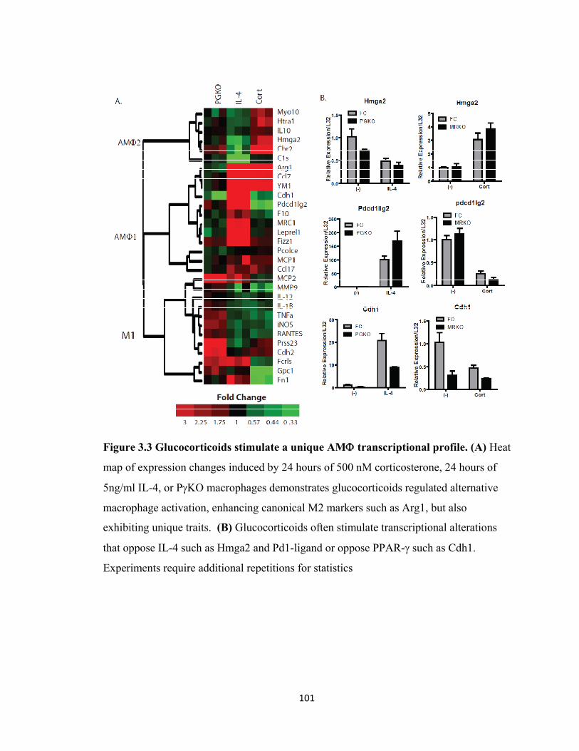
101
Figure 3.3 Glucocorticoids stimulate a unique AMΦ transcriptional profile. (A) Heat
map of expression changes induced by 24 hours of 500 nM corticosterone, 24 hours of
5ng/ml IL-4, or PγKO macrophages demonstrates glucocorticoids regulated alternative
macrophage activation, enhancing canonical M2 markers such as Arg1, but also
exhibiting unique traits. (B) Glucocorticoids often stimulate transcriptional alterations
that oppose IL-4 such as Hmga2 and Pd1-ligand or oppose PPAR-γ such as Cdh1.
Experiments require additional repetitions for statistics

102
First we show that glucocorticoids broadly repress IL-4 driven AMΦ markers.
For example, E-cadherin a gene induced by IL-4 and PPAR-γ, and necessary for
multinucleated giant cell formation in granulomatous inflammation was potently
repressed by glucocorticoids. Other genes critical for AMΦ function such as PD-1
ligand2, PTGS1 (COX1), Fibronectin, IL-27 receptor, and ChK were also potently
repressed by GR activation. Conversely, we demonstrate that many genes upregulated by
glucocorticoids such as Htra1, Hmga2, Myosin X, and Ctla2b, and Cbr2 are repressed by
IL-4. Consistent with the overlap between PPAR-γ and IL-4, we find that genes
upregulated by glucocorticoids and repressed by IL-4, were also increased in expression
in PγKO macrophages.
Taken together these results illustrate the complexity that underlies macrophage
polarization. Glucocorticoids on one hand, and IL-4 and PPAR-γ on the other hand drive
opposing transcriptional responses despite similar anti-inflammatory effects.
Interestingly, standard markers for alternative activation are similarly activated by both
glucocorticoid and IL-4 stimulation. Solely using these markers creates an illusion of
simplicity that macrophage polarization falls along a single spectrum where clearly that is
not the case. Future in vivo studies which investigate macrophage polarization should
take into account unique markers such as IL-27 receptor, E-cadherin which specifically
identify each macrophage subtype.
MR in nuclear factor balance:
Having shown that the nuclear receptor PPAR-γ and GR guide macrophage
polarization into two discrete states, it was important place MR into this context. In the

103
Figure 3.5 Nuclear receptor balance in macrophage polarization.
Comparison of MR, GR, PPAR-γ, and IL-4 targets demonstrates how
opposition between each can guide the transcriptional profile of alternative
macrophage activation stimulated by IL-4.

104
previous chapter we showed that MRKO enhanced the polarizing effects of PPAR-γ
activation, and some IL-4 responses. Additionally, we showed that MR activation
enhances the expression of markers of classical macrophage activation. This lead to the
next question, where GR and IL-4 act antagonistically, how does MR impact this
dynamic.
Unilaterally, MRKO always enhanced glucocorticoid effects despite antagonism
by PPAR-γ and IL-4. For example, E-cadherin and IL-27 Receptor were enhanced by
IL-4 in a PPAR-γ dependant manner, and synergistically repressed by corticosterone and
MRKO (Figure 3.3, Figure 3.4). Conversely, factors such as Cbr2 and Htra1 which were
repressed by IL-4 in a PPAR-γ dependant fashion were synergistically enhanced by
MRKO and corticosterone. While MR did enhance IL-4 responses, these were only in
genes where GR and IL-4 produced the same response, or in genes stimulated by IL-4
that were glucocorticoid insensitive.
To conclude MR guides classical macrophage activation through two
mechanisms. First, it enhances some aspects of classical macrophage activation such as
the pro-inflammatory cytokine IL-1β. Second, it represses aspects of both AMΦ
subtypes. This creates a clearer picture of the role of nuclear factors in macrophage
polarization. While MR, GR, and PPAR-γ enhance some aspects of each macrophage
subtype, these actions are balanced by overlapping repressive activity. This is primarily
true with PPAR-γ which only enhanced a few genes, but was necessary for the repression
of glucocorticoid responsive genes (Figure 3.6).

105
Figure 3.6: MR control of macrophage polarization Heat map and hierarchical
clustering of qRT-PCR expression M1 and AMΦ markers shows that MR not only
enhances M1 expression, but represses multiple facets of two AMΦ clusters

106
Conclusions
Is macrophage polarization a useful paradigm?
Historically, research into macrophage activation has followed a pattern of
increasing appreciation for complexity. Initially, macrophage activation was thought to
resemble a simple on-off switch. The current dogma focuses on a spectrum of activation
between classical and alternatively active states, though the field recognizes this as an
oversimplification. Our comprehensive investigation into the cross talk between the
nuclear receptors MR, GR, and PPAR-γ and stimulants of classical and alternative
activation illustrates macrophage polarization is far more nuanced and dynamic than has
been previously reported. Moreover, changes induced by these three nuclear receptors
did not perfectly overlap with other published macrophage subtypes. If simple categories
such as M1 and AMФ do not accurately exemplify macrophage activation in vitro or in
vivo, then how useful are they?
Macrophage polarization is a useful paradigm because it addresses the
observation that macrophage activation profiles cluster by their regulation. These
clusters can be used to identify specific underlying inflammatory mechanisms to correlate
with disease processes and effective treatments. However, most research to this point has
attempted to fit complex changes observed in inflammatory disease models such as
obesity into simple paradigms likely to the exclusion of AMΦ markers to don’t change
consistently. This is a detriment to the field as it prevents identification of specific
transcriptional programs which correlate with disease and may be used as unique
identifiers in the future for diagnosis or prediction of efficacious treatments.

107
References 1. Gordon, S., Alternative activation of macrophages. Nat Rev Immunol, 2003. 3(1): p. 23‐
35. 2. Stout, R.D. and J. Suttles, Functional plasticity of macrophages: reversible adaptation to
changing microenvironments. J Leukoc Biol, 2004. 76(3): p. 509‐13. 3. Schulz, E.G., et al., Sequential Polarization and Imprinting of Type 1 T Helper
Lymphocytes by Interferon‐gamma and Interleukin‐12. Immunity, 2009. 4. Hart, P.H., et al., Potential antiinflammatory effects of interleukin 4: suppression of
human monocyte tumor necrosis factor alpha, interleukin 1, and prostaglandin E2. Proc Natl Acad Sci U S A, 1989. 86(10): p. 3803‐7.
5. Kodelja, V., et al., Alternative macrophage activation‐associated CC‐chemokine‐1, a novel structural homologue of macrophage inflammatory protein‐1 alpha with a Th2‐associated expression pattern. J Immunol, 1998. 160(3): p. 1411‐8.
6. Martinez, F.O., et al., Transcriptional profiling of the human monocyte‐to‐macrophage differentiation and polarization: new molecules and patterns of gene expression. J Immunol, 2006. 177(10): p. 7303‐11.
7. Munder, M., K. Eichmann, and M. Modolell, Alternative metabolic states in murine macrophages reflected by the nitric oxide synthase/arginase balance: competitive regulation by CD4+ T cells correlates with Th1/Th2 phenotype. J Immunol, 1998. 160(11): p. 5347‐54.
8. Welch, J.S., et al., TH2 cytokines and allergic challenge induce Ym1 expression in macrophages by a STAT6‐dependent mechanism. J Biol Chem, 2002. 277(45): p. 42821‐9.
9. Martinez, F.O., L. Helming, and S. Gordon, Alternative activation of macrophages: an immunologic functional perspective. Annu Rev Immunol, 2009. 27: p. 451‐83.
10. Martinez, F.O., et al., Macrophage activation and polarization. Front Biosci, 2008. 13: p. 453‐61.
11. Odegaard, J.I., et al., Macrophage‐specific PPARgamma controls alternative activation and improves insulin resistance. Nature, 2007. 447(7148): p. 1116‐20.
12. Caglayan, E., et al., Differential roles of cardiomyocyte and macrophage peroxisome proliferator‐activated receptor gamma in cardiac fibrosis. Diabetes, 2008. 57(9): p. 2470‐9.
13. Ogawa, S., et al., Molecular determinants of crosstalk between nuclear receptors and toll‐like receptors. Cell, 2005. 122(5): p. 707‐21.
108
CHAPTER IV:
MACROPHAGE MR AND THE CONTROL OF INNATE AND ADAPTIVE IMMUNITY
Overview
This thesis is the first comprehensive approach to understanding the biology of
MR in an inflammatory cell type. We have conclusively demonstrated that MR regulates
macrophage activation and is critical for control of cardiovascular inflammation. MR in
macrophages binds glucocorticoid with high affinity, and coordinates with the actions of
other nuclear receptors including PPAR-γ and GR. As in any initial foray into a new
biological system, this study has led to many new questions. First, as the mechanisms of
MR transcriptional regulation in non-epithelial tissue are poorly understood, MR in
macrophages may be an ideal paradigm for investigating the biochemistry of MR in a
biologically relevant system. Second, while we show that MR is important in the
development of cardiovascular inflammation, data indicates MR may play a broad role in
regulating immune responses. Finally, our approach which utilized macrophage specific
deletion of MR yielded surprising novel roles for macrophages in regulating fundamental
physiologic and immunologic responses. This chapter outlines how initial investigation
of MR in macrophages has yielded novel insight into basic mechanisms of MR signaling
and its impact on the immune system.

109
Introduction
Understanding of how MR regulates inflammatory responses can be approached
from many angles. First, understanding the basic mechanisms by which MR controls
transcription in macrophages may help identify key interactions which help predict MR’s
role in regulating specific inflammatory responses. Second, macrophage MRKO resulted
in broad dysregulation of macrophage function. This can be utilized as a tool to separate
macrophage action in inflammatory responses from other cell types. Finally, since the
mechanisms by which macrophages coordinate immune response, MΦMRKO will be a
useful model in dissecting the interaction of macrophages with other immune cell types.
Mechanisms of control of control of gene transcription by MR
The high affinity of the mineralocorticoid receptor for glucocorticoids in
macrophages paired with the lack of 11βHSD2 which protects MR from saturating
concentrations of glucocorticoids make it unique among steroid hormone receptors.
Based on the known physiologic concentrations of glucocorticoids, and high level of
11βHSD1 which increases local concentrations of corticosterone/cortisol, MR is likely
fully activated even at low physiologic glucocorticoid concentrations.
Glucocorticoid bound MR has been shown to be important in renal epithelia[2],
the central nervous system[3], and adipose tissue[4] where it modulates physiologic
responses. However, direct investigation into MR’s actions when bound to
glucocorticoids has yielded paradoxical results. For example, glucocorticoids have been
shown to blunt aldosterone mediated sodium transport in renal epithelium, even in the
presence of 11βHSD2 [5-7]. However, if 11βHSD2 is blocked, then suddenly
110
glucocorticoids stimulate sodium transport [8, 9]. If glucocorticoids are effectively
inactivated by 11βHSD2 then how do they antagonize the activating effects of
aldosterone? If MR is still responsive to glucocorticoids even in tissues with 11βHSD2
activity, as some binding studies suggest, then how does aldosterone act differently and
how does 11βHSD2 antagonism confer activating properties on glucocorticoid? One
possible explanation is that catalytic action of 11βHSD2, such as cortisone or
NAD/NADH balance perturbation impacts glucocorticoid-MR complex activity[10-13].
However, similar paradoxical results have been observed in tissues lacking 11βHSD2
such as the brain where intracranial injection of glucocorticoids antagonizes the
hypertensive effects of aldosterone at similar concentrations.
In tissues lacking 11βHSD2, where local concentrations of glucocorticoids yield
tonic activation of nuclear MR, how does MR regulate transcription? There are a few
examples of nuclear receptors which are insensitive to physiologic changes in ligands,
which act largely through interactions with co-activator and co-repressors in a promoter
and context specific manner, similar in mechanism to orphan nuclear receptors.
In the brain this is thought to likely be the case, where MR is responsible for
maintaining essential functions and creating a set-point for the physiologic stress
response[14]. However, the specific functions that MR maintains and how it regulates
these responses is not understood. Since antagonism of glucocorticoid occupied MR
seems to be critically important for the beneficial actions of MR antagonists, specific
understanding of mechanisms by which MR-glucocorticoid complexes regulate
glucocorticoid is an important question.

111
Macrophage/Monocyte differentiation
Direct, acute activation is not the only mechanism which drives macrophage
heterogeneity. Macrophages are a part of a network of myeloid derived, closely related
granulocytes and dendritic cells commonly referred to as the mononuclear phagocyte
system. Recent research has demonstrated that myeloid progenitor and circulating
monocyte populations are highly heterogeneous. Some evidence suggests that monocytes
subpopulations are subsequently pre-disposed to polarize into classically activated and
alternatively activated states (Figure 4.1) [1]. Specifically, the CX3CR1hi, CCR2lo
subpopulation is suspected to drive recruitment of alternatively activated macrophages.
Conversely, the CX3CR1lo, CCR2hi, Ly6Chi population which stains brightly with P/E is
associated with the recruitment of classically activated macrophages[15-18]. Finally,
glucocorticoid treatment of monocytes results in the development of an anti-
inflammatory CCR2lo, IL-10hi, monocyte differentiation which resembles myeloid
suppressor cells[19].
Changes in monocyte populations are indicative of cardiovascular inflammatory
responses. Myocardial ischemia results in two phases of monocyte recruitment. First
Ly6Chi/CCR2hi, M1 precursor cells which contained high matrix metalloprotease activity
are recruited to the site of ischemia. The second phase involved specific recruitment of
CX3CR1hi/Ly6Clo AMФ precursors which were anti-inflammatory and pro-
angiogenic[16]. Similar differential mobilization and recruitment has been observed in
diet induced obesity where recruitment of CCR2 positive cells drives obesity and insulin
resistance. Atherosclerosis appears to recruit both subtypes monocyte/macrophage

112
Figure 4.1: Monocyte heterogeneity Differentiation and maturation of different
monocyte populations primes different macrophage and dendritic cell subtypes. (Taken
from [1])

113
[15, 17]. While the specific roles of each monocyte/macrophage subtype is unknown, the
high degree of MMP activity in M1 cells[16] implicates CCR2 positive monocytes and
M1 macrophages in unstable plaque generation[20], whereas CX3CR1hi monocytes and
AMΦ macrophages express a high degree of growth factors and BMPs that may be
important in the generation of neo-intimal thickening and stable plaque generation[21-
24]. Interestingly, polymorphisms in CX3CR1 which diminish the receptors activity are
associated with reduced risk of coronary disease[25-27]. Double knockout of CX3CR1
and CCR2 results in abolishment of atherosclerosis in Apoe -/- mice.
It is not clear to what degree monocyte heterogeneity contributes to heterogeneity
of tissue and recruited macrophages. Nor is it clear to what degree the expression
changes which occur with monocyte differentiation are permanent upon further
differentiation into macrophages. However, understanding how MR controls monocyte
polarization maybe important in fully understanding how MR controls inflammatory
responses.
MR in macrophages and the control of adaptive immunity
While it is clear that macrophages and innate immunity are critical in the
development of cardiovascular disease, recent evidence has demonstrated that adaptive
immunity may is also likely to be important. It has long been known that auto-immune
disease such as rheumatoid arthritis and lupus erythematosus result in significant
increases in cardiac risk. Increases in circulating Th1 population strongly negatively
correlated with cardiac function in heart failure patients with either ischemic or idiopathic
dilated cardiomyopathy[28-30]. Coronary atherosclerosis results in recruitment of both
114
Th1 and Th17 lymphocytes, contributing to the generation of oxidative stress, vascular
smooth muscle proliferation, and macrophage recruitment and activation[31, 32].
Conversely, IL-33 which has been demonstrated to induce Th2 recruitment reduces the
progression of atherosclerosis in ApoE -/- mice[33]. Thus, the control of lymphocyte
proliferation and recruitment is critical to both the development of cardiovascular disease
and its treatment.
Macrophages and other granulocytes play a central role in the development of the
adaptive immune response. One other critical function of macrophages is in antigen
processing, presentation, and control of the adaptive inflammatory response.
Lymphocyte proliferation in response to antigen is central to combating repeated
infection, identifying self versus non self epitopes, and down-regulating acute innate
immune responses. Improper lymphocyte proliferation in response to antigen is
associated with many chronic inflammatory diseases such as lupus, rheumatoid arthritis,
and atherosclerosis. Additionally, recruitment of effector T cells to peri-vascular spaces
in a model of mineralocorticoid excess has been shown to be critical to the production of
ROS and development of injury and fibrosis[34].
Control of the adaptive response by macrophages occurs at many levels. First,
macrophages act as professional antigen presenters, expressing class I and II MHC, co-
activating receptors, and many cytokines which specifically stimulate T and B cell
expansion and recruitment. Additionally, functionally distinct classes of macrophages
are found in different locations within an expanding follicle, though the roles of these
macrophages are not well understood.

115
The dynamics between macrophage and other facets of innate immunity and the
development of adaptive immune responses still remains poorly understood. Beyond a
few identified cytokines from macrophages such as IL-12 and IL-33 respectively and co-
activator regulation, how macrophage polarization drives specific evolution of
lymphocyte proliferation and recruitment is an active area of research. Since the
MΦMRKO mouse demonstrates a dramatic inflammatory phenotype, it may yield insight
into the interactions between innate and adaptive immune responses occurring with
cardiovascular injury.
To summarize, MR in macrophages likely regulates inflammatory responses
through multiple mechanisms: through transcriptional regulation of macrophage
activation, control of monocyte/macrophage differentiation, and through coordinated
regulation of macrophage and lymphocyte responses. Understanding MR’s role in each
not only will provide new insights into the biology of MR, but greater understanding of
the macrophage’s role in inflammatory signaling.
Results
MR controls macrophage transcription through multiple mechanisms
When investigating the differences between glucocorticoid bound MR and
aldosterone bound MR in their ability to enhance inflammation we observe paradoxical
results which mirror observations in the literature. Aldosterone enhances classical
macrophage activation in an MR dependant manner. Similarly, deletion or antagonism of
glucocorticoid occupied MR is anti-inflammatory. Aldosterone administration at
supraphysiologic concentrations was able to further increase classical macrophage
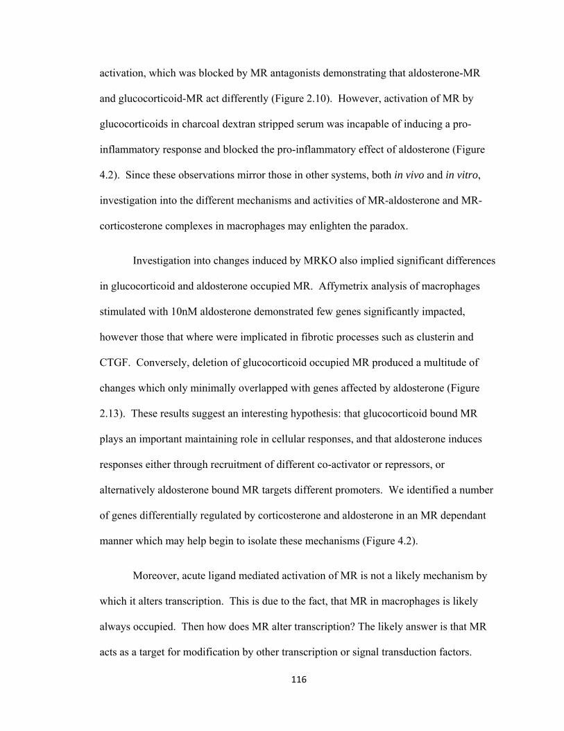
116
activation, which was blocked by MR antagonists demonstrating that aldosterone-MR
and glucocorticoid-MR act differently (Figure 2.10). However, activation of MR by
glucocorticoids in charcoal dextran stripped serum was incapable of inducing a pro-
inflammatory response and blocked the pro-inflammatory effect of aldosterone (Figure
4.2). Since these observations mirror those in other systems, both in vivo and in vitro,
investigation into the different mechanisms and activities of MR-aldosterone and MR-
corticosterone complexes in macrophages may enlighten the paradox.
Investigation into changes induced by MRKO also implied significant differences
in glucocorticoid and aldosterone occupied MR. Affymetrix analysis of macrophages
stimulated with 10nM aldosterone demonstrated few genes significantly impacted,
however those that where were implicated in fibrotic processes such as clusterin and
CTGF. Conversely, deletion of glucocorticoid occupied MR produced a multitude of
changes which only minimally overlapped with genes affected by aldosterone (Figure
2.13). These results suggest an interesting hypothesis: that glucocorticoid bound MR
plays an important maintaining role in cellular responses, and that aldosterone induces
responses either through recruitment of different co-activator or repressors, or
alternatively aldosterone bound MR targets different promoters. We identified a number
of genes differentially regulated by corticosterone and aldosterone in an MR dependant
manner which may help begin to isolate these mechanisms (Figure 4.2).
Moreover, acute ligand mediated activation of MR is not a likely mechanism by
which it alters transcription. This is due to the fact, that MR in macrophages is likely
always occupied. Then how does MR alter transcription? The likely answer is that MR
acts as a target for modification by other transcription or signal transduction factors.

117
Figure 4.2 MR regulates transcription through multiple mechanisms. (A) 10 nM
Aldosterone induces MCP-1 expression in macrophages cultured in C/D media, abolished by
the MR antagonist eplerenone. 10 nM corticosterone produces a GR dependant pro-
inflammatory effect and antagonizes the actions of Aldosterone. (B) Differences in regulation
of Htra1, (C) Clu, and (D) CTGF by 10 nM aldosterone and Corticosterone. (E) Ablation of
glucocorticoid occupied MR reduces IL-1β expression following LPS stimulation and
abolishes glucocorticoid sensitivity (F) *=p<.05 by student’s T-test. Experiments without
statistical data require additional repititions.

118
This can be inferred from the fact effects of MRKO synergize with other stimulants to
enhance or diminish their effects. For example, IL-1β expression was diminished in
MRKO macrophages. LPS stimulation, which induces IL-1β by over a thousand fold,
did not mask the inhibitory effect of IL-1β expression (Figure 4.2). Interestingly,
MRKO abolished the ability of GR to further repress IL-1β induction. These data
illustrate that
MR can synergize with macrophage activation signals such as NFκB, STAT1, or
AP1 to enhance transcription. Additionally, MR is necessary for GR to inhibit this effect.
In these cases, MR’s control of transcription is likely not mediated by acute ligand
activation, but through its ability to interact with inflammatory signals, or the anti-
inflammatory GR.
Many physiologically relevant signals have been proposed to regulate MR’s
activity. However, not until the creation of tissue specific knockouts of MR, has it been
possible to show the necessity of MR for transcriptional regulation of environmental
responses. With the development of the MΦMRKO mouse, these experiments can now
be performed. We have shown the dependency of MR in many glucocorticoid responses;
however, a majority of changes induced by MRKO occurred in genes insensitive to high
doses of corticosterone. If MR regulates transcription primarily through context
dependant interactions with other transcription factors or DNA elements, it is important
to identify cellular functions MR regulates and thereby identify candidate processes
which are likely to impact MR’s actions. MR’s cellular roles predicted by gene ontology

119
Table 4.1: Biological and cellular functions upregulated by
MRKO. Gene ontological analysis of genes induced < 2 fold by
MRKO by GoMINER yielded many important macrophage
functions
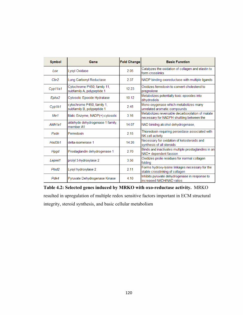
120
Table 4.2: Selected genes induced by MRKO with oxo-reductase activity. MRKO
resulted in upregulation of multiple redox sensitive factors important in ECM structural
integrity, steroid synthesis, and basic cellular metabolism

121
analysis from factors affected by MRKO, suggest its control of inorganic anion transport,
cell adhesion, and redox activity (Table 4.1). Redox activity is especially interesting as it
has been long proposed that MR acts as a redox sensitive transcription factor. We show
that MR regulates a large cluster of factors which involve control of NADP+/NADPH
levels, such as malic enzyme(Me1)[35, 36], and pyruvated dehydrogenase kinase 4
(PDK4) [37], and a number of NADPH requiring aldo-keto reductases of unknown
physiologic function (Table 4.2). Conversely, it has been hypothesized that
NADP+/NADPH and NAD+/NADH are important in MR responses, but never been
conclusively shown[10]. .
In order to investigate specific action of MR at the level of the promoter, first a
putative binding sequences needed to be predicted. While it has always been assumed
that MR and GR bind to the same sequence, it has never been observed experimentally.
We utilized multiple sequence alignment of type II nuclear hormone receptor response
elements identified in promoters of the 15 most upregulated and downregulated genes to
predict a consensus binding sequence (Figure 4.3) . The consensus binding sequence of
upregulated and downregulated genes were identical except contained the opposite
polarity relative to the promoter start site. The MRE also resembled the predicted GRE
and PRE halfsite, but contained significant differences in both polarity and sequence
from the actual GRE identified by ChIP on ChIP assays [38].
Another interesting interaction which is likely important to the cellular role of
MR, occurs between MR and GR. As discussed in chapters 2 and 3, MR plays a critical
role in regulating responses to changing glucocorticoid levels. We identified a large

122
Figure 4.3 Bioinformatic prediction of the mineralocorticoid receptor
response element. (A) Multiple sequence alignment of type II steroid
receptor response elements found in the top 20 genes up and down regulated
yielded strong similarity. (B) Putative MREs identified in genes upregulated
vs downregulated in MRKO macrophages share the same sequence ,but are
opposite in polarity. They also have differences with the canonical GRE.

123
number of genes regulated by MR and GR in different ways (Figures 2.20-2.22).
Understanding the biophysical mechanisms by which MR and GR coordinately regulate
gene transcription will provide new understanding into how glucocorticoids regulate
physiologic processes.
Macrophage MR regulates antigen recruitment and lymphocyte proliferation
Broad transcriptional analysis of MRKO macrophages indicated MHC class II,
and antigen presentation as functions regulated by MR in macrophages. Critical class II
MHC factors such as MHC-A1, and A2, as well as co-activator complexes were
upregulated in the MRKO. In vivo, MΦMRKO mice demonstrated a trend toward
splenomegaly, and qualitatively enlarged follicles with an abnormal expansion of
plasmacytoid cells in the center (Figure 4.4). Abnormal lymphocyte proliferation was
also observed in peyer’s patches in the colon of MΦMRKO mice.
L-NAME/Ang-II treatment also resulted in dramatic splenic structural changes
that were partially mitigated in MΦRKO mice. L-NAME/Ang-II treatment abolished the
clear delineation of red and white pulp with significant infiltration of lymphocytes into
the red pulp, destruction of normal follicular structure, and significant splenic vascular
remodeling. Vascular remodeling in MΦMRKO mice was abolished and follicular
structure was conserved, despite increases in cellularity of the red pulp (Figure 4.5).
MR in monocyte differentiation
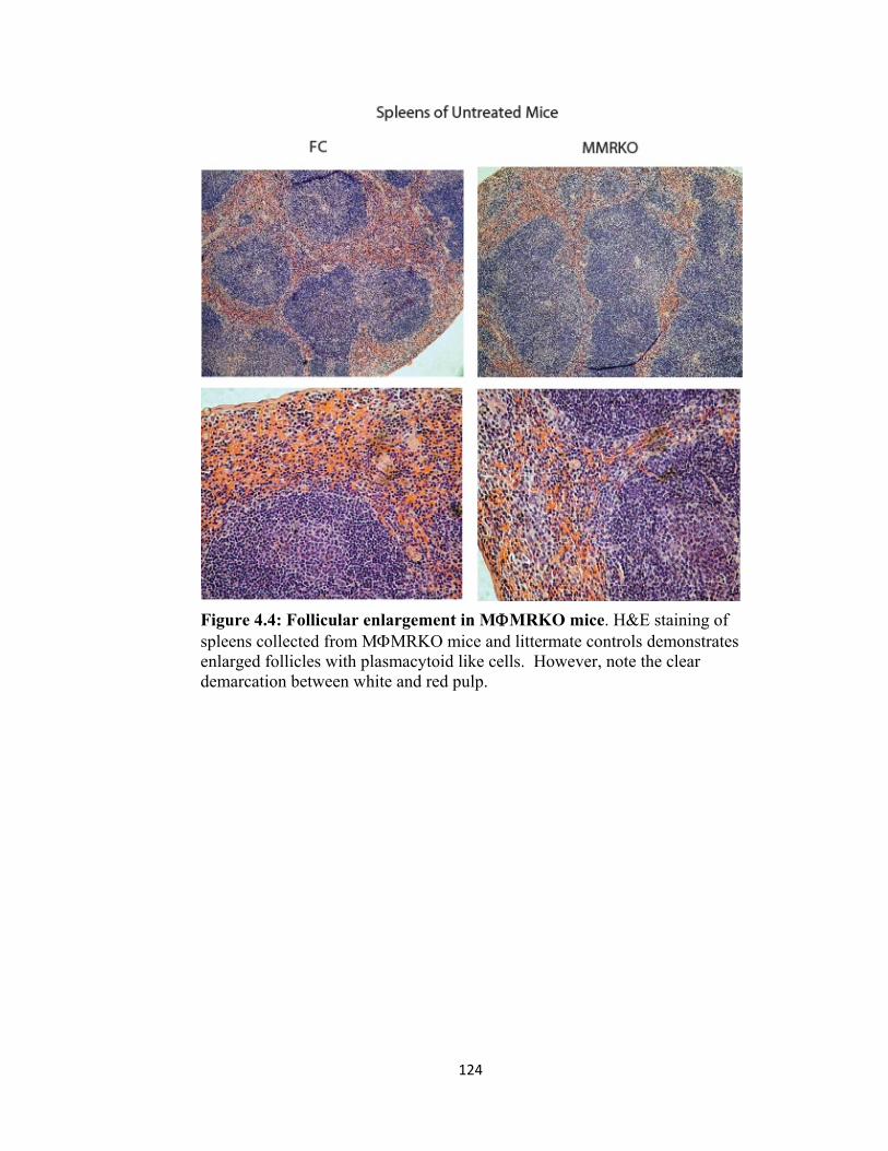
124
Figure 4.4: Follicular enlargement in MΦMRKO mice. H&E staining of spleens collected from MΦMRKO mice and littermate controls demonstrates enlarged follicles with plasmacytoid like cells. However, note the clear demarcation between white and red pulp.

125
Figure 4.5: Splenic Structure is disrupted by L-NAME/Ang-II H&E
staining from mice treated with L-NAME/Ang-II. Normal follicular
structure is abolished following L-NAME/Ang-II treatment, and restored
by MΦMRKO. Hyper-cellularity of red pulp induced by L-NAME/Ang-
II was not abolished by MΦMRKO

126
Preliminary evidence suggests MR plays a critical role in the differentiation of
monocyte subspecies. FACS sorting of circulating monocytes demonstrated a
statistically significant reduction in P/E mid, CCR2 low monocytes which correspond to
the AMΦ precursor. This reduction was completely abolished in MΦMRKO
macrophages. These data implicate MR may not only be important in the repression of
M2 activation, but may be important in the inhibition of differentiation of CX3CR1hi,
GR-1lo, CCR2 lo AMΦ precursor (Figure 4.6).
MRKO also resulted in induction of genes which are not normally expressed in
macrophages. MRKO macrophages expressed high levels of steroidogenic enzymes such
as CYP1b1 and CYP11a1, both of which could not be detected in wildtype macrophages.
These changes could not be mimicked by removal of steroid containing media, or MR
antagonism, or specific MR activation (Figure 4.6). One potential explanation for these
effects is that differentiation which triggers terminal transcriptional changes is
fundamentally altered by MR deletion, and thus genes which are supposed to be
repressed are not.
Discussion and Future Directions
These data illustrate the multiple ways by which MR regulates inflammation.
For the most part these observations only provide a superficial account of associations
without providing specific mechanisms. However, these observations provide
experimental avenues for investigation of very fundamental mechanism of transcriptional
control by MR, monocyte/macrophage differentiation, and innate-adaptive immune
regulation.
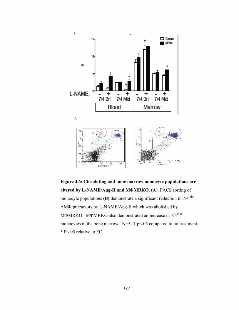
127
Figure 4.6: Circulating and bone marrow monocyte populations are
altered by L-NAME/Ang-II and MΦMRKO. (A). FACS sorting of
monocyte populations (B) demonstrate a significant reduction in 7/4mid
AMΦ precursors by L-NAME/Ang-II which was abolished by
MΦMRKO. MΦMRKO also demonstrated an increase in 7/4mid
monocytes in the bone marrow. N=5, p<.05 compared to no treatment,
* P<.05 relative to FC.

128
MR mediated transcriptional control in macrophages
Comparison of aldosterone and corticosterone occupied MR produced paradoxical
results. There are a number of possible mechanisms by which glucocorticoid and
promoters may selectively bind glucocorticoid or aldosterone bound MR. This has been
aldosterone causes different macrophage responses. First, response elements of different
shown to be the case with the glucocorticoid receptor, where binding of a response
element sequences alters the binding affinity of GR for chemically modified ligands.
Conversely, different ligands confer different binding affinities of GR for specific
sequences.
The second mechanism is that aldosterone and glucocorticoid bound MR interact
differently with activator and co-repressor complexes. Again, this has been shown to be
relevant with GR, where transcriptional activation or repression is dramatically altered by
ligands of different structures. This difference is altered by the presence of different co-
activator and co-repressor complexes as well as different GRE sequences.
Finally, aldosterone bound and glucocorticoid bound MR may be differentially
targeted for post-translational modification. It has been shown that covalent modification
of MR, such as SUMOylation, and ubiquitination alters MR’s ability to affect
transcription, either directly altering its interaction with DNA, or altering protein-protein
interactions. Clearly, aldosterone and glucocorticoid bound MR have different
biochemical properties and interact with proteins differently.
To test these possible mechanisms, or conversely, confirm these effects to be indirect,
demonstration of MR occupancy at relevant promoter is necessary. The identification of
129
genes regulated by MR and GR, and a predicted binding sequence allows for this
possibility in macrophages. This is a technically difficult approach, as no MR antibodies
are specific enough to make ChIP meaningful, and simple epitope tags have been proven
to be insufficient. A few new techniques, one which co-opts bacterial biotinylation
machinery to efficiently label and thus pull down specific proteins, or else using tandem
epitope tags such as a 9X flag tag, have been used to perform challenging ChIP
experiments. ChIP experiments for MR will be performed in a transfectable
macrophage cell line, which has mimicked primary macrophage cell cultures in respect to
MR’s ability enhance or inhibit activation. Occupancy of promoters identified to be
preferentially sensitive to glucocorticoid occupied MR (Cyr61) or aldosterone (CTGF) or
respond to both (Clu, IL-1β) in an MR dependant manner, to see if binding of MR to
these sites occurs differently to aldosterone or glucocorticoid. Additional ChIP
experiments can be performed with known co-activator or co-repressors to see if binding
is impacted by MR activation. Finally, mutational analysis of conserved phosphorylation
sites [39], ubiquitination sites[40], or SUMOylation sites [41] can be utilized to see if
mutations can mask or enhance the differences between aldosterone and corticosterone.
Not only is the mechanism of MR mediated transcriptional regulation unclear, but
also the mechanism by which MR antagonists exert their specific effects. While we show
that MR antagonist only exert visible effects in comparison to activated MR, this could
be due to two reasons. First, MR antagonists may act as classical antagonists, blocking
the nuclear import and DNA binding activity of MR, thereby completely blocking its
nuclear actions. The other potential mechanism is that MR antagonists act as reverse
agonists which require DNA bound MR to act. For example, spironolactone may bind

130
MR on the IL-1β promoter and disrupt co-activator complexes or recruit repressor
complexes onto the promoter. This has been shown to be an important mechanism of
action for many nuclear hormone receptor antagonists including the GR/PR antagonist
RU486[42, 43]. ChIP analysis will allow us to differentiate between these two
mechanisms.
ChIP experiments can be used to identify a number of interesting interactions and
mechanisms of glucocorticoid occupied MR. For example PDK4 is a gene significantly
upregulated in MRKO macrophages. It is known that PDK4 is transcriptionally regulated
by GR, PPAR-γ, redox sensitive transcription factors SIRT1 and FOXO1, and is sensitive
to NADP/NADPH balance[44]. Mutational analysis and other promoter bashing
techniques of conserved response elements in the PDK4 promoter, along with ChIP,
overexpression, siRNA knockdown, and mutations of MR and other signaling molecules
can be utilized to identify critical features and mechanisms which drive the crosstalk of
each important molecular signal.
MR control of monocyte/macrophage differentiation
The observation that MΦMRKO mice exhibit differences in monocyte
populations following L-NAME/Ang-II administration indicates that MR may control
differentiation. This is not surprising given that glucocorticoids which are opposed by
MR stimulate a novel anti-inflammatory monocyte similar to myeloid suppressor cells.
However, these data only provide a single snapshot into the presence of circulating and
bone marrow monocytes. Further characterization of monocyte subpopulations must be
performed over regular intervals to determine the point when reduction in CX3CR1 hi

131
monocytes start to recede, and at what point do differences occur between FC and
MΦMRKO mice.
Changes in circulating monocyte populations may be due to altered differentiation
or recruitment into the tissues. Results from the bone marrow implicate differentiation as
the potential mechanism since a moderate but significant increase in CX3CR1hi was also
observed in MΦMRKO bone marrow; however to confirm this, characterization of
monocytes recruited into cardiac and peri-vascular spaces is important. Since other
models of cardiac injury demonstrate clear phases in monocyte differentiation and
recruitment, it would be interesting to compare to models of mineralocorticoid excess to
see if similar phases exist.
MR in macrophages and the control of adaptive immunity
It has been recently demonstrated that T cell proliferation and recruitment is an
important step in peri-vascular inflammation stimulated by mineralocorticoid excess[34].
We have confirmed that macrophage MR is important in the regulation of lymphocyte
proliferation and recruitment, although the specific mechanisms and immunologic
significance of this has yet to be determined. To further investigate MR in macrophages
control of antigen presentation and lymphocyte proliferation, first the nature of
lymphocyte changes in MΦMRKO mice and following L-NAME/Ang-II treatment must
be elucidated. This may be done through FACS sorting of splenic and recruited
lymphocytes, to identify specific population expansion. Lymphocyte proliferation in
MΦMRKO mice may either be due to T cell or B cell proliferation. B-cell proliferation
occurs following stimulation by antigen presentation, CD40ligand engagement and
132
activation by a number of cytokines such as IL-2 and IL-4. Depending on the local
cytokine environment, B-cells undergo a class switch to produce more IgE and IgM
under Th2 response vs IgG with a Th1 response[45]. Specific increases in antibody
isotype which can be determined through bioplex assay would be an indication of the
underlying inflammatory response which occurs with L-NAME/Ang-II treatment and is
altered by MΦMRKO.
Subsequently, mixed lymphocyte macrophage co-culture experiments may be
utilized to compare the ability of MΦMRKO macrophages to control the proliferation, or
conversely the anergy of specific T and B cell populations. The in vivo significance of
this interaction may be further studied by investigating the robustness of the adaptive
immune response to various repeat infectious challenges.
MR in macrophages in immune responses and inflammatory disease states.
Inflammation is often a double edged sword. On one side, immune responses are
necessary to combat invading pathogens and coordinate the metabolic and cellular
responses to tissue injury and cell death. On the other side, misdirected inflammatory
responses exacerbate tissue injury, promote metabolic derangements which lead to
disease, and can result in irreversible pathology in every biological system.
Inflammatory responses are not simple on-off switches: different stimuli result in
activation of different arms of immunity. Alternative macrophage activation is enhanced
in helminth infections, pulmonary and hepatic fibrosis, but is also associated with
protection in insulin resistance, diabetic nephropathy, hepatic steatosis, and in our study
cardiac fibrosis. For the most part, the macrophage diversity in inflammation has been

133
limited to correlation of specific markers of macrophage activation. We have created a
novel model which blunts classical macrophage activation and enhances specific aspects
of alternative macrophage activation. This can be remarkably useful in dissecting
protective vs exacerbatory roles of macrophage subtypes in immune responses.
Additionally, since MΦMRKO phenocopies MR antagonist treatment in many ways,
identification of inflammatory pathologies protected by MΦMRKO may indicate new
diseases that might be mitigated by MR antagonism.
We have shown that MRKO in macrophages results in a novel alternatively active
state. This state partially overlaps with IL-4 stimulation and PPAR-γ activation. This
state also partially overlaps with glucocorticoid stimulation. This provides a unique tool
for identifying critical programs in inflammatory responses. For example, it has been
shown that TZDs[46], glucocorticoids[47], and MR antagonists[48] are protective in
treating rheumatoid arthritis models. It is also well known that macrophage recruitment
and activation are critical in the pathogenesis of rheumatoid arthritis. It is not known
however, the specific cell types which are critical targets for each therapeutic agent. We
have obtained macrophage specific knockouts of PPAR-γ and MR, which can now be
used to determine if these drugs work through manipulation of macrophage polarization.
We hypothesize that MR knockout will be likely protective in models of adjuvant
or bovine collagen induced murine rheumatoid arthritis model. In a similar manner we
expect PPAR-γ knockout to abolish the protective effects of TZDs. If this true than the
critical components which modify rheumatoid arthritis would be represented in overlap
between PPAR-γ activation and MR inactivation in macrophages. The macrophage
134
specific GR knockout is also available. This mouse model could also be added in to the
comparison of beneficial effects, and would further help focus the known critical
components of modifying rheumatoid arthritis pathogenesis.
As in rheumatoid arthritis, which is primarily thought to be a Th1 mediated
inflammatory disease, MΦMRKO and MΦPγKO can be used to dissect the role of AMΦ
in Th2 or Th17 mediated inflammatory responses. For example pulmonary diseases such
as allergic asthma are mediated by Th2 responses[49]. The role of AMΦ polarization in
the pathogenesis of this disorder has not been directly investigated. We would
hypothesize that MΦMRKO would likely exacerbate allergic asthma models such as
ovalbumin challenge and demonstrate increased sensitivity to methylcholine mediated
airway constriction.
Conversely, in models of pulmonary fibrosis, the contributing roles of Th1, Th2,
Th17 and polarized macrophages is more complex[50, 51]. Th2 cytokines such as IL-13
are necessary for the development of bleomycin induced pulmonary fibrosis[52].
However, unlike other models of Th2 mediated inflammation where Th1 cytokines can
be protective, Th1 polarization enhances rather than diminishes the fibrotic response[53,
54]. Moreover, CCR4 knockout, which diminishes Th2 recruitment[55], resulted in
recruitment of AMΦ polarized macrophages, and was protected in pulmonary
fibrosis[56]. How is this possible if Th2 cytokines are necessary for the development of
AMΦ macrophage? One possible explanation is that the protective macrophage in this
case is not an IL-4 stimulated macrophage, but the third subtype discussed in previous
chapters. This AMΦ resembles the standard AMФ macrophage in many aspects,
135
however contains discrete properties that are repressed by both Th1 and Th2 cytokines.
If this macrophage was important we would expect MΦMRKO to be protective in
models of pulmonary fibrosis despite its AMФ like phenotype. We would also expect in
both CCR4KO and MΦMKRO to express increases in these novel AMΦ markers such as
Htra1, Cdh2, Hmga2, and decreases in PD-1 lig2, E-cadherin, and others. This would
also explain the increases in M2 markers such as Arg1, Ym1, and Ym2, despite decreases
in IL-4, as these genes overlap between different AMΦ subtypes.
A similar, but more focused approach can also be applied. For example we show
that MR and PPAR-γ similarly regulated E-cadherin expression. E-cadherin expression
is abolished in both MRKO and PγKO macrophages. In macrophages, E-cadherin is a
necessary factor in macrophage fusion stimulated by IL-4[57]. This is an important step
in combating fungal and parasitic infections[58], but is also a pathogenic factor in
uncontrolled granulomatous inflammation[59]. We hypothesize that MRKO and PγKO
would demonstrate reduced multi-nucleated giant cell formation in response to IL-4 in
vitro, and would be similarly susceptible to helminth or other parasitic infection which
requires multi-nucleated giant cells to combat infection.
Interestingly, E-cadherin is also necessary in osteoclastogenesis[60]. It has been
recently shown that PPAR-γ is critical for the generation of osteoclasts[61], and may be
one mechanism by which TZDs enhance the risk of osteoporosis [62]. We would expect
a similar phenotype in MΦMRKO mice. This may present a contraindication for MR
antagonists in patients with osteoporosis; conversely this may indicate MR antagonists in

136
the treatment of primary granulomatous diseases such as giant cell arteritis. These
connections have never been investigated.
Summary
Inflammation is involved in almost every human disease process. Treatment of
inflammation is fraught with pitfalls given its importance in preventing infection. This
may, in part, be due to the fact that the clinical approach to treatment of inflammation is
to turn it off. Clearly, the complexities of inflammatory responses warrant more nuanced
treatment strategies. Treatment protocols for other chronic multifactoral disorders such
as cancer generally take multiple approaches from different directions. Even within
macrophage activation, we show marked heterogeneity which can be manipulated in
coordinated fashion by commonly used pharmacologic agents. Research into the
molecular and immunologic cross talk between receptors which directly regulate
inflammation may shed light on potential combination of drugs which may synergize to
produce more effective treatments, increase therapeutic index, or provide novel therapies.
While we have begun to understand MR’s role in the macrophage, and
macrophage MR’s role in immunity, there are many unanswered questions. Coordination
of ChIP, mutational analysis, and expression analysis has been shown to be powerful
tools to dissect molecular crosstalk on promoters. This is a very important project as it
would provide insight into MR’s actions while occupied by glucocorticoids in
biologically relevant tissue. Additionally, since we show that antagonism of
glucocorticoid occupied MR is important in the cardioprotective effects of spironolactone
and eplerenone, identification of specific biochemical attributes which distinguish

137
aldosterone and glucocorticoid occupied MR may begin to allow for next generation MR
antagonists. MR antagonists that only block glucocorticoid occupied MR may provide
cardioprotective effects while diminishing the important limiting complication of MR
antagonists: hyperkalemia.
Since direct transcriptional regulation is likely not the only mechanism by which
MΦMRKO provides alteration in macrophage responses and cardioprotection. We
provide preliminary evidence that monocyte/macrophage differentiation and adaptive
immunity are also impacted both by MΦMRKO and L-NAME/Ang-II. Chronic
inflammatory diseases including the states which drive cardiovascular risk involve
coordinated activation of innate and adaptive immune responses. Antigen processing, T-
cell activation, and autoimmune antibody production, in addition to innate activation of
the myeloid phagocytic system have been shown to be critical for the development of
atherosclerosis. Understanding how MR controlled transcriptional programs in
macrophages lead to changes in innate adaptive immunity in the presence and absence of
cardiovascular inflammation may shed light on critical processes which exacerbate CVD
and provide additional targets for therapy.
To conclude, MR guides inflammatory responses through multiple mechanisms
which are only beginning to be addressed (Figure 4.7). Specific mechanisms by which
MR regulates macrophage transcription and mechanisms by which macrophages regulate
innate immune responses will provide unique insight into the pleiotropic beneficial
actions of MR antagonists.

138
Figure 4.7 Pleiotropic actions of myeloid MR on innate and adaptive
immunity Evidence suggests MR plays a role in the repression of AMΦ
precursor monocyte (A). MR drives M1 macrophage polarization (B): repressing
different AMΦ transcriptional programs driven by GR or PPAR-γ, and enhancing
M1 responses. This occurs in part by synergizing with M1 stimulants including
LPS and likely IFNγ (C), and repression of the action of Th2 cytokine IL-4 (D).
Finally, observations that MΦMRKO results in plasmacytoid cell expansion in
splenic follicle paired with increases in dendritic cell markers such as MHC-II,
LR8b, and dc-SIGN implicate MR in the repression of antigen presentation,
lymphocyte proliferation, and potentially dendritic cell proliferation (E).
Together these data suggest MR’s primary role is to lock a pro-inflammatory, Th-
1 type of innate immune response which is exacerbatory in cardiovascular
disease. Downregulated factors by MMRKO (blue) and upregulated (red).

139

140
References: 1. Gordon, S. and P.R. Taylor, Monocyte and macrophage heterogeneity. Nat Rev Immunol,
2005. 5(12): p. 953‐64. 2. Funder, J. and K. Myles, Exclusion of corticosterone from epithelial mineralocorticoid
receptors is insufficient for selectivity of aldosterone action: in vivo binding studies. Endocrinology, 1996. 137(12): p. 5264‐8.
3. Arriza, J.L., et al., The neuronal mineralocorticoid receptor as a mediator of glucocorticoid response. Neuron, 1988. 1(9): p. 887‐900.
4. Caprio, M., et al., Pivotal role of the mineralocorticoid receptor in corticosteroid‐induced adipogenesis. Faseb J, 2007. 21(9): p. 2185‐94.
5. Brem, A.S. and D.J. Morris, Interactions between glucocorticoids and mineralocorticoids in the regulation of renal electrolyte transport. Mol Cell Endocrinol, 1993. 97(1‐2): p. C1‐5.
6. Morris, D.J., S.A. Latif, and A.S. Brem, Interactions of mineralocorticoids and glucocorticoids in epithelial target tissues revisited. Steroids, 2009. 74(1): p. 1‐6.
7. Ross, E.J., Modification of the effects of aldosterone on electrolyte excretion in man by simultaneous administration of corticosterone and hydrocortisone. Relevance to Conn's syndrome. J Clin Endocrinol Metab, 1960. 20: p. 229‐37.
8. Husted, R.F., J.R. Laplace, and J.B. Stokes, Enhancement of electrogenic Na+ transport across rat inner medullary collecting duct by glucocorticoid and by mineralocorticoid hormones. J Clin Invest, 1990. 86(2): p. 498‐506.
9. Souness, G.W. and D.J. Morris, The "mineralocorticoid‐like" actions conferred on corticosterone by carbenoxolone are inhibited by the mineralocorticoid receptor (type I) antagonist RU28318. Endocrinology, 1991. 129(5): p. 2451‐6.
10. Funder, J.W., Translational research goes both ways: lessons from clinical studies. Clin Exp Pharmacol Physiol, 2008. 35(4): p. 526‐9.
11. Funder, J.W., et al., Mineralocorticoid action: target tissue specificity is enzyme, not receptor, mediated. Science, 1988. 242(4878): p. 583‐5.
12. Makino, Y., et al., Direct association with thioredoxin allows redox regulation of glucocorticoid receptor function. J Biol Chem, 1999. 274(5): p. 3182‐8.
13. Mercer, W.R. and Z.S. Krozowski, Localization of an 11 beta hydroxysteroid dehydrogenase activity to the distal nephron. Evidence for the existence of two species of dehydrogenase in the rat kidney. Endocrinology, 1992. 130(1): p. 540‐3.
14. Joels, M., et al., The coming out of the brain mineralocorticoid receptor. Trends Neurosci, 2008. 31(1): p. 1‐7.
15. Swirski, F.K., et al., Ly‐6Chi monocytes dominate hypercholesterolemia‐associated monocytosis and give rise to macrophages in atheromata. J Clin Invest, 2007. 117(1): p. 195‐205.
16. Nahrendorf, M., et al., The healing myocardium sequentially mobilizes two monocyte subsets with divergent and complementary functions. J Exp Med, 2007. 204(12): p. 3037‐47.
17. Tacke, F., et al., Monocyte subsets differentially employ CCR2, CCR5, and CX3CR1 to accumulate within atherosclerotic plaques. J Clin Invest, 2007. 117(1): p. 185‐94.
18. Tsou, C.L., et al., Critical roles for CCR2 and MCP‐3 in monocyte mobilization from bone marrow and recruitment to inflammatory sites. J Clin Invest, 2007. 117(4): p. 902‐9.

141
19. Varga, G., et al., Glucocorticoids induce an activated, anti‐inflammatory monocyte subset in mice that resembles myeloid‐derived suppressor cells. J Leukoc Biol, 2008. 84(3): p. 644‐50.
20. Johnson, J.L., et al., Divergent effects of matrix metalloproteinases 3, 7, 9, and 12 on atherosclerotic plaque stability in mouse brachiocephalic arteries. Proc Natl Acad Sci U S A, 2005. 102(43): p. 15575‐80.
21. Landsman, L., et al., CX3CR1 is required for monocyte homeostasis and atherogenesis by promoting cell survival. Blood, 2009. 113(4): p. 963‐72.
22. Liu, P., et al., CX3CR1 deficiency confers protection from intimal hyperplasia after arterial injury. Arterioscler Thromb Vasc Biol, 2006. 26(9): p. 2056‐62.
23. Liu, P., et al., CX3CR1 deficiency impairs dendritic cell accumulation in arterial intima and reduces atherosclerotic burden. Arterioscler Thromb Vasc Biol, 2008. 28(2): p. 243‐50.
24. Saederup, N., et al., Fractalkine deficiency markedly reduces macrophage accumulation and atherosclerotic lesion formation in CCR2‐/‐ mice: evidence for independent chemokine functions in atherogenesis. Circulation, 2008. 117(13): p. 1642‐8.
25. Apostolakis, S., et al., Genetic diversity of CX3CR1 gene and coronary artery disease: New insights through a meta‐analysis. Atherosclerosis, 2009.
26. McDermott, D.H., et al., Association between polymorphism in the chemokine receptor CX3CR1 and coronary vascular endothelial dysfunction and atherosclerosis. Circ Res, 2001. 89(5): p. 401‐7.
27. Moatti, D., et al., Polymorphism in the fractalkine receptor CX3CR1 as a genetic risk factor for coronary artery disease. Blood, 2001. 97(7): p. 1925‐8.
28. Cheng, X., et al., TH1/TH2 functional imbalance after acute myocardial infarction: coronary arterial inflammation or myocardial inflammation. J Clin Immunol, 2005. 25(3): p. 246‐53.
29. Wang, Z., et al., Analysis of specific Th1/Th2 helper cell responses and IgG subtype antibodies in anti‐CD4 monoclonal antibody treated mice with autoimmune cardiomyopathy. J Huazhong Univ Sci Technolog Med Sci, 2008. 28(4): p. 409‐14.
30. Yi, A., et al., The prevalence of Th17 cells in patients with dilated cardiomyopathy. Clin Invest Med, 2009. 32(2): p. E144‐50.
31. Cheng, X., et al., The Th17/Treg imbalance in patients with acute coronary syndrome. Clin Immunol, 2008. 127(1): p. 89‐97.
32. Eid, R.E., et al., Interleukin‐17 and interferon‐gamma are produced concomitantly by human coronary artery‐infiltrating T cells and act synergistically on vascular smooth muscle cells. Circulation, 2009. 119(10): p. 1424‐32.
33. Miller, A.M., et al., IL‐33 reduces the development of atherosclerosis. J Exp Med, 2008. 205(2): p. 339‐46.
34. Guzik, T.J., et al., Role of the T cell in the genesis of angiotensin II induced hypertension and vascular dysfunction. J Exp Med, 2007. 204(10): p. 2449‐60.
35. Yang, X., et al., Validation of candidate causal genes for obesity that affect shared metabolic pathways and networks. Nat Genet, 2009. 41(4): p. 415‐23.
36. Hanukoglu, I. and R. Rapoport, Routes and regulation of NADPH production in steroidogenic mitochondria. Endocr Res, 1995. 21(1‐2): p. 231‐41.
37. Holness, M.J. and M.C. Sugden, Regulation of pyruvate dehydrogenase complex activity by reversible phosphorylation. Biochem Soc Trans, 2003. 31(Pt 6): p. 1143‐51.
38. Wang, J.C., et al., Chromatin immunoprecipitation (ChIP) scanning identifies primary glucocorticoid receptor target genes. Proc Natl Acad Sci U S A, 2004. 101(44): p. 15603‐8.
142
39. Alnemri, E.S., et al., Overexpression and characterization of the human mineralocorticoid receptor. J Biol Chem, 1991. 266(27): p. 18072‐81.
40. Tirard, M., et al., Sumoylation and proteasomal activity determine the transactivation properties of the mineralocorticoid receptor. Mol Cell Endocrinol, 2007. 268(1‐2): p. 20‐9.
41. Yokota, K., et al., Coactivation of the N‐terminal transactivation of mineralocorticoid receptor by Ubc9. J Biol Chem, 2007. 282(3): p. 1998‐2010.
42. Zhang, X., et al., A nuclear receptor corepressor modulates transcriptional activity of antagonist‐occupied steroid hormone receptor. Mol Endocrinol, 1998. 12(4): p. 513‐24.
43. Hodgson, M.C., et al., Structural basis for nuclear receptor corepressor recruitment by antagonist‐liganded androgen receptor. Mol Cancer Ther, 2008. 7(10): p. 3187‐94.
44. Sugden, M.C. and M.J. Holness, Mechanisms underlying regulation of the expression and activities of the mammalian pyruvate dehydrogenase kinases. Arch Physiol Biochem, 2006. 112(3): p. 139‐49.
45. Nezlin, R., Combinatorial events in generation of antibody diversity. Comb Chem High Throughput Screen, 2001. 4(5): p. 377‐83.
46. Shiojiri, T., et al., PPAR gamma ligands inhibit nitrotyrosine formation and inflammatory mediator expressions in adjuvant‐induced rheumatoid arthritis mice. Eur J Pharmacol, 2002. 448(2‐3): p. 231‐8.
47. Makrygiannakis, D., et al., Monocytes are essential for inhibition of synovial T‐cell glucocorticoid‐mediated apoptosis in rheumatoid arthritis. Arthritis Res Ther, 2008. 10(6): p. R147.
48. Bendtzen, K., P.R. Hansen, and K. Rieneck, Spironolactone inhibits production of proinflammatory cytokines, including tumour necrosis factor‐alpha and interferon‐gamma, and has potential in the treatment of arthritis. Clin Exp Immunol, 2003. 134(1): p. 151‐8.
49. Passalacqua, G. and G. Ciprandi, Allergy and the lung. Clin Exp Immunol, 2008. 153 Suppl 1: p. 12‐6.
50. Sumida, A., et al., TH1/TH2 immune response in lung fibroblasts in interstitial lung disease. Arch Med Res, 2008. 39(5): p. 503‐10.
51. Simonian, P.L., et al., Th17‐polarized immune response in a murine model of hypersensitivity pneumonitis and lung fibrosis. J Immunol, 2009. 182(1): p. 657‐65.
52. Fichtner‐Feigl, S., et al., IL‐13 signaling through the IL‐13alpha2 receptor is involved in induction of TGF‐beta1 production and fibrosis. Nat Med, 2006. 12(1): p. 99‐106.
53. Chen, E.S., et al., Attenuation of lung inflammation and fibrosis in interferon‐gamma‐deficient mice after intratracheal bleomycin. Am J Respir Cell Mol Biol, 2001. 24(5): p. 545‐55.
54. Huaux, F., et al., A profibrotic function of IL‐12p40 in experimental pulmonary fibrosis. J Immunol, 2002. 169(5): p. 2653‐61.
55. Mikhak, Z., et al., Contribution of CCR4 and CCR8 to antigen‐specific T(H)2 cell trafficking in allergic pulmonary inflammation. J Allergy Clin Immunol, 2009. 123(1): p. 67‐73 e3.
56. Trujillo, G., et al., A novel mechanism for CCR4 in the regulation of macrophage activation in bleomycin‐induced pulmonary fibrosis. Am J Pathol, 2008. 172(5): p. 1209‐21.
57. Moreno, J.L., et al., IL‐4 promotes the formation of multinucleated giant cells from macrophage precursors by a STAT6‐dependent, homotypic mechanism: contribution of E‐cadherin. J Leukoc Biol, 2007. 82(6): p. 1542‐53.
143
58. Smythies, L.E., P.S. Coulson, and R.A. Wilson, Monoclonal antibody to IFN‐gamma modifies pulmonary inflammatory responses and abrogates immunity to Schistosoma mansoni in mice vaccinated with attenuated cercariae. J Immunol, 1992. 149(11): p. 3654‐8.
59. Wang, X., et al., Giant cell arteritis. Rheumatol Int, 2008. 29(1): p. 1‐7. 60. Mbalaviele, G., et al., The role of cadherin in the generation of multinucleated
osteoclasts from mononuclear precursors in murine marrow. J Clin Invest, 1995. 95(6): p. 2757‐65.
61. Wan, Y., L.W. Chong, and R.M. Evans, PPAR‐gamma regulates osteoclastogenesis in mice. Nat Med, 2007. 13(12): p. 1496‐503.
62. Schwartz, A.V. and D.E. Sellmeyer, Effect of thiazolidinediones on skeletal health in women with Type 2 diabetes. Expert Opin Drug Saf, 2008. 7(1): p. 69‐78.
144
CHAPTER V
MACROPHAGE MINERALOCORTICOID RECPTOR IN CARDIOVASCULAR PHYSIOLOGY
Overview
We have shown that MR drives detrimental inflammatory signals in response to
cardiac injury. While there is some evidence that acute administration of MR antagonists
may provide benefit following myocardial infarction, for the most part MR antagonists
provide cardioprotection that becomes more visible over time and includes many risk
factors for cardiac events. In the previous chapter we show how studies about MR in
macrophages may be extrapolated in to understanding basic immune mechanisms. In the
same way, MΦMRKO mice can be utilized to better understand how macrophage actions
are integrated into cardiovascular responses. In this chapter we demonstrate that
macrophage MR is important in multiple facets cardiovascular physiology including
circadian rhythms, sympathetic drive, cardiac hypertrophy, and cerebral ischemia.
Introduction
Macrophage MR and Cardiac Hypertrophy
Mineralocorticoid receptor antagonists, indicated in the treatment of heart failure
have been demonstrated to reduce left ventricular hypertrophy in at risk patients[2]. MR
antagonists are also effective in reducing pressure overload hypertrophy models such as

145
chronic angiotensin II administration and aortic constriction[3-5]. Eplerenone not only
reduces cardiac hypertrophy in these models, but also improves survivability and
diastolic function, reduces oxidative stress, peri-vascular and intracardial fibrosis [5].
These benefits appear to be independent of aldosterone antagonism and independent of
load reduction.
Cardiac response to hypertension has long been associated with inflammatory
signaling. Aortic constriction results in upregulation of MCP-1, TGFβ, IL-1β, and the
endothelial adhesion molecule necessary for macrophage recruitment ICAM1[6-8].
Abolishment of MCP-1 signaling reduces recruitment of macrophages, reduces oxidative
stress, and normalizes diastolic dysfunction following pressure overload hypertrophy [9].
Additionally, MCP-1 neutralizing antibodies reduces cardiac hypertrophy following L-
NAME administration[8].
Additionally, recruited macrophages and monocytes play an important role in the
regulation of extracellular matrix remodeling through the secretion of MMPs. Classically
activated macrophages and CCR2hi pro-inflammatory monocytes shown to be recruited
following cardiac injury produce high levels of MMP9[10]. Deletion of MMP9
attenuates left ventricular enlargement following myocardial ischemia[11]. MMP
secretion by macrophages breaks down normal extracellular matrix structure necessary
for efficient contractile function which is subsequently replaced by highly crosslinked
collagen I and III. Increases in Collagen I and III correlated with decreases in ventricular
function and hypertrophy [12].

146
Macrophages regulate cell growth, and extracellular remodeling through many
mechanisms, each which can potentially contribute to remodeling observed in
hypertrophic hearts. However, to this point most studies implicating macrophages in
cardiac hypertrophy have been correlative in nature and not directly show how
macrophages contribute. As we have shown in previous chapters, cardiovascular
inflammation in hypertrophy correlates in an M1 polarized macrophage response. The
direct impact of macrophage polarization in the hypertrophic response has not been
comprehensively investigated.
Macrophage MR and Cardiovascular Circadian Rhythms
The circadian rhythm plays a prominent role in cardiovascular physiology and
injury. Blood pressure and heart rate exhibit a 24 hour cycle which rises in the early
morning and dips in the evening [1]. Risk for cardiovascular events such as hemorrhagic
stroke or myocardial infarction mirrors this circadian pattern (Figure 5.1) [1]. Non-
dipping status, where patients demonstrate reduced or absent reduction in nighttime blood
pressure is a significant cardiovascular risk factor [13].
Circadian control of the cardiovascular system involves a complex coordination
of environmental cues such as light, behavioral inputs, metabolic and endocrine signals
which are integrated with cell autonomous molecular oscillators. Ablation of the supra-
chiasmatic nucleus which abolishes physiologic circadian control abolishes the blood
pressure circadian rhythm[14]. Similarly, systemic disruption of molecular oscillators
such as Per-2, NPAS2, and BMAL-1, demonstrate alterations in hemodynamic circadian
rhythms[15]. However, it is not clear to what degree neuro-hormones, environmental,
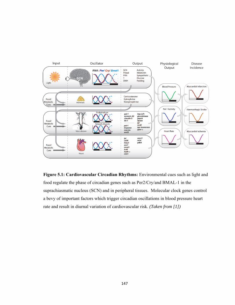
147
Figure 5.1: Cardiovascular Circadian Rhythms: Environmental cues such as light and
food regulate the phase of circadian genes such as Per2/Cry/and BMAL-1 in the
suprachiasmatic nucleus (SCN) and in peripheral tissues. Molecular clock genes control
a bevy of important factors which trigger circadian oscillations in blood pressure heart
rate and result in diurnal variation of cardiovascular risk. (Taken from [1])
148
and metabolic signals contribute to these variations. Some evidence suggests
autonomous circadian oscillators in vascular tissue play an important role in blood
pressure regulation. Endothelial PPAR-γ knockout results in abolishment of the BMAL-
1 circadian rhythm and resulted in diminished 24 hour blood pressure cycling[16].
Interestingly, BMAL-1 disruption results in altered catecholamine metabolism and
enhanced presser response following immobilization stress implicating clock genes in
direct regulation in blood pressure responses [15].
Diurnal variation of inflammation is of similar physiologic importance. Circadian
changes in allergic asthma and febrile responses have been well documented; however
the mechanisms of such changes remain poorly understood. Macrophage activation is
responsive to many endocrine signals regulated in circadian fashion such as epinephrine
and glucocorticoids[17]. Macrophages also express core clock genes such as PER2, and
BMAL-1 which have been shown to directly regulate phagocytic responses, NFκB
activity, and MCP-1 expression[18]. Moreover, pro-inflammatory cytokines and
chemokines have been known to show strong circadian cycling in both cardiac tissue and
the thoracic aorta [19, 20]. Attenuation of circadian signaling, through BMAL-1
deletion, results in endothelial dysfunction which mirrors changes which occur with
inflammation [21]. The contribution of macrophages to physiologic and molecular
biological rhythms is not known, but it remains an intriguing possibility that circadian
changes in inflammatory responses may be responsible for some of the diurnal variation
in cardiovascular risk.
MR has also been linked to control of circadian rhythms. While its expression is
not altered over time[22], it is thought to be important in the control of biological

149
rhythms. Aldosterone, which is produced to a greater degree during the morning than the
night[23] has been shown to directly alter the expression of core clock genes in
cardiomyocytes[24]. Patients with idiopathic primary hyperaldosteronism on average
demonstrate a blunted reduction in night time blood pressure[25]. Whether this in vivo
control of circadian genes is coordinated by aldosterone or glucocorticoid occupied MR
in vivo has not been established. Additionally, it has been shown that both MR and GR
coordinate to control the diurnal variation of ACTH production[26, 27]. We demonstrate
a remarkable change in the circadian rhythm of blood pressure and heart rate in
MΦMRKO mice which illustrates a novel interaction between MR, inflammatory
signaling, and diurnal blood pressure variation.
Macrophage MR and Stroke
Another important cardiac event linked to MR signaling is ischemic stroke.
Studies connecting stroke risk with MR activity mirror that of other cardiac risk factors:
aldosterone appears to exacerbate and MR antagonism confers protection beyond merely
blocking aldosterone. Hyperaldosteronism significantly increases ischemic stroke
risk[28]. Similarly, DOCA administration to animals prior to middle cerebral artery
occlusion resulted in increased vascular remodeling and increased stroke volume[29].
Conversely, MR antagonism is protective in models of stroke. Eplerenone and
spironolactone pretreatment prior to middle cerebral artery occlusion resulted in reduced
stroke volume, reduced neuronal cell death, reduced microglia recruitment, and improved
neurological function[30-32]. This is remarkable given that MR plays an anti-apoptotic
role in neurons in part through upregulation of Bcl2, which is inhibited by MR
150
antagonism[33]. MR antagonism increased collateral circulation, myogenic tone, and
reduced oxidative stress suggesting pleiotropic protective mechanisms [34, 35].
Ultimately, the role of MR in stroke is likely to be multi-factoral. MR activation
by aldosterone stimulates vascular remodeling which decreases collateral flow, increases
inflammatory cell recruitment, and induces oxidative stress. Conversely, glucocorticoid
occupied MR in astrocytes and microglia may enhance inflammatory responses similar to
our observations in cardiovascular injury. As our results demonstrated that deletion of
MR in macrophages yielded remarkable protection in cardiac injury, we hypothesized
that it may have a similar beneficial effect following cerebral ischemia-reperfusion
damage.
Macrophage MR and Diet Induced Obesity
Metabolic syndrome categorized by presentation of a number of highly comorbid
cardiovascular risk factors obesity, type II diabetes mellitus, dyslipidemia, and
hypertension which synergistically increase the likelihood of a cardiac event [36]. The
mechanisms which contribute to the concordance of these risk factors remains poorly
understood.
One contributing factor to each is inflammation. It has been well documented that
a high fat diet and subsequent increase in visceral adipocity contributes to inducing a pro-
inflammatory response with many facets. Circulating pro-inflammatory cytokines such
as TNFα, IL-1β, and IL-6 are all significantly enhanced following 14 weeks of a high fat
diet[37, 38]. A similar pro-inflammatory state is observed in patients with central
obesity. The mechanisms by which diet induced obesity enhances inflammation are
151
diverse. Adipose tissue following a high fat diet has been shown to increase the
expression of pro-inflammatory adipokines and reduce the expression of the anti-
inflammatory adiponectin [39]. Metabolic alterations with high fat diet which include
elevated FFA have been shown to directly activate macrophages though engagement of
TLR4 [40]. Similarly, covalently modified LDL which is increased with high fat diet has
also been shown to promote classical macrophage activation through multiple
mechanisms [41, 42].
Conversely, classical macrophage activation has been shown to be necessary for
pathologies induced by high fat diet. Deletion of CCR2 a chemokine receptor important
in the recruitment of M1 macrophages and monocytes protects mice against induced
obesity and insulin resistance [43]. The population of adipose tissue macrophages
undergoes a dramatic shift from an alternatively activated state to a M1 state during
dietinduced obesity[44, 45]. The mechanism of this effect is largely due to the
recruitment of classically activated macrophages. The contribution of greater M1
macrophages versus reduced AMΦ activity to insulin resistance and other metabolic
changes associated with obesity have not been dissected.
Recent studies have demonstrated that direct action of TZDs on macrophage
function to be a central component to their physiologic effects. Deletion of PPAR-γ in
macrophages results in an increase in pro-inflammatory cytokine expression, reduction
ATP-binding cassette G1 expression, suggesting an impairment of reverse cholesterol
transport [46, 47]. These studies have also demonstrated that macrophage PPAR-γ is
necessary for the full insulin sensitizing effects of TZDs and that modulation of
macrophage function is important in vascular disorders[46, 47].
152
PPAR-δ has also recently been shown to have a similar role to PPAR-γ in controlling the
M1/AMΦ polarization [48, 49]. One group has identified PPAR-δ as being more
important in the liver macrophage like, Kupffer cells[49]. These groups also showed that
cytokines that stimulate AMΦ polarization have beneficial effects on insulin resistance
and glucose and lipid metabolism.
MR antagonists demonstrate similar, but less well documented beneficial effects
on diet induced obesity. One study showed that eplerenone administration to db/db mice
reduced weight gain, improved insulin sensitivity, and reduced expression of pro-
inflammatory markers [50]. Due to the striking overlap in the in vivo activities that MR
antagonists have with the TZDs in inflammatory models that feature macrophage
infiltration as an important component [44, 45] we hypothesized that MR antagonists
may act via a similar macrophage dependant manner mechanism. Moreover, since
MΦMRKO is a novel model with an intrinsic AMΦ shift, it would be useful to further
investigate the ability of alternative macrophage polarization in the metabolic
deraignment associated with high fat diets.
To summarize the cardiovascular risk can be attributed to multiple highly
integrated systems. Likewise, cardioprotective drugs often trigger pleiotropic beneficial
effects toward many factors which promote cardiovascular disease. The specific
mechanisms which connect hemodynamic, cardiac, neural, endocrine, inflammatory, and
metabolic aspects of cardiovascular pathogenesis remain poorly understood. MΦMRKO
has provided a novel window to observe the coordinated regulation of inflammatory

153
signaling and cardiac hypertrophy, hemodynamic circadian rhythms, stroke, and diet
induced obesity.
Results
Macrophage MR in Cardiac Hypertrophy
Despite increases in blood pressure and cardiac work in MΦMRKO mice relative
to controls following L-NAME/Ang-II administration we observed a decrease in cardiac
hypertrophy (Figure 5.2). Remarkably this result occurred despite increases in cardiac
work, due to daytime increases in systolic pressure and heart rate relative to controls. In
many ways this result phenocopies the effect of MR antagonists: protecting against
cardiac hypertrophy and improving contractile function without reducing load. These
results indicate macrophages are likely important in the control of the hypertrophic
response to cardiac injury.
Additionally we observe that PPAR-γ agonists protect against pressure overload
hypertrophy in a cardiomyocyte independent manner. Given that PPAR-γ agonists
protect against cardiac fibrosis and inflammation in response to angiotensin II through
their actions on macrophages, and PPAR-γ agonists and MR antagonists mirror their
effects on macrophage polarization, we hypothesize that macrophage polarization is
critical to the protective effects of TZDs and MR antagonists. In previous chapters we
showed that cardiac hypertrophy in response to L-NAME/Ang-II resulted in recruitment
of M1 macrophages and a reduction in expression of AMΦ macrophage markers.
However, this result could either be due to direct classical macrophage activation by L-
NAME/Ang-II or due to differential recruitment and activation secondary to cardiac

154
Figure 5.2 Macrophage action and cardiac hypertrophy. (A) L-NAME/Ang-II
resulted in cardiac hypertrophy (Measured by heart weight/body weight ratio, HW/BW)
partially abolished by MΦMRKO. Reduction in hypertrophy correlated with protective
alterations in hypertrophy markers. (B) Pressure overload hypertrophy measured by
ventricular weight/body weight ratio (VW/BW) stimulated by abdominal aortic
constriction in wildtype mice resulted in a time dependant M1 shift in cardiac tissue (C).

155
hypertrophy. We investigated the macrophage polarization in the following direct
mechanical pressure overload by aortic constriction to determine if cardiac hypertrophy
resulted in a similar response. As in the L-NAME/Ang-II model we observed a similar
time dependant M1 polarized response, with increases in M1 markers and decreases in
AMΦ markers (Figure 5.2c).
Macrophage MR in hemodynamic circadian rhythms
Investigation of blood pressure changes in MΦMRKO mice in response to high
salt, L-NAME, and Angiotensin II yielded a remarkable phenotype. First, at baseline,
macrophage MR knockout abolished the circadian rhythm of pulse pressure. This pulse
pressure difference was enhanced following high salt, L-NAME and angiotensin II
administration. Second, we observed a daytime only increase in systolic and mean
arterial blood pressure in response to a high salt diet, and L-NAME administration.
Interestingly, we did not observe an increase in diastolic blood pressure. Finally, we
observed a day time only increase in heart rate which corresponded to the increases in
systolic pressure (Figure 5.3). These results indicate a novel role for macrophages in the
control of circadian cycling of heart rate, pulse pressure, and systolic pressure. Currently,
there is no physiologic paradigm which accounts for the possibility that macrophages
may be regulating hemodynamic responses. Moreover, we are not observing an absolute
change in blood pressure or heart rate, only a daytime increase, indicating that
macrophage MR is either responsible for directly regulating a circadian signal which then
feeds back on blood pressure and heart rate responses, or macrophage MR is necessary
for the hemodynamic response to a circadian signal.

156
Figure 5.3: MΦMRKO results in diminished day time reductions in heart rate, pulse
pressure, and systolic pressure: Two day averages of blood pressure and heart rate
collected by telemetry collected over 6 days of treatment of a combination of high-salt,
L-NAME or ANG-II. MΦMRKO (blue - ) resulted in diminished day reductions in heart
rate, and systolic blood pressure under all treatment conditions relative to floxed
littermate controls (FC) (Red -). MΦMRKO also abolished circadian cycling of pulse
pressure. N=4

157
One critical aspect of this phenotype is the physiologic response to salt. It is
recognized that high salt diets are a necessary aspect of hypertension, but also insufficient
to induce it alone. Salt loading is also necessary for the hypertensive and fibrotic
responses of the DOCA-salt model and L-NAME/Ang-II model. The indispensable role
of salt in hypertension and vascular inflammation has been a great mystery since the birth
of the field. Recently it has been shown that high salt diets stimulate peripheral
macrophage accumulation and subsequent secretion of vascular endothelial growth factor
c (VEGF-c). Blockade of VEGF-c signaling induced by high salt diet resulted in plasma
volume expansion and subsequent hypertension. It was concluded based on this
observation that macrophage responses following high salt diets are critical to the
development of salt dependant hypertension[51].
While the diminishment of diurnal pulse pressure variation was visible at
baseline, MΦMRKO did not differ from wildtype under normal salt conditions.
However, the difference in pulse pressure between knockout and controls was rapidly
accentuated following increased salt loading. High salt diet stimulated no significant
difference in pulse pressure or heart rate in controls. High salt in MΦMRKO mice,
however, induced marked increases in both heart rate and pulse pressure. While this
observation may be due to the combination of plasma volume changes and reduced
compliance, the circadian aspect of this phenotype coupled with an increase as opposed
to decrease in heart rate makes it unlikely. These results implicate macrophages in an
adaptive response to high salt which is MR dependant (Figure 5.4).

158
Figure 5.4: MΦMRKO results in altered hemodynamic response to high salt.
Two day averages of blood pressure and heart rate collected by telemetry
collected over 6 days of treatment. High salt diet induced no alterations in pulse
pressure (A) and heart rate (B) in floxed controls (FC), but induced significant
increases in both in MΦMKRO. (C) Increases in pulse pressure in MΦMRKO
mice were visible within 1 day of beginning a high salt diet.

159
These results may occur by numerous potential mechanisms. First, they may be
due to increased daytime sympathetic drive or reduced parasympathetic drive. Second,
they may be due to an alteration of glucocorticoid secretion or sensitivity. Finally, they
may be due to direct regulation of circadian oscillators within the macrophage which then
drive physiologic responses. Each possibility has merit and must be approached
individually.
Macrophage MR in ischemic stroke
We have shown that macrophage MR is an important target for the protective
effects of MR antagonists in cardiovascular inflammation. It has been recently shown
that MR antagonists are also neural protective in models of ischemic stroke, and ischemia
reperfusion damage. Spironolactone treatment interestingly resulted in diminished
macrophage recruitment to ischemic areas and diminished stroke volume.
Spironolactone also appeared to enhance collateral blood circulation to ischemic sites.
As in models of mineralocorticoid excess, the protective effects of MR
antagonists is linked to macrophage recruitment. Also interestingly, alternative
macrophage polarization is linked to angiogenesis [52]. We thus hypothesized that the
protective effects of MR antagonists may be through modification of macrophage or
microglia activation. To test the beneficial effects of MR antagonists, in collaboration
with Mike Wang, we treated MΦMRKO mice with 1 hour of middle cerebral artery
occlusion, and one day recovery. Stroke volume in MΦMRKO mice was significantly
reduced in response to ischemia reperfusion damage (Figure 5.5). Histological analysis
demonstrated that MΦMRKO mice also had micro hemorrhages which were absent in

160
Gene NameFold
ChangeCellular Role Cardiovascular Role
Npr2Atrial Nurietic Peptide Receptor II
2.73Responsible for binding and responding to C type ANP
Represses cardiac hypertrophy and Cardiac Remodelling and can increase vascular blood flow
Adm Adrenomedullin 2.31Vasoactive Peptide upregulated in heart failure
Vasodilatory, angiogenic, anti‐fibrotic protective in both myocardial ischmia and stroke
Cyr61 Cystine rich‐61 3.71BMP inhibitor, upregulated in wound healing and hypoxia
Is an important pro‐angiogenic molecule induced following tissue injury
Sfrp2Secreted Frizzled‐Related Protein 2
4.18Wnt signalling modulador upregulated in infarction
Upregulation is protects ventricular function , reduces apoptosis and limits vascular remodeling
Figure 5.5: Infarct Volume following Middle Cerebral
Artery (MCA) occlusion is reduced in MΦMRKO mice.
Infarct volume calculated based on crystal violet staining of
brain slices following 1 hour MCA occlusion and 24 hr
recovery. (Courtesy of the Wang Lab)
Table 5.1: Novel MR targets upregulated in MRKO macrophages are involved in
cardiovascular protection
161
floxed littermate controls.
We identified a number of factors which may play a role in stroke protection in
isolated MRKO macrophages (Table 5.1). For example, adrenomedullin, a secreted
protein which has been shown to directly enhance cerebral blood flow, was markedly
upregulated by MR deletion and antagonism [53]. Secondly, as has been previously
mentioned MRKO and antagonism resulted in a reduction of pro-inflammatory cytokines
and chemokines reduced by MRKO which are associated neuronal death. Finally, there
is a well documented that NO production following ischemic damage stimulates neural
death [54]. We have shown that MR inhibition upregulates arginase I and II expression
which would siphon nitrogen from NO synthase thereby exerting a protective effect.
Macrophage MRKO does not protect against diet induced obesity and insulin
resistance
To test this hypothesis, obesity was induced by 21 weeks of very high fat feeding
(60% Fat by weight) in male MΦMRKO mice and floxed controls. Caloric intake,
weight gain, and insulin resistance measured by GTT, and ITT and fasting and fed insulin
levels were all measured. We find that MΦMRKO did not mitigate any aspect of diet
induced obesity. Insulin levels, weight gain hepatic steatosis, and insulin sensitivity were
all unaffected by MΦMRRKO (Figure 5.6). Sorting of adipose tissue macrophages
indicated a trend toward reduced M1 macrophages in superficial and visceral fat,
however the results were not consistent across all measures of macrophage recruitment.

162
Figure 5.6: MΦMRKO does not protect against diet induced obesity or
insulin resistance. Weight gain nor insulin sensitivity as detected by insulin
levels or fasting GTT demonstrated protection by MΦMRKO. FC=Floxed
Littermate Control, MMRKO=macrophage MRKO, CD= control diet,
HFD=High fat diet. N=4. 2-tailed ANOVA demonstrated no significant
difference between MMRKO and FC

163
Discussion and Future directions
These data demonstrate the remarkable contribution of macrophage function into
cardiovascular physiology. Perturbation of macrophage function by specific MR
knockout resulted in remarkable physiologic phenotypes: circadian alterations in blood
pressure and heart rate, resistance to cardiac hypertrophy, and protection from cerebral
ischemia reperfusion. However, despite a robust AMΦ shift, MΦMRKO did not improve
insulin sensitivity or obesity. As in the previous chapter while these results imply direct
interactions between macrophage action and cardiovascular physiology they do not
provide mechanism. At the same time these observations present a unique opportunity to
begin to dissect the mechanism, which coordinate cardiovascular and inflammatory
systems.
Macrophage MR and Cardiac Hypertrophy
To determine if MΦMRKO specifically protects against pressure overload
hypertrophy instead of indirectly through modifying the inflammatory response to
angiotensin II, pressure overload will be stimulated by abdominal aortic constriction
above the renal bifurcation. The degree of pressure overload will be measured by
telemetry. Our lab has shown a linear relationship between carotid pressure following
aortic constriction, and the degree of cardiac hypertrophy [55]. A protective effect of
MΦMRKO would result in flattening of the slope of the relationship between pressure
and hypertrophy. We observe this change with TZD stimulation, which occurs
independently of cardiomyocyte PPAR-γ. Interestingly, TZD stimulation also improves

164
survival of animals undergoing this procedure, we can look for a similar protective effect
in MΦMRKO.
L-NAME/Ang-II administration, MΦMKRO mitigated but did not abolish cardiac
hypertrophy. This partial protective effect is similar to what is observed with MR
antagonism in many models of pressure overload hypertrophy. This partial protection
can be exploited experimentally to determine if addition of MR antagonists to
MΦMRKO mice following pressure overload hypertrophy provides any additional
benefit. If it does, this would demonstrate that MR antagonists mitigate hypertrophy
through multiple mechanisms. However, MΦMRKO may abolish additional benefit
demonstrating that eplerenone and spironolactone require MR in macrophages to improve
cardiac hypertrophy.
Protection from cardiac hypertrophy in MΦMRKO was linked to a reduction in
the recruitment of M1 macrophages and mitigation in repression of M2 markers. The
similarity in the macrophage effects of TZDs and MR antagonists paired with the
observation that they have similar cardioprotective effects suggests macrophage
polarization may play an important role in regulating cardiac hypertrophy. A comparison
of the cardiac macrophage specific effects of TZDs and MR antagonists can be identified
through comparison of MΦMRKO and MΦPγKO with and without TZD and eplerenone
treatment following pressure overload. Finally, inflammatory responses can be skewed
by systemic administration of either a Th1 or Th2 stimulus.
Interestingly, since the ability of MR antagonists to improve cardiac contractile
function is tied to the degree of cardiac remodeling in patients with dilated

165
cardiomyopathy, it may be that macrophage regulation of cardiac extracellular matrix
remodeling may be one important mechanism which contributes to a reduction in cardiac
contractility and stimulates growth.
There are two other interesting aspects to this experiment. First, it has been
shown that abdominal aortic constriction results in remodeling of the thoracic aortic wall.
This would provide another model for determining MR in macrophages role in
controlling vascular remodeling and smooth muscle hyperplasia[56]. Second, we have
demonstrated remarkable hemodynamic changes in MΦMRKO mice, that are not fully
understood. Measuring the blood pressure and heart rate change in response to
experientially, but locally induced pressure overload, may yield new insights into
modifying factors of the phenotype.
Macrophage MR and hemodynamic circadian rhythms
High salt, L-NAME, and Ang-II administration result in a coordinated daytime
increase in systolic blood pressure and heart rate. These data taken together implicate
macrophage MR in regulating sympathetic drive, which coordinately enhances heart rate
and systolic blood pressure. Sympathetic drive is regulated in circadian fashion, and is
responsive to inflammatory cytokines. For example peripheral injection of IL-1β induces
a long lasting increase in activity of some sympathetic nerves such as the splenic nerve,
as well as an increase in serum neurepinephrine (NE) turnover[57-59]. However, local
induction of TNFα and IL-1β in the atria, and other peripheral tissues is associated with
repressed NE release, and increased NE turnover in part through modulation of
prostaglandin synthesis[60-62]. Since TNF-α and IL-1β production is reduced by

166
MRKO this poses a possible mechanism into how sympathetic tone is increased; however
it does not explain how these responses are limited to the day. It is possible the circadian
control of sympathetic tone may be directly modulated by macrophage derived cytokines
produced in a circadian pattern. A number of cytokines regulated in a circadian pattern in
aortas such as CXCL16, are produced by macrophages, and are enhanced by MRKO[20].
The hypothesis that MΦMRKO alters sympathetic drive can be confirmed by
measuring heart rate variability from ECG tracings. An increase in heart rate variability
would be an indication of increased sympathetic drive[63]. If sympathetic drive where
similarly affected as observed in heart rate and blood pressure, this would indicate direct
control of sympathetic drive by MR controlled macrophage transcriptional programs.
These results could be confirmed through pharmacologic manipulation of sympathetic or
parasympathetic stimulation.
However, heart rate variability would only confirm whether sympathetic drive is
affected, not the mechanism. HRV could be altered either because of increased day time
sympathetic activity, or due to increased daytime response to sympathetic stimulation.
This could be discerned through measurement of heart rate, hear rate variability, and
blood pressure following acute catecholamine stimulation or through receptor blockade.
Additionally, direct responses to catecholamine stimulation can be directly measured
through aortic rings. Alteration in aortic contraction following phenylephrine
stimulation, or reduced relaxation to acetylcholine would indicate that it is not an increase
in sympathetic nervous system stimulation, but instead an increased tissue sensitivity.

167
Other circadian signals may also be responsible for this phenotype. One example
of an endocrine circadian signal which MR modulates is corticosterone. Glucocorticoid
levels vary dramatically comparing morning and nighttime. In addition, glucocorticoids
have been demonstrated to alter cardiac function, contractility, and blood pressure.
Glucocorticoids have been shown to stimulate a phase shift of circadian genes in
peripheral tissues [64]. We have shown that MR is important in regulating glucocorticoid
responses both as a high affinity glucocorticoid receptor and directly regulating
macrophage responses to high levels of glucocorticoids. Adrenalectomy can be used to
test the role of HPA axis circadian cycling in this phenotype. Abolishment of
physiologic corticosterone secretion with recovery at low and high doses of
glucocorticoids through subcutaneous infusion to mimic circadian concentrations will test
this hypothesis directly.
A third possible mechanism involves direct modulation of macrophage rhythms.
Macrophages express many core factors responsible for setting cellular circadian rhythms
such as CLOCK, NPAS2 (CLOCK2), BMAL1, and BMAL2[18]. It has been recently
shown that molecular regulation of clock signaling factors, can regulate 24 hour
molecular cycles in parenchymal tissues in the absence of endocrine signals. We show
that MRKO causes an alteration in core clock genes NPAS2, and BMAL2. Additionally,
MRKO causes alterations in factors known to be regulated in circadian cycles such as
SERPINE2. It is possible that MRKO is necessary for circadian cycling of specific
factors in macrophages important in blood pressure and heart rate.
This hypothesis would generally be derived from exclusion of exogenous
stimulants regulated in circadian fashion. Additionally, this possibility could be

168
confirmed in part by isolating resident macrophages at different times of day to identify
MR targets which undergo circadian oscillations, specifically the core clock genes
NPAS2 and B-MAL2.
MΦMRKO protection in ischemic stroke
Preliminary evidence demonstrates that MΦMRKO is protective in ischemic
reperfusion damage by middle cerebral artery occlusion. This could either be due to
ablation of MR in inflammatory cells of the CNS such as microglia, or alterations in
perfusion and hypoxic responses in vascular tissue.
The possibility that microglia polarize similarly to macrophages is intriguing and
may play an important role in modifying inflammatory responses in the central nervous
system. It is well known that NO produced by iNOS in microglia and astrocytes is
potently neuro-toxic [54]. It is not known if the increase in arginase or other AMΦ
markers plays a protective role. Expression analysis of contralateral and ipsilateral brain
slices at the site of ischemia to test the hypothesis that ischemia results in microglia
polarization that mirrors macrophage responses to cardiovascular injury.
Additionally, these data again implicate macrophage MR in acute hemodynamic
control. Whether this is due to increased collateral circulation because there are more
blood vessels, or collateral flow is increased by acute vasodilatation, needs to be further
investigated through laser doppler studies. Additionally, one particular MR in
macrophages increased adrenomedullin which has been shown to acutely enhance blood
flow in cerebral vessels and protect from ischemic stroke. If confirmed in vivo, the role

169
of adrenomedullin can be investigated through additional infusions to mask the
phenotype, or through neutralizing antibodies prior to MCA occlusion.
Macrophage MR in diet induced obesity
The result that MΦMRKO does not protect from diet induced obesity and insulin
resistance is important because it indicates that discrete actions of PPAR-γ which do not
overlap with MR are the important mechanism by which TZDs protect against insulin
resistance. These results exclude canonical AMΦ markers YM1, YM2, Arginase which
are induced by both MRKO and IL-4 or TZDs as protective factors. Interestingly, we
identified a cluster of factors where MRKO and PγKO macrophages produce similar
effects which may play pathogenic roles in the development of insulin resistance (table).
Tissue analysis of specific M1 and AMΦ markers in MΦMRKO will potentially
demonstrate a consistent AMΦ shift which is not protective. This experiment can then be
repeated in MΦPγKO mice, and TZD treated MΦPγKO mice to see if insulin resistance
induced by MΦPγKO mice resulted in a similar upregulation of genes such as Cbr2,
Timp3, Cyr61, and Htra1 among others, and if TZDs repressed these factors in a
macrophage PPAR-γ dependant manner. Comparison of expression profiles between
MΦMRKO adipose tissue, MΦPγKO adipose tissue, and TZD treated MΦPγKO adipose
tissue and their floxed control can be utilized to characterize major differences that will
hopefully help identify the unique expression profile which correlates with protection for
insulin resistance.

170
Summary:
This study clearly demonstrates the remarkable degree that macrophage activation
is tied to basic cardiovascular physiology. The reduced cardiac hypertrophy, circadian
and salt-sensitive hemodynamic alterations caused by MΦMRKO presents a unique
opportunity to investigate these physiologic interactions.
Ultimately, the mechanism by which MΦMRKO results in a diminishment of
daytime reductions in blood pressure and heart rate are likely to be complex. However,
underlying this phenotype is a clear interaction with inflammatory signaling, salt
sensitivity, circadian variation, and neural responses. It has long been hypothesized that
the neural immune interface may be an important contributor to cardiac risk; however
there have been few experimental models to begin to isolate specific mechanisms and
consequences of this interaction. This phenotype provides a potential model to begin to
address these questions.
Moreover, recent observations indicate that the transcriptional regulation of
circadian genes plays an important role in many physiologic responses. This appears to
be true not just in the suprachiasmaic nucleus, the neural center for generating many
circadian signals, but also in the periphery. We demonstrate for the first time that
macrophages play a role in physiologic diurnal rhythms. Understanding the mechanism
by which this occurs will yield many additional avenues of research.
Finally, MΦMRKO has allowed us to begin to isolate the critical cellular targets
for the beneficial actions of spironolactone and eplerenone. MΦMRKO phenocopied
some of the beneficial effects of MR antagonists including cardiac hypertrophy and
171
fibrosis. However, MΦMRKO did not mimic the ability of spironolactone and
eplerenone to protect against the metabolic deraignments which occur with obesity. The
power of the condition knockout approach allows us to look at MR’s role in other tissues
including adipose tissue to better understand its role in controlling metabolism.
Conclusions based on utilization of tissue specific knockouts such as lysM cre
must be made with caution, due to the chance that a phenotype may be due to deletion in
another cell type. While recombination in LysM-cre mice has not been shown to occur in
non-granulocytes, it remains a possibility that deletion of MR in a minor neural cell
population may be a potential mechanism. Additionally, since it is clear the macrophages
play a role in cell death responses, extracellular matrix remodeling, as well as neural
growth, it also remains possible that these results are developmental in nature. However,
since we do not observe many physiologic changes at baseline, coupled with recent
observations that macrophages are necessary for the control of blood pressure in high salt
diets make these possibilities unlikely.

172
References 1. Reilly, D.F., E.J. Westgate, and G.A. FitzGerald, Peripheral circadian clocks in the
vasculature. Arterioscler Thromb Vasc Biol, 2007. 27(8): p. 1694‐705. 2. Funder, J.W. and A.S. Mihailidou, Aldosterone and mineralocorticoid receptors:
Clinical studies and basic biology. Mol Cell Endocrinol, 2009. 301(1‐2): p. 2‐6. 3. Fiebeler, A., et al., Mineralocorticoid receptor affects AP‐1 and nuclear factor‐kappab
activation in angiotensin II‐induced cardiac injury. Hypertension, 2001. 37(2 Part 2): p. 787‐93.
4. Franco, V., et al., Eplerenone prevents adverse cardiac remodelling induced by pressure overload in atrial natriuretic peptide‐null mice. Clin Exp Pharmacol Physiol, 2006. 33(9): p. 773‐9.
5. Kuster, G.M., et al., Mineralocorticoid receptor inhibition ameliorates the transition to myocardial failure and decreases oxidative stress and inflammation in mice with chronic pressure overload. Circulation, 2005. 111(4): p. 420‐7.
6. Kai, H., et al., Diastolic dysfunction in hypertensive hearts: roles of perivascular inflammation and reactive myocardial fibrosis. Hypertens Res, 2005. 28(6): p. 483‐90.
7. Kuwahara, F., et al., Roles of intercellular adhesion molecule‐1 in hypertensive cardiac remodeling. Hypertension, 2003. 41(3 Pt 2): p. 819‐23.
8. Kuwahara, F., et al., Hypertensive myocardial fibrosis and diastolic dysfunction: another model of inflammation? Hypertension, 2004. 43(4): p. 739‐45.
9. Ishibashi, M., et al., Critical role of monocyte chemoattractant protein‐1 receptor CCR2 on monocytes in hypertension‐induced vascular inflammation and remodeling. Circ Res, 2004. 94(9): p. 1203‐10.
10. Nahrendorf, M., et al., The healing myocardium sequentially mobilizes two monocyte subsets with divergent and complementary functions. J Exp Med, 2007. 204(12): p. 3037‐47.
11. Moshal, K.S., et al., Targeted deletion of MMP‐9 attenuates myocardial contractile dysfunction in heart failure. Physiol Res, 2008. 57(3): p. 379‐84.
12. Berk, B.C., K. Fujiwara, and S. Lehoux, ECM remodeling in hypertensive heart disease. J Clin Invest, 2007. 117(3): p. 568‐75.
13. Kawano, Y., et al., Masked hypertension: subtypes and target organ damage. Clin Exp Hypertens, 2008. 30(3): p. 289‐96.
14. Witte, K., et al., Effects of SCN lesions on circadian blood pressure rhythm in normotensive and transgenic hypertensive rats. Chronobiol Int, 1998. 15(2): p. 135‐45.
15. Curtis, A.M., et al., Circadian variation of blood pressure and the vascular response to asynchronous stress. Proc Natl Acad Sci U S A, 2007. 104(9): p. 3450‐5.
16. Wang, N., et al., Vascular PPARgamma controls circadian variation in blood pressure and heart rate through Bmal1. Cell Metab, 2008. 8(6): p. 482‐91.
17. Elenkov, I.J., et al., The sympathetic nerve‐‐an integrative interface between two supersystems: the brain and the immune system. Pharmacol Rev, 2000. 52(4): p. 595‐638.
18. Hayashi, M., S. Shimba, and M. Tezuka, Characterization of the molecular clock in mouse peritoneal macrophages. Biol Pharm Bull, 2007. 30(4): p. 621‐6.
19. Storch, K.F., et al., Extensive and divergent circadian gene expression in liver and heart. Nature, 2002. 417(6884): p. 78‐83.
20. Rudic, R.D., et al., Bioinformatic analysis of circadian gene oscillation in mouse aorta. Circulation, 2005. 112(17): p. 2716‐24.
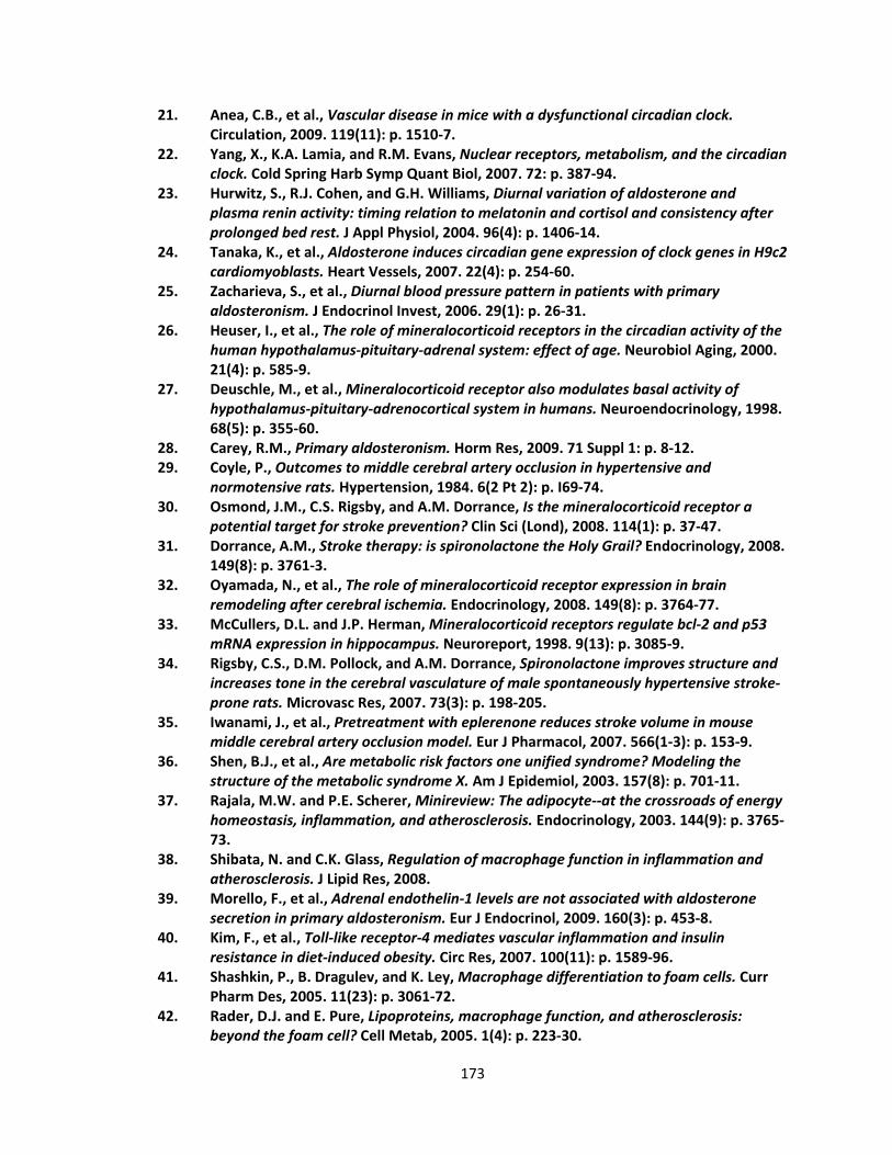
173
21. Anea, C.B., et al., Vascular disease in mice with a dysfunctional circadian clock. Circulation, 2009. 119(11): p. 1510‐7.
22. Yang, X., K.A. Lamia, and R.M. Evans, Nuclear receptors, metabolism, and the circadian clock. Cold Spring Harb Symp Quant Biol, 2007. 72: p. 387‐94.
23. Hurwitz, S., R.J. Cohen, and G.H. Williams, Diurnal variation of aldosterone and plasma renin activity: timing relation to melatonin and cortisol and consistency after prolonged bed rest. J Appl Physiol, 2004. 96(4): p. 1406‐14.
24. Tanaka, K., et al., Aldosterone induces circadian gene expression of clock genes in H9c2 cardiomyoblasts. Heart Vessels, 2007. 22(4): p. 254‐60.
25. Zacharieva, S., et al., Diurnal blood pressure pattern in patients with primary aldosteronism. J Endocrinol Invest, 2006. 29(1): p. 26‐31.
26. Heuser, I., et al., The role of mineralocorticoid receptors in the circadian activity of the human hypothalamus‐pituitary‐adrenal system: effect of age. Neurobiol Aging, 2000. 21(4): p. 585‐9.
27. Deuschle, M., et al., Mineralocorticoid receptor also modulates basal activity of hypothalamus‐pituitary‐adrenocortical system in humans. Neuroendocrinology, 1998. 68(5): p. 355‐60.
28. Carey, R.M., Primary aldosteronism. Horm Res, 2009. 71 Suppl 1: p. 8‐12. 29. Coyle, P., Outcomes to middle cerebral artery occlusion in hypertensive and
normotensive rats. Hypertension, 1984. 6(2 Pt 2): p. I69‐74. 30. Osmond, J.M., C.S. Rigsby, and A.M. Dorrance, Is the mineralocorticoid receptor a
potential target for stroke prevention? Clin Sci (Lond), 2008. 114(1): p. 37‐47. 31. Dorrance, A.M., Stroke therapy: is spironolactone the Holy Grail? Endocrinology, 2008.
149(8): p. 3761‐3. 32. Oyamada, N., et al., The role of mineralocorticoid receptor expression in brain
remodeling after cerebral ischemia. Endocrinology, 2008. 149(8): p. 3764‐77. 33. McCullers, D.L. and J.P. Herman, Mineralocorticoid receptors regulate bcl‐2 and p53
mRNA expression in hippocampus. Neuroreport, 1998. 9(13): p. 3085‐9. 34. Rigsby, C.S., D.M. Pollock, and A.M. Dorrance, Spironolactone improves structure and
increases tone in the cerebral vasculature of male spontaneously hypertensive stroke‐prone rats. Microvasc Res, 2007. 73(3): p. 198‐205.
35. Iwanami, J., et al., Pretreatment with eplerenone reduces stroke volume in mouse middle cerebral artery occlusion model. Eur J Pharmacol, 2007. 566(1‐3): p. 153‐9.
36. Shen, B.J., et al., Are metabolic risk factors one unified syndrome? Modeling the structure of the metabolic syndrome X. Am J Epidemiol, 2003. 157(8): p. 701‐11.
37. Rajala, M.W. and P.E. Scherer, Minireview: The adipocyte‐‐at the crossroads of energy homeostasis, inflammation, and atherosclerosis. Endocrinology, 2003. 144(9): p. 3765‐73.
38. Shibata, N. and C.K. Glass, Regulation of macrophage function in inflammation and atherosclerosis. J Lipid Res, 2008.
39. Morello, F., et al., Adrenal endothelin‐1 levels are not associated with aldosterone secretion in primary aldosteronism. Eur J Endocrinol, 2009. 160(3): p. 453‐8.
40. Kim, F., et al., Toll‐like receptor‐4 mediates vascular inflammation and insulin resistance in diet‐induced obesity. Circ Res, 2007. 100(11): p. 1589‐96.
41. Shashkin, P., B. Dragulev, and K. Ley, Macrophage differentiation to foam cells. Curr Pharm Des, 2005. 11(23): p. 3061‐72.
42. Rader, D.J. and E. Pure, Lipoproteins, macrophage function, and atherosclerosis: beyond the foam cell? Cell Metab, 2005. 1(4): p. 223‐30.

174
43. Weisberg, S.P., et al., CCR2 modulates inflammatory and metabolic effects of high‐fat feeding. J Clin Invest, 2006. 116(1): p. 115‐24.
44. Lumeng, C.N., et al., Increased inflammatory properties of adipose tissue macrophages recruited during diet‐induced obesity. Diabetes, 2007. 56(1): p. 16‐23.
45. Lumeng, C.N., J.L. Bodzin, and A.R. Saltiel, Obesity induces a phenotypic switch in adipose tissue macrophage polarization. J Clin Invest, 2007. 117(1): p. 175‐84.
46. Hevener, A.L., et al., Macrophage PPAR gamma is required for normal skeletal muscle and hepatic insulin sensitivity and full antidiabetic effects of thiazolidinediones. J Clin Invest, 2007. 117(6): p. 1658‐69.
47. Odegaard, J.I., et al., Macrophage‐specific PPARgamma controls alternative activation and improves insulin resistance. Nature, 2007. 447(7148): p. 1116‐20.
48. Kang, K., et al., Adipocyte‐derived Th2 cytokines and myeloid PPARdelta regulate macrophage polarization and insulin sensitivity. Cell Metab, 2008. 7(6): p. 485‐95.
49. Odegaard, J.I., et al., Alternative M2 activation of Kupffer cells by PPARdelta ameliorates obesity‐induced insulin resistance. Cell Metab, 2008. 7(6): p. 496‐507.
50. Guo, C., et al., Mineralocorticoid receptor blockade reverses obesity‐related changes in expression of adiponectin, peroxisome proliferator‐activated receptor‐gamma, and proinflammatory adipokines. Circulation, 2008. 117(17): p. 2253‐61.
51. Machnik, A., et al., Macrophages regulate salt‐dependent volume and blood pressure by a vascular endothelial growth factor‐C‐dependent buffering mechanism. Nat Med, 2009. 15(5): p. 545‐52.
52. Pucci, F., et al., A distinguishing gene signature shared by tumor‐infiltrating Tie2‐expressing monocytes (TEMs), blood "resident" monocytes and embryonic macrophages suggests common functions and developmental relationships. Blood, 2009.
53. Miyashita, K., et al., The neuroprotective and vasculo‐neuro‐regenerative roles of adrenomedullin in ischemic brain and its therapeutic potential. Endocrinology, 2006. 147(4): p. 1642‐53.
54. Saijo, K., et al., A Nurr1/CoREST pathway in microglia and astrocytes protects dopaminergic neurons from inflammation‐induced death. Cell, 2009. 137(1): p. 47‐59.
55. Duan, S.Z., et al., Direct monitoring pressure overload predicts cardiac hypertrophy in mice. Physiol Meas, 2007. 28(11): p. 1329‐39.
56. Kwon, J.S., et al., Effect of pressure overload and its recovery on the rat carotid artery: change of vascular reactivity and remodeling process. Heart Vessels, 2006. 21(1): p. 48‐55.
57. Dunn, A.J., Systemic interleukin‐1 administration stimulates hypothalamic norepinephrine metabolism parallelling the increased plasma corticosterone. Life Sci, 1988. 43(5): p. 429‐35.
58. Katafuchi, T., T. Hori, and S. Take, Central administration of interferon‐alpha enhances rat sympathetic nerve activity to the spleen. Neurosci Lett, 1991. 125(1): p. 37‐40.
59. Besedovsky, H.O., et al., Immunoregulation mediated by the sympathetic nervous system. Cell Immunol, 1979. 48(2): p. 346‐55.
60. Foucart, S. and C. Abadie, Interleukin‐1 beta and tumor necrosis factor‐alpha inhibit the release of [3H]‐noradrenaline from mice isolated atria. Naunyn Schmiedebergs Arch Pharmacol, 1996. 354(1): p. 1‐6.
61. Hurst, S. and S.M. Collins, Interleukin‐1 beta modulation of norepinephrine release from rat myenteric nerves. Am J Physiol, 1993. 264(1 Pt 1): p. G30‐5.

175
62. Hurst, S.M. and S.M. Collins, Mechanism underlying tumor necrosis factor‐alpha suppression of norepinephrine release from rat myenteric plexus. Am J Physiol, 1994. 266(6 Pt 1): p. G1123‐9.
63. Castiglioni, P., et al., Mechanisms of blood pressure and heart rate variability: an insight from low‐level paraplegia. Am J Physiol Regul Integr Comp Physiol, 2007. 292(4): p. R1502‐9.
64. Le Minh, N., et al., Glucocorticoid hormones inhibit food‐induced phase‐shifting of peripheral circadian oscillators. Embo J, 2001. 20(24): p. 7128‐36.
176
CHAPTER VI:
SUMMARY AND CONCLUSIONS
MR in macrophages is an important target of MR antagonists
The original goal of this project was to identify important cellular targets for the
MR antagonist spironolactone and eplerenone in the mitigation of cardiovascular disease.
We focused on the macrophage because of similarities of MR antagonists with PPAR-γ
agonists and statins, both of which had been shown to modulate inflammatory responses
through their actions on macrophages[1-3]. To test this hypothesis we investigated the
ability of MR to directly manipulate macrophage activation in vitro. We showed that in
macrophages MR acts as a high affinity glucocorticoid receptor given the absence of
11βHSD2. In this manner, MR acts to enhance classical macrophage activation, while
repressing alternate activation programs. These conclusions were supported by the
concordant effects of pharmacologic MR antagonism and genetic MR deletion which
enhanced alternative macrophage activation, and repressed classical activation.
To test the biological relevance of MR in macrophages we developed a
macrophage specific knockout of MR. MΦMRKO afforded a similar protection as MR
antagonists in a model of vascular and cardiac fibrosis. This demonstrated that
antagonism of glucocorticoid occupied MR in macrophages is likely an important
177
mechanism by which MR antagonists protect against cardiovascular disease. This
protection correlated with a reversal in the repression of alternative macrophage
activation and recruitment of classically activated macrophages.
The hypothesis that MR antagonists and PPAR-γ agonists act through parallel
mechanisms was born out by the similarity in expression profile induced by PPAR-γ
agonists and MRKO. However, there were a number of key differences. These
differences allowed us to identify a novel alternative activation profile induced by
glucocorticoids, enhanced by MRKO, and repressed by IL-4 and PPAR-γ. The specific
mechanisms by which these macrophage subtypes contribute or protect in models of
cardiovascular disease and other inflammatory disorders is an important future direction
which can be addressed through comparison of pharmacologic activation and
macrophage deletions of PPAR-γ, MR, and GR.
On Macrophage Polarization:
Originally macrophage polarization was illustrated by contributions of Th1 (IFNγ)
and Th2 (IL4/13) cytokines on macrophage activation, with the observation that they
acted in mutually antagonistic manners[4-7]. Subsequently, other stimulants such as IL-
10, PPAR-γ, and FCγR engagement have been shown to result in related but distinct
expression profiles on the basis of differential expression of chemokines and cytokines.
This suggests that macrophage polarization is more complex than a dichotomous choise.
We provide support for this view by demonstrating that two alternative activation
stimulants, glucocorticoid and IL-4 are also mutually antagonistic, with GR inhibiting IL-
4 responses and vice versa. Corticosterone repressed IL-4 targets such as E-cadherin and

178
IL-27 receptor to the same degree as the pro-inflammatory M1 markers IL-1β and TNFα.
Conversely, IL-4 repressed genes induced by corticosterone. These data indicated at least
three distinct populations of macrophages.
Interestingly, glucocorticoids and IL-4 produced similar effects on a number of
genes. Overlap consisted primarily of canonical markers of alternative macrophage
activation such as YM1, YM2, Arg1, and F13a1. These genes are almost always the only
genes used to determine macrophage polarizing effects in vivo. Clearly, they do not
accurately represent the level of heterogeneity which exists among macrophages. It will
be more useful in the future to utilize markers which are distinct in each population of
AMΦ.
Macrophages regulate hemodynamic responses
Recently, macrophage specific deletion of nuclear receptors has been used to
identify novel roles for macrophages in regulating physiologic responses. Macrophage
deletion of PPAR-γ revealed a specific role of macrophages in regulating fatty acid
metabolism and insulin sensitivity in skeletal muscle [1, 2]. Similar studies with PPAR-δ
identified a role of macrophages in controlling liver metabolism and steatosis [8, 9].
Our study of MR in macrophages revealed a novel role for macrophages in
controlling cardiac hypertrophy, heart rate, blood pressure, and blood flow in response to
ischemia. Currently, there are few physiologic paradigms that account for macrophage
control of sympathetic drive, pulse pressure, or circadian rhythms in the absence of a
potent immune challenge. This dissection demonstrates an interaction between

179
inflammatory signaling and a number of factors linked to cardiovascular disease, such as
high salt diet, non-dipper status, mineralocorticoid excess, and sympathetic drive.
In this case however, the changes in dipper status and elevation in pulse pressure
induced by MΦMRKO would indicate a pathogenesic instead of protective role for
macrophage MR [10]. The fact that these effects are not replicated by MR antagonism
may be due to specific mechanisms of MR regulation, or are masked by the effects of
eplerenone and spironolactone in other tissues such as the kidney. It is unlikely however,
that the effects of MR will always exacerbate disease states. If MR were always
pathogenic, its action in macrophages would likely be selected against.
In any case, these observations clearly highlight the importance of MR in
macrophages. Elucidating the mechanism of circadian hemodynamic rhythm
dysregulation in MΦMRKO mice will yield truly novel insights into the interaction
between inflammatory signaling and hypertensive responses.
On the clinical benefit of spironolactone and eplerenone:
We have shown that one likely mechanism by which spironolactone benefits
cardiovascular disease is through antagonism of glucocorticoid occupied MR in
macrophages. This mechanism is likely independent of the renin-angiotensin-aldosterone
system, and is independent of blood pressure lowering effects. Currently, outside of high
risk heart failure patients, MR antagonists are fourth line in the treatment of hypertension.
Our data suggest that MR antagonists will provide additional protective benefit and
should potentially be used in patients with moderate cardiovascular risk and evidence of
elevated inflammatory burden, such as patients with obesity or type II diabetes.
180
Additionally, we show that MΦMRKO protects against cardiac hypertrophy, again
through mechanisms independent of protective hemodynamic changes. This data would
suggest additional benefit of MR antagonists for patients with left ventricular
hypertrophy even if their hypertension is well controlled.
Finally, we show that MRKO broadly impacts inflammatory processes.
MΦMRKO provided a specific benefit in a model of fibrosis. Other animal models of
fibrosis have been protected by administration of MR antagonists. It is likely that MR
antagonists will prove to be a useful adjuvant in the treatment of other inflammatory
diseases, especially diseases where fibrosis in the context of Th1 responses occurs. One
example of this is interstitial lung disease, a group of disorders involving fibrosis of lung
interstitium which often leads to quick reduction of respiratory function, and is poorly
controlled.
Ultimately, the efficacy of MR antagonists in the treatment of fibrotic diseases
can only be confirmed by carefully controlled clinical trials. Fortunately, the MR
antagonist spironolactone is very inexpensive and widely tolerated making it an optimal
drug for clinical studies. The common side effect of spironolactone is due to off target
anti-androgen effects which are absent in the more expensive but more specific
antagonist eplerenone. It is important to mention that many fibro-proliferative disorders
have few if any medical treatments. Even if MR antagonists provide only marginal
benefit, this would still be a step in the right direction.
MR and immune control:
181
We have demonstrated the broad potential impact of MR regulation of
inflammatory signaling on physiologic and pathophysiologic responses. Although
undertaking the studies proposed in Chapter IV and V would require a single lab over a
decade of effort, we have demonstrated that casting a wide net in understanding how
macrophage programs are coordinately regulated can yield important conclusions.
Comparing polarization states between PPAR-γ, MR, and GR and other macrophage
specific knockouts in different inflammatory disorders is likely to lead to a more
complete understanding of how macrophages impact disease.
MR the target of well tolerated therapeutics which may provide benefit in other
clinical settings by the way of its immunomodulatory activity. Understanding how MR
macrophages and other cell types controls transcription in various disease states such as
pulmonary fibrosis, or diet induced obesity will yield novel mechanisms of how nuclear
hormone receptors modulate disease processes, and potentially predict clinical benefit of
MR antagonists in human disease. Specifically, the study of MR in macrophages may
help dissect the critical contributing inflammatory factors which promote cardiovascular
risk and enhance other inflammatory reactions. Identification of these markers may
justify the expansion of studies into disease states which demonstrate similar changes,
and thus may similarly respond to MRKO or MR antagonists.
Some of the beneficial anti-inflammatory activity of MR antagonists may occur in
immune cells other than macrophages. This can be further studied through selective
knockout of MR in other important cell types such as dendritic cells, and lymphocytes.
We identified many targets of MR in macrophages which are expressed in these other cell
types and are important in functions such as antigen presentation and lymphocyte
182
proliferation. This provides a foothold into understanding the function of MR in broad
immune regulation.
On the potential for aldosterone action on macrophages
A central component of this thesis is the occupancy of MR in macrophages. We
have shown that macrophage MR can be similarly bound and activated by both
glucocorticoids and aldosterone. Since at all times, glucocorticoids are in marked excess
to aldosterone, and MR has such a high affinity for corticosteroids, it seems likely that a
majority of MR is occupied by glucocorticoids in vivo and under normal culture
conditions. This is based on a number of assumptions that should be considered. First,
that local concentrations of corticosteroids is always in overwhelming excess to
aldosterone. Since local concentrations of glucocorticoids vary widely, this is not always
necessarily going to be the case, especially in patients with hyperaldosteronism. Second,
it is assumed that the affinity for glucocorticoids and aldosterone is always constant.
Allosteric regulation of MR through DNA-protein and protein-protein interacions may
alter its binding affinity and allow for greater selectivity for aldosterone. The final
assumption is that significant occupancy is a requirement for important cellular effects.
Promoters may have high sensitivity to aldosterone occupied MR, allowing for
significant alteration in transcription even when there are few aldosterone occupied MR
complexes.
Aldosterone action on macrophages may be one mechanism for its pro-
inflammatory effects in vivo. While the pro-inflammatory action of aldosterone may be
indirect, through its effects on other tissues, we do observe a marked induction of pro-

183
inflammatory cytokines in aldosterone stimulated macrophages. However, we were
unable to identify any significant effect of physiologic concentrations of aldosterone on
macrophages cultured in the presence of media steroids. This does not mean that
aldosterone or unoccupied may have important effects on macrophage function in vivo,
especially over the long course of cardiovascular disease progression.
However, the macrophage MRKO mouse phenotype is complex and likely
involves the dysregulation of multiple macrophage functions. A majority of the roles that
MR plays in macrophages is linked to its glucocorticoid occupancy based on the
assumptions listed above. Thus, the most likely explanation for the macrophage MRKO
phenotype, especially its protection in cardiovascular inflammation is due to its role as a
high affinity glucocorticoid receptor.
Macrophage polarization as a paradigm for drug discovery:
We have shown that two agents used in the treatment in cardiovascular disease,
MR antagonists and PPAR-γ agonists act via parallel mechanisms on macrophage
polarization. These data implicate macrophage polarization as a clinically important
target for therapeutic design.
We additionally provide the added insight into the nuances of macrophage
polarization by identifying specific markers of three macrophage subtypes. These studies
are not exhaustive as many other stimulants likely drive macrophages toward novel
states. Comparing the effects of macrophage specific manipulation of drug targets both
in vivo and in vitro as done in this study may help define important markers for
pathogenesis and protective inflammatory processes. This knowledge can then be
184
applied to the development of new therapies. Specific to MR, next generation
antagonists which block only glucocorticoid occupied MR may provide cardioprotective
effects without increasing potassium levels, which is an important side effect
contraindicating their use in patients with renal disease. Investigating the ability of new
MR antagonists to alter macrophage polarization may be a way to predict clinical
efficacy.
Remarkably, we identified interactions between three well tolerated
pharmacologic agents which drive macrophage polarization in different directions. The
clinical benefit of glucocorticoids, PPAR-γ agonists, and MR antagonists can in part, be
attributed to their effects on macrophages. Providing drugs in combination can result in a
dramatically diverse spectrum of macrophage activation profiles. In addition it has been
shown that many secreted inflammatory factors have dual actions, such that at low levels
they can be pro-inflammatory, whereas anti-inflammatory at high levels and vice versa.
It may be that the dramatic upregulation of arginase by IL-4 is pathogenic in fibrosis, but
the less potent upregulation by MR antagonism or PPAR-γ activation is protective. This
may seem impossibly complex, but it is the nuances and complexities of inflammatory
signaling that provide a unique opportunity to tailor treatment regimens. It is not the
processes that different inflammatory disease states have in common that will be the
determining factors deciding future treatments; it is the unique properties that are specific
to a narrow range of macrophage activities that will be important.
The future of healthcare is in personalized medicine. Unique disease states will
be identified not just by the clinical presentation, but through measurement of risk as
determined by an individual’s genetic make-up, and environment, and confirmed through

185
specific molecular characteristics of the disease. Once the disorder or risk is identified,
all the available data can be integrated so a tailored and personal therapeutic approach
can be applied. This approach is beginning to be applied successfully in cancer
treatment, where cancers are characterized through genetic and molecular means, and
their susceptibility to a combination of therapies identified and used.
Disorders with inflammatory components can be approached in the same way. As
initiated with this project, specific factors associated with modulation of an inflammatory
disease can be identified through a comprehensive characterization of the cells involved.
That inflammatory response can then be manipulated in a very specific manner by a
combination of pharmacological agents that manipulate immune polarization in different
directions. For example, glucocorticoids have long been used to combat immune
diseases. A major limiting side effect is susceptibility to infection due to its potent anti-
inflammatory activities. We have shown that MR antagonizes GR in a very specific
subset of genes. It may be that a combination of MR antagonists and glucocorticoids
used in combination may enhance the beneficial effects, thereby increasing the
therapeutic index and diminishing the side effects of high dose glucocorticoids.
MR in parenchymal tissues
MR plays a critical role in multiple physiologic processes. These functions
largely cluster into two major categories. On one side, MR acts to regulate salt and water
balance. This is not a newly emerged function, as MR regulates salt retention and
excretion in the gills of early marine vertebrates. However, this function is independently
regulated through the recent emergence of aldosterone synthase, and 11βHSD2 which

186
confers aldosterone sensitivity in tissues key to the regulation of salt and water
homeostasis such as renal and colonic epithelium and vascular endothelium.
On the other hand, as has been discussed in previous chapters, MR is nearly
ubiquitously expressed, including many tissues that are insensitive to aldosterone due to a
lack of 11βHSD2 expression. MR’s actions and mechanisms in these tissues have not
been comprehensively investigated outside of the central nervous system. We show
significant parallels between the mechanisms of action of MR in macrophages and in the
brain by driving a counter-regulatory but independent transcriptional program and
directly interacting in only a minority of targets. This framework allows for bi-
directional signaling which is context specific.
The dynamic interplay between MR and GR is of great physiologic importance.
Corticosterone and cortisol concentrations which vary in a circadian pattern and are
dramatically increased during emotional and physiologic stress regulate inflammatory,
hemodynamic, and metabolic circadian rhythms and stress responses. MR’s role not only
in baseline regulation of transcription, but its necessity in mediating glucocorticoid
responses will likely be observed in other tissues. Due to the counter-regulatory actions
of MR and GR, we can predict MR’s biological role in other cell types.
It has been often proposed that structural variation among steroid metabolites
such as glucocorticoids may stimulate differing activity upon their receptors. This is
clearly true in macrophage MR. We have demonstrated significant functional differences
between glucocorticoid and aldosterone occupied MR. The role of glucocorticoid
occupied MR in tissues expressing 11βHSD2 under native conditions, and the role of

187
aldosterone occupied MR in tissues lacking 11βHSD2 in patients with
hyperaldosteronism is not clear. Identifying unique markers for the specific action of
either aldosterone or glucocorticoid occupied MR may help unravel this dynamic in vivo.
The pleiotropic roles of MR in regulating physiology are a consequence of its
conserved glucocorticoid affinity. Aldosterone synthase and 11βHSD2 in terrestrial
animals evolved to begin to allow for independent regulation of salt and hemodynamic
homeostasis from the basic functions MR plays when glucocorticoid bound. The reason
for this is clear, as stress in fish, reduces fitness under low salt environments; this would
be especially problematic in terrestrial animals where salt retention is critical for life.
The divergence of glucocorticoid signaling and HPA axis regulation, and
aldosterone action is incomplete by the nature of its evolution. This may explain the
reason that glucocorticoids may still regulate some MR actions in renal epithelium, and
why aldosterone has pro-inflammatory effects in non-epithelial tissue. We observe this
effect directly as ablation of glucocorticoid occupied MR in macrophages results in a
dysregulation of the hemodynamic response to high salt.
This incomplete divergence of physiologic systems places MR at the center of
many cardiovascular risk factors. On one side glucocorticoid bound MR regulates basic
biological functions such as neural excitability, acute inflammatory response,
adipogenesis . One the other MR is necessary in coordinating the hemodynamic response
to salt loading and depletion though aldosterone. These diverse actions likely synergize
to enhance cardiovascular risk and dictate the pleiotropic beneficial effects enacted by

188
MR antagonists (Figure 6.1), and provide a bright future in investigating the molecular
mechanisms which promote disease.

189
Figure 6.1: MR occupancy and the positive feedback mechanisms which drive
cardiovascular disease. The development of cardiovascular disease is derived from
pathology from multiple systems which feeds forward into a decompensated state. Diet
induced obesity and metabolic disease promotes inflammatory responses including
monocytosis and endothelial dysfunction which in turn enhances insulin resistance.
Macrophage activation in response to inflammatory signals such as covalently modified
LDL, promotes vascular remodeling and neo-intimal expansion which increases
peripheral resistance, reduces distal flow, and creates risk for embolization. Cardiac
tissue attempts to compensate for increased load despite reduced perfusion by inducing a
hypertrophic response. We show cardiac hypertrophy involves monocyte/macrophage
recruitment and activation which in turn stimulates cardiac remodeling which further
reduces ventricular function and tissue perfusion. Physiologic responses to reduced
tissue perfusion including activation of the R-A-A-S system, HPA axis, and increased
autonomic drive increase cardiac stress peripheral resistance exacerbate this condition.
The two mineralocorticoid ligands aldosterone and cortisol (corticosterone) play a central
role at many levels of this process.
Antagonism of MR causes pleiotropic beneficial effects in patients with cardiovascular
risk. Dogmatically, the actions of MR antagonists have been assumed to be through the
blockade of aldosterone action. Clearly, based on the absence of 11βHSD-2 in many key
tissues, that antagonism of glucocorticoid occupied MR is also important. We show that
macrophage MR, which is glucocorticoid bound plays a central part in the pathogenesis
of cardiovascular disease and mediates many of the beneficial effects of spironolactone
and eplerenone.

190

191
References 1. Hevener, A.L., et al., Macrophage PPAR gamma is required for normal skeletal muscle
and hepatic insulin sensitivity and full antidiabetic effects of thiazolidinediones. J Clin Invest, 2007. 117(6): p. 1658‐69.
2. Odegaard, J.I., et al., Macrophage‐specific PPARgamma controls alternative activation and improves insulin resistance. Nature, 2007. 447(7148): p. 1116‐20.
3. Yano, M., et al., Statins activate peroxisome proliferator‐activated receptor gamma through extracellular signal‐regulated kinase 1/2 and p38 mitogen‐activated protein kinase‐dependent cyclooxygenase‐2 expression in macrophages. Circ Res, 2007. 100(10): p. 1442‐51.
4. Gordon, S., Alternative activation of macrophages. Nat Rev Immunol, 2003. 3(1): p. 23‐35.
5. Gordon, S. and P.R. Taylor, Monocyte and macrophage heterogeneity. Nat Rev Immunol, 2005. 5(12): p. 953‐64.
6. Martinez, F.O., L. Helming, and S. Gordon, Alternative activation of macrophages: an immunologic functional perspective. Annu Rev Immunol, 2009. 27: p. 451‐83.
7. Martinez, F.O., et al., Macrophage activation and polarization. Front Biosci, 2008. 13: p. 453‐61.
8. Odegaard, J.I., et al., Alternative M2 activation of Kupffer cells by PPARdelta ameliorates obesity‐induced insulin resistance. Cell Metab, 2008. 7(6): p. 496‐507.
9. Kang, K., et al., Adipocyte‐derived Th2 cytokines and myeloid PPARdelta regulate macrophage polarization and insulin sensitivity. Cell Metab, 2008. 7(6): p. 485‐95.
10. Kawano, Y., et al., Masked hypertension: subtypes and target organ damage. Clin Exp Hypertens, 2008. 30(3): p. 289‐96.

192
APPENDIX
193
APPENDIX 1: METHODS
Cell Culture
Raw 264.7 murine macrophage cells (TIB-71, ATCC) are cultured in DMEM (Gibco) with high glucose no sodim bicarbonate, supplemented with 200 nM L-Glutamine, and 10% heat inactivated FBS (Gibco) or 10% Charcoal/Dextran stripped FBS (Hyclone). RAW 264.7 cells are passaged when they reach 90% confluence, confluence results in activation of the macrophages and takes at least one additional passage for them to reach baseline. RAW 264.7 cells are maintained in tissue culture treated T75 flasks and split 1:9 every 5-7 days. To split RAW cells are suspended in 5 mLs of fresh media using a cell scraper (trypsin is not effective) and diluted into a new flask appropriately.
Pharmacology experiments
RAW 264.7 cells were plated at a density of 5 * 10^5 cells per well in 12 well plates and allowed to recover for 24 hours. To test the effects of various ligands Eplerenone is diluted into DMSO, and Spironolactone, RU26752, and RU486 diluted into ethanol and kept at -20C for no longer than 3 months. Substocks of a 200X concentration is made by diluting the stock into PBS just prior to the experiment. After the 24 recovery from plating, RAW cells are treated with various concentrations of ligands for a period of 24 hours. To investigate the effects of MR ligands on macrophage activation, after an initial treatment of 18 hours macrophages were subsequently treated with 100 ng/mL LPS for 3 hours, or 5 ng/mL IL-4 for 24 hours.
Transfection of RAW 264.7 cells
RAW 264.7 cells are plated at a density of 2*10^5 cells per well in 12 well plates. After 24 hours, cells are transfected with Superfect (Qiagen) per manufacturer instructions. Plasmids purified using the Endo-Free Maxi kit (Qiagen) are mixed with 1:3 (ug to mLs of superfect) using 1.5 ug plasmid/4.5 uL of superfect, and applied to the macrophages for 3 hours. RAW cells are then washed gently three times with room temperature PBS containing calcium and magnesium. Transfection with Superfect causes macrophages to lose their adherence so washing too harshly will cause you to lose a significant portion of the transfected macrophages. 24 hours later, RAW cells are washed again 3X in room temperature PBS, and fed fresh, pre-warmed media, and experiment begun. Optimal

194
transfection was quantified to be approximately 40% of cells using a GFP control plasmid.
Over-expression of MR
Macrophages were transfected with a plasmid containing either a FLAG-tagged murine MR cDNA or a full length human MR cDNA (Origine) driven by a CMV constitutive promoter or using pCDNA 3.1(+) as a negative control. Following 24 hours, macrophages were placed in fresh serum and experiment begun as described. 48 hours post transfected yielded the greatest increase in MR activity and expression.
Luciferase Reporter Assay
RAW 264.7 cells were transfected with 1.25 µg of a luciferase reporter (obtained from Iniguez-Lluhi lab), in each well containing the MMTV-LTR harboring multiple steroid responsive elements which drive the expression of luciferase and .25 µg of a Renilla luciferase (obtained from Metzger lab) as a constant control. Following the three hour transfection, RAW cells were allowed to recover in media containing 10% charcoal/Dextran stripped FBS to minimize background. Following 24 hours, macrophages were treated with various ligands for an additional 24 hour (however significant induction of MMTV was observed with only a 3 hour treatment). Luciferase production was measured using Dual-Luciferase Assay (Promega) as per manufacturer instruction using 100 uL of passive lysis buffer, and 20 ul of cell lysate per reaction.
MR western blot
Protein was run on a 12% SDS page gel and blotted onto PVDF membrane (Millipore) by wet transfer. Western Blot for MR was performed with monoclonal antibodies 2D6 and B7 (gift from Gomez-Sanchez Lab) diluted 1:200 in blocking buffer containing PBS, 1% Milk, and .02% Tween 20 which was incubated at 4 C overnight. Secondary HPR conjugated goat anti-mouse antibody () in blocking buffer was applied for 2 hours at room temperature. Following 5 washes in PBS containing .02% tween for 5 minutes per wash, antibody was detected by chemiluminescence ().
Expression analysis
Broad expression analysis was performed primarily using quantitative real-time PCR of cDNA generated from isolated macrophages and tissues. RNA was isolated from macrophages cultured as described using RNAeasy (Qiagen) column purification with on column DNAse digestion. Subsequently, 20 uL of RNA (which is below accurate detectable limit by UV spectrometry) was then used to generate a single cDNA strand using Taqman reverse transcriptase kit (Applied Biosystems). qRT-PCR was performed either utilizing the cyber green method of detection, or specifically designed Taqman

195
Primer-probe pairs (Roche). Primers for the cyber green were either picked using PrimerBank (http://pga.mgh.harvard.edu/primerbank/) or using IDT-DNA primerquest, picking a primer set with an optimal Tm of 60 C, 150 bp amplicon, and spanning an intron-exon boundary and specificity confirmed via BLAST. An accurate primer set was determined by a product with a single melting temperature, and appropriate sized band upon gel electrophoresis. Expression of a specific gene was quantified by identification of Ct, and then normalized to an internal housekeeping gene and a negative control. Multiple housekeeping genes should be utilized in each experiment. For isolated macrophages, ribosomal RNAs such as L32 or 18S are best as they are unaffected by macrophage activation. In tissues, GAPDH and Act6 are also used, however, it is important to be sure they are not significantly altered by the experiment.
Peritoneal macrophage isolation:
Mice are first given an intraperitoneal injection of aged (at least 1 month) of 1 mL 3% Brewer’s Thioglicolate (Sigma) using an insulin syringe. Peritoneal macrophages may then be isolated 3-6 days following the injection. Waiting longer improves the purity of the macrophage isolation, but reduces the yield. All experiments in this thesis were performed from macrophages isolated either 4 or 5 days following injection.
Peritoneal macrophages were isolated through the following procedure:
1. Place sterile dPBS (-Mg/Ca) on Ice and let cool down, also place 15 mL conical tubes on ice (one conical tube per mouse)
2. When dPBS is cool sacrifice mouse a. While mouse is dying use a 10 mL syringe with an 18g needle to pull up 4
mls of PBS, replace 18g needle with a 25g needle, and put the syringe on ice.
3. After mouse is dead, immobilize, and remove the skin over the abdomen while preserving the peritoneal wall, beginning with a vertical midline incision and then exposing the right half of the abdomen so that you can see the spleen clearly.
a. If you nick the peritoneal wall during this step its not worth continuing b. It actually helps to only expose half the peritoneal wall, this helps it
bubble out away from the colon 4. Spray the peritoneal wall with 70% EtOH 5. Insert the syringe with the 25g needle, beveled edge facing up, midline just
superior to the perioverian fat/bladder. a. When you insert the needle be careful not to nick the colon, intestine or
bladder, if you do, move on to the next mouse because that sample will be contaminated.
6. Inject 4 mLs of ice cold dPBS (you do not have to be gentle in this step)

196
7. Remove the syringe and massage the abdomen, moving the organs around to loosen the cells
8. Reinsert the syringe, beveled end up, approximately mid clavicular line, (basically in an area where there is no fat) typically the best area is around the spleen (there is fat there but it doesn’t move if you are careful
9. Slowly take the up the PBS, a. Doing it too fast and you will dislodge the fat and it will clog the needle b. I these mice are injected with thioglycolate the PBS should be cloudy and
white, if they aren’t injected it will look clear c. If the PBS is red, it means you hit something during the injection, not a
big deal, if it is a lot of blood you will have to lyse with hypotonic buffer, (I use sterile H2O for 7 sec)
d. If the PBS is green or brown, it means you nicked the colon . . .move on to the next mouse
e. You will typically get 2-3 mls after the first peritoneal wash 10. Place the pbs in the appropriate conical tube (leave on ice), then change needles
back to the 18g, and draw another 4 mls of PBS 11. Repeat steps 5 through 10 2 more times (washing with a total of 12 mls)
a. At the end I generally get between 9 and 11 mls of PBS 12. During the last mouse, set the tabletop centrifuge to 4 C and let cool down 13. Spin cells at 1300 rpm for 10 minutes to pellet 14. Re-elute in 1 mL of media
a. If thioglycolate illicited macrophages, then you will need to dilute 1:10 to count
b. Typically I will get 2.5-10*10^5 cells per mouse for resident macrophages c. Typically I will get .5-10 *10^6 cells per mouse for thioglycolate
macrophages 15. I plate generally 2*10^5 cells/ml/well of a 12 well plate for RNA isolation
a. I would probably do closer to 1-2*10^6 cells per plate for the conditioned media experiments
16. Media = just DMEM + 10%FBS +1xPen/Strep 17. Initial plating I use Ice cold media 18. After plating, I will leave the media out in the hood to warm up for 2 hours, for
the first wash 19. 2 hours after plating, wash with room temperature sterile dPBS +Ca +Mg and
replace media a. Be gentle when doing this (add the media to the side of the well when you
shoot it in) 20. Let the cells recover overnight before treating them

197
L-NAME/Ang-II Model
L-NG-Nitroarginine methyl ester (L-NAME) a non specific nitric oxide synthase inhibitor (Sigma) is administered at a dose of 40 mg/kg/day in the drinking water with .9%NaCl. Dosage needs to be adapted during the experiment, so measure the weight of the mice and assume they drink 4 mLs of water per day to start (this is generally about .25 mg/ml). When adding to the drinking water, measure the volume, and measure it 2 days later. L-NAME/salt water needs to be replaced at a minimum of every 3 days (it looses activity at room temp).
At day 10 prepare the Alzet pumps, one per mouse. The model for this experiment is 1007D, which pumps either .48 - .50 ul/hr for one week. After one week it needs to be removed, so if you want to do a longer treatment you need to use a different model. Angiotensin II, diluted in to sterile water is further diluted in water to provide a .7 mg/kg/day dose at a.5 ul/hr rate. For each mouse make the proper dilution in 150 uL in a sterile 1.5 mL tube.
In the hood, use forceps sprayed with EtoH, and not hands to open the packet, and use good sterile technique when filling them. Briefly, use a 1 mL syringe and the applicator which comes with the alzet pumps to draw up the angiotensin II, be careful not to get any bubbles (however, you cannot invert and tap to get the bubbles out because of the way the applicator interfaces with the syringe, you get more bubbles that way, so just be gentle).
Use forceps to remove the pump, and then gently push the applicator down into the bottom of the pump and begin to inject the angiotensin mixture. Slowly inject the Ang II while removing the applicator in a single motion, so as to avoid getting air bubbles. When a bead of solution appears at the top completely remove the syringe. Then use forceps to remove the top of the pump and slowly insert it into the bottom (don’t use your hands, you can see this in the instructions as well). When the top has been pushed all the way in, the bead of solution should appear on the outside of the top of the pump.
To insert the pump into the mouse, anesthetize them with 5% isofluorane for 1.5 minutes, and then keep them on 3% during the duration of the surgery. Position them face down, horizontally and tape down their arms and tail. Remove the hair on one side of their upper back. Make a superficial incision perpendicular to the spine, and then use the scissors to blunt dissect downward, parallel to the spine to make space for the pump. Dip the pump in saline and then insert it facing away from the incision. Close the incision with a staple, and glue.

198
After 4 days the mice should look very sick, not moving much, drinking, or eating. At this point the mice can be sacrificed, and analyzed.
Affymetrix analysis and statistics
In all affymetrix experiments in this thesis, primary thioglycolate macrophages pooled from three animals of the same genotype and background were utilized following culturing at a density 5*10^6 cells per well. RNA was isolated by RNAeasy column. RNA from three individual wells was then pooled and provided to the affymetrix core (Cancer center). Two affymetrix chips were then utilized for each condition. This provides limited statistical power to determine significance. In retrospect, despite the increased cost it would have been better to utilize an N of at least 3 chips per condition to allow proper statistical analysis.
The Affymetrix core confirmed RNA quality and concentration by NanoDrop UV spec analysis and then utilized Nano-bead purification () for cRNA synthesis and subsequent affymetrix analysis. The Affymetrix core provided rudimentary statistical analysis which included 3’ to 5’ probe analsysis which is a measure of cRNA fidelity. In one control condition this analysis demonstrated that RNA instability, and that sample was thrown out, further limiting the statistical analsysis that could be performed.
Identification of genes changed by condition
Statistical analysis in these experimentes was limited by low statistical power. In this case, genes expressed in macrophages (identified as having a raw expression score of greater than 2^5) which were altered greater than two fold relative to the control sample were deemed changed. This is somewhat an arbitrary threshold, but a majority of genes changed greater than two fold were shown to be similarly changed by qRT-PCR.
However, due to the lack of statistical power, genes changed less than two fold may also be changed. In this case, individual genes with confirmed roles in macrophage activation, polarization, or cardiovascular disease progression were individually chosen and tested by qRT-PCR in multiple different experiments.
Venn Diagram
Genes identified as changed greater than two fold were then put into a single list, and compared using GeneVenn, which selects out common genes and presents them in Venn Diagram form. However, using a hard threshold of two fold presents the problem of false negatives. For example, if a gene is changed 2 fold in one condition, and only 1.9 fold in the other, then generally we draw the wrong conclusion that it is only uniquely affected by one condition. To alleviate this, genes which were 2 fold effected by one condition

199
and greater than 1.75 in another condition were considered to be commonly altered by both conditions.
Gene Ontology analysis
Genes altered greater than 2 fold (both induced and repressed) were compiled into lists and input into GOminer High throughput (http://discover.nci.nih.gov/gominer/htgm.jsp) against a list of genes expressed in macrophages, with a P value threshold of .05, and minimum of 5 changed genes per category to be listed. A similar analysis was performed utilizing Ingenuity Pathways Analysis (http://www.ingenuity.com/products/pathways_analysis.html) which demonstrated similar results.
Prediction of MR consensus binding sequence
Proximal promoters consisting of the upstream 500 bp, and downstream 50 bp were isolated from the 20 strongest genes increased and decreased by MRKO using the Genomatix bioinformatics suite (Gene2Promoter) including every alternative transcriptional start site from each gene. MatInspector (Genomatix) then demonstrated that in both promoters from upregulated and downregulated genes contained type-II nuclear hormone receptor response elements in greater than 75% of promoters. Twenty five bps surrounding each type-II HRE was then manually isolated from each promoter and then compared via multiple sequence alignment using DiAlign (Genomatix) and presented graphically using Web Logo (http://weblogo.berkeley.edu/logo.cgi)
Cluster Analysis
Cluster Analysis is a tool which allows for comparison of multiple of expression changes and allows for viewing of general patterns of gene expression and regulation. To perform this analysis, gene expression changes identified by qRT-PCR, or by affymetrix (changed greater than 2 fold) were presented in a tab delineated matrix gene names in rows and conditions in columns. Cluster 3.0 was then utilized to organize genes and conditions based on a similarity score generated by un-centered hierarchical clustering. Tree diagram generated by the score, and heat-map of the actual expression changes relative to controls presented in log2 transformed form was presented by TreeView.
Statistics
Pairwise comparisons were utilized to determine statistical significance using a student’s T-Test with a threshold of p<.05. Results were deemed significant if observed in experiments done with an N<3 and repeated at least 3 times. In this thesis there are a few experiments which were not repeated three times and their statistics not reported.

200
Multiple comparisons within a single experiment were performed by a T-tailed ANOVA using Prism 5.0 (Graphpad). To control for a large number of experiments, a bonferoni post-test was performed to control for false positives where indicated.


![arXiv:physics/0312071v1 [physics.class-ph] 11 Dec 2003 · tum variational principle for excited states, and the connection to classical action principles. In Classical Mechanics,](https://img.pdfslide.us/doc/110x75/5ecf35464ccdb1799a4b690f/arxivphysics0312071v1-11-dec-2003-tum-variational-principle-for-excited-states.jpg)