Embed Size (px)
Citation preview
Nodopathies in the central nervous system
Axonal domains in MS
Catherine Lubetzki
Salpêtrière Hospital
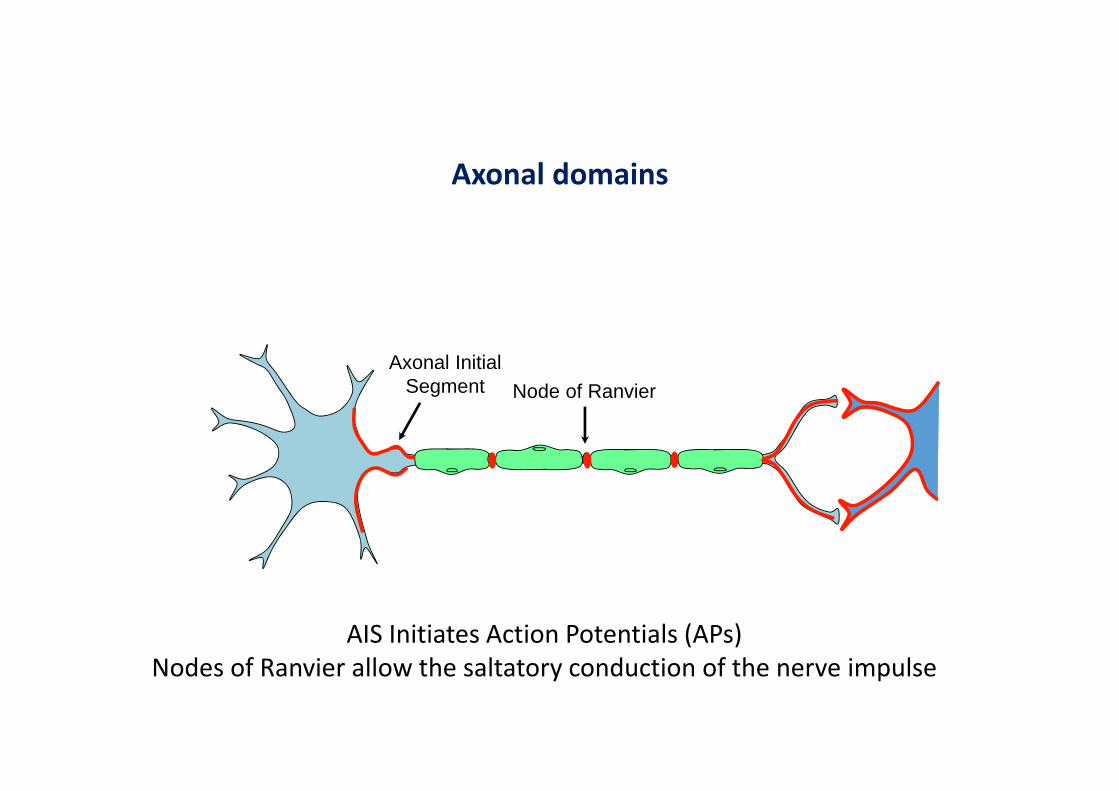
Axonal domains
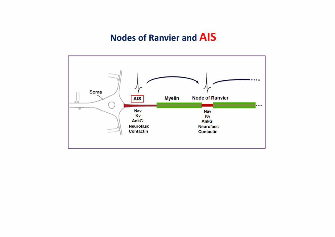
AIS Initiates Action Potentials (APs)Nodes of Ranvier allow the saltatory conduction of the nerve impulse
Node of RanvierAxonal Initial
Segment

Nodes of Ranvier and AIS : a common molecular organization
NeurofascContactin
NeurofascContactin
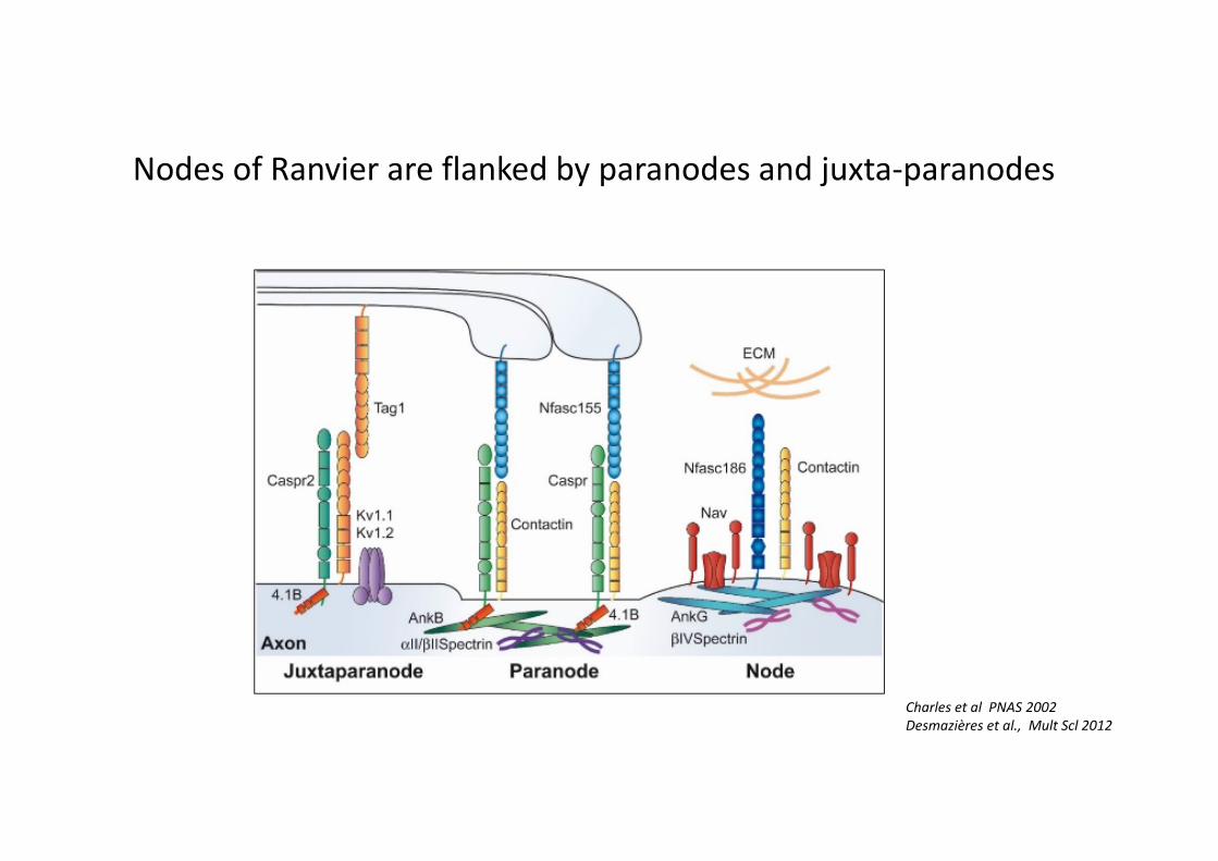
Charles et al PNAS 2002Desmazières et al., Mult Scl 2012
Nodes of Ranvier are flanked by paranodes and juxta-paranodes
What is the evidence for an immune response directed against axonal domains proteins in MS?

Antibodies directed against Neurofasc 155 in MS patients
Purified anti-Neurofasc 155 antibodies binding to bothisoforms (155 and 186)
Mathey et al, J exp Med 2007
Neurofascin as a novel target for autoantibody-mediated axonal injury.

Evidence 2017• Nodal antigens
• CSF and serum immunoreactivity to Neurofasc 186 not reproduced using 2 D electrophoresis and mass spectrocopy(Lovato et al 2008)
• Paranodal antigens• Anti-neurofascin 155 antibodies detected in
MS patients (Mathey et al 2007)• Higher prevalence of anti-Neurofasc 155 Ab
in PPMS to RR-MS (Stich et al 2016)• But not different from controls!
• Juxtaparanodal antigens• Anti-contactin 2 antibodies in 7,8% of MS
patients (Boronat et al 2012)• but anti-TAG1 (rat ortholog) antibodies by use
of a specific cell based assay unsuccessful
But there are major changes of axonal domains in MS!
Nodes of Ranvier and AIS
NeurofascContactin
NeurofascContactin
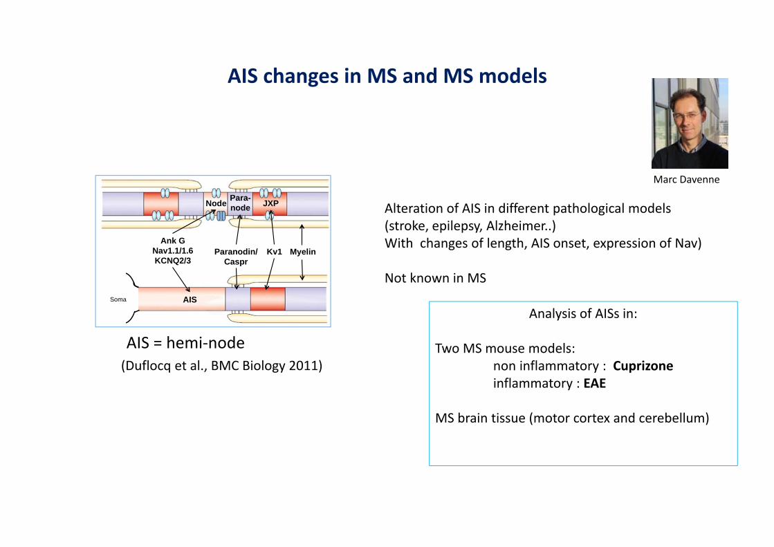
AIS changes in MS and MS models
AIS = hemi-node(Duflocq et al., BMC Biology 2011)
Ank G Nav1.1/1.6 KCNQ2/3
Paranodin/Caspr
Kv1
AIS
JXP Node Para-node
Soma
Myelin
Analysis of AISs in:
Two MS mouse models:non inflammatory : Cuprizoneinflammatory : EAE
MS brain tissue (motor cortex and cerebellum)
Marc Davenne
Alteration of AIS in different pathological models(stroke, epilepsy, Alzheimer..) With changes of length, AIS onset, expression of Nav)
Not known in MS
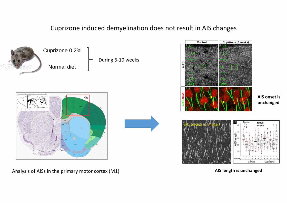
Cuprizone 0,2%
Normal diet
Analysis of AISs in the primary motor cortex (M1)
AIS onset is unchanged
During 6-10 weeks
Cuprizone induced demyelination does not result in AIS changes
3-D tracing in Image J
AIS length is unchanged
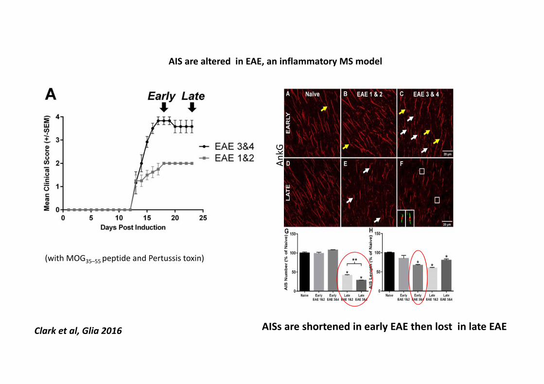
(with MOG35–55 peptide and Pertussis toxin)
AIS are altered in EAE, an inflammatory MS model
AISs are shortened in early EAE then lost in late EAE
AnkG
Clark et al, Glia 2016

AIS changes in MS
• MS lesions• Motor cortex (layer 2-6)• Cerebellum
• Characterized as • Active• Inactive• Normal appearing grey matter
• Compared to control tissue
Con
trol
NA
GM
Act
ive
Lesi
onIn
activ
e Le
sion
AnkGSMI32 Merge AnkGSMI32 Merge
AnkGSMI32 Merge AnkGSMI32 Merge
a b
g
g’
dc
j
j’
fe
k lih
a’ b’ c’ d’
h’ i’
e’ f’
k’ l’
Pyramidal neurons
Ayshegul Dilsizoglu
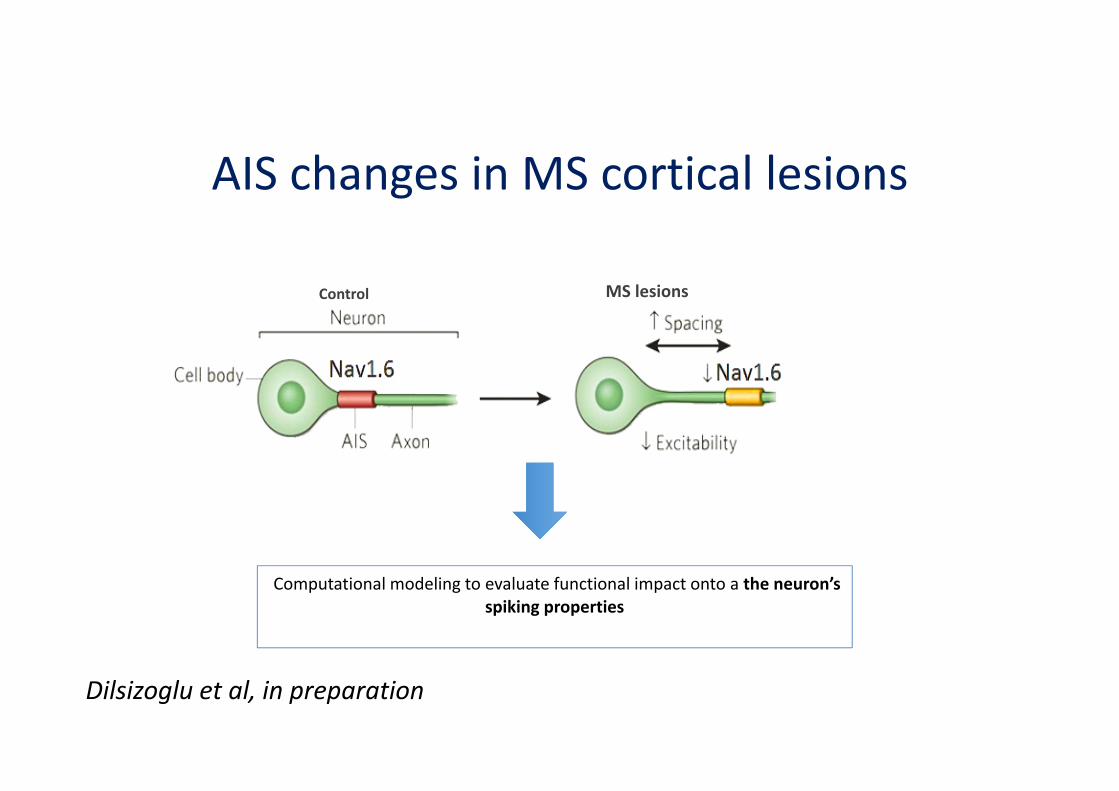
AIS changes in MS cortical lesions
Control MS lesions
Computational modeling to evaluate functional impact onto a the neuron’sspiking properties
Dilsizoglu et al, in preparation

Nodes of Ranvier and AIS
NeurofascContactin
NeurofascContactin

Kv channels Caspr Nav Caspr Kv channels
Nodal and perinodal proteins in MS brain tissue
Periplaque
10µ
Immuno-histochemical analysis ofplaquesperiplaques and NAWMpartially remyelinated plaquesfully remyelinated plaques
London MS brain bankParis MS brain bank
–
periplaque
MBP
plaque
Irène Coman
Juxtaparanode paranode node
N aV
E
N eu rofi lam en t
F
DDe m yeli n at ed pl.
GC A LS P P D
38 92 122 141N =
0
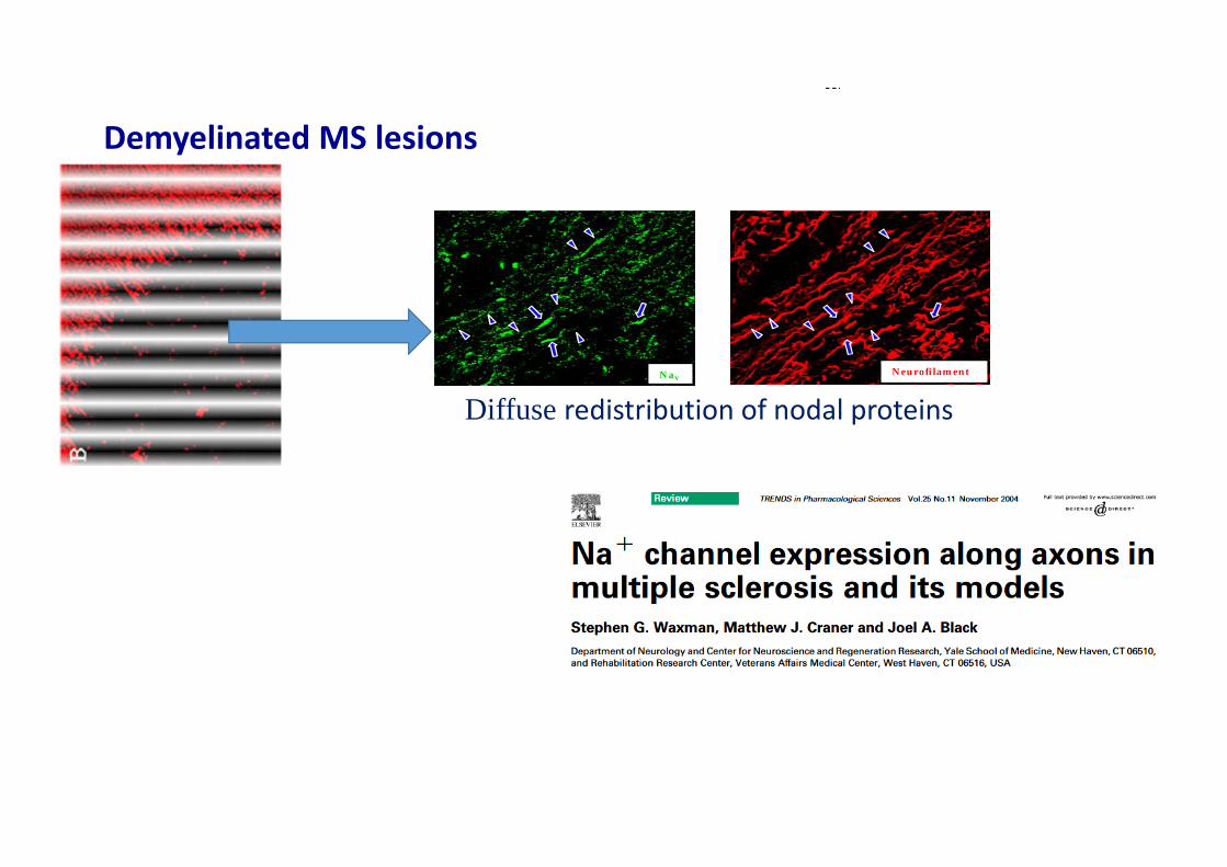
Diffuse redistribution of nodal proteins
Demyelinated MS lesions
Craner et al, PNAS 2004
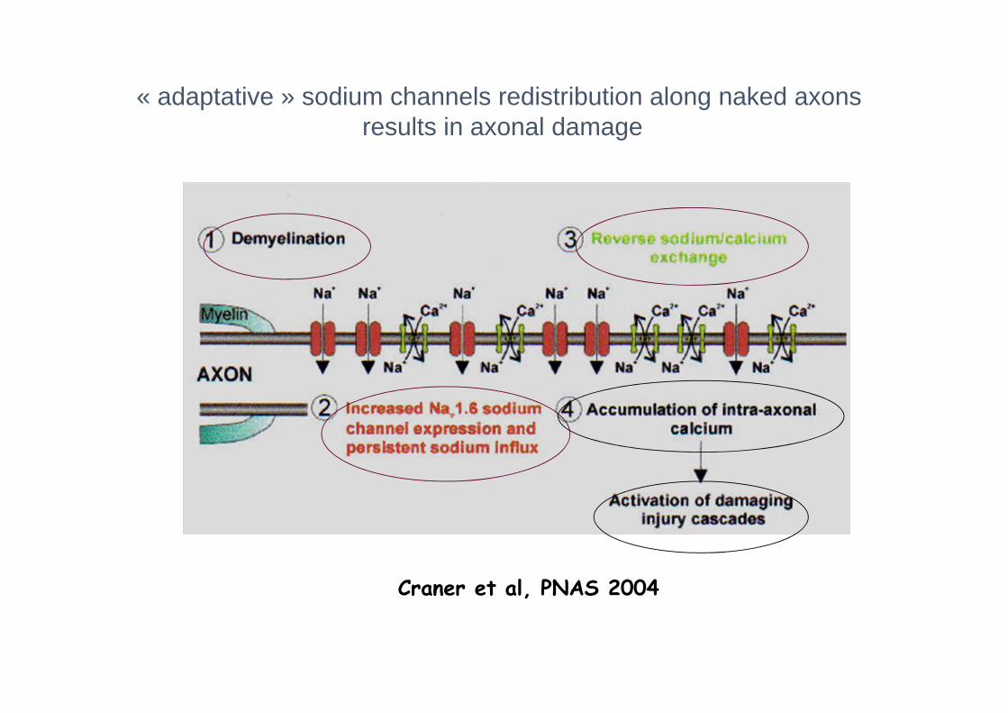
« adaptative » sodium channels redistribution along naked axons results in axonal damage
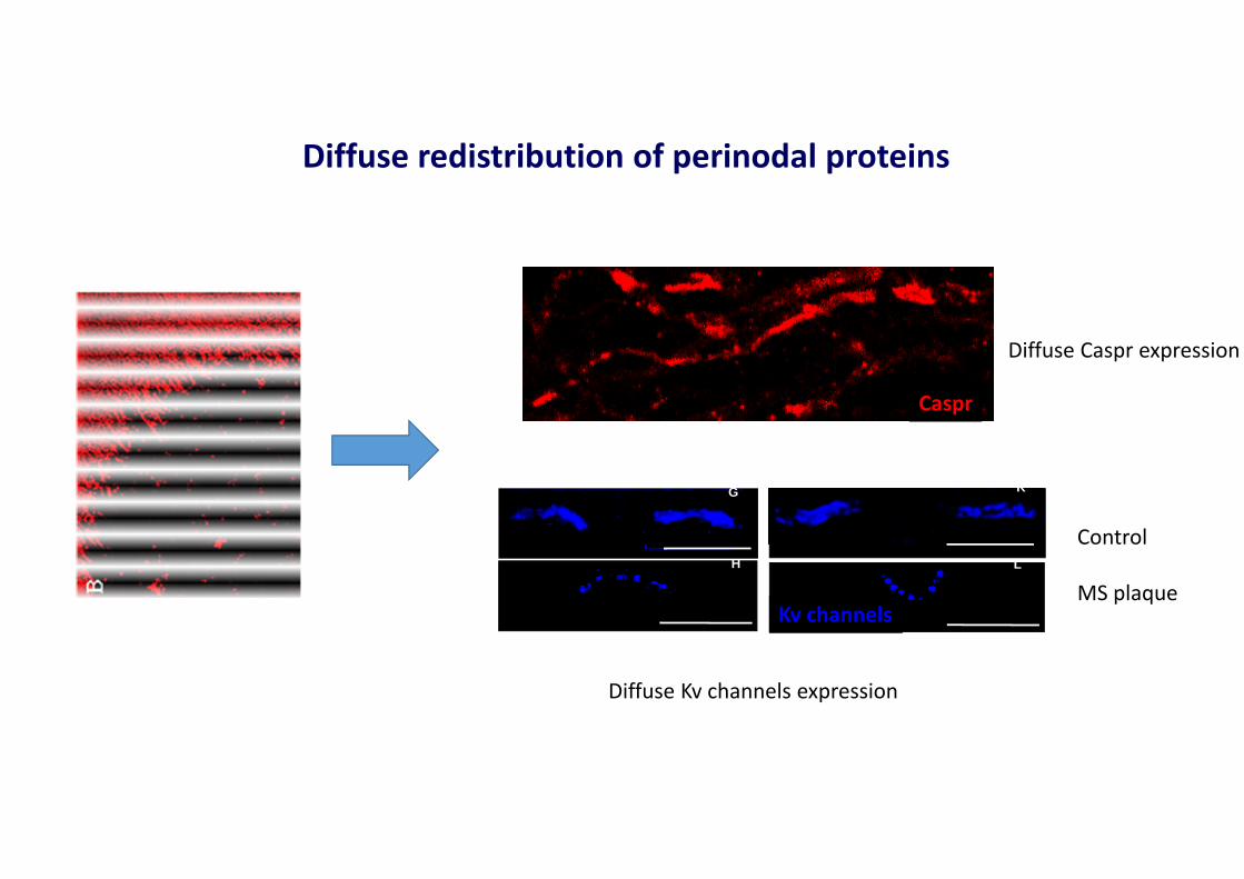
Diffuse redistribution of perinodal proteins
L
KG
H
Caspr
Kv channels
Diffuse Caspr expression
Diffuse Kv channels expression
Control
MS plaque
Howell et al , J Neuropathol Exp Neurol. 2010
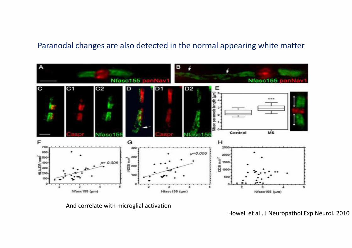
Paranodal changes are also detected in the normal appearing white matter
And correlate with microglial activation

Periplaque
A
NaV,/Caspr
BShadow plaque
Remyelinated MS lesions : nodal re-agregation
E
Kv MBP
G
Caspr2 MBP
nA
C
paranodin/Caspr MBP
Clusters
Nodal clusters are detected prior to remyelination
Do they play a role in remyelination?
Partially remyelinated lesions
Myelinated axon unmyelinated axon
Nav, MBP
Coman et al, Brain 2006

What are the mechanisms of early nodal (re)formation in the CNS?

Nodal proteins agregation beforemyelination
NavAnkGPLP
Nav AnkG
Nav Nfasc PLP NfascNav
Smi31AnkG
Smi31AnkG
Nav Nfasc PLP
Agregates of nodal proteins prior to myelination: prenodes or nascent nodes
Nathalie Sol-Foulon
Mixed hippocampal
cultures

Pampaloni et al, 2007
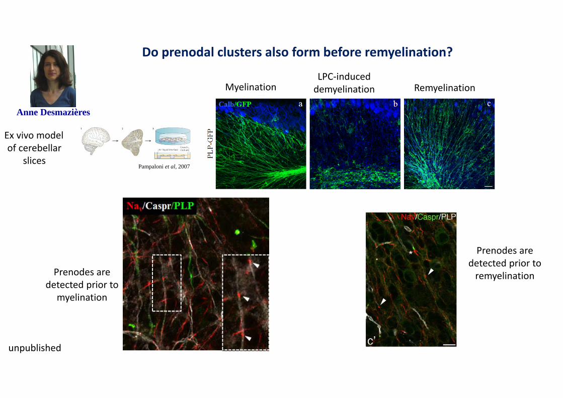
Do prenodal clusters also form before remyelination?
Ex vivo modelof cerebellar
slices
Prenodes are detected prior to
myelination
MyelinationLPC-induced
demyelination Remyelination
Prenodes are detected prior to
remyelination
Anne Desmazières
unpublished

Axonal domains changes in MS
Re-establishment of impulse conduction?
Neuroprotective influence?
Redistribution of nodal proteins along denuded axonsAltered impulse conduction/ conduction block
Axonal irreversible damage
Axonal initial segment changes Altered axonal electrophysiology(reduced excitability)
Agregation of nodal structures prior to remyelination
What are the mechanisms of axonal domains changes in MS?
• Are these changes related to a targeted adaptative immune response?
• No evidence in 2017
• Are these changes induced by demyelination?• Likely for nodal proteins redistribution
• But not for AIS changes?
• Are these changes induced by cells of the innate immunity?

Microglia and axonal domains : an emerging story

Quantification of microglia-AIS contacts in MS cortex ongoing
Ankyrin G Iba1 merge
What is the role of microglia in AIS changes?
Microglial cell aligned along AIS
Human (control) brainMarc Davenne
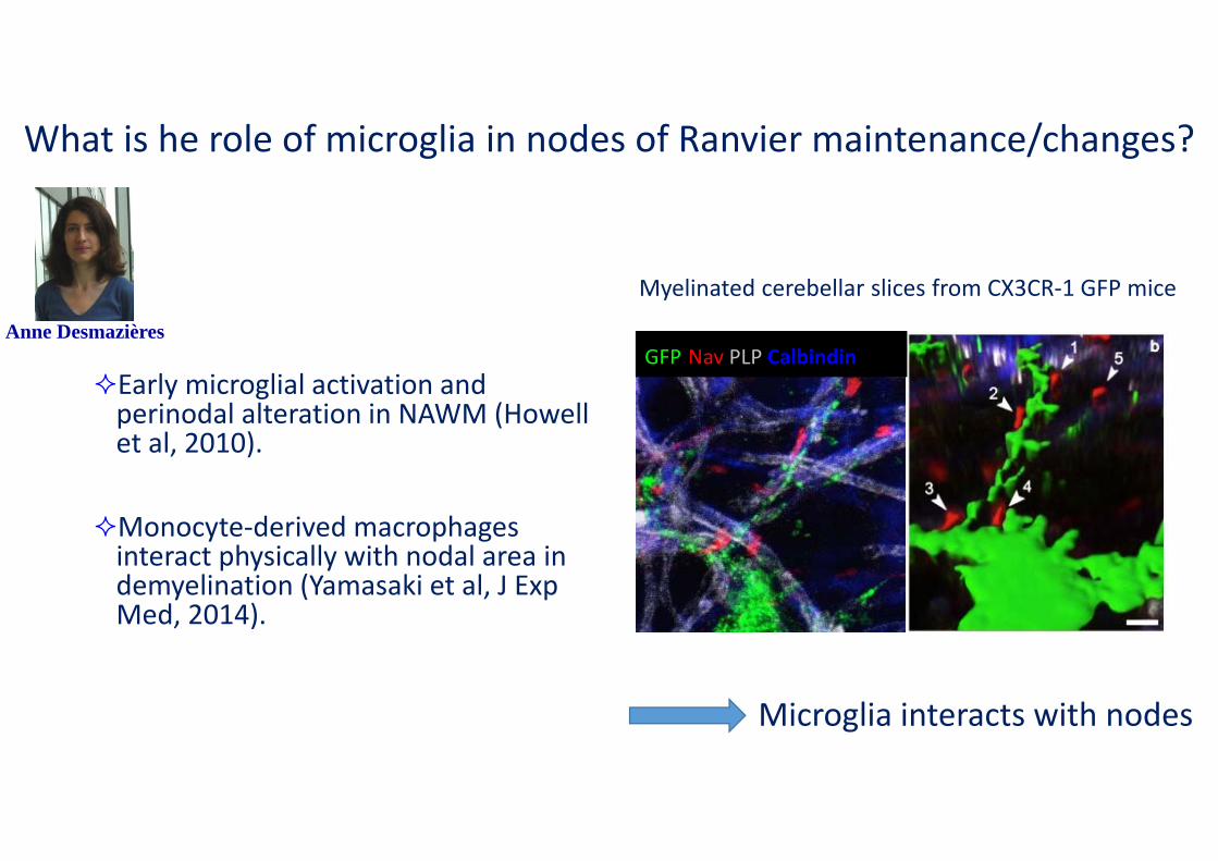
Early microglial activation and perinodal alteration in NAWM (Howellet al, 2010).
Monocyte-derived macrophages interact physically with nodal area in demyelination (Yamasaki et al, J ExpMed, 2014).
What is he role of microglia in nodes of Ranvier maintenance/changes?
GFP Nav PLP Calbindin
InMyelinated cerebellar slices from CX3CR-1 GFP miceP mi
Microglia interacts with nodes
Anne Desmazières

interaction between nodal structures and microglia during remyelination : a live imaging study
t=0
t=0’30
t=1’00
t=1’30
t=2’00
t=2’30
t=3’00
t=3’30
t=4’00
t=4’30
t=5’00
t=5’30
t=6’00
t=6’30
t=7’00
t=7’30
t=8’00
t=8’30
t=9’00
t=9’30
t=10’00
Microglia interacts with prenodal structures during remyelination(10 min video, one image/30’’)
CX3CR1 GFP cerebellar slices
Transduced with 1Nav cherry
Demyelination induced by LPC
Analysis prior to remyelination
Anne Desmazieres and Melina Thethiot,work in progress

Microglial and nodes of Ranvier interactions: an emerging story
• During development and remyelination, microglia interacts with nodal structures
• Role of microglia-nodal interaction?
• Sensing axonal « activity »?
• Influencing stabilization of nodes of Ranvier?
• Role in repair capacity?
microglia

Conclusion
• Changes in axonal domains in MS +++
• Resulting in altered axonal function
• No clear evidence of antibody-mediated nodopathy
• But influence of • Demyelination• Microglial contacts
New pathophysiological concepts
New therapeutic perspectives
favoring (re) clustering of nodal structures
Modulating innate immune response

Repair in MS : from biology to clinical translation
Catherine Lubetzki and Bruno StankoffMarc Davenne, PhD
Anne Desmazières, PhDNathalie Sol-Foulon, PhD
Bernard Zalc, DR emeritus, MD-PhDMarie-Stéphane Aigrot, Engineer
Elizabeth Maillart, MDCaroline Papeix, MDAyshegul Dilsizoglu
Benedetta BodiniMatteo TonietoCéline LouapreSean Freeman
Anne-Laure DubessyEmilie Poirion
Melina Thetiot
Post-docs
PhD students
Desdemona Fricker, Paris VBoris Barbour, ENS, Paris
José Sahel, IDV, ParisPeter Brophy, Edinburgh, ScotlandJeffrey Dupree, Richmond, VA, USA
Christina Stadelman, Goettingen, GermanyRichard Reynolds, London UK
Raul Krause, Vertex, Cambridge MABouvet Labruyere award

WELCOME TO PARIS!
Catherine Lubetzki David MillerChair MSParis2017 ECTRIMS President
Bernhard Hemmer Jack AntelECTRIMS Vice-President ACTRIMS President
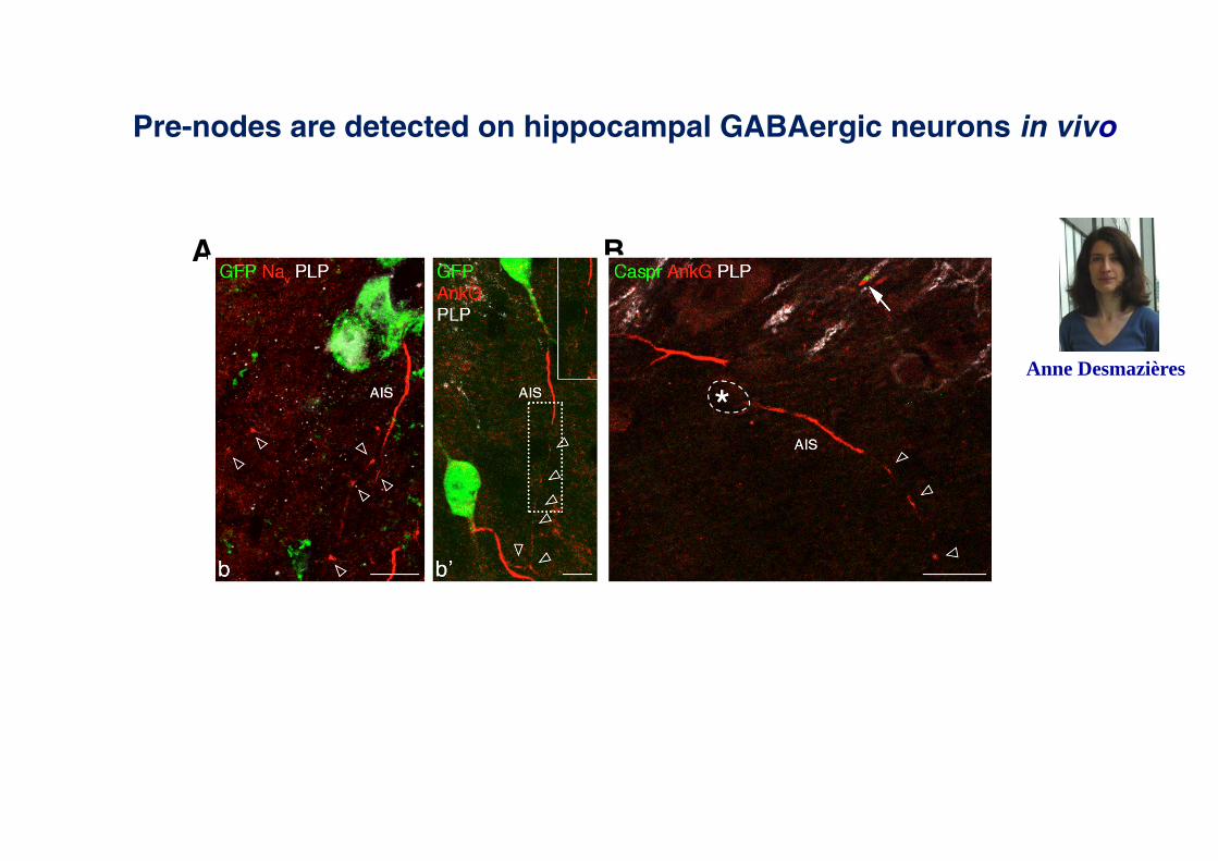
Pre-nodes are detected on hippocampal GABAergic neurons in vivo
A B
Anne Desmazières
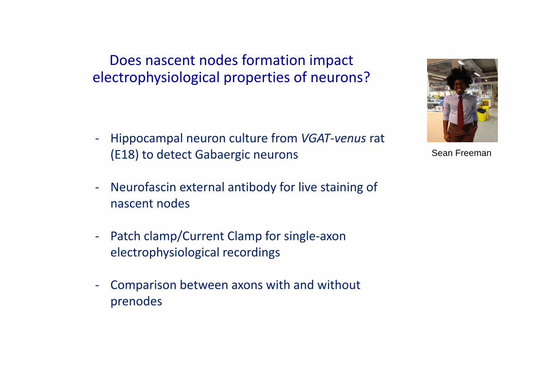
Does nascent nodes formation impact electrophysiological properties of neurons?
- Hippocampal neuron culture from VGAT-venus rat (E18) to detect Gabaergic neurons
- Neurofascin external antibody for live staining of nascent nodes
- Patch clamp/Current Clamp for single-axon electrophysiological recordings
- Comparison between axons with and withoutprenodes
Sean Freeman

Single cell electrophysiological recording in neurons with and without pre-nodes
A B
C D Latency Latency

0.0
0.5
1.0
1.5
2.0
Neurons without
pre-nodes(0.72±0.05 m/s),
Neurons withpre-nodes
(1.23±0.09 m/s)
Con
duct
ion
Vel
ocity
(m/s
)
***
Increased conduction velocity in neurons with pre-nodes
1.5-fold increase in conduction velocity along axons with prenodes
Freeman et al PNAS 2015
Pampaloni et al, 2007
Do prenodal clusters also form before remyelination?
Ex vivo modelof cerebellar
slices
Prenodes are detected prior to
myelination
MyelinationLPC-induced
demyelination Remyelination
Prenodes are detected prior to
remyelination
Anne Desmazières
unpublished
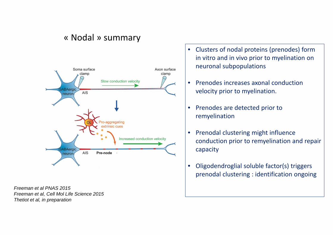
• Clusters of nodal proteins (prenodes) form in vitro and in vivo prior to myelination on neuronal subpopulations
• Prenodes increases axonal conduction velocity prior to myelination.
• Prenodes are detected prior to remyelination
• Prenodal clustering might influence conduction prior to remyelination and repair capacity
• Oligodendroglial soluble factor(s) triggers prenodal clustering : identification ongoing
Freeman et al PNAS 2015Freeman et al, Cell Mol Life Science 2015Thetiot et al, in preparation
« Nodal » summary

AnkG% Iba1% Merge%
Mou
se%
Hum
an%
Are microglia involved in MS-induced AIS alterations ?
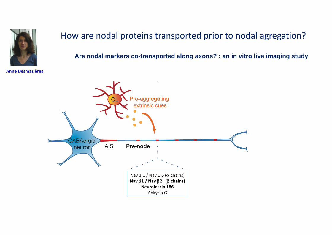
Nav 1.1 / Nav 1.6 ( chains)Nav 1 / Nav 2 (chains)
Neurofascin 186Ankyrin G
How are nodal proteins transported prior to nodal agregation?
Anne Desmazières
Are nodal markers co-transported along axons? : an in vitro live imaging study
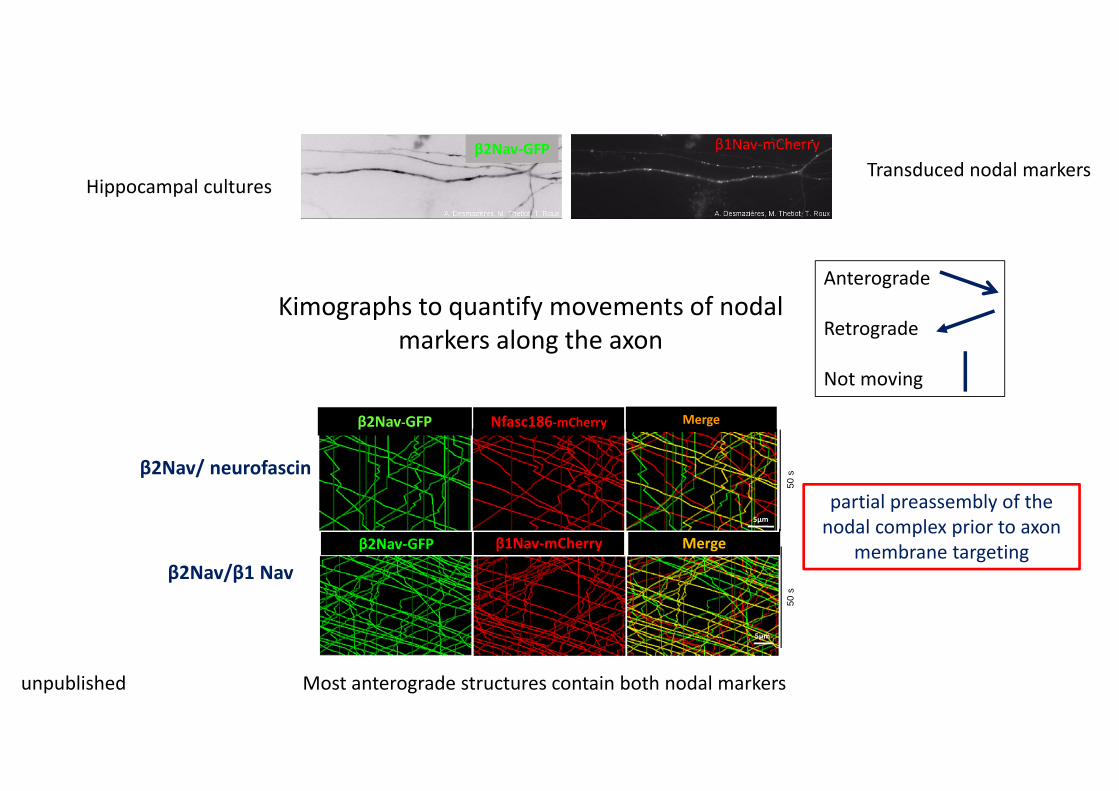
β1Nav-mCherryβ2Nav-GFP
Most anterograde structures contain both nodal markers
50 s
50 s
β2Nav-GFP β1Nav-mCherry Merge
5µm
β2Nav-GFP Nfasc186-mCherry Merge
5µm
5µm
Transduced nodal markersHippocampal cultures
β2Nav/ neurofascin
β2Nav/β1 Nav
Kimographs to quantify movements of nodal markers along the axon
Anterograde
Retrograde
Not moving
unpublished
partial preassembly of the nodal complex prior to axon
membrane targeting
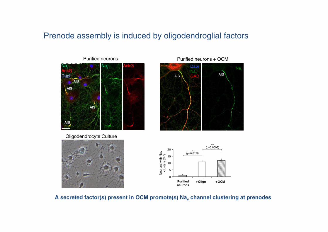
Prenode assembly is induced by oligodendroglial factors
A secreted factor(s) present in OCM promote(s) Nav channel clustering at prenodes
Nav
AISAIS
DapiNavGAD
Purified neurons + OCM
Oligodendrocyte Culture
Purified neurons
Neu
rons
with
Nav
clus
ters
(%°)
Purifiedneurons
Oligo OCM
*(p=0,0179)
***(p=0,0003)
+ +
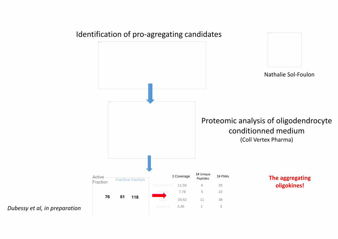
NrCAM 7,78 5 22
Contactin-1 11,56 6 26
ChL1 20,62 11 38
Noelin 2 2,46 1 3
Σ Coverage Σ# UniquePeptides Σ# PSMs
Impossible d’afficher l’image.
Inactive fraction
76 61 118
Active Fraction
Impossible d’afficher l’image.
Impossible d’afficher l’image.
Identification of pro-agregating candidates Impossible d’afficher l’image.
Proteomic analysis of oligodendrocyte conditionned medium
(Coll Vertex Pharma)
The aggregatingoligokines!
Nathalie Sol-Foulon
Dubessy et al, in preparation

The Axon Initial Segment (AIS)
Functions:1. Initiates Action Potentials (APs)2. Maintains axonal integrity/ neuronal polarity
Axon hillock

Impossible d’afficher l’image.
© 2016 American Academy of Neurology. ??diteur American Academy of Neurology. 2
Impossible d’afficher l’image.
Nav 1.8 in MS cerebellumSodium channel Nav1.8: Emerging links to human disease.Han, Chongyang; Huang, Jianying; Waxman, Stephen; MD, PhD
Neurology. 86(5):473-483, February 02, 2016.DOI : 10.1212/WNL.0000000000002333
Na V1.8 is abnormally expressed in Purkinje neurons in multiple sclerosis (MS)Figure 4. NaV1.8 protein (demonstrated by immunocytochemistry, A) and mRNA (demonstrated by in situ hybridization, B) in Purkinje neurons from rapid-autopsy tissue from patients with MS. NaV1.8 protein and mRNA are not detectable in control subjects without neurologic disease (C, protein; D, mRNA, arrow points to Purkinje neuron). From Black et al.17 (E) Purkinje neuron complex spikes lose regularity in experimental autoimmune encephalomyelitis (EAE) (E, top), but regularity is restored following treatment with NaV1.8 blocker A-803467 (E, bottom row). The shaded area on the left depicts 10 consecutive complex spikes from a representative animal from each group superimposed; to the right, 3 individual complex spikes are shown singly. (F) Wild-type mice were administered A-803467 (800 nmol icv) or vehicle after EAE induction and scored for behavioral manifestations of MS-like deficits. Vehicle group showed no changes compared to preinjectionbaseline (p > 0.05 at all time points; n = 19). A803467 partially reversed symptoms 2-4 hours after injection (1 hour, p = 0.607; 2 hours, p p p = 0.001; 5 hours, p = 0.331; Wilcoxon signed-rank test; n = 19). From Shields et al.41

Periplaque
A
NaV, paranodin/Caspr
B
Shadow plaque
Réapparition des aggrégats nodaux et périnodaux lors de la remyélinisation
Coman et al, Brain 2006

Remyélinisation : aspects anormaux
Impossible d’afficher l’image.
NaV, paranodin/Caspr
Impossible d’afficher l’image.
Héminode
Nœud de Ranvier large

Remyélinisation : aspects anormaux
paranodin/Caspr, MBP
Impossible d’afficher l’image.
Internoeuds très courts
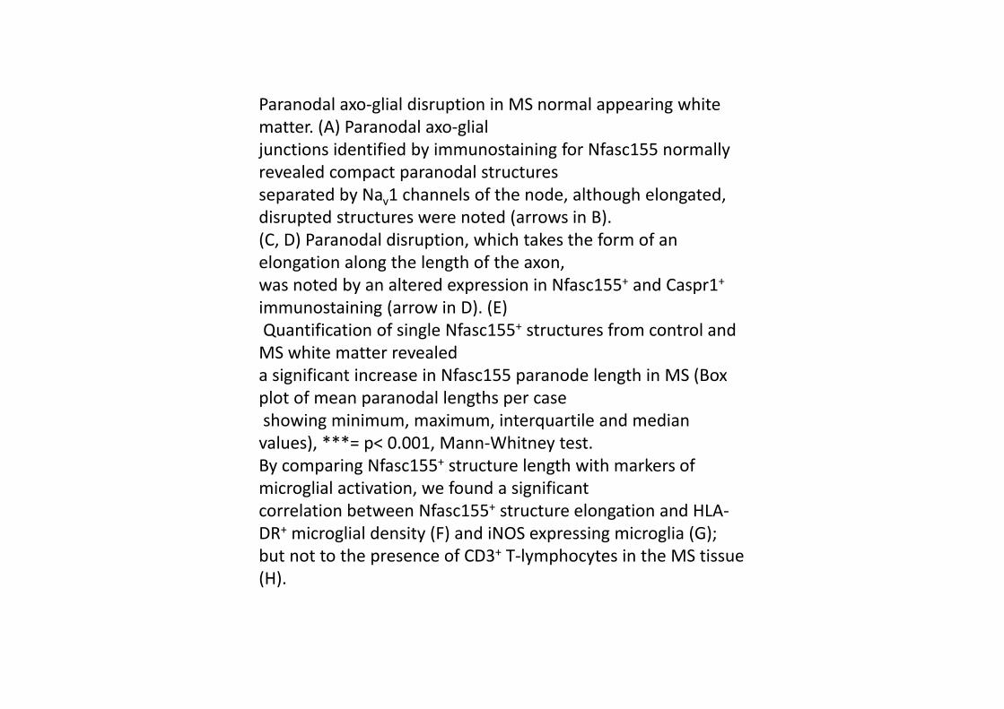
Paranodal axo-glial disruption in MS normal appearing white matter. (A) Paranodal axo-glial junctions identified by immunostaining for Nfasc155 normally revealed compact paranodal structures separated by Nav1 channels of the node, although elongated, disrupted structures were noted (arrows in B). (C, D) Paranodal disruption, which takes the form of an elongation along the length of the axon, was noted by an altered expression in Nfasc155+ and Caspr1+
immunostaining (arrow in D). (E)Quantification of single Nfasc155+ structures from control and
MS white matter revealed a significant increase in Nfasc155 paranode length in MS (Box plot of mean paranodal lengths per caseshowing minimum, maximum, interquartile and median
values), ***= p< 0.001, Mann-Whitney test. By comparing Nfasc155+ structure length with markers of microglial activation, we found a significant correlation between Nfasc155+ structure elongation and HLA-DR+ microglial density (F) and iNOS expressing microglia (G); but not to the presence of CD3+ T-lymphocytes in the MS tissue (H).