Embed Size (px)
Citation preview
THE NITROUS OXIDE METHODFOR THE QUANTITATIVEDETERMINATIONOF CEREBRALBLOODFLOWIN MAN:
THEORY, PROCEDUREANDNORMALVALUES1
By SEYMOURS. KETY AND CARL F. SCHMIDT
(From the Department of Pharmacology, University of Pennsylvania, Philadelphia)
(Received for publication December 4, 1947)
In 1945 the authors reported the determinationof cerebral blood. flow in man by the use of nitrousoxide in low concentration, a technique whichpermitted for the first time quantitative clinicalmeasurement of this important physiologic func-tion (1). Since that time numerous modificationshave been made in the procedure (2) and the un-derlying theory has been subjected to extensive ex-perimental evaluation. The present report con-stitutes a description of the technique as we havenow employed it in over 300 determinations, anexamination of its underlying theory and validity,and values obtained with its use in 34 studies on14 normal young men.
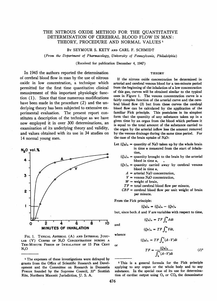
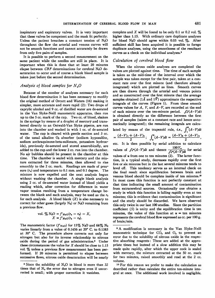
N20 vol.%
4
3
2
0MINUTES OF INHALATION
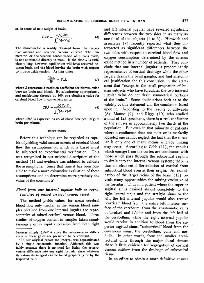
FIG. 1. TYPICAL ARTERIAL (A) AND INTERNAL JUGU-LAR (V) CURVES OF N2O CONCENTRATIONDURING A
TEN-MINUTE PERIOD OF INHALATION OF 15 PER CENTN20
1 The expenses of these investigations were defrayed bygrants from the Office of Scientific Research and Devel-opment and the Committee on Research in DementiaPrecox founded by the Supreme Council, 330 ScottishRite. Northern Masonic Jurisdiction, U. S. A.
THEORY
If the nitrous oxide concentration be determined inarterial and cerebral venous blood for a ten-minute periodfrom the beginning of the inhalation of a low concentrationof this gas, curves will be obtained similar to the typicalones in Figure 1. The venous concentration curve is afairly complex function of the arterial curve and the cere-bral blood flow (3) but from these curves the cerebralblood flow can be calculated by the application of thefamiliar Fick principle. This postulates in its simplestform that the quantity of any substance taken up in agiven time by an organ from the blood which perfuses itis equal to the total amount of the substance carried tothe organ by the arterial inflow less the amount removedby the venous drainage during the same time period. Forthe case of the brain uptake of N20:
Let (QB). = quantity of N20 taken up by the whole brainin time u measured from the start of inhala-tion,
(QA)" = quantity brought to the brain by the arterialblood in time u,
(Qv)u = quantity carried away by cerebral venousblood in time u,
A = arterial N20 concentration,V = venous N20 concentration,W= weight of brain,
TF = total cerebral blood flow per minute,CBF = cerebral blood flow per unit weight of brain
per minute.
From the Fick principle:
QB), = (QA)u - (QV).but, since both A and Vare variables with respect to time,
(QA)U = TFJfAdtand
whence
or
(Qv). = TF fJ Vdt,
(QB)u = TF fJ (A-V)dt
TF= (QB)UJfu(A-V)dt
(1)2
2 This is a general formula for the Fick principleapplying to any organ or the whole body and to anysubstance. In the special case of its use for determina-tion of cardiac output using 02 or CO2 the denominator
476
I
DETERMINATION OF CEREBRALBLOODFLOW IN MAN
or, in terms of unit weight of brain,
CBF= (QB)/W (2)(A-V)dt2
The denominator is readily obtained from the respec-tive arterial and cerebral venous curves.3 The nu-merator, or the cerebral concentration of nitrous oxide,is not obtainable directly in man. If the time u is suffi-ciently long, however, equilibrium will have occurred be-tween brain and the blood leaving the brain with respectto nitrous oxide tension. At that time:
(QB)U = VMS, (3)
where S represents a partition coefficient for nitrous oxidebetween brain and blood. By substituting appropriatelyand multiplying through by 100, one obtains a value forcerebral blood flow in convenient units:
CBF = 100Vu*S (4)fu(A-V)dt
where CBF is expressed as cc. of blood flow per 100 g. ofbrain per minute.
DISCUSSION
Before this technique can be regarded as capa-ble of yielding valid measurements of cerebral bloodflow the assumptions on which it is based mustbe subjected to experimental verification. Thiswas recognized in our original description of themethod (1) and evidence was adduced to validatethe assumptions. Since that time it has been pos-sible to make a more exhaustive evaluation of theseassumptions and to determine more precisely thevalue of the constant S.
Blood from one internal jugular bulb as repre-sentative of mixed cerebral venous bloodThe method yields values for mean cerebral
blood flow only insofar as the venous blood sam-ples obtained from one internal jugular are repre-sentative of mixed cerebral venous blood. Threestudies of oxygen content in samples taken simul-taneously or in rapid succession from both right
becomes simply (A-V) t since the arteriovenous differ-ences of these gases are presumed to be constant.
3 In our original report the integral was approximatedby a single exponential function. Although this wasfairly accurate there is no need for fitting the arterio-venous difference into any rigid formula, since whateverits nature its integral can be found graphically or by thetrapezoid rule.
and left internal jugular have revealed significantdifferences between the two sides in as many asone third of the subjects (4 to 6). Himwich andassociates (7) recently reported what they in-terpreted as significant differences between thetwo sides with respect to cerebral blood flow andoxygen consumption determined by the nitrousoxide method in a number of patients. They con-clude that one internal jugular is predominantlyrepresentative of cortical drainage while the otherlargely drains the basal ganglia, and find anatomi-cal justification for this conclusion in the state-ment that "except in the small proportion of hu-man subjects who have torculars, the two internaljugular veins do not drain symmetrical portionsof the brain." Some doubt arises both as to thevalidity of this statement and the conclusion basedupon it. According to the results of Edwards(8), Manno (9), and Riggs (10) who studieda total of 125 specimens, there is a real confluenceof the sinuses in approximately two thirds of thepopulation. But even in that minority of patientswhere a confluence does not exist or is markedlylopsided one cannot neglect the fact that the torcu-lar is only one of many means whereby mixingmay occur. According to Cobb (11), the venuleswhich emerge from the cortex are continuous withthose which pass through the subcortical regionsto drain into the internal venous system; there isthus no clear-cut differentiation of cortical fromsubcortical blood even at their origin. An exami-nation of the larger veins of the brain (12) re-veals many opportunities for mixing exclusive ofthe torcular. Thus in a patient where the superiorsagittal sinus drained almost completely to theright lateral sinus and the straight sinus to theleft, the left internal jugular would also receive"cortical" blood from the entire left inferior sur-face of the cerebrum, from the anastomotic veinsof Trolard and L'abbe and from the left half ofthe cerebellum, while the right internal jugularwould receive in addition to blood from the su-perior sagittal sinus, "subcortical" blood from thecavernous sinus, the cerebellum, pons and me-dulla. In other words, from the smaller archi-tectural units through the major dural sinusesthere is little evidence for segregation of corticalvenous outflow from the drainage of subcorticaltissue.
In an effort to obtain a more definitive answer
477
SEYMOURS. KETY AND CARL F. SCHMIDT
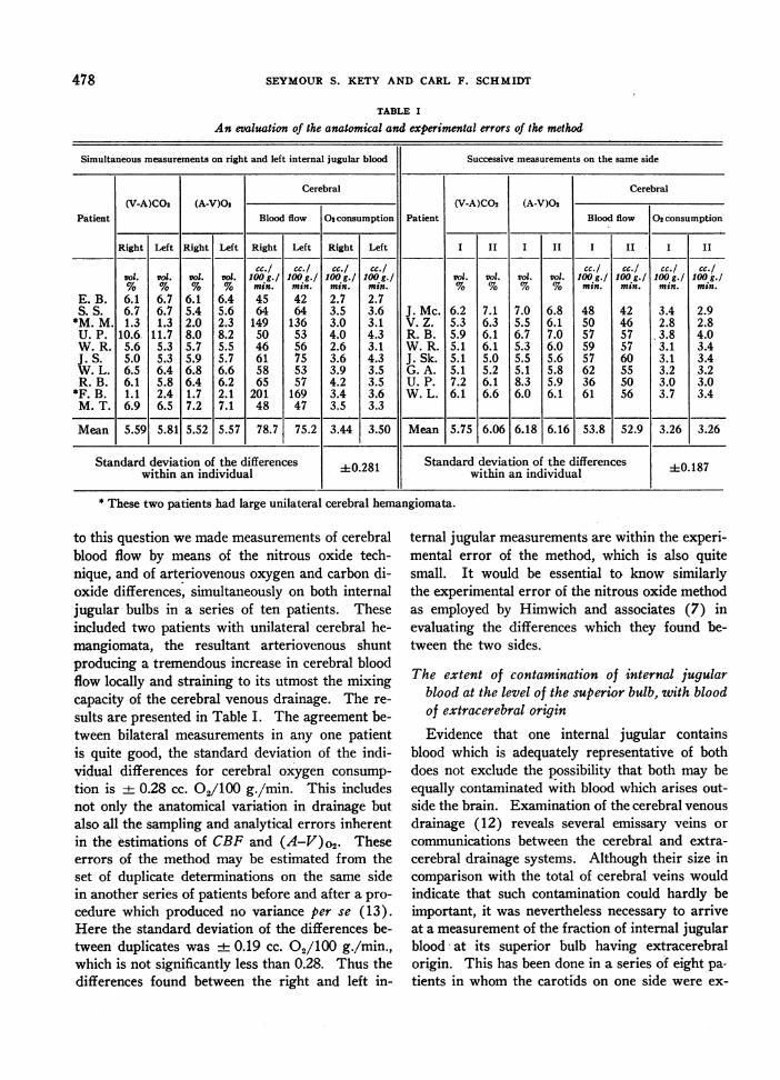
TABLE I
An evaluation of the anatomical and experimental errors of the method
Simultaneous measurements on right and left internal jugular blood Successive measurements on the same side
Cerebral Cerebral(V-A)CO2 (A-V)02 (V-A)CO2 (A-V)O2
Patient Blood flow Os consumption Patient Blood flow 02 consumption
Right Left Right Left Right Left Right Left I II I II I II I II
cc./ cc./ cc./ cc./ cc./ cc./ cc.! cc.!vol. Vol. Vol. Vol. 100g.1 100g.1 100g./ 100g . Vol. Vol. Vol. Vol. 100g.! 100g./ 100g./ 100k./% % % % min. min . min. mins. % % % % min. min. min. min.
E. B. 6.1 6.7 6.1 6.4 45 42 2.7 2.7S. S. 6.7 6.7 5.4 5.6 64 64 3.5 3.6 J. Mc. 6.2 7.1 7.0 6.8 48 42 3.4 2.9
*M. M. 1.3 1.3 2.0 2.3 149 136 3.0 3.1 V. Z. 5.3 6.3 5.5 6.1 50 46 2.8 2.8U. P. 10.6. 11.7 8.0 8.2 50 53 4.0 4.3 R. B. 5.9 6.1 6.7 7.0 57 57 3.8 4.0W. R. 5.6 5.3 5.7 5.5 46 56 2.6 3.1 W. R. 5.1 6.1 5.3 6.0 59 57 3.1 3.4J. S. 5.0 5.3 5.9 5.7 61 75 3.6 4.3 J. Sk. 5.1 5.0 5.5 5.6 57 60 3.1 3.4W. L. 6.5 6.4 6.8 6.6 58 53 3.9 3.5 G. A. 5.1 5.2 5.1 5.8 62 55 3.2 3.2R. B. 6.1 5.8 6.4 6.2 65 57 4.2 3.5 U. P. 7.2 6.1 8.3 5.9 36 50 3.0 3.0
*F. B. 1.1 2.4 1.7 2.1 201 169 3.4 3.6 W. L. 6.1 6.6 6.0 6.1 61 56 3.7 3.4M. T. 6.9 6.5 7.2 7.1 48 47 3.5 3.3
Mean 5.59 5.81 5.52 5.57 78.7 75.2 3.44 3.50 Mean 5.75 6.06 6.18 6.16 53.8 52.9 3.26 3.26
Standard deviation of the differences ±0.281 Standard deviation of the differences 40.187within an individual within an individual
* These two patients had large unilateral cerebral hemangiomata.
to this question we made measurements of cerebralblood flow by means of the nitrous oxide tech-nique, and of arteriovenous oxygen and carbon di-oxide differences, simultaneously on both internaljugular bulbs in a series of ten patients. Theseincluded two patients with unilateral cerebral he-mangiomata, the resultant arteriovenous shuntproducing a tremendous increase in cerebral bloodflow locally and straining to its utmost the mixingcapacity of the cerebral venous drainage. The re-
sults are presented in Table I. The agreement be-tween bilateral measurements in any one patientis quite good, the standard deviation of the indi-vidual differences for cerebral oxygen consump-
tion is + 0.28 cc. O0/100 g./min. This includesnot only the anatomical variation in drainage butalso all the sampling and analytical errors inherentin the estimations of CBF and (A-V)o2. Theseerrors of the method may be estimated from theset of duplicate determinations on the same sidein another series of patients before and after a pro-
cedure which produced no variance per se (13).Here the standard deviation of the differences be-tween duplicates was + 0.19 cc. 02/100 g./min.,which is not significantly less than 0.28. Thus thedifferences found between the right and left in-
ternal jugular measurements are within the experi-mental error of the method, which is also quitesmall. It would be essential to know similarlythe experimental error of the nitrous oxide methodas employed by Himwich and associates (7) inevaluating the differences which they found be-tween the two sides.
The extent of contamination of internal jugularblood at the level of the superior bulb, with bloodof extracerebral originEvidence that one internal jugular contains
blood which is adequately representative of bothdoes not exclude the possibility that both may beequally contaminated with blood which arises out-side the brain. Examination of the cerebral venousdrainage (12) reveals several emissary veins orcommunications between the cerebral and extra-cerebral drainage systems. Although their size incomparison with the total of cerebral veins wouldindicate that such contamination could hardly beimportant, it was nevertheless necessary to arriveat a measurement of the fraction of internal jugularblood* at its superior bulb having extracerebralorigin. This has been done in a series of eight pa-tients in whom the carotids on one side were ex-
-478
DETERMINATION OF CEREBRALBLOODFLOW IN MAN
posed in preparation for cerebral angiography(12). In these patients a dye (T-1824) couldbe injected into an external carotid artery whilesamples were slowly taken from the internal jugu-lar bulbs and the external jugular vein. By com-
paring the dye concentration in the internal jugularwith that in the external jugular, it was possible toarrive at a fairly quantitative measure of the ex-
tent of this contamination. It averaged 2.6%7with a maximum value of 6.5%c. It is interestingto note that when the procedure was reversed andthe dye injected into the internal carotid, signifi-cant amounts appeared in the external jugular in-
dicating that on the average about 20%o of exter-nal jugular blood is of cerebral origin. The olderanatomists who named these communicationsemissary veins anticipated these results. A pos-
sible source of significant contamination is thecommon facial vein which joins the internal jugu-lar several centimeters below the superior bulb.For this reason it is important that the needle beplaced high in the superior bulb and that bloodsamples be taken from this needle at a rate slowenough to insure against the possibility of drawingblood in a retrograde direction from the lowerparts of the vein.
From the results of these two studies, neces-
sarily limited in number by the obvious technicaldifficulties involved, it is possible to conclude thatin the great majority of individuals blood from one
internal jugular at the level of the superior bulbis fairly representative of mixed cerebral venous
blood not significantly contaminated with bloodfrom extracerebral sources. Exceptions may oc-
cur, but are unlikely to constitute a significant frac-tion of the population. This is also borne out bythe comparatively small spread of our data on
cerebral oxygen consumption, in a series of 34 ob-servations on normals (Table III) and 30 studieson schizophrenics (13). Although the possibilityof gross anatomical variation in any individualcase still remains, it is not great and does not com-
promise the validity of results obtained or con-
clusions drawn from a statistically significant seriesof cases.
4 This would not include, however, that part of thevenous return from the eye supplied by the internal caro-
tid and draining into the cavernous sinus.
Equilibration between brain and cerebral venousblood
Although it is obvious that there must be a timeduring the inhalation of a constant tension of inertgas when the brain is in equilibrium with the bloodleaving it with respect to this gas, this time in-terval must be evaluated. In our original reportwe presented indirect evidence that after ten min-utes practical equilibrium between brain and cere-bral venous blood had been achieved. Since thattime we have been able to make direct analysesof the nitrous oxide contents of brain and cerebralvenous blood in dogs exposed to nitrous oxide forvarying times (14). These studies have demon-strated that ten minutes is sufficient for the at-tainment of equilibrium between brain and cere-bral venous blood with respect to nitrous oxidetension. The value for u in Equation 4 maytherefore be taken as ten minutes.
The partition coefficient (S) of N20 between brainand blood
The same experiments yield a value for S whichis very close to unity (0.98). These in vivo re-sults compare very well with the partition coeffi-cients obtained in vitro for dog (1.03) and hu-man (1.06) brains (14).5
Although the nitrous oxide curves are capable ofyielding a value for flow per unit N2O capacity:
CBF/S= Vujw(A-V)dt
regardless of the absolute value of S. flow in termsof unit weight of brain is dependent on the con-stancy of,the partition coefficient among differentindividuals and in the same individual at different
5 On the basis of these more accurate studies it appearsthat our previous tentative value of 1.3 for this factorwas in error. This value had been derived from N20curves on monkeys obtained simultaneously with directmeasurement of cerebral blood flow using the bubble-transfer flowmeter (1). Re-examination of these ex-periments revealed the source of error: the length oftime which the arterial blood spent in the meter beforereaching the brain. This rendered the samples taken fromthe artery and internal jugular vein not really simul-taneous. Recalculation of these curves to correct thiserror yields a mean value for the factor S of 1.07, morecomparable with that obtained by the other methods.
479
SEYMOURS. KETY AND CARL F. SCHMIDT
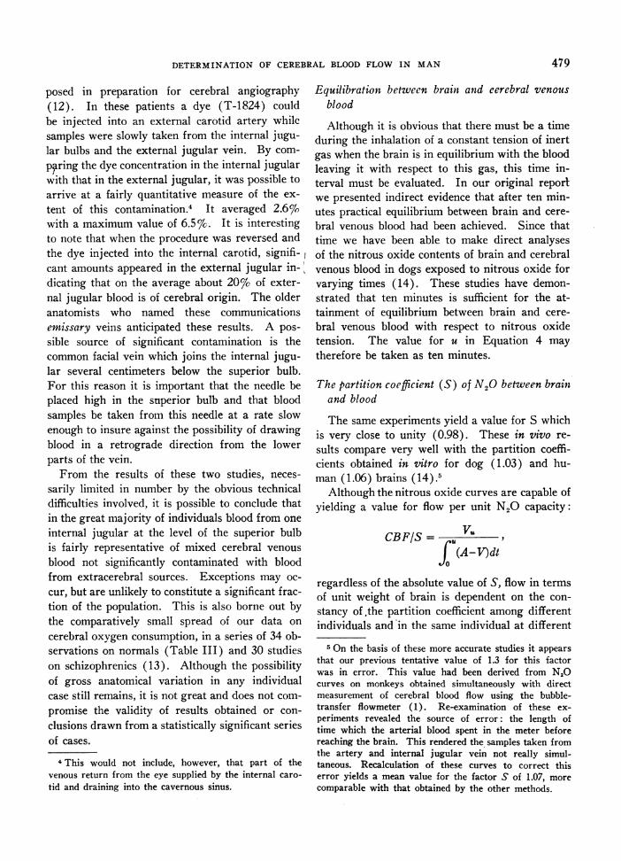
TABLE II
Comparison between the nitrous oxide method andsimultaneous direct measurement of CBF
Rhesus or Spider Monkeys
Experiment Bubble meter flow N20 flow
cc./100 g./min. cc./100 g./min.24 37 3127 42 3328 17 2030 1 46 3730 II 60 5430 III 31 3431 I 38 3431 11 76 7131 III 32 37
times. This coefficient, however, depending ongross physico-chemical constitution would be ex-pected to change significantly only with suchchanges in brain or blood composition as would beincompatible with life (14).
Final corroboration of the validity of the ni-trous oxide method is to be found in comparisonbetween it and the direct flow measurement by
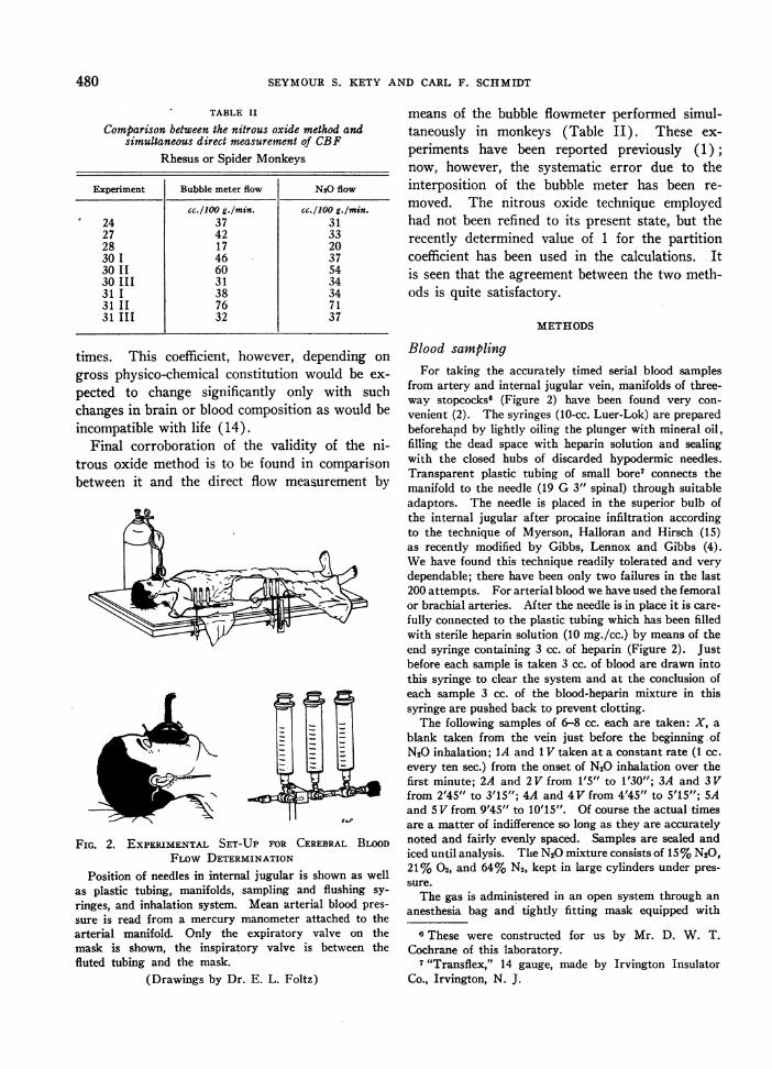
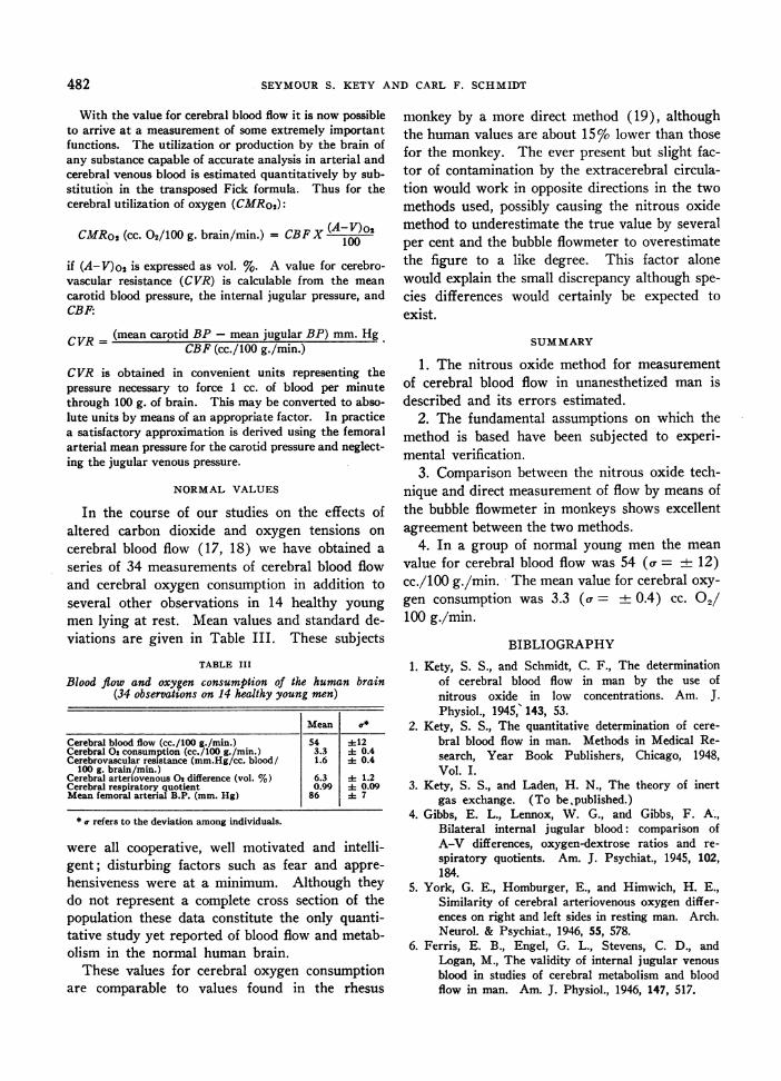
FIG. 2. EXPERIMENTAL SET-UP FOR CEREBRAL BLOODFLOWDETERMINATION
Position of needles in internal jugular is shown as wellas plastic tubing, manifolds, sampling and flushing sy-ringes, and inhalation system. Mean arterial blood pres-sure is read from a mercury manometer attached to thearterial manifold. Only the expiratory valve on themask is shown, the inspiratory valve is between thefluted tubing and the mask.
(Drawings by Dr. E. L. Foltz)
means of the bubble flowmeter performed simul-taneously in monkeys (Table II). These ex-periments have been reported previously (1);now, however, the systematic error due to theinterposition of the bubble meter has been re-moved. The nitrous oxide technique employedhad not been refined to its present state, but therecently determined value of 1 for the partitioncoefficient has been used in the calculations. Itis seen that the agreement between the two meth-ods is quite satisfactory.
METHODS
Blood samplingFor taking the accurately timed serial blood samples
from artery and internal jugular vein, manifolds of three-way stopcocks6 (Figure 2) have been found very con-venient (2). The syringes (10-cc. Luer-Lok) are preparedbeforehand by lightly oiling the plunger with mineral oil,filling the dead space with heparin solution and sealingwith the closed hubs of discarded hypodermic needles.Transparent plastic tubing of small bore7 connects themanifold to the needle (19 G 3" spinal) through suitableadaptors. The needle is placed in the superior bulb ofthe internal jugular after procaine infiltration accordingto the technique of Myerson, Halloran and Hirsch (15)as recently modified by Gibbs, Lennox and Gibbs (4).Wehave found this technique readily tolerated and verydependable; there have been only two failures in the last200 attempts. For arterial blood we have used the femoralor brachial arteries. After the needle is in place it is care-fully connected to the plastic tubing which has been filledwith sterile heparin solution (10 mg./cc.) by means of theend syringe containing 3 cc. of heparin (Figure 2). Justbefore each sample is taken 3 cc. of blood are drawn intothis syringe to clear the system and at the conclusion ofeach sample 3 cc. of the blood-heparin mixture in thissyringe are pushed back to prevent clotting.
The following samples of 6-8 cc. each are taken: X, ablank taken from the vein just before the beginning ofN20 inhalation; 1A and 1 V taken at a constant rate (1 cc.every ten sec.) from the onset of N20 inhalation over thefirst minute; 2A and 2V from 1'5" to 1'30"; 3A and 3Vfrom 2'45" to 3'15"; 4A and 4V from 4'45" to 5'15"; 5Aand 5 V from 9'45" to 10'15". Of course the actual timesare a matter of indifference so long as they are accuratelynoted and fairly evenly spaced. Samples are sealed andiced until analysis. The N20 mixture consists of 15%N20,21% 02, and 64% N2, kept in large cylinders under pres-sure.
The gas is administered in an open system through ananesthesia bag and tightly fitting mask equipped with
6 These were constructed for us by Mr. D. W. T.Cochrane of this laboratory.
7 "Transflex," 14 gauge, made by Irvington InsulatorCo., Irvington, N. J.
480
DETERMINATION OF CEREBRALBLOODFLOW IN MAN
inspiratory and expiratory valves. It is very importantthat these valves be competent and the mask fit perfectly.Unless the patient breathes a constant tension of N20throughout the flow the arterial and venous curves willnot be smooth functions and cannot accurately be drawnfrom only five pairs of samples.
It is possible to perform a second measurement on thesame patient while the needles are still in place. It isimportant when this is done that at least 20 minuteselapse between CBF determinations for cerebral N20 de-saturation to occur and of course a blank blood sample istaken just before the second determination.
Analysis of blood samples for N20
Because of the number of analyses necessary for eachblood flow determination it has been necessary to modifythe original method of Orcutt and Waters (16) making itsimpler, more accurate and more rapid (2): Two drops ofcaprylic alcohol and 9 cc. of distilled water are de-aeratedin the Van Slyke-Neill manometric apparatus, then runup to the 5 cc. mark of the cup. Two cc. of blood, shakenin the syringe by means of a droplet of mercury and trans-ferred directly to an Ostwald-Van Slyke pipette, are runinto the chamber and washed in with 1 cc. of de-aeratedwater. The cup is cleared with gentle suction and 3 cc.of the usual alkaline 02 absorber (sodium hyposulfite,sodium anthraquinone-beta-sulfonate, potassium hydrox-ide), previously de-aerated and stored anaerobically, areadded to the cup and the lower 2 cc. run into the chamber.No air bubbles should be present in the chamber at thistime. The chamber is sealed with mercury and the mix-ture extracted for three minutes, then allowed to risesmoothly to the 2 cc. mark. Readings are made of pres-sure (r@) and temperature to 0.1 mm. and 0.1 degree. Themixture is now expelled and the next analysis begunwithout washing the chamber. A single blank analysisusing 2 cc. of de-aerated water instead of blood yields areading which, after correction for difference in watervapor tension resulting from a temperature change be-tween the blank and each analysis, may be used as the rofor each analysis. A blood blank (X) is also necessary tocorrect for other gases (largely N2) or N20 remaining froma previous flow.
vol. %N20 = f'N2o(r - ro) -X,X =f'N20(rz - ro).
The manometric factor (f'N2o) for 15% N20 and 64% N2varies linearly from a value of 0.1456 at 200 C. to 0.1383at 300 C. The procedure above corrects not only fornitrogen but also for its inverse relationship to nitrousoxide during the period of gas administration.8 Underthese circumstances the value for X should be close to 1.15vol. %unless a previous flow measurement has been per-formed. If 20 minutes is permitted to elapse betweensuccessive flows, nitrous oxide desaturation will be nearly
8 Since the solubility of N20 in blood is more than 32times that of N2 the error due to nitrogen even if uncor-rected is small; with proper correction it vanishes.
complete and X will be found to be only 0.1 or 0.2 vol. %higher than 1.15. With ordinary care duplicate analysesfor blood N20 should agree within 0.05 vol. %. Aftersufficient skill has been acquired it is possible to foregoduplicate analyses, using the smoothness of the resultantcurves as a check on the individual analyses.'
Calculation of cerebral blood flowWhen the nitrous oxide analyses are completed the
values are plotted against time. The time of each sampleis taken as the mid-time of the interval over which thesample was taken except for the first pair, taken at a con-stant rate over the first minute (and therefore alreadyintegrated) which are plotted as lines. Smooth curvesare then drawn through the arterial and venous pointsand so constructed over the first minute that the averagesamples obtained (1A and IV) approximate the respectiveintegrals of the curves (Figure 1). From these smoothcurves values for A, V, and A-V, are recorded at the endof each minute over the ten-minute period. Jfo(A-V)dtis obtained directly as the difference between the firstpair of samples (taken at a constant rate and hence auto-matically integrated); the remaining integrals are calcu-lated by means of the trapezoid rule, i.e., f (A-V)dt
(A- V) I+ (A- V)2 ('3(A-iV)dt = (A-V)2 + (A-V)32 ;f'2
etc. It is then possible by serial addition to calculate
values of f0u(A-V)dt and thence 100 V. for serial.fo(A-V)dtvalues of u from one to ten minutes (2). The latter func-tion, in a typical study, decreases rapidly over the firstfive or six minutes but in the last several minutes tends tolevel off. This tendency serves as an internal check onthe final result since equilibration between brain andvenous blood should be complete inside of ten minutes.10In most cases this function is not perfectly constant atthat time indicating the small amount of contaminationfrom extracerebral sources. Occasionally one obtains astudy in which this function is falling rapidly even at tenminutes; this is evidence that contamination is significantand the study should be discarded. We have observedthis only twice in our last 100 studies. Since the partitioncoefficient (S) is unity and the equilibration time is tenminutes, the value of this function at u = ten minutesrepresents the cerebral blood flow expressed as cc. per 100 g.of brain/min.
9A modification is necessary in the Van Slyke-Neillmanometric technique for CO. and 02 to prevent anerror due to the solubility of nitrous oxide in the respec-tive absorbing reagents: These are added at the appro-priate times but instead of a slow addition this may bemade quite rapidly, after which the upper cock is sealedwith mercury, the mixture extracted at the 50 cc. markfor two minutes, raised smoothly and read at the 2 cc.volume.
10 For this reason we prefer to make the calculation asdescribed rather than calculate the entire ten-minute inte-gral at once. The additional work involved is negligible.
481
SEYMOURS. KETY AND CARL F. SCHMIDT
With the value for cerebral blood flow it is now possibleto arrive at a measurement of some extremely importantfunctions. The utilization or production by the brain ofany substance capable of accurate analysis in arterial andcerebral venous blood is estimated quantitatively by sub-stitution in the transposed Fick formula. Thus for thecerebral utilization of oxygen (CMRo2):
CMRo2(cc. 02/100 g. brain/min.) = CBFX (A-V)o2100
if (A-V)O is expressed as vol. %. A value for cerebro-vascular resistance (CVR) is calculable from the meancarotid blood pressure, the internal jugular pressure, andCBF:
CVR - (mean carotid BP - mean jugular BP) mm. HgCBF (cc./100 g./min.)
CVR is obtained in convenient units representing thepressure necessary to force 1 cc. of blood per minutethrough 100 g. of brain. This may be converted to abso-lute units by means of an appropriate factor. In practicea satisfactory approximation is derived using the femoralarterial mean pressure for the carotid pressure and neglect-ing the jugular venous pressure.
NORMALVALUES
In the course of our studies on the effects ofaltered carbon dioxide and oxygen tensions oncerebral blood flow (17, 18) we have obtained aseries of 34 measurements of cerebral blood flowand cerebral oxygen consumption in addition toseveral other observations in 14 healthy youngmen lying at rest. Mean values and standard de-viations are given in Table III. These subjects
TABLE III
Blood flow and oxygen consumption of the human brain(34 observations on 14 healthy young men)
Mean a*
Cerebral blood flow (cc./100 g./min.) 54 +12Cerebral O consumption (cc./100 g./min.) 3.3 + 0.4Cerebrovascular resistance (mm.Hg/cc. blood/ 1.6 + 0.4
100 g. brain/min.)Cerebral arteriovenous 02 difference (vol. %) 6.3 + 1.2Cerebral respiratory quotient 0.99 + 0.09Mean femoral arterial B.P. (mm. Hg) 86 A 7
* a refers to the deviation among individuals.
were all cooperative, well motivated and intelli-gent; disturbing factors such as fear and appre-
hensiveness were at a minimum. Although theydo not represent a complete cross section of thepopulation these data constitute the only quanti-tative study yet reported of blood flow and metab-olism in the normal human brain.
These values for cerebral oxygen consumptionare comparable to values found in the rhesus
monkey by a more direct method (19), althoughthe human values are about 15%o lower than thosefor the monkey. The ever present but slight fac-tor of contamination by the extracerebral circula-tion would work in opposite directions in the twomethods used, possibly causing the nitrous oxidemethod to underestimate the true value by severalper cent and the bubble flowmeter to overestimatethe figure to a like degree. This factor alonewould explain the small discrepancy although spe-cies differences would certainly be expected toexist.
SUMMARY
1. The nitrous oxide method for measurementof cerebral blood flow in unanesthetized man isdescribed and its errors estimated.
2. The fundamental assumptions on which themethod is based have been subjected to experi-mental verification.
3. Comparison between the nitrous oxide tech-nique and direct measurement of flow by means ofthe bubble flowmeter in monkeys shows excellentagreement between the two methods.
4. In a group of normal young men the meanvalue for cerebral blood flow was 54 (a = ± 12)cc./100 g./min. 'The mean value for cerebral oxy-gen consumption was 3.3 (a = ± 0.4) cc. 02/100 g./min.
BIBLIOGRAPHY1. Kety, S. S., and Schmidt, C. F., The determination
of cerebral blood flow in man by the use ofnitrous oxide in low concentrations. Am. J.Physiol., 1945,' 143, 53.
2. Kety, S. S., The quantitative determination of cere-bral blood flow in man. Methods in Medical Re-search, Year Book Publishers, Chicago, 1948,Vol. I.
3. Kety, S. S., and Laden, H. N., The theory of inertgas exchange. (To bepublished.)
4. Gibbs, E. L., Lennox, W. G., and Gibbs, F. A.,Bilateral internal jugular blood: comparison ofA-V differences, oxygen-dextrose ratios and re-spiratory quotients. Am. J. Psychiat., 1945, 102,184.
5. York, G. E., Homburger, E., and Himwich, H. E.,Similarity of cerebral arteriovenous oxygen differ-ences on right and left sides in resting man. Arch.Neurol. & Psychiat., 1946, 55, 578.
6. Ferris, E. B., Engel, G. L., Stevens, C. D., andLogan, M., The validity of internal jugular venousblood in studies of cerebral metabolism and bloodflow in man. Am. J. Physiol., 1946, 147, 517.
482
DETERMINATION OF CEREBRALBLOODFLOW IN MAN
7. Himwich, W. A., Homburger, E., Maresca, R., andHimwich, H. E., Brain metabolism in man: un-anesthetized and in pentothal narcosis. Am. J.Psychiat., 1947, 103, 689.
8. Edwards, E. A., Anatomic variations of the cranialvenous sinuses. Arch. Neurol. & Psychiat., 1931,26, 801.
9. Manno, quoted by (8).10. Riggs, H. E., quoted by (1).11. Cobb, S., The cerebrospinal blood vessels. Cytology
and Cellular Pathology of the Nervous System,W. Penfield, Ed., Paul Hoeber, Inc., N. Y., 1932.
12. Shenkin, H. A., Harmel, M. H., and Kety, S. S.,The dynamic anatomy of the cerebral circulation.Arch. Neurol. & Psychiat., 1948 (in press).
13. Kety, S. S., Woodford, R. B., Harmel, M. H., Frey-han, F. A., Appel, K. E., and Schmidt, C. F.,Cerebral blood flow and metabolism in schizo-phrenia. Am. J. Psychiat., 1948 (in press).
14. Kety, S. S., Harmel, M. H., Broomell, H. T., andRhode, C. B., The solubility of nitrous oxide inbrain and blood. J. Biol. Chem., 1948, 173, 487.
15. Myerson, A., Halloran, R. D., and Hirsch, H. L.,Technique for obtaining blood from the internaljugular vein and carotid artery. Arch. Neurol. &Psychiat., 1927, 17, 807.
16. Orcutt, F. S., and Waters, R. M., A method for thedetermination of cyclopropane, ethylene, and nitrousoxide in the blood with the Van Slyke-Neill mano-metric apparatus. J. Biol. Chem., 1937, 117, 509.
17. Kety, S. S., and Schmidt, C. F., The effects of activeand passive hyperventilation on cerebral blood flow,cerebral oxygen consumption, cardiac output, andblood pressure of normal young men. J. Clin.Invest., 1946, 25, 107.
18. Kety, S. S., and Schmidt, C. F., The effects of alteredarterial tensions of carbon dioxide and oxygen oncerebral blood flow and cerebral oxygen consump-tion of normal young men. J. Clin. Invest., 1948,27, 484.
19. Schmidt, C. F., Kety, S. S., and Pennes, H. H., Thegaseous metabolism of the brain of the monkey.Am. J. Physiol., 1945, 143, 33.
483