Embed Size (px)
Citation preview
Nitrification in freshwater sediments as studied with
microsensors and fluorescence in situ hybridization
Dissertation zur Erlangung des Grades eines Doktors der
Naturwissenschaften
- Dr. rer. nat. –
dem Fachbereich der Biologie der Universität Bremen vorgelegt von
Dörte Altmann
Bremen
April 2003
Die vorliegende Arbeit wurde in der Zeit von März 2000 bis April 2003 am Max-Planck-Insitut für Marine Mikrobiologie in Bremen angefertigt.
1. Gutachter: Prof. Dr. Bo Barker Jørgensen
2. Gutachter: Prof. Dr. Rudolf Amann
Prüfer:
Prof. Dr. Juliane Filser
Dr. Peter Stief
Tag des Promotionskolloquiums: 6. 6. 2003

Danksagung
Die vorliegende Arbeit wurde mit Mitteln der Deutschen Forschungsgemeinschaft
(DGF, Projekt-Nr. STI202/1) sowie der Max-Planck-Gesellschaft angefertigt.
Prof. Dr. Bo Barker Jørgensen und Prof. Dr. Rudolf Amann danke ich für die
Erstellung der Gutachten zu dieser Arbeit. Allen weiteren Mitgliedern der
Prüfungskommission danke ich für ihre Zeit und Mühe.
Den Leitern der Arbeitsgruppen Mikrosensoren und Molekulare Ökologie, Dr. Dirk de
Beer und Prof. Dr. Rudolf Amann, danke ich dafür, dass sie mir Laborplätze sowie
Arbeitsmaterialien zur Verfügung stellten und mir somit diese interdisziplinäre Arbeit
ermöglichten. Ihnen sei auch gedankt dafür, dass sie mir auf wichtige Fragen zu
meiner Arbeit Antwort gaben.
Ganz besonderer Dank gilt meinem Kollegen und Betreuer Dr. Peter Stief dafür, dass
er mich in sein Projektboot geholt hat sowie für viele gemeinsam verbrachte Messtage
in Kühlkammer und Rheinlabor – jede Menge Humor sorgte hier für eine echt „coole“
Arbeitsatmosphäre. Dr. Peter hat mir außerdem die Anfertigung der Mikrosensoren
und das Messen mit ihnen beigebracht, was mir viel Spaß bereitet hat. Außerdem hat
er sich stets Zeit genommen, anstehende Probleme ausführlich zu diskutieren. Ich habe
mich außerordentlich gut betreut gefühlt – großen Dank dafür!
Ein ebenfalls sehr wichtiger Berater während der Zeit der Anfertigung dieser
Dissertation war Dr. Andreas Schramm, der einen Teil zum Konzept des Projektes
beigetragen hat und für dessen konstruktive Kritik ich sehr dankbar bin.
Mein Dank gilt auch allen Mitarbeitern des Max-Planck-Institutes für Marine
Mikrobiologie, die mit zum Gelingen dieser Arbeit beigetragen haben: Gabi Eickert,
Anja Eggers, Ines Schröder für die Konstruktion von O2-Mikrosensoren und ihre
sympatische Art sich stets für alle Dinge zu interessieren, den TA´s der Molekularen
Ökologie Silke Wetzel und Jörg Wulf für viele praktische Hinweise zum
Zurechtfinden in den Labors der Gruppe; Dr. Armin Gieseke und Dr. Enrique Llobet-
Brossa, dass sie mir das FISHen beigebracht haben, ihnen sowie Dr. Bernhard Fuchs,
Dr. Katrin Knittel und Christine Beardsley für die Beantwortung wichtiger
methodischer Fragen; den Hiwis für ihre Mithilfe bei der Bewältigung der Proben.
Ganz besonders großer Dank gilt meinen lieben Mitstreitern und Bürogenossen
Rebecca, Uschi, Felix sowie Peter für ihre tatkräftige und geduldige Unterstützung vor
allem während der letzten heißen Phase – Ihr wart toll bei der Überlistung und
Beseitigung “kleinerer Unregelmäßigkeiten”. Und überhaupt, was hätte ich nur ohne
Euch all die Zeit gemacht!
Allen Kollegen der Mikrosensorgruppe und Christine Beardsley danke ich dafür, daß
mir die tolle Zeit an diesem Institut unvergeßlich sein wird.
Meinen Eltern danke ich für ihr Intersse an meiner Arbiet sowie die Förderung und
Unterstützung auf meinem bisherigen Weg.
Sven möchte ich sehr dafür danken, daß er mir besonders während der Endphase der
Arbeit den Rücken freihielt und mich mit liebevoller Zuneigung und klugen Worten
stets wieder aufgebaut und mich sehr motiviert hat.

Table of contents Chapter 1…………………………………………………………………………..7
General Introduction………………………………………………………………..7
Thesis outline………………………………………………………………………21
Chapter 2………………………………………………………………………….41
In situ distribution and activity of nitrifying bacteria in freshwater sediment
Chapter 3………………………………………………………………………….57
Distribution and activity of nitrifying bacteria in natural and manipulated stream
sediment as determined with in situ techniques
Chapter 4………………………………………………………………………….81
Nitrification in freshwater sediments as influenced by insect larvae:
Quantification by microsensors and fluorescence in situ hybridization
Chapter 5………………………………………………………………………...101
Summary………………………………………………………………………….101
Zusammenfassung………………………………………………………………..105
Appendix………...……………………………………………………………….109
List of publications

Chapter 1
General introduction
Chapter 1 General Introduction _____________________________________________________________________
8
The benthic nitrogen cycle and nitrification
Over the last decades the global nitrogen cycle (N-cycle) has received substantial
attention since the amount of N that is cycled on a global scale has greatly increased
due to human activities. Moreover, a variety of negative consequences for the living
world have been noticed. From a human point of view the eutrophication of aquatic
ecosystems by excess N is a major problem. The nutrients NO3- and NH4
+ can reach
ground and surface waters by leaching from fertilized soils and insufficient treatment
of wastewater can additionally supply NH4+ to surface waters. As a consequence of the
high external loading with NO3- and NH4
+ growth of especially planktonic primary
producers may be enhanced, which can have profound effects on the quality of
receiving waters (Smith, 2003). The enrichment of NH4+ itself is critical due to the
toxicity of its uncharged form NH3 to aquatic life (Hargreaves, 1998; Pandey, 1999;
Oliviero et al., 2003). Moreover, NO2- which is an intermediate of several microbial N
conversions in the sediment and which is toxic towards animal and human life
(Bruning-Fann and J.B., 1993; Kelso et al., 1999; Neumann et al., 2001) can be
enriched in ground and surface waters as consequence of a high external NO3- load
(Kelso et al., 1997; Stief et al., 2002). Fossil fuel combustion and biomass burning are
major contributors to the emission of gaseous NO + NO2 (summarized as NOx) into the
atmosphere where this gas mix participates in the photochemically induced formation
of tropospheric ozone (Kerr, 1990). Another gaseous N-species, N2O, is an important
greenhouse gas and is therefore associated with global climate changes (Dalal et al.,
2003). N2O is a byproduct of nitrification and an intermediate of denitrification which
is emitted e.g. from irrigated rice soils (Ghosh et al., 2003) and also from estuarine and
coastal sediments with high organic loading (Usui et al., 2001).
In addition to the anthropogenic input of N to aquatic environments internal sources
of N in benthic habitats are mostly originating from the degradation of organic matter
within the sediment. Degradation of organic matter leads to the formation of NH4+,
which is either lost to the overlying water or oxidized to NO3- via nitrification at the
oxic sediment-water interface. At the oxic-anoxic interface within the sediment NO3-
can be reduced to N2 via the process of denitrification. Alternatively NO3- can again be
converted to NH4+ via the dissimilatory pathway of NO3
- ammonification (DNRA).

Chapter 1 General Introduction _____________________________________________________________________
9
Fig. 1: The N cycle showing the chemical forms and key processes involved in the biogeochemical
cycling of N (Herbert, 1999).
Denitrification and DNRA can be supplied with NO3- from either the nitrification
layer or the overlying water. The quantitative dominance of either nitrification or
denitrification strongly depends on the concentrations of O2 and NO3- in the overlying
water (Jørgensen, 2001): At low NO3- concentrations in the overlying water
denitrification depends almost exclusively on the NO3- formed by nitrification and
increases with O2 concentrations and hence the thickness of the nitrification layer. The
close coupling between nitrification and denitrification bears a great potential for the
elimination of NH4+ and NO3
- via gaseous N2 from aquatic ecosystems (Jenkins and
Kemp, 1984; Jensen et al., 1993; Jensen et al., 1994; Herbert, 1999).
Moreover, evidence increases for the performance of anaerobic NH4+-oxidation in
O2-limited or even completely anoxic environments (Jetten, 2001; Mortimer et al.,
2002; Schmidt et al., 2002). Physiological studies demonstrate complete anoxic
conversion of NH4+ by known aerobic NH4
+-oxidizing bacteria (Bock et al., 1995;
Schmidt and Bock, 1997, 1998). Further potential of ammonia oxidation lies in the
activity of newly identified anaerobic NH4+-oxidizers affiliated within the group of
Planctomycetales (Strous et al., 1999) oxidizing NH4+ with NO2
- to N2 (Vandegraaf et
al., 1995). This process has been called Anammox (Mulder et al., 1995) and has so far
Chapter 1 General Introduction _____________________________________________________________________
10
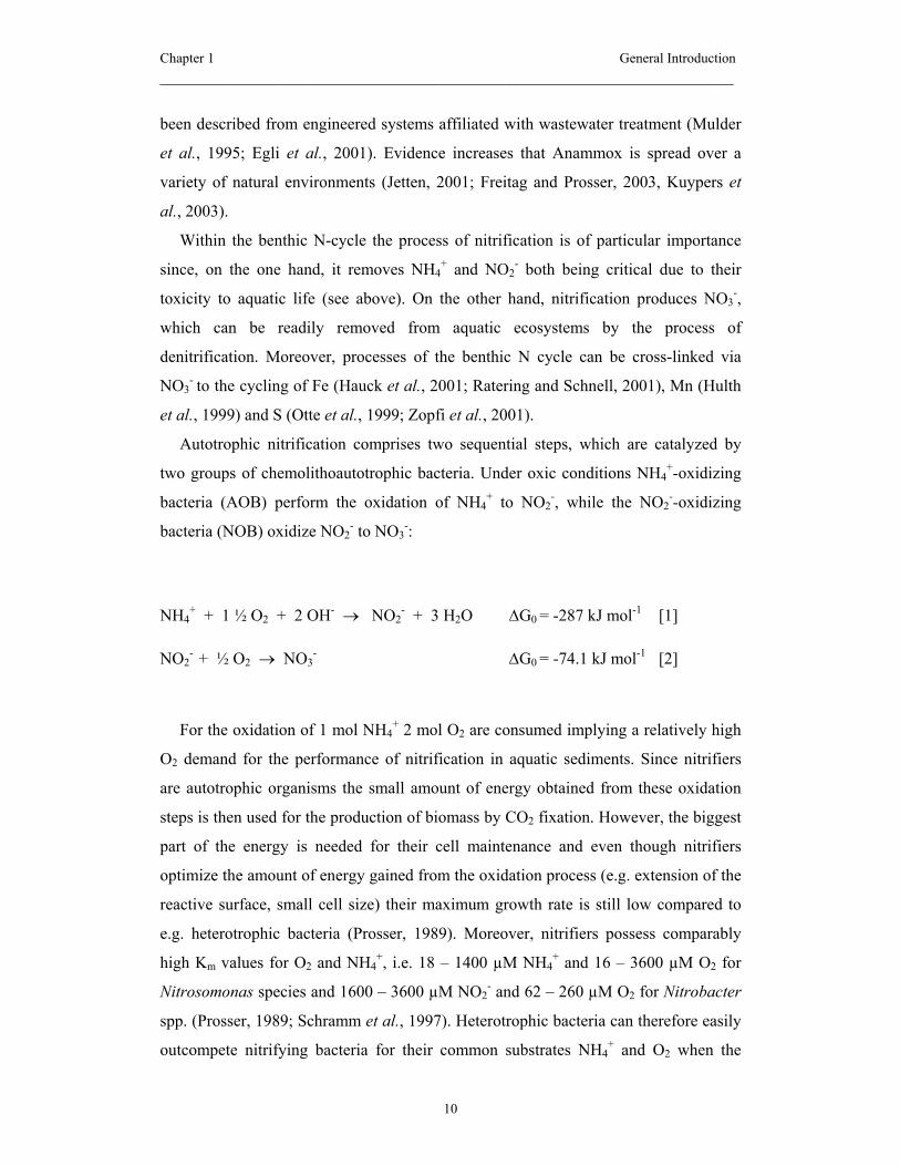
been described from engineered systems affiliated with wastewater treatment (Mulder
et al., 1995; Egli et al., 2001). Evidence increases that Anammox is spread over a
variety of natural environments (Jetten, 2001; Freitag and Prosser, 2003, Kuypers et
al., 2003).
Within the benthic N-cycle the process of nitrification is of particular importance
since, on the one hand, it removes NH4+ and NO2
- both being critical due to their
toxicity to aquatic life (see above). On the other hand, nitrification produces NO3-,
which can be readily removed from aquatic ecosystems by the process of
denitrification. Moreover, processes of the benthic N cycle can be cross-linked via
NO3- to the cycling of Fe (Hauck et al., 2001; Ratering and Schnell, 2001), Mn (Hulth
et al., 1999) and S (Otte et al., 1999; Zopfi et al., 2001).
Autotrophic nitrification comprises two sequential steps, which are catalyzed by
two groups of chemolithoautotrophic bacteria. Under oxic conditions NH4+-oxidizing
bacteria (AOB) perform the oxidation of NH4+ to NO2
-, while the NO2--oxidizing
bacteria (NOB) oxidize NO2- to NO3
-:
NH4+ + 1 ½ O2 + 2 OH- → NO2
- + 3 H2O ∆G0 = -287 kJ mol-1 [1]
NO2- + ½ O2 → NO3
- ∆G0 = -74.1 kJ mol-1 [2]
For the oxidation of 1 mol NH4+ 2 mol O2 are consumed implying a relatively high
O2 demand for the performance of nitrification in aquatic sediments. Since nitrifiers
are autotrophic organisms the small amount of energy obtained from these oxidation
steps is then used for the production of biomass by CO2 fixation. However, the biggest
part of the energy is needed for their cell maintenance and even though nitrifiers
optimize the amount of energy gained from the oxidation process (e.g. extension of the
reactive surface, small cell size) their maximum growth rate is still low compared to
e.g. heterotrophic bacteria (Prosser, 1989). Moreover, nitrifiers possess comparably
high Km values for O2 and NH4+, i.e. 18 – 1400 µM NH4
+ and 16 – 3600 µM O2 for
Nitrosomonas species and 1600 – 3600 µM NO2- and 62 – 260 µM O2 for Nitrobacter
spp. (Prosser, 1989; Schramm et al., 1997). Heterotrophic bacteria can therefore easily
outcompete nitrifying bacteria for their common substrates NH4+ and O2 when the

Chapter 1 General Introduction _____________________________________________________________________
11
availability is low and organic C is not limiting (Strauss and Lamberti, 2000, 2002)
(Verhagen et al., 1992).
Most commonly isolated and described AOB and NOB are the genera
Nitrosomonas and Nitrobacter, respectively. Since methods are now available to detect
and quantify nitrifiers in their natural environment (reviewed in (Schramm and
Amann, 1999) evidence increases that other nitrifying organisms like the genera
Nitrosospira and Nitrospira might be of greater importance in some environments, e.g.
at low concentrations of NH4+, NO2
- and O2 (Schramm et al., 1998; Okabe et al., 1999;
Schramm et al., 2000).
In sediments O2 is often depleted within the first few millimeters of the surface.
Thus nitrification is limited to a very narrow zone. Nitrifiers therefore face aggravating
conditions, i.e. they have to deal with a limited supply of O2 and they have to compete
with metabolically superior microorganisms for their sources O2 and NH4+. Compared
to other functional groups of bacteria like sulfate reducers, which can make up to
around 9% of the total bacterial community of marine sediments (Ravenschlag et al.,
2000), cell numbers of nitrifying bacteria in sediments are relatively low. Cell numbers
determined with the most probable number method range from 103 – 106 ml-1 making
up only around 1% of total bacterial cells (Smorczewski and Schmidt, 1991; Bianchi
and Lefevre, 1999; Whitby et al., 2001). It is therefore striking that the contribution of
nitrifying bacteria to the microbial N conversions and to the overall O2 consumption in
sediments is so substantial. Nitrification can consume up to 90% of the total
sedimentary O2 consumption (see own data, Chapter 4) and nitrification rate is often
comparable to the rate of denitrification (Lorenzen et al., 1998; Meyer et al., 2001).
For the fine scale investigation of nitrification measuring tools are needed that
allow a sufficiently high spatial resolution for the exact determination of nitrification
rates and for the identification and quantification of the nitrifying community. For this
purpose, the combination of selective microsensors with fluorescence in situ
hybridization provides a useful tool to target function and structure of nitrification also
in sediments (Amann and Kühl, 1998). This approach has already been successfully
applied in biofilms and aggregates of wastewater treatment plants (Schramm et al.,
1997; Okabe et al., 1999; Gieseke et al., 2001).
Chapter 1 General Introduction _____________________________________________________________________
12
Investigating the ecology of nitrifying bacteria with microsensors
and FISH
The activity of nitrifying bacteria as determined with microsensors
Because of their small size microsensors are useful tools to investigate processes
occurring at a very small scale as it is the case in microbial mats, biofilms, and
sediments (de Beer, 1999), where diffusion and reaction processes create steep
physicochemical gradients within µm distances. The application of microsensors
minimizes the disturbance of these gradients, of the three dimensional organization of
microbial communities, and of other microenvironmental conditions such as boundary
layers, diffusion and flow patterns on the microscale (Amann and Kühl, 1998).
Moreover, the fine spatial resolution of the microsensors allows the functional
separation of different layers and hence the spatially differentiated interpretation of
processes that occur in close vicinity (e.g. nitrification and denitrification). Thus, the
microsensor technique has great advantages compared to other methods of
investigating the processes of the benthic N-cycle: Mass balances (Hansen et al., 1981;
Rysgaard et al., 1995; Van Luijn et al., 1999; Christensen et al., 2000), potential
nitrification rates (Hansen et al., 1981) (Henriksen et al., 1993) (Vouve et al., 2000),
and 15N tracer techniques (Rysgaard et al., 1994; Rysgaard et al., 1995; Christensen et
al., 2000) (Usui et al., 2001) (Vouve et al., 2000) do either average across several
microbial processes or they do not measure in situ activities. Microsensor
measurements on the other hand only give net conversion rates and each of the
alternative methods bears also some advantages, e.g. 15N techniques are useful for the
investigation of the coupled nitrification-denitrification since the source of NO3- can be
differentiated properly (Nielsen, 1992).
In the present thesis microsensors for O2 (Revsbech, 1989), NH4+, and NO3
- (de
Beer and Sweerts, 1989; de Beer et al., 1991) have been applied to determine the
activity of nitrifying bacteria in freshwater sediments (compare reaction equations [1]
and [2]).

Chapter 1 General Introduction _____________________________________________________________________
13
The microsensors for O2, NH4+, and NO3
- belong to two different types of
electrochemical sensors that are commonly used for ecological studies: (1)
potentiometric electrodes (e.g. for H+, Ca2+, NH4+, NO2
-, and NO3-) measure an
electrochemical potential difference across a liquid membrane and (2) amperometric
electrodes (e.g. for O2, N2O, and H2S) measure the current that is generated by the
reduction of the respective substrate at the cathode of the sensor.
Potentiometric electrodes for NH4+ and NO3
-. These electrodes contain an ion
exchanging liquid membrane within the tip of the sensor (liquid ion exchanger – LIX).
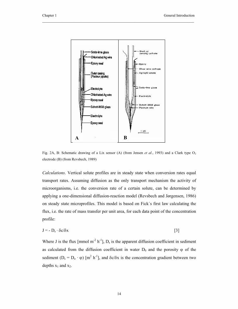
LIX sensors can be constructed with a capillary tip of 1 – 10 µm thus allowing an
equivalent spatial resolution of the measurements. A typical LIX sensor is sketched in
Fig. 2 A. Both, NH4+ and NO3
- microsensors have been successfully used in freshwater
sediments (de Beer and Sweerts, 1989; de Beer et al., 1991). Their sensitivity towards
interference from other ions, such as K+ for NH4+ and HCO3
- and Cl- for NO3-,
impedes their use in saline waters. Even in freshwater sediments the accuracy of
vertical NO3- profiles can be affected by increasing concentrations of HCO3
- with
depth (Kühl and Revsbech, 2001). To overcome this problem a biosensor for NO3- has
recently been developed that allows fine scale measurements in marine environments
and in biologically reactive environments with high concentrations of HCO3- (Larsen
et al., 1997).
Amperometric Clark type O2 electrodes. The Clark type O2 sensor is the best known
gas microsensor (Revsbech, 1989). This sensor consists of a gold-coated cathode,
which is situated behind a silicone membrane. O2 diffusing through the gas-permeable
membrane is reduced at the cathode when a polarization voltage is impressed. O2
microsensors can be constructed with a tip size of 1-100 µm, they are further
characterized by the absence of interferences, linear calibration curves, fast response
times (≥ 0.1 s) and low sensitivity to stirring of the external medium (Kühl and
Revsbech). An O2 microsensor with a guard cathode is sketched in Fig. 2 B.
Chapter 1 General Introduction _____________________________________________________________________
14
Fig. 2A, B: Schematic drawing of a Lix sensor (A) (from Jensen et al., 1993) and a Clark type O2
electrode (B) (from Revsbech, 1989)
Calculations. Vertical solute profiles are in steady state when conversion rates equal
transport rates. Assuming diffusion as the only transport mechanism the activity of
microorganisms, i.e. the conversion rate of a certain solute, can be determined by
applying a one-dimensional diffusion-reaction model (Revsbech and Jørgensen, 1986)
on steady state microprofiles. This model is based on Fick´s first law calculating the
flux, i.e. the rate of mass transfer per unit area, for each data point of the concentration
profile:
J = - Ds · δc/δx [3]
Where J is the flux [mmol m-2 h-1], Ds is the apparent diffusion coefficient in sediment
as calculated from the diffusion coefficient in water D0 and the porosity ϕ of the
sediment (Ds = Do · ϕ) [m2 h-1], and δc/δx is the concentration gradient between two
depths x1 and x2.
A B
Chapter 1 General Introduction _____________________________________________________________________
15
The fluxes calculated for the single points in a concentration profile can be used for
the calculation of local volumetric conversion rates [mmol m-3 h-1] after (de Beer,
1999):
R = Jab – Jbc / (0.5(xa + xb) – 0.5(xb + xc)) [4]
The calculation procedure thus comprises the differentiation of the concentration
profile using adjacent values. As a result a vertical profile of local conversion rates is
obtained. The subsequent averaging of consecutive conversion values within a certain
zone can partly reduce noise that may result from the differentiation procedure and
which can sometimes strongly affect the calculated conversion rates.
The nitrifying activity within a given sediment can be expressed as: (i) the local
volumetric conversion rates in single layers of the nitrification zone and (ii) the bulk
areal conversion rate in mol m-2 h-1 within this zone as calculated by integration of
volumetric conversion rates over the considered depth interval, i.e. the oxic layer. By
integration over the NO3- consumption zone within the anoxic layer rates within in the
anoxic layer the areal rate of denitrification activity can be determined. Thereby the
quantitative importance of nitrification and denitrification for the budget of N
conversions in the sediment can be estimated, i.e. net influx to or efflux of NO3- from
the sediment. Alternatively, the total flux of a solute into or out of the sediment is
represented by the linear concentration gradient within the diffusive boundary layer.
Identity and distribution of nitrifying bacteria: the application of FISH
Hybridization of intact cells with fluorescently labeled, 16S rRNA targeted
oligonucleotides (fluorescence in situ hybridization, FISH) has been introduced to
microbial ecology approximately a decade ago as an accurate method of characterizing
microbial populations in their in situ environment (De Long et al., 1989; Amann et al.,
1990). The ribosomal 16S rRNA is considered a powerful target because (i) ribosomes
are a common feature of all organisms, (ii) ribosomes are of homologous origin and
show functional constancy and hence the phylogeny of the organisms can be
reconstructed based on the 16S rRNA molecules, (iii) the differentiation at any
taxonomic level, i.e. from species to domains, is allowed because of a different degree
of conservation within the rRNA molecules, and (iv) 16S rRNA sequences have been
Chapter 1 General Introduction _____________________________________________________________________
16
described for many species and are available from public databases (e.g.
http://www.ncbi.nlm.nih.gov/) (Schramm and Amann, 1999).
The method of FISH is part of the so called full cycle rRNA approach which
comprises the extraction of DNA from environmental samples, the amplification of
rDNA using PCR and specific primers, the cloning and sequencing of the amplified
rDNA fragments and the subsequent design of specific probes to enumerate whole
fixed cells in the original sample by in situ hybridization (Olsen et al., 1986; Amann et
al., 1995). Thereby, quantitative information on the community structure can be
obtained. Oligonucleotide probes that are commercially available are either provided
with a 32P label or with a fluorescent dye (Cy3, Cy5, Fluorescein). Radio labeled
probes are commonly used for hybridization of extracted and immobilized rRNA (dot
blot hybridization). The relative abundance of a certain 16S rRNA sequence is then
expressed as the fraction of total 16S rRNA in the sample (Schramm et al., 1997). The
dot blot hybridization has been applied for the investigation of sulfate reducing
bacteria in arctic sediments in which a low cellular RNA content was expected
(Ravenschlag et al., 2000). The hybridization of intact cells with fluorescently labeled
oligonucleotides (fluorescence in situ hybridization (FISH)) enables the detection of a
certain rRNA sequence within morphologically intact cells in situ, i.e. in their natural
environment (Schramm et al., 1997). Data about in situ distribution and abundance of
the cells are thereby complemented by information on the morphology and on the
spatial organization of the cells. Since a low cellular rRNA content limits the
detectability of cells and thus the application of FISH in environmental samples the
FISH method has been extended by the use of multilabeled oligonucleotides (DeLong
et al., 1999) or by the enzymatic amplification of the hybridization signal (Schönhuber
et al., 1997).
The FISH technique constitutes a major advantage compared to classical methods
like the most probable number technique or viable plate counts since it enables the in
situ detection and a reliable quantification of bacterial populations. In contrast, the use
of isolation and cultivation techniques for quantitative purposes is limited by the “great
plate count anomaly” (Staley and Konopka, 1985)): Species will be overlooked for
which the cultivation conditions are not suitable or which have entered a non-
culturable stage. Thus, by the application of cultivation-dependent techniques the

Chapter 1 General Introduction _____________________________________________________________________
17
actual size and diversity of microbial communities can be underestimated dramatically
(Amann et al., 1995).
An important precondition for the in situ detection of a larger functional group of
microorganisms like nitrifiers is their monophyletic origin allowing the detection of
group specific signatures within the rRNA (Schramm, in press). The nitrifying bacteria
are at present grouped into two monophyletic lineages of NH4+-oxidizing bacteria and
four phylogenetically distinct groups of NO2--oxidizing bacteria. NH4
+-oxidizing
bacteria comprising the genera Nitrosospira, Nitrosovibrio, Nitrosolobus,
Nitrosomonas and also the species Nitrosococcus mobilis are closely related organisms
within the β subclass of the Proteobacteria. Two other species of this genus
Nitrosococcus oceani and Nitrosococcus halophilus are located within the γ subclass
of the Proteobacteria. NO2--oxidizing bacteria were assigned to the α, γ, and δ
subclasses of Proteobacteria (genera Nitrobacter, Nitrococcus, and Nitrospina). A
distinct phylum close to the δ subclass of the Proteobacteria comprises the genus
Nitrospira (Koops and Pommerening-Roser, 2001). Based on 16S rRNA sequences of
nitrifying bacteria a comprehensive set of probes targeting these nitrifiers has been
developed and tested for FISH (Schramm, in press).
Alternatively to the 16S rRNA approach NH4+-oxidizing bacteria can be identified
by targeting the functional gene encoding for the NH4+ monooxygenase (AmoA) which
catalyzes the first step of NH4+ oxidation (McTavish et al., 1993; Rotthauwe et al.,
1997; Phillips et al., 2000). However, the enzyme catalyzing NO2--oxidation (NO2
--
oxidoreductase, NOR) has so far only been detected and quantified by immunological
methods (Spieck et al., 1996; Bartosch et al., 1999; Maron et al., 2003).
In the present thesis fluorescence in situ hybridization with monolabeled probes
was used to address the identification and in situ distribution of nitrifying bacteria in
several freshwater sediments.
Influence of sediment disturbances on nitrification
Benthic nitrification is controlled by a number of physico-chemical factors which
include temperature, pH, salinity, O2, NH4+ and dissolved CO2 concentrations (Herbert,
1999) and the presence of inhibitory compounds like H2S (Bagarinao, 1992).
Moreover burrowing macrofauna can have profound effects on the performance of

Chapter 1 General Introduction _____________________________________________________________________
18
nitrification (Pelegri and Blackburn, 1994, 1995a; Rysgaard et al., 2000; Svensson et
al., 2001). Mixing the sediment artificially, as it is commonly used for factorial
sediment experiments (van de Bund et al., 1994; Svensson and Leonardson, 1996;
Strauss and Lamberti, 2002) is also thought to influence nitrification activity
(Svensson et al., 2001). Sediments in sheltered areas and streams are influenced by
movements of the overlying water body, which can have enormous effects on the
sediment loading and hence the stratification of microbiota within the sediment (Wulff
et al., 1997). In such dynamic habitats motile organisms can have advantages
compared to non-motile forms since they can easily recolonize newly formed surfaces
(Wulff et al., 1997). Moreover, nitrifiers may be severely handicapped under such
conditions due to their low competitiveness with other organisms, i.e. their slow
growth and low substrate affinities (Prosser, 1989; Schramm et al., 1997).
Natural disturbance: sediment bioturbation
A variety of investigations have been carried out addressing the influence of
bioturbation and bioirrigation on N mineralization in marine (Pelegri and Blackburn,
1994; Gilbert et al., 1998; Bartoli et al., 2000), estuarine (Pelegri et al., 1994; Pelegri
and Blackburn, 1995b; Rysgaard et al., 1995), and freshwater sediments (Pelegri and
Blackburn, 1996; Svensson et al., 2001; Stief and de Beer, 2002). Macroinvertebrates
living in or at the surface of sediments intensively act on their physical, chemical, and
microbiological environment. While feeding on sediment particles and by their
locomotive activity the animals actively rework the sediment thereby translocating
sediment particles and associated microorganisms to more or less favorable
microenvironments (Matisoff and Wang, 2000). The construction of burrows, which
can reach deep into anoxic sediment layers, and the ventilation of these burrows lead
to an extension of the oxidized sediment surface and to a 3-D microstratification as a
consequence of steep gradients around the burrow walls (Kristensen et al., 1985;
Fenchel, 1996). Moreover, the animals may stimulate the generation of suitable
organic substrates for bacteria by ingestion, digestion and the subsequent excretion of
dissolved compounds which can be used for bacterial growth (Goedkoop et al., 1997).
By the use of common methods 15N tracer techniques and mass balance studies
bioirrigation of sediments was often shown to stimulate the activity of bacteria
Chapter 1 General Introduction _____________________________________________________________________
19
involved in benthic N conversions (Kristensen et al., 1985; Pelegri et al., 1994;
Svensson, 1997; Gilbert et al., 1998; Bartoli et al., 2000). The deposit feeding of
several animals on the other hand, i.e. the ingestion and digestion of particles from the
sediment surface causes significant grazing losses of particle-associated bacteria
(Johnson et al., 1989; Rouf and Rigney, 1993; Plante, 2000).
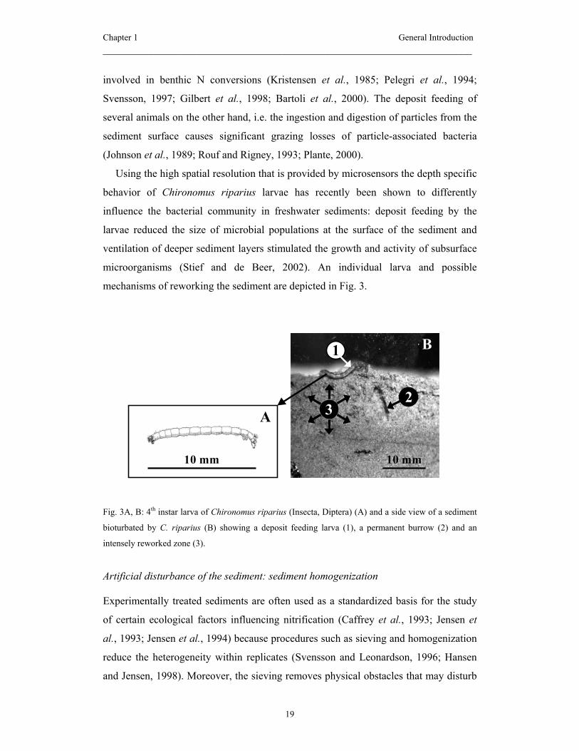
Using the high spatial resolution that is provided by microsensors the depth specific
behavior of Chironomus riparius larvae has recently been shown to differently
influence the bacterial community in freshwater sediments: deposit feeding by the
larvae reduced the size of microbial populations at the surface of the sediment and
ventilation of deeper sediment layers stimulated the growth and activity of subsurface
microorganisms (Stief and de Beer, 2002). An individual larva and possible
mechanisms of reworking the sediment are depicted in Fig. 3.
Fig. 3A, B: 4th instar larva of Chironomus riparius (Insecta, Diptera) (A) and a side view of a sediment
bioturbated by C. riparius (B) showing a deposit feeding larva (1), a permanent burrow (2) and an
intensely reworked zone (3).
Artificial disturbance of the sediment: sediment homogenization
Experimentally treated sediments are often used as a standardized basis for the study
of certain ecological factors influencing nitrification (Caffrey et al., 1993; Jensen et
al., 1993; Jensen et al., 1994) because procedures such as sieving and homogenization
reduce the heterogeneity within replicates (Svensson and Leonardson, 1996; Hansen
and Jensen, 1998). Moreover, the sieving removes physical obstacles that may disturb
10 mm
1
10 mm
23
B
A
Chapter 1 General Introduction _____________________________________________________________________
20
microsensor measurements. Nevertheless, the microbial stratification within the
sediment is revoked during homogenization and sedimentary processes can be
destabilized. During the settlement of homogenized sediment in experimental
containers the particulate organic matter may accumulate at the sediment surface and
thereby favor the thriving of heterotrophic bacteria (Stief et al., accepted). Sieving and
homogenization were shown to initially reduce microbial growth (Findlay et al., 1990)
and especially slowly growing organisms as nitrifying bacteria (Prosser, 1989) are
assumed to recover more slowly from disturbance than other microorganisms
(Svensson et al., 2001). However, the sediments are allowed to restratify and stabilize
prior to the start of the actual investigation (Rysgaard et al., 1994; Pelegri and
Blackburn, 1995a; Svensson et al., 2001; Stief et al., 2002). Steady state in the
respective sediment is assumed to have reestablished when fluxes of the nutrients of
interest have stabilized (Rysgaard et al., 1994; Svensson et al., 2001; Stief et al.,
2002). To clarify the potential problem of biased nitrification in such sediment
incubations a precise investigation of the activity and distribution of nitrifiers is thus
important.

Chapter 1 Introduction: Thesis outline _____________________________________________________________________
21
Thesis outline
Sediment-dwelling macroinvertebrates influence benthic microbial processes by
various mechanisms, e.g. by feeding, locomotive activities and by the construction of
burrow-like structures. The process of nitrification plays a crucial role in freshwater
sediments because it removes NH4+ and NO2
- both being potentially toxic to aquatic
live. Against the background of high loading of freshwater systems with NH4+ and
NO2- bioturbated sediments bear ecological potentials as sinks for inorganic N
compounds. The influence of bioturbation and bioirrigation on nitrification in
freshwater sediments was therefore the main focus of this thesis. Special attention was
given to the burrows of the investigated invertebrates where a high microbial activity
(burrows as “microbial hot spots”) and intense animal-microbe interactions are
assumed.
Animal-related changes in nitrification might be based on changes in the structure and
function of the nitrifying community, i.e. changes of the taxonomic composition,
abundance, distribution and the activity of functional groups. The application of in situ
techniques, i.e. fluorescence in situ hybridization (FISH) and microsensors allowed the
determination of these parameters independent from cultivation. Moreover, the high
spatial resolution of the two methods is indispensable, as nitrification is restricted to a
very narrow zone of the sediment surface (around 2 mm). Depending on the concrete
aim of single experiments FISH and microsensors have been supplemented by
additional methods of microbial ecology (e.g. determination of nitrification potentials,
total fluxes, and exoenzyme activities).
The objectives of this thesis were to i) evaluate the potential of the combined
application of microsensors and FISH in freshwater sediments, ii) to obtain first
insights into the in situ abundance, distribution and activity of nitrifying bacteria in
model freshwater sediment, iii) to investigate burrows of macroinvertebrates as
potential hot spots of nitrification and other mechanisms by which nitrifying bacteria
are linked to bioturbating and bioirrigating macroinvertebrates.
In the following a short summary of the experiments that are included in this thesis is
given (Experiment 1 – 3). Moreover, a short summary of additional experiments,
which resulted in manuscripts with a co-authorship is given. It is complemented by a
Chapter 1 Introduction: Thesis outline _____________________________________________________________________
22
more detailed description of experimental parts that were accompanied by some
difficulties (Experiment 4 –6).
The first experiment (Chapter 2) was dedicated to evaluate the potentials and
limitations of the combination of the two in situ methods (FISH and microsensors) for
the investigation of nitrifying bacteria in freshwater sediments. The combined
approach proved to be applicable to freshwater model sediment. The two functional
groups of nitrifiers, i.e. NH4+- and NO2
--oxidizing bacteria (AOB and NOB) were
quantified independent from cultivation by FISH and their in situ metabolic activity
was successfully measured with microsensors. For the first time, Nitrospira spp. was
identified as the dominant NO2--oxidizing bacterium in a freshwater sediment. With
the detection of Nitrospira spp. in various other environments this stresses the
importance of Nitrospira spp. and not Nitrobacter spp. for in situ NO2--oxidation.
In the second experiment (Chapter 3) the experimental approach of the first
experiment was applied to compare model sediment (´manipulated´) with untreated
(´natural´) stream sediment. Since the pre-treatment of sediment is commonly
accomplished prior to factorial experiments the impact of this artificial disturbance on
the performance of nitrification was investigated. Using FISH and microsensors the
activity and distribution of nitrifying bacteria was successfully determined:
measurements in the natural sediment revealed comparably low activities and
abundances of AOB and NOB and were considered as a snap-shot in a probably
dynamic and heterogeneous environment. The manipulated sediment revealed higher
activities and abundances of nitrifiers compared to the natural sediment, which was
ascribed to a better substrate availability within the uppermost sediment layer.
Moreover, we observed that in the manipulated sediment NOB persisted longer than
AOB did under unsuitable conditions, i.e. at depths where the substrate and O2 supply
was low.
In the third experiment (Chapter 4) FISH and microsensors were used to address the
impact of sediment-dwelling Chironomus riparius larvae (Insecta, Diptera) on
nitrification in two freshwater sediments with different organic content. C. riparius
larvae are known to affect microbial processes by their movements, their ventilation
behavior and their feeding. With this they change the O2 microdistribution within the
Chapter 1 Introduction: Thesis outline _____________________________________________________________________
23
sediment and the distribution of particle-associated microorganisms. Nitrification was
thus expected to be affected by C. riparius. The experiment revealed that the sediment
type was decisive for the larval behavior and thus for the animal impact on benthic
nitrification: in the organic poor sediment the extensive sediment reworking and the
feeding of the larvae reduced the nitrification activity measured therein. In contrast, in
the organic rich sediment net nitrification was neither influenced in the bulk sediment
or due to the presence of burrows, which the larvae constructed in this sediment type.
The abundances of AOB and NOB were not influenced by the larvae neither in the
organic-poor nor in the organic-rich sediment.
The methodological design and the results of experiment 1 – 3 have been submitted for
publication.
Why reduced nitrification activity was not reflected by reduced abundances of the
nitrifiers (result of experiment 3) was investigated in experiment 4. It was assumed
that the larval feeding (gut passage of particles and associated bacteria) reduced the
metabolically active part of the nitrifying population but that the ribosome content of
this inactive fraction was still high enough for detection with FISH. Thus, an
experiment was conducted to quantify metabolically active vs. inactive nitrifiers on the
fecal pellets of the larvae. Microsensor profiles were successfully recorded at the
surface of the fecal pellets (300 µm in diameter) to determine the nitrification activity.
For the visualization of metabolically active cells FISH was combined with a live-
dead-stain, which was supposed to allow a microscopic differentiation of metabolic
active vs. inactive cells of a certain nitrifier population. The live-dead-stain which was
chosen is based on the esterase substrate cFDA yielding the fluorescent
carboxyfluorescein (cF) upon hydrolysis (Bunthof et al. 2001). When applied
separately each stain (cF and FISH/DAPI) yielded signals, which could be quantified
by epifluorescence microscopy. However, cells of the killed control and living cells
showed the same cF signal intensity. Moreover, cF and DAPI signals were
overlapping, meaning that cF containing objects could not be undoubtedly identified as
bacterial cells. In future investigations metabolic activity of single cells could be
targeted by showing DNA synthesis in general (Pernthaler et al. 2002a) or by
monitoring the precursor rRNA of certain bacterial populations (Oerther et al. 2000,
Schmid et al. 2001).
Chapter 1 Introduction: Thesis outline _____________________________________________________________________
24
In Experiment 5 the contribution of single burrows of the mayfly Ephoron virgo
(Insecta, Ephemeroptera) to microbial mineralization and to the N cycle was
investigated. Investigations with Ephoron virgo larvae were made in a bypass flow
channel of the river Rhine (Ecological lab of the University of Cologne). E. virgo
burrows reach up to 10 cm deep into the sediment and are typically lined with fine
particulate organic matter that is trapped inside the burrows due to permanent
ventilation of the burrow lumen (E. virgo as filter-feeder). For experimental purposes
larvae from lab cultures had been inserted into the sediment of the flow channel. The
burrows were visible through the walls of the channel thus allowing microsensor
measurements not only within the lumen but also within the walls of the burrows.
Measurements showed that concentrations of O2 and NO3- within the lumen are nearly
as high as in the water column. Nitrification rates as well as denitrification rates were
distinctly higher in the walls than in the surrounding sediment. Also exoenzyme
activities were higher than in the burrow-free sediment. The results showed that E.
virgo larvae changed the physico-chemical and microbial properties of the sediment.
With this they could have a considerable impact on large scale biogeochemical
processes in the sediments of the river Rhine. The results of this part of the experiment
have been submitted for publication.
In a supplementary part, data on the microbial activity along the burrow walls were
supposed to be complemented by the identification and quantification of total bacterial
numbers (DAPI) and nitrifiers in particular (FISH/DAPI). In order to get such
´microbial imprints` of the burrow linings a variety of approaches was tested. Small
sediment cores containing one branch of a burrow were taken and were sliced
horizontally. The aim of this approach was to get thin slices or an imprint of a
sediment cross-section on slides or filters which could be hybridized and then
evaluated microscopically. Though clearly visible as distinct compartments, the
burrow walls were very fragile and their sampling turned out to be difficult, i.e.
sampling resulted in a collapse of the burrows when the cores were sliced. Freezing of
the whole cores was supposed to overcome this problem. The perfusion of the samples
with a fixative prior to the freezing (to maintain an intact cell structure) was likewise
impeded by the fragility of the sediment structure. In a second approach several rows
of microscopic slides had been exposed at the wall of the transparent flow channel at
the time when the incubation had started. At the time of the activity measurements

Chapter 1 Introduction: Thesis outline _____________________________________________________________________
25
(after 3 months) several burrows were visible behind the slides and it was hoped that
after the recovering, fixation and embedding of the slides and of the attached particles
and after FISH the burrow linings and bacteria therein would be visible by
microscopy. This succeeded with one of the slides, which bore a biofilm with the
shape of two opposite lines. Unfortunately, no cells were visible within this biofilm.
This was either due to washout during the treatment of the slides or because the
surface of the slides were not suitable for adhesion of sedimentary bacteria.
Despite the problem of getting the microbial imprint of the burrow walls also the
quantification of bacteria in bulk sediment (treatment of samples as described in the
method sections of the manuscripts (Chapters 2 – 4)) was difficult. This was due to
low signal intensities and misleading DAPI signals (a lot of yellow fluorescent objects
were interfering). Low signal intensities could not be overcome by the application of
HRP probes (Pernthaler et al. 2002a). Finally, the samples could not be evaluated.
Experiment 6 was aimed to get microsensor profiles and microbial imprints of the
burrow linings of Chironomus riparius and Chironomus plumosus larvae. Both can
densely colonize (highly eutrophic) sediments of small streams and lakes. The
incubations were made in slender cuvette-form aquaria. Microsensor measurements
within the lumen allowed calculations of N conversions within this sediment
compartment. In contrast, high concentration changes and the fragility of the burrow
walls hampered the recording of steady state profiles within the burrow walls.
Microscopic slides that had been exposed to the sediment did not bear biofilms of the
burrow linings. Only the water-exposed part of the slides was densely colonized by
bacteria. To correlate measured activities of the lumen with bacterial abundances it
was necessary to compare cores with and without burrows (method see Chapter 4).
The data were supplemented by measuring the nitrification potentials of sediment
samples, which integrate over the (potential) activity and abundance of nitrifiers. In
addition to the diffusive fluxes of NH4+ and NO3
- measured with the microsensors,
total fluxes were measured in order to determine the effective contribution of the
burrows to the exchange of N between water and sediment.

Chapter 1 Introduction: Thesis outline _____________________________________________________________________
26
A manuscript on the microsensor measurements within Chironomus burrows,
nitrification potentials, as well as on diffusive and total fluxes in the sediments will be
written.
Chapter 1 Introduction: References _____________________________________________________________________
27
References
Amann, R., and Kühl, M. 1998. In situ methods for assessment of microorganisms and
their activities. Current Opinion in Microbiology 1: 352-358.
Amann, R., Ludwig, W., and Schleifer, K.-H. 1995. Phylogenetic identification and in
situ detection of individual microbial cells without cultivation. Microbiological
Reviews 59: 143-169.
Amann, R.I., Krumholz, L., and Stahl, D.A. 1990. Fluorescent oligonucleotide probing
of whole cells for determinative, phylogenetic and environmental studies in
microbiology. Journal of Bacteriology 172: 762-770.
Bagarinao, T. 1992. Sulfide as an environmental factor and toxicant: tolerance and
adaptions in aquatic organisms. Aquatic Toxicology 24: 21-62.
Bartoli, M., Nizzoli, D., Welsh, D.T., and Viaroli, P. 2000. Short-term influence of
recolonisation by the polycheate worm Nereis succinea on oxygen and nitrogen
fluxes and denitrification: a microcosm simulation. Hydrobiologia 431: 165-
174.
Bartosch, S., Wolgast, I., Spieck, E., and Bock, E. 1999. Identification of nitrite-
oxidizing bacteria with monoclonal antibodies recognizing the nitrite
oxidoreductase. Applied and Environmental Microbiology 65: 4126-4133.
Bianchi, M., and Lefevre, F.D. 1999. Regulatin of nitrification in the land-ocean
contact area of the Rhone River plume (NW Mediterranean). Aquatic Microbial
Ecology 18: 301-312.
Bock, E., Schmidt, I., Stüven, R., and D., Z. 1995. Nitrogen loss caused by denitrifying
Nitrosomonas cells using ammonium of hydrogen as electron donors and nitrite
as electron acceptor. Archives of Microbiology 163: 16-20.
Bruning-Fann, C.S., and Kaneene, J.B. 1993. The effects of nitrate, nitrite and N-
nitroso compounds on human health: a review. Veterinary and Human
Toxicology 35: 521-538.
Chapter 1 Introduction: References _____________________________________________________________________
28
Bunthof ,C.J., Bloemen, K., Breeuwer, P., Rombouts, F.M., and Abee, T. 2001. Flow
cytometric assessment of viability of lactic acid bacteria. Applied and
Environmental Microbiology 67: 2326-2335.
Caffrey, J.M., Sloth, N.P., Kaspar, H.F., and Blackburn, T.H. 1993. Effect of organic
loading on nitrification and denitrification in a marine sediment microcosm.
FEMS Microbiology Ecology 12: 159-167.
Christensen, P.B., Rysgaard, S., Sloth, N.P., Dalsgaard, T., and Schwaerter, S. 2000.
Sediment minaralization, nutrient fluxes, denitrification and dissimilatory
nitrate reduction to ammonium in an eastuarine fjord with sea cage trout farms.
Aquatic Microbial Ecology 21: 73-84.
Dalal, R.C., Wang, W.J., Robertson, G.P., and Parton, W.J. 2003. Nitrous oxide
emission from Australian agricultural lands and mitigation options: a review.
Australian Journal of Soil Research 41: 165-195.
de Beer, D. 1999. Use of micro-electrodes to measure in situ microbial activities in
biofilms, sediments, and microbial mats. In Molecular Microbial Ecology
Manual. Akkermans, A.D.L., van Elsas, J.D., de Bruijn, F.J. (eds). Kluwer
Academic Press, pp. 1-23.
de Beer, D., and Sweerts, J.-P.R.A. 1989. Measurement of nitrate gradients with an
ion-selective microelectrode. Analytica Chimica Acta 219: 351-356.
de Beer, D., Sweerts, J.-P.R.A., and van den Heuvel, J.C. 1991. Microelectrode
measurement of ammonium profiles in freshwater sediments. FEMS
Microbiology Ecology 86: 1-6.
DeLong, E.F., Wickham, G.S., and Pace, N. 1989. Phylogenetic stains: ribosomal
RNA-based probes for the identification of single cells. Science 243: 1360-
1363.
DeLong, E.F., Taylor, L.T., Marsh, T.L., and Preston, C.M. 1999. Vizualization and
enumeration of marine planktonic bacteria by using polyribonucleotide probes
and fluorescence in situ hybridization. Applied and Environmental
Microbiology 65: 5554-5563.
Chapter 1 Introduction: References _____________________________________________________________________
29
Egli, K., Fanger, U., Alvarez, P.J.J., Siegrist, H., van der Meer, J.R., and Zehnder,
A.J.B. 2001. Enrichment and characterization of an anammox bacterium from a
rotating biological contactor treating ammonium-rich leachate. Archive of
Microbiology 175: 198-207.
Fenchel, T. 1996. Worm burrows and oxic microniches in marine sediments. 1. Spatial
and temporal scales. Marine Biology 127: 289-295.
Findlay, R.H., Trexler, M.B., Guckert, J.B., and White, D.C. 1990. Laboratory study of
disturbance in marine sediments: response of a microbial community. Marine
Ecology Progress Series 62: 121-133.
Freitag, T.E., and Prosser, J.I. 2003. Community structure of ammonia-oxidizing
bacteria within anoxic marine sediments. Applied and Environmental
Microbiology 69: 1359-1371.
Ghosh, S., Majumdar, D., and Jain, M.C. 2003. Methane and nitrous oxide emissions
from an irrigated rice of North India. Chemosphere 51: 181-195.
Gieseke, A., Purkhold, U., Wagner, M., Amann, R., and Schramm, A. 2001.
Community structure and activity dynamics of nitrifying bacteria in a
phosphate-removing Biofilm. Applied and Environmental Microbiology 67:
1351-1362.
Gilbert, F., Stora, G., and Bonin, P. 1998. Influence of bioturbation on denitrification
activity in Mediterranean coastal sediments: an in situ approach. Marine
Ecology Progress Series 163: 99-107.
Goedkoop, W., Gullberg, K.R., Johnson, R.K., and Ahlgren, I. 1997. Microbial
response of a freshwater benthic community to a simulated diatom
sedimentation event: Interactive effects of benthic fauna. Microbial Ecology
34: 131-143.
Hansen, J.I., Hendriksen, K., and Blackburn, T.H. 1981. Seasonal distribution of
nitrifying bacteria andrates of nitrification in coastal marine sediments.
Microbial Ecology 7: 297-304.

Chapter 1 Introduction: References _____________________________________________________________________
30
Hansen, K., and Jensen, E. 1998. The impact of the polychaete Nereis diversicolor and
enrichment with macroalgal (Chaetomorpha linum) detritus on benthic
metabolism and nutrient dynamics in organic-poor and organic rich sediment.
Journal of Experimental Marine Biology and Ecology 231: 201-223.
Hargreaves, J.A. 1998. Nitrogen biogeochemistry of aquaculture ponds. Aquaculture
166: 181-212.
Hauck, S., Benz, M., Brune, A., and Schink, B. 2001. Ferrous iron oxidation by
denitrifying bacteria in profundal seidments of a deep lake (Lake Constance).
FEMS Microbiology Ecology 37: 127-134.
Henriksen, K., Blackburn, T.H., Lomstein, B.A., and McRoy, C.P. 1993. Rates of
nitrification, distribution of nitrifying abcteria and inorganic N-fluxes in
northern Bering-Chukchi shelf sediments. Continental Shelf Research 13: 629-
651.
Herbert, R.A. 1999. Nitrogen cycling in coastal marine ecosystems. FEMS
Microbiology Reviews 23: 563-590.
Hulth, S., Aller, R.C., and Gilbert, F. 1999. Coupled anoxic nitrification manganese
reduction in marine sediments. Geochimica et Cosmochimica Acta 63: 49-66.
Jenkins, M.C., and Kemp, W.M. 1984. The coupling of nitrification and denitrification
in two estuarine sediments. Limnology and Oceanography 29: 609-619.
Jensen, K., Revsbech, N.P., and Nielsen, L.P. 1993. Microscale distribution of
nitrification activity in sediment determined with a shielded microsensor for
nitrate. Applied and Environmental Microbiology 59: 3287-3296.
Jensen, K., Sloth, N.P., Risgaard-Petersen, N., Rysgaard, S., and Revsbech, N.P. 1994.
Estimation of nitrification and denitrification from microprofiles of oxygen and
nitrate in model sediment systems. Applied and Environmental Microbiology
60: 2094-2100.
Jetten, M.S.M. 2001. New pathways for ammonia conversion in soil and aquatic
systems. Plant and Soil 230: 9-19.
Chapter 1 Introduction: References _____________________________________________________________________
31
Johnson, R.K., Boström, B., and van de Bund, W. 1989. Interactions between
Chironomus riparius (L.) and the microbial community in surficial sediments of
a shallow, eutrophic lake. Limnology and Oceanography 34: 992-1003.
Jørgensen, B.B. 2001. Diagenesis and sediment-water exchange. In The benthic
boundary layer - transport processes and biogeochemistry. Boudreau, B.P., and
Jørgensen, B.B. (eds). New York: Oxford University Press, pp. 219-222.
Kelso, B.H.L., Glass, D.M., and Smith, R.V. 1999. Toxicity of nitrite to freshwater
invertebrates. In Managing risks of nitrates to humans and environment.
Wilson, W.S., Ball, A.S., and Hinton, R.H. (eds). Cambridge: Royal Society of
Chemistry, pp. 175-188.
Kelso, B.H.L., Smith, R.V., Laughlin, R.J., and Lennox, S.D. 1997. Dissimilatory
nitrate reduction in anaerobic sediments leading to river nitrite accumulation.
Applied and Environmental Microbiology 63: 4679-4685.
Kerr, A. 1990. The chemistry of gaseous nitrogen compounds in the troposphere.
Mitteilungen der Eidg. Anstalt für Wasserversorgung, Abwasserreinigung und
Gewässerschutz 30: 23-24.
Koops, H.P., and Pommerening-Roser, A. 2001. Distribution and ecophysiology of the
nitrifying bacteria emphasizing cultured species. FEMS Microbiology Ecology
37: 1-9.
Kristensen, E., Jensen, M.H., and Andersen, T.K. 1985. The impact of polychaete
(Nereis-virens Sars) burrows on nitrification and nitrate reduction in estuarine
sediments. Journal of Experimental Marine Biology and Ecology 85: 75-91.
Kühl, M., and Revsbech, N.P. 2001. Biogeochemical microsensors for boundary layer
studies. In The benthic boundary layer - transport processes and
biogeochemistry. Boudreau, B.P., and Jørgensen, B.B. (eds). New York:
Oxford University Press, pp. 180-210.
Kuypers, M.M.M., Sliekers, A.O., Lavik, G., Schmid, M., Jørgensen, B.B., Kuenen,
J.G., Damste, J.S.S., Strouss, M., and Jetten, M.S.M. 2003. Anaerobic

Chapter 1 Introduction: References _____________________________________________________________________
32
ammonium oxidation by anammox bacteria in the Black Sea. Nature 422: 608-
611.
Larsen, L.H., Kjaer, T., and Revsbech, N.P. 1997. A microscale NO3- biosensor for
environmental applications. Analytical Chemistry 69: 3527-3531.
Lorenzen, J., Larsen, L.H., Kjaer, T., and Revsbech, N.-P. 1998. Biosensor
determination of the microscale distribution of nitrate, nitrate assimilation,
nitrification, and denitrification in a Diatom-inhabited freshwater sediment.
Applied Environmental Microbiology 64: 32.
Maron, P.-A., Coeur, C., Pink, C., Clays-Josserand, A., Lensi, R., Richaume, A., and
Potier, P. 2003. Use of polyclonal antibodies to detect and quantify the NOR
protein of nitrite oxidizers in complex environments. Journal of
Microbiological Methods 53: 87-95.
Matisoff, G., and Wang, X. 2000. Particle mixing by freshwater infaunal bioirrigators:
Midges (Chironomidae : Diptera) and mayflies (Ephemeridae :
Ephemeroptera). Journal of Great Lakes Research 26: 174-182.
McTavish, H., Fuchs, J.A., and Hooper, A.B. 1993. Sequence of the gene coding for
ammonia monooxygenase in Nitrosomonas europaea. Journal of Bacteriology
175: 2436-2444.
Meyer, R.L., Kjaer, T., and Revsbech, N.P. 2001. Use of NOx- microsensors to
estimate the activity of sediment nitrification and NOx- consumption along an
estuarine salinity, nitrate and light gradient. Aquatic Microbiology and
Ecology 26: 181-193.
Mortimer, R.J.G., Krom, M.D., Harris, S.J., Hayes, P.J., Davies, I.M., Davison, W.,
and Zhang, H. 2002. Evidence for suboxic nitrification in recent marine
sediments. Marine Ecology Progress Series 236: 31-35.
Mulder, A., Vandegraaf, A.A., Robertson, L.A., and Kuenen, J.G. 1995. Anaerobic
ammonium oxidation discovered in a denitrifying fluidized bed reactor. FEMS
Microbiology Ecology 16: 177-183.

Chapter 1 Introduction: References _____________________________________________________________________
33
Neumann, D., Kramer, M., Raschke, I., and Gräfe, B. 2001. Detritimental effects of
nitrite on the development of benthic Chironomus larvae, in relation to their
settlement in muddy sediments. Archive of Hydrobiology 153: 103-128.
Nielsen, L.P. 1992. Denitrification in sediment determined from nitrogen isotope
pairing. FEMS Microbiology Ecology 86: 357-362.
Oerther, D.B., Pernthaler, J., Schramm, A., Amann, R., and Raskin, L. 2000.
Monitoring precursor 16S rRNAs of Acinetobacter spp. in activated sludge
wastewater treatment systems. Applied and Environmental Microbiology 66:
2154-2165.
Okabe, S., Satoh, H., and Watanabe, Y. 1999. In situ analysis of nitrifying biofilms as
determined by in situ hybridization and the use of microelectrodes. Applied and
Environmental Microbiology 65: 3182-3191.
Oliviero, L., Barbier, J., and Duprez, D. 2003. Wet air oxidation of nitrogen-containing
organic compounds and ammonia in aqueous media. Applied Catalysis B-
Environmental 40: 163-184.
Olsen, G.J., Lane, D.J., Giovannoni, S.J., Pace, N.R., and Stahl, D.A. 1986. Microbial
ecology and evolution: a ribosomal RNA approach. Annual Reviews
Microbiology 40: 337-365.
Otte, S., Kuenen, J.G., Nielsen , L.P., Paerl, H.W., Zopfi, J., Schulz, H.N. Teske, A.,
Strotmann, B., Gallardo, V., and Jørgensen, B.B. 1999. Nitrogen, carbon, and
sulfur metabolism in natural Thioploca samples. Applied and Environmental
Microbiology 65: 3148-3157.
Pandey, A.C. 1999. Agrofertilizers in relation to fish and fisheries (review article).
Journal of Advanced Zoology 20: 44-48.
Pauer, J.J., and Auer, M.T. 2000. Nitrification in the water column and sediment of a
hypereutrophic lake and adjoining river system. Water Research 34: 1247-
1254.

Chapter 1 Introduction: References _____________________________________________________________________
34
Pelegri, S.P., and Blackburn, T.H. 1994. Bioturbation effects of the amphipod
Corophium volutator on microbial nitrogen transformations in marine-
sediments. Marine Biology 121: 253-258.
Pelegri, S.P., and Blackburn, T.H. 1995a. Effects of Tubifex tubifex (Oligochaeta:
Tubificidae) on N - mineralization in freshwater sediments, measured with 15N
isotopes. Aquatic Microbial Ecology 9: 289-294.
Pelegri, S.P., and Blackburn, T.H. 1995b. Effect of bioturbation by Nereis sp, Mya
arenaria and Cerastoderma sp on nitrification and denitrification in estuarine
sediments. Ophelia 42: 289-299.
Pelegri, S.P., and Blackburn, T.H. 1996. Nitrogen cycling in lake sediments
bioturbated by Chironomus plumosus larvae under different degrees of
oxygenation. Hydrobiologia 325: 231-238.
Pelegri, S.P., Nielsen, L.P., and Blackburn, T.H. 1994. Denitrification in estuarine
sediment stimulated by the irrigation activity of the amphipod Corophium
volutator. Marine Ecology Progress Series 105: 285-290.
Pernthaler, A., Pernthaler, J. Schattenhofer, M., and Amann, R. 2002a. Identification
of DNA-synthesizing bacterial cells in coastal North Sea plankton. Applied and
Environmental Microbiology 68: 5728-5736.
Pernthaler, A., Pernthaler, J., and Amann, R. 2002b. Fluorescence in situ hybridization
and catalyzed reporter deposition for the identification of marine bacteria.
Applied and Environmental Microbiology 68: 3094-3101
Phillips, C.J., Paul, E.A., and Prosser, J.I. 2000. Quantitative analysis of ammonia
oxidising bacteria using competitive PCR. FEMS Microbiology Ecology 32:
167-175.
Plante, C.J. 2000. Role of bacterial exopolymeric capsules in protection from deposit-
feeder digestion. Aquatic Microbial Ecology 21: 211-219.
Prosser, J.I. 1989. Autotrophic nitrification in bacteria. In Advances in microbial
physiology. Rose, A.H., and Tempest, D.W. (eds). London, San Diego, New
Chapter 1 Introduction: References _____________________________________________________________________
35
York, Berkeley, Boston, Sydney, Tokyo, Toronto: Academic Press, Harcourt
Brace Jovanovich.
Ratering, S., and Schnell, S. 2001. Nitrate-dependent iron(II) oxidation in paddy soil.
Environmental Microbiology 3: 100-109.
Ravenschlag, K., Sahm, K., Knoblauch, C., Jørgensen, B.B., and Amann, R. 2000.
Community structure, cellular rRNA content, and activity of sulfate-reducing
bacteria in marine Arctic sediments. Applied and Environmental Microbiology
66: 3592-3602.
Revsbech, N.P. 1989. An oxygen microsensor with a guard cathode. Limnology and
Oceanography 34: 474-478.
Revsbech, N.P., and Jørgensen, B.B. 1986. Microelectrodes: their use in microbial
ecology. Advances in Microbial Ecology 9: 293-352.
Rotthauwe, J.H., Witzel, K.P., and Liesack, W. 1997. The ammonia monooxygenase
structural gene amoA as a functional marker: Molecular fine-scale analysis of
natural ammonia- oxidizing populations. Applied and Environmental
Microbiology 63: 4704-4712.
Rouf, M.A., and Rigney, M.M. 1993. Bacterial florae in larvae of the lake fly
Chironomus plumosus. Applied and Enivronmental Microbiology 59: 1236-
1241.
Rysgaard, S., Christensen, P.B., and Nielsen, L.P. 1995. Seasonal variation in
nitrification and denitrification in estuarine sediment colonized by benthic
microalgae and bioturbating infauna. Marine Ecology Progress Series 126:
111-121.
Rysgaard, S., Risgaardpetersen, N., Sloth, N.P., Jensen, K., and Nielsen, L.P. 1994.
Oxygen regulation of nitrification and denitrification in sediments. Limnology
and Oceanography 39: 1643-1652.
Rysgaard, S., Christensen, P.B., Sorensen, M.V., Funch, P., and Berg, P. 2000. Marine
meiofauna, carbon and nitrogen mineralization in sandy and soft sediments of
Disko Bay, West Greenland. Aquatic Microbial Ecology 21: 59-71.

Chapter 1 Introduction: References _____________________________________________________________________
36
Schmid, M., Schmitz-Esser, S., Jetten, M., and Wagner, M. 2001. 16S-23S rDNA
intergenic spacer and 23S rDNA of anaerobic ammonium-oxidizing bacteria:
implications for phylogeny and in situ detection. Environmental Microbiology
3: 450-459.
Schmidt, I., and Bock, E. 1997. Anaerobic ammonia oxidation with nitrogen dioxide
by Nitrosomonas eutropha. Archives of Microbiology 167: 106-111.
Schmidt, I., and Bock, E. 1998. Anaerobic ammonia oxidation by cell-free extracts of
Nitrosomonas eutropha. Antonie Leeuwenhoek 73: 271-278.
Schmidt, I., Sliekers, O., Schmid, M., Cirpus, I., Strous, M., Bock, E. Kuenen, J.G.,
and Jetten, M.S.M. 2002. Aerobic and anaerobic ammonia oxidizing bacteria -
competitors or natural partners? FEMS Microbiology Ecology 39: 175-181.
Schönhuber, W., Fuchs, B., Juretschko, S., and Amann, R. 1997. Improved sensitivity
of whole-cell hybridization by the combination of horseradish peroxidase-
labeled oligonucleotides and tyramide signal amplification. Applied and
Environmental Microbiology 63: 3268-3273.
Schramm, A. in press. In situ analysis of structure and activity of the nitrifying
community in biofilms, aggregates, and sediments. Geomicrobiological
Journal.
Schramm, A., and Amann, R. 1999. Nucleic acid-based techniques for analyzing the
diversity, structure, and dynamics of microbial communities in wastewater
treatment. In. Winter, J. (ed). Weinheim: WILEY-VCH Verlag GmbH.
Schramm, A., Larsen, L.H., Revsbech, N.P., and Amann, R.I. 1997. Structure and
function of a nitrifying biofilm as determined by microelectrodes and
fluorescent oligonucleotide probes. Water Science and Technology 36: 263-
270.
Schramm, A., de Beer, D., Wagner, M., and Amann, R. 1998. Identification and
activities in situ of Nitrosospira and Nitrospira spp. as dominant populations in
Chapter 1 Introduction: References _____________________________________________________________________
37
a nitrifying fluidized bed reactor. Applied and Environmental Microbiology 64:
3480-3485.
Schramm, A., de Beer, D., Gieseke, A., and Amann, R. 2000. Microenvironments and
distribution of nitrifying bacteria in a membrane-bound biofilm. Environental
Microbiology 2: 680-686.
Smith, V.H. 2003. Eutrophication of freshwater and coastal marine ecosystems.
Environmental Science and Pollution Research 10: 126-139.
Smorczewski, W.T., and Schmidt, E.L. 1991. Numbers, activities, and diversity of
autotrophic ammonia-oxidizing bacteria in a fresh-water, eutrophic lake
sediment. Canadian Journal of Microbiology 37: 828-833.
Spieck, E., Aamand, J., Bartosch, S., and Bock, E. 1996. Immunocytochemical
detection and location of the membrane-bound nitrite oxidoreductase in cells of
Nitrobacter and Nitrospira. FEMS Microbiology Letters 139: 71-76.
Staley, J.T., and Konopka, A. 1985. Measurement of in situ activities of
nonphotosynthetic microorganisms in aquatic and terrestrial habitats. Annual
Review of Microbiology 39: 321-346.
Stief, P., and de Beer, D. 2002. Bioturbation effects of Chironomus riparius on the
benthic N-cycle as measured using microsensors and microbiological assays.
Aquatic Microbial Ecology: International Journal 27: 175-185.
Stief, P., de Beer, D., and Neumann, D. 2002. Small scale distribution of interstitial
nitrite in freshwater sediment microcosms: the role of nitrate and oxygen
availability and sediment permeability. Microbial Ecology 43: 367-378.
Stief, P., Schramm, A., Altmann, D., and de Beer, D. accepted. Decreasing nitrification
rates, despite substrate-enrichment, in freshwater sediment microcosms: A
microsensor study. FEMS Microbiology Ecology.
Strauss, E.A., and Lamberti, G.A. 2000. Regulation of nitrification in aquatic
sediments by organic carbon. Limnology and Oceanography 45: 1854-1859.
Chapter 1 Introduction: References _____________________________________________________________________
38
Strauss, E.A., and Lamberti, G.A. 2002. Effect of dissolved organic carbon quality on
microbial decomposition and nitrification rates in stream sediments. Freshwater
Biology 47: 65-74.
Strous, M., Fuerst, J.A., Kramer, E.H.M., Logemann, S., Myuzer, G., van de Pas-
Schoonen, K.T., Webb, R., Kuenen, J.G., and Jetten, M.S.M. 1999. Missing
lithotroph identified as new planctomycete. Nature 400: 446-449.
Svensson, J.M. 1997. Influence of Chironomus plumosus larvae on ammonium flux
and denitrification (measured by the acetylene blockage- and the isotope
pairing-technique. in eutrophic lake sediment. Hydrobiologia 346: 157-168.
Svensson, J.M., and Leonardson, L. 1996. Effects of bioturbation by tube-dwelling
chironomid larvae on oxygen uptake and denitrification in eutrophic lake
sediments. Freshwater Biology 35: 289-300.
Svensson, J.M., Enrich-Prast, A., and Leonardson, L. 2001. Nitrification and
denitrification in a eutrophic lake sediment bioturbated by oligochaetes.
Aquatic Microbial Ecology 23: 177-186.
Usui, T., Koike, I., and Ogura, N. 2001. N2O production, nitrification and
denitrification in an estuarine sediment. Estuarine Coastal and Shelf Science
52: 769-781.
van de Bund, W., Goedkoop, W., and Johnson, R.K. 1994. Effects of deposit-feeder
activity on bacterial production and abundance in profundal lake sediment.
Journal of North American Benthological Society 13: 532-539.
Van Luijn, F., Boers, P.C.M., Lijklema, L., and Sweerts, J.P. 1999. Nitrogen fluxes
and processes in sandy and muddy sediments from a shallow eutrophic lake.
Water Research 33: 33-42.
Vandegraaf, A.A., Mulder, A., Debruijn, P., Jetten, M.S.M., Robertson, L.A., and
Kuenen, J.G. 1995. Anaerobic oxidation of ammonium is a biologically
mediated process. Applied and Environmental Microbiology 61: 1246-1251.

Chapter 1 Introduction: References _____________________________________________________________________
39
Verhagen, F.J.M., Duyts, H., and Laanbroek, H.J. 1992. Competition for ammonium
between nitrifying and heterotrophic bacteria in continuously percolated soil
columns. Applied and Environmental Microbiology 58: 3303-3311.
Vouve, F., Guiraud, G., Marol, C., Girard, M., Richard, P., and Laima, M.J.C. 2000.
NH4+ turnover in intertidal sediments of Marennes-Oleron Bay (France): effect
of sediment temperature. Oceanologica Acta 23: 575-584.
Whitby, C.B., Saunders, J.R., Pickup, R.W., and McCarthy, A.J. 2001. A comparison
of ammonia-oxidiser populations in eutrophic and oligotrophic basins of a large
freshwater lake. Antonie Van Leeuwenhoek International Journal of General
and Molecular Microbiology 79: 179-188.
Wulff, A., Sundbäck, K., Nilsson, C., Carlson, L., and Jönsson, B. 1997. Effect of
sediment load on the microbenthic community of a shallow-water sandy
sediment. Estuaries 20: 547-558.
Zopfi, J., Kjaer, T., Nielsen, L.P., and Jørgensen, B.B. 2001. Ecology of Thioploca
spp.: Nitrate and Sulfur storage in relation to chemical microgradients and
influence of Thioploca spp. on the sedimentary nitrogen cycle. Applied and
Environmental Microbiology 67: 5530-5537.

Chapter 2
In situ distribution and activity of nitrifying bacteria
in freshwater sediment
Dörte Altmann 1, Peter Stief 1, Rudolf Amann 1, Dirk de Beer 1,
and Andreas Schramm 2
1 Max Planck Institute for Marine Microbiology, D-28359 Bremen, Germany
2 Department of Ecological Microbiology, BITOEK, University of Bayreuth,
D-95440 Bayreuth, Germany
Accepted for publication in Environmental Microbiology
Chapter 2 Nitrifying bacteria in a model sediment _____________________________________________________________________
42
Summary
Nitrification was investigated in a model freshwater sediment by the combined use of
microsensors and fluorescence in situ hybridization with rRNA-targeted
oligonucleotide probes. In situ nitrification activity was restricted mainly to the upper
2 mm of the sediment and coincided with the maximum abundance of nitrifying
bacteria, i.e. 1.5·107 cells cm-3 for ammonia-oxidizing Beta-proteobacteria (AOB) and
8.6·107 cells cm-3 for Nitrospira-like nitrite-oxidizing bacteria (NOB). Cell numbers of
AOB decreased more rapidly with depth than numbers of NOB. For the first time,
Nitrospira-like bacteria could be quantified and correlated with in situ nitrite oxidation
rates in a sediment. Estimated cell-specific nitrite oxidation rates were 1.2-2.7 fmol
NO2- cell-1 h-1.
Introduction
Nitrification in sediments has been shown to occur in narrow zones, i.e., within a few
or sometimes even less than one mm (Jensen et al., 1993; Jensen et al., 1994;
Lorenzen et al., 1998). This observation provides a challenge for the exact
determination of rates and for the quantification and identification of the nitrifying
community with sufficiently high spatial resolution. For the analysis of processes and
populations in nitrifying biofilms and aggregates, the combined use of microsensors
and fluorescence in situ hybridization (FISH) has been successfully applied (reviewed
by Schramm, 2003).
This approach has so far not been used for the investigation of nitrification in
freshwater sediments, partially because the low abundance of nitrifiers and the
background fluorescence of sediment material were assumed to reduce the
applicability of FISH (Prosser and Embley, 2002). Although numerous studies have
addressed the diversity of nitrifying bacteria in sediments with molecular methods
(e.g., Hastings et al., 1998; Kowalchuk et al., 1998; Whitby et al., 2001), information
about abundance and fine-scale distribution of ammonia-oxidizing bacteria (AOB) in
sediments is scarce, and nitrite-oxidizing bacteria (NOB) have been widely neglected.

Chapter 2 Nitrifying bacteria in a model sediment _____________________________________________________________________
43
The objectives of the present study were therefore (i) to evaluate the potentials and
limitations of the combined use of microsensors and FISH in freshwater sediments,
and (ii) to obtain first insights into the in situ abundance, vertical distribution, and
activity of AOB and NOB in a model freshwater sediment.
Results and Discussion
Sediment pre-incubation
Sediment from a small lowland stream (fine sand, low organic content) that had been
sieved to remove macrofauna was allowed to settle in cylindrical chambers (Jensen et
al., 1994). Sediment chambers were incubated in the dark with stream water close to
the in situ conditions, i.e., O2 adjusted to air saturation, 15°C, NH4+ (50 µM) and NO3
-
(500 µM) added to concentrations only slightly higher than in situ (5-16 µM NH4+,
370 µM NO3-). After two weeks, the sediment NH4
+ conversions had reached steady
state which was proven by repeated bulk measurements of NH4+ in the overlying water
(data not shown), demonstrating that NH4+ fluxes into the sediment remained stable
over time. Macrofauna had been removed to allow for a stable stratification and to
avoid disturbance of measurements. However, after this initial perturbation, care was
taken to maintain near-in situ conditions during re-stratification of the sediment; it is
therefore anticipated that the original nitrifying community re-established in the
sediment columns, and that similar communities and distributions of nitrifiers are to be
found under similar conditions in other freshwater sediments.
Activity of nitrifying bacteria
Nitrification activity as determined by microsensor measurements was restricted to the
upper 2 mm of the sediment (Fig. 1A, B). Maximum conversion rates were found at
the sediment surface, and nitrate production rates (0.2 µmol cm-3 h-1) were comparable
to rates obtained in other freshwater sediments by microsensor measurements (Jensen
et al., 1993; Jensen et al., 1994; Lorenzen et al., 1998).

Chapter 2 Nitrifying bacteria in a model sediment _____________________________________________________________________
44
-4
-2
0
2
4
6
8
10
0 200 400 600
concentration [µmol l-1]
sedi
men
t dep
th [m
m]
A
-0.6 -0.3 0.0 0.3 0.6
0-1
2-3
4-5
6-7
8-9
volumetric conversion rates [µmol cm-3 h-1]
Fig. 1A, B: Mean values + standard deviation for concentrations (A) and volumetric conversion rates
(B) of O2 (n = 5), NH4+ (n = 4), and NO3
- (n = 4) along the depth of the freshwater model sediment.
Dotted line corresponds to the sediment surface.
Microsensors for O2 , NO3-, and NH4
+ were prepared and calibrated according to published protocol
(Revsbech, 1989; de Beer et al., 1997). Vertical concentration profiles in the sediment chambers were
recorded using a measuring setup as previously described (Stief et al., 2002). Concentration profiles
were used to calculate volumetric conversion rates of O2, NH4+ and NO3
- (de Beer and Stoodley, 1999;
Stief and de Beer, 2002).
However, benthic macrofauna can alter conversion rates of processes within the
nitrogen cycle, and stimulation (e.g., Pelegri and Blackburn, 1995, 1996) as well as
inhibition (e.g., Stief and de Beer, 2002; Pelegri and Blackburn, 1996) of nitrification
by macroinvertebrates have been reported. Therefore, the exclusion of macrofauna in
our model sediment may have biased the obtained rates compared to undisturbed
sediments. Depth-integrated reaction rates of spatially coinciding O2 consumption,
NH4+ consumption, and NO3
- production were 1.00 ± 0.04 mmol m-2 h-1, 0.18 ± 0.04
mmol m-2 h-1, and 0.34 ± 0.14 mmol m-2 h-1, respectively. The production of NO3- at a
two-fold higher rate than consumption of NH4+ might be explained by two processes:
(i) simultaneous NH4+ production by mineralization (de Beer et al., 1991) leads to an
underestimation of NH4+ consumption by nitrification (Okabe et al., 1999); and (ii)
NO3- reduction in anoxic sediment layers might provide additional NO2
- for NO3-
production in the oxic zone independent of NH4+ (Stief et al., 2002). Indeed, NO3
-
O2 NH4
+
NO3-
O2 NH4
+ NO3
-
1-2
3-4
5-6
7-8
9-10
B
Chapter 2 Nitrifying bacteria in a model sediment _____________________________________________________________________
45
consumption was detected between a sediment depth of 2.5 mm (i.e., after O2 had
disappeared) and 8.5 mm at rates of 0.02-0.06 µmol cm-3 h-1, which is at the lower end
of rates measured in other freshwater sediments (e.g., Jensen et al., 1994; Lorenzen et
al., 1998).
Abundance and vertical distribution of nitrifying bacteria.
Total cell numbers were 2.4-3.4·109 cm-3, with highest numbers at the sediment
surface. Of these cells, 80% at the surface and about 50% in the deepest layers could
be detected by FISH with a combination of probes EUB338 (Amann et al., 1990),
EUB338-II, and EUB338-III (Daims et al., 1999). Unspecific signals (due to binding
of the negative control probe NON338 (Manz et al., 1992) or autofluorescent cells)
were detected for only ≤ 0.04% of all cells, which means that theoretically microbial
populations with an abundance of less than 8·105 cells cm-3 were not detectable by
FISH. Ammonia-oxidizing Beta-proteobacteria (β-AOB) as detected by probe
Nso1225 (Mobarry et al., 1996) (Fig. 2A, B) accounted for only about 0.5% of all
cells. Their cell numbers decreased even further with depth (Fig. 3). Due to these low
numbers, further identification of β-AOB by FISH was not attempted.
NOB could be identified by hybridization with probe Ntspa662 (Daims et al.,
2001a) as members of the genus Nitrospira (Fig. 2C, D) which accounted for maximal
2.7% of all cells. Identification was confirmed by hybridization with probe Ntspa712
specific for the phylum Nitrospira (Daims et al., 2001a) which yielded similar
numbers (data not shown). Nitrobacter, the most commonly isolated NOB, was not
detected by hybridization with probe Nit3 (Wagner et al., 1996). After identification
of Nitrospira as key nitrite oxidizer in engineered systems like freshwater aquaria
(Hovanec et al., 1998), activated sludge (Juretschko et al., 1998; Daims et al., 2001a)
and bioreactors (e.g., Schramm et al., 1998; Okabe et al., 1999; Gieseke et al., 2001),
this is yet another example of the importance of Nitrospira not Nitrobacter for nitrite
oxidation in situ. Nitrospira-like 16S rRNA gene sequences have also been retrieved
from several natural aquatic habitats like deltaic mud, caves or lakes (summarized in
Daims et al., 2001a), and Nitrospira spp. can be enriched from a wide range of
habitats, including soils and hot springs (Bartosch et al., 2002; Alawi et al., 2003).

Chapter 2 Nitrifying bacteria in a model sediment _____________________________________________________________________
46
Fig. 2 A – D: Epifluorescence micrographs of a cluster of ammonia-oxidizing β-proteobacteria
hybridized with probe Nso1225 (A) and Nitrospira-like bacteria hybridized with probe Ntspa662 (C),
and the respective DAPI counterstain (B, D). Scale bars are 5 µm; arrows indicate Nitrospira-like cells.
Sediment cores (diameter 2.5 cm, taken from sediment columns after microsensor measurements)
were sectioned horizontally and fixed with paraformaldehyde (Llobet-Brossa et al., 1998). Sediment
sections were diluted 30-fold in a 1:1 mixture of 1× phosphate-buffered saline (10 mM sodium
phosphate [pH 7.2], 130 mM NaCl) and 96% Ethanol, sonicated ( 3 x 60 s, 20% pulse, 109 µm
amplitude) with a type UW70 probe (Sonopuls HD70; Bandelin, Berlin, Germany), and aliquots of 20-
30 µl were immobilized on gelatine-coated microscopic slides. FISH with CY3-labeled oligonucleotide
probes (Hybaid Interactiva, Ulm, Germany), counter staining of all cells with 4´,6-diamino-2-
phenylindole (DAPI; 0.5 µg ml-1), and microscopic analysis was according to published protocol
(Pernthaler et al., 2001).
However, this is the first report of not just detection but also quantification and
distribution of Nitrospira-like NOB in a quasi natural system, and to correlate these
data with in situ activity measurements. With a size of 0.4 - 0.8 µm (Fig. 2C,D) the
cells were somewhat larger than previously detected by FISH (e.g., Schramm et al.,
1998), but still well in the range of cultured Nitrospira (Bock, 1992; Ehrich et al.,
1995).
A B
C D

Chapter 2 Nitrifying bacteria in a model sediment _____________________________________________________________________
47
Although cell numbers of Nitrospira-like NOB also decreased with depth, their
abundance remained relatively high (0.4-1.0 % of all cells), even in the deeper, anoxic
layers (Fig. 3). These results are in agreement with previous studies showing that (i)
the abundance of nitrifying bacteria decreases with depth in freshwater sediments
(Whitby et al., 2001); (ii) the numbers of NOB might be 3-30 times higher than that of
AOB in sediments (Smorczewski and Schmidt, 1991), nitrifying aggregates (Schramm
et al., 1999), or biofilms (Gieseke et al., 2001); and (iii) NOB of the genus Nitrospira
maintain a ribosome content high enough for detection by FISH even in the absence of
O2 (Schramm et al., 1998; Okabe et al., 1999; Schramm et al., 2000; Gieseke et al.,
2001). The latter might indicate an adaptation of Nitrospira sp. to survive or even
thrive under anoxic conditions, or be simply a result of low turnover/slow decay of
ribosomes in Nitrospira sp. (Morgenroth et al., 2000). Compared to the enumeration of
nitrifying bacteria in freshwater sediments by the most probable number method (Hall,
1986; Smorczewski and Schmidt, 1991; Hastings et al., 1998; Pauer and Auer, 2000;
Whitby et al., 2001), cell numbers determined by FISH in the present study are 1-4
orders of magnitude higher. This finding is consistent with the general observation that
cultivation-dependent methods severely underestimate the actual size of microbial
populations (Amann et al., 1995), which is also true for nitrifying bacteria (Hall,
1986).
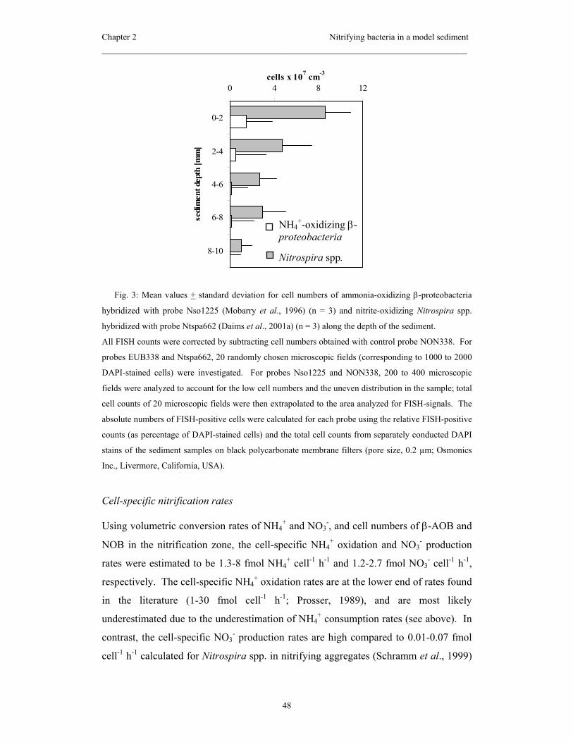
Chapter 2 Nitrifying bacteria in a model sediment _____________________________________________________________________
48
Fig. 3: Mean values + standard deviation for cell numbers of ammonia-oxidizing β-proteobacteria
hybridized with probe Nso1225 (Mobarry et al., 1996) (n = 3) and nitrite-oxidizing Nitrospira spp.
hybridized with probe Ntspa662 (Daims et al., 2001a) (n = 3) along the depth of the sediment.
All FISH counts were corrected by subtracting cell numbers obtained with control probe NON338. For
probes EUB338 and Ntspa662, 20 randomly chosen microscopic fields (corresponding to 1000 to 2000
DAPI-stained cells) were investigated. For probes Nso1225 and NON338, 200 to 400 microscopic
fields were analyzed to account for the low cell numbers and the uneven distribution in the sample; total
cell counts of 20 microscopic fields were then extrapolated to the area analyzed for FISH-signals. The
absolute numbers of FISH-positive cells were calculated for each probe using the relative FISH-positive
counts (as percentage of DAPI-stained cells) and the total cell counts from separately conducted DAPI
stains of the sediment samples on black polycarbonate membrane filters (pore size, 0.2 µm; Osmonics
Inc., Livermore, California, USA).
Cell-specific nitrification rates
Using volumetric conversion rates of NH4+ and NO3
-, and cell numbers of β-AOB and
NOB in the nitrification zone, the cell-specific NH4+ oxidation and NO3
- production
rates were estimated to be 1.3-8 fmol NH4+ cell-1 h-1 and 1.2-2.7 fmol NO3
- cell-1 h-1,
respectively. The cell-specific NH4+ oxidation rates are at the lower end of rates found
in the literature (1-30 fmol cell-1 h-1; Prosser, 1989), and are most likely
underestimated due to the underestimation of NH4+ consumption rates (see above). In
contrast, the cell-specific NO3- production rates are high compared to 0.01-0.07 fmol
cell-1 h-1 calculated for Nitrospira spp. in nitrifying aggregates (Schramm et al., 1999)
0 4 8 12
0-2
2-4
4-6
6-8
8-10
sedi
men
t dep
th [m
m]
cells x 107 cm-3
NH4+-oxidizing β-
proteobacteria
Nitrospira spp.
Chapter 2 Nitrifying bacteria in a model sediment _____________________________________________________________________
49
and biofilms (Gieseke et al., 2001). This observation might be explained in several
ways: (i) different strains of Nitrospira might be present in the sediment and in the
bioreactors, and, considering the broad phylogenetic diversity of the genus (Daims et
al., 2001a), the specific NO2- oxidation rates might greatly differ between these
different strains. To approach this hypothesis, Nitrospira-like 16S rRNA sequence
data need to be retrieved from the sediment, a difficult task due to the relative low
abundance of Nitrospira. Although in principle Nitrospira-specific PCR can be used,
the published primers (Dionisi et al., 2002) do not target the whole genus Nitrospira
and yield only sequence information of about 150 bp, too little for meaningful
phylogenetic analysis. Therefore, further identification of Nitrospira-like NOB in the
sediment has to await further method development. (ii) the cell-specific NO2-
oxidation rates of Nitrobacter are much higher (5-40 fmol cell-1 h-1 ; Prosser, 1989)
than those reported for Nitrospira. Even though undetectable by FISH in the sediment,
i.e. at cell numbers ≤ 8·105 cells cm-3, Nitrobacter might significantly contribute to the
production of NO3-; and (iii) the occurrence of other NOB, e.g., of the genus
Nitrococcus or Nitrospina, which have so far only been detected in marine
environments, cannot completely be ruled out.
Concluding remarks
The combined use of microsensors and FISH proved to be applicable to freshwater
sediments. For the first time, the in situ abundance of nitrifiers in a sediment could be
determined with sufficiently high spatial resolution to be combined with fine-scale
activity measurements. Nitrospira-like bacteria were the dominant if not sole nitrite-
oxidizers in the sediment. Limitations for the application of the combined approach to
sediments are the potential occurrence of bioturbating macrofauna that may disturb
microsensor measurements and interpretation of profiles in a real natural sediment, and
the extremely time-consuming counting procedure during FISH analysis; despite an
enormous counting effort, populations with lower abundance (<106 cells cm-3 of
sediment) may remain undetectable by FISH due to the background of unspecific
binding and autofluorescence. While the application of automated image analysis
(e.g., Daims et al., 2001b) will in the future speed up FISH analysis even in difficult
samples like sediments, it will most likely not improve its detection limit.
Chapter 2 Nitrifying bacteria in a model sediment _____________________________________________________________________
50
Acknowledgements
We thank Enrique Llobet-Brossa and Armin Gieseke for their advice and helpful
comments regarding FISH in sediments. Gabi Eickert, Anja Eggers, and Ines Schröder
are acknowledged for the construction of the O2 microelectrodes.
Financial support was provided by the German Research Foundation (STI202/1), by
the Max Planck Society, Germany, and by an Otto Hahn Award of the Max Planck
Society to A.S.

Chapter 2 Nitrifying bacteria in a model sediment _____________________________________________________________________
51
References Alawi, M., Spieck, E., Pommerening-Röser, A., and Koops, H.-P. 2003. Molecular
characterization of new nitrite-oxidizing bacteria using DGGE analysis.
BIOspektrum special issue 2003 (VAAM): abstract PH011, p. 96.
Amann, R.I., Krumholz, L., and Stahl, D.A. 1990. Fluorescent-oligonucleotide probing
of whole cells for determinative, phylogenetic, and environmental studies in
microbiology. J Bacteriol 172: 762-770.
Amann, R.I., Ludwig, W., and Schleifer, K.H. 1995. Phylogenetic identification and in
situ detection of individual microbial cells without cultivation. Microbiol Rev
59: 143-169.
Bartosch, S., Hartwig, C., Spieck, E., and Bock, E. 2002. Immunological detection of
Nitrospira-like bacteria in various soils. Microb Ecol 43: 26-33.
Bock, E.K., and Koops, H.-P. 1992. The genus Nitrobacter and related genera. In
Balows, A., Trüper, H.G., Dworkin, M., Harder, M., and Schleifer, K.-H. (eds.)
The Prokaryotes. Springer Verlag, New York, pp. 2302-2309.
Daims, H., Brühl, A., Amann, R., Schleifer, K.-H., and Wagner, M. 1999. The domain-
specific probe EUB338 is insufficient for the detection of all Bacteria:
Development and evaluation of a more comprehensive probe set. Syst Appl
Microbiol 22: 434-444.
Daims, H., Nielsen, J.L., Nielsen, P.H., Schleifer, K.-H., and Wagner, M. 2001a. In
situ characterization of Nitrospira-like nitrite oxidizing bacteria active in
wastewater treatment plants. Appl Environ Microbiol 67: 5273-5284.
Daims, H., Ramsing, N.B., Schleifer, K.-H., and Wagner, M. 2001b. Cultivation-
independent, semiautomated determination of absolute bacterial cell numbers
in environmental samples by fluorescence in situ hybridization. Appl Environ
Microbiol 67: 5810-5818.

Chapter 2 Nitrifying bacteria in a model sediment _____________________________________________________________________
52
de Beer, D., Schramm, A., Santegoeds, C.M., and Kühl, M. 1997. A nitrite
microsensor for profiling environmental biofilms. Appl Environ Microbiol 63:
973-977.
de Beer, D., and Stoodley, P. 1999. Microbial biofilms. In Balows, A., Trüper, H.G.,
Dworkin, M., Harder, W., and Schleifer, K.-H. (eds.), The Prokaryotes: an
evolving electronic resource for the microbiological community. Springer, New
York, p. 267.
de Beer, D., Sweerts, J.-P.R.A., and van den Heuvel, J.C. 1991. Microelectrode
measurement of ammonium profiles in freshwater sediment. FEMS Microbiol
Ecol 86: 1-6.
Dionisi, H.M., Layton, A.C., Harms, G., Gregory, I.R., Robinson, K.G., and Sayler,
G.S. 2002. Quantification of Nitrosomonas oligotropha-like ammonia-
oxidizing bacteria and Nitrospira spp. from full-scale wastewater treatment
plants by competitive PCR. Appl Environ Microbiol, 68, 245-253.
Ehrich, S., Behrens, D., Lebedeva, E., Ludwig, W., and Bock, E. 1995. A new
obligately chemolithoautotrophic, nitrite-oxidizing bacterium, Nitrospira
moscoviensis sp. nov. and its phylogenetic relationship. Arch Microbiol 164:
16-23.
Gieseke, A., Purkhold, U., Wagner, M., Amann, R., and Schramm, A. 2001. structure
and activity dynamics of nitrifying bacteria in a phosphate-removing biofilm.
Appl Environ Microbiol 67: 1351-1362.
Hall, G.H. 1986. Nitrification in lakes. In Prosser, J.I. (ed.) Nitrification. IRL Press,
Oxford, Washington D.C., pp. 127-156.
Hastings, R.C., Saunders, J.R., Hall, G.H., Pickup, R.W., and McCarthy, .J. 1998.
Application of molecular biological techniques to a seasonal study of ammonia
oxidation in a eutrophic freshwater lake. Appl Environ Microbiol 64: 3674-
3682.
Chapter 2 Nitrifying bacteria in a model sediment _____________________________________________________________________
53
Hovanec, T.A., Taylor, L.T., Blakis, A., and DeLong, E.F. 1998. Nitrospira-like
bacteria associated with nitrite oxidation in freshwater aquaria. Appl Environ
Microbiol 64: 258-264.
Jensen, K., Revsbech, N.P., and Nielsen, L.P. 1993. Microscale distribution of
nitrification activity in sediment determined with a shielded microsensor for
nitrate. Appl Environ Microbiol 59: 3287-3296.
Jensen, K., Sloth, N.P., Risgaard-Petersen, N., Rysgaard, S., and Revsbech, N.P. 1994.
Estimation of nitrification and denitrification from microprofiles of oxygen and
nitrate in model sediment systems. Appl Environ Microbiol 60: 2094-2100.
Juretschko, S., Timmermann, G., Schmidt, M., Schleifer, K.-H., Pommerening-Röser,
A., Koops, H.-P., and Wagner, M. 1998. Combined molecular and conventional
analysis of nitrifying bacterial diversity in activated sludge: Nitrosococcus
mobilis and Nitrospira-like bacteria as dominant populations. Appl Environ
Microbiol 64: 3042-3051.
Kowalchuk, G.A., Bodelier, P.L.E., Heilig, G.H.J., Stephen, J.R., and Laanbroek, H.J.
1998. Community analysis of ammonia-oxidising bacteria, in relation to
oxygen availability in soils and root-oxygenated sediments, using PCR, DGGE
and oligonucleotide probe hybridisation. FEMS Microbiol Ecol 27: 339-350.
Llobet-Brossa, E., Rossello-Mora, R., and Amann, R. 1998. Microbial community
composition of wadden sea sediments as revealed by fluorescence in situ
hybridization. Appl Environ Microbiol 64: 2691-2696.
Lorenzen, J., Larsen, L.H., Kjaer, T., and Revsbech, N.-P. 1998. Biosensor
determination of the microscale distribution of nitrate, nitrate assimilation,
nitrification, and denitrification in a diatom-inhabited freshwater sediment.
Appl Environ Microbiol 64: 3264-3269.
Manz, W., Amann, R., Ludwig, W., Wagner, M., and Schleifer, K.-H. 1992.
Phylogenetic oligodeoxynucleotide probes for the major subclasses of
proteobacteria: problems and solutions. Syst Appl Microbiol 15: 593-600.
Chapter 2 Nitrifying bacteria in a model sediment _____________________________________________________________________
54
Mobarry, B.K., Wagner, M., Urbain, V., Rittmann, B.E., and Stahl, D.A. 1996.
Phylogenetic probes for analyzing abundance and spatial organization of
nitrifying bacteria. Appl Environ Microbiol 62: 2156-2162.
Morgenroth, E., Obermayer, A., Arnold, E., Brühl, A., Wagner, M., and Wilderer, P.E.
2000. Effect of long-term idle periods on the performance of sequencing batch
reactors. Water Sci Technol 41(1): 105-113.
Okabe, S., Satoh, H., and Watanabe, Y. 1999. In situ analysis of nitrifying biofilms as
determined by in situ hybridization and the use of microelectrodes. Appl
Environ Microbiol 65: 3182-3191.
Pauer, J.J., and Auer, M.T. 2000. Nitrification in the water column and sediment of a
hypereutrophic lake and adjoining river system. Water Res 34: 1247-1254.
Pelegri, S.P., and Blackburn, T.H. 1995. Effects of Tubifex tubifex (Oligichaeta:
Tubificidae) on N-mineralization in freshwater sediments, measured with N-15
isotopes. Aquat Microb Ecol 9: 289-294.
Pelegri, S.P., and Blackburn, T.H. 1996. Nitrogen cycling in lake sediments
bioturbated by Chironomus plumosus larvae, under different degrees of
oxygenation. Hydrobiologia 325: 231-238.
Pernthaler, J., Glöckner, F.O., Schönhuber, W., and Amann, R. 2001. Fluorescence in
situ hybridization (FISH) with rRNA-targeted oligonucleotide probes. In Paul,
J. (ed.) Methods in Microbiology. Academic Press Inc., San Diego, Vol. 30, pp.
207-226.
Prosser, J.I. 1989. Autotrophic nitrification in bacteria. Adv Microb Physiol 30: 125-
181.
Prosser , J.I., and Embley, T. 2002. Cultivation-based and molecular approaches to
characterisation of terrestrial and aquatic nitrifiers. Antonie van Leeuwenhoek
81:165-179.
Revsbech, N.P. 1989. An oxygen microelectrode with a guard cathode. Limnol
Oceanogr 34: 474-478.
Chapter 2 Nitrifying bacteria in a model sediment _____________________________________________________________________
55
Schramm, A. 2003. In situ analysis of structure and activity of the nitrifying
community in biofilms, aggregates, and sediments. Geomicrobiol J 20: in press.
Schramm, A., de Beer, D., Gieseke, A., and Amann, R. 2000. Microenvironments and
distribution of nitrifying bacteria in a membrane-bound biofilm. Environ
Microbiol 2: 680-686.
Schramm, A., de Beer, D., van den Heuvel, J.C., Ottengraf, S., and Amann, R. 1999.
Microscale distribution of populations and activities of Nitrosospira and
Nitrospira spp. along a macroscale gradient in a nitrifying bioreactor:
quantification by in situ hybridization and the use of microsensors. Appl
Environ Microbiol 65: 3690-3696.
Schramm, A., de Beer, D., Wagner, M., and Amann, R. 1998. Identification and
activity in situ of Nitrosospira and Nitrospira spp. as dominant populations in a
nitrifying fluidized bed reactor. Appl Environ Microbiol 64: 3480-3485.
Smorczewski, W.T., and Schmidt, E.L. 1991. Numbers, activities, and diversity of
autotrophic ammonia-oxidizing bacteria in a freshwater, eutrophic lake
sediment. Can J Microbiol 37: 828-833.
Stief, P., and de Beer, D. 2002. Bioturbation effects of Chironomus riparius on the
benthic N-cycle as measured using microsensors and microbiological assays.
Aquat Microb Ecol 27: 175-185.
Stief, P., de Beer, D., and Neumann, D. 2002. Small-scale distribution of interstitial
nitrite in freshwater sediment microcosms: the role of nitrate and oxygen
availability, and sediment permeability. Microb Ecol 43: 367-378.
Wagner, M., Rath, G., Koops, H.-P., Flood, J., and Amann, R. 1996. In situ analysis of
nitrifying bacteria in sewage treatment plants. Water Sci Technol 34(1-2): 237-
244.
Whitby, C.B., Saunders, J.R., Pickup, R.W., and McCarthy, A.J. 2001. A comparison
of ammonia-oxidiser populations in eutrophic and oligotrophic basins of a large
freshwater lake. Antonie van Leeuwenhoek 79:179-188.


Chapter 3
Distribution and activity of nitrifying bacteria in
natural and manipulated stream sediment as
determined with in situ techniques
Dörte Altmann, Peter Stief, Rudolf Amann, and Dirk de Beer
Max Planck Institute for Marine Microbiology, D-28359 Bremen, Germany
Submitted to Aquatic Microbial Ecology
Chapter 3 Nitrifying bacteria in natural and manipulated sediment _____________________________________________________________________
58
Abstract
Nitrification was studied with a combination of microsensors and fluorescence in situ
hybridization (FISH) in sandy sediment of a small lowland stream. Comparative
measurements were performed in both the intact field sediment (‘natural sediment’)
and after processing sediment from the same site for a laboratory incubation
experiment (‘manipulated sediment’). In the natural sediment nitrification activity and
abundances of nitrifiers were markedly low. In contrast, nitrification activity in the
manipulated sediment (sieved, homogenized, and incubated for five weeks in the
laboratory with NH4+-enriched stream water) was significantly higher than in the
natural sediment. Abundances of NH4+-oxidizing bacteria (AOB) and Nitrospira sp. in
the 0-2 mm layer were 12 and 3-5.5 times higher than in the natural sediment,
respectively. AOB disappeared faster from anoxic sediment layers than Nitrospira sp.,
indicating that the latter was more persistent under anoxic conditions. Higher activities
and abundances of nitrifiers near the sediment-water interface of the manipulated
sediment were explained by (i) the additional NH4+ supply via the overlying water and
(ii) by adverse conditions for nitrification in the field. In conclusion, the snapshot
measurement in the natural sediment revealed the spatial heterogeneity created by
stream dynamics, whereas the sediment manipulation provided semi-natural
microcosms with reduced heterogeneity ready for factorial experiments.
Introduction
Metabolic activities of nitrification, the microbial oxidation of NH4+ to NO3
-, have
been measured with microsensors in intact field sediments (Sweerts and de Beer, 1989;
Lorenzen et al., 1998; Meyer et al., 2001) as well as in model, i.e. sieved and
homogenized sediments (Jensen et al., 1993; Jensen et al., 1994; Stief et al., 2002).
Only fine scale measurements of this type allowed to precisely localize the narrow
surface layer of active nitrification that may be overlooked by conventional pore water
analysis (Henriksen et al., 1993; Rysgaard et al., 1995) or mass balance studies
(Christensen et al., 2000; Van Luijn et al., 1999). Using microsensors the process of
nitrification could be localized in the upper few mm of the sediment and the influence
of other sedimentary processes like O2 production by photosynthesis on nitrification
and the coupling of nitrification and denitrification was demonstrated. However, the
Chapter 3 Nitrifying bacteria in natural and manipulated sediment _____________________________________________________________________
59
pure process analysis of sedimentary nitrification leaves in the dark which
microorganisms perform nitrification in this particular habitat, in which quantities
these microorganisms occur, and how they are spatially organized in the sediment. So
far the identification and quantification of nitrifying bacteria in natural sediments has
been addressed, for instance, by the most probable number technique (Pauer and Auer,
2000; Smorczewski and Schmidt, 1991; Whitby et al., 2001; Bianchi and Lefevre,
1999). Cell numbers detected in these studies were 104 –105 g-1 for AOB and 103 g-1
for NOB and quantified nitrifiers could be identified as members of the genera
Nitrosospira, Nitrosomonas and Nitrobacter using immunofluorescence analysis and
by enrichment cultures. However, using this cultivation-dependent technique, the
actual size and structure of microbial communities in general (Amann et al., 1995) and
of nitrifying populations in particular (Hall et al., 1996) can be severely misjudged due
to non-culturability of some microorganisms and selective enrichment of others. To
date, this possible bias can be circumvented by using cultivation-independent
techniques such as FISH (Amann and Kühl, 1998). The combined application of
microsensors and FISH for nitrification studies has so far been used exclusively in
wastewater biofilms (Schramm et al., 1997; Okabe et al., 1999; Gieseke et al., 2001)
and in a model freshwater sediment (Altmann et al., accepted), but not in intact field
sediments as presented here.
While measurements in naturally heterogeneous field sediments help depict the
spatial and temporal dynamics of a particular habitat, working with sieved and
homogenized sediments is a commonly used approach for factorial experiments under
defined conditions. Sieving removes physical obstacles that may disturb microsensor
measurements and homogenization produces a high degree of similarity between
replicate sediment cores (Svensson and Leonardson, 1996; Davidsson et al., 1997;
Hansen and Jensen, 1998). On the other hand, the harsh pretreatment of sediments has
been questioned because it destroys the vertical microbial stratification and has a
negative effect on the persistence of slowly growing members of the microbial
community, e.g. nitrifiers mixed into anoxic subsurface layers (Findlay et al., 1990;
Svensson et al., 2001). Recovery of these microorganisms and the establishment of a
close-to-natural microbial stratification may take as long as 2-3 weeks under
laboratory conditions (Tuominen et al., 1999). For this reason a preincubation of the
Chapter 3 Nitrifying bacteria in natural and manipulated sediment _____________________________________________________________________
60
manipulated sediments is usually scheduled prior to the actual experimental treatments
(Svensson et al., 2001; Stief et al., accepted).
The major aim of our study was to investigate for the first time the activity and
distribution of nitrifying bacteria in situ, i.e. in a natural stream sediment with
microsensors and FISH. Secondly, we studied the performance of nitrification after the
same natural sediment was sieved, homogenized and incubated in natural stream water
and at ambient temperature. The same set of methods was used to follow the
reorganization of nitrification in this manipulated sediment during a 5-week incubation
in the laboratory.
Materials and Methods
Sampling site. Sandy sediment for i) measurements in the natural sediment and ii) for
those in the manipulated sediment was sampled in May 2002 from a small lowland
stream, Rittrumer Mühlenbach (Northern Germany). The stream crosses an area of
glacial sand accumulation in the Wildeshausener Geest. Average flow velocity was 0.2
m s-1 causing ripple formation on the sediment surface, with detritus mainly
accumulating in the troughs of this ripple system. Concentrations of NH4+ and NO3
- in
the overlying water at the day of sampling were 10 and 500 µmol l-1, respectively. The
organic content of the sediment ranged between 1.0 and 5.1 % (n = 15) and the
sediment was only sparsely inhabited by macrofauna.
Experimental design. For the analysis of the natural sediment intact sediment cores of
15 cm length and 7.5 cm ∅ were taken in replicates with Plexiglas cylinders from a
water depth of 50 cm. To avoid disturbances of the sediment surface the enclosed
water phase was hermetically sealed and the cores were carefully transported to the lab
within 45 min. The microsensor measurements were performed immediately in natural
stream water and at in situ temperature (9.6°C). During the measurements the
overlying water was held turbulent to ensure the formation of a diffusive boundary
layer. For the analysis of the manipulated sediment, in the same reach surface sediment
(0-5 cm) was collected with a flat shovel and transferred into buckets. In the laboratory
the sediment was immediately sieved to remove macrofauna, large detritus and
pebbles, filled into beakers (∅ 9 cm) up to a height of 13 cm, and allowed to settle
over night (Jensen et al., 1994). Six beakers were submersed in 3 basins containing 15

Chapter 3 Nitrifying bacteria in natural and manipulated sediment _____________________________________________________________________
61
l of unfiltered stream water and were incubated in the dark at in situ temperature
(9.6°C). O2 concentration in the overlying water was adjusted to air saturation. To
avoid NH4+ limitation of nitrification the concentration of NH4
+ in the overlying water
was kept constant by repeated additions of 50 µmol l-1 of a NH4Cl stock solution.
Concentrations of NH4+, NO3
- and NO2- in the basins were checked regularly using
photometric test kits by Merck (Germany). After 3 and 5 weeks of incubation
microsensor measurements and sediment samples for FISH were taken.
Microsensor measurements. Microsensors for O2 (Revsbech, 1989), NO3- (de Beer and
Sweerts, 1989), and NH4+ (de Beer and van den Heuvel, 1988) were prepared,
calibrated, and operated in a measuring setup as previously described (Stief et al.,
2002). Replicate profiles were recorded at randomly chosen spots of the sediment
surface down to a depth of 10 mm. Information on the nitrification activity was
obtained by calculation of the fluxes of O2, NO3-, and NH4
+ at the sediment water
interface (Jswi) and at the oxic/anoxic interface (Janx) using Fick´s first law of diffusion.
Diffusion coefficients for of O2, NO3-, and NH4
+ in the overlying water (Dw) were
taken from the literature (Schramm et al., 1999) and corrected for the measuring
temperature (9.6 °C) by applying the Stokes-Einstein-relation. The sediment diffusion
coefficients of O2, NO3-, and NH4
+ (Ds) were calculated from an averaged profile of
diffusivity as measured with a H2 diffusivity sensor (Unisense A/S, Denmark)
(Revsbech et al., 1998; Stief et al., accepted). Net nitrification activity, i.e. the areal
conversion rate of NO3- and NH4
+ within the oxic layer, was defined as the sum of Jswi
and Janx of each solute.
FISH and total cell counts. After the microsensor profiles had been recorded one
sediment core (∅ 2.5 cm) was taken from each sediment cylinder or beaker,
respectively, and was sectioned horizontally into 2 mm-thick slices down to a depth of
10 mm. Sediment slices were prepared for FISH as described in (Altmann et al.,
accepted). For FISH a set of oligonucleotide probes specific for i) Eubacteria (probe
mix EUB 338 (Amann et al., 1990), EUB II and EUB (Daims et al., 1999)), ii) NH4+-
oxidizing β-proteobacteria (Nso1225, (Mobarry et al., 1996)), and iii) for the NO2--
oxidizing genera Nitrobacter (Nit3, (Wagner et al., 1996)) and Nitrospira (Ntspa662,
(Daims et al., 2000)) was used. Probes Nit3 and Ntspa662 were used with an
equimolar amount of a competitor oligonucleotide as indicated in the references. To

Chapter 3 Nitrifying bacteria in natural and manipulated sediment _____________________________________________________________________
62
check for unspecific binding samples were hybridized with control probe Non338
(Manz et al., 1992). All probes were purchased labeled with the fluorescent dye CY3
(Hybaid Interactiva, Ulm, Germany). FISH and counter staining of all cells with 4´,6-
diamino-2-phenylindole (DAPI; 0.5 µg ml-1) were performed according to published
protocols (Pernthaler et al., 1998). Counting was adapted to the low numbers of FISH-
positive cells and their uneven distribution in the aliquots as described in (Altmann et
al., accepted). To correct for cell losses during hybridization total bacterial cell
numbers were determined separately by DAPI-staining of sonicated and diluted
sediment samples on black membrane polycarbonate filters (pore size, 0.2 µm;
Osmonics Inc., Livermore, California, USA). The absolute numbers of FISH-positive
cells were calculated for each probe using the relative FISH-positive counts (as
percentage of DAPI-stained cells) and the total cell counts.
Results
Natural sediment: Microsensor measurements. Concentration profiles of O2, NH4+,
and NO3- as measured in the natural sediment are presented in Fig. 1 A-C. To highlight
the heterogeneity between replicate cores, profiles are shown for each individual core.
Fig. 1 Vertical concentration profiles of O2 (A), NH4+ (B), and NO3
- (C) as measured in the naturally
stratified sediment of a lowland stream. Average profiles (+ 1 SD, n ≥ 3) of three replicate sediment
cores (c1-3) are plotted separately.
A-2
0
2
4
6
8
10
0 100 200 300 400
cO2 [µmol l-1]
dept
h [m
m]
B-2
0
2
4
6
8
10
0 250 500 750
cNO3- [µmol l-1]
C-2
0
2
4
6
8
10
0 50 100 150
cNH4+ [µmol l-1]

Chapter 3 Nitrifying bacteria in natural and manipulated sediment _____________________________________________________________________
63
In all three cores O2 became depleted within the uppermost 2 mm of the sediment, but
in core 3 this decrease in O2 concentration was less steep indicating lower bacterial
activity compared to the other cores. NO3- was continuously depleted over a depth of
approximately 6 mm in cores 1 and 2, but did not reach 0 µmol l-1 within the sampled
sediment depth of core 3. NH4+ concentration in the cores 2 and 3 decreased slightly
within the upper 2 mm and increased again in the deeper layers. In contrast, core 1 was
characterized by comparably high NH4+ concentration in deeper layers and a steep
concentration gradient at the oxic/anoxic interface. The local fluxes of O2, NH4+, and
NO3- were directed into the oxic layer of the natural sediment (Tab. 1).
Table 1. Local fluxes of O2, NH4+, and NO3
- [µmol m-2 h-1] within the presumable nitrification layer of
the sediment from a lowland stream. Natural sediment: Means (+ 1 SD, n ≥ 3) within each of three
replicate cores. Manipulated sediment: Means of three replicate cores within one sampling date (+ 1 SD,
n ≥ 9). Positive values = production, negative values = consumption.
sediment type core time JO2 JNH4 JNO3
natural 1 week 0 -796 (62) -89 (27) -123 (86)
natural 2 week 0 -925 (121) -10 (3) +24 (179)
natural 3 week 0 -600 (60) -12 (5) -34 (15)
manipulated 1-3 week 3 -425 (57) -101 (19) +249 (152)
manipulated 1-3 week 5 -494 (75) -77 (21) +247 (53)

Chapter 3 Nitrifying bacteria in natural and manipulated sediment _____________________________________________________________________
64
Natural sediment: Nitrifier abundances. Total cell numbers in the natural sediment
were 8 – 13.9 ⋅ 109 cells cm-3 showing almost equal distribution throughout the
sampled sediment depth. Overall detection rate with FISH, as determined with
combined oligonucleotide probes EUB 338, EUB II, and EUB III, was 40-50% of total
bacterial cells. Unspecific signals (i.e. binding of probe NON338 and autofluorescent
cells) were not detected in this natural sediment. Absolute abundances and the vertical
distribution of NH4+- oxidizing β-proteobacteria (AOB) and of the NO2
--oxidizing
bacteria (NOB) are shown in Fig. 2.
Fig. 2 Vertical distribution of NH4+-oxidizing β-proteobacteria (AOB, A) and NO2
--oxidizing
Nitrospira sp. (NOB, B) as revealed with FISH in the naturally stratified sediment. Average abundances
(+ 1 SD) of three replicate sediment cores are given.
AOB abundance was highest in the 2-4 mm layer but peaked again in the deepest
sampled layer, i.e. in 8-10 mm depth, accounting for 0.26% and 0.16% of total cells,
respectively. FISH indicated that in the natural sediment NOB were represented by the
genus Nitrospira, whereas the commonly isolated genus Nitrobacter spp. was not
detected. Cells of Nitrospira ssp. were 5-9 times more abundant than the NH4+-
oxidizing β-proteobacteria and here too the maximum numbers were found in the 2-4
mm layer where they made up 1.2% of all bacterial cells. Cells of Nitrospira ssp. were
also detected in remarkably high abundances in the anoxic layers of the sediment.
A
0 1 2 3 4 5
0-2
2-4
4-6
6-8
8-10
dept
h [m
m]
AOB [cells x107 cm-3]
B
0 10 20 30
0-2
2-4
4-6
6-8
8-10
NOB [cells x107 cm-3]

Chapter 3 Nitrifying bacteria in natural and manipulated sediment _____________________________________________________________________
65
Manipulated sediment: Microsensor measurements. In contrast to the natural sediment,
the high similarity between replicate cores of the manipulated sediment allowed
averaging of all measured profiles within one sampling date. Averaged concentration
profiles of O2, NH4+, and NO3
- recorded after 3 and 5 weeks of incubation in the
laboratory are shown in Fig. 3.
Fig. 3: Vertical concentration profiles of O2, NH4+, and NO3
- as measured in the manipulated sediment
after a 3-week (A-C) and 5-week (D-F) incubation in the laboratory. Average profiles of three replicate
sediment cores are plotted (+ 1 SD, n ≥ 9).
O2 penetration depth after 3 weeks was 7 mm. NH4+ concentration that was held
constant at 50 µmol l-1 in the overlying water, decreased within the upper 1 mm of the
sediment and increased slightly in the deeper layers. The NO3- concentration peak in
the upper 1 mm of sediment was followed by an extended zone of slightly decreasing
NO3- concentration, but NO3
- was not completely consumed within the sampled
sediment depth. After 5 weeks of incubation the concentrations of O2, NH4+, and NO3
-
showed the same vertical patterns than 2 weeks before, but O2 penetration depth
B-0.20
0.00
0.20
0.40
0.60
0.80
1.00
0 250 500 750
cNO3- [µmol l-1]
C-0.20
0.00
0.20
0.40
0.60
0.80
1.00
0 25 50 75
cNH4+ [µmol l-1]
D
-0.20
0.00
0.20
0.40
0.60
0.80
1.00
0 100 200 300 400
dept
h [m
m]
E
-0.20
0.00
0.20
0.40
0.60
0.80
1.00
0 250 500 750
F
-0.20
0.00
0.20
0.40
0.60
0.80
1.00
0 25 50 75
A-0.20
0.00
0.20
0.40
0.60
0.80
1.00
0 200 400
cO2 [µmol l-1]
dept
h [m
m]
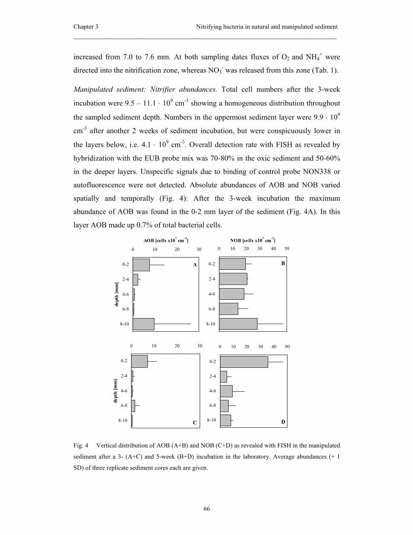
Chapter 3 Nitrifying bacteria in natural and manipulated sediment _____________________________________________________________________
66
increased from 7.0 to 7.6 mm. At both sampling dates fluxes of O2 and NH4+ were
directed into the nitrification zone, whereas NO3- was released from this zone (Tab. 1).
Manipulated sediment: Nitrifier abundances. Total cell numbers after the 3-week
incubation were 9.5 – 11.1 ⋅ 109 cm-3 showing a homogeneous distribution throughout
the sampled sediment depth. Numbers in the uppermost sediment layer were 9.9 ⋅ 109
cm-3 after another 2 weeks of sediment incubation, but were conspicuously lower in
the layers below, i.e. 4.1 ⋅ 109 cm-3. Overall detection rate with FISH as revealed by
hybridization with the EUB probe mix was 70-80% in the oxic sediment and 50-60%
in the deeper layers. Unspecific signals due to binding of control probe NON338 or
autofluorescence were not detected. Absolute abundances of AOB and NOB varied
spatially and temporally (Fig. 4): After the 3-week incubation the maximum
abundance of AOB was found in the 0-2 mm layer of the sediment (Fig. 4A). In this
layer AOB made up 0.7% of total bacterial cells.
Fig. 4 Vertical distribution of AOB (A+B) and NOB (C+D) as revealed with FISH in the manipulated
sediment after a 3- (A+C) and 5-week (B+D) incubation in the laboratory. Average abundances (+ 1
SD) of three replicate sediment cores each are given.
A
0 10 20 30
0-2
2-4
4-6
6-8
8-10
dept
h [m
m]
AOB [cells x107 cm-3]
C
0 10 20 30
0-2
2-4
4-6
6-8
8-10
dept
h [m
m]
D
0 10 20 30 40 50
0-2
2-4
4-6
6-8
8-10
B
0 10 20 30 40 50
0-2
2-4
4-6
6-8
8-10
NOB [cells x107 cm-3]
Chapter 3 Nitrifying bacteria in natural and manipulated sediment _____________________________________________________________________
67
In deeper layers their proportion decreased to 0.04% of total cells. In the 8-10 mm
layer their averaged abundance was exceptionally high, but this was due to the
detection of two cell clusters (70 and 90 cells, respectively) in just one out of three
replicate cores. After another 2 weeks of incubation the absolute abundances and the
vertical distribution of the AOB were the same as before, except for the absence of a
secondary abundance peak in the 8-10 mm layer (Fig. 4C). NOB in the manipulated
sediment were also represented by the genus Nitrospira, whereas Nitrobacter spp. was
not detected at any time of the incubation. Cells of Nitrospira spp. were evenly
distributed over the whole sampled sediment depth after 3 weeks of incubation and
made up approximately 1.9% of total bacterial cells (Fig. 4B). After 5 weeks, however,
a distinct abundance maximum had developed at the sediment surface and cell
numbers there had increased to 3.6% of total cells (Fig 4D).
Discussion
Natural sediment
The investigated stream sediment was characterized by negligible low nitrification
activity as indicated by the absence of distinct oxic NO3- peaks in the majority of the
measured profiles. On average NO3- was consumed throughout the whole sampled
sediment depth. Dissimilatory NO3- reduction was probably the principal pathway of
NO3- consumption in this sediment. Additionally performed measurements in
illuminated sediments (light intensity 500 µE m-2 s-1, data not shown) did not reveal
measurable photosynthetic capacity and hence, in our sediments photosynthesis can be
ruled out as a significant sink for NO3-. However, the areal rates of NO3
- consumption
were at the lower end of the rates reported in the literature (Sweerts and de Beer, 1989;
Lorenzen et al., 1998; Meyer et al., 2001). These low rates of N cycling in our
sediment (i.e., nitrification and denitrification) were probably due to its low content of
readily usable organic matter, which limited the supply of NH4+ and CO2 for
nitrification and of electron donors for dissimilatory NO3- reduction. Secondly, the low
rates may be indicative of the physical dynamics in natural stream sediment, as the
water currents permanently reshape the sediment surface, e.g. cut off formerly oxic
layers from O2 (Wulff et al., 1997). Such a disturbance may inhibit nitrification in
Chapter 3 Nitrifying bacteria in natural and manipulated sediment _____________________________________________________________________
68
particular, since O2 is needed for the performance of this process (Caffrey et al., 1993;
Sloth et al., 1995). Moreover, nitrifying bacteria are characterized by low growth
kinetics (Prosser, 1989) meaning that recovery from disturbances may be particularly
slow (Findlay et al., 1990; Svensson et al., 2001). Supportive of the dynamic character
of the natural sediment is also the heterogeneity of solute conversions within and
between the replicate sediment cores.
Applying oligonucleotide probes specific for NH4+-oxidizing β-proteobacteria
(AOB) and the NO2--oxidizing genera Nitrospira and Nitrobacter (NOB) we localized
and quantified members of the two physiological groups of nitrifying bacteria in situ,
i.e. in a natural freshwater sediment that was sampled directly in the field. Most
probable number counts (MPN) of culturable nitrifying bacteria in other natural
freshwater sediments led to abundances in the order of 104 to 105 cells per g (Pauer
and Auer, 2000; Smorczewski and Schmidt, 1991; Whitby et al., 2001) and were thus
1-2 orders of magnitude lower than those detected in our in situ study. This
discrepancy is apparently due to the fact that methods like MPN or enrichment cultures
are highly biased because they are cultivation-dependent and thus often underestimate
the actual size of bacterial communities in general (Amann et al., 1995) and of
nitrifying populations in particular (Hall et al., 1996). However, we are also aware of
possible disadvantages of the FISH technique: With FISH the total population size of
nitrifiers can be accurately estimated, but a discrimination of active vs. inactive cells is
difficult because nitrifiers are able to maintain a relatively high ribosome content even
if they are not active (Batchelor et al., 1997; Morgenroth et al., 2000). In our study this
problem was overcome though by additionally using microsensors in order to give the
precise site and the magnitude of nitrifying activity.
In the investigated natural sediment the genus Nitrobacter, which is the most
commonly isolated NO2--oxidizer, was not detected with the in situ technique of FISH.
Instead, we identified and quantified the genus Nitrospira as a major representative of
NOB. This genus has recently been detected in several habitats by means of
comparative 16S rRNA analysis (Hovanec et al., 1998; Juretschko et al., 1998; Cho
and Kim, 2000; Li and Horikoshi, 1999; Todorov et al., 2000). Our finding thus
further confirms the recently assumed dominance of Nitrospira rather than Nitrobacter
not only in engineered systems and model sediments, but also in intact freshwater
sediment.
Chapter 3 Nitrifying bacteria in natural and manipulated sediment _____________________________________________________________________
69
Maximum abundances of both the AOB and NOB in the natural sediment occurred
in the 2-4 mm horizon that was apparently anoxic during our microsensor
measurements. In a previous study applying microsensors and FISH to model sediment
(Altmann et al., accepted) highest abundances of AOB and NOB were found in the
uppermost sediment layer and coincided well with the conversion maxima of O2, NH4+
and NO3-. However, in the present study, the abundance peak of AOB and NOB in the
2-4 mm layer clearly did not coincide with concentration minima or maxima of
nitrification substrates or products. Two main reasons can be considered for the
presence of the abundance peak in this apparently anoxic layer: i) In the field, i.e. just
before the cores were taken and measured in the laboratory, O2 may have also been
present in the 2-4 mm layer. O2 penetration depth may have decreased due to the lower
current velocity during our measurements and hence advective transport of O2 into the
sediment (Huettel and Rusch, 2000) was probably less significant. If this scenario was
true, then under field conditions the nitrifier population has met ideal growth
conditions at the lower end of the oxic zone in which the supply of NH4+ from deeper
layers was higher compared to the upper end of the oxic zone. ii) The layer of
maximum abundances of AOB and NOB may represent the former surface of the
sediment which has recently been slopped and cut off from O2 supply by the horizontal
shifting of ripple structures. The newly reshaped sediment surface may not have been
recolonized by nitrifiers to form a new abundance maximum.
Manipulated sediment
In contrast to the natural sediment the sieved and homogenized sediment was
characterized by a distinct nitrification zone in the uppermost layer of the sediment.
Even though, nitrifiers obviously did not tap the full potential of nitrification, as was
indicated by incomplete depletion of NH4+ within the oxic sediment layer. Since O2
was present to a depth of 7 mm, nitrification was limited neither by NH4+ and O2, but
rather by other factors like a population size too small to consume the continuously
supplied NH4+ to its limit or a shortage of CO2 which was internally produced by
mineralization. However, nitrification activity, i.e. the areal production of NO3- within
the oxic zone was comparable to other freshwater studies applying microsensors to
experimentally manipulated sediment (Jensen et al., 1993; Jensen et al., 1994; Stief et
al., 2002). In contrast to the higher nitrification rates in the manipulated sediment,
Chapter 3 Nitrifying bacteria in natural and manipulated sediment _____________________________________________________________________
70
dissimilatory NO3- consumption was lower than in the natural sediment as it was
indicated by the incomplete consumption of NO3- within the sampled sediment depth.
This was apparently due to a lack of electron donors for NO3- reduction as a
consequence of the low sedimentary organic content. It seems likely that by sieving
the sediment dissolved and fine particulate organic matter has partially been washed
out from the sediment. Moreover, after the sediments had settled a sharp separation
into the sandy sediment and a fluffy organic matter layer at the sediment surface of 2-3
mm thickness was observed suggesting that the sandy sediment below this fluffy layer
became poor in at least particulate organics. Indeed organic content of the manipulated
sediment was lower compared to the natural sediment and decreased even further with
sediment depth (data not shown). Both nitrification and denitrification activity did not
change during the 2 week incubation indicating the stability of the manipulated
sediment during this period. Moreover, the sieving and homogenization of the
sediment clearly reduced heterogeneity within and between the sediment cores as was
indicated by the low variability between concentration profiles. From this point of
view this pretreatment of sediment and a time course of 2 weeks after initial
restratification hence presents a suitable basis for the performance of factorial
experiments.
Sediment manipulation has most likely created also a homogeneous vertical
distribution of both AOB and NOB at the start of the experiment. During the following
laboratory incubation both populations showed a different pattern of vertical
restratification: AOB were found at highest abundances in the 0-2 mm layer already
after 3 weeks and this was still the case after 5 weeks of incubation. In contrast, NOB
were still homogeneously distributed after 3 weeks of incubation, but finally showed
signs of vertical stratification after 5 weeks of incubation. The AOB population
quickly established an abundance maximum near the sediment-water interface at the
expense of deeper layers, even though O2 penetrated 7 mm deep into the sediment and
was thus not limiting. This preferential growth within the uppermost sediment layer
was probably due to the continuous supply of NH4+ via the overlying water, while in
the deeper oxic layers AOB were probably limited by the low supply of NH4+ from the
anoxic zone. A spatial “plasticity” of the microdistribution of nitrification activity, i.e.
the shifting of nitrification activity in response to the NH4+ source was also
demonstrated by (Jensen et al., 1993). Given that NO2- production by AOB was the
Chapter 3 Nitrifying bacteria in natural and manipulated sediment _____________________________________________________________________
71
only significant source of NO2- in the sediment, NOB was expected to thrive better at
the sediment water interface than in deeper layers, and so it was near the end of the
experiment. Obviously though, NOB in this experiment persisted longer the unsuitable
conditions in the deeper layers (i.e., lack of O2 and NO2-) than did AOB. This superior
persistence may have contributed to the delayed stratification of NOB compared to
that of AOB. Similarly, a slower decay of Nitrospira spp. compared to that of ß-AOB
was observed in anoxic layers by (Altmann et al., accepted). This feature of Nitrospira
sp. was assumed to be either due to its adaptation to survive or even thrive under
anoxic conditions or to be a result of the slow decay of ribosomes in this NO2--
oxidizer. Supportive of the superior persistence of NOB in deeper sediment layers
were also the higher NO2-- compared to NH4
+-oxidation rates which could be evoked
in slurry incubations of various sediments (P. Stief, unpublished results).
Comparison of natural and manipulated sediment
Despite the destruction of the microbial stratification by sieving and homogenizing,
the manipulated sediment was finally characterized by higher abundances of AOB and
NOB as well as by conspicuously higher nitrification activity. This was probably due
to the additional supply with NH4+, which can be a major factor limiting nitrification in
sediments (de Beer et al., 1991; Jensen et al., 1993). On the other hand, sediment
pretreatment and laboratory incubation have obviously produced a loss of electron
acceptors and NH4+ within the sediments, which resulted in lower consumption of O2
and lower dissimilatory consumption of NO3-. Possible reasons for this loss are the
washout of dissolved substances during sieving and the “dilution” of otherwise
patchily distributed particulate organic matter during homogenization and by the
accumulation of fine particulate organic matter at the sediment surface. This may
constitute non-favorable conditions for the performance of both nitrification and
denitrification because NH4+, CO2, and electron donors may become limiting. In our
case though, at least NH4+ limitation was counteracted by supplying it via the
overlying water.
As expected, the natural sediment was more heterogeneous than the manipulated
sediment giving an idea of the spatial and temporal dynamics in the sampled stream
habitat. Measurements conducted directly in the field are useful for investigations of
the complexity of a particular habitat, for comparisons between different natural
Chapter 3 Nitrifying bacteria in natural and manipulated sediment _____________________________________________________________________
72
habitats and for investigations of the seasonality within certain habitats. The
pretreatment of the sediment, however, produced a high degree of similarity between
replicate cores which is useful for the performance of factorial experiments under
defined conditions. In our case the complementary use of both approaches also
allowed to address the adaptive capacities of the sedimentary microorganisms to
changed conditions.
Conclusions
The application of microsensors combined with FISH for the investigation of
nitrification in a natural stream sediment was challenging because of the generally
lower activities and cell abundances compared to e.g. waste water biofilms. Still, to
date it is the only acceptable in situ approach to study nitrification activity with the
appropriate spatial resolution and to determine the abundance of nitrifiers without the
bias of cultivation-dependent techniques. With the use of FISH, the two physiological
groups of nitrifiers, AOB and NOB, could be separately studied with respect to both
their prevalence in a natural sediment and their fate upon sediment manipulation and
incubation. Microsensor data, on the other hand, helped to reveal which of the
identified nitrifiers were actually active and what effect their activity had on the
nutrient exchange between the sediment and the overlying water. In this sense, our
field measurements represent a snapshot of the situation in a small stream at a given
time of the year, whereas the laboratory incubation revealed the potential of the same
sediment for nitrification and the ability of the inhabiting nitrifiers to adapt to
changing conditions.
Chapter 3 Nitrifying bacteria in natural and manipulated sediment _____________________________________________________________________
73
Acknowlegdements
We thank Enrique Llobet-Brossa and Armin Gieseke for their advice and helpful
comments regarding FISH in sediments. Gaby Eickert, Anja Eggers, and Ines Schröder
are acknowledged for the construction of O2 microelectrodes. For critical comments on
the paper we thank Rebecca Ludwig. Financial support was provided by the German
Research Foundation (STI202/1), and by the Max Planck Society, Germany.

Chapter 3 Nitrifying bacteria in natural and manipulated sediment _____________________________________________________________________
74
References
Altmann, D., Stief, P., Amann, R., de Beer, D., and Schramm, A. accepted. In situ
distribution and activity of nitrifying bacteria in freshwater sediment.
Environmental Microbiology.
Amann, R. I., Krumholz, L. and Stahl, D. A. 1990. Fluorescent-oligonucleotide
probing of whole cells for determinative, phylogenetic, and environmental
studies in microbiology. Journal of Bacteriology 172:762-770.
Amann, R. and Kühl, M. 1998. In situ methods for assessment of microorganisms and
their activities. Current Opinion in Microbiology 1:352-358.
Amann, R. Ludwig, W., and Schleifer, K.-H. 1995. Phylogenetic identification and in
situ detection of individual microbial cells without cultivation. Microbiological
Reviews 59:143-169.
Batchelor, S. E., Cooper, M., Chhabra, S. R., Glover, L. A., Stewart, G., Williams, P.
and Prosser, J. I. 1997. Cell density-regulated recovery of starved biofilm
populations of ammonia-oxidizing bacteria. Applied and Environmental
Microbiology 63:2281-2286.
Bianchi, M. and Lefevre, F. D. 1999. Regulation of nitrification in the land-ocean
contact area of the Rhone River plume (NW Mediterranean). Aquatic
Microbial Ecology 18:301-312.
Caffrey, J. M., Sloth, N. P., Kaspar, H. F., and Blackburn, T. H. 1993. Effect of
organic loading on nitrification and denitrification in a marine sediment
microcosm. FEMS Microbiology Ecology 12:159-167.
Cho, J.-C. and Kim, S.-J. 2000. Increase in bacterial community diversity in
subsurface aquifers receiving livestock wastewater input. Applied and
Environmental Microbiology 66:956-965.
Christensen, P. B., Rysgaard, S., Sloth, N. P., Dalsgaard, T., and Schwaerter, S. 2000.
Sediment mineralization, nutrient fluxes, denitrification and dissimilatory
nitrate reduction to ammonium in an eastuarine fjord with sea cage trout farms.
Aquatic Microbial Ecology 21:73-84.
Daims, H., Nielsen, P., Nielsen, J. L., Juretschko, S., and Wagner M. 2000. Novel
Nitrospira-like bacteria as dominant nitrite-oxidizers in biofilms from
Chapter 3 Nitrifying bacteria in natural and manipulated sediment _____________________________________________________________________
75
wastewater treatment plants: diversity and in situ-physiology. Water Sci
Technol. 41:85-90.
Daims, H., Brühl, A., Amann, R., Schleifer, K.-H., and Wagner, M. 1999. The
domain-specific probe EUB338 is insufficient for the detection of all bacteria:
development and evaluation of a more comprehensive probe set. Systematic
and Applied Microbiology 22:434-444.
Davidsson, T. E., Stepanauskas, R., and Leonardson, L. 1997. Vertical patterns of
nitrogen transformations during infiltration in two wetland soils. Applied and
Environmental Microbiology 63:3648-3656.
de Beer, D., and Sweerts, J.-P. R. A. 1989. Measurement of nitrate gradients with an
ion-selective microelectrode. Analytica Chimica Acta 219:351-356.
de Beer, D. and van den Heuvel, J. C. 1988. Response of ammonium-selective
microelectrodes based on the neutral carrier nonactin. Talanta 35:728-730.
de Beer, D., Sweerts, J.-P. R. A., and van den Heuvel, J. C. 1991. Microelectrode
measurement of ammonium profiles in freshwater sediments. FEMS
Microbiology Ecology 86:1-6.
Findlay, R. H., Trexler, M. B., Guckert, J. B., and White, D.C. 1990. Laboratory study
of disturbance in marine sediments: response of a microbial community.
Marine Ecology Progress Series 62:121-133.
Gieseke, A., Purkhold, U., Wagner, M., Amann, R. and Schramm, A. 2001.
Community structure and activity dynamics of nitrifying bacteria in a
phosphate-removing biofilm. Applied and Environmental Microbiology
67:1351-1362.
Hall, P. O. J., Hulth, S., Hulthe, G., Landen, A., and Tengberg, A. 1996. Benthic
nutrient fluxes on a basin-wide scale in the Skagerak (north-eastern North
Sea). Journal of Sea Research 35:123-137.
Hansen, K., and Jensen, E. 1998. The impact of the polychaete Nereis diversicolor
and enrichment with macroalgal (Chaetomorpha linum) detritus on benthic
metabolism and nutrient dynamics in organic poor and organic rich sediment.
Journal of Experimental Marine Biology and Ecology 231:201-223.
Henriksen, K., Blackburn, T. H., Lomstein, B. A., and McRoy, C. P. 1993. Rates of
nitrification, distribution of nitrifying bacteria and inorganic N fluxes in
Chapter 3 Nitrifying bacteria in natural and manipulated sediment _____________________________________________________________________
76
northern Bering-Chukchi shelf sediments. Continental Shelf Research 13:629-
651.
Hovanec, T. A., Taylor, L. T., Blakis, A. and Delong, E. F. 1998. Nitrospira-like
bacteria associated with nitrite oxidation in freshwater aquaria. Applied and
Environmental Microbiology 64:258-264.
Huettel, M., and Rusch, A. 2000. Transport and degradation of phytoplankton in
permeable sediment. Limnology and Oceanography 45:534-549.
Jensen, K., Revsbech, N. P., and Nielsen, L. P. 1993. Microscale distribution of
nitrification activity in sediment determined with a shielded microsensor for
nitrate. Applied and Environmental Microbiology 59:3287-3296.
Jensen, K., Sloth, N. P., Risgaard-Petersen, N., Rysgaard, S., and Revsbech, N. P.
1994. Estimation of nitrification and denitrification from microprofiles of
oxygen and nitrate in model sediment systems. Applied and Environmental
Microbiology 60:2094-2100.
Juretschko, S., Timmermann, G., Schmid, M., Schleifer, K. H., Pommerening-Roser,
A., Koops, H. P. and Wagner, M. 1998. Combined molecular and conventional
analyses of nitrifying bacterium diversity in activated sludge: Nitrosococcus
mobilis and Nitrospira-like bacteria as dominant populations. Applied and
Environmental Microbiology 64:3042-3051.
Li, L. C. K. and Horikoshi, K. 1999. Bacterial diversity in deep-sea sediments from
different depths. Biodiversity and Conservation 8:659-677.
Lorenzen, J. Larsen, L. H., Kjaer, T., and Revsbech, N.-P. 1998. Biosensor
determination of the microscale distribution of nitrate, nitrate assimilation,
nitrification, and denitrification in a diatom-inhabited freshwater sediment.
Applied and Environmental Microbiology 64:3264-3269.
Manz, W., Amann, R., Ludwig, W., Wagner, M., and Schleifer, K.-H. 1992.
Phylogenetic oligodeoxynucleotide probes for the major subclasses of
proteobacteria: problems and solutions. Systematic and Applied Microbiology
15:593-600.
Meyer, R.L., Kjaer, T., and Revsbech, N.-P. 2001. Use of NOx- microsensors to
estimate the activity of sediment nitrification and NOx- consumption along an

Chapter 3 Nitrifying bacteria in natural and manipulated sediment _____________________________________________________________________
77
estuarine salinity, nitrate, and light gradient. Aquatic Microbial Ecology
26:181-193.
Mobarry, B. K., Wagner, M., Urbain, V., Rittmann, B. E. and Stahl, D. A. 1996.
Phylogenetic probes for analyzing abundance and spatial organization of
nitrifying bacteria. Applied and Environmental Microbiology 62:2156-2162.
Morgenroth, E., Obermayer, A., Arnold, E., Brühl, A., Wagner, M., and Wilderer,
P.A. 2000. Effect of long-term idle periods on the performance of sequencing
batch reactors. Water Science and Technology 41:105-113.
Okabe, S., Satoh, H., and Watanabe, Y. 1999. In situ analysis of nitrifying biofilms as
determined by in situ hybridization and the use of microelectrodes. Applied
and Environmental Microbiology 65:3182-3191.
Pauer, J. J. and Auer, M. T. 2000. Nitrification in the water column and sediment of a
hypereutrophic lake and adjoining river system. Water Research 34:1247-
1254.
Pernthaler, J., Glockner, F. O., Unterholzner, S., Alfreider, A., Psenner, R. and
Amann, R. 1998. Seasonal community and population dynamics of pelagic
bacteria and archaea in a high mountain lake. Applied and Environmental
Microbiology 64:4299-4306.
Prosser, J. I. 1989. Autotrophic nitrification in bacteria. In Rose, A. H. and Tempest,
D. W.(eds.) Advances in Microbial Physiology, vol. 30. Academic Press,
London.
Revsbech, N.-P. 1989. An oxygen microsensor with a guard cathode. Limnology and
Oceanography 34:474-478.
Revsbech, N. P., Nielsen, L. P., and Ramsing, N. B. 1998. A novel microsensor for
determination of apparent diffusivity in sediments. Limnology and
Oceanography 43:986-992.
Rysgaard, S., Christensen, P. B., and Nielsen, L. P. 1995. Seasonal variation in
nitrification and denitrification in estuarine sediment colonized by benthic
microalgae and bioturbating infauna. Marine Ecology Progress Series
126:111-121.
Schramm, A., Larsen, L. H., Revsbech, N. P., and Amann, R. 1997. Structure and
function of a nitrifying biofilm as determined by microelectrodes and

Chapter 3 Nitrifying bacteria in natural and manipulated sediment _____________________________________________________________________
78
fluorescent oligonucleotide probes. Water Science and Technology 36:263-
270.
Schramm, A., Santegoeds, C. M., Nielsen, H. K., Ploug, H., Wagner, M., Pribyl, M.,
Wanner, J., Amann, R. and De Beer, D. 1999. On the occurrence of anoxic
microniches, denitrification, and sulfate reduction in aerated activated sludge.
Applied and Environmental Microbiology 65:4189-4196.
Sloth, N. P., Blackburn, H., Hansen, L. S., Risgaardpetersen, N., and Lomstein, B. A.
1995. Nitrogen cycling in sediments with different organic loading. Marine
Ecology Progress Series 116:163-170.
Smorczewski, W. T. and Schmidt, E. L. 1991. Numbers, activities, and diversity of
autotrophic ammonia-oxidizing bacteria in a fresh-water, eutrophic lake
sediment. Canadian Journal of Microbiology 37:828-833.
Stief, P., de Beer, D., and Neumann, D. 2002. Small scale distribition of interstitial
nitrite in freshwater sediment microcosms: the role of nitrate and oxygen
availability, and sediment permeability. Microbial Ecology 43:367-378
Stief, P., Schramm, A., Altmann, D., and de Beer D. accepted. Decreasing nitrification
rates, despite substrate-enrichment, in freshwater sediment microcosms: A
microsensor study. FEMS Microbiology Ecology.
Svensson, J. M., Enrich-Prast, A., and Leonardson, L. 2001. Nitrification and
denitrification in an eutrophic lake sediment bioturbated by oligochaetes.
Aquatic Microbial Ecology 23:177-186.
Svensson, J. M. and Leonardson, L. 1996. Effects of bioturbation by tube-dwelling
chironomid larvae on oxygen uptake and denitrification in eutrophic lake
sediments. Freshwater Biology 35:289-300.
Sweerts, J. P. R. A. and de Beer, D. 1989. Microelectrode measurements of nitrate
gradients in the littoral and profundal sediments of a meso-eutrophic lake
(Lake Vechten, the Netherlands). Applied and Environmental Microbiology
55:754-757.
Todorov, J. R., Chistoserdov, A. Y. and Aller, J. Y. 2000. Molecular analysis of
microbial communities in mobile deltaic muds of southeastern Papua New
Guinea. FEMS Microbiology Ecology 33:147-155.
Tuominen, L., Malela, K. and Kuparinen, J. 1999. Nutrient fluxes, porewater profiles
and denitrification in sediment influenced by algal sedimentation and
Chapter 3 Nitrifying bacteria in natural and manipulated sediment _____________________________________________________________________
79
bioturbation by Monoporeia affinis. Estuarine, Coastal and Shelf Science
49:83-97.
Van Luijn, F., Boers, P. C. M., Lijklema, L., and Sweerts, J. 1999. Nitrogen fluxes and
processes in sandy and muddy sediments from a shallow eutrophic lake. Water
Research 33:33-42.
Wagner, M., Rath, G., Koops, H.-P., Flood, J., and Amann, R. 1996. In situ analysis of
nitrifying bacteria in sewage treatment plants. Water Science and Technology
34:237-244.
Whitby, C. B., Saunders, J. R., Pickup, R. W., and McCarthy, A. J. 2001. A
comparison of ammonia-oxidiser populations in eutrophic and oligotrophic
basins of a large freshwater lake. Antonie Van Leeuwenhoek International
Journal of General and Molecular Microbiology 79:179-188.
Wulff, A., Sundbäck, K., Nilsson, C., Carlson, L., and Jönsson, B. 1997. Effect of
sediment load on the microbenthic community of a shallow-water sandy
sediment.Estuaries20:547-558.

Chapter 4
Nitrification in freshwater sediments as influenced
by insect larvae: Quantification by microsensors
and fluorescence in situ hybridization
Dörte Altmann, Peter Stief, Rudolf Amann, and Dirk de Beer
Max Planck Institute for Marine Microbiology, D-28359 Bremen, Germany
Submitted to Microbial Ecology
Chapter 4 Nitrification influenced by insect larvae _____________________________________________________________________
82
Abstract
Sediment-reworking macroinvertebrates can stimulate nitrification by increasing the
oxygen penetration into sediments or reduce it by grazing on nitrifying bacteria. We
investigated the influence of Chironomus riparius larvae (Insecta: Diptera) on the
activity, abundance and distribution of the two groups of nitrifiers, i.e. NH4+-oxidizing
(AOB) and NO2--oxidizing bacteria (NOB), in two freshwater sediments with a
combination of microsensor measurements and fluorescence in situ hybridization
(FISH). In the organic-poor sediment nitrifying activity was reduced by the animals,
whereas no effect was detected in the organic-rich sediment. We interpret this
difference as a result of the variable larval grazing behavior in the two sediment types:
In the organic-poor sediment larval feeding activity on the sediment surface was
laterally extensive, while it was locally restricted to the immediate surroundings of
permanent burrows in the organic-rich sediment. Surprisingly, the animals did not
cause any significant changes of the abundances of AOB and NOB. This implies that
the observed reduction of nitrification activity was not density-regulated, but rather a
matter of lowered metabolic activity due to partial digestion and re-deposition of
particle-associated bacteria into the sediment.
Introduction
Nitrification, the microbially mediated oxidation of NH4+ to NO3
-, plays a central role
in the N cycle of aquatic environments, since it links the mineralization of organic
matter (formation of NH4+) to the recycling of N to the atmosphere (denitrification of
NO3-). Especially with respect to the growing eutrophication of aquatic environments
this link inheres a great significance for the elimination of N from polluted ecosystems
(Herbert 1999). The impact of sediment-dwelling macroinvertebrates on benthic
microorganisms has been studied intensively. This resulted in a rather complex picture
of the interactions between the animals and the sedimentary microbial community. The
burrowing and bioirrigation of sediment-dwellers increases the surface area and
substrate availability for benthic nitrifiers (Kristensen et al. 1985; Pelegri and
Blackburn 1994; Svensson 1997; Gilbert et al. 1998; Bartoli et al. 2000). On the other
hand, grazing by deposit-feeding invertebrates can also significantly reduce the
abundance or the metabolic activity of particle-associated bacteria (Johnson et al.
Chapter 4 Nitrification influenced by insect larvae _____________________________________________________________________
83
1989; Rouf and Rigney 1993; Plante 2000). Depth specific behavior of benthic
macrofauna, i.e. larval grazing at the sediment surface, vertical particle relocation, and
ventilation of deeper layers, can lead to simultaneous stimulation of bacterial activity
and reduction of microbial biomass in different layers of the same sediment (Stief and
de Beer 2002).
While measuring the activity of the nitrifying bacteria their identity and distribution
in bioturbated freshwater sediments have been ignored so far. However, the qualitative
and quantitative analysis of the nitrifying populations at a high spatial resolution can
provide further information on the mechanisms of how benthic macroinvertebrates
influence nitrification, since the nitrifiers may respond differently to the presence of
animals than other bacterial groups. Fluorescence in situ hybridization (FISH)
(DeLong et al. 1989) has been successfully applied in the past to identify and quantify
bacterial populations in aquatic environments and to determine their spatial
distribution (Llobet-Brossa et al. 1998; Pernthaler et al. 1998). The combination of
FISH with microsensors has proved as a powerful tool to investigate in situ structure
and function of nitrifying communities in biofilms (Schramm et al. 1997; Gieseke et
al. 2001) and in a freshwater sediment (Altmann et al. accepted).
In our laboratory experiments, we combined the use of microsensors and FISH to
evaluate the influence of Chironomus riparius larvae on the activity, composition and
distribution of NH4+- and NO2
--oxidizing populations. We hypothesized that larval
grazing and ventilation would be accompanied by spatially coincident changes of AOB
and NOB abundances and activities.
Materials and Methods
Sediment sampling and incubation. Surface sediment (0 to 5 cm) was collected from
two field sites differing in grain size and organic content: (i) Sandy sediment (mean
grain size 200 µm) was sampled from a small lowland stream with a sandy bed,
Rittrumer Mühlenbach (near Wildeshausen, Northern Germany). The sediment had a
low organic content (i.e., a mean weight loss on combustion of 2.5% (SD = ± 0.3%, n
= 20)) and was only sparsely inhabited by chironomids of not identified species, (ii)
silty sediment (mean grain size <100 µm) was collected from the Hetter, a man-made
brook in the former floodplains of the River Rhine near Rees, Northwest Germany.

Chapter 4 Nitrification influenced by insect larvae _____________________________________________________________________
84
The organic content of this sediment averaged 12.7% (SD = + 2.2%, n = 20) and
contained moderate densities of Chironomus spp. The sediments were sieved (mesh
size of 1 mm) to remove macrofauna and large detritus, filled into Perspex cylinders
(∅ 9 cm, height 13 cm) and allowed to settle over night. Replicate cylinders were pre-
incubated in the dark at 15 °C in basins containing 24 l of aerated stream water until
the sediment had reached steady state, i.e. NH4+ fluxes into the sediment remained
stable over time (Stief et al. 2002). NO3- and NH4
+ concentrations in the water were
kept constant at 500 and 50 µmol l-1, respectively, by repeated addition of aliquots
from NaNO3 and NH4Cl stock solutions. After pre-incubation Chironomus riparius
larvae (4th larval stage, mean body length 1 cm), taken from a laboratory-bred
population, were introduced into the sediment cylinders at an abundance of 1
individual per cm2 (Stief and de Beer 2002). Cylinders were checked regularly for
dead and emerged larvae, which were then replaced.
Six sediment cylinders each were incubated with organic-poor, sandy sediment and
organic-rich, silty sediment. Three sediment cylinders of each experimental run served
as controls, i.e. they did not receive larvae, while the remaining cylinders served as
animal treatments. After another 14 days of incubation at 15 °C and in the dark,
microsensor measurements and fixation of sediment samples for FISH were done
within three days.
Microsensor measurements. Microsensors for O2 (Revsbech 1989), NO3-, and NH4
+
(de Beer and v. d. Heuvel 1988; de Beer and Sweerts 1989) were prepared, calibrated,
and operated in a measuring setup as previously described (Stief et al. 2002). In the
organic-poor sediment measurements were performed at random spots on the sediment
surface, whereas in organic-rich sediment spots in between animal burrows were
chosen. Replicate profiles were recorded down to a depth of 10 mm. Local volumetric
conversion rates at different depths were calculated based on the second derivative of
the concentration profiles. Within each conversion rate profile five consecutive values
were averaged to obtain a spatial resolution of 1 mm. This was done to more clearly
arrange data, to reduce noise resulting from the calculation of the second derivative,
and to approximate the spatial resolution of the FISH. Standard deviations for the
mean of replicate profiles were then calculated by error propagation. To obtain the
depth-integrated rate of nitrification the local conversion rates of O2 and NO3- were
Chapter 4 Nitrification influenced by insect larvae _____________________________________________________________________
85
averaged across the nitrification zone, i.e. the overlap of the oxic and NO3- production
zone (Stief and de Beer 2002).
Total flux measurements. In additional microcosms of organic-rich sediment the total
solute exchange (affected by diffusion plus advective transport of solutes and reaction
processes) was determined from the concentration changes in the overlying water over
time. The water was aerated and adjusted to 100 µmol l-1 NH4+ and 500 µmol l-1 NO3
-.
The NH4+ and NO3
- concentrations in the overlying water were analyzed at the start
and after 4 h using photometric test kits by Merck (Germany). Linearity of the
concentration changes during the first 4 h of the incubation was ascertained in one
cylinder by sampling the overlying water every hour. Total fluxes (J) were calculated
as:
Jtotal = (∆C V) / (∆t A),
where ∆C is the concentration change over the time interval ∆t, V is the volume of the
overlying water, and A is the cross section area of the sediment surface.
Estimation of heterotrophic O2 consumption. Based on the depth-integrated conversion
rates of O2 and NO3- in the nitrification zone and the stoichiometry of nitrification, i.e.
O2 : NH4+ : NO3
- = 2 : 1 : 1, the heterotrophic O2 consumption was estimated. This was
done by subtraction of the O2 consumption due to nitrification from the overall
consumption of O2.
FISH and total cell counts. One sediment core (∅ 2.5 cm) was taken from each
sediment cylinder, and sectioned horizontally into 2 mm-thick slices down to a depth
of 10 mm. Sediment slices were fixed with paraformaldehyde (Llobet-Brossa et al.
1998) and diluted 30-fold in a 1:1 mixture of phosphate-buffered saline (composed of
130 mmol l-1 NaCl, 3 mmol l-1 NaH2PO4 and 7 mmol l-1 Na2HPO4 [pH 7.2]) and 96 %
ethanol. Suspensions were sonicated with a type UW70 probe (3 x 60 s, 20 % pulse,
109 µm amplitude, Sonopuls HD70; Bandelin, Berlin, Germany). Aliquots of 20-30 µl
of a ten-fold dilution of the sonicated samples were immobilized on gelatine-coated
microscopic slides. For FISH oligonucleotide probes specific for i) NH4+-oxidizing β-
proteobacteria (Nso1225, (Mobarry et al. 1996)) and ii) for the NO2--oxidizing genera
Nitrobacter (Nit3, (Wagner et al. 1996)) and Nitrospira (Ntspa662, (Daims et al.
2001)) were used. Probes Nit3 and Ntspa662 were used with equimolar amounts of
Chapter 4 Nitrification influenced by insect larvae _____________________________________________________________________
86
competitor oligonucleotides as indicated in the references. Correction for unspecific
binding was achieved by subtracting cell numbers hybridized with control probe
Non338 (Manz et al. 1992) from the FISH counts. All probes were purchased labeled
with the fluorescent dye CY3 (Hybaid Interactiva, Ulm, Germany). FISH and counter
staining of all cells with 4´, 6-diamino-2-phenylindole (DAPI; 0.5 µg ml-1) were
performed according to published protocols (Pernthaler et al. 1998). Counting was
adapted to the low numbers of FISH-positive cells and their uneven distribution in the
aliquots as described in (Altmann et al. accepted). Total cell counts were determined
separately by DAPI-staining of sonicated and diluted sediment samples on black
membrane polycarbonate filters (pore size, 0.2 µm; Osmonics Inc., Livermore,
California, USA). The absolute numbers of FISH-positive cells were calculated for
each probe using the relative FISH-positive counts (as percentage of DAPI-stained
cells) and the total cell counts.
Results
Larval behavior in the two sediment types
In both sediments the larvae of C. riparius buried quickly into the sediment. However,
the larvae behaved differently in the two investigated sediment types. In the organic-
poor sandy sediment, they reworked the top 5 mm and made the surface look fluffier
than in the larvae-free controls. The larval fecal pellets (diameter of 300 µm)
extensively covered the surface of the sediment and were also found in the subsurface
layer down to a depth of approximately 5 mm. In the organic rich sediment, however,
the larvae constructed a high number of permanent burrows, which they aerated
actively by undulating body movements. Because of this localized activity pattern,
particle redistribution was not as intense as in the organic-poor sediment. The fecal
pellets were mainly deposited around the burrow openings.

Chapter 4 Nitrification influenced by insect larvae _____________________________________________________________________
87
Larval effects on nitrification activity
After a two-week incubation with C. riparius larvae, vertical gradients of O2, NH4+,
and NO3-, representing steady state conditions, were recorded. O2 penetrated 1.8 mm
into the organic-poor control sediment, while the presence of the larvae enhanced O2
penetration to 3.8 mm (Fig. 1A). Local O2 consumption rates were significantly
reduced in the presence of animals (Fig. 1D).
Fig. 1A-F: Vertical concentraton profiles (A-C) and local conversion rates (D-F) in the organic-poor
sediment after a two-week incubation with and without C. riparius larvae. Means and standard
deviation of 3 replicate sediment beakers are given. Dotted line corresponds to the sediment surface.
Welch corrected t-test between animal and control treatments revealed significant differences at p <
0.05 (*) and p < 0.01 (**). Positive values = production, negative values = consumption.
NO3-, the end product of nitrification, was produced down to a sediment depth of 4-5
mm in the organic-poor sediment (Fig. 1F), thereby exceeding the O2 penetration
depth, both in the animal and in the control treatments. Superficial NO3- production
was greater than the NO3- consumption below the oxic zone, rendering the organic-
poor sediment a source for NO3- to the overlying water (data not shown). Although
some animal-related decrease of superficial nitrification seems obvious the larvae did
D
-1000 -500 0
0
2
4
6
8
10
sedi
men
t dep
th [m
m]
RO2 [mmol m-3 h-1]
B
-2
0
2
4
6
8
10
0 50 100 150cNH4
+ [µmol l-1]
C
-2
0
2
4
6
8
10
0 150 300 450 600
cNO3- [µmol l-1]
E
-150 -50 50
0
2
4
6
8
10
RNH4+ [mmol m-3 h-1]
F
-100 50 200 350 500
0
2
4
6
8
10
RNO3- [mmol m-3 h-1]
A
-2
0
2
4
6
8
10
0 100 200 300
cO2 [µmol l-1]
sedi
men
t dep
th [m
m]
control animals
***
control animals
**
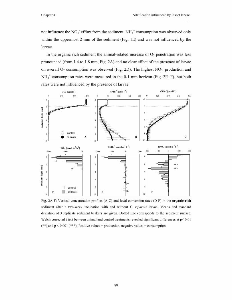
Chapter 4 Nitrification influenced by insect larvae _____________________________________________________________________
88
not influence the NO3- efflux from the sediment. NH4
+ consumption was observed only
within the uppermost 2 mm of the sediment (Fig. 1E) and was not influenced by the
larvae.
In the organic rich sediment the animal-related increase of O2 penetration was less
pronounced (from 1.4 to 1.8 mm, Fig. 2A) and no clear effect of the presence of larvae
on overall O2 consumption was observed (Fig. 2D). The highest NO3- production and
NH4+ consumption rates were measured in the 0-1 mm horizon (Fig. 2E+F), but both
rates were not influenced by the presence of larvae.
Fig. 2A-F: Vertical concentration profiles (A-C) and local conversion rates (D-F) in the organic-rich
sediment after a two-week incubation with and without C. riparius larvae. Means and standard
deviation of 3 replicate sediment beakers are given. Dotted line corresponds to the sediment surface.
Welch corrected t-test between animal and control treatments revealed significant differences at p< 0.01
(**) and p < 0.001 (***). Positive values = production, negative values = consumption.
D
-800 -400 0
0
2
4
6
8
10
sedi
men
t dep
th [m
m]
RO2 [mmol m-3 h-1]
E
-200 -100 0 100
0
2
4
6
8
10
RNH4+ [mmol m-3 h-1]
F
-200 -100 0 100 200
0
2
4
6
8
10
RNO3- [mmol m-3 h-1]
A
-2
0
2
4
6
8
10
0 100 200 300
cO2 [µmol l-1]
sedi
men
t dep
th [m
m]
C
-2
0
2
4
6
8
10
0 125 250 375 500
cNO3- [µmol l-1]
B
-2
0
2
4
6
8
10
0 50 100 150 200
cNH4+ [µmol l-1]
control animals
control animals
**
********
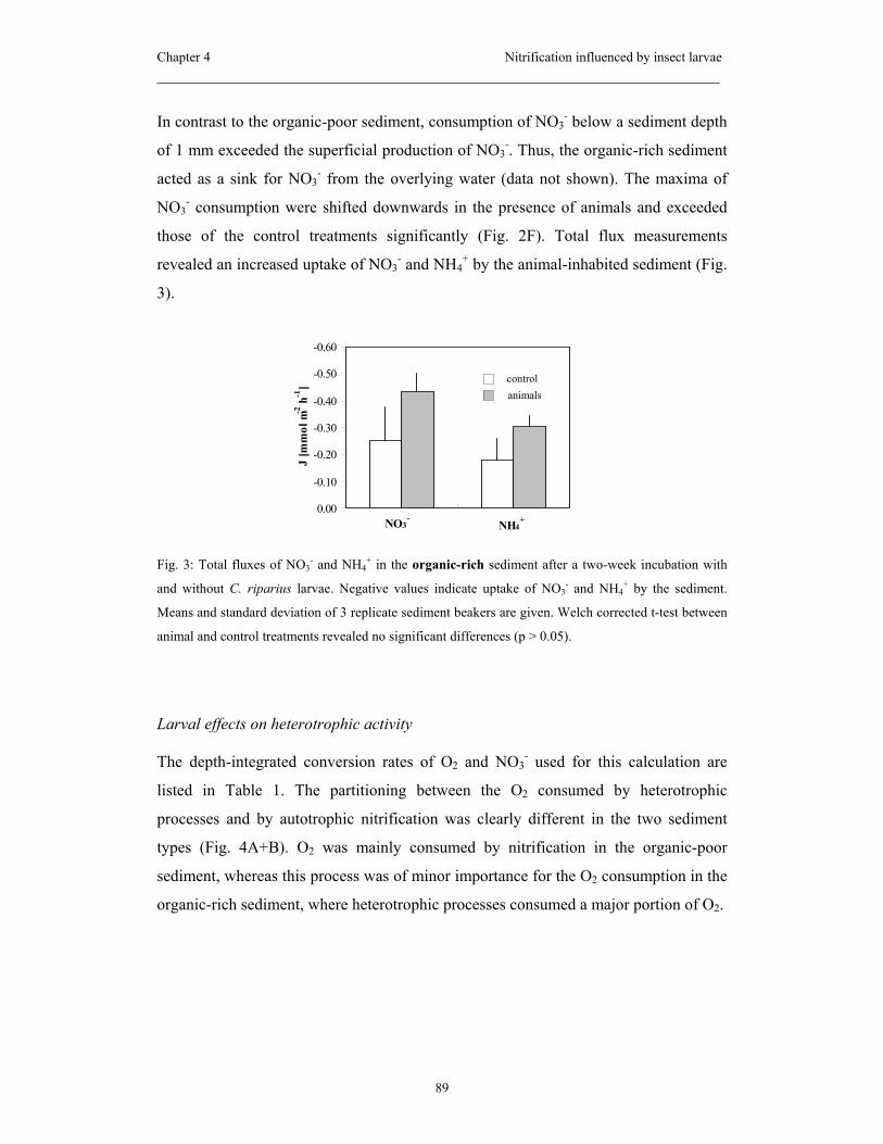
Chapter 4 Nitrification influenced by insect larvae _____________________________________________________________________
89
In contrast to the organic-poor sediment, consumption of NO3- below a sediment depth
of 1 mm exceeded the superficial production of NO3-. Thus, the organic-rich sediment
acted as a sink for NO3- from the overlying water (data not shown). The maxima of
NO3- consumption were shifted downwards in the presence of animals and exceeded
those of the control treatments significantly (Fig. 2F). Total flux measurements
revealed an increased uptake of NO3- and NH4
+ by the animal-inhabited sediment (Fig.
3).
Fig. 3: Total fluxes of NO3- and NH4
+ in the organic-rich sediment after a two-week incubation with
and without C. riparius larvae. Negative values indicate uptake of NO3- and NH4
+ by the sediment.
Means and standard deviation of 3 replicate sediment beakers are given. Welch corrected t-test between
animal and control treatments revealed no significant differences (p > 0.05).
Larval effects on heterotrophic activity
The depth-integrated conversion rates of O2 and NO3- used for this calculation are
listed in Table 1. The partitioning between the O2 consumed by heterotrophic
processes and by autotrophic nitrification was clearly different in the two sediment
types (Fig. 4A+B). O2 was mainly consumed by nitrification in the organic-poor
sediment, whereas this process was of minor importance for the O2 consumption in the
organic-rich sediment, where heterotrophic processes consumed a major portion of O2.
-0.60
-0.50
-0.40
-0.30
-0.20
-0.10
0.00
J [m
mol
m-2
h-1
]
NO3-
NH4+
controlanimals

Chapter 4 Nitrification influenced by insect larvae _____________________________________________________________________
90
Table 1. Depth-integrated conversion rates of O2 and NO3- (J) within the nitrification zone of organic-
poor and organic-rich sediment either devoid or inhabited by C. riparius larvae. Organic-poor: Means
of three replicate cores within one sampling date (+ 1 SD, n ≥ 3), organic-rich: Means of three replicate
cores within one sampling date (+ 1 SD, n ≥ 5). Positive values = production, negative values =
consumption.
organic-poor organic-rich J [mmol m-2 h-1] control animals control animals
O2 -0.915 ± 0.236 -0.489 ± 0.183 -0.699 ± 0.117 -0.957 ± 0.206
NO3- 0.404 ± 0.027 0.199 ± 0.121 0.105 ± 0.098 0.016 ± 0.110
Moreover, in the organic-poor sediment the presence of the animals caused a decrease
in O2 consumption by nitrification. The heterotrophic O2 consumption remained
unchanged. In the organic-rich sediment, we found no animal-related change in the O2
consumption due to nitrification but the heterotrophic O2 uptake was enhanced
significantly in the presence of the larvae.
Fig. 4A, B: O2 consumption by nitrification and by heterotrophic processes in the organic-poor (A) and
organic-rich sediment (B) after a two-week incubation with and without C. riparius larvae. Means and
standard deviation of 3 replicate sediment beakers are given. Welch corrected t-test between animal and
control treatments revealed significant differences at p< 0.001 (***).
dept
h in
tegr
ated
O2 c
onsu
mpt
ion
[mm
ol m
-2 h
-1]
***
A: poor
0.0
0.2
0.4
0.6
0.8
1.0controlanimals
B: rich
0.00.2
0.40.6
0.80.0
nitrifiers heterotrophs
controlanimals
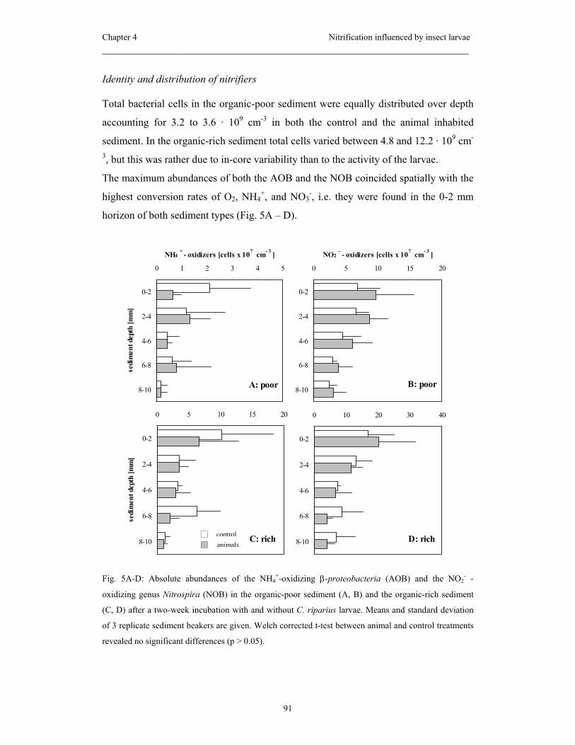
Chapter 4 Nitrification influenced by insect larvae _____________________________________________________________________
91
Identity and distribution of nitrifiers
Total bacterial cells in the organic-poor sediment were equally distributed over depth
accounting for 3.2 to 3.6 · 109 cm-3 in both the control and the animal inhabited
sediment. In the organic-rich sediment total cells varied between 4.8 and 12.2 · 109 cm-
3, but this was rather due to in-core variability than to the activity of the larvae.
The maximum abundances of both the AOB and the NOB coincided spatially with the
highest conversion rates of O2, NH4+, and NO3
-, i.e. they were found in the 0-2 mm
horizon of both sediment types (Fig. 5A – D).
Fig. 5A-D: Absolute abundances of the NH4+-oxidizing β-proteobacteria (AOB) and the NO2
- -
oxidizing genus Nitrospira (NOB) in the organic-poor sediment (A, B) and the organic-rich sediment
(C, D) after a two-week incubation with and without C. riparius larvae. Means and standard deviation
of 3 replicate sediment beakers are given. Welch corrected t-test between animal and control treatments
revealed no significant differences (p > 0.05).
A: poor
0 1 2 3 4 5
0-2
2-4
4-6
6-8
8-10
sedi
men
t dep
th [m
m]
NH4+ - oxidizers [cells x 107 cm- 3 ]
D: rich
0 10 20 30 40
0-2
2-4
4-6
6-8
8-10C: rich
0 5 10 15 20
0-2
2-4
4-6
6-8
8-10
sedi
men
t dep
th [m
m]
B: poor
0 5 10 15 20
0-2
2-4
4-6
6-8
8-10
NO2- - oxidizers [cells x 107 cm- 3 ]
controlanimals
Chapter 4 Nitrification influenced by insect larvae _____________________________________________________________________
92
In this layer the NH4+-oxidizing ß-proteobacteria, as determined with probe Nso1225,
accounted for 0.6 and 1.2% of all cells in the organic-poor and the organic-rich
sediment, respectively. NO2--oxidizing bacteria of the genus Nitrospira as detected
with probe Ntspa662 accounted for about 2% of all cells in both sediment types. The
abundances of Nitrospira sp. remained relatively high even with increasing depth,
making up at least 1.0% of all cells even in the lowest sampled sediment layer.
Nitrobacter sp., the most commonly isolated NOB, was not detected with probe Nit3
in any of the sediment treatments. In both sediments the presence of the chironomids
had no significant influence on the abundances and the distribution patterns of AOB
and NOB.
Discussion
In our experiment we combined the use of microsensors with FISH to evaluate the
influence of C. riparius larvae on both the activity and the distribution of the NH4+-
and NO2--oxidizing bacteria (AOB and NOB). Based on the results of a previous study
we hypothesized that the grazing of C. riparius would decrease the nitrifying activity
in the top layer of the sediment and that ventilation of deeper layers would increase the
nitrifying activity (Stief and de Beer 2002). These differences in nitrifying activity
were assumed to be regulated by the abundance of nitrifiers. The presence of larvae
reduced the nitrification activity in the top layer of organic-poor, but not in the
organic-rich sediment. The abundance of neither AOB nor NOB was affected by the
presence of animals in any of the two sediments.
Larval behavior in the two sediment types
The differential behavior of the chironomids, i.e. the extensive lateral sediment
reworking in the organic-poor sediment and the locally restricted activity in the
organic-rich sediment has major implications for interpretation of the microsensor
measurements. In the organic-poor sediment the vertical profiles recorded at random
spots of the sediment surface can be taken as representative of the average distribution
of solutes (see also Stief and de Beer 2002) because of the larval-induced
homogenization of the surface. In contrast, in the organic-rich sediment the
microsensor technique revealed only the animal impact on the bulk sediment in

Chapter 4 Nitrification influenced by insect larvae _____________________________________________________________________
93
between animal burrows, while the additional N conversions inside the burrows were
excluded. In this case, the overall effect of the chironomid larvae on total benthic N
conversions was more accurately represented by the total fluxes as determined with
whole core incubations (see also Matisoff et al. 1985; Archer and Devol 1992).
Larval effects on nitrification and heterotrophic O2 demand
The stimulation of benthic nitrification by chironomid larvae has been demonstrated in
several previous studies and was discussed to be a consequence of increased O2
penetration due to ventilation and extended surface area and the release of NH4+ by the
animals (Mayer et al. 1995; Pelegri and Blackburn 1996; Svensson 1998). However, in
our experiment, the animals lowered the overall nitrification rates in the organic-poor
sediment despite an improved O2 supply. Because the abundance of nitrifiers remained
almost unchanged in the presence of animals this decrease can be interpreted as a
reduction of the metabolic activity of single cells or, what is more likely, a decrease of
the active proportion of the nitrifying population. We assume that the ingestion and
digestion of organic particles by the larvae and the subsequent deposition and
distribution of fecal pellets within the surface layer of the sediment reduced the overall
metabolic activity of the particle-associated bacteria. The reduction of activity and
abundances of particle-associated bacteria by deposit-feeding invertebrates has also
been demonstrated in several other studies (Johnson et al. 1989; Rouf and Rigney
1993; Plante 2000). The metabolic recovery of different groups of bacteria, which have
survived gut passage, occurs likely at different rates. Nitrifiers, for example, are poor
competitors with heterotrophic bacteria because of their slow growth (Prosser 1989)
and comparably high Km values for O2 (Schramm et al. 1997) and may thus recover
less fast from the gut passage than heterotrophs. Indeed, the O2 consumption due to
nitrification was reduced in the presence of the larvae, whereas heterotrophic O2
consumption remained unchanged. Microsensor measurements on single fecal pellets
would here be helpful to further illuminate the role of the feces in overall sedimentary
nitrification activity.
In the bulk sediment between the burrows pervading the organic-rich sediment the
microsensor approach did not reveal an animal-induced effect on nitrification. In
contrast, the animal-inhabited sediment was characterized by a higher total uptake of
both NO3- and NH4
+ as revealed by the total fluxes. This was probably due to the high
Chapter 4 Nitrification influenced by insect larvae _____________________________________________________________________
94
number of permanent burrows displaying a high microbial activity (Kristensen et al.
1985; van de Bund et al. 1994; Svensson 1997). Although the presence of burrows
enhanced the uptake of NH4+ their contribution to nitrification in particular remains
unclear because at the same time no net production of NO3- occurred. The burrows
even acted as an additional sink for NO3- probably due to dissimilative NO3
-
consumption in the burrow walls and the surrounding sediment. Though the
microsensor approach did not reveal an animal effect on nitrification the heterotrophic
O2 consumption in the sediment between the burrows was increased by the animals.
This could be a consequence of the fertilization of heterotrophic microbes, but not
nitrifiers, with dissolved compounds released into the overlying water during the
feeding activities of the chironomids (Goedkoop et al., 1997). Similar observations
were made during an experiment with chironomids: Control sediment cores that were
exposed to the same overlying water as the animal-inhabited cores showed increasing
O2 consumption rates (M. Koschorreck, pers. comm.).
Identity and distribution of nitrifiers
In our investigation the presence of C. riparius larvae influenced the activity of
nitrifiers but not the abundance of AOB and NOB as it was revealed by FISH. Thus, C.
riparius larvae did neither efficiently decimate the nitrifying population, nor did they
significantly promote the growth of nitrifiers in the ventilation layer of the sediments
during the course of the experiment (2 weeks). The vertical patterns of activity and
abundance of nitrifiers do not necessarily match within the sediment column because
metabolically inactive cells may survive in both the oxic and even in the anoxic
sediment layers for extended periods (Gieseke et al. 2001; Morgenroth et al. 2000;
Schramm et al. 2000). Due to their ability to maintain a relatively high ribosome
content even at a stage of low metabolic activity, as given under starvation and other
stress conditions, nitrifiers are detectable with FISH even if no nitrifying activity can
be recorded with the microsensors. Applying methods for the detection of the single
cell activity may solve this dilemma in the future. For example, precursor rRNA has
been monitored and shown to be a promising tool for the determination of not only
size but also in situ activity of certain bacterial populations (Oerther et al. 2000;
Schmid et al. 2001). DNA-synthesizing cells can be detected by the uptake of the
Chapter 4 Nitrification influenced by insect larvae _____________________________________________________________________
95
halogenated thymidine analogue bromodeoxyuridine and consecutive FISH (Pernthaler
et al. 2002).
Summary and conclusions
Depending on the sediment type, the larvae showed a different behavior in terms of
structuring the sediment by their feeding and burrowing activities. As a consequence
nitrification was influenced differently: (i) in the organic-poor sediment its activity
was reduced by the extensive lateral larval grazing while (ii) its activity remained
rather unaffected in the space between animal burrows constructed in the organic-rich
sediment. In the latter case however, total flux measurements revealed the larval
burrows as additional sinks of NO3- rather than a site of net nitrification activity.
Extrapolated to a real world benthic ecosystem the sediment type may be decisive in
regulating the impact of C. riparius on the N cycle and hence the recycling of N to the
atmosphere.
This work stresses the need of further work applying a range of techniques to better
understand the complex interactions between sediment-dwelling animals and the
microbial community involved in benthic N cycle. Microsensor measurements i)
directly within the burrows and ii) at the surface of the larval feces would greatly
enhance our knowledge of local effects of bioturbation activities on nitrification. This
knowledge would even be further improved by the quantification of the actually active
portion of nitrifying bacteria.
Acknowledgements
We thank Enrique Llobet-Brossa and Armin Gieseke for their advice and helpful
comments regarding FISH in sediments. Gabi Eickert, Anja Eggers, and Ines Schröder
are acknowledged for the construction of the O2 microelectrodes. For critical
comments on the paper we thank Rebecca Ludwig and Armin Gieseke. For statistical
advice we are grateful to Werner Wosniok (University of Bremen).
Financial support was provided by the German Research Foundation (STI202/1),
and by the Max Planck Society, Germany.

Chapter 4 Nitrification influenced by insect larvae _____________________________________________________________________
96
References
Altmann, D., Stief, P., Amann, R., de Beer, D., and Schramm A. accepted. In situ
distribution and activity of nitrifying bacteria in freshwater sediment.
Environmental Microbiology.
Archer, D. and Devol, A. 1992. Benthic oxygen fluxes on the Washington Shelf and
Slope - a comparison of in situ microelectrode and chamber flux measurements.
Limnology and Oceanography 37: 614-629
Bartoli, M., Nizzoli, D., Welsh, D. T., and Viaroli, P. 2000. Short-term influence of
recolonisation by the polycheate worm Nereis succinea on oxygen and nitrogen
fluxes and denitrification: a microcosm simulation. Hydrobiologia 431: 165-
174
Daims, H., Nielsen, J. L., Nielsen, P. H., Schleifer, K. H., and Wagner, M. 2001. In
situ characterization of Nitrospira-like nitrite oxidizing bacteria active in
wastewater treatment plants. Applied and Environmental Microbiology 67:
5273-5284
de Beer, D. and Sweerts, J.-P. R. A. 1989. Measurement of nitrate gradients with an
ion-selective microelectrode. Analytica Chimica Acta 219: 351-356
de Beer, D. and van den Heuvel, J. C. 1988. Response of ammonium-selective
microelectrodes based on the neutral carrier nonactin. Talanta 35: 728-730
DeLong, E. F., Wickham, G. S., and Pace N. 1989. Phylogenetic stains: ribosomal
RNA--based probes for the identification of single cells. Science 243: 1360-
1363
Gieseke, A., Purkhold, U., Wagner, M., Amann, R., and Schramm, A. 2001.
Community structure and activity dynamics of nitrifying bacteria in a
phosphate-removing biofilm. Applied and Environmental Microbiology 67:
1351-1362
Gilbert, F., Stora, G., and Bonin, P. 1998. Influence of bioturbation on denitrification
activity in mediterranean coastal sediments: an in situ approach. Marine
Ecology Progress Series 163: 99-107
Chapter 4 Nitrification influenced by insect larvae _____________________________________________________________________
97
Goedkoop, W., Gullberg, K. R., Johnson, R. K., and Ahlgren, I. 1997. Microbial
response of a freshwater benthic community to a simulated diatom
sedimentation event: Interactive effects of benthic fauna. Microbial Ecology
34:131-143.
Herbert, R. A. 1999. Nitrogen cycling in coastal marine ecosystems. FEMS
Microbiology Reviews 23: 563-590
Johnson, R. K.; Boström, B., and van de Bund, W. 1989. Interactions between
Chironomus riparius (L.) and the microbial community in surficial sediments
of a shallow, eutrophic lake. Limnology and Oceanography 34: 992-1003
Kristensen, E., Jensen, M. H., and Andersen, T. K. 1985. The impact of polychaete
(Nereis virens Sars) burrows on nitrification and nitrate reduction in estuarine
sediments. Journal of Experimental Marine Biology and Ecology 85: 75-91
Llobet-Brossa, E., Rossello-Mora, R., and Amann, R. 1998. Microbial community
composition of Wadden Sea sediments as revealed by fluorescence in situ
hybridization. Applied and Environmental Microbiology 64: 2691-2696
Manz, W., Amann, R., Ludwig, W., Wagner, M., and Schleifer, K.-H. 1992.
Phylogenetic oligodeoxynucleotide probes for the major subclass of
proteobacteria: problems and solutions. Systematic and Applied Microbiology
15: 593-600
Matisoff, G., Fisher, J. B., and Matis, S. 1985. Effects of benthic macroinvertebrates
on the exchange of solutes between sediments and fresh-water. Hydrobiologia
122: 19-33
Mayer, M. S., Schaffner, L., and Kemp, W. M. 1995. Nitrification potentials of benthic
macrofaunal tubes and burrow walls: effects of sediment NH4+ and animal
irrigation behavior. Marine Ecology Progress Series 121: 157-169
Mobarry, B. K., Wagner, M., Urbain, V., Rittmann, B. E., and Stahl, D. A. 1996.
Phylogenetic probes for analyzing abundance and spatial organization of
nitrifying bacteria. Applied and Environmental Microbiology 62: 2156-2162

Chapter 4 Nitrification influenced by insect larvae _____________________________________________________________________
98
Morgenroth, E., Obermayer, A., Arnold, E., Brühl, A., Wagner, M., and Wilderer, P.
A. 2000. Effect of long-term idle periods on the performance of sequencing
batch reactors. Water Science and Technology 41: 105-113
Oerther, D. B., Pernthaler, J., Schramm, A., Amann, R., and Raskin, L. 2000.
Monitoring precursor 16S rRNAs of Acinetobacter spp. in activated sludge
wastewater treatment systems. Applied and Environmental Microbiology 66:
2154-2165
Pelegri, S. P. and Blackburn, T. H. 1994. Bioturbation effects of the amphipod
Corophium volutator on microbial nitrogen transformations in marine
sediments. Marine Biology 121: 253-258
Pelegri, S. P. and Blackburn, T. H. 1996. Nitrogen cycling in lake sediments
bioturbated by Chironomus plumosus larvae, under different degrees of
oxygenation. Hydrobiologia 325: 231-238
Pernthaler, A., Pernthaler, J., Schattenhofer, M., and Amann, R. 2002. Identification of
DNA-sythesizing bacterial cells in coastal North Sea plankton. Applied and
Environmental Microbiology 68: 5728-5736
Pernthaler, J., Glockner, F. O., Unterholzner, S., Alfreider, A., Psenner, R., and
Amann, R. 1998. Seasonal community and population dynamics of pelagic
bacteria and archaea in a high mountain lake. Applied and Environmental
Microbiology 64: 4299-4306
Plante, C. J. 2000. Role of bacterial exopolymeric capsules in protection from deposit-
feeder digestion. Aquatic Microbial Ecology 21: 211-219
Prosser, J. I. 1989. Autotrophic nitrification in bacteria. In: Rose A. H. and
Tempest D. W. (eds.) Advances in Microbial Physiology. Academic Press,
London
Revsbech, N. P. 1989. An oxygen microsensor with a guard cathode. Limnology and
Oceanography 34: 474-478

Chapter 4 Nitrification influenced by insect larvae _____________________________________________________________________
99
Rouf, M. A. and Rigney, M. M. 1993. Bacterial florae in larvae of the lake fly
Chironomus plumosus. Applied and Environmental Microbiology 59: 1236-
1241
Schmid, M., Schmitz-Esser, S., Jetten, M., and Wagner, M. 2001. 16S-23S rDNA
intergenic spacer and 23S rDNA of anaerobic ammonium-oxidizing bacteria:
implications for phylogeny and in situ detection. Environmental Microbiology
3: 450-459
Schramm, A., De Beer, D., Gieseke, A., and Amann, R. 2000. Microenvironments and
distribution of nitrifying bacteria in a membrane-bound biofilm. Environmental
Microbiology 2: 680-686
Schramm, A., Larsen, L. H., Revsbech, N. P., and Amann, R. 1997. Structure and
function of a nitrifying biofilm as determined by microelectrodes and
fluorescent oligonucleotide probes. Water Science and Technology 36: 263-270
Stief, P., and de Beer, D. 2002. Bioturbation effects of Chironomus riparius on the
benthic N-cycle as measured using microsensors and microbiological assays.
Aquatic Microbial Ecology 27: 175-185
Stief, P., de Beer, D., and Neumann, D. 2002. Small scale distribition of interstitial
nitrite in freshwater sediment microcosms: the role of nitrate and oxygen
availability, and sediment permeability. Microbial Ecology 43: 367-378
Svensson, J. M. 1997. Influence of Chironomus plumosus larvae on ammonium flux
and denitrification (measured by the acetylene blockage- and the isotope
pairing-technique) in eutrophic lake sediment. Hydrobiologia 346: 157-168
Svensson, J. M. 1998. Emission of N2O, nitrification and denitrification in a eutrophic
lake sediment bioturbated by Chironomus plumosus. Aquatic Microbial
Ecology 14: 289-299
van de Bund, W., Goedkoop,W., and Johnson, R. K. 1994. Effects of deposit feeder
activity on bacterial production and abundance in profundal lake sediment.
Journal of the North American Benthological Society 13: 532-539
Chapter 4 Nitrification influenced by insect larvae _____________________________________________________________________
100
Wagner, M., Rath, G., Koops, H.-P., Flood, J., and Amann, R. 1996. In situ analysis of
nitrifying bacteria in sewage treatment plants. Water Science and Technology
34: 237-244
Chapter 5
Summary
Zusammenfassung
Chapter 5 Summary _____________________________________________________________________
102
Summary
Nitrification is a central process of the benthic N cycle, which sets the preconditions
for the removal of excess N from anthropogenically impaired aquatic environments.
The fact that nitrification in freshwater sediments usually occurs within a thin oxic
surface layer necessitates techniques with sufficiently high spatial resolution for the in
situ investigation of this important microbial process. In previous studies of nitrifying
biofilms in wastewater treatment systems microsensors and fluorescence in situ
hybridization (FISH) served this purpose within even thinner oxic surface layers than
in sediments. Stimulated by this great achievement, the combination of microsensors
and FISH was also used in the present thesis to study some of the major ecological
features of nitrifying bacteria in freshwater sediments in situ.
Microsensors alone have been applied in some investigations of freshwater sediments
before, but FISH applicability was assumed to be hampered by low cell numbers of
nitrifiers and high background fluorescence in the sediment. Thus, the first experiment
(Chapter 2) was dedicated to evaluate the potential of this combined approach for the
investigation of in situ distribution and activity of nitrifying bacteria in model
freshwater sediment. Nitrification activity in this sediment as measured with
microsensors was highest in the uppermost 2 mm of the sediment, i.e. 0.2 µmol cm-3 h-
1, and comparable to rates measured in other freshwater sediments. Maximum
abundances of both NH4+- and NO2
--oxidizing bacteria (AOB and NOB) as determined
with FISH were 1.5 x 10⁷ cells cm-3 and 8.6 x 10⁷ cells cm-3, respectively, and
coincided spatially with maximum nitrification activity. Cell numbers of AOB and
NOB were 1-4 orders of magnitude higher compared to previous studies using the
most probable number technique. The present study also demonstrated the relevance of
the genus Nitrospira (and not Nitrobacter spp.) for in situ NO2--oxidation not only in
wastewater treatment systems, but also in freshwater sediments. For the first time, cell-
specific nitrification rates of the genus Nitrospira could be calculated for freshwater
sediment and added up to 1.2-2.7 fmol NO2- cell-1 h-1.
In a second experiment (Chapter 3) the combined approach of microsensors and FISH
was applied to investigate the activity and distribution of nitrifying bacteria in intact
field sediment (‘natural sediment’). The results were compared to sediment from the
Chapter 5 Summary _____________________________________________________________________
103
same sampling site, which had been sieved and homogenized in preparation for a
laboratory incubation experiment (‘manipulated sediment’). The genus Nitrospira was
the prevalent NO2--oxidizer not only in the model sediment used in the first study, but
also in the natural stream sediment used here. Nitrification activity in the natural
sediment was negligible and abundances of nitrifiers in the apparently oxic layer were
low. However, maximum abundances of both AOB and NOB were found in the 2-4
mm layer, which was anoxic at the time of the microsensor measurements. This may
have been a consequence of the downward shift of the abundance maximum towards
better availability of NH4+ in the field and the reduction of the O2 penetration depth
due to a lower current velocity during the laboratory measurements. Alternatively, it
may be indicative of the physical dynamics in the stream sediment, e.g. the horizontal
shifting of sediment ripples that slop formerly oxic layers. In the manipulated sediment
nitrification activity and abundances of both AOB and NOB were conspicuously
higher than in the natural sediment which was ascribed to the better availability of
NH4+ throughout the incubation. Nitrospira spp. could be detected with FISH for an
extended period of time in anoxic layers, into which they had been transported due to
homogenization. In contrast, the AOB population was quickly restricted to the oxic
surface layer in which they were supplied with O2 and NH4+ from the overlying water.
The pretreated sediment was characterized by a lower spatial heterogeneity compared
to the natural sediment, which is beneficial for the conduction of factorial experiments.
In the third experiment microsensors and FISH were applied to investigate the impact
of Chironomus riparius larvae (Insecta: Diptera) on the activity and distribution of
AOB and NOB in two sediments with different organic contents. C. riparius burrowed
within the upper 10 mm of the sediments, changed the O2 microdistribution therein and
grazed on detritus particles inclusive of the attached microorganisms. However, in
dependence on the sediment type the larvae behaved differently and thereby influenced
the nitrification activity in a different way: In the organic poor sediment the larvae
extensively reworked the sediment and overall nitrification activity was significantly
lowered. Since the larvae did not cause any significant changes in the abundances of
AOB or NOB, the reduction of nitrifying activity probably resulted from a general loss
of metabolic activity of the nitrifiers. This may have been a consequence of larval
deposit feeding on attached bacteria and the defecation of metabolically inactivated
cells back into the sediment. In the organic rich sediment larval feeding was locally
Chapter 5 Summary _____________________________________________________________________
104
restricted to the immediate surroundings of the sediment burrows. Hence, the
microsensor measurements did not reveal an animal-related impact on nitrification
activity in the sediment between the burrows. Abundances of AOB and NOB were
again not influenced by the presence of C. riparius. However, total fluxes of NH4+ and
NO3- into the sediment were higher in the presence of larvae. Thus, microbial activities
were indeed apparent in the burrows, but they did not contribute to net nitrification in
the sediment as a whole. This study adds the plasticity of larval behavior to the
complexity of factors influencing benthic nitrification in bioturbated sediments.
Within the scope of this thesis microsensors and fluorescence in situ hybridization
(FISH) were successfully applied to investigate in situ activities and distribution of
NH4+-oxidizing bacteria (AOB) and NO2
--oxidizing bacteria (NOB) at a fine scale in
freshwater sediments. While with FISH these two physiological groups of nitrifiers
were identified and quantified, the microsensor measurements revealed the sediment
layers in which the nitrifiers were actually active and what effect their activity had on
the N exchange between the sediment and the overlying water. Findings of special
interest were (i) the quantitative prevalence of the NO2--oxidizing genus Nitrospira, so
far only known for its occurrence mainly in wastewater treatment systems, in both
model and natural stream sediments, (ii) the detectability of AOB and NOB with FISH
in anoxic sediment layers as a consequence of their high ribosome content even when
not actively nitrifying, and (iii) the plasticity of larval behavior in dependence on the
sediment type which adds even more complexity to the array of factors influencing
benthic nitrification in bioturbated sediments.
Chapter 5 Zusammenfassung _____________________________________________________________________
105
Zusammenfassung
Nitrifikation ist ein zentraler Prozess innerhalb des benthischen N-Kreislaufes, welcher
die Voraussetzungen für die Entfernung von überschüssigem N aus anthropogen
beeinträchtigten aquatischen Lebensräumen schafft. Die Tatsache, dass Nitrifikation in
limnischen Sedimenten normalerweise in einer sehr dünnen oxischen
Oberflächenschicht vorkommt, erfordert Messtechniken mit einer ausreichend hohen
räumlichen Auflösung, um in situ Untersuchungen dieses wichtigen mikrobiellen
Prozesses vornehmen zu können. In früheren Studien, die sich mit der Untersuchung
nitrifizierender Biofilme aus Kläranlagen befassen, konnten Mikrosensoren und die
Fluoreszenz in situ Hybridisierung (FISH) in sogar noch dünneren oxischen
Oberflächenschichten als im Sediment erfolgreich angewendet werden. Angeregt
durch diese Ergebnisse wurde die Kombination der Mikrosensoren mit der FISH in der
vorliegenden Arbeit genutzt, um einige der hauptsächlichen ökologischen Aspekte
nitrifizierender Bakterien in Süßwassersedimenten zu untersuchen.
Mikrosensoren wurden bereits in früheren Untersuchungen von Süßwassersedimenten
eingesetzt, die Anwendbarkeit von FISH wurde bisher jedoch aufgrund sehr geringer
Zellzahlen der Nitrifizierer und hoher Hintergrundfluoreszenz im Sediment in Frage
gestellt. Aus diesem Grund bestand das Ziel der ersten Studie (Kapitel 2) darin, das
Potential dieses kombinierten Methodenansatzes für die Untersuchung der in situ
Verteilung und Aktivität nitrifizierender Bakterien in einem Modell-Süßwasser-
Sediment zu erkunden. Die Nitrifikationsaktivität in diesem Sediment, gemessen mit
Mikrosensoren, war am höchsten innerhalb der obersten 2 mm des Sedimentes, d. h.
0.2 µmol cm-3 h-1, und war vergleichbar mit Raten, die in anderen
Süßwassersedimenten gemessen wurden. Maximale Abundanzen von NH4+- und NO2
--
oxidierenden Bakterien (AOB and NOB), bestimmt mit FISH, betrugen 1.5 x 10⁷
Zellen cm-3 and 8.6 x 10⁷ Zellen cm-3, und stimmten räumlich mit der maximalen
Nitrifikationsaktivität überein. Die Zellzahlen von AOB und NOB waren 1-4
Größenordnungen höher als die in früheren Studien mit Hilfe der „Most Probable
Number“- Methode ermittelten Abundanzen. In der vorliegenden Untersuchung wurde
eine quantitative Bedeutung der Gattung Nitrospira (und nicht Nitrobacter spp.) für
die in situ NO2--Oxidation festgestellt. Dies galt bisher vor allem für Biofilme aus
Chapter 5 Zusammenfassung _____________________________________________________________________
106
Kläranlagen. Zum ersten Mal konnten auch zellspezifische Nitrfikationsraten für die
Gattung Nitrospira im Süßwassersediment berechnet werden, sie betrugen bis zu 1.2-
2.7 fmol NO2- Zelle-1 h-1.
Innerhalb der zweiten Studie (Kapitel 3) wurde diese Methodenkombination
angewendet, um die Aktivität und Verteilung nitrifizierender Bakterien in ungestörtem
Sediment im Freiland (‘natural sediment’) zu untersuchen. Die Ergebnisse wurden mit
einem Sediment vom gleichen Standort verglichen, welches in Vorbereitung auf ein
Laborinkubations-Experiment jedoch gesiebt und homogenisiert wurde (‘manipulated
sediment’). Die Gattung Nitrospira war nicht nur im Modell-Sediment aus der ersten
Studie, sondern auch in dem natürlichen Fließgewässersediment der vorherrschende
NO2--Oxidierer. Die Nitrifikationsaktivität im natürlichen Sediment war unbedeutend
und die Abundanzen der Nitrifikanten in der oxischen Oberflächenschicht waren
gering. Die maximalen Abundanzen von AOB und NOB waren dagegen in der
darunterliegenden 2-4 mm Schicht zu finden, welche zur Zeit der Mikrosensor-
Messungen anoxisch war. Dies könnte durch eine Verlagerung des
Abundanzmaximums an den unteren Rand der oxischen Schicht (mit höherer NH4+-
Verfügbarkeit) und eine Verringerung der Eindringtiefe von O2 im Labor verursacht
worden sein. Eine andere Erklärungsmöglichkeit bieten die dynamischen
Umlagerungsprozesse im Fließgewässersediment, z.B. horizontal wandernde
Sedimentrippel, die ehemals oxische Zonen überlagern. Im behandelten Sediment
waren sowohl die Nitrifikationsaktivitäten als auch die Abundanzen der AOB und
NOB auffallend höher als im natürlichen Sediment, was auf die bessere NH4+-
Verfügbarkeit während der Laborinkubation zurückgeführt werden kann. Bakterien der
Gattung Nitrospira waren mit der FISH über einen längeren Zeitraum in anoxischen
Sedimentschichten detektierbar, in die sie durch die Sedimenthomogenisierung
verlagert worden waren. Im Gegensatz dazu war die AOB Population bereits nach
kurzer Inkubationszeit auf die oxische Schicht beschränkt, in der sie mit O2 und NH4+
aus der Wassersäule versorgt wurden. Das vorbehandelte Sediment war durch eine,
verglichen mit dem natürlichen Sediment, niedrigere räumliche Heterogenität
gekennzeichnet (Abundanzen und Aktivitäten der Nitrifikanten), was es für
Experimente geeignet macht, bei denen die Untersuchung ausgewählter Faktoren im
Vordergrund steht.
Chapter 5 Zusammenfassung _____________________________________________________________________
107
In der dritten Studie (Kapitel 4) wurden Mikrosensoren und FISH genutzt, um die
Auswirkung von Chironomus riparius Larven (Insecta: Diptera) auf die Aktivität und
Verteilung von AOB und NOB in zwei Sedimenten mit unterschiedlichem organischen
Gehalt zu untersuchen. Die Fress- und Grabaktivität von C. riparius beschränkte sich
auf die oberen 10 mm des Sediments und führte dort zu Veränderungen der
kleinräumigen O2-Verteilung. Die Larven ernährten sich von Detrituspartikeln und den
daran angehefteten Mikroorganismen. In Abhängigkeit vom Sedimenttyp verhielten
sich die Larven unterschiedlich und beeinflußten so die Nitrifikationsaktivität auf
verschiedene Weise: Im organisch-armen Sediment wurde das Sediment durch die
Fress- und Grabtätigkeit intensiv und weiträumig umgelagert; dies führte zu einer
signifikanten Verringerung der Nitrifikationsaktivität. Da es gleichzeitig zu keiner
signifikanten Änderung der Abundanzen von AOB und NOB kam, ist der Rückgang
der Nitrifikationsaktivität wahrscheinlich nur mit einer allgemeinen Reduktion der
metabolischen Aktivität der Nitrifikanten zu erklären. Dies könnte eine Folge der
Aufnahme und Verdauung partikelassoziierter Bakterien sein, die anschließend -
metabolisch inaktiviert - wieder in das Sediment ausgeschieden wurden. Im organisch-
reichen Sediment beschränkte sich die Nahrungsaufnahme der Larven auf die
unmittelbare Umgebung ihrer permanenten Wohnröhren. Aus diesem Grund konnten
mit Mikrosensormessungen zwischen den Wohnröhren keine durch Larven
verursachten Veränderungen der Nitrifikationsaktivität festgestellt werden. Auch in
diesem Sedimenttyp waren die Abundanzen von AOB und NOB nicht durch die
Larven beeinflußt. In Gegenwart der Larven waren allerdings die Gesamtfluxe von
NH4+ und NO3
- vom Wasser ins Sediment erhöht. Das heißt, dass zwar mikrobielle
Umsetzungen in den Wohnröhren stattfanden, dass diese aber keine Auswirkungen auf
die Nettonitrifikation des Gesamtsediments hatten. Diese Studie zeigt, dass die
Plastizität des Larvenverhaltens ein weiterer wichtiger Faktor ist, der die Nitrifikation
im Sediment beeinflusst.
Im Rahmen dieser Doktorarbeit wurden Mikrosensoren und Fluoreszenz in situ
Hybridisierung (FISH) erfolgreich für die Untersuchung der in situ Aktivitäten und
der Verteilung von NH4+-oxidierenden (AOB) und NO2
--oxidierenden Bakterien
(NOB) mit hoher räumlicher Auflösung in Süßwassersedimenten verwendet. Während
mit FISH die beiden physiologischen Gruppen der Nitrifikanten identifiziert und
Chapter 5 Zusammenfassung _____________________________________________________________________
108
quantifiziert werden konnten, lieferten die Mikrosensormessungen Informationen
darüber, in welchen Sedimentschichten die Nitrifikanten tatsächlich aktiv waren und
welche Auswirkungen diese Aktivität auf den Austausch von N-Verbindungen
zwischen Sediment und Wassersäule hatte. Von besonderem Interesse waren folgende
Untersuchungsergebnisse: (i) die quantitative Bedeutung der NO2--oxidierenden
Gattung Nitrospira, deren Vorkommen bisher vor allem in Kläranlagen bekannt war,
(ii) die Detektion von inaktiven AOB und NOB in anoxischen Sedimentschichten mit
FISH aufgrund ihres hohen Ribosomengehaltes, und (iii) das komplexe
Larvenverhalten von Chironomus riparius als regulierender Faktor für die benthische
Nitrifikation.
Appendix List of Publications _____________________________________________________________________
109
List of Publications
Contributions to the manuscripts presented in this thesis
1) Altmann, D., Stief, P., Amann, R., de Beer, D. and A. Schramm.
In situ distribution and activity of nitrifying bacteria in freshwater sediment.
This manuscript has been accepted for publication in Environmental Microbiology
Outline and concept of the project by P.S. and A.S., experimental design by P.S. and D.A., microsensor construction and measurements by P.S., fluorescence in situ hybridisation by D.A., writing by D.A. with help of coauthors
2) Altmann, D., Stief, P., Amann, R. and D. de Beer
Distribution and activity of nitrifying bacteria in natural and manipulated stream
sediment
as determined with in situ techniques.
This manuscript will be submitted to Aquatic Microbial Ecology
Experimental design by D.A. and P.S., microsensor construction by D.A., microsensor measurements by D.A., fluorescence in situ hybridisation by D.A., writing by D.A. with help of coauthors
3) Altmann, D., Stief, P., Amann, R., and D. de Beer
Nitrification in freshwater sediments as influenced by insect larvae: Quantification
by
microsensors and fluorescence in situ hybridization.
This manuscript has been submitted to Microbial Ecology
Outline and concept of the project by P.S. and D.A., experimental design by P.S. and D.A., microsensor construction by D.A. and P.S., microsensor measurements by D.A. and P.S., fluorescence in situ hybridisation by D.A., writing by D.A. with help of coauthors

Appendix List of Publications _____________________________________________________________________
110
Further publications
1) Stief, P., Schramm, A., Altmann, D., and D. de Beer
Decreasing nitrification rates, despite substrate-enrichment, in freshwater sediment
microcosms: A microsensor study.
This manuscript has been accepted for publication in FEMS Microbiology Ecology.
2) Stief, P., Altmann, D., Kureck, A., and D. de Beer
Microbial activities in the burrow environment of the potamal fly Ephoron virgo.
This manuscript has been submitted for publication to Freshwater Biology.