Embed Size (px)
Citation preview
NIST Air-Kerma Standard for Electronic Brachytherapy Calibrations
Michael G. Mitch, Ph.D.
Leader, Dosimetry Group
Radiation Physics Division
Physical Measurement Laboratory
National Institute of Standards and Technology
Disclaimers
Certain commercial equipment, manufacturers, instruments, or materials are identified in this presentation in order to specify the experimental procedure adequately. Such identification is for informational purposes only and is not intended to imply recommendation or endorsement by the National Institute of Standards and Technology, nor is it intended to imply that the manufacturer, materials, or equipment are necessarily the best available for the purpose. Xoft, Inc. provided funding for the development of the NIST electronic brachytherapy facility and supplied systems for source control.
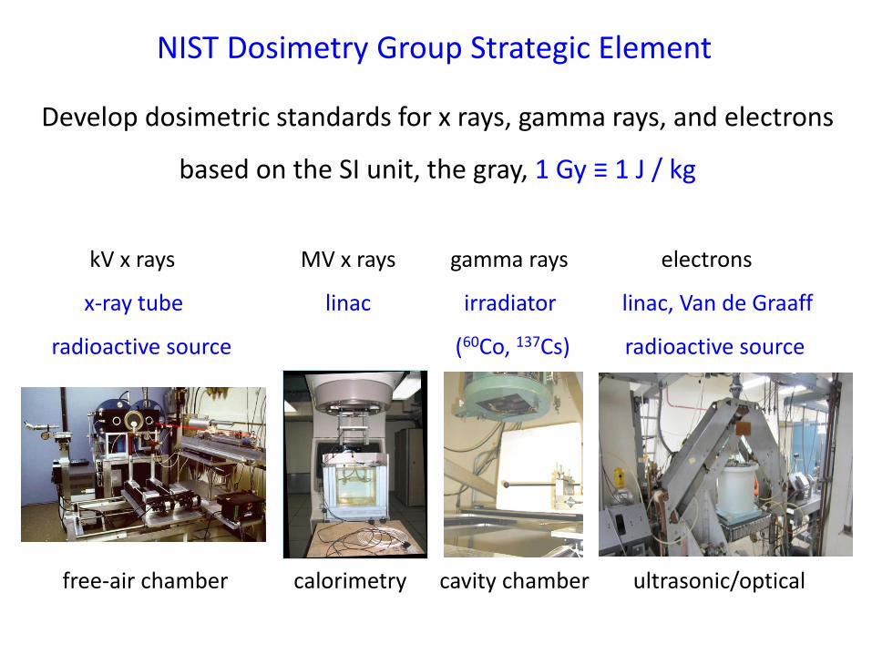
Develop dosimetric standards for x rays, gamma rays, and electrons
based on the SI unit, the gray, 1 Gy ≡ 1 J / kg
kV x rays MV x rays gamma rays electrons
x-ray tube linac irradiator linac, Van de Graaff
radioactive source (60Co, 137Cs) radioactive source
NIST Dosimetry Group Strategic Element
free-air chamber calorimetry cavity chamber ultrasonic/optical
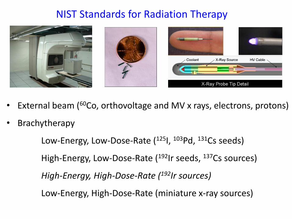
NIST Standards for Radiation Therapy
• External beam (60Co, orthovoltage and MV x rays, electrons, protons)
• Brachytherapy
Low-Energy, Low-Dose-Rate (125I, 103Pd, 131Cs seeds)
High-Energy, Low-Dose-Rate (192Ir seeds, 137Cs sources)
High-Energy, High-Dose-Rate (192Ir sources)
Low-Energy, High-Dose-Rate (miniature x-ray sources)
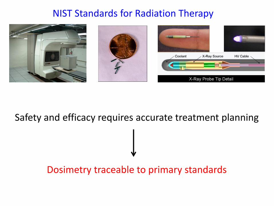
Safety and efficacy requires accurate treatment planning
Dosimetry traceable to primary standards
NIST Standards for Radiation Therapy
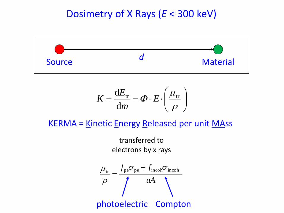
Dosimetry of X Rays (E < 300 keV)
KERMA = Kinetic Energy Released per unit MAss
d Source Material
trtr
d
dE
m
EK
uA
ff incohincohpepetr
photoelectric Compton
transferred to electrons by x rays
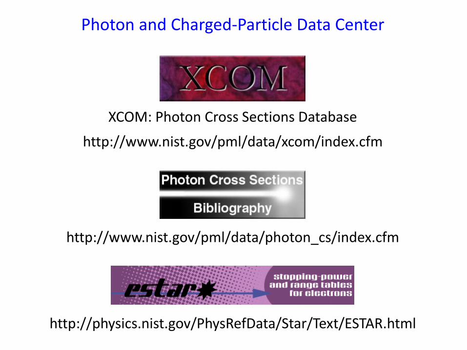
http://physics.nist.gov/PhysRefData/Star/Text/ESTAR.html
XCOM: Photon Cross Sections Database
http://www.nist.gov/pml/data/xcom/index.cfm
Photon and Charged-Particle Data Center
http://www.nist.gov/pml/data/photon_cs/index.cfm
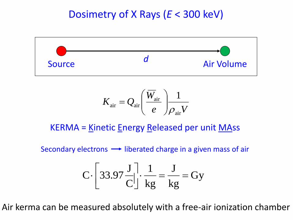
Air kerma can be measured absolutely with a free-air ionization chamber
d Source Air Volume
Ve
WQK
air
airairair
1
Secondary electrons liberated charge in a given mass of air
Gykg
J
kg
1
C
J33.97 C
Dosimetry of X Rays (E < 300 keV)
KERMA = Kinetic Energy Released per unit MAss

Free-Air Ionization Chamber (E < 300 keV)
i
ikVe
WQK
air
airairair
1
Air Kerma
W anode x-ray tube
filters
0
200
400
600
800
1000
0 20 40
E (keV)
Co
un
ts
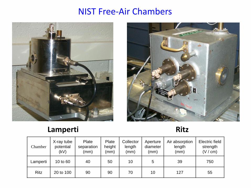
NIST Free-Air Chambers
Lamperti Ritz
Chamber
X-ray tube
potential
(kV)
Plate
separation
(mm)
Plate
height
(mm)
Collector
length
(mm)
Aperture
diameter
(mm)
Air absorption
length
(mm)
Electric field
strength
(V / cm)
Lamperti 10 to 60 40 50 10 5 39 750
Ritz 20 to 100 90 90 70 10 127 55
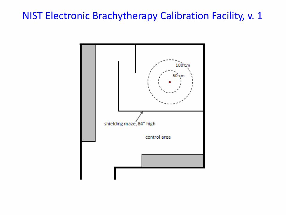
NIST Electronic Brachytherapy Calibration Facility, v. 1
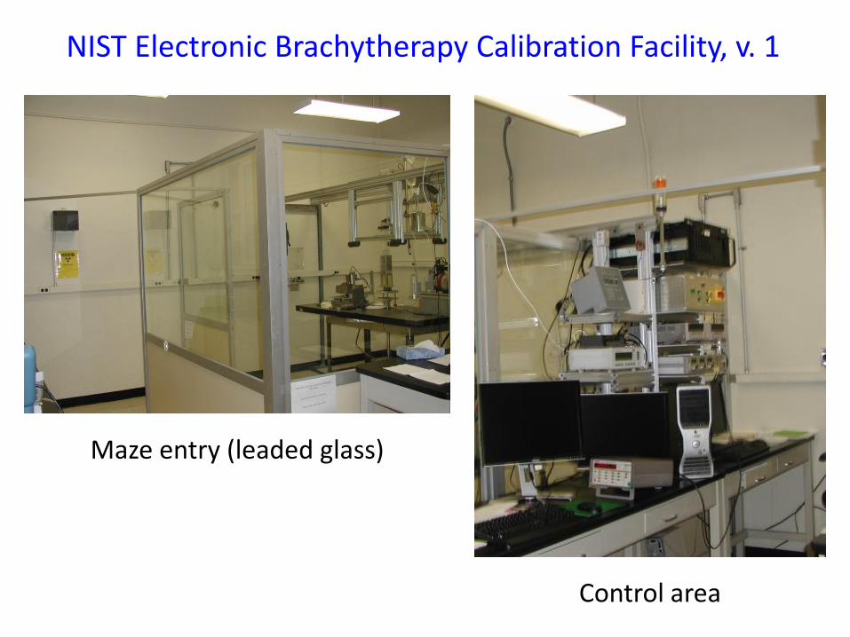
NIST Electronic Brachytherapy Calibration Facility, v. 1
Control area
Maze entry (leaded glass)
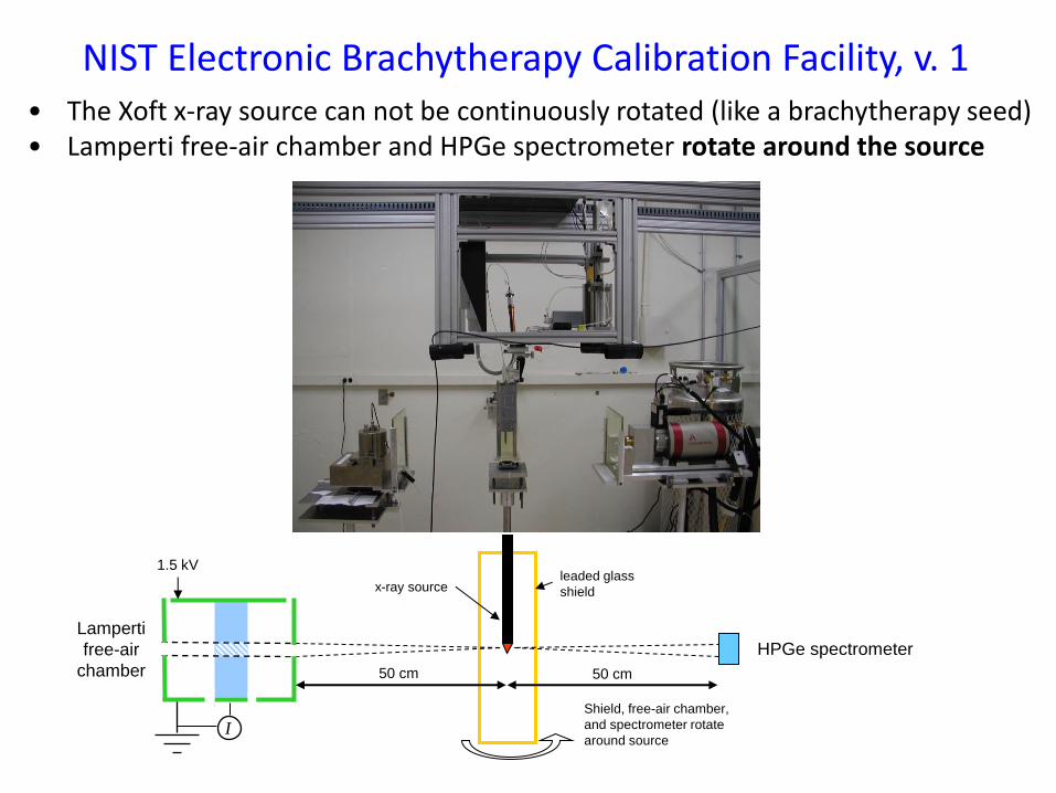
NIST Electronic Brachytherapy Calibration Facility, v. 1
leaded glass
shield x-ray source
Shield, free-air chamber,
and spectrometer rotate
around source
HPGe spectrometer
I
1.5 kV
50 cm
Lamperti
free-air
chamber 50 cm
• The Xoft x-ray source can not be continuously rotated (like a brachytherapy seed) • Lamperti free-air chamber and HPGe spectrometer rotate around the source
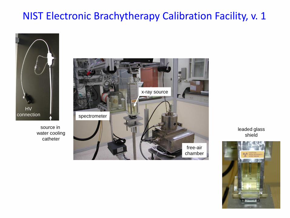
spectrometer
free-air
chamber
x-ray source
HV
connection
source in
water cooling
catheter
leaded glass
shield
NIST Electronic Brachytherapy Calibration Facility, v. 1
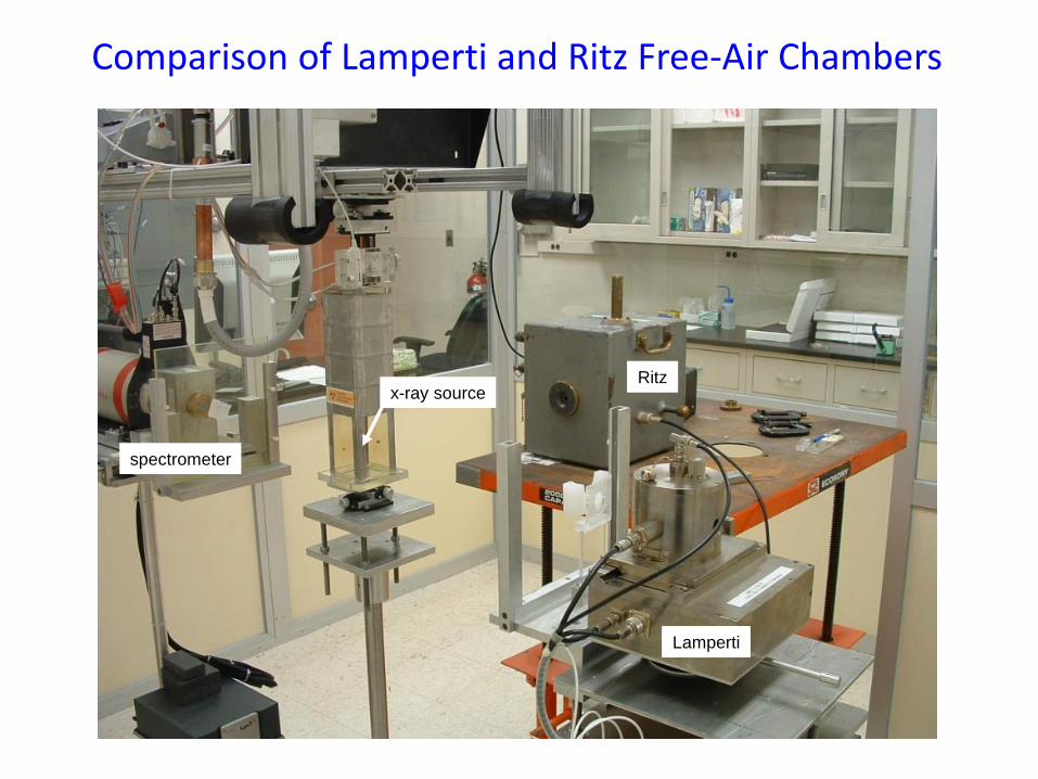
Ritz
Lamperti
spectrometer
x-ray source
Comparison of Lamperti and Ritz Free-Air Chambers
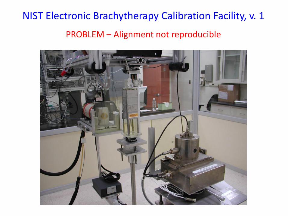
PROBLEM – Alignment not reproducible
NIST Electronic Brachytherapy Calibration Facility, v. 1
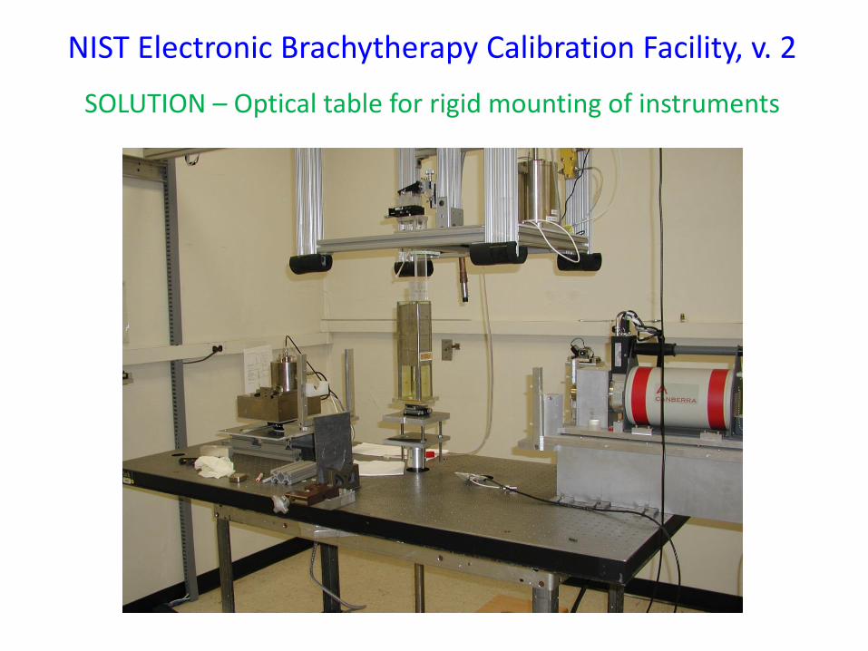
NIST Electronic Brachytherapy Calibration Facility, v. 2
SOLUTION – Optical table for rigid mounting of instruments
NIST Electronic Brachytherapy Calibration Facility, v. 2
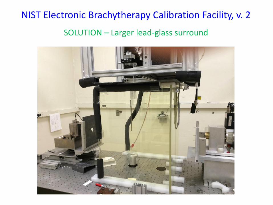
SOLUTION – Larger lead-glass surround
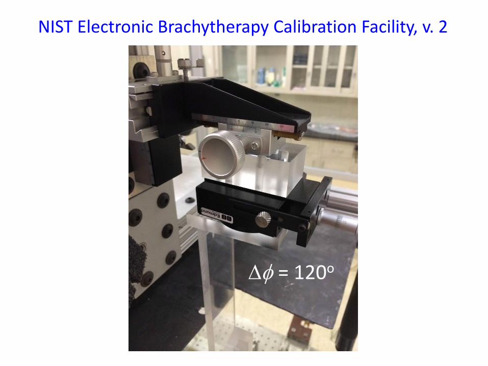
NIST Electronic Brachytherapy Calibration Facility, v. 2
Df = 120o
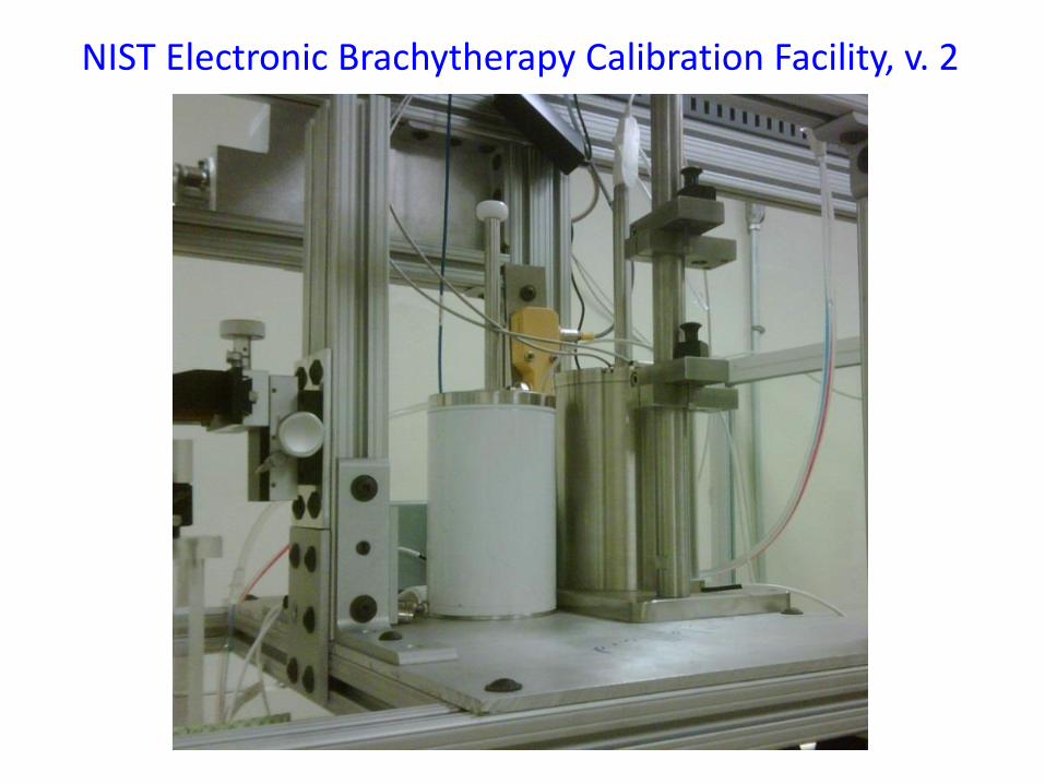
NIST Electronic Brachytherapy Calibration Facility, v. 2
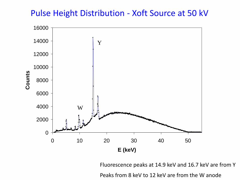
Pulse Height Distribution - Xoft Source at 50 kV
0
2000
4000
6000
8000
10000
12000
14000
16000
0 10 20 30 40 50
E (keV)
Co
un
ts
Fluorescence peaks at 14.9 keV and 16.7 keV are from Y
Peaks from 8 keV to 12 keV are from the W anode
Y
W
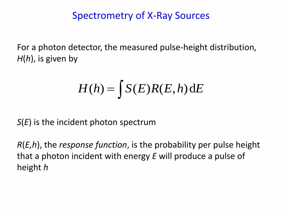
EhEREShH d),()()(
S(E) is the incident photon spectrum R(E,h), the response function, is the probability per pulse height that a photon incident with energy E will produce a pulse of height h
For a photon detector, the measured pulse-height distribution, H(h), is given by
Spectrometry of X-Ray Sources
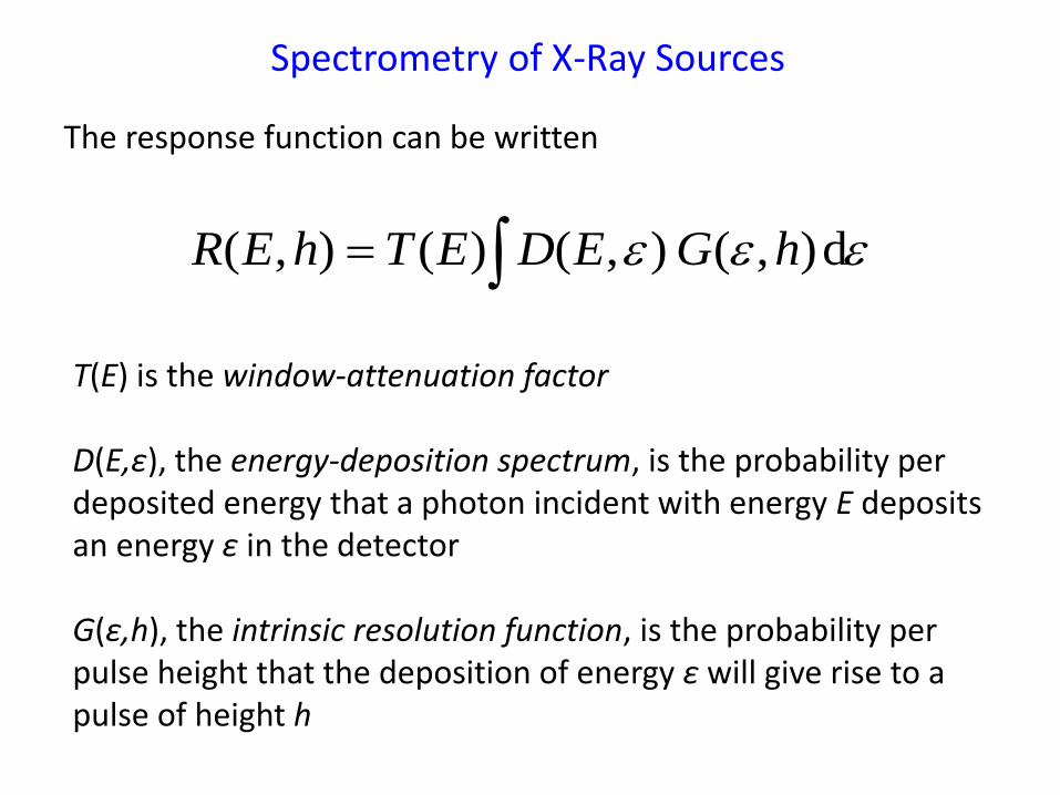
T(E) is the window-attenuation factor D(E,ε), the energy-deposition spectrum, is the probability per deposited energy that a photon incident with energy E deposits an energy ε in the detector G(ε,h), the intrinsic resolution function, is the probability per pulse height that the deposition of energy ε will give rise to a pulse of height h
d),(),()(),( hGEDEThER
The response function can be written
Spectrometry of X-Ray Sources
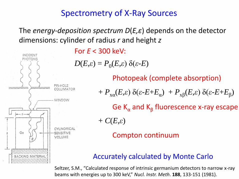
The energy-deposition spectrum D(E,ε) depends on the detector dimensions: cylinder of radius r and height z
Accurately calculated by Monte Carlo
Spectrometry of X-Ray Sources
For E < 300 keV:
D(E,ε) = P0(E,ε) δ(ε-E)
+ Pxα(E,ε) δ(ε-E+Eα) + Pxβ(E,ε) δ(ε-E+Eβ)
+ C(E,ε)
Photopeak (complete absorption)
Ge Kα and Kβ fluorescence x-ray escape
Compton continuum
Seltzer, S.M., “Calculated response of intrinsic germanium detectors to narrow x-ray beams with energies up to 300 keV,” Nucl. Instr. Meth. 188, 133-151 (1981).
10
100
1000
10000
0 10 20 30 40 50
Counts
Energy, keV
Pulse Height Distribution
True Photon Spectrum
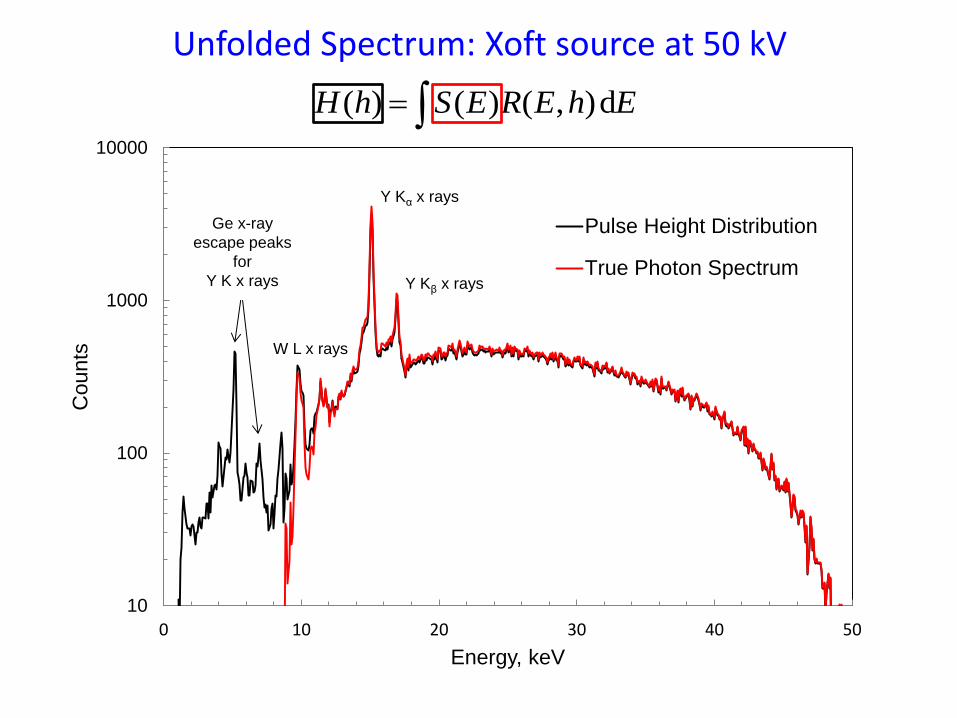
Unfolded Spectrum: Xoft source at 50 kV
EhEREShH d),()()(
Ge x-ray
escape peaks
for
Y K x rays Y Kβ x rays
Y Kα x rays
W L x rays
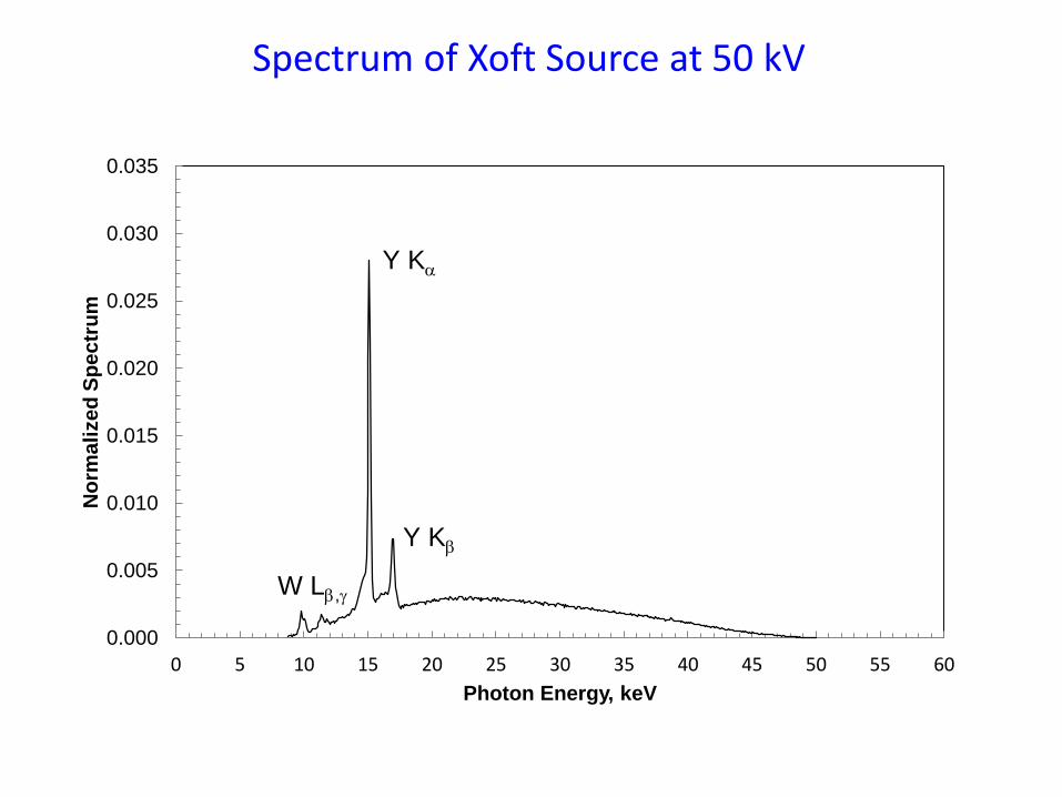
Spectrum of Xoft Source at 50 kV
0.000
0.005
0.010
0.015
0.020
0.025
0.030
0.035
0 5 10 15 20 25 30 35 40 45 50 55 60
No
rma
lize
d S
pe
ctr
um
Photon Energy, keV
Y Ka
Y Kb
W Lb,g
i
ikVe
WIK
air
airairair
1
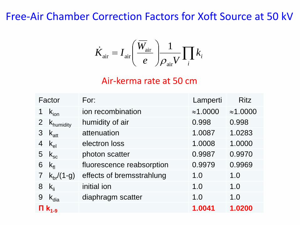
Factor For: Lamperti Ritz
1 kion ion recombination 1.0000 1.0000
2 khumidity humidity of air 0.998 0.998
3 katt attenuation 1.0087 1.0283
4 kel electron loss 1.0008 1.0000
5 ksc photon scatter 0.9987 0.9970
6 kfl fluorescence reabsorption 0.9979 0.9969
7 kbr/(1-g) effects of bremsstrahlung 1.0 1.0
8 kii initial ion 1.0 1.0
9 kdia diaphragm scatter 1.0 1.0
П k1-9 1.0041 1.0200
Free-Air Chamber Correction Factors for Xoft Source at 50 kV
Air-kerma rate at 50 cm
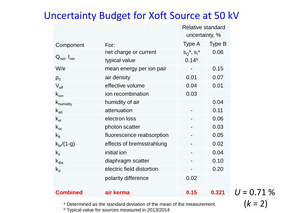
a Determined as the standard deviation of the mean of the measurement. b Typical value for sources measured in 2013/2014
Uncertainty Budget for Xoft Source at 50 kV
Component For:
Relative standard
uncertainty, %
Type A Type B
Qnet, Inet net charge or current sQ
a, sIa 0.06
typical value 0.14b
W/e mean energy per ion pair - 0.15
ρ0 air density 0.01 0.07
Veff effective volume 0.04 0.01
kion ion recombination 0.03
khumidity humidity of air 0.04
katt attenuation - 0.11
kel electron loss - 0.06
ksc photon scatter - 0.03
kfl fluorescence reabsorption - 0.05
kbr/(1-g) effects of bremsstrahlung - 0.02
kii initial ion - 0.04
kdia diaphragm scatter - 0.10
kd electric field distortion - 0.20
polarity difference 0.02
Combined air kerma 0.15 0.321 U = 0.71 % (k = 2)
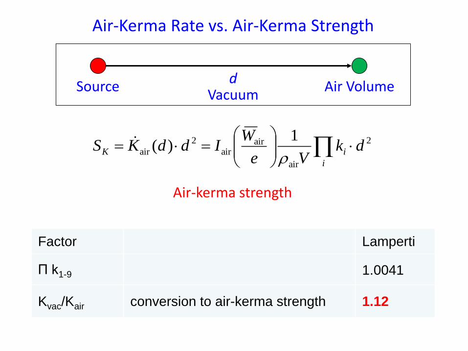
Air-Kerma Rate vs. Air-Kerma Strength
2
air
airair
2
air
1)( dk
Ve
WIddKS
i
iK
Air-kerma strength
Vacuum d
Source Air Volume
Factor Lamperti
П k1-9 1.0041
Kvac/Kair conversion to air-kerma strength 1.12
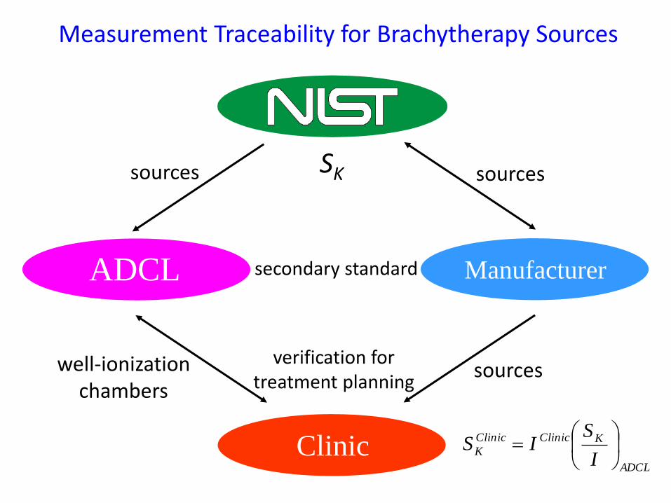
Manufacturer
sources sources
well-ionization chambers
sources
secondary standard
verification for treatment planning
ADCL
Clinic
Measurement Traceability for Brachytherapy Sources
SK
ADCL
KClinicClinic
KI
SIS
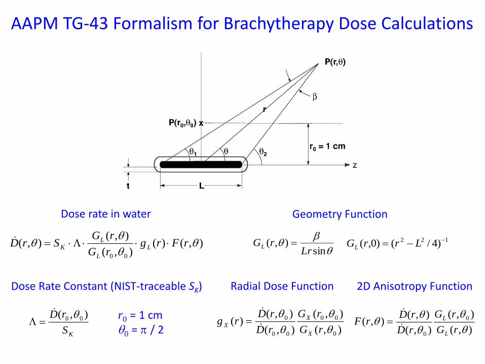
),()(),(
),(),(
00
rFrg
rG
rGSrD L
L
LK
Dose rate in water
KS
rD ),( 00
Dose Rate Constant (NIST-traceable SK)
b
sin),(
LrrGL 122 )4/()0,( LrrGL
Geometry Function
),(
),(
),(
),()(
0
00
00
0
rG
rG
rD
rDrg
X
X
X
Radial Dose Function
),(
),(
),(
),(),( 0
0
rG
rG
rD
rDrF
L
L
2D Anisotropy Function
r0 = 1 cm 0 = p / 2
AAPM TG-43 Formalism for Brachytherapy Dose Calculations
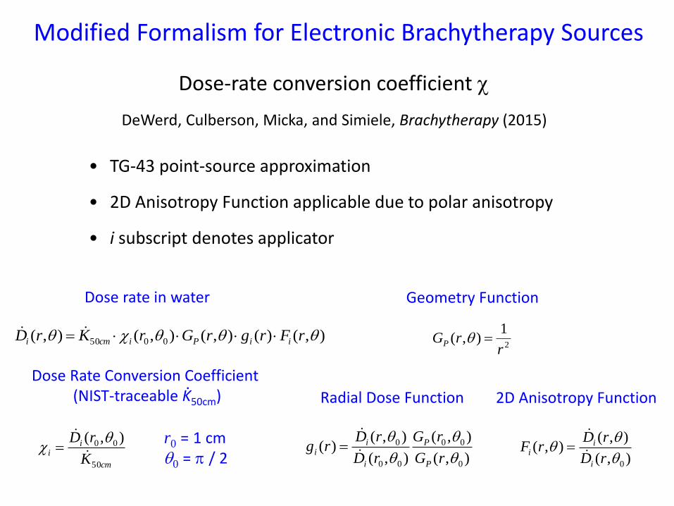
),()(),(),(),( 0050 rFrgrGrKrD iiPicmi
Dose rate in water
cm
ii
K
rD
50
00 ),(
Dose Rate Conversion Coefficient (NIST-traceable K50cm)
2
1),(
rrGP
Geometry Function
),(
),(
),(
),()(
0
00
00
0
rG
rG
rD
rDrg
P
P
i
ii
Radial Dose Function
),(
),(),(
0
rD
rDrF
i
ii
2D Anisotropy Function
r0 = 1 cm 0 = p / 2
Modified Formalism for Electronic Brachytherapy Sources
Dose-rate conversion coefficient
DeWerd, Culberson, Micka, and Simiele, Brachytherapy (2015)
.
• TG-43 point-source approximation
• 2D Anisotropy Function applicable due to polar anisotropy
• i subscript denotes applicator
AAPM Dosimetric Prerequisites
LDR Brachytherapy - Air-kerma strength calibrations traceable to NIST - TG-43 parameters published (experimental and Monte Carlo) - NIST standard transferred to the ADCLs - Annual comparisons between NIST and ADCLs
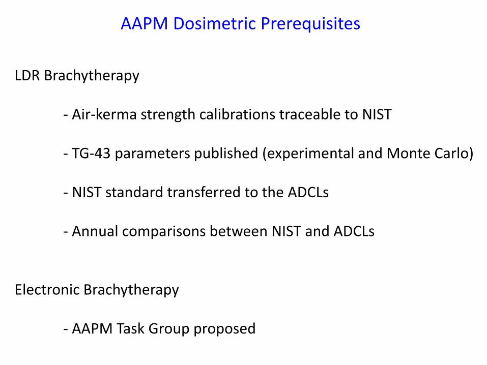
LDR Brachytherapy - Air-kerma strength calibrations traceable to NIST - TG-43 parameters published (experimental and Monte Carlo) - NIST standard transferred to the ADCLs - Annual comparisons between NIST and ADCLs Electronic Brachytherapy - AAPM Task Group proposed
AAPM Dosimetric Prerequisites
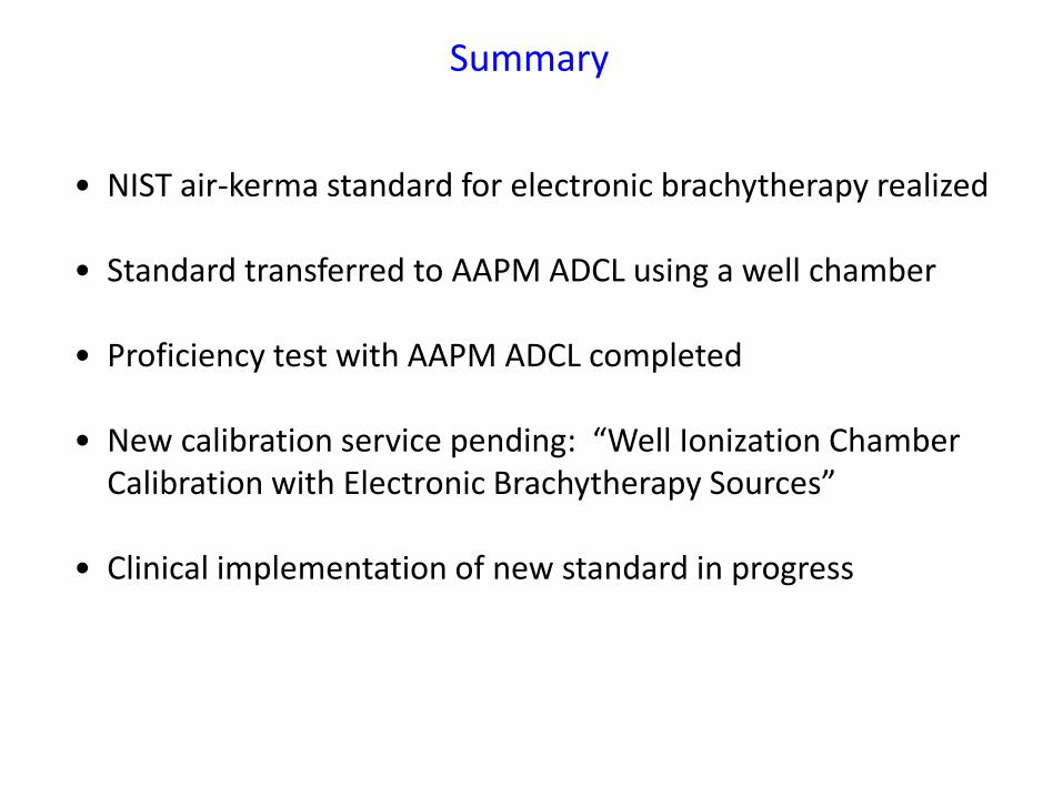
• NIST air-kerma standard for electronic brachytherapy realized
• Standard transferred to AAPM ADCL using a well chamber
• Proficiency test with AAPM ADCL completed • New calibration service pending: “Well Ionization Chamber
Calibration with Electronic Brachytherapy Sources”
• Clinical implementation of new standard in progress
Summary
Acknowledgements
• Xoft, Inc. – funding for the development of the NIST electronic brachytherapy facility; sources and associated equipment
• Mel McClelland and Dave Eardley – design, fabrication, and
installation of mechanical, electrical, and electronic systems
• Ron Tosh – computer control code
• Jason Walia – spectrometer calibration


![Calibration for Brachytherapy Sources€¦ · • Photon Source: Air kerma strength - symbol S k. Given as unit “U” where U[=] μGy- m2/h. Values of S k can be up to 260 U. •](https://img.pdfslide.us/doc/110x75/605c00080d3b994973099b6b/calibration-for-brachytherapy-sources-a-photon-source-air-kerma-strength-symbol.jpg)