Embed Size (px)
Citation preview

Enhanced midbrain response at 6-month follow-up in cocaineaddiction, association with reduced drug-related choice
Scott J. Moeller1, Dardo Tomasi2, Patricia A. Woicik1, Thomas Maloney1, Nelly Alia-Klein1,Jean Honorio3, Frank Telang2, Gene-Jack Wang1, Ruiliang Wang1, Rajita Sinha4, DeniCarise5,6, Janetta Astone-Twerell7, Joy Bolger1, Nora D. Volkow2,8, and Rita Z. Goldstein1,*
1Brookhaven National Laboratory, Upton, NY 119732National Institute on Alcohol Abuse and Alcoholism, Bethesda, MD 208923Stony Brook University, Stony Brook, NY 117944Yale University School of Medicine, New Haven, CT 065115Phoenix House Foundation, Inc., New York, NY 100356University of Pennsylvania, Philadelphia, PA 191047Samaritan Village, Inc., Briarwood, NY 114358National Institute on Drug Abuse, Bethesda, MD 20892
AbstractDrug addiction is characterized by dysregulated dopamine neurotransmission. Although dopaminefunctioning appears to partially recover with abstinence, the specific regions that recover andpotential impact on drug seeking remain to be determined. Here we used functional magneticresonance imaging (fMRI) to study an ecologically valid sample of 15 treatment-seeking cocaineaddicted individuals at baseline and 6-month follow-up. At both study sessions, we collected fMRIscans during performance of a drug Stroop task, clinical self-report measures of addiction severity,and behavioral measures of cocaine seeking (simulated cocaine choice); actual drug use inbetween the two study sessions was also monitored. At 6-month follow-up (compared withbaseline) we predicted functional enhancement of dopaminergically-innervated brain regions,relevant to the behavioral responsiveness toward salient stimuli. Consistent with predictions,whole-brain analyses revealed responses in the midbrain (encompassing the ventral tegmentalarea/substantia nigra complex) and thalamus (encompassing the mediodorsal nucleus) that werehigher (and more positively correlated) at follow-up than baseline. Increased midbrain activityfrom baseline to follow-up correlated with reduced simulated cocaine choice, indicating thatheightened midbrain activations in this context may be marking lower approach motivation forcocaine. Normalization of midbrain function at follow-up was also suggested by exploratorycomparisons with active cocaine users and healthy controls (who were assessed only at baseline).Enhanced self-control at follow-up was suggested by a trend for the commonly hypoactive dorsal
*Correspondence and requests for materials should be addressed to: Rita Z. Goldstein, Medical Research, Brookhaven NationalLaboratory, 30 Bell Ave., Bldg. 490, Upton, NY, 11973-5000; tel. (631) 344-2657; fax (631) 344-5260; [email protected].
Author ContributionsSJM, DT, NDV, and RZG designed research; DT, PAW, TM, NAK, FT, GJW, and RW performed research; PAW, RS, DC, JAT, andJB coordinated research recruitment; SJM, DT, JH, and RW analyzed data; SJM, DT, NDV, and RZG wrote the paper. All authorscritically reviewed content and approved final version for publication.
Disclosure/Conflict of InterestNone declared.
NIH Public AccessAuthor ManuscriptAddict Biol. Author manuscript; available in PMC 2013 November 01.
Published in final edited form as:Addict Biol. 2012 November ; 17(6): 1013–1025. doi:10.1111/j.1369-1600.2012.00440.x.
NIH
-PA Author Manuscript
NIH
-PA Author Manuscript
NIH
-PA Author Manuscript

anterior cingulate cortex to increase response during a drug-related context. Together, these resultssuggest that fMRI could be useful in sensitively tracking follow-up outcomes in drug addiction.
Keywordscocaine addiction; follow-up outcome; fMRI; midbrain; thalamus; dopamine; anterior cingulatecortex; drug Stroop
INTRODUCTIONA core feature of drug addiction is dysregulated dopaminergic neurotransmission, includingdecreased dopamine receptor availability and release (Volkow et al., 2004), and functionalimpairments in frontal brain regions innervated by dopamine (Goldstein and Volkow, 2002).Such dopaminergic dysregulation in addiction is associated with abnormalities in how brainreward circuits respond to drugs/drug-related stimuli (Robinson and Berridge, 2003) andnatural reinforcers (Garavan et al., 2000; Martin-Sölch et al., 2001), such that the pursuit ofdrugs surpasses that of other reinforcers (Goldstein and Volkow, 2002). For example, themore blunted the striatal dopaminergic release to a stimulant challenge (Martinez et al.,2007) and the lower the striatal dopamine D1 receptor availability (Martinez et al., 2009) incocaine abusers, the higher is the choice to self-administer cocaine over receiving money.However, as indicated by non-human primate studies (Beveridge et al., 2009; Melega et al.,1997) and human positron emission tomography (PET) studies (Volkow et al., 2001), suchstriatal dopaminergic dysregulation may partly recover with abstinence [but see (Iyo et al.,2004)]. Other studies using different imaging modalities and targeting different regions/systems have similarly revealed partial recovery with abstinence (Bell et al., 2011; Ernst andChang, 2008; Kim et al., 2006; Nordahl et al., 2005). However, the extent to which suchresults translate to an ecologically valid sample of treatment-seeking drug addictedindividuals remains to be determined.
The goal of the present functional magnetic resonance imaging (fMRI) study was to testwhether the blood oxygen level dependent (BOLD) response in dopaminergically-innervatedbrain regions changes over a six-month follow-up period in initially treatment-seekingcocaine addicted individuals. We were especially interested in functional enhancements ofthese regions, which we expected to correlate with measures of reduced drug seeking. Atbaseline and again at 6-month follow-up, subjects performed an emotionally salient (drugStroop) task while undergoing fMRI. In previous fMRI investigations of this drug Strooptask, active cocaine abusers showed midbrain response to the drug cues (Goldstein et al.,2009b), consistent with this region’s role in mediating the behavioral reactivity tomotivationally salient stimuli through phasic and tonic dopamine release (Montague et al.,2004; Schultz, 2010). Also on this task, active cocaine abusers showed hypoactivity indorsal and rostro-ventral subregions of the anterior cingulate cortex (ACC) (Goldstein et al.,2009a). These ACC subregions were normalized after administration of an indirectdopamine agonist (methylphenidate) as associated with better control of behavior (Goldsteinet al., 2010), consistent with these regions’ respective roles in cognitive control(Ridderinkhof et al., 2004) and emotional conflict resolution (Etkin et al., 2006).Collectively, these findings speak to the ability of this drug Stroop task to interrogatedopaminergically-innervated brain regions, which mediate functions of high pertinence to atreatment-seeking population (e.g., cognitive functioning, motivational salience). Theseprevious fMRI studies, in combination with previous PET studies showing recoveredmidbrain (Volkow et al., 2001), striatum (Volkow et al., 2001), and thalamic (Wang et al.,2004) function after abstinence in methamphetamine abusers, guided our currenthypotheses. We predicted that at 6-month follow-up (compared with baseline), subjects
Moeller et al. Page 2
Addict Biol. Author manuscript; available in PMC 2013 November 01.
NIH
-PA Author Manuscript
NIH
-PA Author Manuscript
NIH
-PA Author Manuscript
would show increased activity in the midbrain, ACC, thalamus, and striatum, as associatedwith reduced cocaine choice behavior (a measure previously shown to correlate with actualcocaine use (Moeller et al., 2009)].
METHODS & MATERIALSSubjects
Our main sample included 15 treatment-seeking cocaine abusers (11 males, 41.4 ± 9.1 yearsold), all right-handed, native English speakers, and free of sustained/maintenancemedications that could affect the BOLD signal for > 30 days prior and throughout the study(see Supplementary Information for subject exclusion criteria). All subjects twice completedour emotionally salient fMRI drug Stroop task (described below) without loss of data due tomotion or technical difficulties: at baseline after detoxification (≥ 3 weeks after last druguse: 111.5 ± 151.4 days) and then again at 6-month follow-up. The average time betweenscanning sessions for these subjects was 198 days (6.4 months) ± 30.5 days (1 month).Subjects were referred from three drug treatment facilities located in the New York Tri-Statearea (for information on these facilities, see Supplementary Information): Phoenix House(N=8), Samaritan Village (N=2), and Yale Cocaine Research Clinic (N=5). Subjects metcriteria for current cocaine dependence (N=11), or cocaine dependence in partial (N=3) orsustained remission (N=1), as determined by a comprehensive diagnostic interviewconducted at baseline (see Supplementary Information for interview components and subjectcomorbidities). We tracked subjects’ abstinence between the two scanning sessions (seeSupplementary Information for our three-tiered tracking system), which revealed that eightsubjects remained abstinent (they had been abstinent 190.9 ± 17.3 days at second studysession) while seven subjects had at least one lapse where cocaine was used between the twosessions. The last drug use for these seven relapsing subjects occurred 71.3 ± 61.8 days priorto the second study session. There were no differences between relapsers and abstainers inbaseline abstinence (i.e., the number of days of abstinence prior to the first scan) [t(7.0)=2.1,p>0.07]. There were also no differences in the amount of days elapsed between scans amongthose who relapsed versus those who remained abstinent [t(13)=1.0, p>0.3].
To establish norms, generalizability of results, and the prospect of normalization of function,we also compared these 15 treatment-seeking cocaine subjects with 13 actively usingcocaine subjects (verified by positive cocaine urine screen on study day) and 13 healthycontrol subjects with no history of substance abuse. Although comparisons between theseactive users and controls were reported previously (Goldstein et al., 2009a; Goldstein et al.,2009b), these analyses are entirely novel because these previous subjects have never beencompared with a treatment-seeking sample; and most importantly, here for the first time wereport direct comparisons between baseline and follow-up in these treatment-seekingsubjects. Because these matched active users and controls completed the drug Stroop taskonly once, all analyses that included active users and controls were considered exploratory,therefore primarily reported in Supplementary Information. All subjects provided writtenconsent to participate in accordance with the Stony Brook University Institutional ReviewBoard and the associated treatment facility’s Institutional Review Board.
TaskAfter training (Supplementary Information), subjects viewed 40 drug words and 40 matchedneutral (household) words (2000 ms per word), in a blocked on–off or off–on order (i.e.,drug–neutral or neutral–drug), counterbalanced between subjects and sessions (Goldstein etal., 2009a; Goldstein et al., 2010) (Figure S1). We selected a blocked design because priorbehavioral studies have demonstrated that salient stimuli (here, drug words) become morepotent when grouped together into blocks, rather than when intermixed with neutral trials
Moeller et al. Page 3
Addict Biol. Author manuscript; available in PMC 2013 November 01.
NIH
-PA Author Manuscript
NIH
-PA Author Manuscript
NIH
-PA Author Manuscript

(Holle et al., 1997); blocked designs, where the BOLD signal is measured between blocks oftrials and not individual trials (D’Esposito et al., 1999), are also more robust than event-related designs against vascular pulsatility effects on the midbrain [one of our main regionsof interest (ROIs)], which is an acknowledged concern (D’Ardenne et al., 2008). There wereeight 3.4 min task repetitions (four drug, four neutral), each containing two blocks of 20drug or neutral words, interleaved with a 20 s white fixation cross overlaid on a blackbackground. Each word trial consisted of a 500 ms fixation cross, a 2000 ms wordpresentation (for word reading), a 500 ms response window, and a 500 ms feedback slide(correct/incorrect). During the 500 ms response window, subjects had to press one of fourbuttons (yellow, blue, red, green) on a commercially available MRI response pad (Cedrusbrand Lumina model LP-400), matching the ink color of the word they had just read; wordcolor order was pseudorandomized across all task runs. Note that low response variability onthis task was expected [driven by the current blocked design and by the extended separationbetween word reading and button press that reduced working memory concerns but alsoreduced cognitive conflict (Goldstein et al., 2007)]. To further increase saliency of this task(beyond that already afforded by the salient drug words, see Supplementary Information),subjects performed each word sequence under one of four counterbalanced monetary rewardamounts (50¢, 25¢, 1¢, or 0¢), gained for correct performance for up to $75 of real money.The monetary value to be gained for correct performance was displayed at the beginning ofeach run and at the end of each trial. Because contrasting these task conditions would havebeen overly punitive in this relatively small sample size, and because money did not interactwith session or word for either task accuracy or reaction time (see SupplementaryInformation, where there was only a main effect of money on task performance that furtherattests to this task’s emotional salience and ability to tap into reward responsiveness), wecollapsed results across the money conditions throughout the behavioral and fMRI analyses.Therefore, and although our MRI scanner provides sufficient coverage of this region (exceptfor perhaps its most ventral aspects) (see Figure S2), we did not have specific hypotheses forthe orbitofrontal cortex.
Behavioral MeasuresTask Performance and Targeted Neuropsychological Drug-Choice Tasks—Reaction time and accuracy data were collected across all trials. The total money earnedthroughout the task was also ascertained. Since all subjects were treatment-seeking (andmore than half of the sample was abstinent throughout the study), targetedneuropsychological tasks of simulated implicit and explicit drug-seeking were adopted inlieu of actual drug use variables (the latter included a preponderance of zeros, complicatingdata analysis; nevertheless, we still explored baseline prediction of subsequent drug use, asdescribed in Results and Supplementary Information). These tasks were previouslyassociated with/modulated by actual drug use [cocaine urine status or frequency of recentcocaine use (Moeller et al., 2010; Moeller et al., 2009)], therefore of particular utility in thecurrent treatment-seeking population. In the current study, although not reachingsignificance likely due to sample size, cocaine choice was also positively associated withactual cocaine use (Supplementary Information). In brief, both drug-seeking tasks measurethe extent to which cocaine images are selected for viewing compared with pleasant,unpleasant, or neutral images. In the explicit task, choice is made between two fully visibleside-by-side images; in the implicit task, selections are made between pictures hidden underflipped-over cards (and thus location of the drug stimuli needs to be acquired throughexperience) (Moeller et al., 2009). Dependent variables were the total number of cocaineimages selected for viewing in each task (Table 1).
Moeller et al. Page 4
Addict Biol. Author manuscript; available in PMC 2013 November 01.
NIH
-PA Author Manuscript
NIH
-PA Author Manuscript
NIH
-PA Author Manuscript

MRI Data AcquisitionMRI scanning was performed on a 4T whole-body Varian/Siemens MRI scanner, equippedwith a self-shielded whole-body Siemens Sonata EPI hardware [maximum gradient strengthper channel 44mT/m, slew rate 176 mT/(m·ms)]. A standard quadrature head resonator isused for all studies. The BOLD responses were measured as a function of time using a T2*-weighted single-shot gradient-echo echoplanar imaging sequence (echo time/repetitiontime=20/1600 ms, 3.125×3.125 mm2 in-plain voxel size, 4 mm slice thickness, 1 mm gap,typically 33 coronal slices, 20×20×16.5 cm3 field of view, 64×64 matrix size, 90° flip angle,200 kHz bandwidth with ramp sampling, 128 time points, and 4 dummy scans to bediscarded to avoid nonequilibrium effects in the fMRI signal). Padding minimized motion,which was also monitored immediately after each fMRI run (Caparelli et al., 2003).Earplugs and headphones minimized the interference effect of scanner noise (Tomasi et al.,2005). Anatomical MRI started with the acquisition of a sagittal T1-weighted gradient-echolocalizer (TE/TR 10/100 ms, 5 mm slice thickness, 20 cm FOV, matrix size=256×192, 128phase encoding steps, 13 sec scan time). This was followed by an axial T1-weighted 3D-MDEFT (three-dimensional modified driven equilibrium Fourier transform) sequence (TE/TR=7/15ms, 0.94×0.94×1 mm spatial resolution, 256 readout and 192×96 phase-encodingsteps, partial k-space acquisition, 16 minutes scan time) (Lee et al., 1995), and a modifiedT2-weighted hyperecho sequence (Hennig and Scheffler, 2001). Images were reconstructedin IDL (Interactive Data Language, Research Systems, Boulder, CO) using a Hammingfilter, a phase correction method that produces minimal ghost artifacts, and an iterativephase correction to partially recover the signal loss due to susceptibility effects (conductedby an MRI physicist). Anatomical images were reviewed by a neurologist to rule out grossmorphological abnormalities.
MRI Data ProcessingAnalyses were performed with Statistical Parametric Mapping (SPM2) (Wellcome TrustCentre for Neuroimaging, London, UK). A six-parameter rigid body transformation (3rotations, 3 translations) was used for image realignment and for correction of head motion;criteria for acceptable motion were 2 mm displacement and 2° rotation in any of the axes inany of the task repetitions. The realigned datasets were spatially normalized to the standardstereotactic space of the Montreal Neurological Institute (MNI) using a 12-parameter affinetransformation (Ashburner et al., 1997) and a voxel size of 3×3×3 mm. An 8 mm full-width-half-maximum Gaussian kernel was used to spatially smooth the data.
BOLD fMRI AnalysesA general linear model (Friston et al., 1995) and a box-car design convolved with acanonical hemodynamic response function and high-pass filter (cut-off frequency: 1/520 s)was used to calculate the individual BOLD-fMRI maps. Four contrast maps per subject werecalculated, reflecting percent signal change from fixation for each of two task conditionsduring two scan sessions (drug and neutral epochs at baseline and follow-up). Theseindividual contrast maps were included in a second-order (random-effects) 2 (session:baseline, follow-up) × 2 (word: drug, neutral) repeated measures ANOVA SPM2 model. Wealso inspected whole-brain correlations (simple regressions), where the task performanceand clinical variables listed in Table 1 were entered as covariates regressed against the drugStroop contrasts. To specifically test the hypothesis of change, these whole-braincorrelations were conducted using follow-up>baseline difference scores. Considering themain effects reported below, correlations with the drug choice tasks and clinical variableswere conducted using the averaged drug Stroop contrasts (averaged across the drug andneutral contexts); correlations with the task specific accuracy and reaction time (RT)variables were conducted separately for the drug and neutral contexts. Thus, we performednine targeted whole-brain correlations, which employed similar whole-brain correction
Moeller et al. Page 5
Addict Biol. Author manuscript; available in PMC 2013 November 01.
NIH
-PA Author Manuscript
NIH
-PA Author Manuscript
NIH
-PA Author Manuscript

procedures as the ANOVA analyses (described below). In addition, to explore abstinence asa potential mechanism of change, we (A) repeated our main analyses while also includingrelapse (yes, no) as a between-subjects factor; and (B) conducted preliminary analyseswhere we used fMRI data (at baseline and change at follow-up) to predict actual drug usethat occurred between study sessions (these prediction analyses are primarily reported inSupplementary Information).
Brain activation clusters were corrected for multiple comparisons using the continuousrandom field calculation (Adler, 1981), here based on the expected Euler characteristics ofthe regions above a p<0.001 voxel uncorrected threshold, where clusters with at least 15contiguous voxels that were p<0.05 cluster-level corrected were considered significant forall analyses. This conservative threshold was chosen to minimize Type I error in this samplesize. An even more stringent statistical threshold (whole-brain p<0.05 family-wise errorcorrection at the voxel level, and p<0.05 error correction at the cluster level) was employedfor the between-group analyses because the groups were compared pairwise and post-hoc(see also Supplementary Information). Anatomical specificity was corroborated with theMRIcron software. Brain activation and deactivation clusters were further evaluated withcomplementary ROI analyses to identify outliers and to report (and graphically represent)average values in a volume comparable to the image smoothness [e.g., the volume of theresolution elements or “resels” (Worsley et al., 1992)], rather than single-voxel peak values.Thus, 9-mm isotropic cubic masks were created and centered at the exact coordinates inTable 2 and were kept invariant across subjects; the average BOLD fMRI signal amplitudesin these regions were computed using a custom program written in IDL. These ROIs, whichgive precise spatial localization of the functional responses (Tomasi et al., 2007a, b), wereanalyzed with the appropriate (e.g., ANOVA, correlation) analyses in SPSS, and were usedto rule out potentially confounding covariates (Supplementary Information).
RESULTSBelow, we report within-subjects (follow-up versus baseline) comparisons and brain-behavior correlations in the primary sample of 15 treatment-seeking cocaine subjects (TableS3 provides all task activations and deactivations in this sample across both study sessions).We also report between-group comparisons among the treatment-seeking subjects, activelyusing subjects, and healthy controls – but only those comparisons that directly bear on our apriori ROIs; all other effects from these between-group analyses are reported inSupplementary Information (see also Tables S4–S5; Figures S4–S5).
Behavior (Table 1)Task accuracy and RT (for the correct trials) were separately analyzed with 2 (session:baseline, follow-up) × 2 (word: drug, neutral) repeated measures ANOVAs. The clinicalself-report measures and neuropsychological drug-related tasks were separately analyzedwith paired t-tests. These behavioral measures generally did not differ between baseline andfollow-up (Table 1). Of all possible effects, only the word × session interaction for RTreached significance [F(1,14)=8.3, p<0.05], driven by faster performance at follow-up thanbaseline, but only during the neutral trials (Table 1).
SPM fMRI (Table 2)Whole-Brain Within-Subjects Analyses—The follow-up>baseline contrast revealedmain effects in the bilateral midbrain (ventral tegmental area/substantia nigra complex)(Figure 1A–1C) and right thalamus (mediodorsal nucleus) (Figure 1A, 1D). Table 2 lists theother regions that showed either follow-up>baseline or follow-up<baseline main effects; thelatter contrast revealed activations in regions of the control network (e.g., pre-supplementary
Moeller et al. Page 6
Addict Biol. Author manuscript; available in PMC 2013 November 01.
NIH
-PA Author Manuscript
NIH
-PA Author Manuscript
NIH
-PA Author Manuscript

motor area, inferior parietal cortex). At the voxel level, there was a word × sessioninteraction in the left dorsal ACC/midcingulate, explained by higher drug than neutralresponses at follow-up but a reverse pattern at baseline. However, this interaction did notreach cluster level significance (Table 2). There were no drug>neutral or drug<neutral maineffects (i.e., effects of word that were independent of session), and no effects for the rostro-ventral ACC or striatum.
Brain-Behavior Correlations—There was a negative correlation between changedresponse (follow-up>baseline) in the right midbrain and changed (follow-up>baseline)explicit cocaine choice behavior (Figure 2A), indicating that higher midbrain response wasassociated with reduced cocaine-seeking behavior. In contrast, changed cerebellum response(despite lack of main effects in this region) correlated with increased explicit cocaine choice(Table 2). Brain-behavior correlations with the other clinical severity measures were notsignificant.
In addition to these primary correlation analyses, preliminary correlation analyses revealedthat one midbrain ROI at baseline (x=12, y=−21, z=−15 mm) negatively predicted thenumber of days of any drug use (cocaine, alcohol, or marijuana) during the three monthspreceding the second study session (r=−0.59, p<0.05); this effect was driven by midbrainBOLD response to neutral words (r=−0.69, p<0.01). However, this effect was not confirmedwith whole-brain analysis. For all other results of these BOLD-fMRI correlations with actualdrug use, see Supplementary Information.
Midbrain-Thalamus Correlations—Given similar signal increases in midbrain andthalamus, and following our a priori hypotheses regarding enhanced BOLD signal in theseregions at follow-up, we undertook exploratory correlation analyses that tested for theirincreased correlation at follow-up. Masks around the two midbrain and one thalamus peakcoordinates from Table 2 (the follow-up>baseline contrast) were entered as seeds into threewhole brain correlation analyses, separately at baseline and follow-up (thus totaling sixanalyses). Contrasts for these analyses were response to averaged drug and neutral trialscompared with fixation. There were no correlations at baseline, but, as expected, one of thetwo midbrain seeds was significantly correlated with the bilateral thalamus at follow-up(Figure 2B–2C) (Table 2). Note that this midbrain-thalamus correlation remained significanteven after excluding the potential outlier in the upper right quadrants (p<0.05).
Effects of Relapse—We re-analyzed task accuracy and RT using 2 (session: baseline,follow-up) × 2 (word: drug, neutral) × 2 (relapse: yes, no) mixed ANOVAs; money gainedand the clinical variables were analyzed with 2 (session: baseline, follow-up) × 2 (relapse:yes, no) mixed ANOVAs. Including relapse into these analyses did not alter results; groupalso did not interact with any of these variables (p>0.1), indicating that change in taskperformance and clinical severity from baseline to follow-up did not differ as a function ofabstinence.
Similarly, adding the same between-subjects variable (relapse: yes, no) into ROI analyses(the extracted BOLD signals of the two midbrain ROIs and the one thalamic ROI asimplemented in SPSS) indicated that the enhanced activations in these regions from baselineto follow-up were not differentially driven by either the relapsers or non-relapsers (allp>0.05). Taken together, these analyses justify the inclusion of all treatment-seekers into asingle group.
Comparisons with Active Users or Healthy Controls: Treatment-Seekers at Baseline:At baseline, the treatment-seekers showed lower left midbrain activation than controls, butdid not differ from active users. This result indicates that the treatment-seekers may have
Moeller et al. Page 7
Addict Biol. Author manuscript; available in PMC 2013 November 01.
NIH
-PA Author Manuscript
NIH
-PA Author Manuscript
NIH
-PA Author Manuscript

had initially impaired functioning of this region, therefore suggesting that these treatment-seekers were representative of cocaine abusers at baseline (e.g., not initially different due toenhanced motivation to abstain) (Figure 3).
Comparisons with Active Users or Healthy Controls: Treatment-Seekers at Follow-Up:At follow-up, the treatment-seekers activated the midbrain more than active users (who wereassessed at baseline). In contrast, treatment-seekers at follow-up did not differ from healthycontrols (who were assessed at baseline) (Figure 3) (note, however, that when relaxing thethreshold to p<0.001 voxel uncorrected, controls continued to show higher midbrain activitythan the treatment-seekers: peak voxel: x=−12, y=−15, z=−9 mm, x=−9, 49 voxels, Z=4.4,p=0.021 cluster-level uncorrected). These results in the treatment-seekers at follow-up, incombination with results in the treatment-seekers at baseline (above) and the meanspresented in Figure 1, could collectively indicate that initial midbrain hypoactivations werepartially normalized after six months.
DISCUSSIONThe present longitudinal fMRI study revealed enhanced BOLD-fMRI activations in thedopaminergic midbrain and thalamus at 6-month follow-up compared with baseline ininitially treatment-seeking cocaine addicted individuals. Because this midbrain response todrug and neutral words was (A) enhanced at 6-month follow-up compared with baseline,and (B) at baseline, lower than healthy controls but not different from active users, whereasat follow-up, higher than active users but still somewhat lower than controls, we interpretthis increased BOLD response to indicate a partial restoration of response in this region,which was initially deactivated. Also consistent with this interpretation were thebehaviorally meaningful correlations between increased midbrain activity and reducedcocaine-seeking behavior, and between midbrain response at baseline and actual drug useover the next six months. Although the latter correlation was not confirmed with whole-brain analysis, it nonetheless supports the idea that baseline midbrain activity during thissalient task predicts positive abstinence-related outcomes; more generally, it also contributesto a growing body of literature in which baseline neuroimaging assessments are used topredict future drug-related outcomes (Brewer et al., 2008; Janes et al., 2010; Jia et al., 2011;Martinez et al., 2011; Paulus et al., 2005). Because midbrain dopamine heavily innervatesthe mediodorsal nucleus of the thalamus (Sanchez-Gonzalez et al., 2005), and because bothregions participate in conditioned reinforcement and reward expectation (Corbit et al., 2003;Mitchell et al., 2007; Volkow et al., 2003) and general reward processing (Liu et al., 2011),one could indeed expect these regions to show parallel functional enhancement. In furthersupport, we observed higher (more positive) correlations between the midbrain and thalamusat follow-up than baseline, which may suggest that over this 6-month study period there waspartial restoration of connectivity between these regions – a connection that is disrupted inactive users (Tomasi et al., 2010). Taken together, these midbrain and thalamus results echoprevious studies in substance abusers showing that increased responsiveness to positivereinforcers predicted better clinical outcome (Heinz et al., 2007; Lubman et al., 2009). Ourresults are also relevant to the concept of allostasis in addiction, such that theneuroadaptations from chronic exposure to addictive drugs, which change the threshold forrewards (Koob and Le Moal, 2008), may have partially recovered during this 6-monthfollow-up period. Behaviorally, such recovery might manifest as enhanced responsivenessduring a non-drug related context, possibly indicated in the current study by faster RTduring the neutral trials at follow-up. Overall, although BOLD-fMRI activity is an indirectmarker of dopaminergic neurotransmission (Tomasi et al., 2009) and although other (non-dopamine) systems may also be involved, we interpret these collective midbrain andthalamus effects, and correlations with reduced drug-seeking behavior and drug use, aspossibly marking improved dopaminergic functioning at follow-up.
Moeller et al. Page 8
Addict Biol. Author manuscript; available in PMC 2013 November 01.
NIH
-PA Author Manuscript
NIH
-PA Author Manuscript
NIH
-PA Author Manuscript

This study also revealed several unexpected/null findings. First, although we initiallyexpected higher cognitive control at follow-up compared with baseline, the only support forthis hypothesis was a word × session interaction in the dorsal ACC, which was significant atthe uncorrected voxel level but not the corrected cluster level. This trend, showing higheractivity to the drug words at follow-up, may reflect increased effort/control specificallyduring a drug-related context among individuals putatively motivated to remain abstinent(here, treatment-seekers at 6-month follow-up) (Nestor et al., 2011), befitting the positedrole of this region in response selection (Bush et al., 2002; Vogt, 2005) and in generalconflict resolution (Egner et al., 2008). Other regions relevant to cognitive control [e.g., pre-supplementary motor area and inferior parietal cortex (Seeley et al., 2007)] showed follow-up<baseline main effects, possibly reflective of higher cognitive demands when the task wasnovel (i.e., more activity was needed to achieve comparable performance). Anotherunexpected result was the lack of effects in the rostro-ventral ACC. Nevertheless, given themore pronounced group differences in this region at follow-up than baseline (i.e., whentreatment-seekers were compared with active users or controls) (SupplementaryInformation, Figure S5), within-subjects differences may indeed emerge with larger samplesizes. Finally, given that the striatum showed partial recovery following abstinence in bothanimal (Beveridge et al., 2009; Melega et al., 1997) and human (Volkow et al., 2001)studies, and given that the dorsal striatum was proposed as a mechanism of cocaine craving(Volkow et al., 2006), one could have anticipated the striatum to reduce activity to drugwords at follow-up. However, because this drug Stroop task did not reveal striatum effectsin our previous reports (Goldstein et al., 2009a; Goldstein et al., 2009b), null results for thisregion become less surprising.
The primary limitation of this study is the potential for practice effects. We attempted toreduce this concern by (A) controlling for sleepiness, which did not attenuate the currentfindings (Supplementary Information); and (B) performing exploratory between-group SPManalyses, which supported the idea that our midbrain main effect could reflect normalizationof an initially impaired function (Results) (although we acknowledge that these analyses,though buttressing the within-subject analyses, are subject to the same practice effects). Inaddition to these analyses, this concern of practice effects is lessened when considering thatmidbrain activity negatively correlated with sleepiness (Supplementary Information), therewas a substantial (6-month) interval between scanning sessions to minimize habituation, andenhanced midbrain response (that would indicate increased dopamine firing) more likelyreflects task engagement/motivation than disengagement (Boehler et al., 2011; Satoh et al.,2003; Volkow et al., 2010). Nevertheless, future longitudinal investigations should includecontrol subjects who also complete the task twice, and alternate versions of the task at thetwo time points. A second limitation is that our primary effects emerged averaged across thedrug and neutral words, complicating interpretations vis-à-vis cognitive control or reactivityto drug-related stimuli. Future studies with more subjects (power) and/or more lenientstatistical thresholds might be needed to detect differential effects as a function of word, aswas the case in our previous report (Goldstein et al., 2009b). A third limitation is that themidbrain correlation with drug choice, although cluster-level significant at p<0.05, was oneof nine whole-brain analyses. Given that this correlation would not have reachedsignificance with an additional Bonferroni correction (i.e., p<0.005 cluster-level corrected),it needs to be replicated in future studies. A final limitation is that the precise mechanism ofthe midbrain and thalamic effects remains to be studied further; in particular, the respectivecontributions of therapy, abstinence, and subject self-selection into the study need to bedisentangled in larger samples. Yet this latter limitation is balanced by ecological validity(studying treatment-seekers exposed to various treatment modalities), which increasesgeneralizability.
Moeller et al. Page 9
Addict Biol. Author manuscript; available in PMC 2013 November 01.
NIH
-PA Author Manuscript
NIH
-PA Author Manuscript
NIH
-PA Author Manuscript

In summary, to our knowledge the current results show for the first time elevated midbrainand thalamic fMRI response, and correlation between these regions, in initially treatment-seeking cocaine addicted individuals at a 6-month follow-up, and with associated decreasesin drug seeking. By scanning study subjects twice, the current study extends previous workthat has used baseline neural response to predict clinical outcome (Brewer et al., 2008; Janeset al., 2010; Jia et al., 2011; Martinez et al., 2011; Paulus et al., 2005). Consistent withprevious studies (Nestor et al., 2011; Volkow et al., 2001; Wang et al., 2004), the presentstudy found improvement effects in the more subcortical regions; in the more prefrontalcortical regions, impairments may be more intractable, as supported by studies revealingfunctional or structural abnormalities in addicted individuals even after ≥ 1 year abstinenceespecially in prefrontal cortical regions (Durazzo et al., 2011; Ersche et al., 2005; Nestor etal., 2011). The robust effects in the current study are striking in light of the markedheterogeneity of this treatment-seeking sample (e.g., length of abstinence, comorbidity, in-patient status, remission status, etc.), and the associated potential for increased variabilityand reduced power. Overall, our results suggest that functional imaging, and brain-behaviorcorrelations, could provide sensitive biomarkers of abstinence-related outcomes in drugaddiction – biomarkers that are detectable even in the general absence of behavioraldifferences.
Supplementary MaterialRefer to Web version on PubMed Central for supplementary material.
AcknowledgmentsThis study was supported by grants from the National Institute on Drug Abuse (to RZG: 1R01DA023579; to SJM:1F32DA030017-01).
We are grateful to staff at Phoenix House, Samaritan Village, and Yale for subject referrals and help with subjectfollow-ups.
Notice: This manuscript has been authored by Brookhaven Science Associates, LLC under Contract No. DE-AC02-98CHI-886 with the U.S. Department of Energy. The United States Government retains, and the publisher,by accepting the article for publication, acknowledges, a world-wide license to publish or reproduce the publishedform of this manuscript, or allow others to do so, for the United States Government purposes.
ReferencesAdler, RJ. The geometry of random fields. Wiley; Chichester: 1981.
Ashburner J, Neelin P, Collins DL, Evans A, Friston K. Incorporating prior knowledge into imageregistration. Neuroimage. 1997; 6:344–352. [PubMed: 9417976]
Bell RP, Foxe JJ, Nierenberg J, Hoptman MJ, Garavan H. Assessing white matter integrity as afunction of abstinence duration in former cocaine-dependent individuals. Drug Alcohol Depend.2011; 114:159–168. [PubMed: 21075564]
Beveridge TJ, Smith HR, Nader MA, Porrino LJ. Abstinence from chronic cocaine self-administrationalters striatal dopamine systems in rhesus monkeys. Neuropsychopharmacology. 2009; 34:1162–1171. [PubMed: 18769473]
Boehler CN, Hopf JM, Krebs RM, Stoppel CM, Schoenfeld MA, Heinze HJ, Noesselt T. Task-load-dependent activation of dopaminergic midbrain areas in the absence of reward. J Neurosci. 2011;31:4955–4961. [PubMed: 21451034]
Brewer JA, Worhunsky PD, Carroll KM, Rounsaville BJ, Potenza MN. Pretreatment brain activationduring stroop task is associated with outcomes in cocaine-dependent patients. Biological Psychiatry.2008; 64:998–1004. [PubMed: 18635157]
Moeller et al. Page 10
Addict Biol. Author manuscript; available in PMC 2013 November 01.
NIH
-PA Author Manuscript
NIH
-PA Author Manuscript
NIH
-PA Author Manuscript

Bush G, Vogt BA, Holmes J, Dale AM, Greve D, Jenike MA, Rosen BR. Dorsal anterior cingulatecortex: a role in reward-based decision making. Proc Natl Acad Sci U S A. 2002; 99:523–528.[PubMed: 11756669]
Caparelli EC, Tomasi D, Arnold S, Chang L, Ernst T. k-Space based summary motion detection forfunctional magnetic resonance imaging. Neuroimage. 2003; 20:1411–1418. [PubMed: 14568510]
Corbit LH, Muir JL, Balleine BW. Lesions of mediodorsal thalamus and anterior thalamic nucleiproduce dissociable effects on instrumental conditioning in rats. Eur J Neurosci. 2003; 18:1286–1294. [PubMed: 12956727]
D’Ardenne K, McClure SM, Nystrom LE, Cohen JD. BOLD responses reflecting dopaminergicsignals in the human ventral tegmental area. Science. 2008; 319:1264–1267. [PubMed: 18309087]
D’Esposito M, Zarahn E, Aguirre GK. Event-related functional MRI: implications for cognitivepsychology. Psychol Bull. 1999; 125:155–164. [PubMed: 9990848]
Durazzo TC, Tosun D, Buckley S, Gazdzinski S, Mon A, Fryer SL, Meyerhoff DJ. Cortical Thickness,Surface Area, and Volume of the Brain Reward System in Alcohol Dependence: Relationships toRelapse and Extended Abstinence. Alcohol Clin Exp Res. 2011
Egner T, Etkin A, Gale S, Hirsch J. Dissociable neural systems resolve conflict from emotional versusnonemotional distracters. Cereb Cortex. 2008; 18:1475–1484. [PubMed: 17940084]
Ernst T, Chang L. Adaptation of brain glutamate plus glutamine during abstinence from chronicmethamphetamine use. J Neuroimmune Pharmacol. 2008; 3:165–172. [PubMed: 18521756]
Ersche KD, Fletcher PC, Lewis SJ, Clark L, Stocks-Gee G, London M, Deakin JB, Robbins TW,Sahakian BJ. Abnormal frontal activations related to decision-making in current and formeramphetamine and opiate dependent individuals. Psychopharmacology (Berl). 2005; 180:612–623.[PubMed: 16163533]
Etkin A, Egner T, Peraza DM, Kandel ER, Hirsch J. Resolving emotional conflict: a role for the rostralanterior cingulate cortex in modulating activity in the amygdala. Neuron. 2006; 51:871–882.[PubMed: 16982430]
Friston KJ, Holmes AP, Worsley KJ, Poline JB, Frith CD, Frackowiak RS. Statistical parametric mapsin functional imaging: a general approach. Hum Brain Mapp. 1995; 2:189–210.
Garavan H, Pankiewicz J, Bloom A, Cho JK, Sperry L, Ross TJ, Salmeron BJ, Risinger R, Kelley D,Stein EA. Cue-induced cocaine craving: neuroanatomical specificity for drug users and drugstimuli. Am J Psychiatry. 2000; 157:1789–1798. [PubMed: 11058476]
Goldstein RZ, Alia-Klein N, Tomasi D, Honorio Carrillo J, Maloney T, Woicik PA, Wang R, TelangF, Volkow ND. Anterior cingulate cortex hypoactivations to an emotionally salient task in cocaineaddiction. Proc Natl Acad Sci USA. 2009a; 106:9453–9458. [PubMed: 19478067]
Goldstein RZ, Tomasi D, Alia-Klein N, Honorio Carrillo J, Maloney T, Woicik PA, Wang R, TelangF, Volkow ND. Dopaminergic response to drug words in cocaine addiction. J Neurosci. 2009b;29:6001–6006. [PubMed: 19420266]
Goldstein RZ, Tomasi D, Rajaram S, Cottone LA, Zhang L, Maloney T, Telang F, Alia-Klein N,Volkow ND. Role of the anterior cingulate and medial orbitofrontal cortex in processing drug cuesin cocaine addiction. Neuroscience. 2007; 144:1153–1159. [PubMed: 17197102]
Goldstein RZ, Volkow ND. Drug addiction and its underlying neurobiological basis: neuroimagingevidence for the involvement of the frontal cortex. Am J Psychiatry. 2002; 159:1642–1652.[PubMed: 12359667]
Goldstein RZ, Woicik PA, Maloney T, Tomasi D, Alia-Klein N, Shan J, Honorio J, Samaras D, WangR, Telang F, Wang GJ, Volkow ND. Oral methylphenidate normalizes cingulate activity incocaine addiction during a salient cognitive task. Proc Natl Acad Sci U S A. 2010
Heinz A, Wrase J, Kahnt T, Beck A, Bromand Z, Grusser SM, Kienast T, Smolka MN, Flor H, MannK. Brain activation elicited by affectively positive stimuli is associated with a lower risk of relapsein detoxified alcoholic subjects. Alcohol Clin Exp Res. 2007; 31:1138–1147. [PubMed: 17488322]
Hennig J, Scheffler K. Hyperechoes. Magn Reson Med. 2001; 46:6–12. [PubMed: 11443704]
Holle C, Neely JH, Heimberg RG. The effects of blocked versus random presentation and semanticrelatedness of stimulus words on response to a modified Stroop task among social phobics. CognitTher Res. 1997; 21:681–697.
Moeller et al. Page 11
Addict Biol. Author manuscript; available in PMC 2013 November 01.
NIH
-PA Author Manuscript
NIH
-PA Author Manuscript
NIH
-PA Author Manuscript

Iyo M, Sekine Y, Mori N. Neuromechanism of developing methamphetamine psychosis: aneuroimaging study. Ann N Y Acad Sci. 2004; 1025:288–295. [PubMed: 15542729]
Janes AC, Pizzagalli DA, Richardt S, de BFB, Chuzi S, Pachas G, Culhane MA, Holmes AJ, Fava M,Evins AE, Kaufman MJ. Brain reactivity to smoking cues prior to smoking cessation predictsability to maintain tobacco abstinence. Biol Psychiatry. 2010; 67:722–729. [PubMed: 20172508]
Jia Z, Worhunsky PD, Carroll KM, Rounsaville BJ, Stevens MC, Pearlson GD, Potenza MN. AnInitial Study of Neural Responses to Monetary Incentives as Related to Treatment Outcome inCocaine Dependence. Biol Psychiatry. 2011
Kim SJ, Lyoo IK, Hwang J, Chung A, Hoon Sung Y, Kim J, Kwon DH, Chang KH, Renshaw PF.Prefrontal grey-matter changes in short-term and long-term abstinent methamphetamine abusers.Int J Neuropsychopharmacol. 2006; 9:221–228. [PubMed: 15982446]
Koob GF, Le Moal M. Addiction and the brain antireward system. Annu Rev Psychol. 2008; 59:29–53. [PubMed: 18154498]
Lee JH, Garwood M, Menon R, Adriany G, Andersen P, Truwit CL, Ugurbil K. High contrast and fastthree-dimensional magnetic resonance imaging at high fields. Magn Reson Med. 1995; 34:308–312. [PubMed: 7500867]
Liu X, Hairston J, Schrier M, Fan J. Common and distinct networks underlying reward valence andprocessing stages: a meta-analysis of functional neuroimaging studies. Neurosci Biobehav Rev.2011; 35:1219–1236. [PubMed: 21185861]
Lubman DI, Yucel M, Kettle JW, Scaffidi A, Mackenzie T, Simmons JG, Allen NB. Responsivenessto drug cues and natural rewards in opiate addiction: associations with later heroin use. Arch GenPsychiatry. 2009; 66:205–212. [PubMed: 19188543]
Martin-Sölch C, Magyar S, Kunig G, Missimer J, Schultz W, Leenders KL. Changes in brainactivation associated with reward processing in smokers and nonsmokers. A positron emissiontomography study. Exp Brain Res. 2001; 139:278–286. [PubMed: 11545466]
Martinez D, Carpenter KM, Liu F, Slifstein M, Broft A, Friedman AC, Kumar D, Van Heertum R,Kleber HD, Nunes E. Imaging dopamine transmission in cocaine dependence: link betweenneurochemistry and response to treatment. Am J Psychiatry. 2011; 168:634–641. [PubMed:21406463]
Martinez D, Narendran R, Foltin RW, Slifstein M, Hwang DR, Broft A, Huang YY, Cooper TB,Fischman MW, Kleber HD, Laruelle M. Amphetamine-induced dopamine release: Markedlyblunted in cocaine dependence and predictive of the choice to self-administer cocaine. Am JPsychiatry. 2007; 164:622–629. [PubMed: 17403976]
Martinez D, Slifstein M, Narendran R, Foltin RW, Broft A, Hwang DR, Perez A, Abi-Dargham A,Fischman MW, Kleber HD, Laruelle M. Dopamine D1 receptors in cocaine dependence measuredwith PET and the choice to self-administer cocaine. Neuropsychopharmacology. 2009; 34:1774–1782. [PubMed: 19177067]
Melega WP, Raleigh MJ, Stout DB, Lacan G, Huang SC, Phelps ME. Recovery of striatal dopaminefunction after acute amphetamine- and methamphetamine-induced neurotoxicity in the vervetmonkey. Brain Res. 1997; 766:113–120. [PubMed: 9359594]
Mitchell AS, Browning PG, Baxter MG. Neurotoxic lesions of the medial mediodorsal nucleus of thethalamus disrupt reinforcer devaluation effects in rhesus monkeys. J Neurosci. 2007; 27:11289–11295. [PubMed: 17942723]
Moeller SJ, Maloney T, Parvaz MA, Alia-Klein N, Woicik PA, Telang F, Wang GJ, Volkow ND,Goldstein RZ. Impaired insight in cocaine addiction: laboratory evidence and effects on cocaine-seeking behaviour. Brain. 2010; 133:1484–1493. [PubMed: 20395264]
Moeller SJ, Maloney T, Parvaz MA, Dunning JP, Alia-Klein N, Woicik PA, Hajcak G, Telang F,Wang GJ, Volkow ND, Goldstein RZ. Enhanced choice for viewing cocaine pictures in cocaineaddiction. Biol Psychiatry. 2009; 66:169–176. [PubMed: 19358975]
Montague PR, Hyman SE, Cohen JD. Computational roles for dopamine in behavioural control.Nature. 2004; 431:760–767. [PubMed: 15483596]
Nestor L, McCabe E, Jones J, Clancy L, Garavan H. Differences in “bottom-up“ and “top-down“ neural activity in current and former cigarette smokers: Evidence for neural substrateswhich may promote nicotine abstinence through increased cognitive control. Neuroimage. 2011
Moeller et al. Page 12
Addict Biol. Author manuscript; available in PMC 2013 November 01.
NIH
-PA Author Manuscript
NIH
-PA Author Manuscript
NIH
-PA Author Manuscript

Nordahl TE, Salo R, Natsuaki Y, Galloway GP, Waters C, Moore CD, Kile S, Buonocore MH.Methamphetamine users in sustained abstinence: a proton magnetic resonance spectroscopy study.Arch Gen Psychiatry. 2005; 62:444–452. [PubMed: 15809412]
Paulus MP, Tapert SF, Schuckit MA. Neural activation patterns of methamphetamine-dependentsubjects during decision making predict relapse. Arch Gen Psychiatry. 2005; 62:761–768.[PubMed: 15997017]
Ridderinkhof KR, Ullsperger M, Crone EA, Nieuwenhuis S. The role of the medial frontal cortex incognitive control. Science. 2004; 306:443–447. [PubMed: 15486290]
Robinson TE, Berridge KC. Addiction. Annu Rev Psychol. 2003; 54:25–53. [PubMed: 12185211]
Sanchez-Gonzalez MA, Garcia-Cabezas MA, Rico B, Cavada C. The primate thalamus is a key targetfor brain dopamine. J Neurosci. 2005; 25:6076–6083. [PubMed: 15987937]
Satoh T, Nakai S, Sato T, Kimura M. Correlated coding of motivation and outcome of decision bydopamine neurons. J Neurosci. 2003; 23:9913–9923. [PubMed: 14586021]
Schultz W. Dopamine signals for reward value and risk: basic and recent data. Behav Brain Funct.2010; 6:24. [PubMed: 20416052]
Seeley WW, Menon V, Schatzberg AF, Keller J, Glover GH, Kenna H, Reiss AL, Greicius MD.Dissociable intrinsic connectivity networks for salience processing and executive control. JNeurosci. 2007; 27:2349–2356. [PubMed: 17329432]
Tomasi D, Caparelli EC, Chang L, Ernst T. fMRI-acoustic noise alters brain activation during workingmemory tasks. Neuroimage. 2005; 27:377–386. [PubMed: 15893942]
Tomasi D, Goldstein RZ, Telang F, Maloney T, Alia-Klein N, Caparelli EC, Volkow ND. Thalamo-cortical dysfunction in cocaine abusers: implications in attention and perception. Psychiatry Res.2007a; 155:189–201. [PubMed: 17582746]
Tomasi D, Goldstein RZ, Telang F, Maloney T, Alia-Klein N, Caparelli EC, Volkow ND. Widespreaddisruption in brain activation patterns to a working memory task during cocaine abstinence. BrainRes. 2007b; 1171:83–92. [PubMed: 17765877]
Tomasi D, Volkow ND, Wang R, Carrillo JH, Maloney T, Alia-Klein N, Woicik PA, Telang F,Goldstein RZ. Disrupted functional connectivity with dopaminergic midbrain in cocaine abusers.PLoS ONE. 2010; 5:e10815. [PubMed: 20520835]
Tomasi D, Volkow ND, Wang R, Telang F, Wang GJ, Chang L, Ernst T, Fowler JS. Dopaminetransporters in striatum correlate with deactivation in the default mode network during visuospatialattention. PLoS ONE. 2009; 4:e6102. [PubMed: 19564918]
Vogt BA. Pain and emotion interactions in subregions of the cingulate gyrus. Nat Rev Neurosci. 2005;6:533–544. [PubMed: 15995724]
Volkow ND, Chang L, Wang GJ, Fowler JS, Franceschi D, Sedler M, Gatley SJ, Miller E, HitzemannR, Ding YS, Logan J. Loss of dopamine transporters in methamphetamine abusers recovers withprotracted abstinence. J Neurosci. 2001; 21:9414–9418. [PubMed: 11717374]
Volkow ND, Fowler JS, Wang GJ, Swanson JM. Dopamine in drug abuse and addiction: results fromimaging studies and treatment implications. Mol Psychiatry. 2004; 9:557–569. [PubMed:15098002]
Volkow ND, Wang GJ, Ma Y, Fowler JS, Zhu W, Maynard L, Telang F, Vaska P, Ding YS, Wong C,Swanson JM. Expectation Enhances the Regional Brain Metabolic and the Reinforcing Effects ofStimulants in Cocaine Abusers. J Neurosci. 2003; 23:11461–11468. [PubMed: 14673011]
Volkow ND, Wang GJ, Newcorn JH, Kollins SH, Wigal TL, Telang F, Fowler JS, Goldstein RZ, KleinN, Logan J, Wong C, Swanson JM. Motivation deficit in ADHD is associated with dysfunction ofthe dopamine reward pathway. Mol Psychiatry. 2010
Volkow ND, Wang GJ, Telang F, Fowler JS, Logan J, Childress AR, Jayne M, Ma Y, Wong C.Cocaine cues and dopamine in dorsal striatum: mechanism of craving in cocaine addiction. JNeurosci. 2006; 26:6583–6588. [PubMed: 16775146]
Wang GJ, Volkow ND, Chang L, Miller E, Sedler M, Hitzemann R, Zhu W, Logan J, Ma Y, FowlerJS. Partial recovery of brain metabolism in methamphetamine abusers after protracted abstinence.Am J Psychiatry. 2004; 161:242–248. [PubMed: 14754772]
Moeller et al. Page 13
Addict Biol. Author manuscript; available in PMC 2013 November 01.
NIH
-PA Author Manuscript
NIH
-PA Author Manuscript
NIH
-PA Author Manuscript
Worsley KJ, Evans AC, Marrett S, Neelin P. A three-dimensional statistical analysis for CBFactivation studies in human brain. J Cereb Blood Flow Metab. 1992; 12:900–918. [PubMed:1400644]
Moeller et al. Page 14
Addict Biol. Author manuscript; available in PMC 2013 November 01.
NIH
-PA Author Manuscript
NIH
-PA Author Manuscript
NIH
-PA Author Manuscript

Figure 1.Bilateral midbrain and thalamic response at 6-month follow-up. (A) mean % BOLD signalchange in the midbrain and thalamus at 6-month follow-up compared with initial baseline(≥3 weeks detoxified). (B, C, D) Means and standard errors of these peak BOLD responses,separately for drug and neutral trials, showing the follow>baseline main effects (N=15).Images are in neurological convention (left=left).
Moeller et al. Page 15
Addict Biol. Author manuscript; available in PMC 2013 November 01.
NIH
-PA Author Manuscript
NIH
-PA Author Manuscript
NIH
-PA Author Manuscript

Figure 2.Midbrain correlations with behavior and thalamus. (A) Negative correlation between changein midbrain activity during drug and neutral trials and change in cocaine choice behavior.(B, C) Positive correlation between one of the midbrain seeds (x=−3, y=−18, z=−18) andbilateral thalamus activity at follow-up (B: right thalamus; C: left thalamus). The parallelcorrelations between midbrain and thalamus at baseline were not significant (data notshown).
Moeller et al. Page 16
Addict Biol. Author manuscript; available in PMC 2013 November 01.
NIH
-PA Author Manuscript
NIH
-PA Author Manuscript
NIH
-PA Author Manuscript
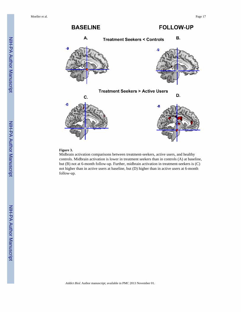
Figure 3.Midbrain activation comparisons between treatment-seekers, active users, and healthycontrols. Midbrain activation is lower in treatment seekers than in controls (A) at baseline,but (B) not at 6-month follow-up. Further, midbrain activation in treatment-seekers is (C)not higher than in active users at baseline, but (D) higher than in active users at 6-monthfollow-up.
Moeller et al. Page 17
Addict Biol. Author manuscript; available in PMC 2013 November 01.
NIH
-PA Author Manuscript
NIH
-PA Author Manuscript
NIH
-PA Author Manuscript

NIH
-PA Author Manuscript
NIH
-PA Author Manuscript
NIH
-PA Author Manuscript
Moeller et al. Page 18
Table 1
Task Performance and Clinical Outcome Variables at Initial Baseline and 6-Month Follow-up in 15 treatment-seeking subjects with cocaine use disorder.
Paired t Baseline Follow-up
TASK PERFORMANCE
Total Correct (max: 20): Drug Words 1.0 17.3 ± 1.6 17.0 ± 1.6
Total Correct (max: 20): Neutral Words 0.3 17.1 ± 1.9 17.0 ± 1.5
Reaction Time (Correct Trials) (ms): Drug WordsA 0.6 253.9 ± 15.9 252.2 ± 17.1
Reaction Time (Correct Trials) (ms): Neutral Words 2.2* 258.7 ± 12.9 250.8 ± 16.9
NEUROPSYCHOLOGICAL DRUG TASKS
Implicit Choice: Drug Images −0.5 13.5 ± 8.4 14.5 ± 10.0
Explicit Choice: Drug Images 1.1 76.5 ± 83.4 62.3 ± 68.9
CLINICAL VARIABLES
Cocaine Selective Severity Assessment Scale (withdrawal/craving symptoms; range: 0–126) 1.7 13.9 ± 12.2 10.6 ± 10.3
Severity of Dependence Scale (range: 0–15) 2.1 10.0 ± 3.8 7.5 ± 3.8
Cocaine Craving Questionnaire (range: 0–45) 0.5 8.1 ± 5.8 7.4 ± 8.7
Note: Numbers are M ± SD; consistent with the text, task performance variables are averaged across four money conditions;
Asignificantly differs from the neutral words at baseline;
*p<0.05.
Addict Biol. Author manuscript; available in PMC 2013 November 01.

NIH
-PA Author Manuscript
NIH
-PA Author Manuscript
NIH
-PA Author Manuscript
Moeller et al. Page 19
Tabl
e 2
Tas
k ac
tivat
ions
, dea
ctiv
atio
ns, a
nd c
orre
latio
ns w
ith b
ehav
ior
at b
asel
ine
vers
us a
t 6-m
onth
fol
low
-up
in 1
5 tr
eatm
ent-
seek
ing
subj
ects
with
coc
aine
use
diso
rder
.
BA
Side
Vox
els
Zp
clus
ter
leve
l cor
rect
edx
yz
Fol
low
-up>
Bas
elin
e
Mid
brai
n (V
TA
/sub
stan
tia n
igra
com
plex
)L
754.
9.0
24−
3−
18−
18
R4.
212
−21
−15
Tha
lam
usR
125
3.7
.002
9−
1512
Prec
uneu
s7
L77
3.9
.022
−6
−60
39
Cun
eus
18M
3.7
0−
8415
Fol
low
-up<
Bas
elin
e
Med
ial F
ront
al G
yrus
/Pre
-SM
A6
M
529
4.8
.000
0−
357
Post
cent
ral G
yrus
4R
4.2
39−
2154
Prec
entr
al G
yrus
6L
3.4
−42
054
Infe
rior
Par
ieta
l Lob
ule
40R
674.
3.0
3645
−36
42
Infe
rior
Occ
ipita
l Gyr
us19
R20
04.
7.0
0039
−66
−15
Fusi
form
Gyr
us37
3.8
33−
42−
15
Lin
gual
Gyr
us19
L16
3
4.7
.001
−30
−81
−12
Lin
gual
Gyr
us18
3.7
−6
−87
−9
Fusi
form
Gyr
us19
3.5
−27
−66
−9
Cor
rela
tion
s w
ith
Beh
avio
r: F
ollo
w-u
p>B
asel
ine
Dru
g an
d N
eutr
al W
ords
Exp
licit
Coc
aine
Cho
ice
C
ereb
ellu
mL
178
+4.
3.0
00−
18−
57−
12
M
idbr
ain
R30
−4.
4.0
503
−12
−15
Mid
brai
n-T
hala
mus
Cor
rela
tion
s: F
ollo
w-u
p
Dru
g an
d N
eutr
al W
ords
Addict Biol. Author manuscript; available in PMC 2013 November 01.

NIH
-PA Author Manuscript
NIH
-PA Author Manuscript
NIH
-PA Author Manuscript
Moeller et al. Page 20
BA
Side
Vox
els
Zp
clus
ter
leve
l cor
rect
edx
yz
Mid
brai
n se
ed: −
3, −
18, −
18
T
hala
mus
B12
8+
3.6
.000
9−
186
−9
−18
6
Not
e. A
ll re
sults
wer
e p<
0.05
clu
ster
-lev
el c
orre
cted
and
p<
0.00
1 vo
xel-
leve
l unc
orre
cted
, ≥15
con
tiguo
us v
oxel
s; Z
(+
) va
lue,
pos
itive
cor
rela
tion;
Z (
−)
valu
e, n
egat
ive
corr
elat
ion;
VT
A=
vent
ral
tegm
enta
l are
a, A
CC
= a
nter
ior
cing
ulat
e co
rtex
, SM
A=
supp
lem
enta
ry m
otor
are
a, L
=le
ft s
ide,
R=
righ
t sid
e, B
=bi
late
ral,
M=
med
ial (
neur
olog
ical
con
vent
ion)
; for
a li
stin
g of
all
regi
ons
that
sho
wed
task
-re
late
d ac
tivat
ions
or
deac
tivat
ions
, see
Tab
le S
3.
Addict Biol. Author manuscript; available in PMC 2013 November 01.