Embed Size (px)
Citation preview
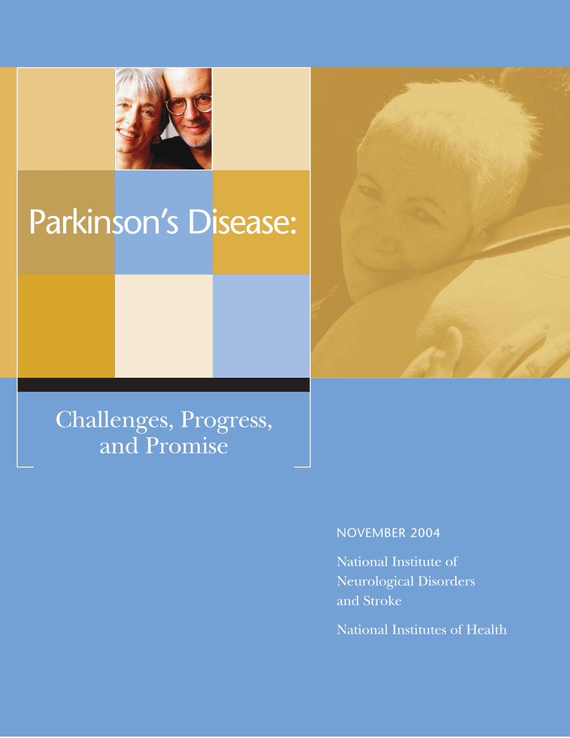
Parkinson’s Disease:
Challenges, Progress, and Promise
NOVEMBER 2004
National Institute ofNeurological Disorders and Stroke
National Institutes of Health

Parkinson’s Disease: Challenges, Progress, and Promise 1
Joel,a retired university professor,...
...went for regular walks with his wife. Ten years
ago, his wife began to notice that he was
shuffling his feet and was not swinging his arms
as most people do while walking. She also
noticed some changes in his posture and
unusual movements of his left arm. Joel’s
primary doctor referred him to a neurologist,
who told him he had Parkinson’s disease and
prescribed a combination of drugs to treat the
symptoms. He later saw a movement disorder
specialist as well.
Joel’s symptoms have gradually grown worse
with time. He now has tremors and rigidity
of movements on both sides of his body, and
balance problems when his medication wears off.
The disease also makes it difficult to project his
voice. He takes a combination of drugs that
help him to walk and to perform daily tasks.
However, he can no longer work in the
woodworking shop he had developed as a
retirement hobby. His loss of mobility and
speech impairment limit his social interactions.
He and his wife also have had to give up many
of their retirement travel plans.
Joel and Margaret are two of the many people in theUnited States who are living with Parkinson’s disease(PD), a complex disorder of the central nervous system.PD is the second most common neurodegenerativedisease in the United States, after Alzheimer’s disease.The defining characteristics of PD include tremor,slowness of movement (bradykinesia), rigidity, andimpaired balance and coordination. As these symptomsbecome more pronounced, patients may have difficultywalking, talking, or completing simple tasks. Theyalso may experience depression, difficulty sleeping, and other problems.
Margaret,a woman in her thirties,...
...began to experience tremors and stiffness
of her left arm while she walked. When these
symptoms continued, she saw several
neurologists to determine what was going on.
Because she was young, the doctors initially
thought she might have a brain tumor or
multiple sclerosis. However, after a brain
scan and neurological examinations, she was
diagnosed with Parkinson’s disease.
Margaret did not take any medications for the
disease until several years after her diagnosis.
She then began medication that reduced her
symptoms but did not stop the disease from
getting worse. She eventually developed
involuntary movements called dyskinesias,
which are a common side effect of levodopa,
the most common Parkinson’s drug. Because
of this, she decided to undergo a new therapy
called deep brain stimulation (DBS), which
provides electrical stimulation to the brain
through surgically implanted wires. The DBS
immediately reduced the amount of levodopa
Margaret needed to take, which stopped the
dyskinesias. The stimulation has since been
adjusted externally many times in order
to improve control of her symptoms. While
Margaret still takes medication, she needs
fewer pills than before the DBS.
Now, almost 25 years after her diagnosis,
Margaret is able to function very well in the
morning and early afternoon. Late in the day,
however, problems with balance, speech, and
fatigue return. She manages by planning her
activities carefully, avoiding many commitments
during the late afternoon and evening, and
increasing her medication when necessary.
Parkinson’s Disease: Challenges, Progress, and Promise 1
Introduction__________________________________________________________________________________________________________ 2
Brain and Movement: The Basics____________________________________________________________________ 5
What Goes Wrong In Parkinson’s Disease?___________________________________________________ 6
How Can We Treat Parkinson’s Disease?_______________________________________________________ 8
Drug Treatments ___________________________________________________________________________________________________________ 8
Surgical Treatments________________________________________________________________________________________________________ 8
Complementary and Supportive Therapies________________________________________________________________________ 10
Research Findings and New Directions________________________________________________________ 11
Genetics______________________________________________________________________________________________________________________ 11
Environmental Factors ___________________________________________________________________________________________________ 18
Pathways to Parkinson’s Disease ______________________________________________________________________________________ 20
Models for Parkinson’s Disease ________________________________________________________________________________________ 26
Therapeutic Approaches ________________________________________________________________________________________________ 29
Other Clinical Research __________________________________________________________________________________________________ 36
Conclusion____________________________________________________________________________________________________________ 39
4
3
2
1
Contents
“ A U G U S T U S D I V I N U S C O N U B I U M N O K S HParkinson’s Disease: Challenges, Progress, and Promise
Introduction
Parkinson’s Disease: Challenges, Progress, and Promise2 3
major neurodegenerative disorder, and defeating
PD remains a significant challenge.
The National Institute of Neurological Disorders
and Stroke (NINDS), part of the National
Institutes of Health (NIH), has a long history of
support for PD research. In 1997, recognizing
the need to accelerate the pace of PD research,
Congress signed the Morris K. Udall Parkinson’s
Disease Research Act into law. The Udall Act
directed the NIH to expand and coordinate
Parkinson’s research with the purpose of finding
a cure or treatment for this disease, and to award
Core Center Grants — designated as Morris K.
Udall Centers of Excellence for Parkinson's
Disease (PD) Research — to encourage
complementary research and to provide training
for scientists undertaking PD research.
The Udall Centers of Excellence have embarked
on diverse research avenues, but they all share a
common goal: scientific research to improve the
diagnosis and treatment of patients with PD and
related neurodegenerative disorders and to gain
a better understanding of the fundamental
cause(s) of the disease. These centers, along
with many other labs funded by the NIH, have
Ever since PD was first described in 1817,
scientists have pursued the causes and
treatment of the disease. In the early 1960s,
scientists identified the primary problem
underlying the disease: the loss of brain cells
that produce a chemical called dopamine, which
helps to coordinate and control muscle activity.
This discovery led to the first successful
treatment for PD and suggested ways of devising
new and even more effective therapies.
Parkinson’s research continues to be a very active
field, with new and intriguing findings reported
every day.
Research suggests that PD affects at least 500,000
people in the United States, and some estimates
are much higher. Society pays an enormous price
for PD. The total cost to the nation is estimated
to exceed $6 billion annually. The financial and
public health impact of this disease is expected
to increase as the population ages.
In recent years, Parkinson’s research has
advanced to the point that halting the
progression of PD, restoring lost function, and
even preventing the disease are all considered
realistic goals. However, we cannot yet cure any
As part of the implementation process for the
PD research agenda, NIH convened a summit
with several outstanding scientists in July 2002
to gain a better sense of where the field of PD
research stands internationally and to collect
information on "roadblocks" that impede
progress. This meeting led to a set of
recommendations that were compiled into a
matrix (see p. 4) of short-to-long range and low-
to-high risk action items in order to address
some of these roadblocks. The matrix identifies
goals that can help to advance PD research and
places them within a general time frame of when
progress might reasonably be expected. This
matrix helps to guide research planning at NIH,
but is also designed as a tool for the entire PD
community. It includes important responsibilities
for voluntary and private funding organizations,
and will lead to opportunities for collaboration
with other government agencies and the
international community as well. This matrix is
intended to be a living document that can be
revised and expanded as current goals are
achieved and new goals are identified.
Twelve different NIH Institutes and Centers fund
research on PD, including the National Institute
of Neurological Disorders and Stroke, National
Institute on Aging, National Institute of Mental
Health, National Institute of Environmental
Health Sciences, National Human Genome
made substantial progress in understanding PD.
Some highlights of this research include the
discovery and characterization of several genes
linked to familial PD; the expansion of research
on potential environmental factors that may
underlie PD; and studies of potential new
therapies, including growth factor
administration, drug therapy, and deep
brain stimulation.
As part of its mission to reduce the burden of
neurological disease, NINDS is committed to
expanding translational research – studies that
translate or develop promising findings in basic
research into effective treatments in the clinic.
PD research is considered prime territory for
translation because exciting new discoveries in
basic science have accelerated our understanding
of the disease over the past few years. The
Institute is supporting three coordinated
programs to encourage translational research
projects that focus on PD and other neurological
disorders. These programs provide tools and
resources for therapy development, support work
necessary to begin clinical testing, and offer
career development and training experience for
investigators interested in translational research.
The NIH conducts a vigorous and expanding
program of research focused on PD. In 2000, the
agency convened a workshop to create an
agenda for PD research. The attendees included
intramural, extramural, and industry scientists;
representatives from several Parkinson's advocacy
groups; and ethicists. They developed an agenda
that comprises four major areas: understanding
PD, developing new treatments, creating new
research capabilities, and enhancing the
research process.
The PD research agenda is also relevant for
other diseases. PD research can lead the way in
the fight against all forms of neurodegeneration,
and research on other types of neuro-
degeneration also can provide vital clues about
how PD may be cured.
“ A U G U S T U S D I V I N U S C O N U B I U M N O K S HParkinson’s Disease: Challenges, Progress, and Promise4 Parkinson’s Disease: Challenges, Progress, and Promise 5
HighRisk
MediumRisk
LowRisk
Parkinson’s Disease Research Community Goals Matrix
Research Institute, National Institute on
Deafness and Other Communication Disorders,
National Institute of Nursing Research, National
Institute on Drug Abuse, National Institute of
Biomedical Imaging and Bioengineering,
National Institute of Child Health and Human
Development, National Center for
Complementary and Alternative Medicine, and
National Center for Research Resources.
Representatives from each of these Institutes, as
well as from the Department of Defense and the
Department of Veterans Affairs, meet twice a
year under the leadership of NINDS to discuss
research and initiatives for PD, to plan meetings
and workshops, and to coordinate research
efforts across federal agencies. This committee
is called the Parkinson’s Disease Coordinating
Committee (PDCC). Parkinson’s is a
multifaceted disease, and each of these Institutes
brings a valuable perspective to confronting the
biological complexities of the disorder, to
pursuing the diverse therapeutic strategies
showing promise, and to providing the resources
necessary to carry out a research agenda of this
breadth.
The NINDS also tracks PD research conducted
in other countries. This worldwide tracking
activity helps researchers identify potential areas
for international collaboration and reduces the
chance of duplicated activities.
This report serves to highlight and update the
substantial progress made in PD research during
the past 5 years. While the report highlights the
work of the Udall Centers, many contributions
have been made by other NINDS grantees and
researchers around the world.
• Understanding Gene/Environment Interactions• Biomarker Development
• General Roadblocks toAdvancing PD Research
• Translational Research/TherapeuticsDevelopment
Short Term (0-3 years)
Medium Term (4-6 years)
Long Term(7-10 years)
• Core Facilities and Resources • Integration of PD Research Centers• Public-Private Partnerships• Gene Discovery• Translational Research/Therapeutics Development
• Core Facilities and Resources • General Resources
– Gene Therapy Resources– Animal Models– Brain Banks
• Integration of PD Research Centers• Public-Private Partnerships• Basic Cell Biology Research• General Roadblocks to Advancing PD Research• Understanding Gene/Environment Interactions
• Core Facilities andResources
• Brain Banks• Clinical Trials of
Non-motor Symptoms
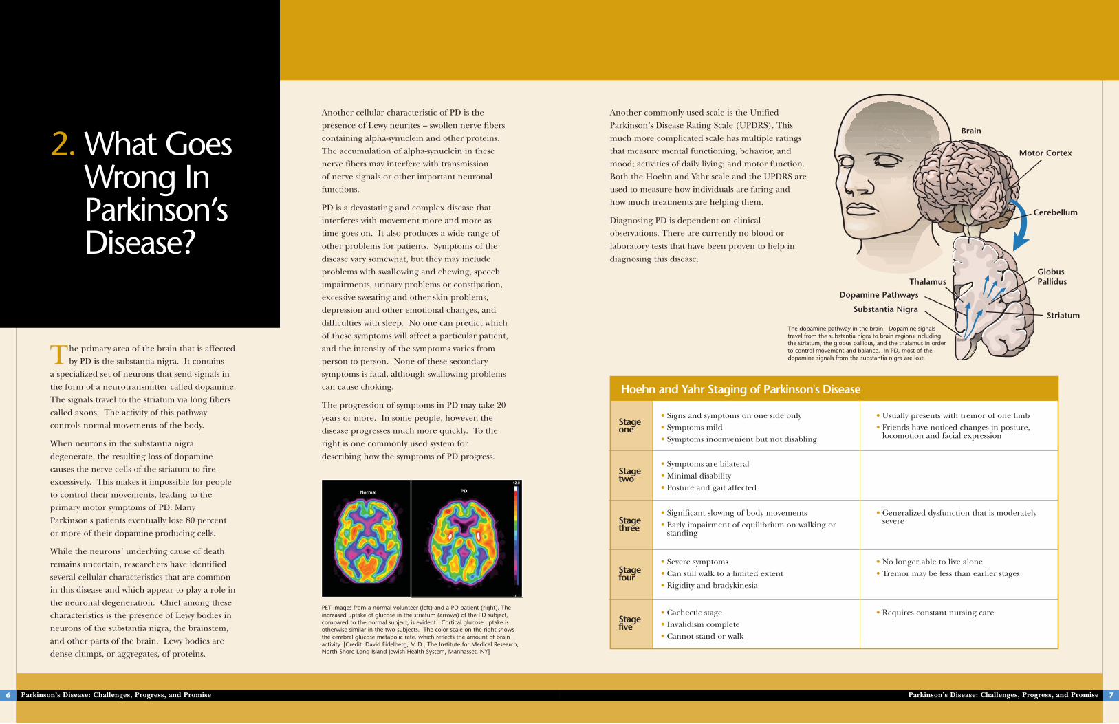
cell is also a neuron, it will carry the signal on
to the next cell. If the receiving cell is a muscle
fiber, it will react to the stimulation by
contracting, which creates movement.
When a person initiates a movement,
information from the senses, from parts
of the brain that control planning, and from
other brain regions travels to a region called the
striatum. The striatum then interacts with other
areas of the brain — the substantia nigra, globus
pallidus, and thalamus — to send out signals that
control balance and coordination. These signals
travel to the cerebellum, which controls muscle
coordination, and then finally down the spinal
cord to peripheral nerves in the limbs, head,
and torso, where they control the muscles.
The molecules that carry information through
the brain and spinal cord are called
neurotransmitters. Neurotransmitters are special
chemicals produced by neurons that accumulate
in tiny sacs at the end of nerve fibers. When
stimulated, these sacs release neurotransmitters
into the gap between neurons, called a synapse.
The neurotransmitters cross the synapse and
attach to proteins called receptors on the
neighboring cell. These signals change the
properties of the receiving cell. If the receiving
Neurons transmit signals across tiny spaces called synapses. Signalingchemicals called neurotransmitters are gathered into sacs called synapticvesicles. When the nerve receives a signal, these sacs release theirneurotransmitters into the synapse. Proteins called receptors, found onthe receiving neuron, bind to the neurotransmitters and trigger a newnerve impulse.
1. Brain and Movement:The Basics
“ A U G U S T U S D I V I N U S C O N U B I U M N O K S H Parkinson’s Disease: Challenges, Progress, and Promise 7Parkinson’s Disease: Challenges, Progress, and Promise6
Another cellular characteristic of PD is the
presence of Lewy neurites – swollen nerve fibers
containing alpha-synuclein and other proteins.
The accumulation of alpha-synuclein in these
nerve fibers may interfere with transmission
of nerve signals or other important neuronal
functions.
PD is a devastating and complex disease that
interferes with movement more and more as
time goes on. It also produces a wide range of
other problems for patients. Symptoms of the
disease vary somewhat, but they may include
problems with swallowing and chewing, speech
impairments, urinary problems or constipation,
excessive sweating and other skin problems,
depression and other emotional changes, and
difficulties with sleep. No one can predict which
of these symptoms will affect a particular patient,
and the intensity of the symptoms varies from
person to person. None of these secondary
symptoms is fatal, although swallowing problems
can cause choking.
The progression of symptoms in PD may take 20
years or more. In some people, however, the
disease progresses much more quickly. To the
right is one commonly used system for
describing how the symptoms of PD progress.
The primary area of the brain that is affected
by PD is the substantia nigra. It contains
a specialized set of neurons that send signals in
the form of a neurotransmitter called dopamine.
The signals travel to the striatum via long fibers
called axons. The activity of this pathway
controls normal movements of the body.
When neurons in the substantia nigra
degenerate, the resulting loss of dopamine
causes the nerve cells of the striatum to fire
excessively. This makes it impossible for people
to control their movements, leading to the
primary motor symptoms of PD. Many
Parkinson's patients eventually lose 80 percent
or more of their dopamine-producing cells.
While the neurons’ underlying cause of death
remains uncertain, researchers have identified
several cellular characteristics that are common
in this disease and which appear to play a role in
the neuronal degeneration. Chief among these
characteristics is the presence of Lewy bodies in
neurons of the substantia nigra, the brainstem,
and other parts of the brain. Lewy bodies are
dense clumps, or aggregates, of proteins.
2. What GoesWrong InParkinson’sDisease?
PET images from a normal volunteer (left) and a PD patient (right). Theincreased uptake of glucose in the striatum (arrows) of the PD subject,compared to the normal subject, is evident. Cortical glucose uptake isotherwise similar in the two subjects. The color scale on the right showsthe cerebral glucose metabolic rate, which reflects the amount of brainactivity. [Credit: David Eidelberg, M.D., The Institute for Medical Research,North Shore-Long Island Jewish Health System, Manhasset, NY]
Another commonly used scale is the Unified
Parkinson’s Disease Rating Scale (UPDRS). This
much more complicated scale has multiple ratings
that measure mental functioning, behavior, and
mood; activities of daily living; and motor function.
Both the Hoehn and Yahr scale and the UPDRS are
used to measure how individuals are faring and
how much treatments are helping them.
Diagnosing PD is dependent on clinical
observations. There are currently no blood or
laboratory tests that have been proven to help in
diagnosing this disease.
The dopamine pathway in the brain. Dopamine signalstravel from the substantia nigra to brain regions includingthe striatum, the globus pallidus, and the thalamus in orderto control movement and balance. In PD, most of thedopamine signals from the substantia nigra are lost.
• Signs and symptoms on one side only • Symptoms mild • Symptoms inconvenient but not disabling
• Usually presents with tremor of one limb • Friends have noticed changes in posture,
locomotion and facial expression
• Symptoms are bilateral • Minimal disability • Posture and gait affected
• Significant slowing of body movements • Early impairment of equilibrium on walking or
standing
• Generalized dysfunction that is moderatelysevere
• Severe symptoms • Can still walk to a limited extent • Rigidity and bradykinesia
• No longer able to live alone • Tremor may be less than earlier stages
• Cachectic stage • Invalidism complete • Cannot stand or walk
• Requires constant nursing care
Stageone
Stagetwo
Stagethree
Stagefour
Stagefive
Hoehn and Yahr Staging of Parkinson's Disease
Brain
Motor Cortex
Thalamus
Dopamine Pathways
Substantia Nigra
GlobusPallidus
Striatum
Cerebellum
“ A U G U S T U S D I V I N U S C O N U B I U M N O K S H Parkinson’s Disease: Challenges, Progress, and PromiseParkinson’s Disease: Challenges, Progress, and Promise8
example, anticholinergic drugs decrease the
activity of the neurotransmitter acetylcholine.
These drugs help to reduce tremors and muscle
stiffness, which can result from having more
acetylcholine than dopamine.
The third category of drugs prescribed for PD
includes medications that help control the non-
motor symptoms of the disease. For example,
people with PD-related depression may be
prescribed antidepressants.
— — — — — — — — — — — — — — — — — — — — — — — — — — — — — — — — — — — — — — —
Surgical TreatmentsAt present, there are two commonly used
surgical treatments for PD: pallidotomy and
deep brain stimulation. Because these
procedures are invasive, they are usually reserved
for severely afflicted Parkinson's patients who do
not get adequate relief from medications.
There are currently two main types of
treatment for PD: drug treatments and
surgery.
Drug TreatmentsMedications for PD fall into three categories.
The first category includes drugs that work
directly or indirectly to increase the level of
dopamine in the brain. People cannot simply
take dopamine pills because dopamine does not
easily pass through blood vessels into the brain.
The most common drugs for PD are dopamine
precursors – substances such as levodopa that
cross the blood-brain barrier and are then
changed into dopamine. Other drugs mimic
dopamine, prevent or slow its breakdown, or
increase the amount of it that is released.
The second category of PD drugs affects other
neurotransmitters in the body in order to ease
some of the symptoms of the disease. For
3. How Can We TreatParkinson’sDisease?
Brain surgery was one of the first treatments for
PD. Surgeons discovered that, by removing or
destroying parts of the brain that were
“misfiring,” some of the symptoms of PD could
be alleviated. The most common early brain
operations for PD were pallidotomy, which
destroyed part of the globus pallidus, and
thalamotomy, which destroyed part of the
thalamus. These procedures were irreversible
and often led to complications. Clinicians have
improved these techniques a great deal, but
while they are much safer now, they are still
irreversible.
In recent years, scientists have found that they
can mimic the effects of pallidotomy and
thalamotomy by deep brain stimulation (DBS).
With DBS, an electrode is implanted in the brain
in a way that calms the abnormal neuronal
firing. This procedure is much safer than
pallidotomy or thalamotomy because the
electrodes can be turned off if the patient
9
Deep BrainStimulation
For many years, the only surgical treatments for PD
were pallidotomy and thalamotomy – procedures in
which surgeons selectively destroy small portions of
the brain in order to relieve tremor and rigidity. The
tissue destruction is irreversible. Because these
procedures often led to troubling side effects,
surgery was largely replaced with drug therapy once
levodopa became available for PD in the 1960s.
In the 1980s, researchers in France discovered that
chronic stimulation (now termed deep brain
stimulation or DBS) of a brain region called the
thalamus could block tremors in patients with
essential tremor. Studies in a monkey model for PD
also revealed the brain circuits that are altered in
this disease and pointed to a brain region called the
subthalamic nucleus as a key target. This discovery
opened the door to a new era of surgical
treatments. Investigators then examined the effects
of stimulating the subthalamic nucleus in patients
with PD and found that the stimulation had
profound effects on patients’ tremor, slowness, and
stiffness. In DBS, electrodes are implanted into the
brain and connected to a small electrical device
called a pulse generator that can be externally
programmed. DBS reduces the need for levodopa
and related drugs, which in turn decreases the
involuntary movements called dyskinesias that are a
common side effect of levodopa. It also helps to
alleviate fluctuations of symptoms and to reduce
tremors, slowness of movements, and gait problems.
Unlike pallidotomy and thalamotomy, DBS is
reversible. However, it requires careful programming
of the stimulator device in order to work correctly.
DBS has now been approved by the U.S. Food and
Drug Administration, and it is widely used as a
treatment for PD. It also is used to treat dystonia
and essential tremor, and it is being tested for
disorders such as Tourette syndrome, epilepsy, and
depression. Researchers are continuing to study DBS
and to develop ways of improving it. They are
conducting clinical studies to determine the best
part of the brain to receive stimulation and to
determine the long-term effects of this therapy.
They also are working to improve the technology
available for DBS.
One surgical treatment for PD is called deep brain stimulation (DBS).DBS can be performed on either one side (unilateral) or both sides ofthe body (bilateral). In bilateral DBS, electrodes are implanted in thesubthalamic nucleus or the globus pallidus of the brain. Insulatedwires are then passed under the skin of the head, neck, and shoulderto connect the electrodes to battery-operated neurostimulators thatare implanted under the skin, usually near the collarbones. Impulsesfrom the neurostimulators interfere with and block the brain signalsthat cause PD symptoms.
“ A U G U S T U S D I V I N U S C O N U B I U M N O K S HParkinson’s Disease: Challenges, Progress, and Promise10
and put underused and rigid muscles through a
full range of motion. Exercise cannot stop
disease progression, but it may improve body
strength so that the person can better cope with
his or her disability. Researchers are studying
whether exercise also may improve the response
to levodopa and/or increase levels of beneficial
compounds called neurotrophic factors in the
brain. Targeted exercises also may improve
balance, help people overcome gait problems,
and strengthen certain muscles so that people
can speak and swallow better. Although
structured exercise programs help many patients,
more general physical activity, such as walking,
gardening, swimming, and using exercise
machines, is also beneficial.
Some early reports suggested that dietary
supplements may be protective in PD. In
addition, a phase II clinical trial of a supplement
called coenzyme Q10 suggested that large doses
of this substance can slow disease progression in
patients with early-stage PD. The NINDS and the
National Center for Complementary and
Alternative Medicine (NCCAM) are funding
research to determine if folate, coffee, dietary
antioxidants, fat, alcohol, and/or dairy products
are beneficial. While there is currently no
evidence that any specific dietary factor is
beneficial in PD, a normal, healthy diet can
promote overall well-being for PD patients just as
it would for anyone else.
Other complementary therapies that are used by
some individuals with PD include massage
therapy, yoga, tai chi, acupuncture, ginkgo
biloba (for concentration problems), and the
Alexander technique, which optimizes posture
and muscle activity. There have been limited
studies suggesting mild benefits with many of
these therapies, but they do not slow PD and
there is no convincing evidence that they are
beneficial.
experiences problems. The stimulation also can
be adjusted to match the patient’s needs.
Because of this, DBS is now the primary surgical
intervention for PD. In 1997, the U.S. Food and
Drug Administration (FDA) approved DBS for
the treatment of essential tremor using a single
implanted electrode on one side of the brain. In
January 2002, the FDA approved DBS for PD
using two implanted electrodes — one on each
side of the brain. Recently, the FDA also
approved a technologically advanced electrode
apparatus that can be controlled by the patient
through use of a remote control device.
— — — — — — — — — — — — — — — — — — — — — — — — — — — — — — — — — — — — — — —
Complementary andSupportive TherapiesA wide variety of complementary and supportive
therapies may be used for PD. Among these
therapies are standard rehabilitation techniques,
which can help with problems such as gait and
voice disorders, tremors and rigidity, and
cognitive decline. Exercise may help people
improve their mobility. Physical therapy or
muscle-strengthening exercises may tone muscles
inherited and sporadic (non-hereditary) cases of
the disease. The same genes and proteins that
are altered or missing in inherited cases may also
be altered in sporadic cases by environmental
toxins or other factors.
Identifying gene defects can also help
researchers understand how PD occurs, develop
animal models that accurately mimic the
neuronal death in human PD, identify new drug
targets, and improve diagnosis. The genetic
approach has been very successful, with new
discoveries occurring at an unprecedented pace.
The following summary highlights current
knowledge about the genes known to be involved
in PD, and the functions of the proteins these
genes produce.
■ alpha-synucleinThe first PD-related gene to be identified was
alpha-synuclein. Researchers at NIH and
other institutions studied the genetic profiles
of a large Italian family and three Greek
families with familial PD and found that their
disease was related to a mutation in this gene.
During the past five years, researchers
have made substantial advances in our
understanding of the biological factors involved
in PD. They are beginning to decipher the roles
of individual genes and environmental factors in
PD and to learn how the interplay of these
factors can lead to the disease. Each abnormal
gene or environmental factor that is identified
provides another clue to help solve the mystery
of PD.
— — — — — — — — — — — — — — — — — — — — — — — — — — — — — — — — — — — — — — —
GeneticsUntil the last decade, many researchers believed
that PD was caused solely by environmental
factors. However, the discovery of gene
mutations in familial, or inherited, forms of PD
has led to an explosion of research on PD genes
and the function of the proteins that are
encoded by these genes.
Although most people do not inherit PD,
studying the genes responsible for the inherited
cases can help researchers understand both
Parkinson’s Disease: Challenges, Progress, and Promise 11
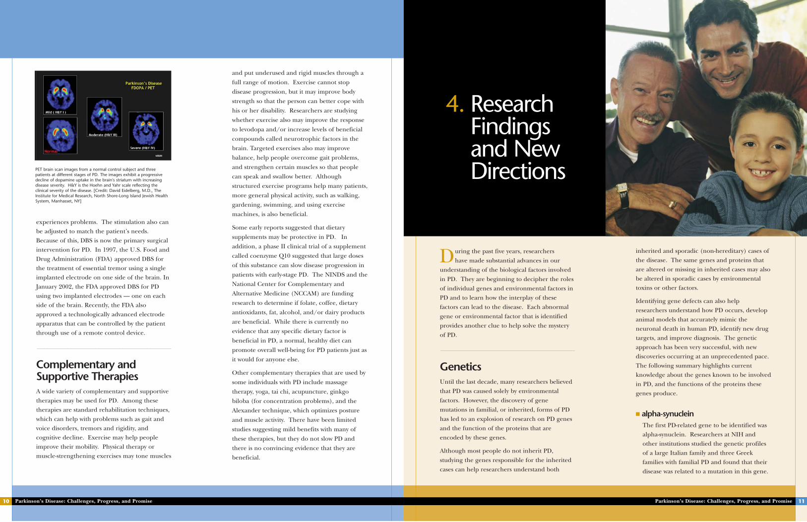
PET brain scan images from a normal control subject and threepatients at different stages of PD. The images exhibit a progressivedecline of dopamine uptake in the brain's striatum with increasingdisease severity. H&Y is the Hoehn and Yahr scale reflecting theclinical severity of the disease. [Credit: David Eidelberg, M.D., TheInstitute for Medical Research, North Shore-Long Island Jewish HealthSystem, Manhasset, NY]
4. ResearchFindings and NewDirections
“ A U G U S T U S D I V I N U S C O N U B I U M N O K S HParkinson’s Disease: Challenges, Progress, and Promise12
protein disposal system and cause neurons to
die. A group of researchers at NIH and other
institutions investigated a rare familial form of
early-onset PD and discovered that a
multiplication of the normal alpha-synuclein
gene, and a corresponding increase in alpha-
synuclein protein, can cause the disease. The
researchers analyzed blood samples from a
family, the "Iowa kindred," in which many
relatives developed PD or related neurological
diseases. In the relatives with PD, the
researchers found four copies of the alpha-
synuclein gene — an abnormal triplication of
three alpha-synuclein genes on one copy of
chromosome 4 and one gene on the other
chromosome 4 — instead of the usual two
copies of the alpha-synuclein gene. This
multiplication resulted in an abnormally large
amount of alpha-synuclein in the cells.
A third theory proposes that mutant alpha-
synuclein interferes with the normal
housekeeping functions of cells and lets
proteins build up to toxic levels. Researchers
They found a second alpha-synuclein mutation
in a German family with PD. These findings
prompted studies of the role of alpha-
synuclein in PD, which led to the discovery
that Lewy bodies from people with the
sporadic form of PD contained clumps of
alpha-synuclein proteins. This discovery
revealed a potential link between hereditary
and sporadic forms of the disease and sparked
investigations into the normal function of
alpha-synuclein as well as the possible effects
of alpha-synuclein mutations on normal
cellular activity.
One theory about how alpha-synuclein is
associated with PD holds that the mutated
protein interferes with cell membranes.
Within the cell body, individual molecules of
alpha-synuclein join together to form tiny
protein threads called fibrils; this process is
called fibrillization. Investigators at the
Brigham and Women’s Hospital Udall Center
and elsewhere have shown that mutations in
the alpha-synuclein gene disrupt the
fibrillization process and lead to the
accumulation of protofibrils, an intermediate
step in alpha-synuclein fibrillization. They
found that alpha-synuclein protofibrils have
protein structures which resemble bacterial
and insect toxins that make membranes leaky.
This could trigger cell death and may explain
the toxicity of Lewy body proteins. This idea is
supported by studies from the Massachusetts
General Hospital and Massachusetts Institute
of Technology Udall Center showing that
alpha-synuclein is located near cell
membranes in postmortem brain tissue from
people with diffuse Lewy body disease.
Another study suggests that a buildup of
normal alpha-synuclein may clog up the cell’s
■ ParkinGenetic studies on a rare, juvenile-onset form
of PD led to the discovery of the parkin gene.
Originally, this form of PD was not linked to
Lewy bodies. However, Mayo Clinic Udall
Center scientists have found parkin mutations
that are accompanied by Lewy body pathology.
Further studies have shown that parkin is a
part of the so-called ubiquitin-proteasome
system, which breaks down proteins in the cell.
This suggests that parkin mutations may lead
to accumulation of toxic proteins within
neurons. Researchers also have shown that
parkin interacts with synphilin-1 and alpha-
synuclein and mediates an important step in
protein handling. When alpha-synuclein,
another protein called synphilin-1, and parkin
are injected together into cells in culture, they
form inclusions in the cell that are similar to
Lewy bodies. This suggests that parkin may be
important in both inherited and sporadic
forms of the disease. Several studies have
suggested that normal parkin protects neurons
from diverse threats, including alpha-synuclein
toxicity, proteasomal dysfunction, and
excitotoxicity. Other evidence indicates that
at the Columbia University Udall Center,
along with colleagues at Brigham and
Women’s Hospital and the Albert Einstein
College of Medicine, have found that normal
alpha-synuclein is broken down by lysosomes,
which act as the cell’s garbage disposal system.
Mutant alpha-synuclein, however, blocks the
pathway into the lysosomes. This inhibits the
breakdown of alpha-synuclein as well as other
proteins. This may trigger a toxic buildup of
protein “garbage” inside the cell.
Researchers are continuing to study the alpha-
synuclein gene to clarify how it affects PD.
For example, Mayo Clinic Udall Center
researchers are assessing the alpha-synuclein
gene in a large group of people with PD, and
in a control group of healthy people who
match the PD patients in age, gender, and
demographics, in order to look for variations
in the gene that may affect susceptibility to the
disease. Investigators at the Johns Hopkins
University Udall Center have developed mice
with alpha-synuclein gene mutations and
found that the mice accumulate alpha-
synuclein in the midbrain, cerebellum,
brainstem, and spinal cord and develop an
adult-onset neurodegenerative disease with
symptoms resembling human PD, including
motor dysfunction, bradykinesia, and dystonia.
The discovery of alpha-synuclein has paved the
way for other genetic linkage studies in
families with PD (see Gene Discoveries, p. 15).
Within the past 5 years, many regions of the
genome have been linked to PD and four
additional PD genes have been identified,
including parkin, DJ-1, PINK1, and DRDN.
Parkinson’s Disease: Challenges, Progress, and Promise 13
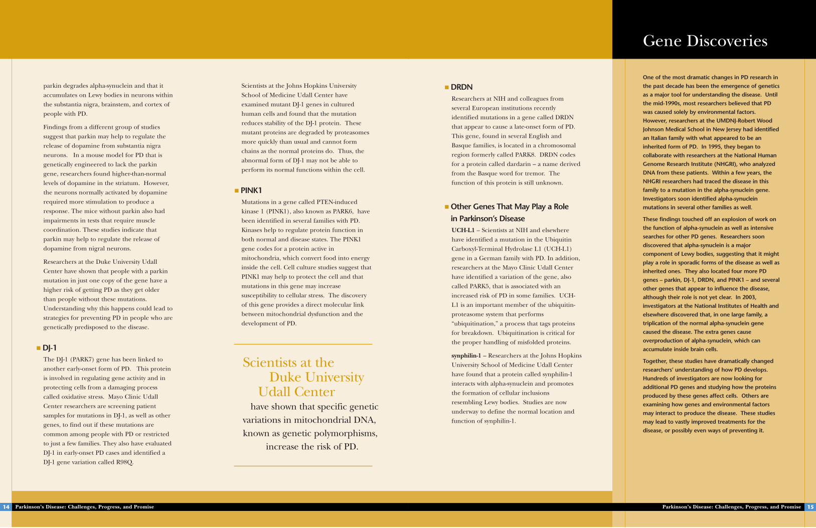
Researchers studying a family with a hereditary form of PD discoveredthat members of this family had a triplication of the normal alpha-synuclein gene on one chromosome. This image shows the threealpha-synuclein genes (pink) on one copy of chromosome 4.
Parkinson'sGene Location
“ A U G U S T U S D I V I N U S C O N U B I U M N O K S HParkinson’s Disease: Challenges, Progress, and Promise14
Scientists at the Johns Hopkins University
School of Medicine Udall Center have
examined mutant DJ-1 genes in cultured
human cells and found that the mutation
reduces stability of the DJ-1 protein. These
mutant proteins are degraded by proteasomes
more quickly than usual and cannot form
chains as the normal proteins do. Thus, the
abnormal form of DJ-1 may not be able to
perform its normal functions within the cell.
■ PINK1Mutations in a gene called PTEN-induced
kinase 1 (PINK1), also known as PARK6, have
been identified in several families with PD.
Kinases help to regulate protein function in
both normal and disease states. The PINK1
gene codes for a protein active in
mitochondria, which convert food into energy
inside the cell. Cell culture studies suggest that
PINK1 may help to protect the cell and that
mutations in this gene may increase
susceptibility to cellular stress. The discovery
of this gene provides a direct molecular link
between mitochondrial dysfunction and the
development of PD.
parkin degrades alpha-synuclein and that it
accumulates on Lewy bodies in neurons within
the substantia nigra, brainstem, and cortex of
people with PD.
Findings from a different group of studies
suggest that parkin may help to regulate the
release of dopamine from substantia nigra
neurons. In a mouse model for PD that is
genetically engineered to lack the parkin
gene, researchers found higher-than-normal
levels of dopamine in the striatum. However,
the neurons normally activated by dopamine
required more stimulation to produce a
response. The mice without parkin also had
impairments in tests that require muscle
coordination. These studies indicate that
parkin may help to regulate the release of
dopamine from nigral neurons.
Researchers at the Duke University Udall
Center have shown that people with a parkin
mutation in just one copy of the gene have a
higher risk of getting PD as they get older
than people without these mutations.
Understanding why this happens could lead to
strategies for preventing PD in people who are
genetically predisposed to the disease.
■ DJ-1 The DJ-1 (PARK7) gene has been linked to
another early-onset form of PD. This protein
is involved in regulating gene activity and in
protecting cells from a damaging process
called oxidative stress. Mayo Clinic Udall
Center researchers are screening patient
samples for mutations in DJ-1, as well as other
genes, to find out if these mutations are
common among people with PD or restricted
to just a few families. They also have evaluated
DJ-1 in early-onset PD cases and identified a
DJ-1 gene variation called R98Q.
■ DRDNResearchers at NIH and colleagues from
several European institutions recently
identified mutations in a gene called DRDN
that appear to cause a late-onset form of PD.
This gene, found in several English and
Basque families, is located in a chromosomal
region formerly called PARK8. DRDN codes
for a protein called dardarin – a name derived
from the Basque word for tremor. The
function of this protein is still unknown.
■ Other Genes That May Play a Role in Parkinson’s DiseaseUCH-L1 – Scientists at NIH and elsewhere
have identified a mutation in the Ubiquitin
Carboxyl-Terminal Hydrolase L1 (UCH-L1)
gene in a German family with PD. In addition,
researchers at the Mayo Clinic Udall Center
have identified a variation of the gene, also
called PARK5, that is associated with an
increased risk of PD in some families. UCH-
L1 is an important member of the ubiquitin-
proteasome system that performs
“ubiquitination,” a process that tags proteins
for breakdown. Ubiquitination is critical for
the proper handling of misfolded proteins.
synphilin-1 – Researchers at the Johns Hopkins
University School of Medicine Udall Center
have found that a protein called synphilin-1
interacts with alpha-synuclein and promotes
the formation of cellular inclusions
resembling Lewy bodies. Studies are now
underway to define the normal location and
function of synphilin-1.
Parkinson’s Disease: Challenges, Progress, and Promise 15
Gene Discoveries
One of the most dramatic changes in PD research in
the past decade has been the emergence of genetics
as a major tool for understanding the disease. Until
the mid-1990s, most researchers believed that PD
was caused solely by environmental factors.
However, researchers at the UMDNJ-Robert Wood
Johnson Medical School in New Jersey had identified
an Italian family with what appeared to be an
inherited form of PD. In 1995, they began to
collaborate with researchers at the National Human
Genome Research Institute (NHGRI), who analyzed
DNA from these patients. Within a few years, the
NHGRI researchers had traced the disease in this
family to a mutation in the alpha-synuclein gene.
Investigators soon identified alpha-synuclein
mutations in several other families as well.
These findings touched off an explosion of work on
the function of alpha-synuclein as well as intensive
searches for other PD genes. Researchers soon
discovered that alpha-synuclein is a major
component of Lewy bodies, suggesting that it might
play a role in sporadic forms of the disease as well as
inherited ones. They also located four more PD
genes – parkin, DJ-1, DRDN, and PINK1 – and several
other genes that appear to influence the disease,
although their role is not yet clear. In 2003,
investigators at the National Institutes of Health and
elsewhere discovered that, in one large family, a
triplication of the normal alpha-synuclein gene
caused the disease. The extra genes cause
overproduction of alpha-synuclein, which can
accumulate inside brain cells.
Together, these studies have dramatically changed
researchers’ understanding of how PD develops.
Hundreds of investigators are now looking for
additional PD genes and studying how the proteins
produced by these genes affect cells. Others are
examining how genes and environmental factors
may interact to produce the disease. These studies
may lead to vastly improved treatments for the
disease, or possibly even ways of preventing it.
Scientists at the Duke University
Udall Center have shown that specific genetic
variations in mitochondrial DNA,known as genetic polymorphisms,
increase the risk of PD.

“ A U G U S T U S D I V I N U S C O N U B I U M N O K S H
analysis). Genes identified by the genomic
convergence technique are relatively likely to
play a role in PD. This combination of two
powerful techniques saves investigators time and
effort compared to studying the results of gene
expression or linkage analysis alone. This
approach has been used successfully to identify
the GSTO-1 gene.
Researchers at the Duke University Udall Center
have performed the first large expression studies
to identify genes that are abnormally active or
inactive in areas of the brain affected most by
PD. They also have compared gene activity in
PD with that in similar diseases such as
progressive supranuclear palsy and FTDP-17. In
addition, they have helped develop new gene
analysis techniques that can determine if specific
genetic variants make an individual more
susceptible to PD. These tests, called the
Pedigree Disequilibrium Test (PDT) and Geno-
PDT, have improved on previously available
techniques.
may be linked to PD. They have finished
intensive sequencing of seven mitochondrial
genes in frontal cortex samples from a small
group of people with the disease and an equal
number of controls. They found potentially
important mutations in genes called ND2, ND4L,
and ND5. These findings counter the hypothesis
that PD is caused simply by an increase in age-
related mtDNA mutations. The University of
Virginia researchers also have developed
methods to remove and replace the human
mitochondrial genome. These technologies may
lead to mitochondrial gene replacement as a
method of treating PD and other sporadic
neurodegenerative diseases. They could also be
used to show whether these mitochondrial gene
mutations cause PD and related diseases.
Duke University researchers also are investigating
the role of the mitochondrial genome in PD.
They have discovered that specific DNA regions
and variations are associated with an increased
risk of PD. This work has led to a collaborative
study with the University of Virginia investigators
to look more closely at how these mitochondrial
DNA variations affect cellular functions.
Researchers are developing a variety of new
approaches to speed research on genes and on
the functions of the proteins they produce. For
example, scientists at the Duke University Udall
Center have developed a new approach called
“genomic convergence” to study PD and other
common diseases. This approach identifies and
prioritizes candidate susceptibility genes for PD
by taking data from gene expression studies and
merging it with data from studies that detect
chromosomal regions linked to PD (linkage
Parkinson’s Disease: Challenges, Progress, and Promise 17
apolipoprotein E – Duke University researchers
conducting a genomic screen to identify genes
influencing age of onset for PD and Alzheimer’s
disease have found that normal genetic
variations in the apolipoprotein E protein affect
the age of onset for PD, just as they do for
Alzheimer’s.
PARK3, PARK9, PARK10, and PARK11 – These
are chromosomal regions that have been
implicated in families with PD. The
chromosomal regions have not been narrowed
down to specific genes, but researchers are
working to identify the genes and to determine
their function. People with PARK3 have a
relatively late age of onset, much like sporadic
PD. PARK9 has been identified in one Jordanian
family. PARK10 is linked to age of onset of PD in
Icelandic families. PARK11 was identified in
pairs of siblings and appears to affect
susceptibility to the disease.
The search for additional PD-related genes
continues on many fronts. University of Virginia
Udall Center researchers are working to define
mitochondrial DNA (mtDNA) mutations that
PACRG – This gene, called parkin co-regulated
gene, was identified by researchers at the Mayo
Clinic Udall Center. PACRG appears to interact
with parkin and to be part of the protein
degradation system. This protein also appears
to be a component of Lewy bodies.
GSTO-1 and -2 – A gene called glutathione S-
transferase omega-1 (GSTO-1) appears to affect
the age of onset for PD and Alzheimer’s disease.
GSTO-1 is one of a family of genes that break
down and recycle many compounds in cells,
including drugs, carcinogens, and the products
of oxidative stress. Studies suggest that GSTO-1
may modify an inflammatory compound called
interleukin-1 beta and protect against the
inflammation commonly found in brains from
people with PD. Scientists also have identified a
related gene, glutathione S-transferase omega-2
(GSTO-2).
tau – The tau protein is an important
component of microtubules, which are part of
the cell's structural support system and help to
deliver substances throughout the cell. Recent
studies have linked an abnormal tau protein to a
parkinsonian disorder, frontotemporal dementia
with parkinsonism linked to chromosome 17
(FTDP-17). Additional studies have suggested
that aberrations in the tau protein contribute to
the pathology of sporadic PD. Mayo Clinic Udall
Center researchers are sequencing the tau gene
in samples from their patients with familial
parkinsonism to determine if it plays a role in
these forms of PD.
fibroblast growth factor 2 – This growth factor
helps to maintain neurons. Studies by Duke
University researchers suggest that mutations
in the FGF2 gene may be a risk factor for PD.
Parkinson’s Disease: Challenges, Progress, and Promise16
“ A U G U S T U S D I V I N U S C O N U B I U M N O K S HParkinson’s Disease: Challenges, Progress, and Promise18
study has shown that mutations in the parkin
gene may be a risk factor for late-onset PD as
well as the juvenile-onset disease.
NINDS also is sponsoring a DNA and cell line
repository to enhance gene discovery by
supplying DNA samples, cell lines, and clinical
and pedigree data to the neuroscience
community.
Research on the genetics of PD also receives
funding from other NIH institutes. The National
Human Genome Research Institute is sponsoring
a clinical study to identify people with inherited
PD and to look for gene mutations in these
individuals, and the National Institute of
Environmental Health Sciences is sponsoring a
study to look at nine candidate genes in a sample
of 800 people with PD and their siblings to
clarify what role these genes might play in the
development of this disease.
Environmental FactorsAlthough the importance of genetics in PD is
increasingly recognized, many researchers still
believe that environmental exposures also
increase a person’s risk of developing the
disease. Even when genes are a factor in the
disease, as with many familial cases, exposure
to toxins or other environmental factors may
influence when symptoms of the disease appear
and/or how the disease progresses.
One of the primary pieces of evidence that
environmental factors play a role in the
development of PD is that the relative risk of the
disease is higher in industrialized countries than
in less industrialized ones. In addition, studies
have found that farmers and other agricultural
workers have an increased risk of developing PD.
Taken together, these studies suggest that toxic
chemicals or exposure to other environmental
Mayo Clinic Udall Center researchers have
gathered information and DNA samples from
more than 200 PD families. They also are
collaborating with investigators from other
countries. They have worked with researchers at
the University of British Columbia to study
affected and unaffected PD family members with
positron emission tomography (PET) scans. In
addition, they have screened familial PD cases
for DNA expansion mutations and identified 12
families who have mutations for spinocerebellar
ataxia type 2. They also have worked with the
European Consortium on Parkinson’s Disease to
complete a preliminary genetic analysis of 350
pairs of siblings. This effort has identified five
regions of the genome that appear especially
significant and are being studied for potential
PD genes.
Several additional large-scale efforts to identify
genes that play a role in PD are underway.
NINDS is helping to sponsor PROGENI
(Parkinson’s Research: the Organized Genetics
Initiative), which is looking at genes and other
potential PD risk factors in 900 pairs of siblings
in North America. Among other discoveries, this
A study of people in the World War II Veteran
Twins Registry has suggested that genetic factors
do not play a major role in causing sporadic PD
that begins after age 50. However, genetic
factors do appear to play a role when the disease
begins at or before age 50. A number of other
twin studies have found similar results. The
chance that two siblings will both have PD is
similar for fraternal and identical twins,
suggesting that environmental exposures are
more important than genetics in determining
who will get the disease. Other studies have
found that fraternal and identical twins of
people with PD often have significant loss of
dopamine neurons even when they don’t
experience any symptoms.
In another line of research, investigators are
studying a disorder with a unique combination
of parkinsonian symptoms, dementia, and motor
neuron disease found in some people from the
island of Guam to see if it might be due to an
environmental factor or factors. A similar
syndrome has been identified in people from the
Kii peninsula in Japan. Researchers have long
speculated that the disorder on Guam might be
related to the consumption of animals that eat
toxic cycad seeds found on that island. A 2002
study found neurotoxins in flour from cycad
plants and showed that mice fed the cycad flour
developed behavioral changes and neuron loss
much like those seen in PD.
Viruses are another possible environmental
trigger for PD. People who developed
encephalopathy after a 1918 influenza epidemic
were later stricken with severe, progressive
Parkinson’s-like symptoms. A group of
Taiwanese women developed similar symptoms
after herpesvirus infections. In the latter case,
the symptoms were linked to a temporary
inflammation of the substantia nigra, and later
factors present in industrial and agricultural
areas might increase the risk of PD.
Another piece of evidence comes from
observations of people who have been
accidentally poisoned with the toxin MPTP
(1-methyl-4-phenyl-1,2,5,6-tetrahydropyridine),
which sometimes contaminates street drugs.
MPTP is structurally similar to some pesticides.
A breakdown product of MPTP, called MPP+,
is toxic to substantia nigra neurons — the
neurons that are affected in PD. MPTP
produces a severe, permanent parkinsonian
syndrome in affected people, and is now used to
create animal models of PD. This discovery
demonstrated that a toxic substance can damage
the brain and produce parkinsonian symptoms.
Parkinson’s Disease: Challenges, Progress, and Promise 19
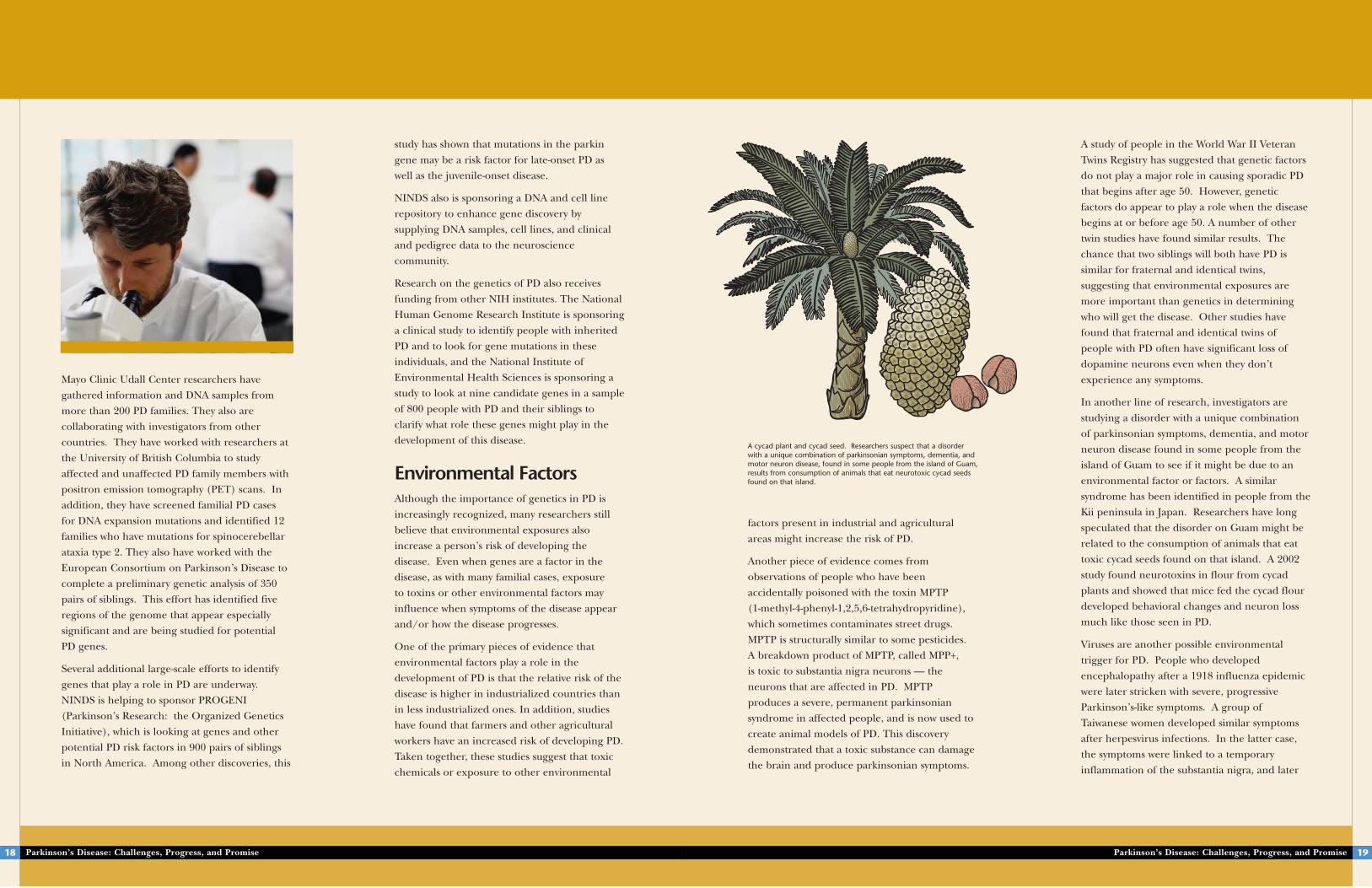
A cycad plant and cycad seed. Researchers suspect that a disorderwith a unique combination of parkinsonian symptoms, dementia, andmotor neuron disease, found in some people from the island of Guam,results from consumption of animals that eat neurotoxic cycad seedsfound on that island.
“ A U G U S T U S D I V I N U S C O N U B I U M N O K S HParkinson’s Disease: Challenges, Progress, and Promise20
Along with genetic studies, these environmental
studies lay the groundwork for a comprehensive
understanding of how PD develops and how it
might be prevented.
— — — — — — — — — — — — — — — — — — — — — — — — — — — — — — — — — — — — — — —
Pathways to Parkinson’sDiseaseMany researchers are working to understand the
complex cellular activities and protein
interactions that may lead to PD. Cellular factors
that have been implicated in PD include
mitochondrial interactions, oxidative stress,
programmed cell death (a biochemical chain of
events by which cells self-destruct), excitotoxicity,
protein aggregation, immune factors, and the
ubiquitin-proteasome protein degradation
system. While these factors represent many
different lines of research, scientists are
beginning to understand how they may fit
together to form a full picture of how PD
develops.
disappeared. However, these cases showed that
viruses can sometimes affect the region of the
brain damaged in PD. Other studies have found
evidence of activated immune cells and the
accumulation of inflammation-associated
proteins in PD. These changes might be
triggered by viruses in some cases.
Scientists are continuing to study environmental
toxins such as pesticides and herbicides that can
cause PD symptoms in animals. Researchers
supported by the NINDS and the National
Institute on Aging have shown that exposing
rodents to the pesticide rotenone can cause
cellular and behavioral changes that mimic those
seen in PD. Work supported by the National
Institute of Environmental Health Sciences has
shown that other agricultural compounds also
can produce abnormalities in cells that are
similar to those seen in PD. This research is
supported through a program called the
Collaborative Centers for Parkinson’s Disease
Environmental Research (CCPDER)
Consortium. This program sponsors a variety of
projects to examine how occupational exposure
to toxins and use of caffeine and other
substances may affect risk, and whether inherited
genetic mutations may predispose certain people
to developing PD after exposure to certain
chemicals.
Researchers at Rush-Presbyterian-St. Luke’s
Medical Center have examined whether prenatal
exposure to toxins may increase the risk of PD.
They found that exposure to a bacterial toxin
called lipopolysaccharide during development in
rats leads to the birth of animals with fewer than
the normal number of dopamine neurons. This
dopamine neuron loss persists into the animals’
adulthood and increases with age, which mimics
the course of human PD.
causes oxidative protein and DNA damage and
increases susceptibility to free radical-induced
cell death. It also leads to the same
pathological, biochemical, and behavioral
features seen in PD. Despite the fact that it
inhibits complex I throughout the brain,
rotenone causes degeneration only in
dopamine neurons. Studies suggest that these
neurons are selectively vulnerable to complex I
impairment.
Scientists at the Duke University Udall Center
have found evidence that specific genetic
variations in mtDNA, known as genetic
polymorphisms, can increase the risk of
getting PD, while other mtDNA variations are
■ Mitochondria, Oxidative Stress, andProgrammed Cell DeathFor years, mitochondria, the “energy plants” of
the cell, have been implicated in the
development of PD. Mitochondria are unique
parts of the cell that have their own DNA
(mtDNA). This DNA is separate from the
genes found in the nucleus of every cell. Most
research on the role of mitochondria in PD
points to abnormalities in the largest
component of the mitochondrial energy
processing machinery — a group of proteins
known as complex I.
Several lines of research suggest a
mitochondrial role in protein aggregation,
Lewy body formation, and neuronal death.
Mitochondria are major sources of free
radicals — highly unstable molecules that
damage components of the cell, such as
membranes, proteins, and DNA. This process
is often referred to as oxidative stress.
Oxidative stress-related changes, including
free radical damage to DNA, proteins, and
fats, have been detected in brains of PD
patients.
Research has shown that an array of toxins,
including MPTP and a pesticide and herbicide
called rotenone, can affect mitochondrial
complex I and increase the number of free
radicals it produces. Researchers at the
Columbia University Udall Center have found
that these free radicals can modify alpha-
synuclein in a way that causes it to aggregate
or clump together into minute fibers, called
fibrils.
Investigators at Emory University have
modeled the process by which mitochondrial
defects produce oxidative stress by using
rotenone. In rats, chronic rotenone exposure
Parkinson’s Disease: Challenges, Progress, and Promise 21
A dopamine-producing neuron that is undergoing programmed celldeath after exposure to the toxin 6-hydroxydopamine. The redstaining demonstrates that this is a dopamine neuron. The greenshows cleavage of a cellular protein called actin due to activity of cell death proteins called caspases. The blue shows clumping of the neuron's nuclear DNA, due to programmed cell death. [Credit: Robert E. Burke, M.D., Columbia University, New York, NY.Reprinted from Experimental Neurology, 2002, vol. 175, withpermission from Elsevier.]
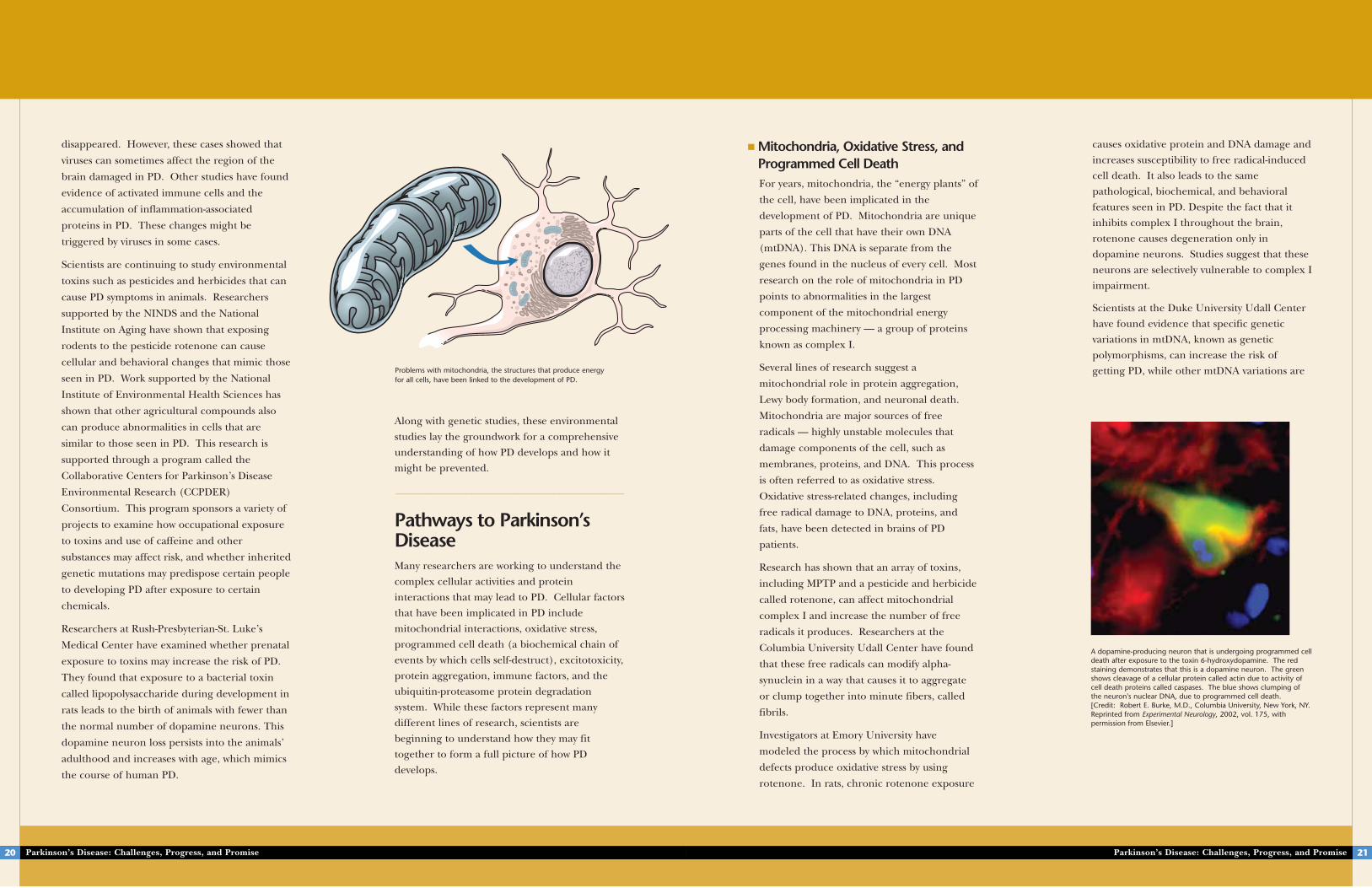
Problems with mitochondria, the structures that produce energyfor all cells, have been linked to the development of PD.
“ A U G U S T U S D I V I N U S C O N U B I U M N O K S HParkinson’s Disease: Challenges, Progress, and Promise22
suggest that programmed cell death plays a
role in animal models for PD and that it also
may be involved in the human disease.
Scientists at the University of Virginia Udall
Center have found that treatment with MPP+,
a toxic derivative of MPTP that inhibits
mitochondrial complex I, influences several
molecules known to play a role in
programmed cell death. Interestingly, some of
these changes required activation of nitric
oxide, a free radical that is often expressed in
injured or damaged cells. The role of nitric
oxide in programmed cell death was
confirmed in an experiment in which cells
were treated with nitric oxide instead of
MPP+. This treatment produced the same
programmed cell death-related changes as
MPP+. Other experiments have shown that
mice lacking Bax and nitric oxide are
protected against MPTP toxicity.
Investigators at the University of Virginia Udall
Center also have developed hybrid cells, called
cybrids, in which mitochondrial DNA from PD
patients is placed in neuroblastoma (cancer)
cells. These cybrids develop Lewy bodies just
like those in the dopamine neurons of PD
patients. The cybrid cell lines with the lowest
associated with a lowered risk of the disorder.
They also have found that PD patients have
more mtDNA variations than patients with
other movement disorders or Alzheimer’s
disease. Researchers still need to define how
these mtDNA variations may lead to PD.
Mitochondria also are thought to initiate
a process called programmed cell death.
Programmed cell death, or apoptosis, is
necessary for normal embryonic development.
Scientists believe programmed cell death
allows cells to die without disturbing their
surrounding environment. However,
programmed cell death also has been
implicated in many neurodegenerative
diseases and conditions, including PD.
Several molecules are known to participate
in the programmed cell death pathway; some
promote cell survival while others promote
cell death. An important topic in
neuroscience research is the relationship
between these pro-death and pro-survival
molecules.
Mitochondria trigger programmed cell death
by releasing a substance called cytochrome c
that activates proteins called caspases and
other cell death factors. Researchers believe
this may occur in response to oxidative stress
and mitochondrial toxins.
The idea that programmed cell death plays a
role in PD has been strengthened by studies
performed at the Udall Centers. For example,
Columbia University scientists have discovered
that, in an MPTP mouse model for PD, a pro-
cell death molecule known as Bax is abundant
in dopamine-producing neurons of the
substantia nigra. They also showed that mice
lacking the Bax gene were protected from
brain damage caused by MPTP. These results
Studies have suggested that UCH-L1 is
involved in the production of ubiquitin.
Mutations in the parkin gene also interfere
with normal proteasomal function. Scientists
at the Johns Hopkins University Udall Center
have shown that treatment with a toxin that
inhibits the ubiquitin-proteasome system
causes cells with mutant alpha-synuclein to be
susceptible to programmed cell death. This
cell death is accompanied by activation of
caspases and by injury to the mitochondria.
These changes could be blocked by
cyclosporin A, which prevents the release of
factors that activate caspases.
Proteasome inhibition results in accumulation
of molecules normally degraded by the
ubiquitin-proteasome pathway, such as p53,
NFKB, and Bax. These molecules help to
promote programmed cell death.
■ Protein Aggregation
PD is characterized by fibrillar inclusions
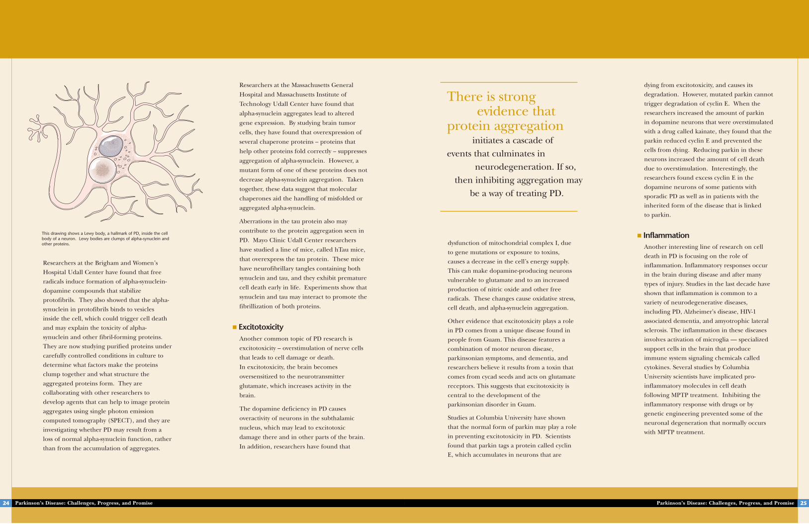
inside the cell called Lewy bodies. Lewy bodies
include clumps (aggregates) of alpha-
synuclein fibrils and other proteins. There is
strong evidence that this protein aggregation
initiates a cascade of events that culminates in
neurodegeneration. If so, then inhibiting
aggregation may be a way of treating PD.
Many researchers are trying to learn the
function of Lewy bodies. Some studies argue
that Lewy bodies are a byproduct of
degenerative processes within neurons, while
others suggest that Lewy bodies are a
protective mechanism by which neurons lock
away abnormal molecules that might otherwise
be harmful. In addition, some research
suggests that protofibrils – an intermediate
step in the development of alpha-synuclein
fibrils – may be damaging to the cell.
complex I activity make the most Lewy bodies.
These findings show that PD mitochondrial
gene expression in a cybrid model is sufficient
to spontaneously cause development of Lewy
bodies, providing strong support for the idea
that mitochondrial defects are key to the
development of sporadic PD.
Collectively, these results demonstrate that
mitochondrial-induced programmed cell
death contributes to the neuronal loss in PD.
This suggests that a possible treatment strategy
for PD is to inhibit the cascade of events
associated with programmed cell death.
Accordingly, scientists have discovered that
inhibiting the release of cytochrome c and
caspases from mitochondria with the drugs
minocycline, pramipexole, and bongkreckic
acid can protect cells from degeneration.
These drugs may be clinically useful as
neuroprotectants to prevent PD.
■ Protein Degradation (Ubiquitin-Proteasome System) Another major area of PD research involves
the cell’s protein disposal system, called the
ubiquitin-proteasome system. Researchers
believe that if this disposal system fails to work
correctly, toxins and other substances may
build up to harmful levels, leading to cell
death.
In the ubiquitin-proteasome system, a
chemical called ubiquitin acts as a “tag” that
marks certain proteins in the cell for
degradation by the proteasomes. The
ubiquitin-proteasome system involves
interactions between several proteins,
including parkin and UCH-L1. This suggests
that disruption of the ubiquitin-proteasome
pathway is part of the mechanism by which
mutations in these genes cause PD.
Parkinson’s Disease: Challenges, Progress, and Promise 23
“ A U G U S T U S D I V I N U S C O N U B I U M N O K S HParkinson’s Disease: Challenges, Progress, and Promise24
Researchers at the Massachusetts General
Hospital and Massachusetts Institute of
Technology Udall Center have found that
alpha-synuclein aggregates lead to altered
gene expression. By studying brain tumor
cells, they have found that overexpression of
several chaperone proteins – proteins that
help other proteins fold correctly – suppresses
aggregation of alpha-synuclein. However, a
mutant form of one of these proteins does not
decrease alpha-synuclein aggregation. Taken
together, these data suggest that molecular
chaperones aid the handling of misfolded or
aggregated alpha-synuclein.
Aberrations in the tau protein also may
contribute to the protein aggregation seen in
PD. Mayo Clinic Udall Center researchers
have studied a line of mice, called hTau mice,
that overexpress the tau protein. These mice
have neurofibrillary tangles containing both
synuclein and tau, and they exhibit premature
cell death early in life. Experiments show that
synuclein and tau may interact to promote the
fibrillization of both proteins.
■ ExcitotoxicityAnother common topic of PD research is
excitotoxicity – overstimulation of nerve cells
that leads to cell damage or death.
In excitotoxicity, the brain becomes
oversensitized to the neurotransmitter
glutamate, which increases activity in the
brain.
The dopamine deficiency in PD causes
overactivity of neurons in the subthalamic
nucleus, which may lead to excitotoxic
damage there and in other parts of the brain.
In addition, researchers have found that
Researchers at the Brigham and Women’s
Hospital Udall Center have found that free
radicals induce formation of alpha-synuclein-
dopamine compounds that stabilize
protofibrils. They also showed that the alpha-
synuclein in protofibrils binds to vesicles
inside the cell, which could trigger cell death
and may explain the toxicity of alpha-
synuclein and other fibril-forming proteins.
They are now studying purified proteins under
carefully controlled conditions in culture to
determine what factors make the proteins
clump together and what structure the
aggregated proteins form. They are
collaborating with other researchers to
develop agents that can help to image protein
aggregates using single photon emission
computed tomography (SPECT), and they are
investigating whether PD may result from a
loss of normal alpha-synuclein function, rather
than from the accumulation of aggregates.
dying from excitotoxicity, and causes its
degradation. However, mutated parkin cannot
trigger degradation of cyclin E. When the
researchers increased the amount of parkin
in dopamine neurons that were overstimulated
with a drug called kainate, they found that the
parkin reduced cyclin E and prevented the
cells from dying. Reducing parkin in these
neurons increased the amount of cell death
due to overstimulation. Interestingly, the
researchers found excess cyclin E in the
dopamine neurons of some patients with
sporadic PD as well as in patients with the
inherited form of the disease that is linked
to parkin.
■ InflammationAnother interesting line of research on cell
death in PD is focusing on the role of
inflammation. Inflammatory responses occur
in the brain during disease and after many
types of injury. Studies in the last decade have
shown that inflammation is common to a
variety of neurodegenerative diseases,
including PD, Alzheimer's disease, HIV-1
associated dementia, and amyotrophic lateral
sclerosis. The inflammation in these diseases
involves activation of microglia — specialized
support cells in the brain that produce
immune system signaling chemicals called
cytokines. Several studies by Columbia
University scientists have implicated pro-
inflammatory molecules in cell death
following MPTP treatment. Inhibiting the
inflammatory response with drugs or by
genetic engineering prevented some of the
neuronal degeneration that normally occurs
with MPTP treatment.
dysfunction of mitochondrial complex I, due
to gene mutations or exposure to toxins,
causes a decrease in the cell’s energy supply.
This can make dopamine-producing neurons
vulnerable to glutamate and to an increased
production of nitric oxide and other free
radicals. These changes cause oxidative stress,
cell death, and alpha-synuclein aggregation.
Other evidence that excitotoxicity plays a role
in PD comes from a unique disease found in
people from Guam. This disease features a
combination of motor neuron disease,
parkinsonian symptoms, and dementia, and
researchers believe it results from a toxin that
comes from cycad seeds and acts on glutamate
receptors. This suggests that excitotoxicity is
central to the development of the
parkinsonian disorder in Guam.
Studies at Columbia University have shown
that the normal form of parkin may play a role
in preventing excitotoxicity in PD. Scientists
found that parkin tags a protein called cyclin
E, which accumulates in neurons that are
Parkinson’s Disease: Challenges, Progress, and Promise 25
There is strong evidence that
protein aggregation initiates a cascade of
events that culminates in neurodegeneration. If so,
then inhibiting aggregation may be a way of treating PD.
This drawing shows a Lewy body, a hallmark of PD, inside the cellbody of a neuron. Lewy bodies are clumps of alpha-synuclein andother proteins.

“ A U G U S T U S D I V I N U S C O N U B I U M N O K S HParkinson’s Disease: Challenges, Progress, and Promise26
Models for Parkinson’sDiseaseMuch of the research that is leading to advances
in understanding and treating PD would not be
possible without research models – cell lines and
animals with features that mimic those of human
PD. Scientists use these models to investigate
questions such as what goes wrong in PD, how
does cellular damage lead to behavioral
symptoms, and how might potential new
treatments affect the disease process. For
example, levodopa, the drug most commonly
used to treat PD, was shown to counteract PD
symptoms in an animal model before it was
tested in humans.
Researchers at the Udall Centers and other
institutions are continually refining existing
research models and developing new ones.
Current models range from specialized, hybrid
cells in culture dishes to rats and other animals
with the same genetic defects identified in
humans with PD. These models fall into three
Although inflammation can be damaging,
studies have shown that activating immune
cells in specific ways also can protect nerve
cells in animal models of spinal cord and
brain injury. Recently, researchers at the
University of Nebraska Medical Center and at
the Columbia University Udall Center in New
York successfully reduced the amount of
neurodegeneration in a mouse model for PD
by using an experimental vaccine to modify
the behavior of microglia in the brain.
Research at the Columbia University Udall
Center has shown that dopamine neurons in
brains from patients with PD have higher
levels of an inflammatory enzyme called COX-
2 than those of people without PD. COX-2
triggers inflammation in damaged tissues. The
scientists also found elevated levels of COX-2
in a mouse model for PD. When they gave
these mice a drug called rofecoxib that
inhibits COX-2, it doubled the number of
neurons that survived. Surprisingly, however,
the researchers did not find reduced
inflammation with this drug. Instead, they
found that the COX-2 inhibitor may protect
neurons by preventing oxidative stress.
Other studies suggest that the protein GSTO-
1, which has been linked to the age of onset of
PD, may modify the inflammatory cytokine
interleukin-1 beta and therefore may reduce
the inflammation found in brains from people
with PD. It also may protect against
programmed cell death.
— — — — — — — — — — — — — — — — — — — — — — — — — — — — — — — — — — — — — — —
6-Hydroxydopamine is another toxin that kills
dopamine neurons, producing neuron death
and parkinsonian symptoms in rats and mice.
The 6-hydroxydopamine model has been used
to evaluate potential PD therapies such as cell
transplantation and neurotrophic factors.
In the late 1990s, researchers at the Emory
University Udall Center developed a new
model using a pesticide and herbicide called
rotenone. Rotenone interferes with the
activity of mitochondrial complex I. The
rotenone model is the first one that produces
selective neuron degeneration, Lewy bodies,
and behavioral changes similar to those seen
in humans with PD. Rats exposed to rotenone
develop large inclusions in substantia nigra
neurons that resemble Lewy bodies and
contain alpha-synuclein and ubiquitin.
Rotenone-treated animals also develop
bradykinesia, rigidity, and gait problems.
A common theme among the toxin-induced
Parkinson models is that the toxins interfere
with activities of mitochondria. This
knowledge has helped researchers develop
additional models using different agents that
act on mitochondria. For example, scientists
have developed new rodent models by
administering the pesticides paraquat and
maneb, and by using a combination of MPTP
and the drug probenecid.
■ Genetic ModelsThe discoveries of genetic mutations in some
hereditary cases of PD have prompted the
development of mouse models genetically
engineered to have mutations or deletions of
PD genes. These so-called transgenic mice
have become excellent models to study how
PD develops. Genetic engineering has also
categories: toxin-induced models that show how
environmental factors trigger parkinsonian
symptoms, genetic models that show how gene
defects can affect the brain, and spontaneously
occurring models that mimic some of the
features of PD. The cellular and behavioral
changes in toxin-induced models often overlap
with those that have gene defects, providing
further evidence that PD results from both
environmental and genetic factors.
■ Toxin-Induced ModelsThe two best-known and widely used animal
models in PD research are the MPTP model
and the 6-hydroxydopamine model. MPTP is
a toxin that kills neurons in the substantia
nigra, causing symptoms that closely resemble
PD. Investigators discovered this reaction in
the 1980s when heroin addicts in California
who had taken a street drug contaminated
with MPTP developed severe parkinsonism.
This discovery allowed researchers to simulate
PD in animals for the first time.
Parkinson’s Disease: Challenges, Progress, and Promise 27
Research at theColumbia University
Udall Center has shown that dopamine neurons
in brains from patients with PD have higher levels of an
inflammatory enzyme called COX-2than those of people without PD.
gait. The second spontaneous mouse model
for PD is called the quaking mouse because it
develops tremors soon after birth. Due to a
chromosomal deletion, these mice lack both
the parkin gene and the PACRG gene. Unlike
humans with parkin deletions, this mouse does
not have a loss of dopamine-producing
neurons or a buildup of alpha-synuclein.
However, it may help investigators understand
the function of the parkin protein.
Therapeutic ApproachesWhile levodopa and other drugs can provide
initial relief from parkinsonian symptoms, none
of these treatments halts the loss of dopamine
neurons and nerve fibers. Thus, new treatments
that slow the underlying disease are desperately
needed. As knowledge about this disorder grows,
potential new ways of preventing and treating
the disease are being revealed. Promising
treatments in development include new drug
therapies (including neurotrophins,
neuroprotectants, and immunotherapy), surgical
therapies, cell transplantation, gene therapy, and
transcranial magnetic stimulation.
■ Drug TherapyCurrent treatment for PD relies primarily on
drugs to control the symptoms. While these
drugs work well early in PD, they progressively
fail as more nerve cells die. Drug-induced
dyskinesias and fluctuations of motor
symptoms also limit drug benefits in many
cases.
Neurotrophic factors – molecules that support
survival, growth, and development of brain
cells – are one focus of new drug research.
These chemicals are being studied as potential
“ A U G U S T U S D I V I N U S C O N U B I U M N O K S H
therapies for many neurological diseases.
Researchers are investigating whether
neurotrophic factors can halt dopamine cell
degeneration and help to repair brain cells in
PD. One such drug, glial cell line-derived
neurotrophic factor (GDNF), has been shown
to protect dopamine neurons and to promote
their survival in models of PD. Researchers at
the University of Kentucky Udall Center have
helped to develop a technique for delivering
these molecules directly into the brain. When
GDNF was given for a period of 3 to 6 months,
it prompted repair of dopamine neurons and
improvement in their function. It also seemed
to protect damaged dopamine cells from
further degeneration. The researchers are
now conducting an FDA-approved Phase I
Parkinson’s Disease: Challenges, Progress, and Promise 29
been used to develop cell lines that model
some of the processes that go awry in PD.
One example of a genetic model — human
neuronal cells that contain mutant alpha-
synuclein — was developed by researchers at
the Mayo Clinic Udall Center. These cells
develop levels of alpha-synuclein 25- to 50-fold
higher than in controls. When these cells are
exposed to rotenone, unusual forms of
synuclein begin to appear.
Researchers at the University of Virginia Udall
Center have developed hybrid cells, called
cybrids, that have mitochondrial DNA from
PD patients inserted into tumor cells. These
cybrids develop Lewy bodies just like those in
the dopamine neurons of PD patients. Before
this model was developed, researchers needed
brain samples from PD patients in order to
study Lewy bodies.
Researchers also have developed strains of
mice with mutations in the alpha-synuclein
and parkin genes. Mice with alpha-synuclein
mutations develop an adult-onset
neurodegenerative disease characterized by
movement dysfunction and pathological
aggregation of alpha-synuclein. Mice without
the parkin gene show abnormal regulation of
dopamine in the striatum and impairments in
behavioral tests that require muscle
coordination. Researchers are now working to
develop animal models without normal DJ-1
function.
■ Spontaneously Occurring ModelsScientists have identified two spontaneously
occurring strains of mice that may be useful as
models for PD. The first strain is known as
reeler mice. These mice have reduced
dopamine activity in the striatum and several
other brain areas, which results in an impaired
Parkinson’s Disease: Challenges, Progress, and Promise28
Neuroprotection
A major goal of PD research is the development of
new therapies. Currently available treatments for
PD can effectively control motor symptoms of the
disease in the early stages, but they don’t slow or
halt the relentless progression of the disease. An
explosion of discoveries during the past decade is
now providing renewed hope of a therapy that will
prevent the underlying nerve damage in this disease
– a strategy known as neuroprotection.
The idea of neuroprotection for PD is not new. A
number of clinical studies have tested different
compounds to see if they might stop the disease
progression. Some studies initially showed small
positive effects, but most of these studies included a
limited number of patients and did not examine the
effects of these therapies over a long period of time.
In addition, researchers have no good way of
measuring whether these or other compounds truly
prevent neuronal damage. Consequently,
researchers cannot be sure that the positive effects
seen in these studies are due to neuroprotection or
if they represent short-term effects on symptoms.
Recently, a wealth of new information about how
neurons may be damaged in PD has allowed
investigators to identify many potential new ways of
treating this disease, including nerve growth factors,
anti-inflammatory drugs, and antioxidants. To
overcome some of the problems that have plagued
previous studies, in 2002 an NINDS-sponsored
committee conducted a systematic review of data on
59 potential neuroprotective agents for PD. This
committee ultimately selected four of the most
promising drugs for study in a series of clinical trials
known as Neuroprotection Exploratory Trials in
Parkinson’s Disease (NET-PD). These drugs —
coenzyme Q10, GPI-1485, creatine, and minocycline
— are now being tested at more than 40 centers in
the United States and Canada. Researchers hope
that this new approach to selecting and testing
compounds will lead to the first proven
neuroprotective therapy or therapies for PD and
revolutionize treatment of this disease.
In recent years,Parkinson’s research
has advanced tothe point
that halting the progression of PD, restoring lost function,
and even preventing the disease are all considered realistic goals.
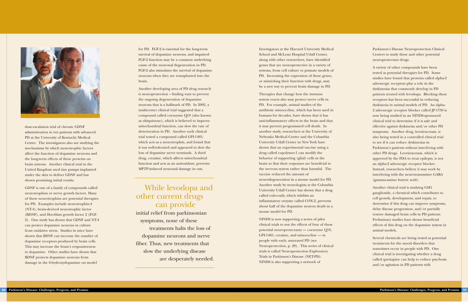
“ A U G U S T U S D I V I N U S C O N U B I U M N O K S HParkinson’s Disease: Challenges, Progress, and Promise30
for PD. FGF-2 is essential for the long-term
survival of dopamine neurons, and impaired
FGF-2 function may be a common underlying
cause of the neuronal degeneration in PD.
FGF-2 also stimulates the survival of dopamine
neurons when they are transplanted into the
brain.
Another developing area of PD drug research
is neuroprotection – finding ways to prevent
the ongoing degeneration of dopamine
neurons that is a hallmark of PD. In 2002, a
multicenter clinical trial suggested that a
compound called coenzyme Q10 (also known
as ubiquinone), which is believed to improve
mitochondrial function, can slow the rate of
deterioration in PD. Another early clinical
trial tested a compound called GPI-1485,
which acts as a neurotrophin, and found that
it was well-tolerated and appeared to slow the
loss of dopamine nerve terminals. A third
drug, creatine, which affects mitochondrial
function and acts as an antioxidant, prevents
MPTP-induced neuronal damage in rats.
dose-escalation trial of chronic GDNF
administration in ten patients with advanced
PD at the University of Kentucky Medical
Center. The investigators also are studying the
mechanisms by which neurotrophic factors
affect the function of dopamine neurons and
the long-term effects of these proteins on
brain systems. Another clinical trial in the
United Kingdom used tiny pumps implanted
under the skin to deliver GDNF and has
shown promising initial results.
GDNF is one of a family of compounds called
neurotrophins or nerve growth factors. Many
of these neurotrophins are potential therapies
for PD. Examples include neurotrophin-4
(NT-4), brain-derived neurotrophic factor
(BDNF), and fibroblast growth factor 2 (FGF-
2). One study has shown that GDNF and NT-4
can protect dopamine neurons in culture
from oxidative stress. Studies in mice have
shown that BDNF can increase the number of
dopamine receptors produced by brain cells.
This may increase the brain’s responsiveness
to dopamine. Other studies have shown that
BDNF protects dopamine neurons from
damage in the 6-hydroxydopamine rat model
Parkinson's Disease Neuroprotection Clinical
Centers to study these and other potential
neuroprotectant drugs.
A variety of other compounds have been
tested as potential therapies for PD. Some
studies have found that proteins called alpha-2
adrenergic receptors play a role in the
dyskinesias that commonly develop in PD
patients treated with levodopa. Blocking these
receptors has been successful in reducing
dyskinesia in animal models of PD. An alpha-
2 adrenergic receptor blocker called JP-1730 is
now being studied in an NINDS-sponsored
clinical trial to determine if it is safe and
effective against dyskinesia and/or other PD
symptoms. Another drug, levetiracetam, is
also being tested in a controlled clinical trial
to see if it can reduce dyskinesias in
Parkinson’s patients without interfering with
other PD drugs. Levetiracetam, which is
approved by the FDA to treat epilepsy, is not
an alpha-2 adrenergic receptor blocker.
Instead, researchers believe it may work by
interfering with the neurotransmitter GABA
(gamma-amino butyric acid).
Another clinical trial is studying GM1
ganglioside, a chemical which contributes to
cell growth, development, and repair, to
determine if this drug can improve symptoms,
delay disease progression, and/or partially
restore damaged brain cells in PD patients.
Preliminary studies have shown beneficial
effects of this drug on the dopamine system in
animal models.
Several chemicals are being tested as potential
treatments for the mood disorders that
sometimes occur in people with PD. One
clinical trial is investigating whether a drug
called quetiapine can help to reduce psychosis
and/or agitation in PD patients with
Investigators at the Harvard University Medical
School and McLean Hospital Udall Center,
along with other researchers, have identified
genes that are neuroprotective in a variety of
systems, from cell culture to primate models of
PD. Increasing the expression of these genes,
or mimicking their function with drugs, may
be a new way to prevent brain damage in PD.
Therapies that change how the immune
system reacts also may protect nerve cells in
PD. For example, animal studies of the
antibiotic minocycline, which has been used in
humans for decades, have shown that it has
anti-inflammatory effects in the brain and that
it may prevent programmed cell death. In
another study, researchers at the University of
Nebraska Medical Center and the Columbia
University Udall Center in New York have
shown that an experimental vaccine using a
drug called copolymer-1 can modify the
behavior of supporting (glial) cells in the
brain so that their responses are beneficial to
the nervous system rather than harmful. The
vaccine reduced the amount of
neurodegeneration in a mouse model for PD.
Another study by neurologists at the Columbia
University Udall Center has shown that a drug
called rofecoxib, which inhibits an
inflammatory enzyme called COX-2, prevents
about half of the dopamine neuron death in a
mouse model for PD.
NINDS is now supporting a series of pilot
clinical trials to test the effects of four of these
potential neuroprotectants — coenzyme Q10,
GPI-1485, creatine, and minocycline — in
people with early, untreated PD (see
Neuroprotection, p. 28). This series of clinical
trials is called Neuroprotection Exploratory
Trials in Parkinson's Disease (NET-PD).
NINDS is also supporting a network of
Parkinson’s Disease: Challenges, Progress, and Promise 31
While levodopa and other current drugs
can provide initial relief from parkinsonian
symptoms, none of these treatments halts the loss of
dopamine neurons and nervefiber. Thus, new treatments that
slow the underlying disease are desperately needed.
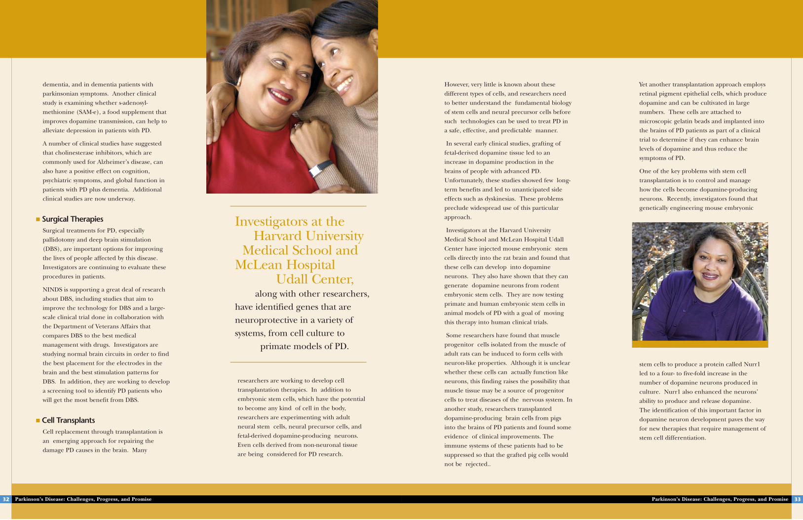
“ A U G U S T U S D I V I N U S C O N U B I U M N O K S H Parkinson’s Disease: Challenges, Progress, and Promise 33Parkinson’s Disease: Challenges, Progress, and Promise32
researchers are working to develop cell
transplantation therapies. In addition to
embryonic stem cells, which have the potential
to become any kind of cell in the body,
researchers are experimenting with adult
neural stem cells, neural precursor cells, and
fetal-derived dopamine-producing neurons.
Even cells derived from non-neuronal tissue
are being considered for PD research.
dementia, and in dementia patients with
parkinsonian symptoms. Another clinical
study is examining whether s-adenosyl-
methionine (SAM-e), a food supplement that
improves dopamine transmission, can help to
alleviate depression in patients with PD.
A number of clinical studies have suggested
that cholinesterase inhibitors, which are
commonly used for Alzheimer’s disease, can
also have a positive effect on cognition,
psychiatric symptoms, and global function in
patients with PD plus dementia. Additional
clinical studies are now underway.
■ Surgical TherapiesSurgical treatments for PD, especially
pallidotomy and deep brain stimulation
(DBS), are important options for improving
the lives of people affected by this disease.
Investigators are continuing to evaluate these
procedures in patients.
NINDS is supporting a great deal of research
about DBS, including studies that aim to
improve the technology for DBS and a large-
scale clinical trial done in collaboration with
the Department of Veterans Affairs that
compares DBS to the best medical
management with drugs. Investigators are
studying normal brain circuits in order to find
the best placement for the electrodes in the
brain and the best stimulation patterns for
DBS. In addition, they are working to develop
a screening tool to identify PD patients who
will get the most benefit from DBS.
■ Cell TransplantsCell replacement through transplantation is
an emerging approach for repairing the
damage PD causes in the brain. Many
Yet another transplantation approach employs
retinal pigment epithelial cells, which produce
dopamine and can be cultivated in large
numbers. These cells are attached to
microscopic gelatin beads and implanted into
the brains of PD patients as part of a clinical
trial to determine if they can enhance brain
levels of dopamine and thus reduce the
symptoms of PD.
One of the key problems with stem cell
transplantation is to control and manage
how the cells become dopamine-producing
neurons. Recently, investigators found that
genetically engineering mouse embryonic
stem cells to produce a protein called Nurr1
led to a four- to five-fold increase in the
number of dopamine neurons produced in
culture. Nurr1 also enhanced the neurons’
ability to produce and release dopamine.
The identification of this important factor in
dopamine neuron development paves the way
for new therapies that require management of
stem cell differentiation.
However, very little is known about these
different types of cells, and researchers need
to better understand the fundamental biology
of stem cells and neural precursor cells before
such technologies can be used to treat PD in
a safe, effective, and predictable manner.
In several early clinical studies, grafting of
fetal-derived dopamine tissue led to an
increase in dopamine production in the
brains of people with advanced PD.
Unfortunately, these studies showed few long-
term benefits and led to unanticipated side
effects such as dyskinesias. These problems
preclude widespread use of this particular
approach.
Investigators at the Harvard University
Medical School and McLean Hospital Udall
Center have injected mouse embryonic stem
cells directly into the rat brain and found that
these cells can develop into dopamine
neurons. They also have shown that they can
generate dopamine neurons from rodent
embryonic stem cells. They are now testing
primate and human embryonic stem cells in
animal models of PD with a goal of moving
this therapy into human clinical trials.
Some researchers have found that muscle
progenitor cells isolated from the muscle of
adult rats can be induced to form cells with
neuron-like properties. Although it is unclear
whether these cells can actually function like
neurons, this finding raises the possibility that
muscle tissue may be a source of progenitor
cells to treat diseases of the nervous system. In
another study, researchers transplanted
dopamine-producing brain cells from pigs
into the brains of PD patients and found some
evidence of clinical improvements. The
immune systems of these patients had to be
suppressed so that the grafted pig cells would
not be rejected..
Investigators at the Harvard University
Medical School andMcLean Hospital
Udall Center, along with other researchers,
have identified genes that areneuroprotective in a variety of systems, from cell culture to
primate models of PD.
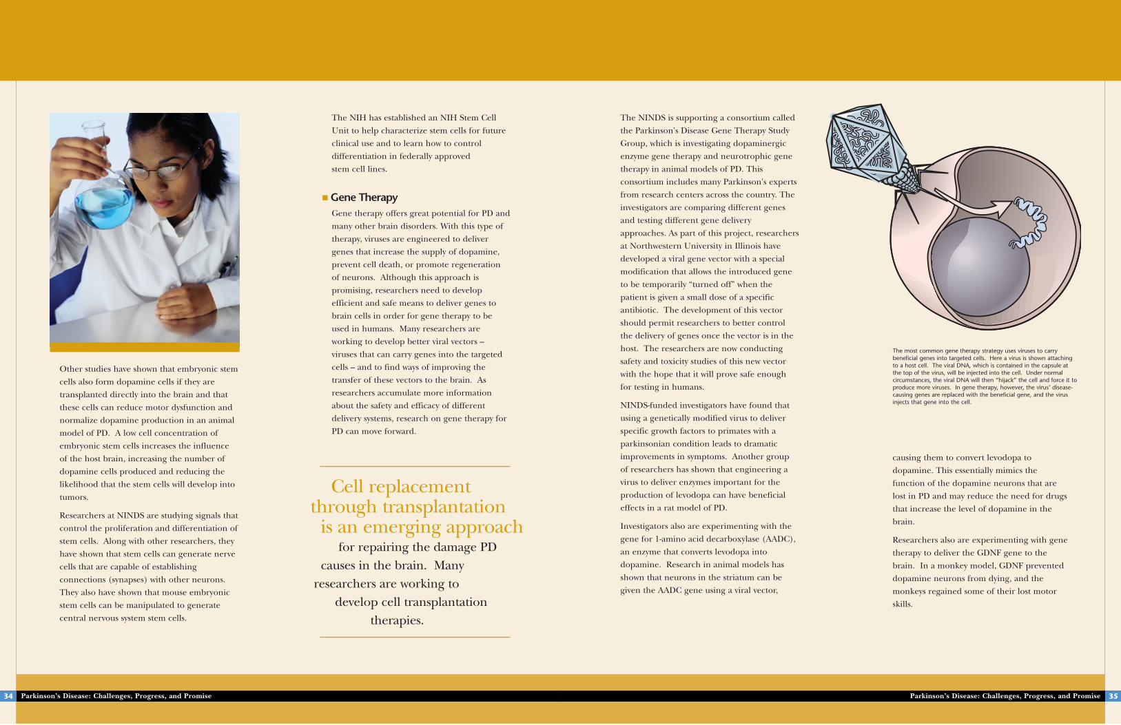
“ A U G U S T U S D I V I N U S C O N U B I U M N O K S H
causing them to convert levodopa to
dopamine. This essentially mimics the
function of the dopamine neurons that are
lost in PD and may reduce the need for drugs
that increase the level of dopamine in the
brain.
Researchers also are experimenting with gene
therapy to deliver the GDNF gene to the
brain. In a monkey model, GDNF prevented
dopamine neurons from dying, and the
monkeys regained some of their lost motor
skills.
The NINDS is supporting a consortium called
the Parkinson's Disease Gene Therapy Study
Group, which is investigating dopaminergic
enzyme gene therapy and neurotrophic gene
therapy in animal models of PD. This
consortium includes many Parkinson's experts
from research centers across the country. The
investigators are comparing different genes
and testing different gene delivery
approaches. As part of this project, researchers
at Northwestern University in Illinois have
developed a viral gene vector with a special
modification that allows the introduced gene
to be temporarily “turned off” when the
patient is given a small dose of a specific
antibiotic. The development of this vector
should permit researchers to better control
the delivery of genes once the vector is in the
host. The researchers are now conducting
safety and toxicity studies of this new vector
with the hope that it will prove safe enough
for testing in humans.
NINDS-funded investigators have found that
using a genetically modified virus to deliver
specific growth factors to primates with a
parkinsonian condition leads to dramatic
improvements in symptoms. Another group
of researchers has shown that engineering a
virus to deliver enzymes important for the
production of levodopa can have beneficial
effects in a rat model of PD.
Investigators also are experimenting with the
gene for 1-amino acid decarboxylase (AADC),
an enzyme that converts levodopa into
dopamine. Research in animal models has
shown that neurons in the striatum can be
given the AADC gene using a viral vector,
Parkinson’s Disease: Challenges, Progress, and Promise 35Parkinson’s Disease: Challenges, Progress, and Promise34
The NIH has established an NIH Stem Cell
Unit to help characterize stem cells for future
clinical use and to learn how to control
differentiation in federally approved
stem cell lines.
■ Gene TherapyGene therapy offers great potential for PD and
many other brain disorders. With this type of
therapy, viruses are engineered to deliver
genes that increase the supply of dopamine,
prevent cell death, or promote regeneration
of neurons. Although this approach is
promising, researchers need to develop
efficient and safe means to deliver genes to
brain cells in order for gene therapy to be
used in humans. Many researchers are
working to develop better viral vectors –
viruses that can carry genes into the targeted
cells – and to find ways of improving the
transfer of these vectors to the brain. As
researchers accumulate more information
about the safety and efficacy of different
delivery systems, research on gene therapy for
PD can move forward.
Other studies have shown that embryonic stem
cells also form dopamine cells if they are
transplanted directly into the brain and that
these cells can reduce motor dysfunction and
normalize dopamine production in an animal
model of PD. A low cell concentration of
embryonic stem cells increases the influence
of the host brain, increasing the number of
dopamine cells produced and reducing the
likelihood that the stem cells will develop into
tumors.
Researchers at NINDS are studying signals that
control the proliferation and differentiation of
stem cells. Along with other researchers, they
have shown that stem cells can generate nerve
cells that are capable of establishing
connections (synapses) with other neurons.
They also have shown that mouse embryonic
stem cells can be manipulated to generate
central nervous system stem cells.
Cell replacementthrough transplantation
is an emerging approachfor repairing the damage PD
causes in the brain. Many researchers are working to
develop cell transplantation therapies.
The most common gene therapy strategy uses viruses to carrybeneficial genes into targeted cells. Here a virus is shown attaching to a host cell. The viral DNA, which is contained in the capsule at the top of the virus, will be injected into the cell. Under normalcircumstances, the viral DNA will then “hijack” the cell and force it toproduce more viruses. In gene therapy, however, the virus’ disease-causing genes are replaced with the beneficial gene, and the virusinjects that gene into the cell.
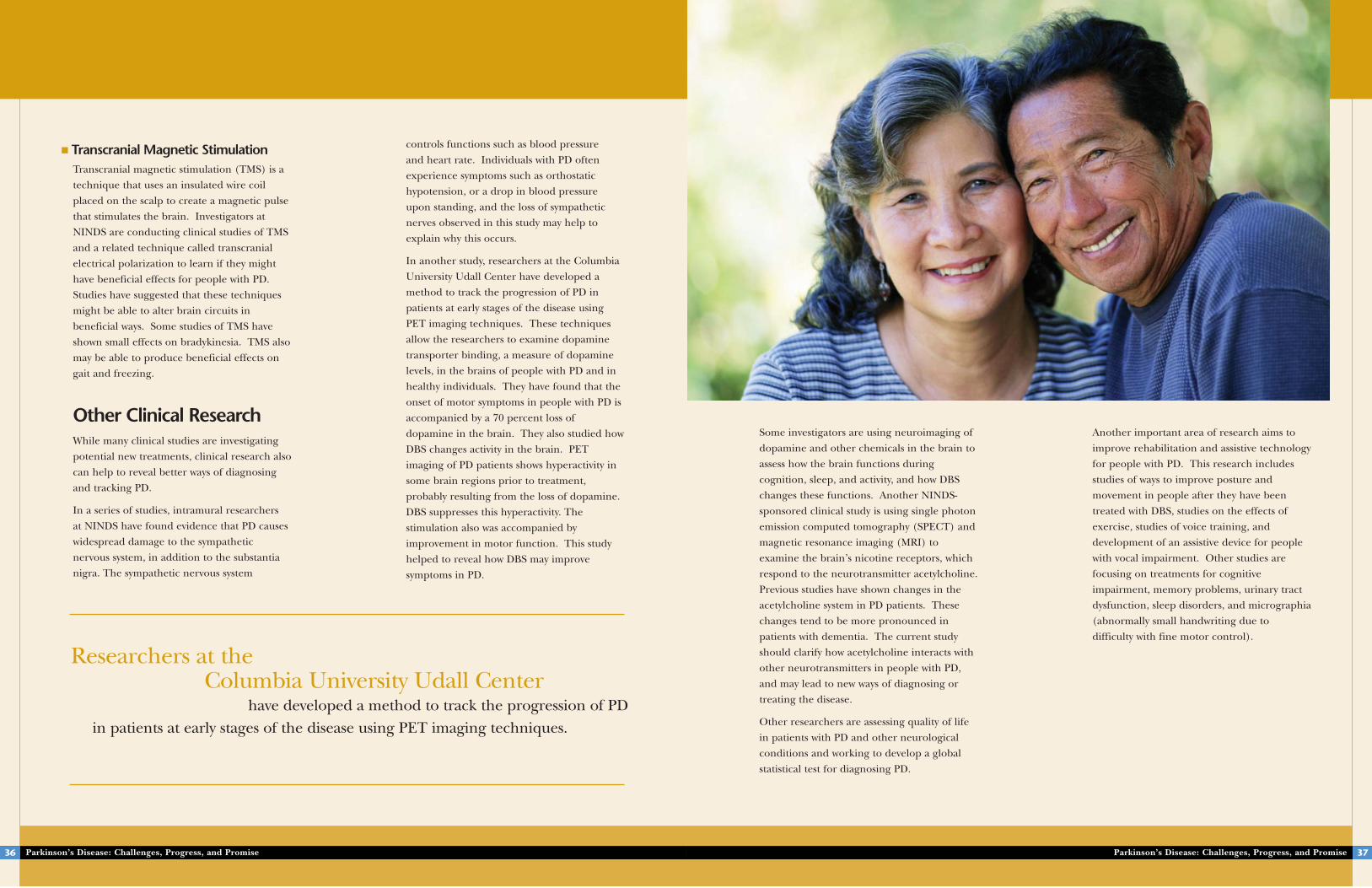
“ A U G U S T U S D I V I N U S C O N U B I U M N O K S H
Another important area of research aims to
improve rehabilitation and assistive technology
for people with PD. This research includes
studies of ways to improve posture and
movement in people after they have been
treated with DBS, studies on the effects of
exercise, studies of voice training, and
development of an assistive device for people
with vocal impairment. Other studies are
focusing on treatments for cognitive
impairment, memory problems, urinary tract
dysfunction, sleep disorders, and micrographia
(abnormally small handwriting due to
difficulty with fine motor control).
Some investigators are using neuroimaging of
dopamine and other chemicals in the brain to
assess how the brain functions during
cognition, sleep, and activity, and how DBS
changes these functions. Another NINDS-
sponsored clinical study is using single photon
emission computed tomography (SPECT) and
magnetic resonance imaging (MRI) to
examine the brain’s nicotine receptors, which
respond to the neurotransmitter acetylcholine.
Previous studies have shown changes in the
acetylcholine system in PD patients. These
changes tend to be more pronounced in
patients with dementia. The current study
should clarify how acetylcholine interacts with
other neurotransmitters in people with PD,
and may lead to new ways of diagnosing or
treating the disease.
Other researchers are assessing quality of life
in patients with PD and other neurological
conditions and working to develop a global
statistical test for diagnosing PD.
Parkinson’s Disease: Challenges, Progress, and Promise 37Parkinson’s Disease: Challenges, Progress, and Promise36
controls functions such as blood pressure
and heart rate. Individuals with PD often
experience symptoms such as orthostatic
hypotension, or a drop in blood pressure
upon standing, and the loss of sympathetic
nerves observed in this study may help to
explain why this occurs.
In another study, researchers at the Columbia
University Udall Center have developed a
method to track the progression of PD in
patients at early stages of the disease using
PET imaging techniques. These techniques
allow the researchers to examine dopamine
transporter binding, a measure of dopamine
levels, in the brains of people with PD and in
healthy individuals. They have found that the
onset of motor symptoms in people with PD is
accompanied by a 70 percent loss of
dopamine in the brain. They also studied how
DBS changes activity in the brain. PET
imaging of PD patients shows hyperactivity in
some brain regions prior to treatment,
probably resulting from the loss of dopamine.
DBS suppresses this hyperactivity. The
stimulation also was accompanied by
improvement in motor function. This study
helped to reveal how DBS may improve
symptoms in PD.
■ Transcranial Magnetic StimulationTranscranial magnetic stimulation (TMS) is a
technique that uses an insulated wire coil
placed on the scalp to create a magnetic pulse
that stimulates the brain. Investigators at
NINDS are conducting clinical studies of TMS
and a related technique called transcranial
electrical polarization to learn if they might
have beneficial effects for people with PD.
Studies have suggested that these techniques
might be able to alter brain circuits in
beneficial ways. Some studies of TMS have
shown small effects on bradykinesia. TMS also
may be able to produce beneficial effects on
gait and freezing.
Other Clinical ResearchWhile many clinical studies are investigating
potential new treatments, clinical research also
can help to reveal better ways of diagnosing
and tracking PD.
In a series of studies, intramural researchers
at NINDS have found evidence that PD causes
widespread damage to the sympathetic
nervous system, in addition to the substantia
nigra. The sympathetic nervous system
Researchers at the Columbia University Udall Center
have developed a method to track the progression of PD in patients at early stages of the disease using PET imaging techniques.
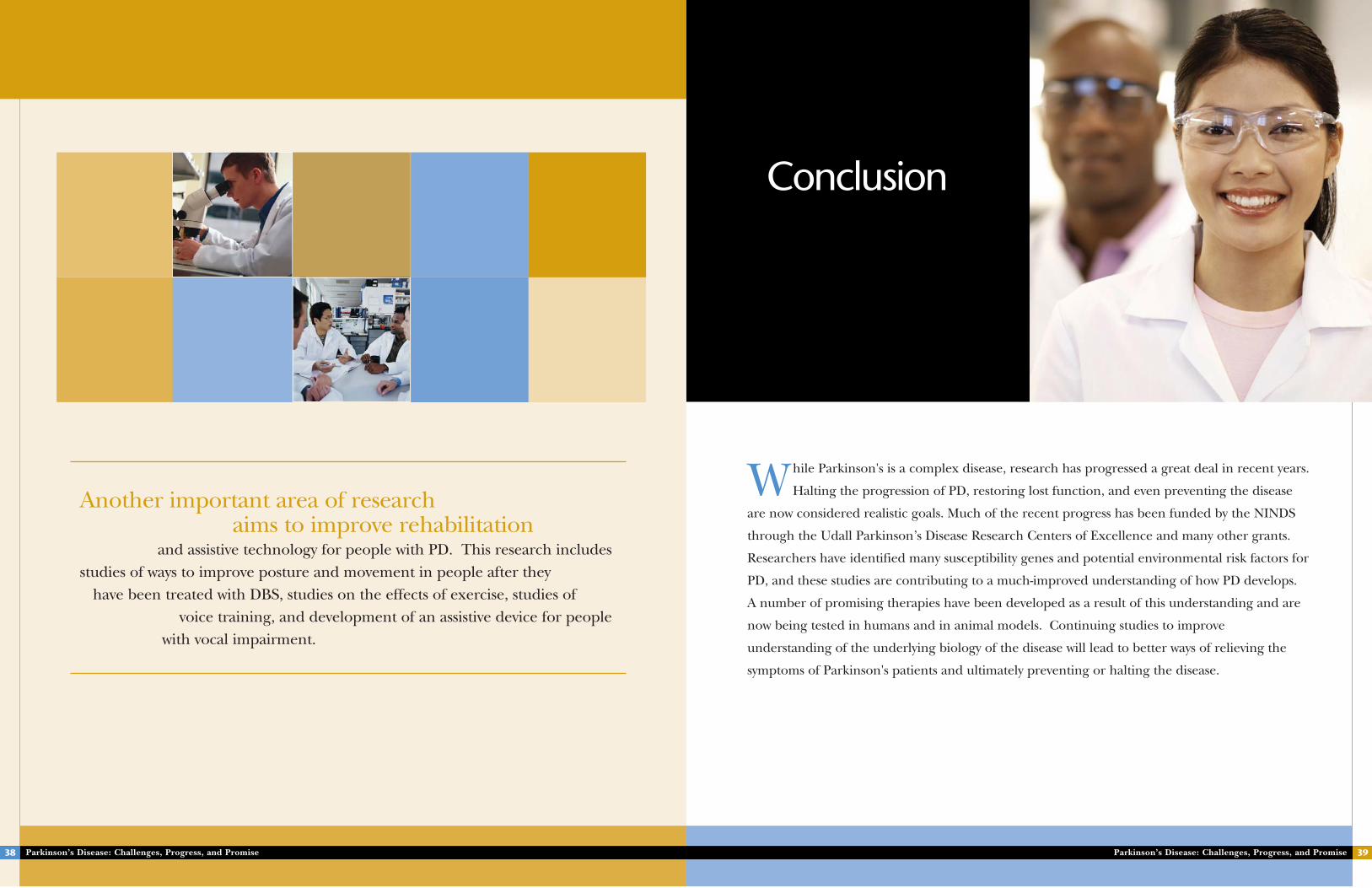
“ A U G U S T U S D I V I N U S C O N U B I U M N O K S HParkinson’s Disease: Challenges, Progress, and Promise38 Parkinson’s Disease: Challenges, Progress, and Promise
Another important area of research aims to improve rehabilitation
and assistive technology for people with PD. This research includesstudies of ways to improve posture and movement in people after they
have been treated with DBS, studies on the effects of exercise, studies of voice training, and development of an assistive device for people
with vocal impairment.
39
Conclusion and Future Directions
While Parkinson's is a complex disease, research has progressed a great deal in recent years.
Halting the progression of PD, restoring lost function, and even preventing the disease
are now considered realistic goals. Much of the recent progress has been funded by the NINDS
through the Udall Parkinson’s Disease Research Centers of Excellence and many other grants.
Researchers have identified many susceptibility genes and potential environmental risk factors for
PD, and these studies are contributing to a much-improved understanding of how PD develops.
A number of promising therapies have been developed as a result of this understanding and are
now being tested in humans and in animal models. Continuing studies to improve
understanding of the underlying biology of the disease will lead to better ways of relieving the
symptoms of Parkinson's patients and ultimately preventing or halting the disease.
Conclusion
OverviewAs part of its efforts to defeat PD, the NINDS
supports a number of Centers of Excellence for
Parkinson’s Disease Research throughout the
country. The Centers’ multidisciplinary research
environment allows scientists to take advantage of
new discoveries in the basic and technological
sciences that can lead to clinical advances, and, in
addition, allows for collaborations across centers
which can expedite the pace of research. Most of
the Centers also provide state-of-the-art training for
young scientists who are preparing for research
careers investigating PD and related neurological
disorders. Some of the topics that the Centers
address include:
• Neuronal and mitochondrial genetic studies to
elucidate key proteins involved in
neurodegeneration and to determine genetic
differences between familial and sporadic PD
• Studies of the structure and function of proteins
involved in cell death and degeneration
• Studies of the anatomical structures and brain
chemicals involved in PD
• Studies to improve animal models of PD
• Imaging studies involving PET
• Studies of PD risk factors in people of different
gender and ethnicity
• Animal studies testing possible PD treatments,
such as neuroprotective therapies, implantation of
genetically engineered cells, DBS, and levodopa
drug therapy.
These Centers have contributed greatly to the PD
research field. Their many significant achievements
include discovery of the UCH-L1, PACRG, and GSTO-
1 genes, development of the rotenone rat model for
PD, the discovery that PD mitochondrial gene
expression is sufficient to spontaneously cause
development of Lewy bodies in cells, and the first
United States clinical trial of chronic GDNF
administration in patients with advanced PD.
Recently, the NINDS has funded a Parkinson’s
Disease Data Organizing Center (PD-DOC) to collect
information from all PD centers, thus allowing for
standardized data, resources, and reagents to be
shared widely within the PD community. The
NINDS hopes that research at these Centers of
Excellence will lead to clinical trials of new
treatments in patients with PD.
HistoryOn November 13, 1997, the President of the United
States signed into law The Morris K. Udall
Parkinson’s Disease Research Act (P.L. 105-78). Prior
to the passage of this Act, the NINDS had already
recognized the need to establish Centers of
Excellence in PD research, and as a result, released
an initial Request for Applications (RFA) to solicit
these centers. Of the applications that were received
in response to this RFA, NINDS selected three
centers for funding. Following the passage of the
Udall Act, NINDS issued a second RFA for PD
Centers of Excellence and funded eight additional
grants. All of the Udall Centers focus on scientific
research designed to improve the diagnosis and
treatment of patients with PD and related
neurodegenerative disorders and on research to
gain a better understanding of the fundamental
cause(s) of the disease. The Centers have lived up to
their expectation to foster an environment that
enhances the research effectiveness of investigators
in a multidisciplinary setting, utilizing specialized
methods relevant to the study of these disorders.
The Centers of Excellence for Parkinson’s Disease
Research program was developed in honor of
former Congressman Morris K. Udall, who died in
1998 after a long battle with PD. Mr. Udall was
elected to the U.S. House of Representatives in 1961
in a special election to replace his brother Stewart
who left the position to become President John F.
Kennedy's Secretary of the Interior. Udall was
diagnosed with PD in 1979, but he remained active
as a Member of Congress until May 1991.
Parkinson’s Disease: Challenges, Progress, and Promise40
Udall Centers Across the CountryBrigham and Women's HospitalBoston, Massachusetts Director: Peter Lansbury, Jr., Ph.D.
Columbia UniversityNew York City, New YorkDirector: Robert Burke, M.D.
Duke UniversityDurham, North CarolinaDirector: Jeffery M. Vance, M.D.
Harvard University Medical School and McLean Hospital Belmont, MassachusettsDirector: Ole Isacson, M.D.
Johns Hopkins University School of MedicineBaltimore, MarylandDirector: Ted M. Dawson, M.D., Ph.D.
Massachusetts General Hospital and Massachusetts Institute of TechnologyBoston, MassachusettsDirector: Anne Young, M.D., Ph.D.
Mayo ClinicJacksonville, FloridaDirector: Dennis W. Dickson, M.D.
Northwestern UniversityChicago, IllinoisDirector: D. James Surmeier, Ph.D.
University of California, Los AngelesLos Angeles, CaliforniaDirector: Marie-Francoise S. Chesselet, M.D.
University of KentuckyLexington, KentuckyDirector: Greg A. Gerhardt, Ph.D.
University of PittsburghPittsburgh, PennsylvaniaDirector: Michael J. Zigmond, Ph.D.
University of VirginiaCharlottesville, VirginiaDirector: G. Fred Wooten, M.D.
Coordinating data and resources from all these centers:Parkinson’s Disease Organizing CenterUniversity of RochesterRochester, New YorkDirector: Roger Kurlan, M.D.
FutureNINDS is committed to continuing and
enhancing the tradition of scientific excellence
that has been fostered by the Udall Centers over
the past 5 years. To this end, NINDS and the
National Institute of Environmental Health
Sciences released a Program Announcement in
October 2002 to renew the Institute’s
commitment to the program and to aid current
and prospective Udall Center investigators in
developing competitive applications for funding.
NINDS has also announced an increase in funds
that may be allotted to the Centers to fund the
types of clinical research needed to capitalize on
the increasing number of findings in the basic
sciences. In addition, NINDS staff members are
working with the research community to develop
a standard, minimum amount of clinical
information to be collected about each PD
patient, allowing information from different
studies to be compared and combined. NINDS
believes that all of these efforts will help to
strengthen the Udall program in the coming
years.
NINDS is committed to continuing
and enhancingthe tradition of scientificexcellence that has been
fostered by the Udall Centersover the past 5 years.
Parkinson’s Disease: Challenges, Progress, and Promise 41
Morris K. Udall Centers of Excellence for PD Research

U.S. DEPARTMENT OF HEALTH AND HUMAN SERVICES
National Institutes of Health
NIH Publication No. 05-5595November 2004