Embed Size (px)
DESCRIPTION
nihms
Citation preview

Bacterial Swarming: A Model System for Studying Dynamic Self-assembly
Matthew F. Copeland and Douglas B. Weibel*Department of Biochemistry, University of Wisconsin-Madison, 433 Babcock Drive, Madison, WI,U.S.A.
AbstractBacterial swarming is an example of dynamic self-assembly in microbiology in which thecollective interaction of a population of bacterial cells leads to emergent behavior. Swarmingoccurs when cells interact with surfaces, reprogram their physiology and behavior, and adapt tochanges in their environment by coordinating their growth and motility with other cells in thecolony. This review summarizes the salient biological and biophysical features of this system anddescribes our current understanding of swarming motility. We have organized this review intofour sections: 1) The biophysics and mechanisms of bacterial motility in fluids and its relevance toswarming. 2) The role of cell/molecule, cell/surface, and cell/cell interactions during swarming. 3)The changes in physiology and behavior that accompany swarming motility. 4) A concludingdiscussion of several interesting, unanswered questions that is particularly relevant to soft matterscientists.
1. IntroductionMicrobiology is replete with examples of dynamic self-assembly that span subcellular,cellular, and multicellular length scales and are characterized by interactions between manycomponents.1 Biofilms, spores, swarms, mycelia, and fruiting bodies are multicellularstructures in which dynamic self-assembly controls the organization and function ofpopulations of cells. We refer to dynamic self-assembly in the context of a system that hasthe following characteristics: 1) it consists of many components that interact spontaneouslyand reversibly via non-covalent forces; 2) it is far from equilibrium; and 3) the componentsform ordered structures (and patterns) only when the system is continuously dissipatingenergy. The emergence of collective behavior or multicellularity in bacteria arises from thedynamic self-assembly of densely packed populations of cells based on stimuli andmechanisms that are poorly understood.2, 3 This phenotype is relevant to a range of fields,including ecology, agriculture, and biomedicine and research in this area has applications inboth fundamental and applied science.2, 4 The principles of emergent behavior that areuncovered in the study of microbial self-assembly may be applied to areas that extend farbeyond biology and microbes into fields such as economics, weather, and populationdynamics.
Eubacteria have a number of characteristics that make them an excellent experimental modelfor studying dynamic self-assembly. 1) Laboratory strains are relatively easy to culture andgrow rapidly into high-density populations of cells in liquid and on surfaces. 2) A wealth ofchemical, biochemical, and genetic data is available for different genera and make itpossible to systematically perturb and study bacteria. 3) The genome of many bacterial
* Author to whom correspondence should be addressed; [email protected].
NIH Public AccessAuthor ManuscriptSoft Matter. Author manuscript; available in PMC 2013 August 05.
Published in final edited form as:Soft Matter. 2009 ; 5(6): 1174–1187. doi:10.1039/B812146J.
NIH
-PA Author Manuscript
NIH
-PA Author Manuscript
NIH
-PA Author Manuscript

species has been sequenced and repositories and genetic stock centers catalog, store, anddistribute mutants of model organisms, including Escherichia coli (E. coli), Bacillus subtilis(B. subtilis), Salmonella enterica serovar Typhimurium (S. typhimurium), and Pseudomonasaeruginosa (P. aeruginosa). Thus it is straightforward to obtain knockouts rapidly for areasonable fee, especially for E. coli where single and double knockouts of the entiregenome have been created.5–7 4) A variety of biophysical techniques are available formanipulating individual or populations of bacterial cells on surfaces and in liquids.8, 9
One of the more interesting examples of dynamic self-assembly in microbiology is aphenotype observed in many strains of flagellated bacteria referred to as ‘swarming’.10 Theintroduction of the phrase ‘swarm’ was probably chosen to capture the dynamic, swirlingpatterns of multicellular movement reminiscent of populations of swarming bees and otherinsects (Movies S1–S4).11 Swarming motility occurs when planktonic cells contact surfacesand replicate to a high-density population of cells. It is unclear whether planktonic cellsinitially adsorb on and remain in close physical contact to surfaces or whether they areseparated from surfaces by a thin layer of fluid; we discuss the latter scenario in more detaillater in the review. The transition from the growth of cells in liquids to surfaces isaccompanied by changes in their phenotype in many species of bacteria. Populations ofswarming bacterial cells migrate collectively across surfaces at an approximate rate of 2–10µm·sec−1 and consume nutrients in the underlying medium.12 There are three basicrequirements for bacterial swarming motility: 1) cells are motile and have functionalflagella; 2) cells are in contact or close proximity to surfaces; and 3) cells are in contact withother motile cells (Table 1).
Much of our understanding of this field was pioneered by Belas, Harshey, Henrichsen,Hughes, Shapiro, Williams, and others and is based on studying the genetics andbiochemistry of swarming.3, 4, 10–17 In this review we focus on introducing scientists andengineers that study soft condensed matter to a system of dynamic bacterial self-assemblythat will benefit greatly from the introduction of polymeric materials and the physicaltechniques of materials science and engineering. We have divided this review into foursections that focus on the following areas: 1) the biophysical components of bacteria cellmotility in fluids and its relevance to swarming; 2) the role of cell/molecule, cell/surface,and cell/cell interactions during swarming; 3) the changes in physiology and behavior thataccompany swarming motility; and 4) a concluding discussion of several interesting,unanswered questions that is particularly relevant to soft matter scientists.
2. An introduction to bacterial motilityMany genera of bacteria use flagella for their motility, which are actuated by the cell andperform work on the surrounding fluid.18 Our current understanding of motility in bacteriaindicates that the mechanisms used by planktonic cells to translate through bulk fluids areclosely related to or are identical to the mechanisms used by swarming cells. To introducethe reader to this area, we provide a description of the motility system in bacteria and thephysics of fluids at the length scale of bacterial cells that plays a fundamental role in theirmotility. We focus on E. coli and S. typhimurium as these model organisms were used todissect much of what we currently know about the role of flagella in bacterial motility.
a. Expression of flagellar proteinsThe biosynthesis, assembly, and function of the flagellum involves more than 50 genes thatare divided across several operons.19 The expression of this flagellar regulon is divided intothree hierarchical, temporally transcribed classes of genes: early (class 1), middle (class 2),and late genes (class 3). Global regulatory signals influence transcription of the early genes,flhD and flhC, which constitute the flhDC operon. FlhD and FlhC are transcriptional
Copeland and Weibel Page 2
Soft Matter. Author manuscript; available in PMC 2013 August 05.
NIH
-PA Author Manuscript
NIH
-PA Author Manuscript
NIH
-PA Author Manuscript

activators that direct transcription of the middle flagellar genes from their upstream class 2promoters. The middle genes are responsible for expression of the basal body and hook ofthe flagella, as well as the class 3 promoter sigma factor, σ28 (FliA), and its correspondinganti-sigma factor, FlgM. Following completion of the basal body-hook structure, FlgM issecreted out of the nascent flagellum, allowing σ28 to activate transcription of the late (class3) flagellar and chemotaxis genes responsible for synthesis of the flagellar filament andmotility components (Figure 1b).19 The structure of the flagellum, the properties of thisorganelle, and its role in motility are described below.
b. Structure and function of the flagellumBacteria have evolved efficient mechanisms of motility in bulk fluids and on surfaces usingflagella. Swimming cells of E. coli and S. typhimurium are peritrichous and actuate 6–8flagella to translate through fluids.20, 21 Each flagellum consists of three regions: 1) helicalfilament; 2) hook; and 3) basal body and motor components (Figure 1b).22 The helicalfilament is a polymer of the flagellin protein (FliC) which terminates in a filament cap(FliD).23 Eleven parallel rows of FliC form a left-handed helical filament, which may be upto 15-µm long with a diameter of 12–25 nm.24, 25 The filament is connected to the flagellarhook, which is a polymer consisting of the FlgE protein and the hook-filament junctionproteins FlgK and FlgL.23 The basal body of the flagellum is embedded in the cell wall ofGram-negative bacteria and consists of four rings and a rod. The rings are located in distinctregions of the membrane and are illustrated in Figure 1a.23, 26
The rotary motor of the bacterial flagellum consists of a stator (MotA and MotB) and a rod(FlgB, FlgC, FlgF, and FlgG).25, 27 The components of the stator are stationary andembedded in the basal body; the rotor consists of FliG attached to the MS-ring (FliF).Together, the rotor and the stator (Mot proteins) generate torque.25, 28 A proton motiveforce, or in some circumstances, a gradient of sodium ions, causes the flagellar motor torotate.29–31 The passage of protons through channels in MotA and MotB creates aconformational change in the stator, which subsequently turns the rotor.32, 33 The C-ringconsists of FliG, FliM, and FliN and functions as a switch complex and rotor to control thedirection of rotation of the motor that actuates the flagella. A recent review by Chevance andHughes provides an excellent overview of the components of the bacterial flagellum.34
Rotation of the flagellar motor in the counterclockwise direction (CCW)—as viewed fromthe distal end of the filament—causes the left-handed helical flagellar filaments to form abundle, which propels the cell forward in a phase of motility referred to as a ‘run’ (Figure1c).35 The torque produced by rotating the bundle of flagella at an angular velocity of ~100Hz is balanced by viscous drag from the clockwise (CW) rotation of the cell body around itslong axis at a frequency of ~10 Hz. The viscous drag on flagella moving normal to its longaxis produces thrust. When one or more motors rotate CW the flagella assume a right-handed waveform, the bundle of flagella unravels, and the cell ‘tumbles’ in place.12, 36–38
Motility in planktonic cells of E. coli consists of two components: 1) periods of runningwhich typically lasts for 1 sec; and 2) randomly interspersed tumbles that last for ~0.1 sec.39
The interplay between smooth swimming and tumbling results in a random walk. Thedirection of motor rotation is biased by extracellular signals, including chemoattractants(e.g. sugars, amino acids, and dipeptides) and chemorepellents (e.g. phenol, Ni2+, andCo2+).23, 40–43 The chemotaxis system and the switching between directions of motorrotation play a role in swarming and are discussed in more detail in section 4c.
c. ViscosityAll motile microorganisms live in a regime in which viscosity dominates over inertial forcesdue to the intrinsic dimensions of cells and the velocity at which they translate through
Copeland and Weibel Page 3
Soft Matter. Author manuscript; available in PMC 2013 August 05.
NIH
-PA Author Manuscript
NIH
-PA Author Manuscript
NIH
-PA Author Manuscript

fluids.44, 45 This regime is characterized by a low Reynolds number (Re) which is a unit-lessparameter that describes the ratio of inertial and viscous forces acting on a particle movingthrough a fluid. The Reynolds number is expressed as Re = lvρ/µ, where l is the length scaleof the moving object (µm), v is the velocity (µm·sec−1), ρ is the density (g·cm−3), and µ isthe viscosity (kg·m−1·sec−1). Swimming cells of E. coli (~2-µm long, 800-nm wide) translatethrough fluids with a velocity approaching 20–30 µm·sec−1 and a Re of ~10−5, which issimilar to most other motile microorganisms.44 Bacteria are motile in fluids that have anarrow range of viscosity and are only sensitive to viscosity modifying agents that have alength scale that is smaller than the dimensions of cells (e.g. Percoll, Ficoll, or otherbranched polymers).
Several groups have investigated the motion of E. coli cells in aqueous solutions ofpolymers and have characterized the affect of the microviscosity of the fluid on cellmotility.46 Greenberg and Canale-Parola observed that motility increased to a maximum of30 µm·sec−1 at 8×10⁻³ Pa·s and then decreased exponentially with increasing viscosity,reaching 0 µm·sec−1 at 6×10⁻² Pa·s.47 As we discuss in Section 3b, swarming isaccompanied by the secretion of large amounts of polymers and other surface-activecompounds, which modify the properties of fluids and surfaces and may increase viscositybeyond the normal range in which cells are motile. An important unanswered question iswhether swarming cells, which are often longer and have more flagella than planktonic cells,are capable of motility in higher viscosity fluids. Can cells increase their torque byexpressing and actuating a more flagella? An answer to this question will help us betterunderstand the motility of cells on surfaces and in densely packed multicellular structures(e.g. biofilms).
3. Cell-surface and cell-cell interactionsSwarming begins with the introduction of one or more cells on a hydrogel surface. In the labthis is typically accomplished by inoculating the center of a gel with a small droplet of aliquid suspension of cells. The excess liquid is absorbed by the hydrogel and the cells arebrought into contact with the surface. Strains of swarming bacteria that are in contact withhydrogel surfaces undergo a complex, and poorly understood developmental cycle ofdifferentiation, motility, and dedifferentiation (Figure 2). When cells differentiate into theswarming phenotype, they generally elongate into multinucleate filaments and the density offlagella on their surface increases 3–50x per unit area of cell surface.10 This is not alwaysthe case, however, as some swarming genera, including species of Bacillus andPseudomonas, do not elongate and may only double the number of flagella per unit area ofcell surface after differentiation.48, 49 The addition of water to a swarming colony causes thecells to rapidly disperse into the fluid and appears to promote dedifferentiation.
Individual, isolated swarming cells are not motile on surfaces. As the cells replicate and thepopulation increases there is a threshold in cell density beyond which the cells begin movingcollectively. Following the inoculation of swarm agar, the timeframe for the development ofswarming motility from a single isolated cell is not universally known, although it has beensuggested that the initiation of swarming motility depends on the water content of thesubstrate.50 The lag phase that occurs before the onset of swarming motility in P. mirabilisinvolves cell growth, differentiation into swarmer cells, and the dynamic self-assembly ofswarmer cells into multicellular packs that migrate collectively across surfaces.51 The lagphase for P. mirabilis swarming is inversely proportional to the concentration of the cells onthe substrate.51, 52 Models of P. mirabilis swarming that are based on experimental datasuggest a threshold value of ~10−3 cells·µm−2 is required to initiate differentiation into theswarmer phenotype.51–53 It is currently unclear why a quorum is required before the cellsbegin translating on substrates.
Copeland and Weibel Page 4
Soft Matter. Author manuscript; available in PMC 2013 August 05.
NIH
-PA Author Manuscript
NIH
-PA Author Manuscript
NIH
-PA Author Manuscript
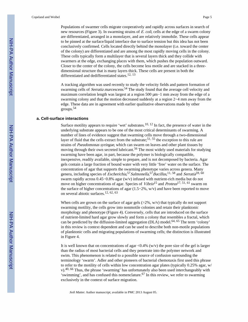
Populations of swarmer cells migrate cooperatively and rapidly across surfaces in search ofnew resources (Figure 3). In swarming strains of E. coli, cells at the edge of a swarm colonyare differentiated, arranged in a monolayer, and are relatively immobile. These cells appearto be pinned at the surface/liquid interface due to surface tension but this idea has not beenconclusively confirmed. Cells located directly behind the monolayer (i.e. toward the centerof the colony) are differentiated and are among the most rapidly moving cells in the colony.These cells typically form a multilayer that is several layers thick and they collide withswarmers at the edge, exchanging places with them, which pushes the population outward.Closer to the center of the colony, the cells become less motile and are stacked in a three-dimensional structure that is many layers thick. These cells are present in both thedifferentiated and dedifferentiated states.12, 13
A tracking algorithm was used recently to study the velocity fields and pattern formation ofswarming cells of Serratia marcescens.54 The study found that the average cell velocity andmaximum correlation length was largest at a region 500 µm−1 mm away from the edge of aswarming colony and that the motion decreased suddenly at a region 2−4 mm away from theedge. These data are in agreement with earlier qualitative observations made by othergroups.54
a. Cell-surface interactionsSurface motility appears to require ‘wet’ substrates.10, 12 In fact, the presence of water in theunderlying substrate appears to be one of the most critical determinants of swarming. Anumber of lines of evidence suggest that swarming cells move through a two-dimensionallayer of fluid that the cells extract from the substrate;51, 55 the exception to this rule arestrains of Pseudomonas syringae, which can swarm on leaves and other plant tissues bymoving through their own secreted lubricant.56 The most widely used materials for studyingswarming have been agar, in part, because the polymer is biologically compatible,inexpensive, readily available, simple to prepare, and is not decomposed by bacteria. Agargels contain a large fraction of bound water with very little ‘free’ water on the surface. Theconcentration of agar that supports the swarming phenotype varies across genera. Manygenera, including species of Escherichia,57Salmonella,57Bacillus,11, 58 and Serratia59, 60
swarm rapidly across 0.45−0.8% agar (w/v) infused with nutrient-rich media but do notmove on higher concentrations of agar. Species of Vibrio15 and Proteus17, 51, 61 swarm onthe surface of higher concentrations of agar (1.5−2%, w/v) and have been reported to moveon several abiotic surfaces.12, 62, 63
When cells are grown on the surface of agar gels (>2%, w/v) that typically do not supportswarming motility, the cells grow into nonmotile colonies and retain their planktonicmorphology and phenotype (Figure 4). Conversely, cells that are introduced on the surfaceof nutrient-limited hard agar grow slowly and form a colony that resembles a fractal, whichcan be predicted by the diffusion-limited aggregation (DLA) model.64, 65 The term ‘colony’in this review is context dependent and can be used to describe both non-motile populationsof planktonic cells and migrating populations of swarming cells; the distinction is illustratedin Figure 4.
It is well known that on concentrations of agar <0.4% (w/v) the pore size of the gel is largerthan the radius of most bacterial cells and they penetrate into the polymer network andswim. This phenomenon is related to a possible source of confusion surrounding theterminology ‘swarm’. Adler and other pioneers of bacterial chemotaxis first used this phraseto refer to the motility of cells within low concentration agar plates (typically 0.25% agar, w/v).40, 66 Thus, the phrase ‘swarming’ has unfortunately also been used interchangeably with‘swimming’, and has confused this nomenclature.67 In this review, we refer to swarmingexclusively in the context of surface migration.
Copeland and Weibel Page 5
Soft Matter. Author manuscript; available in PMC 2013 August 05.
NIH
-PA Author Manuscript
NIH
-PA Author Manuscript
NIH
-PA Author Manuscript

A narrow concentration range of agar (0.45−0.5%, w/v) supports swarming of E. coli K-12strains and may be related to the lack of an intact O-antigen on the lipopolysaccharide (LPS)of these cells. The O-antigen is a component of the outer membrane that may play a role inthe wettability of cells on hydrogel surfaces through its influence on the surface energy ofcells.68–71 Harshey and colleagues used Eiken agar to recover swarming motility in mutantsof S. marcescens that are defective in surfactant production, and mutants in LPSbiosynthesis in S. typhimurium typhimurium.12, 70, 72 The chemical composition of thismaterial differs from other sources of agar on which these cells were not motile.57
Unfortunately, the heterogeneity of agar makes it very difficult to pinpoint how this materialcomplements these strains.
A fundamental unanswered question in this area is what is the limitation of surfaces thatsupport swarming? The systematic investigation of the chemical and physical properties ofmaterials required for swarming motility will be an important step forward for this field. Thedevelopment of chemically characterized hydrogels as substitutes for agar will make itpossible to precisely vary the conditions of substrates and observe how these parametersaffect swarming. A variety of techniques are available for modifying hydrogels that may beparticularly useful in this area of microbiology.73–75 We are not arguing for the globalreplacement of agar. Instead, the introduction of more homogenous polymeric materials maypinpoint physical and chemical ‘triggers’ for the emergence of this behavior.
b. Surface-active moleculesSeveral groups have suggested that swarming cells translate across surfaces using amechanism that resembles swimming motility in a two-dimensional layer of fluid that isextracted from the underlying gel by syneresis (e.g. the contraction of a gel accompanied bythe exudation of liquid).51, 55, 76 This layer of fluid may be formed during or after thesecretion of extracellular biomolecules by swarming cells, which modify the surface tensionof water and makes it possible for cells to become immersed in a thin film of fluid.4, 11, 12
There are primarily four classes of compounds that modify the surface tension of liquids andare secreted by bacteria: polysaccharides, lipopolysaccharides, lipoproteins, and glycolipids.In the following sub-sections we review their role in swarming.
Polysaccharides—Swarmer cells secrete a matrix that consists of polysaccharides,peptides, proteins, and other compounds.12, 70, 77, 78 Polysaccharides are the predominantcomponent of the mixture produced by P. mirabilis during swarming migration.79, 80 Gygi etal. isolated a mutant of P. mirabilis that does not migrate across surfaces.78 This mutationmapped to an open-reading frame responsible for the assembly of the ‘colony migrationfactor’, an acidic capsular polysaccharide that may be responsible for extracting fluid fromthe underlying agar (Figure 5a).51, 81 Boles et al. demonstrated that the increase inproduction of capsular polysaccharides in V. parahaemolyticus decreases swarming motilityand promotes the adhesion of cells to surfaces, suggesting that these polymers may havedifferent roles in surface colonization in different organisms.82
Lipopolysaccharides—Toguchi et al. screened S. typhimurium mutants for deficienciesin swarming motility and isolated several mutants that were defective in LPS biosynthesis.70
The authors suggest that LPS plays a role in surface motility by modulating the wettabilityof the cell surface. S. typhimurium mutants of waaG, a gene involved in the biosynthesis ofthe LPS core structure, produced extensive amounts of extracellular matrix, which led to theearly onset of swarming.70 Chen et al. demonstrated recently that swarming cells of S.typhimurium use an osmotic agent—which may be LPS—to extract fluid from agar.76
Copeland and Weibel Page 6
Soft Matter. Author manuscript; available in PMC 2013 August 05.
NIH
-PA Author Manuscript
NIH
-PA Author Manuscript
NIH
-PA Author Manuscript

Lipopeptides—The differentiation of Serratia liquefaciens MG1 (S. liquefaciens) intoswarmers and their collective spreading is controlled by the flhDC flagellar master regulatorand a quorum-sensing biosynthetic pathway, respectively.59, 83–85 Cells use the quorum-sensing signal molecules, C4-HSL (N-butanoyl-L-homoserine lactone) and C6-HSL (N-hexanoyl-L-homoserine lactone) to sense the density of cells in the nascent swarmingpopulation.83, 86 The S. liquefaciens quorum-sensing system uses SwrI, an N-acyl-L-homoserine lactone (AHL) synthase, and SwrR, a transcriptional repressor and a receptor forthe signal generated by the AHL synthase; collectively, these components activatetranscription of swrA, which encodes a protein complex responsible for production ofserrawettin W2.83, 87–90 Serrawettin W2 is a cyclic lipodepsipentapeptide that reduces thesurface tension of water and is essential for Serratia swarming migration; the addition ofserrawettin W2 rescued the surface motility of swrI and swrI/swrA mutants (Figure5b).83, 85, 88
B. subtilis secretes a surface-active agent, surfactin, during the colonization of agarsurfaces.91–93 Kearns and Losick discovered that surfactin was essential for swarmingmigration in B. subtilis.48 Laboratory strains of B. subtilis that do not swarm contain aframeshift mutation in the sfp gene required for surfactin synthesis.94 In addition to the sfpgene, the srf operon—consisting of srfAA, srfAB, and srfAC—encodes the enzymecomplex, surfactin synthetase, which is essential for swarming (Figure 5c).92, 95,48
Glycolipids—Rhamnolipids are a class of amphiphilic glycolipids secreted primarily bythe Gram-negative opportunistic human pathogen, P. aeruginosa.96 These compoundsreduce the surface tension of liquids and are implicated in pathogenesis. Swarming coloniesof P. aeruginosa have a characteristic pattern of tendrils that radiate away from the center ofthe swarm plate that is partially due to the secretion of rhamnolipids.97,98P. aeruginosaproduces two types of rhamnolipids: L-rhamnosyl-3-hydroxydecanoyl-3-hydroxydecanoate(mono-RL) and L-rhamnosyl-L-rhamnosyl-3-hydroxydecanoyl-3-hydroxydecanoate (di-RL).96 These two compounds and the rhamnolipid precursor, 3-(3-hydroxyalkanoyloxy)alkanoic acid (HAA) are secreted by P. aeruginosa and facilitateswarming motility (Figure 5d).97–99 Tremblay et al. demonstrated recently that thecharacteristic swarming pattern of P. aeruginosa arises from the properties of di-RLs, mono-RLs, and HAAs.97 Their data suggests that di-RLs are chemoattractants, HAAs arechemorepellents, and mono-RLs are wetting agents.97 None of these molecules appears toaffect swimming motility.
c. Cell/cell interactionsIn contrast to the growing body of literature on the role of physical interactions betweenmammalian cells that regulate physiology and behavior 100, 101, our understanding of cell/cell interactions in bacteria is still unclear. Cell-cell interactions are particularly relevant toswarming bacteria in which a large population of cells is packed into a confined volume.Physical interactions between bacterial cells may be responsible for many of the observedphenotypes associated with swarming, including: 1) alignment of adjacent bacterial cells andtheir coordinated movement in ‘rafts’; 2) formation of dynamic, circular vortices; and 3)coordination of the motility of colonies across surfaces. Below we summarize data thatsuggests that contact between cells may be important in swarming motility, behavior, andemergence.
Interactions between translating swarming cells—Physical contact betweenswarming cells is important for surface migration as individual swarming cells isolated froma colony do not move unless the agar is supplemented with a surfactant or has a layer ofliquid on the surface.12 Several early observations of Proteus demonstrated that swarmer
Copeland and Weibel Page 7
Soft Matter. Author manuscript; available in PMC 2013 August 05.
NIH
-PA Author Manuscript
NIH
-PA Author Manuscript
NIH
-PA Author Manuscript
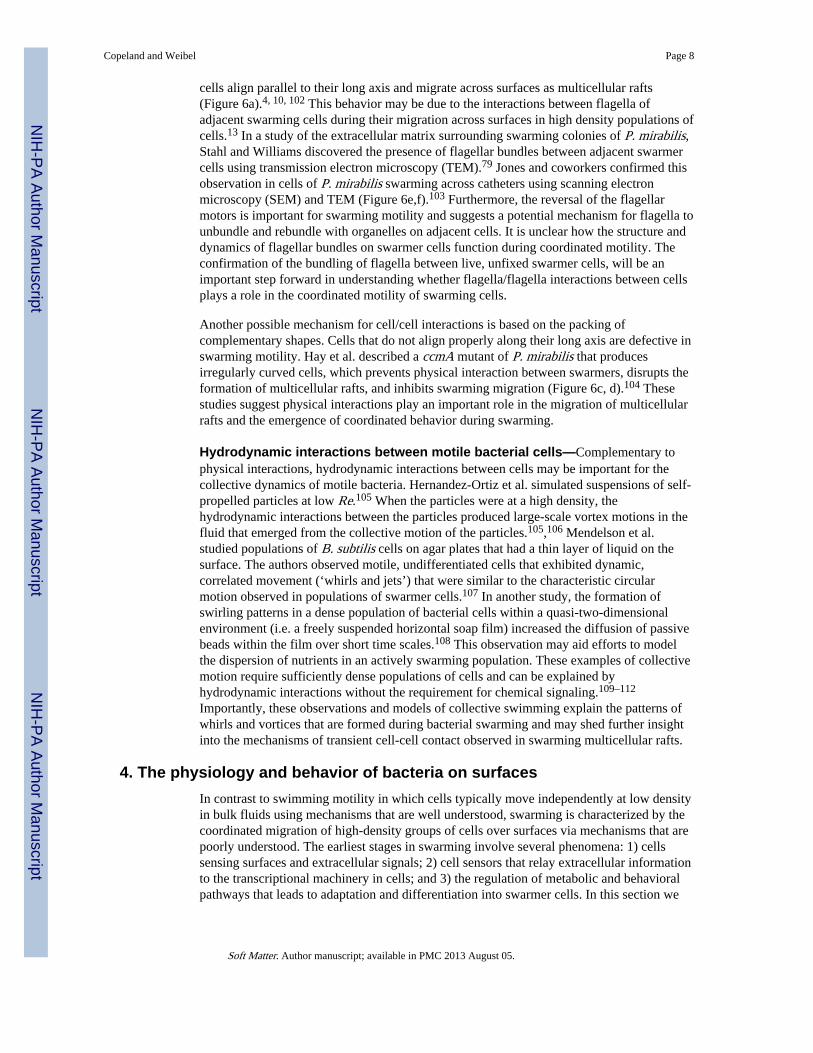
cells align parallel to their long axis and migrate across surfaces as multicellular rafts(Figure 6a).4, 10, 102 This behavior may be due to the interactions between flagella ofadjacent swarming cells during their migration across surfaces in high density populations ofcells.13 In a study of the extracellular matrix surrounding swarming colonies of P. mirabilis,Stahl and Williams discovered the presence of flagellar bundles between adjacent swarmercells using transmission electron microscopy (TEM).79 Jones and coworkers confirmed thisobservation in cells of P. mirabilis swarming across catheters using scanning electronmicroscopy (SEM) and TEM (Figure 6e,f).103 Furthermore, the reversal of the flagellarmotors is important for swarming motility and suggests a potential mechanism for flagella tounbundle and rebundle with organelles on adjacent cells. It is unclear how the structure anddynamics of flagellar bundles on swarmer cells function during coordinated motility. Theconfirmation of the bundling of flagella between live, unfixed swarmer cells, will be animportant step forward in understanding whether flagella/flagella interactions between cellsplays a role in the coordinated motility of swarming cells.
Another possible mechanism for cell/cell interactions is based on the packing ofcomplementary shapes. Cells that do not align properly along their long axis are defective inswarming motility. Hay et al. described a ccmA mutant of P. mirabilis that producesirregularly curved cells, which prevents physical interaction between swarmers, disrupts theformation of multicellular rafts, and inhibits swarming migration (Figure 6c, d).104 Thesestudies suggest physical interactions play an important role in the migration of multicellularrafts and the emergence of coordinated behavior during swarming.
Hydrodynamic interactions between motile bacterial cells—Complementary tophysical interactions, hydrodynamic interactions between cells may be important for thecollective dynamics of motile bacteria. Hernandez-Ortiz et al. simulated suspensions of self-propelled particles at low Re.105 When the particles were at a high density, thehydrodynamic interactions between the particles produced large-scale vortex motions in thefluid that emerged from the collective motion of the particles.105,106 Mendelson et al.studied populations of B. subtilis cells on agar plates that had a thin layer of liquid on thesurface. The authors observed motile, undifferentiated cells that exhibited dynamic,correlated movement (‘whirls and jets’) that were similar to the characteristic circularmotion observed in populations of swarmer cells.107 In another study, the formation ofswirling patterns in a dense population of bacterial cells within a quasi-two-dimensionalenvironment (i.e. a freely suspended horizontal soap film) increased the diffusion of passivebeads within the film over short time scales.108 This observation may aid efforts to modelthe dispersion of nutrients in an actively swarming population. These examples of collectivemotion require sufficiently dense populations of cells and can be explained byhydrodynamic interactions without the requirement for chemical signaling.109–112
Importantly, these observations and models of collective swimming explain the patterns ofwhirls and vortices that are formed during bacterial swarming and may shed further insightinto the mechanisms of transient cell-cell contact observed in swarming multicellular rafts.
4. The physiology and behavior of bacteria on surfacesIn contrast to swimming motility in which cells typically move independently at low densityin bulk fluids using mechanisms that are well understood, swarming is characterized by thecoordinated migration of high-density groups of cells over surfaces via mechanisms that arepoorly understood. The earliest stages in swarming involve several phenomena: 1) cellssensing surfaces and extracellular signals; 2) cell sensors that relay extracellular informationto the transcriptional machinery in cells; and 3) the regulation of metabolic and behavioralpathways that leads to adaptation and differentiation into swarmer cells. In this section we
Copeland and Weibel Page 8
Soft Matter. Author manuscript; available in PMC 2013 August 05.
NIH
-PA Author Manuscript
NIH
-PA Author Manuscript
NIH
-PA Author Manuscript
discuss several mechanisms for extracellular sensing that plays a role in differentiation andthe metabolic changes that accompany swarming in several species.
a. Differentiation into swarmer cellsProteus mirabilis, the archetypal swarmer cell, differentiates into swarmer cells whencultured on hydrogel surfaces (1.5−3% agar, w/v).13, 17, 113, 114 Macroscopically, P.mirabilis colonies alternate between periodic phases of migration (swarming) and phases ofgrowth without movement (consolidation). The cyclic repetition of these stages results inregularly spaced concentric terraces that are geometrically similar to a bulls-eye pattern.14
The migration and consolidation pattern of swarming strains of P. mirabilis as well as that ofother swarming genera have been modeled with reaction-diffusion theory using partialdifferential equations and/or kinetic equations that incorporate a parameter for the age-dependence of swarmer cell behavior.115–118
Swarmer cells of P. mirabilis are 10–80 µm long with approximately 10 to 50 times moreflagella than planktonic cells; the formation of swarmers is accompanied by the inhibition ofcell septation, but not DNA synthesis, as swarmer cells are multinucleate and have the sameratio of DNA/cell length as planktonic cells (Table 1).17, 119,120 Harshey has suggested thatthe large number of flagella on swarmer cells reduces friction between the cell body and thesurface and facilitates swarming.12 The flagella that play a role in surface motility by mostswarming strains of Gram-negative and Gram-positive bacteria are the same organelles usedfor individual swimming motility.4, 11, 13 Some genera of bacteria, including species ofVibrio and Aeromonas, however, encode a separate flagellar system for motility on surfacesand in viscous fluids.121–124 These bacteria use a sheathed polar flagellum for translatingthrough bulk fluids and express unsheathed lateral flagella for motility on surfaces. Thesynthesis and assembly of lateral flagella in Vibrio parahaemolyticus (V. parahaemolyticus)is activated when cells are grown in viscous fluids (e.g. 18% Ficoll 400 w/v, 8×10⁻² Pa·s),on agar surfaces (<2% agar, w/v) or in the presence of a polyclonal antibody that bindsspecifically to the polar flagellum and causes agglutination.121, 122, 125 The induction of thelateral flagella genes in V. parahaemolyticus is also triggered by iron-limited conditions.126
Pioneering research by McCarter et al. demonstrated that the polar flagellum in V.parahaemolyticus acts as a mechanosensor; the inhibition of rotation of the polar flagelluminduces the expression and assembly of lateral flagella and leads to differentiation into theswarming phenotype.113, 122, 127 Alavi et al. showed that this concept extends to P. mirabilisin which planktonic cells differentiate into swarmers when the cells are on surfaces and therotation of the flagellum is inhibited by the substrate.113 Swarmer cells of P. mirabilisdedifferentiate into planktonic cells soon after they are transferred to a liquid, presumably asa result of loss of the surface stimulus.4, 51, 113, 128 Additionally, the inhibition of flagellarotation as a result of surface contact (0.7−0.8% agar, w/v) or growth in a solution of Ficoll400 (5−10%, w/v, 2–5×10⁻³ Pa·s) stimulates increased flagellin production and theswarming phenotype in Serratia marcescens (strain 274) (S. marcescens).60 The flagellum inmany strains of bacteria appears to be a mechanical sensor of the extracellular environment,which relays signals that affect the transcription of genes and leads to differentiation.113
Not surprisingly, mutations of flagellar genes produce abnormal swarmer cell phenotypes,presumably as a result of a perturbation of flagella-based mechanisms of sensingsurfaces.114, 120 Mutants of the flaA gene in P. mirabilis, which encodes the subunit of theflagellar filament, produce cells that are not motile.129 The FlaA mutants do not differentiateinto swarmers when grown using conditions that stimulate swarmer cell development inwild-type Proteus.13, 14, 129 Belas and colleagues identified other flagellar genes in P.mirabilis that result in abnormal swarmer cell development.120 These genes include fliL, acomponent of the flagellar basal body, fliG, part of the flagellar switch, and flgH, which
Copeland and Weibel Page 9
Soft Matter. Author manuscript; available in PMC 2013 August 05.
NIH
-PA Author Manuscript
NIH
-PA Author Manuscript
NIH
-PA Author Manuscript

forms the basal-body L-ring.120 Gygi et al. demonstrated that a P. mirabilis mutant of theflhA gene, encoding a flagellum export protein, does not produce flagella and is unable todifferentiate into a swarmer cell.114, 130 Furthermore, a transposon inserted into the flgNgene in P. mirabilis, which encodes a protein involved in flagella filament assembly,resulted in cells which were motile in liquid, but not on surfaces, potentially due to areduction in the number of flagella on swarmer cells.131 In V. parahaemolyticus, mutantsthat are unable to form a polar flagellum generate a constitutive swarmer phenotype.122, 132
Collectively, these studies demonstrate that the biosynthesis, assembly, and function offlagella in some bacteria are essential for sensing environmental stimuli, including surfacesthat produce the swarming phenotype.
FliL is a protein that is transcribed from the middle class 2 flagellar operon, fliLMNOPQRof S. typhimurium and E. coli that had an unknown function until recently.19 Knockouts ofthis protein in S. typhimurium133 and E. coli134 produce cells that are not motile onsurfaces.135 Flagellar filaments in cells of fliL mutant strains grown on swarm agar (0.6%agar, w/v) are severed at the rod. The flagella are not broken off if the movement of the cellson the swarm agar is disrupted by mutations to the Mot proteins. Attmannspacher andcoworkers suggest that a significant amount of torque is transmitted to the flagellar motorwhen cells are on the surface of swarm agar; this situation requires the stabilization of theflagella by FliL.135 A fliL mutant in P. mirabilis produced cells that had no flagella and werenon-motile. Interestingly, these cells elongated into swarmers during growth in liquid andbecame hyper-elongated on agar.128 These data suggest that FliL may play a role intransmitting an external, surface-sensing stimuli into a change in gene expression within thecell.128
It is clear that flagella play a role in sensing surfaces in species of Proteus, Serratia, andVibrio but it is unclear if this mechanism for relaying information on the extracellularenvironment of cells to the transcriptional machinery is widespread among Eubacteria.Remarkably few other mechanisms that bacteria use to sense physical boundaries andsurfaces are known. Sensing mechanisms based on receptor/ligand binding between cellsand surfaces are commonly used by eukaryotes but were overlooked in bacteria untilrecently. Mignot et al. found that cells of Myxococcus xanthus form transient adhesioncomplexes between the cell surface and the substrate. These complexes play a role in surfacemotility and are reminiscent of mammalian focal adhesions.136 We anticipate that theintroduction of biocompatible polymers and gels with controlled physical properties andsurface chemistry will play an important role in the study of sensors that bacterial cells useto detect changes in their physical environment. These materials will make it possible tostudy the role of a variety of stimuli, including: 1) stiffness (e.g. Young’s modulus); 2)porosity; 3) surface gradients; and 4) ligand/receptor interactions.
b. Metabolism and respirationInoue et al. screened the Keio collection, a set of single-gene knockouts of all thenonessential genes in E. coli K-12,5 and identified 294 knockouts that were no longer able toswarm.137 Seventy of these genes were involved in general metabolism and included genesaffiliated with the tricarboxylic acid cycle, ATP synthase, and the electron-transport chain,suggesting that swarming motility may require an increase in energy production.
Cells of E. coli and S. typhimurium require nutrient-rich media supplemented with a carbonsource, such as glucose, for development into swarmers.57, 138 Using proteomics, Kim andSurette found that enzymes associated with anabolism were upregulated in swarmer cells ofS. typhimurium and enzymes involved in the catabolism of peptides as well as nucleotidesalvage were down-regulated relative to planktonic cells. The permeability of the outermembrane was reduced in swarmer cells, which suggests that these cells may be relying on
Copeland and Weibel Page 10
Soft Matter. Author manuscript; available in PMC 2013 August 05.
NIH
-PA Author Manuscript
NIH
-PA Author Manuscript
NIH
-PA Author Manuscript
de novo biosynthesis pathways to obtain amino acids and nucleotides rather than importingthem.138 Wang and coworkers used DNA-arrays to profile mRNA transcripts of swarmingcells of S. typhimurium and observed that genes involved in iron metabolism andlipopolysaccharide (LPS) biosynthesis were upregulated.139 These observations of generalcellular metabolism suggest that swarming cells are physiologically and biochemicallydifferent from planktonic, swimming cells for reasons that are not yet clear.
c. ChemotaxisThe chemotaxis system consists of sensory receptors linked to a cytoplasmicphosphorylation cascade that alters the rotational bias of the flagella motors and producesnet cell motility toward attractants and away from repellents. This area has been reviewedseveral times,21, 43, 140, 141 including an excellent, recent review by Hazelbauer et al.142 Thechemotaxis pathway is important for differentiation into the swarming phenotype and colonymigration.143–145 The addition of glutamine to minimal media that does not supportswarming motility, triggers the differentiation of cells of P. mirabilis into swarmers andleads to the colonization of surfaces.143 Allison et al. demonstrated that glutamine is specificfor chemotaxis by swarmers and does not elicit a response from swimming cells.143 Thechemotaxis machinery, but not necessarily the act of chemosensing itself, is important forthe development of swarming in E. coli, S. typhimurium, and S. marcescens.57, 144, 145
Wang et al. observed that chemotaxis mutants of S. typhimurium have fewer and shorterflagella and generate bacterial lawns that have less fluid than lawns of wild-type cells.146
Based on these observations and the downregulation of late (class 3) flagellar genes inchemotaxis mutants, Harshey and colleagues suggest that the flagellum is able to senseenvironmental conditions (external wetness) that are favorable for filament assembly.146
Subsequent studies by Mariconda et al. demonstrated that chemotaxis mutants unable toswitch the direction of flagellar motor rotation are defective in their ability to efficientlymigrate across the surface.147 It is currently unknown why these mutants produce swarmcolonies that are drier than wild type colonies. The connection between chemotaxis andswarming is not yet fully understood.
d. Quorum sensing and signal transductionThe population density of cells and signaling molecules influences the development andmigration of swarmer cells. Signaling molecules produced by bacteria are referred to asautoinducers (AIs). Threshold concentrations of AIs produce changes in gene expressionthrough the modulation of transcriptional regulators in a process referred to as ‘quorumsensing’. Multiple quorum-sensing systems exist in bacteria, including the N-acyl-homoserine lactone (AHL) quorum sensing machinery of Gram-negative bacteria and themore recently discovered AI-2 and AI-3 systems for interspecies communication.148, 149
Several species of swarming bacteria use AHLs to trigger the production of surfactants thatplay an important role in surface motility.86, 97 We highlight examples of the role ofsignaling molecules in swarming, such as the induction of surfactant expression, in thissection and section 3b. For an extensive review of the relationship between swarming andquorum sensing, see Daniels et al.150
Motlity and swarming in many species are regulated by two-component phosphorelaysignaling systems that may not be modulated by quorum sensing autoinducers. The Rcsphosphorelay system of the Enterobacteriaceae family is one such system, which regulates avariety of processes, including: expression of genes involved in colanic acid synthesis, celldivision, cell wall integrity, and virulence.151 Importantly, this phosphorelay system is alsoimplicated in regulation of swarming motility, primarily through modulation of the flhDCoperon.152, 153 Two-component signaling systems are used by microorganisms to sense andrespond to environmental changes. The basic constituents of these systems are two signal
Copeland and Weibel Page 11
Soft Matter. Author manuscript; available in PMC 2013 August 05.
NIH
-PA Author Manuscript
NIH
-PA Author Manuscript
NIH
-PA Author Manuscript
transducers, specifically a sensor histidine kinase and a response regulator. Of the signalsactivating the Rcs system, including: osmotic shock, overproduction of a membranechaperone protein, and growth at low-temperature (20°C) in the presence of glucose andhigh zinc concentrations; the activation of RcsC during growth on a solid surface isintriguing because of its potential role in affecting the gene expression of swarmercells.152, 154
Fatty acids can also inhibit swarming in P. mirabilis through RsbA, which encodes ahomologue of the E. coli RcsD protein.155 This pathway was originally implicated inswarming motility in P. mirabilis based on the observation that a rsbA mutant swarmedprecociously.156 This phenotype was also observed in rcsC and rcsD mutants in E. coli aswell as a rcsB mutant in S. typhimurium.153, 157 In agreement with these mutant phenotypes,the RcsCDB phosphorelay system is a negative regulator of the flhDC operon in E. coli.Repression is achieved via binding of the response regulator, RscB, and a cofactor, RcsA, toa RcsAB box in the promoter region of the flhDC operon.158 As described previously,repression of the flhDC operon will negatively affect swarming development and migration.Interestingly, the RcsC/RcsD/RcsB phosphorelay system is also involved in thetranscriptional activation of the Wzz gene, which encodes a protein involved in determiningthe chain length of the O-antigen in S. typhimurium.159 As described earlier, the O-antigenis important for swarming migration in Salmonella.70 The RcsCDB system was alsooriginally noted for its activation of genes involved in the biosynthesis of colanic acid,which is an inhibitor of swarming motility.153, 160 In conclusion, these observations suggestthat the Rcs phosphorelay system plays an important role in swarming.
5. OutlookBacterial swarming may be one of the most tractable models for studying dynamic self-assembly in a biological system. After a century of experiments, however, there are stillmany fundamental unanswered questions about the system, including: 1) the stimuli,sensors, and biochemistry involved in the differentiation of cells into swarmers on surfaces;2) chemical and physical interactions between cells and surfaces; 3) the mechanism of cellmotility on surfaces; 4) the microscopic and macroscopic organization of swarming coloniesand the dynamics of cells in these structures; and 5) the importance of cell/cell and cell/fluidinteractions in the coordination of growth and motility. Microbiological tools may notprovide enough leverage to dissect this system at a level of detail that is required tounderstand how collective behavior emerges and how this phenotype plays a role inmulticellular homeostasis, survival, evolution, and pathogenesis.
Physical scientists and engineers are in a unique position to have an impact on this area ofmicrobiology through the application of materials and techniques from chemistry andmaterials science and engineering. Below we outline and summarize three areas where theintersection of microbiology and materials science and engineering may find synergy inanswering fundamental questions about bacterial swarming and the origins of emergentbehavior.
a. Surface sensingSome cells use their polar flagellum to sense their microenvironment and detect boundariesbetween fluids and surfaces. We described this mechanism for V. parahaemolyticus and P.mirabilis in Section 4a and pointed out that it may not be a sensor that is universallyconserved in Eubacteria. What other mechanisms do bacterial cells use to sense surfaces?Are there examples of mechanisms that are conserved across many different genera? Tounderstand these questions at the molecular level, we need to determine the stimuli that cellssense, the sensors that couple information on the extracellular microenvironment to the
Copeland and Weibel Page 12
Soft Matter. Author manuscript; available in PMC 2013 August 05.
NIH
-PA Author Manuscript
NIH
-PA Author Manuscript
NIH
-PA Author Manuscript

transcription of genes, and the biochemical pathways that play a role in coordinating thedifferentiation of cells into swarmers.
One approach to the study of this problem is to build new platforms for studying swarming.The application of polymer substrates that have defined chemical and physical propertiesthat can be systematically varied will make it possible to explore the stimuli that triggerdifferentiation. The fusion of different classes of soft, biocompatible polymers and geneticand genomic techniques will lead to the study of the signaling pathways and the resultingtranscriptional changes that occur during differentiation at a new level of detail.
b. Surface motilityThe mechanisms that are involved in the movement of swarming cells across surfaces arestill not understood in detail. Several observations have led to the hypothesis that swarmerstranslate through a two-dimensional lay of fluid that is extracted from the hydrogel. Thestudy of the temporal emergence of swarming from a single cell on a surface may provideinsight into this hypothesis. The application of techniques for imaging and measuring thethickness of films of fluids will provide direct evidence of this mechanism. The ability tochange the surface energy of the polymer will make it possible to manipulate the thicknessof the layer of fluid on the substrate and determine how this parameter affects swarmingmotility and collective movement.
Some species of bacteria swarm on materials that provide little or no source of endogenouswater (e.g. silicone catheters). These strains of bacteria differentiate into swarmers thatproduce and secrete significant amounts of extracellular polymers, which may provide thelayer of fluid that the cells move through. Although it has not been determinedexperimentally, it seems reasonable that these fluids have a viscosity that approaches orexceeds the limited range that supports the swimming of planktonic cells (Section 2c). Howdo swarmers move through fluids that have a viscosity that inhibits the motility ofplanktonic cells? Swarmers have a higher density of flagella than planktonic cells. Does anincrease in the number of flagella increase the torque produced by the cell?
c. The dynamics of flagellaThe structure and dynamics of a single bundle of flagella in planktonic cells of E. coli hasbeen the model system for our understanding of the mechanisms of bacterial cell motility.As we have pointed out in this review, swarmers typically have a higher density of flagellathan planktonic cells. Surprisingly little is known about the spatial and temporal structure ofthe bundles of flagella on swarming cells. In contrast to studies of motility of planktonicbacterial cells in bulk fluids in which cell/cell interactions are uncommon, swarmingcolonies are characterized by frequent interactions between cells. What happens to thestructure and dynamics of the flagella when two swarming cells are brought in closeproximity (ie. close enough for their flagella to interact)? Do the flagella on adjacentswarming cells interact and become intertwined? The high density of cells in swarmingcolonies and the large number of flagella per cell makes it seem reasonable that the flagellamay form intercellular bundles. This hypothesis is indirectly supported by the observationthat the motors on swarming cells undergo frequent reversals in direction, which causesintracellular bundles of flagella on cells to break apart and reform. Could changes in thedirection of the motors provide a mechanism for the formation of intercellular bundles offlagella between adjacent swarmers? These observations lead to the untested hypothesis thatthe apparent coordination of motility in swarming populations may be a consequence of thephysical tethering of swarmers to their neighbors.
Copeland and Weibel Page 13
Soft Matter. Author manuscript; available in PMC 2013 August 05.
NIH
-PA Author Manuscript
NIH
-PA Author Manuscript
NIH
-PA Author Manuscript

Swarming is a phenotype that plays a role in bacterial pathogenesis and biofilm formationand may be a common method for motile strains of bacteria adsorbed or in contact withsurfaces to regain motility. Table 2 summarizes genera of swarming bacteria and includes abrief summary of their ‘native’ habitat, although not all of the species within each genus arecapable of surface motility. The table provides a general idea of how widely this behavior isdistributed in Eubacteria. Interestingly, many of the genera included in the table live inhabitats that are not dominated by bulk fluids and may more accurately be thought of inenvironments that are at interfaces between surfaces and liquids. This observation bringsinto question whether cells evolved flagella for swimming in bulk fluids or for moving onsurface. Perhaps the movement of bacterial cells on a surface is the norm and not theexception.
Swarming presents a unique opportunity for studying dynamic self-assembly in a biologicalsystem that is relevant to biomedicine and ecology. The infusion of new techniques based onsoft matter will have an important impact on understanding bacterial surface motility and theorigins of emergent behavior, and will almost certainly inspire new directions in materialsscience and the study of microbiological systems.
Supplementary MaterialRefer to Web version on PubMed Central for supplementary material.
References1. Whitesides GM, Grzybowski B. 2002; vol. 295:2418–2421.
2. Dworkin, M.; Shapiro, JA. Bacteria as multicellular organisms. New York: Oxford University Press;1997.
3. Shapiro JA. Annu Rev Microbiol. 1998; 52:81–104. [PubMed: 9891794]
4. Fraser GM, Hughes C. Curr Opin Microbiol. 1999; 2:630–635. [PubMed: 10607626]
5. Baba T, Ara T, Hasegawa M, Takai Y, Okumura Y, Baba M, Datsenko KA, Tomita M, Wanner BL,Mori H. Mol Syst Biol. 2006; 2 2006 0008.
6. Butland, G.; Babu, M.; Diaz-Mejia, JJ.; Bohdana, F.; Phanse, S.; Gold, B.; Yang, W.; Li, J.;Gagarinova, AG.; Pogoutse, O.; Mori, H.; Wanner, BL.; Lo, H.; Wasniewski, J.; Christopolous, C.;Ali, M.; Venn, P.; Safavi-Naini, A.; Sourour, N.; Caron, S.; Choi, JY.; Laigle, L.; Nazarians-Armavil, A.; Deshpande, A.; Joe, S.; Datsenko, KA.; Yamamoto, N.; Andrews, BJ.; Boone, C.;Ding, H.; Sheikh, B.; Moreno-Hagelseib, G.; Greenblatt, JF.; Emili, A. Nat Methods. 2008.
7. Typas A, Nichols RJ, Siegele DA, Shales M, Collins SR, Lim B, Braberg H, Yamamoto N,Takeuchi R, Wanner BL, Mori H, Weissman JS, Krogan NJ, Gross CA. Nat Methods. 2008
8. Weibel DB, DiLuzio WR, Whitesides GM. Nat Rev Microbiol. 2007; 5:209–218. [PubMed:17304250]
9. Weibel DB, Lee A, Mayer M, Brady SF, Bruzewicz D, Yang J, Diluzio WR, Clardy J, WhitesidesGM. Langmuir. 2005; 21:6436–6442. [PubMed: 15982051]
10. Henrichsen J. Bacteriol Rev. 1972; 36:478–503. [PubMed: 4631369]
11. Harshey RM. Mol Microbiol. 1994; 13:389–394. [PubMed: 7997156]
12. Harshey RM. Annu Rev Microbiol. 2003; 57:249–273. [PubMed: 14527279]
13. Allison C, Hughes C. Sci Prog. 1991; 75:403–422. [PubMed: 1842857]
14. Belas, R.; Shapiro, M.; JA, D., editors. Bacteria as multicellular organisms. edition edn.. Oxford,England: Oxford University Press; 1996.
15. McCarter L. J Mol Microbiol Biotechnol. 1999; 1:51–57. [PubMed: 10941784]
16. Verstraeten N, Braeken K, Debkumari B, Fauvart M, Fransaer J, Vermant J, Michiels J. Trends inMicrobiology. 2008; 16:496–506. [PubMed: 18775660]
17. Williams FD, Schwarzhoff RH. Annu Rev Microbiol. 1978; 32:101–122. [PubMed: 360961]
18. Bray, D. Cell Movements: From Molecules to Motility. Garland Publishing; 2001.
Copeland and Weibel Page 14
Soft Matter. Author manuscript; available in PMC 2013 August 05.
NIH
-PA Author Manuscript
NIH
-PA Author Manuscript
NIH
-PA Author Manuscript

19. Chilcott GS, Hughes KT. Microbiol Mol Biol Rev. 2000; 64:694–708. [PubMed: 11104815]
20. Silverman M, Simon M. Nature. 1974; 249:73–74. [PubMed: 4598030]
21. Manson MD, Armitage JP, Hoch JA, Macnab RM. J Bacteriol. 1998; 180:1009–1022. [PubMed:9495737]
22. Macnab, RM.; Neidhardt, FC.; Curtiss, R.; Ingraham, JL.; Lin, ECC., editors; Neidhardt, FC.;Curtis, R., III; Ingraham, J.; Lin, ECC.; Low, KB.; Magasanik, B.; Reznikoff, WS.; Riley, M.;Schaechter, M.; Umbarger, HE., editors. Vol. 1. 1996. p. 123-145.
23. Berg HC. Annu Rev Biochem. 2003; 72:19–54. [PubMed: 12500982]
24. Yonekura K, Maki-Yonekura S, Namba K. Nature. 2003; 424:643–650. [PubMed: 12904785]
25. Macnab RM. Annu Rev Microbiol. 2003; 57:77–100. [PubMed: 12730325]
26. Jarrell KF, McBride MJ. Nat Rev Microbiol. 2008; 6:466–476. [PubMed: 18461074]
27. Berg HC. Nature. 1975; 254:389–392. [PubMed: 1090851]
28. Zhou J, Lloyd SA, Blair DF. Proc Natl Acad Sci U S A. 1998; 95:6436–6441. [PubMed: 9600984]
29. Larsen SH, Adler J, Gargus JJ, Hogg RW. Proc Natl Acad Sci U S A. 1974; 71:1239–1243.[PubMed: 4598295]
30. Kojima S, Blair DF. Int Rev Cytol. 2004; 233:93–134. [PubMed: 15037363]
31. McCarter LL. J Mol Microbiol Biotechnol. 2004; 7:18–29. [PubMed: 15170400]
32. Blair DF, Berg HC. Cell. 1990; 60:439–449. [PubMed: 2154333]
33. Kojima S, Blair DF. Biochemistry. 2001; 40:13041–13050. [PubMed: 11669642]
34. Chevance FF, Hughes KT. Nat Rev Microbiol. 2008; 6:455–465. [PubMed: 18483484]
35. Berg, HC. E. Coli in Motion. Springer; 2003.
36. Larsen SH, Reader RW, Kort EN, Tso WW, Adler J. Nature. 1974; 249:74–77. [PubMed:4598031]
37. Macnab RM, Ornston MK. J Mol Biol. 1977; 112:1–30. [PubMed: 328893]
38. Turner L, Ryu WS, Berg HC. J Bacteriol. 2000; 182:2793–2801. [PubMed: 10781548]
39. Berg HC, Brown DA. Nature. 1972; 239:500–504. [PubMed: 4563019]
40. Adler J. Science. 1966; 153:708–716. [PubMed: 4957395]
41. Blair DF. Annu Rev Microbiol. 1995; 49:489–522. [PubMed: 8561469]
42. Adler J. J Bacteriol. 1966; 92:121–129. [PubMed: 5328747]
43. Stock, JB.; Surette, MG. Cellular and Molecular Biology. Neidhardt, FC.; Curtiss, RI.; Ingraham,JL.; Lin, ECC.; Low, KB.; Magasanik, B.; Riley, M.; Schaechter, M.; Umbarger, HE., editors.1996. p. 1103-1129.
44. Berg, HC. Random Walks in Biology. Princeton University Press; 1993.
45. Purcell EM. Am. J. Phys. 1977; 45:3–11.
46. Berg HC, Turner L. Nature. 1979; 278:349–351. [PubMed: 370610]
47. Greenberg EP, Canale-Parola E. J Bacteriol. 1977; 132:356–358. [PubMed: 410784]
48. Kearns DB, Losick R. Molecular Microbiology. 2003; 49:581–590. [PubMed: 12864845]
49. Rashid MH, Kornberg A. Proc Natl Acad Sci U S A. 2000; 97:4885–4890. [PubMed: 10758151]
50. Bees MA, Andresen P, Mosekilde E, Givskov M. Bull Math Biol. 2002; 64:565–587. [PubMed:12094409]
51. Rauprich O, Matsushita M, Weijer CJ, Siegert F, Esipov SE, Shapiro JA. J Bacteriol. 1996;178:6525–6538. [PubMed: 8932309]
52. Itoi-i H, Wakita J, Matsuyama T, Matsushita M. Journal of the Physical Society of Japan. 1999;68:1436–1443.
53. Czirók A, Matsushita M, Vicsek T. Physical Review E. 2001; 63:31915.
54. Steager EB, Kim CB, Kim MJ. Physics of Fluids. 2008; 20:73601–73601.
55. Bees MA, Andresen P, Mosekilde E, Givskov M. J Math Biol. 2000; 40:27–63. [PubMed:10663662]
56. Quinones B, Dulla G, Lindow SE. Mol Plant Microbe Interact. 2005; 18:682–693. [PubMed:16042014]
Copeland and Weibel Page 15
Soft Matter. Author manuscript; available in PMC 2013 August 05.
NIH
-PA Author Manuscript
NIH
-PA Author Manuscript
NIH
-PA Author Manuscript
57. Harshey RM, Matsuyama T. Proc Natl Acad Sci U S A. 1994; 91:8631–8635. [PubMed: 8078935]
58. Senesi S, Celandroni F, Salvetti S, Beecher DJ, Wong AC, Ghelardi E. Microbiology. 2002;148:1785–1794. [PubMed: 12055298]
59. Eberl L, Christiansen G, Molin S, Givskov M. J Bacteriol. 1996; 178:554–559. [PubMed:8550481]
60. Alberti L, Harshey RM. J Bacteriol. 1990; 172:4322–4328. [PubMed: 2198253]
61. Jeffries CD, Rogers HE. J Bacteriol. 1968; 95:732–733. [PubMed: 5640395]
62. Stickler D, Hughes G. Eur J Clin Microbiol Infect Dis. 1999; 18:206–208. [PubMed: 10357056]
63. Watterson JD, Cadieux PA, Stickler D, Reid G, Denstedt JD. J Endourol. 2003; 17:523–527.[PubMed: 14565887]
64. Matsuyama T, Matsushita M. Critical Reviews in Microbiology. 1993; 19:117–135. [PubMed:8338618]
65. Shapiro J. BioEssays. 1995; 17:597–607. [PubMed: 7646482]
66. Wolfe AJ, Berg HC. Proc Natl Acad Sci U S A. 1989; 86:6973–6977. [PubMed: 2674941]
67. Garcia-Pichel F. J Bacteriol. 1989; 171:3560–3563. [PubMed: 2498293]
68. Liu D, Reeves PR. Microbiology. 1994; 140((Pt 1)):49–57. [PubMed: 7512872]
69. Stevenson G, Neal B, Liu D, Hobbs M, Packer NH, Batley M, Redmond JW, Lindquist L, ReevesP. J Bacteriol. 1994; 176:4144–4156. [PubMed: 7517391]
70. Toguchi A, Siano M, Burkart M, Harshey RM. J Bacteriol. 2000; 182:6308–6321. [PubMed:11053374]
71. Raetz, CRH. Escherichia coli and Salmonella: cellular and molecular biology. 2nd ed..Washington, DC: ASM Press; 1996. p. 1035-1063.
72. Matsuyama T, Bhasin A, Harshey RM. J Bacteriol. 1995; 177:987–991. [PubMed: 7860610]
73. Campbell CJ, Klajn R, Fialkowski M, Grzybowski BA. Phys. Lett. 2003; 83:4444.
74. Grzybowski BA, Campbell CJ. Materials Today. 2007; 10:38–46.
75. Martin BD, Brandow SL, Dressick WJ, Schull TL. Langmuir. 2000; 16:9944–9946.
76. Chen BG, Turner L, Berg HC. J Bacteriol. 2007; 189:8750–8753. [PubMed: 17905988]
77. Mireles JR 2nd, Toguchi A, Harshey RM. J Bacteriol. 2001; 183:5848–5854. [PubMed: 11566982]
78. Gygi D, Rahman MM, Lai HC, Carlson R, Guard-Petter J, Hughes C. Mol Microbiol. 1995;17:1167–1175. [PubMed: 8594335]
79. Stahl SJ, Stewart KR, Williams FD. J Bacteriol. 1983; 154:930–937. [PubMed: 6341364]
80. Fuscoe FJ. Med Lab Technol. 1973; 30:373–382. [PubMed: 4803945]
81. Rahman MM, Guard-Petter J, Asokan K, Hughes C, Carlson RW. J Biol Chem. 1999; 274:22993–22998. [PubMed: 10438465]
82. Boles BR, McCarter LL. J Bacteriol. 2002; 184:5946–5954. [PubMed: 12374828]
83. Eberl L, Molin S, Givskov M. J Bacteriol. 1999; 181:1703–1712. [PubMed: 10074060]
84. Givskov M, Ostling J, Eberl L, Lindum PW, Christensen AB, Christiansen G, Molin S, KjellebergS. J Bacteriol. 1998; 180:742–745. [PubMed: 9457883]
85. Lindum PW, Anthoni U, Christophersen C, Eberl L, Molin S, Givskov M. J Bacteriol. 1998;180:6384–6388. [PubMed: 9829950]
86. Eberl L, Winson MK, Sternberg C, Stewart GS, Christiansen G, Chhabra SR, Bycroft B, WilliamsP, Molin S, Givskov M. Mol Microbiol. 1996; 20:127–136. [PubMed: 8861211]
87. Van Houdt R, Givskov M, Michiels CW. FEMS Microbiol Rev. 2007; 31:407–424. [PubMed:17459113]
88. Matsuyama T, Kaneda K, Nakagawa Y, Isa K, Hara-Hotta H, Yano I. J Bacteriol. 1992; 174:1769–1776. [PubMed: 1548227]
89. Stachelhaus T, Marahiel MA. FEMS Microbiol Lett. 1995; 125:3–14. [PubMed: 7867917]
90. Turgay K, Krause M, Marahiel MA. Mol Microbiol. 1992; 6:529–546. [PubMed: 1560782]
91. Connelly MB, Young GM, Sloma A. Journal of Bacteriology. 2004; 186:4159–4167. [PubMed:15205417]
92. Kearns DB, Chu F, Rudner R, Losick R. Mol Microbiol. 2004; 52:357–369. [PubMed: 15066026]
Copeland and Weibel Page 16
Soft Matter. Author manuscript; available in PMC 2013 August 05.
NIH
-PA Author Manuscript
NIH
-PA Author Manuscript
NIH
-PA Author Manuscript

93. Mendelson NH, Salhi B. Journal of Bacteriology. 1996; 178:1980. [PubMed: 8606173]
94. Nakano MM, Corbell N, Besson J, Zuber P. Mol Gen Genet. 1992; 232:313–321. [PubMed:1557038]
95. Cosmina P, Rodriguez F, de Ferra F, Grandi G, Perego M, Venema G, van Sinderen D. MolMicrobiol. 1993; 8:821–831. [PubMed: 8355609]
96. Soberón-Chávez G, Lépine F, Déziel E. Applied Microbiology and Biotechnology. 2005; 68:718–725. [PubMed: 16160828]
97. Tremblay J, Richardson AP, Lepine F, Deziel E. Environ Microbiol. 2007; 9:2622–2630.[PubMed: 17803784]
98. Caiazza NC, Shanks RM, O'Toole GA. J Bacteriol. 2005; 187:7351–7361. [PubMed: 16237018]
99. Deziel E, Lepine F, Milot S, Villemur R. Soc General Microbiol (Edition edn.). 2003; vol.149:2005–2013.
100. Liebner S, Cavallaro U, Dejana E. Arterioscler Thromb Vasc Biol. 2006; 26:1431–1438.[PubMed: 16556854]
101. Matsuo K, Irie N. Arch Biochem Biophys. 2008; 473:201–209. [PubMed: 18406338]
102. Morrison RB, Scott A. Nature. 1966; 211:255–257. [PubMed: 5965543]
103. Jones BV, Young R, Mahenthiralingam E, Stickler DJ. Infect Immun. 2004; 72:3941–3950.[PubMed: 15213138]
104. Hay NA, Tipper DJ, Gygi D, Hughes C. J Bacteriol. 1999; 181:2008–2016. [PubMed: 10094676]
105. Hernandez-Ortiz JP, Stoltz CG, Graham MD. Physical Review Letters. 2005; 95:204501.[PubMed: 16384062]
106. Underhill PT, Hernandez-Ortiz JP, Graham MD. Phys Rev Lett. 2008; 100:248101. [PubMed:18643631]
107. Mendelson NH, Bourque A, Wilkening K, Anderson KR, Watkins JC. J Bacteriol. 1999;181:600–609. [PubMed: 9882676]
108. Wu XL, Libchaber A. Physical Review Letters. 2000; 84:3017–3020. [PubMed: 11019000]
109. Dombrowski C, Cisneros L, Chatkaew S, Goldstein RE, Kessler JO. Physical Review Letters.2004; 93:98103.
110. Narayan V, Ramaswamy S, Menon N. Science. 2007; 317:105. [PubMed: 17615353]
111. Riedel IH, Kruse K, Howard J. American Association for the Advancement of Science (Editonedn.). 2005; vol. 309:300–303.
112. Sokolov A, Aranson IS, Kessler JO, Goldstein RE. Physical Review Letters. 2007; 98:158102.[PubMed: 17501387]
113. Alavi M, Belas R. Methods Enzymol. 2001; 336:29–40. [PubMed: 11398406]
114. Gygi D, Bailey MJ, Allison C, Hughes C. Mol Microbiol. 1995; 15:761–769. [PubMed: 7783646]
115. Ayati BP. Journal of Mathematical Biology. 2006; 52:93–114. [PubMed: 16283413]
116. Esipov SE, Shapiro JA. Journal of Mathematical Biology. 1998; 36:249–268.
117. Mimura M, Sakaguchi H, Matsushita M. Physica A: Statistical Mechanics and its Applications.2000; 282:283–303.
118. Zorzano MP, Hochberg D, Cuevas MT, Gomez-Gomez JM. Phys Rev E Stat Nonlin Soft MatterPhys. 2005; 71:031908. [PubMed: 15903460]
119. Hoeniger JF. Can J Microbiol. 1966; 12:113–123. [PubMed: 4162515]
120. Belas R, Goldman M, Ashliman K. J Bacteriol. 1995; 177:823–828. [PubMed: 7836320]
121. Belas R, Simon M, Silverman M. J Bacteriol. 1986; 167:210–218. [PubMed: 3013835]
122. McCarter L, Hilmen M, Silverman M. Cell. 1988; 54:345–351. [PubMed: 3396074]
123. Merino S, Shaw JG, Tomas JM. FEMS Microbiol Lett. 2006; 263:127–135. [PubMed: 16978346]
124. Shimada T, Sakazaki R, Suzuki K. Jpn J Med Sci Biol. 1985; 38:141–145. [PubMed: 4068347]
125. Kawagishi I, Imagawa M, Imae Y, McCarter L, Homma M. Mol Microbiol. 1996; 20:693–699.[PubMed: 8793868]
126. McCarter L, Silverman M. J Bacteriol. 1989; 171:731–736. [PubMed: 2914871]
127. McCarter L, Silverman M. Mol Microbiol. 1990; 4:1057–1062. [PubMed: 2233248]
Copeland and Weibel Page 17
Soft Matter. Author manuscript; available in PMC 2013 August 05.
NIH
-PA Author Manuscript
NIH
-PA Author Manuscript
NIH
-PA Author Manuscript

128. Belas R, Suvanasuthi R. J Bacteriol. 2005; 187:6789–6803. [PubMed: 16166542]
129. Belas R. J Bacteriol. 1994; 176:7169–7181. [PubMed: 7961488]
130. Furness RB, Fraser GM, Hay NA, Hughes C. J Bacteriol. 1997; 179:5585–5588. [PubMed:9287017]
131. Gygi D, Fraser G, Dufour A, Hughes C. Mol Microbiol. 1997; 25:597–604. [PubMed: 9302021]
132. Sar N, McCarter L, Simon M, Silverman M. J Bacteriol. 1990; 172:334–341. [PubMed: 2294089]
133. Schoenhals GJ, Macnab RM. Microbiology. 1999; 145((Pt 7)):1769–1775. [PubMed: 10439416]
134. Raha M, Sockett H, Macnab RM. J Bacteriol. 1994; 176:2308–2311. [PubMed: 8157599]
135. Attmannspacher U, Scharf BE, Harshey RM. Mol Microbiol. 2008; 68:328–341. [PubMed:18284590]
136. Mignot T, Shaevitz JW, Hartzell PL, Zusman DR. Science. 2007; 315:853. [PubMed: 17289998]
137. Inoue T, Shingaki R, Hirose S, Waki K, Mori H, Fukui K. J Bacteriol. 2007; 189:950–957.[PubMed: 17122336]
138. Kim W, Surette MG. Mol Microbiol. 2004; 54:702–714. [PubMed: 15491361]
139. Wang Q, Frye JG, McClelland M, Harshey RM. Mol Microbiol. 2004; 52:169–187. [PubMed:15049819]
140. Bourret RB, Stock AM. J Biol Chem. 2002; 277:9625–9628. [PubMed: 11779877]
141. Falke JJ, Hazelbauer GL. Trends Biochem Sci. 2001; 26:257–265. [PubMed: 11295559]
142. Hazelbauer GL, Falke JJ, Parkinson JS. Trends Biochem Sci. 2008; 33:9–19. [PubMed:18165013]
143. Allison C, Lai HC, Gygi D, Hughes C. Mol Microbiol. 1993; 8:53–60. [PubMed: 8497197]
144. Burkart M, Toguchi A, Harshey RM. Proc Natl Acad Sci U S A. 1998; 95:2568–2573. [PubMed:9482927]
145. O’Rear J, Alberti L, Harshey RM. J Bacteriol. 1992; 174:6125–6137. [PubMed: 1400161]
146. Wang Q, Suzuki A, Mariconda S, Porwollik S, Harshey RM. EMBO J. 2005; 24:2034–2042.[PubMed: 15889148]
147. Mariconda S, Wang Q, Harshey RM. Mol Microbiol. 2006; 60:1590–1602. [PubMed: 16796690]
148. Sperandio V, Torres AG, Giron JA, Kaper JB. Journal of Bacteriology. 2001; 183:5187.[PubMed: 11489873]
149. Sperandio V, Torres AG, Jarvis B, Nataro JP, Kaper JB. Proc Natl Acad Sci U S A. 2003;100:8951–8956. [PubMed: 12847292]
150. Daniels R, Vanderleyden J, Michiels J. FEMS Microbiol Rev. 2004; 28:261–289. [PubMed:15449604]
151. Majdalani N, Gottesman S. Annual Review of Microbiology. 2005; 59:379–405.
152. Huang YH, Ferrières L, Clarke DJ. Research in Microbiology. 2006; 157:206–212. [PubMed:16427772]
153. Wang Q, Zhao Y, McClelland M, Harshey RM. The Journal of Bacteriology. 2007
154. Ferrieres L, Clarke DJ. Molecular Microbiology. 2003; 50:1665–1682. [PubMed: 14651646]
155. Liaw SJ, Lai HC, Wang WB. Infection, Immunity. 2004; 72:6836. [PubMed: 15557604]
156. Belas R, Schneider R, Melch M. J Bacteriol. 1998; 180:6126–6139. [PubMed: 9829920]
157. Takeda S, Fujisawa Y, Matsubara M, Aiba H, Mizuno T. Mol Microbiol. 2001; 40:440–450.[PubMed: 11309126]
158. Francez-Charlot A, Laugel B, Van Gemert A, Dubarry N, Wiorowski F, Castanie-Cornet MP,Gutierrez C, Cam K. Mol Microbiol. 2003; 49:823–832. [PubMed: 12864862]
159. Delgado MA, Mouslim C, Groisman EA. Molecular Microbiology. 2006; 60:39–50. [PubMed:16556219]
160. Gottesman S, Trisler P, Torres-Cabassa A. Journal of Bacteriology. 1985; 162:1111. [PubMed:3888955]
161. Kalir S, McClure J, Pabbaraju K, Southward C, Ronen M, Leibler S, Surette MG, Alon U.Science. 2001; 292:2080. [PubMed: 11408658]
Copeland and Weibel Page 18
Soft Matter. Author manuscript; available in PMC 2013 August 05.
NIH
-PA Author Manuscript
NIH
-PA Author Manuscript
NIH
-PA Author Manuscript

162. Kohler T, Curty LK, Barja F, van Delden C, Pechere JC. J Bacteriol. 2000; 182:5990–5996.[PubMed: 11029417]
163. Senesi S, Ghelardi E, Celandroni F, Salvetti S, Parisio E, Galizzi A. Journal of Bacteriology.2004; 186:1158–1164. [PubMed: 14762011]
164. Magnuson R, Solomon J, Grossman AD. Cell. 1994; 77:207–216. [PubMed: 8168130]
165. Belas R, Erskine D, Flaherty D. Journal of Bacteriology. 1991; 173:6279. [PubMed: 1917860]
166. Sturgill G, Rather PN. Molecular Microbiology. 2004; 51:437–446. [PubMed: 14756784]
167. Stafford GP, Hughes C. Microbiology. 2007; 153:541–547. [PubMed: 17259626]
Copeland and Weibel Page 19
Soft Matter. Author manuscript; available in PMC 2013 August 05.
NIH
-PA Author Manuscript
NIH
-PA Author Manuscript
NIH
-PA Author Manuscript

Figure 1.Structure, expression, assembly, and function of bacterial flagella. A) A cartoon depictingthe components of flagella discussed in this review; some elements have intentionally beenremoved for clarity.34 B) A diagram of the hierarchy of the expression of flagellar genes inE. coli161 C) Fluorescence microscopy images of swimming E. coli cells and correspondingcartoons depicting a cell ‘running’ and ‘tumbling’.38 The cartoon on the top left depicts acell with bundled flagella ‘running’; the image below shows a live cell in this configuration.When the cell ‘runs’ the bundle of flagella rotate CCW (as seen from behind the cell) andthe cell body rotates CW. The cartoon on the top right depicts a cell ‘tumbling’ in which theflagella are splayed outward; the image immediately below shows a live cell in this
Copeland and Weibel Page 20
Soft Matter. Author manuscript; available in PMC 2013 August 05.
NIH
-PA Author Manuscript
NIH
-PA Author Manuscript
NIH
-PA Author Manuscript

configuration. Images are reprinted or modified with permission from A) Nature PublishingGroup, Copyright 2008, B) American Association for the Advancement of Science,Copyright 2001, C) American Society for Microbiology, Copyright, 2000.
Copeland and Weibel Page 21
Soft Matter. Author manuscript; available in PMC 2013 August 05.
NIH
-PA Author Manuscript
NIH
-PA Author Manuscript
NIH
-PA Author Manuscript
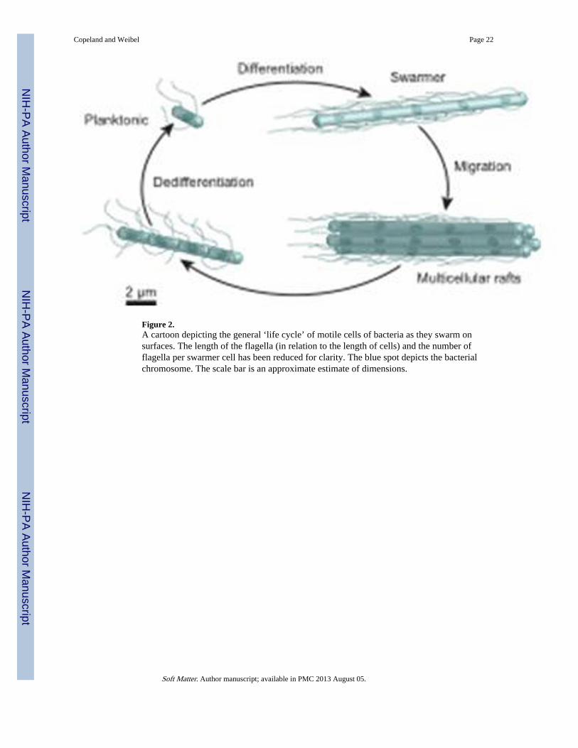
Figure 2.A cartoon depicting the general ‘life cycle’ of motile cells of bacteria as they swarm onsurfaces. The length of the flagella (in relation to the length of cells) and the number offlagella per swarmer cell has been reduced for clarity. The blue spot depicts the bacterialchromosome. The scale bar is an approximate estimate of dimensions.
Copeland and Weibel Page 22
Soft Matter. Author manuscript; available in PMC 2013 August 05.
NIH
-PA Author Manuscript
NIH
-PA Author Manuscript
NIH
-PA Author Manuscript

Figure 3.A-C) Time lapse images depicting the macroscopic migration of a swarming colony of wild-type E. coli strain RP437 on the surface of 0.45% Eiken agar (w/v) infused with nutrientbroth (1.0% peptone, 0.5% NaCl, 0.3% beef extract, and 0.5% glucose). The agar solidifiedovernight at 25 °C before the center of the gel was inoculated with 2 µl of a saturatedovernight culture of bacteria. The plate was incubated at 30 °C. The expansion of the swarmcolony over time is shown at 10, 15, and 20 hours after inoculation. D) A sequence ofmicroscopy images that demonstrate the time-dependent spreading of swarmer cells acrossan agar surface. The panels show the progression of the edge of the swarm colony over athree-minute period. The media and inoculation procedure was identical to the conditions
Copeland and Weibel Page 23
Soft Matter. Author manuscript; available in PMC 2013 August 05.
NIH
-PA Author Manuscript
NIH
-PA Author Manuscript
NIH
-PA Author Manuscript

used for images A-C. The images were acquired using phase contrast microscopy and aCCD camera; the images were inverted to improve the contrast between the cells andbackground. The cells are moving from the right-hand side of the image to the left.
Copeland and Weibel Page 24
Soft Matter. Author manuscript; available in PMC 2013 August 05.
NIH
-PA Author Manuscript
NIH
-PA Author Manuscript
NIH
-PA Author Manuscript

Figure 4.A) A schematic diagram depicting a colony of bacteria grown on the surface of a ‘stiff’ agargel (e.g. 1.5%, w/v) and a characteristic planktonic cell isolated from the colony. The TEMimage shows a planktonic cell of S. liquefaciens MG1.83 B) A swarming colony of bacteriagrown on the surface of a ‘soft’ agar gel (e.g. 0.45%, w/v) and a TEM image of a swarmercell of S. liquefaciens MG1 isolated from the outer edge of a swarm colony.83 The arrowsdepict the radial outward expansion of the colony on the surface. TEM images in A and Bare reprinted with permission from the American Society for Microbiology, Copyright 1999.
Copeland and Weibel Page 25
Soft Matter. Author manuscript; available in PMC 2013 August 05.
NIH
-PA Author Manuscript
NIH
-PA Author Manuscript
NIH
-PA Author Manuscript
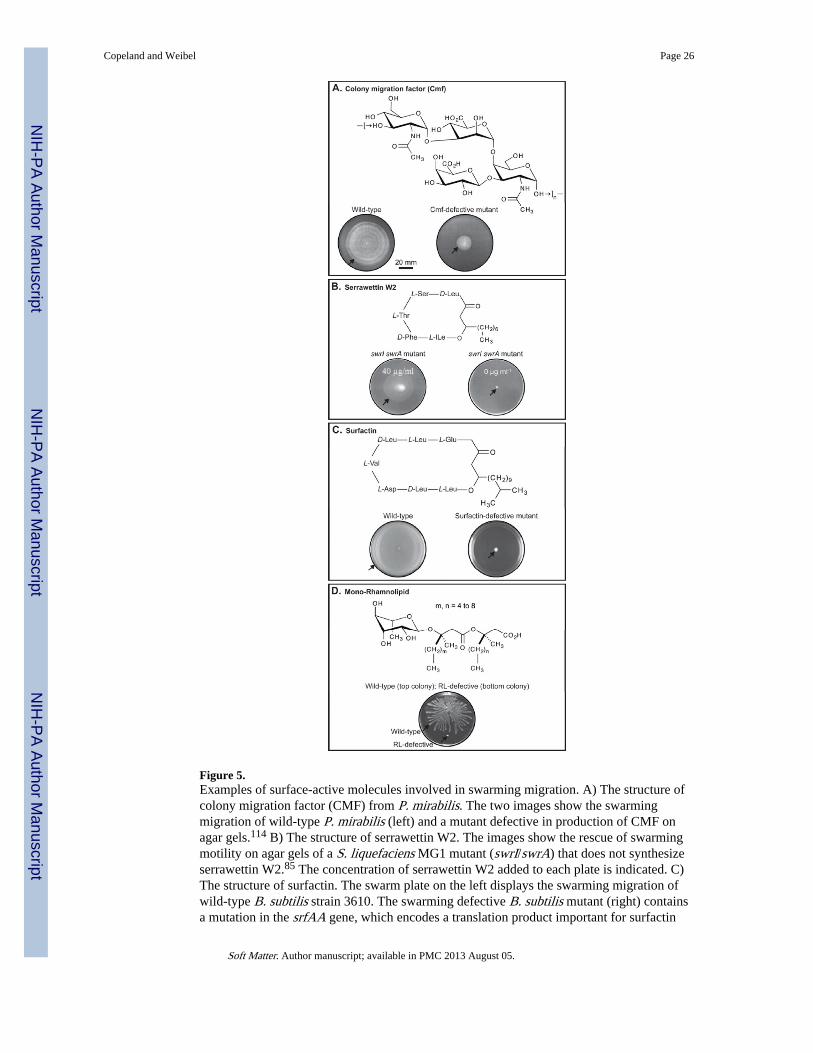
Figure 5.Examples of surface-active molecules involved in swarming migration. A) The structure ofcolony migration factor (CMF) from P. mirabilis. The two images show the swarmingmigration of wild-type P. mirabilis (left) and a mutant defective in production of CMF onagar gels.114 B) The structure of serrawettin W2. The images show the rescue of swarmingmotility on agar gels of a S. liquefaciens MG1 mutant (swrI/swrA) that does not synthesizeserrawettin W2.85 The concentration of serrawettin W2 added to each plate is indicated. C)The structure of surfactin. The swarm plate on the left displays the swarming migration ofwild-type B. subtilis strain 3610. The swarming defective B. subtilis mutant (right) containsa mutation in the srfAA gene, which encodes a translation product important for surfactin
Copeland and Weibel Page 26
Soft Matter. Author manuscript; available in PMC 2013 August 05.
NIH
-PA Author Manuscript
NIH
-PA Author Manuscript
NIH
-PA Author Manuscript
synthesis.92 D) The structure of mono-rhamnolipid. The image shows the swarmingmigration of wild-type P. aeruginosa (top structure) and an rhlA mutant (bottom structure),which is unable to synthesize rhamnolipids, on agar gels.162 The black arrows on each agarplate depict the edge of the swarming colony. Images are reprinted with permission from (A)Blackwell Science Ltd, Copyright 1995, (B) American Society for Microbiology, Copyright1998, (C) Blackwell Publishing Ltd, Copyright 2004, and (D) American Society forMicrobiology, Copyright 2000.
Copeland and Weibel Page 27
Soft Matter. Author manuscript; available in PMC 2013 August 05.
NIH
-PA Author Manuscript
NIH
-PA Author Manuscript
NIH
-PA Author Manuscript

Figure 6.Examples of cell-cell contact in swarming motility. A) An image of the edge of a swarmingcolony of P. mirabilis (300x). The swarmer cells are arranged into tightly packed rafts ofcells.10 B) S. liquefaciens MG1 cells at the leading edge of a swarming colony migratetogether.83 No scale was provided in the original publication for images A) and B). C-D)TEM images of a wild-type P. mirabilis swarmer cell (C) and a mutant of the ccmA gene inP. mirabilis (D).104 The image (inset) shows the swarming motility of colonies of wild-typeP. mirabilis (left) and the ccmA mutant (right) on an agar gel. E-F) SEM images of vapor-fixed swarmer cells of wild-type P. mirabilis (E) and a mutant with reduced swarmingmotility (~85% reduction in surface migration) (F).103 Images are reprinted with permission
Copeland and Weibel Page 28
Soft Matter. Author manuscript; available in PMC 2013 August 05.
NIH
-PA Author Manuscript
NIH
-PA Author Manuscript
NIH
-PA Author Manuscript

from (A) American Society for Microbiology, Copyright 1972, (B) American Society forMicrobiology, Copyright 1999, (C) and (D) American Society for Microbiology, Copyright1999, and (E) and (F) American Society for Microbiology, Copyright 2004.
Copeland and Weibel Page 29
Soft Matter. Author manuscript; available in PMC 2013 August 05.
NIH
-PA Author Manuscript
NIH
-PA Author Manuscript
NIH
-PA Author Manuscript

NIH
-PA Author Manuscript
NIH
-PA Author Manuscript
NIH
-PA Author Manuscript
Copeland and Weibel Page 30
Tabl
e 1
A c
ompa
riso
n of
the
char
acte
rist
ics
of p
lank
toni
c an
d sw
arm
er c
ell f
or s
ever
al s
war
min
g ge
nera
of
Eub
acte
ria.
Abb
revi
atio
ns: l
ipop
olys
acch
arid
e (L
PS);
3-(3
-hyd
roxy
alka
noyl
oxy)
alk
anoi
c ac
ids
(HA
As)
; per
itric
hous
(Pe
r.);
pol
ar (
Pol.)
. Que
stio
n m
arks
indi
cate
unk
now
n or
sug
gest
ed f
acto
rs o
rch
arac
teri
stic
s of
sw
arm
ing
mot
ility
. The
per
cent
age
of a
gar
that
is r
equi
red
to in
duce
sw
arm
ing
in E
. col
i ref
ers
to E
iken
aga
r; E
. col
i K12
str
ains
no
not
swar
m o
n th
e sa
me
perc
enta
ge o
f D
ifco
aga
r.
Pla
nkto
nic
cell
Swar
mer
cel
l
Org
anis
mC
ell
Len
gth
(µm
)
Num
ber
offl
agel
la
Cel
lle
ngth
(µm
)In
crea
sein
fla
gella
rela
tive
to
plan
kton
ic
Rat
e of
surf
ace
mig
rati
on(µ
m m
in−1
)
Secr
eted
surf
acta
nt o
rw
etti
ng a
gent
Aga
r %
(w/v
) th
atSu
ppor
tssw
arm
ing
Mec
hani
sms
that
affe
ct s
war
mer
cel
lde
velo
pmen
t
B. s
ubtil
is(u
ndom
estic
ated
str
ains
)1–
2.548
5–10
(Per
.)3–
64810
×16
316
7–23
348Su
rfac
tin48
0.5–
0.748
Ext
race
llula
r se
rine
prot
ease
(E
pr);
91
Com
X p
hero
mon
e164
E. c
oli K
122–
4574–
7(P
er.)
235–
2057
2–3×
5712
0–60
057L
PS; O
-ant
igen
?70, 7
60.
45–0
.5 (
Eik
en)57
Che
mot
axis
sys
tem
144
P. m
irab
ilis
1–279
6–10
(Pe
r.)16
510
–8079
10–5
0×13
950;
13 7
0010
Col
ony
mig
ratio
n fa
ctor
781.
5–313
Inhi
bitio
n of
flag
ella
r ro
tatio
n;14
Glu
tam
ine;
143
Putr
esci
ne16
6
P. a
erug
inos
a1–
2491
(Pol
.)16
22–
349≥
2× (
Pol.)
49?
Rha
mno
lipid
s(M
ono/
Di)
;H
AA
s;97
–99
Exo
poly
sacc
hari
de (
algi
nate
)49
0.45
–0.5
;49 0
.5–0
.716
2N
itrog
en li
mita
tion16
2 ;Q
uoru
m s
ensi
ng97
V. p
arah
aem
olyt
icus
1.5–
2121
1(P
ol.)
121
30–4
014, 1
2110
–50×
121,
127
12513
?1.
5–214
Inhi
bitio
n of
flag
ella
r ro
tatio
n;12
2
Iron
lim
itatio
n;12
6
Che
mot
axis
sys
tem
14
S. ty
phim
uriu
m2–
4576–
10 (
Per.
)225–
2057
2–3×
5712
0–60
057L
PS;70
Flh
E?16
70.
5–0.
857C
hem
otax
is s
yste
m14
7
S. m
arce
scen
s/S.
liqu
efac
iens
0.5–
2601–
2(P
er.)
605–
3014
, 60
2–3×
59, 8
388
13Se
rraw
ettin
850.
6–0.
813
Inhi
bitio
n of
flag
ella
r ro
tatio
n;14
Quo
rum
sen
sing
;85
Che
mot
axis
sys
tem
14
Soft Matter. Author manuscript; available in PMC 2013 August 05.

NIH
-PA Author Manuscript
NIH
-PA Author Manuscript
NIH
-PA Author Manuscript
Copeland and Weibel Page 31
Table 2
A comparison of the genera of bacteria that have been observed to swarm and their natural habitat.
Swarming Genera of Bacteria Natural habitat
Aeromonas Fresh water; GI tract
Azospirillum Soil; rhizosphere
Bacillus Soil; air (spores)
Burkholderia Soil; fresh water
Clostridium Soil; marine sediment; GI tract
Escherichia GI tract
Myxococcus Soil
Proteus Soil; GI/urinary tract; water;plant surfaces
Pseudomonas Water; soil; plant surfaces
Rhizobium Soil; root
Rhodospirillum Water
Salmonella GI tract; feces
Serratia Soil; water; plant surfaces
Vibrio Salt water
Yersinia Rodent; flea; GI tract
Soft Matter. Author manuscript; available in PMC 2013 August 05.