Embed Size (px)
DESCRIPTION
journal, NIHMS
Citation preview
Mechanistic Aspects of Inflammation and Clinical Managementof Inflammation in Acute Gouty Arthritis
Bruce N. Cronstein, MD andPaul R. Esserman Professor of Medicine, NYU School of Medicine, 550 First Ave., NBV16N1,New York, NY 10016, USA, Telephone: 1-212-263-6404
Prashanth Sunkureddi, MDClear Lake Rheumatology, 2060 Space Park Drive, Suite 208, Nassau Bay, TX 77058, USA,Telephone: 1-281-957-9127; Fax: 1-281-957-9157Bruce N. Cronstein: [email protected]; Prashanth Sunkureddi: [email protected]
AbstractIt has been recently demonstrated that interleukin-1β (IL-1β) plays a central role in monosodiumurate (MSU) crystal-induced inflammation and that the NALP3 inflammasome plays a major rolein IL-1β production. These discoveries have offered new insights into the pathogenesis of acutegouty arthritis. In this review, we discuss the molecular mechanisms by which MSU crystalsinduce acute inflammation and examine the mechanisms of action (MOAs) of traditional anti-inflammatory drugs (eg, nonsteroidal anti-inflammatory drugs [NSAIDs], colchicine, andglucocorticoids) and biologic agents (eg, the IL-1β antagonists anakinra, rilonacept, andcanakinumab) to understand how their MOAs contribute to their safety profiles. Traditional anti-inflammatory agents may act on the IL-1β pathway at some level; however, their MOAs arebroad-ranging, unspecific, and biologically complex. This lack of specificity may explain therange of systemic side effects associated with them. The therapeutic margins of NSAIDs,colchicine, and glucocorticoids are particularly low in elderly patients and in patients withcardiovascular, metabolic, or renal comorbidities that are frequently associated with goutyarthritis. In contrast, the IL-1β antagonists act on very specific targets of inflammation, which maydecrease the potential for systemic side effects, although infrequent but serious adverse events(including infection and administration reactions) have been reported. Because these IL-1βantagonists target an early event immediately downstream from NALP3 inflammasome activation,they may provide effective alternatives to traditional agents with minimal systemic side effects.Results of ongoing trials of IL-1β antagonists will likely provide clarification of their potentialrole in the management of acute gouty arthritis.
Corresponding Author: Bruce N. Cronstein, MD, Paul R. Esserman Professor of Medicine, NYU School of Medicine, 550 First Ave.,NBV16N1, New York, NY 10016, USA, Telephone: 1-212-263-6404, [email protected].
Conflicts of Interest:BN Cronstein, MD: Consultant to: Bristol-Myers Squibb, Novartis, CanFite Biopharmaceuticals, Cypress Laboratories, Regeneron(Westat, DSMB), Endocyte, Protalex, Allos, Inc., Savient, Gismo Therapeutics, Antares Pharmaceutical, and Medivector (all <$10,000). Grants received from: King Pharmaceuticals, NIH, Vilcek Foundation, OSI Pharmaceuticals, URL Pharmaceuticals, Inc.,and Gilead Pharmaceuticals. Board member of: Vilcek Foundation. Stock held in: CanFite Biopharmaceuticals (received formembership in Scientific Advisory Boards). Four patents held on the use of: adenosine A2A receptor agonists to promote woundhealing and to inhibit fibrosis; adenosine A1 receptor antagonists to treat osteoporosis and other diseases of bone; adenosine A1 andA2B receptor antagonists to treat fatty liver; and adenosine A2A receptor agonists to prevent prosthesis loosening.P. Sunkureddi, MD, PA: Speaker’s Bureau/Consultant to: Novartis, Bristol Myers Squibb, UCB, Pfizer (all >$10,000).
NIH Public AccessAuthor ManuscriptJ Clin Rheumatol. Author manuscript; available in PMC 2014 January 01.
Published in final edited form as:J Clin Rheumatol. 2013 January ; 19(1): 19–29. doi:10.1097/RHU.0b013e31827d8790.
NIH
-PA Author Manuscript
NIH
-PA Author Manuscript
NIH
-PA Author Manuscript
Keywordsinterleukin-1β; anakinra; canakinumab; rilonacept; inflammation; gouty arthritis; mechanism ofaction; safety; biologic agents
IntroductionAlthough gouty arthritis has been recognized as a distinct pathologic entity since ancienttimes, the molecular pathogenesis of acute gouty attacks and the sequence of pathologicevents resulting in chronic tophaceous gout are only now becoming fully understood.Clearly, monosodium urate (MSU) crystals are at the center of the pathophysiologicsequence, and although their role in gout was well described by Garrod in the 19th Century[1], their role in the pathogenesis of acute gouty arthritis had to be rediscovered in 1966 [2].The broader understanding of the pathogenesis of acute gouty arthritis has led to a newunderstanding of the mechanism of action (MOA) of agents commonly used to treat goutyarthritis and has resulted in the development of novel therapies for acute gouty arthritis. Inthis review, we will discuss the molecular mechanisms by which MSU crystals induce acuteinflammation, the mechanisms by which current therapies interfere with and suppress theinflammation associated with acute gouty arthritis, the mechanisms associated with typicaladverse events (AEs) associated with traditional gouty arthritis anti-inflammatory therapies,and the potential mechanistic advantages of biologic therapies that specifically target theinterleukin-1β (IL-1β) inflammation pathway.
Inflammation, an OverviewInflammation is characterized by rubor, tumor, calor, dolor, and functio laesa (redness,swelling, heat, pain, and loss of function) [3]. Vascular events, including dilatation,leakiness, and expression of molecules involved in the recruitment of leukocytes, play amajor role in the first three characteristics and result in the accumulation of neutrophils,macrophages/monocytes, and other inflammatory cells at inflamed sites [3]. The vascularendothelium plays a central role in these events and may be influenced by a variety ofintercellular messengers ranging from small molecules (eg, eicosanoids, histamine) topeptide messengers (eg, cytokines and chemokines) [3–6]. In turn, the vascular endotheliumwill secrete agents including eicosanoids and cytokines, which influence the inflammatoryprocess [3]. Vascular endothelial cells recruit leukocytes through the expression of adhesionmolecules at inflamed sites, and different vascular adhesion molecules recruit different celltypes. In acute gouty attacks, neutrophils are the predominant cell type, and these cellsadhere to the endothelial surface proteins E-selectin, P-selectin, and intercellular adhesionmolecule-1 (ICAM-1), which are expressed or upregulated at inflamed sites [7]. Cytokines,such as IL-1β and tumor necrosis factor-α (TNF-α), are the primary stimuli for endothelialexpression and upregulation of these adhesive molecules. Older studies have implicatedMSU-induced release of IL-1 as central to the initiation of inflammation [4,5], and recentstudies indicate that uptake of MSU crystals by cells activates the NALP3 inflammasome,leading to the elaboration of activated IL-1 [8]. In acute gouty attacks, the predominantcellular infiltrate is comprised almost exclusively of neutrophils. IL-8 and its receptor onneutrophils, CXCR2, are required for the development of an acute inflammatory response toMSU crystals [9].
Monosodium Urate Crystals and InflammationIn individuals who suffer from both acute gouty attacks and chronic tophaceous gout, MSUcrystals are present in both symptomatic and asymptomatic joint tissue and joint fluid. Manyevents can set off acute gouty attacks, including overindulgence in alcohol, metabolic
Cronstein and Sunkureddi Page 2
J Clin Rheumatol. Author manuscript; available in PMC 2014 January 01.
NIH
-PA Author Manuscript
NIH
-PA Author Manuscript
NIH
-PA Author Manuscript
stresses such as those that accompany acute myocardial infarctions or surgery, or, mostpredictably, major shifts in serum uric acid levels leading to resorption of MSU crystals,such as occurs after starting urate-lowering therapy (ULT) [10,11]. It is now clear that inresponse to MSU crystals, the cells in the joints that initiate the inflammatory cascade aremacrophages; these cells phagocytose MSU crystals and release chemo-attractants, such asleukotrienes, IL-8, and others, that recruit neutrophils to the site and start the inflammatorycascade [12,13]. Once recruited to the joint, neutrophils phagocytose MSU crystals andfurther contribute to the inflammation that characterizes acute gouty attacks.
The mechanisms by which cells take up MSU crystals and activate the inflammatorycascade have been under study for many years, and a number of mechanisms have beenproposed and investigated to explain uptake of MSU crystals by leukocytes. MSU crystalsare hygroscopic and bind many different proteins to their surface, including immunoglobulinG (IgG) and complement proteins [14–19], which interact with specific receptors onleukocytes to promote leukocyte recruitment and crystal phagocytosis. One experimentalproblem that has hindered our understanding of the mechanism by which MSU crystalsinteract with and activate leukocytes is that many MSU preparations used for in vitro studiesare contaminated by endotoxin, which directly stimulates Toll-like receptors (TLRs) onleukocytes. Subsequent studies in which endotoxin contamination was eliminated indicatedthat MSU crystals directly interacted with CD14, a leukocyte cell-surface molecule thatinteracts with TLR2 and TLR4 to stimulate leukocytes [20], in addition to promotingphagocytosis via complement and immunoglobulin receptors. Regardless of the mechanismby which the MSU crystals are phagocytosed, the crystals interact with TLR2 and TLR4 aswell as with the NALP3 inflammasome to stimulate leukocyte activation, leading to theinflammatory cascade [8].
In 2006, Martinon and colleagues [8] first demonstrated that MSU crystals activate aspecific inflammatory cascade in leukocytes leading to production of IL-1. Essentially, theseauthors demonstrated that crystals (MSU and calcium pyrophosphate dihydrate [the crystalsthat cause calcium pyrophosphate dehydrate disease]) engage the cryopyrin (NALP3)inflammasome, a signaling protein complex in leukocytes that, linked by the adaptor proteinapoptosis-associated speck-like protein (ASC), activates caspase-1. Caspase-1, or theneutrophil protease proteinase 3 (PR3) [21], cleaves pro-IL-1β to generate IL-1β, permittingthe release of IL-1β into the extracellular space. Once secreted, IL-1β leads to activation ofvascular endothelium and production of other chemokines and cytokines, resulting in therecruitment of leukocytes. Based on their homology to caspase-1 and proteinase 3, otherneutrophil proteases are likely to play a role in activation of pro-Il-1 to IL-1β as well [22]. Inaddition to the in vitro evidence of NALP3 activation, increased caspase-1 activation, andincreased IL-1 production by cells exposed to crystals, mice lacking NALP3 did not mountmuch of an inflammatory response to challenge with MSU crystals, with few leukocytespresent in the exudate. Thus, the demonstration that the NALP3 inflammasome plays acentral role in response to pathogenic crystals leading to IL-1β production and that IL-1βplays the central role in crystal-induced inflammation offered a new insight into thepathogenesis of gouty arthritis [8].
Subsequent work has provided evidence that other factors contribute to MSU-mediatedactivation of the inflammasome [23,24]. Joosten and colleagues [24] reported that free fattyacids, which activate TLR2, further drive MSU-induced IL-1β production. Moreover, priorstudies had demonstrated that MSU crystals induce increased expression ofcyclooxygenase-2 (COX-2) with enhanced production of inflammatory prostaglandins thatalso likely contribute to the enhanced IL-1β production [25–28].
Cronstein and Sunkureddi Page 3
J Clin Rheumatol. Author manuscript; available in PMC 2014 January 01.
NIH
-PA Author Manuscript
NIH
-PA Author Manuscript
NIH
-PA Author Manuscript
It did not take long for investigators to carry out confirmatory experiments in patients withacute gouty attacks. Administration of an IL-1β/IL-1α blocker (the recombinant IL-1receptor [IL-1R] antagonist anakinra) [29,30], a chimeric IL-1 TRAP dimeric fusion proteinconsisting of portions of IL-1R and the IL-1R accessory protein that neutralizes both IL-1βand IL-1α (rilonacept) [31], or a fully human monoclonal antibody that neutralizes theactivity of human IL-1β (canakinumab) [32], all have some efficacy in the treatment ofacute gouty arthritis [33]. Intracellular IL-1β signals for inflammation via activation ofnuclear factor-κB (NFκB) and other studies have shown that signaling moleculesdownstream of the IL-1 receptor (MyD88) are required for MSU to activate NFκB and forthe development of crystal-induced arthritis [34].
Termination of Acute Gouty AttacksAcute gouty arthritis attacks are generally self-limited and last less than 3 to 4 weeks. Verylittle is understood at present about how gouty arthritis attacks spontaneously terminatedespite the ongoing presence of the inciting agent. One suggestion has been that monocytes/macrophages that take up the apoptotic neutrophils from the joint secrete transforminggrowth factor-β1 (TGF-β1) and other anti-inflammatory mediators that terminate the attack[35]. Similar uptake of apoptotic neutrophils occurs in rheumatoid arthritis withouttermination or amelioration of inflammation, so there must be other factors at work as well[36]. Recent work has indicated that interaction of free fatty acids with TLR2 is required forurate crystal-mediated activation of the inflammasome, production of active IL-1, and acuteinflammation [24]. This finding has suggested that the elevation of free fatty acids followinga heavy meal plays a critical role in the induction of acute gouty attacks. Interestingly, it haslong been known that low density lipoprotein (LDL; specifically apolipoprotein B in theLDL), commonly elevated in many patients with gout, bind to MSU crystals and preventneutrophil activation by crystals, another potential mechanism for suppression ofinflammation during intercurrent gout.
Clinical Management of Inflammation in Acute Gouty Arthritis: How DoTherapies Work and How Do Their Mechanisms of Action Affect Safety?Overview of Acute Gouty Arthritis Management and Treatment Limitations
The goals of gouty arthritis treatment are 2-fold. First, rapid anti-inflammatory therapy isnecessary to manage the significant pain, swelling, and disability associated with acuteattacks [10,11]. Once an acute attack has terminated, ULT should be initiated to preventfuture acute attacks and long-term complications associated with chronic tophaceous gout(eg, joint destruction) [11].
Long before the development of biologic agents that target IL-1β, other therapies had beenused successfully and are still currently used for the prevention and treatment of acute goutyattacks, including nonsteroidal anti-inflammatory drugs (NSAIDs), colchicine, andglucocorticoids. Figure 1 provides an overview of the sites of action of traditional therapiesand new biologic agents involved in mediating crystal-induced inflammation. Thesetraditional therapies are effective in reducing inflammation, with symptomatic reliefoccurring within 24 hours [37,38]. Research on traditional agents continues, with theAGREE study demonstrating that low doses of colchicines are as effective as high dosesover 24 hours, with safety similar to placebo [38]. Glucocorticoids are a good option inpatients with contraindications to NSAIDs and colchicine or in refractory cases [39],although a meta-analysis showed inconclusive evidence for their effectiveness comparedwith other anti-inflammatory agents [40].
Cronstein and Sunkureddi Page 4
J Clin Rheumatol. Author manuscript; available in PMC 2014 January 01.
NIH
-PA Author Manuscript
NIH
-PA Author Manuscript
NIH
-PA Author Manuscript

Current gouty arthritis treatment guidelines recommend oral NSAIDs or colchicine a first-line systemic treatment for acute attacks [41] However, the MOAs through which NSAIDsand colchicine reduce inflammation are not specific, and the systemic actions of these drugsare known to cause severe AEs in some patients [42,43]. For example, NSAIDs areassociated with adverse gastrointestinal, renal, and cardiovascular effects [43,44], and attherapeutic doses, colchicine is associated with safety concerns such as blood dyscrasias,drug-drug interactions, neuromuscular toxicity, and gastrointestinal AEs [42].
Because gouty arthritis is associated with several comorbidities (including cardiovasculardisease, hypertension, type 2 diabetes mellitus, obesity, hyperlipidemia, metabolicsyndrome, chronic kidney disease [CKD], and nephrolithiasis) [45,46], contraindications toNSAIDs and/or colchicine are common [47]. In a study that reviewed medical records fromthe Department of Veterans Affairs, of 575 patients diagnosed with gouty arthritis, morethan 88% of patients had at least 1 comorbid condition [47]. In the same study, more than90% of patients had at least 1 contraindication to NSAIDs and approximately 50% ofpatients had at least 1 contraindication to colchicines [47].
In patients who cannot tolerate NSAIDs or colchicine, and in patients with polyarticulargouty arthritis or CKD, glucocorticoids are recommended for the management of acutegouty arthritis attacks [48]. However, even with short-term use, glucocorticoids have beenassociated with AEs, such as hypertension and diabetes mellitus [49]. In patients with pre-existing impaired glucose tolerance, the diabetogenic effects associated with glucocorticoidscan be particularly substantial [49].
Below, we present a detailed examination of the MOAs of traditional anti-inflammatorydrugs and how their varied actions may contribute to the safety profiles of these agents. Wethen discuss newer agents that act specifically on the IL-1β pathway.
Nonsteroidal Anti-inflammatory DrugsProstaglandins generated by the inducible COX-2 enzyme play a major role in thestimulation of inflammatory responses and contribute to the development of the cardinalsigns of acute inflammation in gouty arthritis attacks [3,50]. Unlike COX-1, which isexpressed constitutively in most cells and is responsible for homeostatic functions (includingepithelial cytoprotection, platelet aggregation, and regulation of renal blood flow), COX-2 isthe product of an immediate-early gene that is rapidly inducible and tightly regulated [51].COX-2 is the dominant source of prostaglandins in inflammation. However, recent evidencesuggests that both COX-1 and COX-2 may contribute to prostanoid production during bothacute inflammatory responses and the resolution phase of inflammation [3]. COX-1accounts for approximately 10% to 15% of prostanoid formation induced bylipopolysaccharide, and both COX-1 and COX-2 are expressed in circulating inflammatorycells ex vivo [52]. Human data indicate that COX-1–derived prostanoids drive the initialphase of acute inflammation, while COX-2 upregulation may not occur until several hourslater [3,52]. The roles of different prostanoids formed by COX-1 and/or COX-2 areextremely complex; depending on whether a given prostanoid is formed by COX-1 orCOX-2, the same molecule may either stimulate or resolve inflammation [3].
The biological consequences of COX inhibition with NSAIDs are potentially broad-rangingand not well understood. It is hypothesized that NSAID-based COX inhibition can betolerated in most patients because prostanoid formation is a homeostatic response system.Under most physiologic conditions, only small amounts of prostanoids are formed, and theirbiological importance may be minimal. However, under conditions of physiologic stress (ie,in elderly patients and patients with comorbid renal, cardiovascular, or gastrointestinalconditions), alterations in prostanoid expression are associated with increased safety
Cronstein and Sunkureddi Page 5
J Clin Rheumatol. Author manuscript; available in PMC 2014 January 01.
NIH
-PA Author Manuscript
NIH
-PA Author Manuscript
NIH
-PA Author Manuscript
concerns [3]. These safety concerns can be particularly problematic when relatively highdoses of NSAIDs are prescribed, as is common practice in the management of acute goutyarthritis attacks [53].
Safety of Nonsteroidal Anti-inflammatory Drugs—It is well established thatNSAIDs are associated with an increased risk of gastrointestinal side effects. Themechanisms associated with NSAID-induced gastric damage are outlined in Figure 2 [43].Most nonselective NSAIDs that inhibit both COX-1 and COX-2 are organic acids, and theirulcerogenic potential is associated with their pKa and lipophilicity. NSAIDs with pKavalues between approximately 2.8 and 4.4 are most likely to cause ulcers, as are lipophilicNSAIDs that interact with phospholipids and disrupt gastric mucosal membranes. Incontrast, most selective COX-2 inhibitors are not acidic and have much higher pKa values.Thus, selective COX-2 inhibitors are less likely than nonselective NSAIDs to causegastrointestinal mucosal irritation [43]. However, the United States Food and DrugAdministration does not distinguish between the gastrointestinal safety profiles of selectiveand nonselective NSAIDs and applies the same package insert warning labels for allNSAIDs.
The comparative incidence of serious gastrointestinal events associated with selectiveCOX-2 inhibitors is roughly half that of nonselective NSAIDs [3]. However, selectiveCOX-2 inhibitors are still associated with the potential to cause serious gastrointestinalevents in high-risk patients, as these inhibitors block the synthesis of gastroduodenalepithelial COX-2–dependent prostanoids that accelerate ulcer healing [3]. In addition, whenCOX pathways are blocked by NSAIDs, some arachidonic acid is diverted through thelipoxygenase (LOX) pathway, which increases leukotriene synthesis, which can furtherpropagate mucosal damage.
Selective COX-2 inhibitors were developed to reduce the risk of gastrointestinalcomplications associated with nonselective NSAIDs; however, selective COX-2 inhibitorswere found to be associated with an increased risk of cardiovascular events Of note, 2widely used COX-2 inhibitors, rofecoxib and valdecoxib, were withdrawn from the globalmarket due to cardiovascular safety concerns [54]. The cardiovascular risks associated withselective COX-2 inhibitors have been confirmed in several studies [55–57], and thesefindings have recently been extended to some nonselective NSAIDs [44,54,58]. Thecardiovascular risks associated with NSAIDs (including increased risk of recurrentmyocardial infarction and death) have been observed even with short-term use (ie, <1 week),as is common for the management of acute gouty arthritis flares [54].
COX-2 inhibition promotes cardiovascular injury through several mechanisms. SelectiveCOX-2 inhibition leads to a loss of the majority of COX-2–derived vascular prostacyclinsynthesis without alteration in platelet thromboxane synthesis [51]. Prostacyclin is a potentvasodilator and inhibitor of platelet aggregation, leukocyte adhesion, and vascular smoothmuscle cell proliferation in the kidneys, liver, lungs, and heart. Therefore, suppression ofCOX-2–derived prostacyclin by both nonselective NSAIDs and selective COX-2 inhibitorsincreases the risk of thrombosis, hypertension, atherosclerosis, and myocardial infarction[3,51].. It has also been hypothesized that the increase in leukotriene biosynthesis thatresults from the shunting of arachidonic acid to the LOX pathway may increase the risk ofatherosclerosis [51,59].
COX-1 and COX-2 are both expressed in the kidneys. The effects of NSAID-inducedinhibition of renal prostaglandin E2 can result in sodium retention, edema, and exacerbationof hypertension. Inhibition of prostacyclin expression can decrease renal blood flow andglomerular filtration rate, which can lead to hyperkalemia as a result of decreased potassium
Cronstein and Sunkureddi Page 6
J Clin Rheumatol. Author manuscript; available in PMC 2014 January 01.
NIH
-PA Author Manuscript
NIH
-PA Author Manuscript
NIH
-PA Author Manuscript
excretion. COX-2 inhibition can be especially problematic in elderly patients, in whomCOX-2 expression has been detected in the macula [60].
ColchicineThe earliest recorded use of colchicine to treat gouty arthritis dates back to the ancientGreeks and Egyptians (extracts of Colchicum autumnalis), and colchicine has been usedextensively for the treatment of acute gouty arthritis flares since the 18th century [61].Colchicine is an antimitotic alkaloid that binds to specific sites on the cytoskeletal proteintubulin and disrupts microtubule polymerization. This disruption of normal cytoskeletalassembly results in a range of biologic effects on essential cell functions, includinginhibition of intracellular vesicle transport, decreased secretion of chemokines andcytokines, impairment of cell migration, and inhibition of cell division [62].
In line with the current understanding of the pathogenesis of gouty arthritis, in vitro studieshave shown that high concentrations of colchicine suppress inflammation by blocking IL-1βprocessing in monocytes stimulated by MSU, but do not affect IL-1β activation byextracellular adenosine triphosphate. This suggests that colchicine acts upstream of NALP3inflammasome-driven caspase-1 activation [8]. Despite these findings, it is unlikely that thismechanism accounts for colchicine’s therapeutic effects in crystal-driven inflammation,because these effects have only been observed at colchicine concentrations that are 10-foldto 100-fold higher than those achieved in patients during the treatment of acute goutyarthritis and 100-fold to 1000-fold higher than the concentrations required to mediateprophylaxis of chronic gouty arthritis [8,62,63].
Other studies have reported that pharmacologically relevant concentrations of colchicinedirectly inhibit intracellular signaling molecules (eg, tyrosine kinases and phospholipases) inneutrophils that inhibit some, but not all, of the inflammatory actions of these cells (eg,chemotaxis, superoxide anion production, adhesion to cellular substrata, and mobilizationand release of lysosomal enzymes during phagocytosis) [6,64–70]. Evidence also shows thatcolchicine inhibits neutrophil migration following crystal activation without changes inproduction of the chemokine IL-8 [71]. Colchicine induces COX-1 and COX-2 geneexpression and does not inhibit COX-1 or COX-2 in neutrophils [72].
Previous observations have shown that concentrations of colchicine that are similar to thoseachieved during prophylaxis of acute attacks (ie, nanomolar concentrations) alter theexpression of endothelial adhesion molecules (E-selectin) on cells required for therecruitment of neutrophils [73], thereby suppressing the development of acute goutyarthritis. At higher concentrations, colchicine induces shedding of neutrophil adhesionmolecules (L-selectin), thus preventing further neutrophil recruitment [73]. All of the actionsof colchicine in this setting have been attributed to the capacity of colchicine to disruptmicrotubules.
Safety of Colchicine—Colchicine has a narrow therapeutic index between efficacy andtreatment-limiting gastrointestinal side effects, including diarrhea and abdominal paincaused by increased peristaltic activity [42,62]. The pharmacokinetics and safety ofcolchicine are driven in large part by its binding to tubulin. Because tubulin-boundcolchicine has a slow dissociation rate, the half-life of this complex is approximately 20 to30 hours. After colchicine therapy is discontinued, its terminal elimination half-life isroughly 16 hours and the biologic effects of colchicine require 24 to 48 hours to dissipate[62]. Colchicine’s long half-life may contribute to its narrow therapeutic margin(particularly in patients with renal or hepatic impairment), as colchicine is predominantlymetabolized in the liver and up to 20% of an administered dose is cleared by the kidneys.The half-life of colchicine in patients with severe renal impairment is 2-fold to 3-fold longer
Cronstein and Sunkureddi Page 7
J Clin Rheumatol. Author manuscript; available in PMC 2014 January 01.
NIH
-PA Author Manuscript
NIH
-PA Author Manuscript
NIH
-PA Author Manuscript

compared with patients with normal renal function, which significantly increases the risk ofcolchicine accumulation and toxicity [62,74].
Colchicine is metabolized in the liver by the cytochrome P450 enzyme CYP3A4. Therefore,interactions between colchicine and drugs with CYP3A4 inhibitory activity have thepotential to cause colchicine accumulation, increase colchicine’s pharmacologic effects, andpredispose patients to colchicine toxicity. Drug-drug interactions have been observedbetween colchicine and CYP3A4 inhibitors including cimetidine, clarithromycin,erythromycin, fluoxetine, paroxetine, nefazodone, indinavir and other protease inhibitors,tolbutamide, and azole antifungals [62,63]. For example, cimetidine decreases the hepaticclearance of colchicine by roughly 30%, which prolongs the plasma elimination half-life[62], and clarithromycin markedly increases colchicine exposure, which has been associatedwith fatalities in patients with renal insufficiency [75].
Colchicine also binds to P-glycoprotein (P-gp), an adenosine triphosphate-binding proteinwidely distributed in cell membranes in the intestinal epithelium, biliary tract, blood-brainbarrier, and renal proximal tubules. P-gp influences absorption, bioavailability, andelimination of its substrates [62]. These substrates include chemotherapeutic agents,macrolide antibiotics, protease inhibitors, and glucocorticoids, as well as some statins (eg,simvastatin and fluvastatin) and calcium-channel blockers (eg, verapamil) that arecommonly prescribed in patients with cardiovascular comorbidities associated with goutyarthritis [62]. Similar to CYP3A4 inhibitors, the use of P-gp modulating agents incombination with colchicine can lead to intracellular colchicine accumulation, withincreases in pharmacologic effects or toxicity [62].
Colchicine’s interactions with microtubules can cause accumulation of lysosomes andautophagic vacuoles in the cytoskeleton, resulting in pathologic alterations in skeletalmuscle and induction of significant axonal neuropathy. These adverse consequences maypresent as myopathy (eg, rhabdomyolysis), neuropathy, or bone marrow suppression [76].Although the likelihood of serious AEs associated with colchicine is generally considered tobe dose-dependent (ie, mortality rates are estimated at 10% at doses >0.5 mg/kg) [42,62], ananalysis by Wilbur and Makowsky [76] identified cases of colchicine-induced myotoxicityat lower doses. In the 75 cases identified, the mean (standard deviation) cumulative dailydose of colchicine was only 1.4 (0.96) mg [76] (for comparison, standard therapeutic dosesof colchicine for acute gouty arthritis attacks are 1.2 mg at the first sign of a flare followedby 0.6 mg 1 hour later) [63]. In many of the cases, patients had been receiving standarddoses of colchicine for long periods of time but experienced recent declines in renal functionor other underlying conditions [76].
GlucocorticoidsGlucocorticoids have long been used to treat acute gouty arthritis attacks, particularly inpatients who are intolerant to NSAIDs or colchicines [48]. This class of agents has manywell-described anti-inflammatory actions that are mediated through binding interactionswith glucocorticoid receptors, which are localized in the cytoplasm of target cells found inalmost all tissues in the human body [77,78]. The complex molecular architecture ofglucocorticoid-induced antagonism of inflammation is outlined in Figure 3.
The clinical success of glucocorticoids as effective anti-inflammatory agents is largelyattributed to their ability to reduce the expression of pro-inflammatory genes via activationof the glucocorticoid receptor and the concomitant inhibition pro-inflammatory transcriptionfactors (eg, NFκB and activating protein-1 [AP-1]) through a mechanism known astransrepression [78]. In acute gouty arthritis, the most notable anti-inflammatory action ofglucocorticoids is the capacity of these agents to prevent activation of NFκB by either TNF-
Cronstein and Sunkureddi Page 8
J Clin Rheumatol. Author manuscript; available in PMC 2014 January 01.
NIH
-PA Author Manuscript
NIH
-PA Author Manuscript
NIH
-PA Author Manuscript

α or IL-1β. Glucocorticoids increase the expression of the inhibitor of κB (IκB), which isthe cytoplasmic chaperone that prevents translocation of NFκB to the nucleus [79]. Thus,glucocorticoids prevent IL-1β from stimulating the inflammatory cascade.
However, much of the anti-inflammatory efficacy of glucocorticoids results from pleiotropiceffects of the glucocorticoid receptor on diverse signaling pathways through direct andindirect genomic effects. This pleiotropy, along with known nongenomic effects involvingsecond-messenger and membrane-associated receptor signaling, can result in a broad rangeof side effects [80]. In addition to NFκB and AP-1, other transcription factors that arenegatively regulated by the glucocorticoid receptor via transrepression include cyclicadenosine monophosphate (cAMP) response element-binding protein, interferon regulatoryfactor 3, nuclear factor of activated T cells, signal transducer and activator of transcription,T-box expressed in T cells, and GATA-binding protein 3. Target genes involved intransrepression include those encoding for a broad range of inflammatory cytokines,enzymes, receptors, and adhesion molecules, notably, IL-1β, IL-2, IL-3, IL-4, IL-5, IL-6,IL-8, IL-12, COX-2, E-selectin, and TNF-α [77,78]. In addition, glucocorticoids cansuppress inflammation through transactivation of genes that are associated with increasedsynthesis of anti-inflammatory proteins, including lipocortin-1, IL-1 receptor agonist, IκB,and β2-adrenoceptor [77,80].
Safety of GlucocorticoidsSide effects associated with glucocorticoid therapy mainly arise from the ability of thesteroid-activated glucocorticoid receptor to activate target genes involved in the metabolismof sugar, protein, fat, muscle, and bone via transactivation and to suppress the hypothalamic-pituitary-adrenal axis via transrepression [78,81]. The most common AEs associated withsteroid use for the management of acute gouty arthritis attacks are typically related to thecomplex effects of glucocorticoids on metabolism and the endocrine system. The mostclinically relevant metabolic consequence associated with glucocorticoid therapy isgenerally considered to be hyperglycemia related to glucocorticoid-induced upregulation inglucose synthesis. This increase in glucose synthesis results from transactivation of acomplex network of hepatic enzymes that control gluconeogenesis, mobilization anddegradation of proteins, and increased glycogen storage in the liver [78,81].
Glucocorticoid therapy is also associated with adverse cardiovascular effects, most notably,hypertension, dyslipidemia, and reduced fibrinolytic potential [81,82]. These AEs are mostcommon in patients treated with high doses of glucocorticoids and in elderly patients with afamily history of essential hypertension [82]. The relationship between glucocorticoids andblood pressure regulation is complex; however, it is thought that the primary mechanismsinvolved are increases in systemic vascular resistance, extracellular volume, and cardiaccontractility [81,82].
Development of psychiatric problems and aggravation of pre-existing psychoses in patientsreceiving acute glucocorticoid therapy have also been reported; moreover, when acutetreatment is discontinued, patients may experience psychiatric withdrawal symptoms,including fatigue and depression [80,81]. These symptoms are more common in women andusually develop within 2 weeks of beginning treatment, particularly with doses ofprednisolone that are above 40 mg/day. Glucocorticoids can also have reversible adverseeffects on memory and cognition. The underlying mechanisms associated with steroid-induced psychoses are thought to be related to hippocampal damage caused by directglucocorticoid exposure, including decreased dendritic branching, altered synapticstructures, neuron loss, and inhibition of neuronal regeneration. Glucocorticoids can alsocause hypothalamic-pituitary-adrenal axis function abnormalities and dysregulation of theserotonin (5-HT) system. One of the most important mechanisms associated with the
Cronstein and Sunkureddi Page 9
J Clin Rheumatol. Author manuscript; available in PMC 2014 January 01.
NIH
-PA Author Manuscript
NIH
-PA Author Manuscript
NIH
-PA Author Manuscript
pathophysiology of depression is suppression of the 5-HT1A receptor, which occurs viaglucocorticoid receptor-mediated transrepression of NFκB [81].
Despite the use of glucocorticoids in clinical practice, the actual incidence of AEs when theyare used as a short-term course in gouty arthritis is not known. Recently, the EuropeanLeague Against Rheumatism (EULAR) developed recommendations to monitor AEsassociated with low-dose glucocorticoid therapy for rheumatoid arthritis. Per theserecommendations, patients treated with glucocorticoid therapy should be monitored fordiabetes/glucose intolerance, hypertension, electrolyte shifts, infections, mood changes, andmental problems, which may occur as possible side effects of steroid treatment [83].Whether similar recommendations need to be followed while using steroids during an acuteattack of gouty arthritis is not clear; however, since gouty arthritis is often associated withcomorbidities, the occurrence of some of these side effects cannot be ruled out. Also, someof these AEs can occur following single injections of intra-articular or intramuscularcorticosteroids. Therefore, patients receiving glucocorticoids for the management of acutegouty arthritis must be observed carefully.
Biologic Therapies: Interleukin-1β AntagonistsFrom the earlier discussion of the role of IL-1β in inflammation, it is clear that agents thattarget IL-1β or prevent the actions of IL-1β on cells are likely to be useful therapies for thetreatment or prevention of acute gouty attacks. As noted above, binding of IL-1β by eitherrilonacept or canakinumab or binding of IL-1R1 by anakinra may be effective mechanismsfor treating or preventing acute gouty arthritis attacks (Figure 4) [33].
Although all 3 agents target IL-1β, their MOAs are different. Anakinra is an IL-1Rantagonist that binds to IL-1R1 and blocks IL-1β and IL-1α [84]. Rilonacept is arecombinant dimeric fusion protein consisting of portions of IL-1R and the IL-1R accessoryprotein linked to the Fc portion of IgG1. Rilonacept acts as a receptor to neutralize bothIL-1β and IL-1α and as a soluble decoy receptor [85]. Canakinumab is a fully humanmonoclonal antibody that binds to human IL-1β and neutralizes its activity by blocking itsinteraction with IL-1 receptors. However, it does not bind IL-1α or IL-1 receptor antagonist(IL-1RA) [86]. The specificity of these agents in targeting IL-1β may have positiveimplications for safety and tolerability.
Published studies in gout include two phase III trials for canakinumab [89] and one phase IIItrial for rilonacept [90]. Results suggest that these biologic therapies may provideimprovement in pain and inflammation associated with acute gouty arthritis attacks. Thereare no published trials evaluating anakinra in patients with gouty arthritis.
Safety of Interleukin-1β Antagonists—Early safety findings from phase II trials ofcanakinumab and rilonacept (8–16 weeks’ duration) and a pilot study of anakinra (4-weeks’duration) demonstrated that IL-1β antagonists are generally well tolerated in patients withgouty arthritis [29,32,88,91], although infrequent but serious AEs, including infections andadministration reactions, have been reported. Safety results of recently published phase IIItrials (of short duration of 16–24 weeks) for canakinumab and rilonacept are consistent withthese earlier phase II results [89,90]. In the canakinumab phase III trials [89], the mostcommon AEs were back pain, hypertension, and headache. In the rilonacept phase III trial[90], the most common AEs were injection-site reactions, upper respiratory tract infections,and headache. As mentioned above, there are no published phase II or III trials evaluatinganakinra in patients with gouty arthritis.
As with other biologics, the potential for increased bacterial infections is of concern withIL-1 antagonist [92], with the immunosuppressant effect of these agents likely the
Cronstein and Sunkureddi Page 10
J Clin Rheumatol. Author manuscript; available in PMC 2014 January 01.
NIH
-PA Author Manuscript
NIH
-PA Author Manuscript
NIH
-PA Author Manuscript

mechanism for the increased risk [93]. In the phase III trials, infections were reported in20.4% of patients receiving canakinumab and 12.2% of patients receiving the activecomparator (triamcinolone acetonide) [89], and in 18.0% of patients receiving rilonacept and22.8% of patients receiving placebo [90]. No opportunistic infections were reported witheither of these agents. As there are no head-to-head comparative gouty arthritis studiesbetween rilonacept, canakinumab, and anakinra, no safety comparisons can be made. Ofnote, none of these agents are yet approved by the FDA for gouty arthritis. A long-termstudy of rilonacept is ongoing (NCT01459796) and a long-term study of canakinumab hasrecently been completed (NCT00927810), both in gouty arthritis. Results, when available,will provide information on the long-term safety of these agents in this patient population.
ConclusionThe NALP3 inflammasome and IL-1β are crucial players in the inflammatory pathwayassociated with acute gouty arthritis attacks [8]. While the traditional agents, NSAIDs,colchicine, and glucocorticoids, may act on this pathway at some level, their MOAs are notspecific. The anti-inflammatory actions of these agents are broad-ranging and biologicallycomplex. In many cases, these agents target pathways that are high upstream in theinflammatory process [3,8]. Although inhibition of these upstream pathways produces a netbenefit in reducing pain and inflammation for most patients with acute gouty arthritis, thebenefit/risk margin of these agents is generally low and varies substantially from oneindividual to another. The therapeutic margin for colchicine is always very low, with thepotential for serious AEs in patients with renal or hepatic comorbidities and in patientstaking other drugs that inhibit CYP3A4 or P-gp [62]. The therapeutic margin for NSAIDsmay be particularly low in elderly patients and in patients with comorbidities [3]. Theincidence of AEs associated with short courses of glucocorticoid therapy for the treatment ofacute gouty arthritis is not known; however, glucocorticoids act on many body systems andbased on recent guidelines, patients treated with glucocorticoids should be monitored fordiabetes/glucose intolerance, hypertension, electrolyte shifts, infections, mood changes, andmental problems [78,81,83]. NSAIDs, colchicine, and glucocorticoids may have contrastingeffects in different body systems, which may contribute to the wide array of AEs associatedwith these drugs. For these reasons, it has been suggested that more specific, downstreamtargeting of the inflammation pathway may result in safer and more effective therapies [3].
The IL-1β antagonists being studied for the management of acute gouty arthritis attacks(anakinra, rilonacept, and canakinumab) act on very specific targets of inflammation [33].Because these agents target an early event immediately downstream from NALP3inflammasome activation by MSU crystals, it is possible that they will provide effectivealternatives to the less specific traditional agents without the associated systemic sideeffects. Data from long-term studies will further elucidate the potential role of IL-1βantagonism in the management of acute gouty arthritis.
AcknowledgmentsSource of funding: Certain sections of the manuscript, including the “Introduction” and the sections titled“Inflammation, an Overview,” “Monosodium Urate Crystals and Inflammation,” and “Termination of Acute GoutyArthritis,” were written by Dr. Cronstein. Sections focusing on the clinical management of gouty arthritis wereprepared by Dr. Sunkureddi, with editorial assistance provided by Cherie Koch, PhD, of Oxford PharmaGenesisInc. Each author’s contribution was merged together by Dr. Koch who provided editorial assistance to improve theflow and eliminate any redundancies. Dr. Koch also provided assistance with figure preparation and styling of themanuscript for submission. Funding for this assistance was provided by Novartis Pharmaceuticals Corporation. Theauthors were fully responsible for all content and editorial decisions and received no financial support or other formof compensation related to the development of this manuscript. The opinions expressed in the manuscript are thoseof the authors, and Novartis Pharmaceuticals Corporation had no influence on the contents.
Cronstein and Sunkureddi Page 11
J Clin Rheumatol. Author manuscript; available in PMC 2014 January 01.
NIH
-PA Author Manuscript
NIH
-PA Author Manuscript
NIH
-PA Author Manuscript
Reference List1. Garrod AB. Researches on Gout.-Part I. The Urine in the Different Forms of Gout.-Part II. The
Influence of Colchicum upon the Urine. Med Chir Trans. 1858; 41:325–360.
2. Steele AD, McCarty DJ Jr. An experimental model of acute inflammation in man. Arthritis Rheum.1966; 9:430–442. [PubMed: 5328048]
3. Ricciotti E, FitzGerald GA. Prostaglandins and inflammation. Arterioscler Thromb Vasc Biol. 2011;31:986–1000. [PubMed: 21508345]
4. Di Giovine FS, Malawista SE, Nuki G, et al. Interleukin 1 (IL 1) as a mediator of crystal arthritis.Stimulation of T cell and synovial fibroblast mitogenesis by urate crystal-induced IL 1. J Immunol.1987; 138:3213–3218. [PubMed: 3033070]
5. Di Giovine FS, Malawista SE, Thornton E, et al. Urate crystals stimulate production of tumornecrosis factor alpha from human blood monocytes and synovial cells. Cytokine mRNA and proteinkinetics, and cellular distribution. J Clin Invest. 1991; 87:1375–1381. [PubMed: 2010550]
6. Gilbert C, Poubelle PE, Borgeat P, et al. Crystal-induced neutrophil activation: VIII. Immediateproduction of prostaglandin E2 mediated by constitutive cyclooxygenase 2 in human neutrophilsstimulated by urate crystals. Arthritis Rheum. 2003; 48:1137–1148. [PubMed: 12687558]
7. Cronstein BN, Weissmann G. The adhesion molecules of inflammation. Arthritis Rheum. 1993;36:147–157. [PubMed: 8431203]
8. Martinon F, Pétrilli V, Mayor A, et al. Gout-associated uric acid crystals activate the NALP3inflammasome. Nature. 2006; 440:237–241. [PubMed: 16407889]
9. Terkeltaub R, Baird S, Sears P, et al. The murine homolog of the interleukin-8 receptor CXCR-2 isessential for the occurrence of neutrophilic inflammation in the air pouch model of acute uratecrystal-induced gouty synovitis. Arthritis Rheum. 1998; 41:900–909. [PubMed: 9588743]
10. Baker JF, Schumacher HR. Update on gout and hyperuricemia. Int J Clin Pract. 2010; 64:371–377.[PubMed: 19909378]
11. Neogi T. Clinical practice. Gout. N Engl J Med. 2011; 364:443–452. [PubMed: 21288096]
12. Martin WJ, Walton M, Harper J. Resident macrophages initiating and driving inflammation in amonosodium urate monohydrate crystal-induced murine peritoneal model of acute gout. ArthritisRheum. 2009; 60:281–289. [PubMed: 19116939]
13. Schiltz C, Lioté F, Prudhommeaux F, et al. Monosodium urate monohydrate crystal-inducedinflammation in vivo: quantitative histomorphometric analysis of cellular events. Arthritis Rheum.2002; 46:1643–1650. [PubMed: 12115197]
14. Ortiz-Bravo E, Sieck MS, Schumacher HR Jr. Changes in the proteins coating monosodium uratecrystals during active and subsiding inflammation. Immunogold studies of synovial fluid frompatients with gout and of fluid obtained using the rat subcutaneous air pouch model. ArthritisRheum. 1993; 36:1274–1285. [PubMed: 8216421]
15. Cherian PV, Schumacher HR Jr. Immunochemical and ultrastructural characterization of serumproteins associated with monosodium urate crystals (MSU) in synovial fluid cells from patientswith gout. Ultrastruct Pathol. 1986; 10:209–219. [PubMed: 3715998]
16. Russell IJ, Papaioannou C, McDuffie FC, et al. Effect of IgG and C-reactive protein oncomplement depletion by monosodium urate crystals. J Rheumatol. 1983; 10:425–433. [PubMed:6887165]
17. Hasselbacher P, Schumacher HR. Immunoglobulin in tophi and on the surface of monosodiumurate crystals. Arthritis Rheum. 1978; 21:353–361. [PubMed: 646833]
18. Naff GB, Byers PH. Complement as a mediator of inflammation in acute gouty arthritis. I. Studieson the reaction between human serum complement and sodium urate crystals. J Lab Clin Med.1973; 81:747–760. [PubMed: 4349137]
19. Tramontini N, Huber C, Liu-Bryan R, et al. Central role of complement membrane attack complexin monosodium urate crystal-induced neutrophilic rabbit knee synovitis. Arthritis Rheum. 2004;50:2633–2639. [PubMed: 15334478]
20. Scott P, Ma H, Viriyakosol S, et al. Engagement of CD14 mediates the inflammatory potential ofmonosodium urate crystals. J Immunol. 2006; 177:6370–6378. [PubMed: 17056568]
Cronstein and Sunkureddi Page 12
J Clin Rheumatol. Author manuscript; available in PMC 2014 January 01.
NIH
-PA Author Manuscript
NIH
-PA Author Manuscript
NIH
-PA Author Manuscript

21. Joosten LA, Netea MG, Fantuzzi G, et al. Inflammatory arthritis in caspase 1 gene-deficient mice:contribution of proteinase 3 to caspase 1-independent production of bioactive interleukin-1beta.Arthritis Rheum. 2009; 60:3651–3662. [PubMed: 19950280]
22. Dinarello CA. Interleukin-1 in the pathogenesis and treatment of inflammatory diseases. Blood.2011; 117:3720–3732. [Epub 2011 Feb 8]. [PubMed: 21304099]
23. Dinarello CA. How interleukin-1β induces gouty arthritis. Arthritis Rheum. 2010; 62:3140–3144.[PubMed: 20662058]
24. Joosten LA, Netea MG, Mylona E, et al. Engagement of fatty acids with Toll-like receptor 2 drivesinterleukin-1β production via the ASC/caspase 1 pathway in monosodium urate monohydratecrystal-induced gouty arthritis. Arthritis Rheum. 2010; 62:3237–3248. [PubMed: 20662061]
25. Martinez RV, Reval M, Campos MD, et al. Involvement of peripheral cyclooxygenase-1 andcyclooxygenase-2 in inflammatory pain. J Pharm Pharmacol. 2002; 54:405–412. [PubMed:11902807]
26. Pouliot M, James MJ, McColl SR, et al. Monosodium urate microcrystals induce cyclooxygenase-2in human monocytes. Blood. 1998; 91:1769–1776. [PubMed: 9473245]
27. Hasselbacher P. Stimulation of synovial fibroblasts by calcium oxalate and monosodium uratemonohydrate. A mechanism of connective tissue degradation in oxalosis and gout. J Lab ClinMed. 1982; 100:977–985. [PubMed: 6292314]
28. McMillan RM, Hasselbacher P, Hahn JL, et al. Interactions of murine macrophages withmonosodium urate crystals: stimulation of lysosomal enzyme release and prostaglandin synthesis.J Rheumatol. 1981; 8:555–562. [PubMed: 6946240]
29. So A, De Smedt T, Revaz S, et al. A pilot study of IL-1 inhibition by anakinra in acute gout.Arthritis Res Ther. 2007; 9:R28. [PubMed: 17352828]
30. So A, De Meulemeester M, Pikhlak A, et al. Canakinumab for the treatment of acute flares indifficult-to-treat gouty arthritis: results of a multicenter, phase II, dose-ranging study. ArthritisRheum. 2010; 62:3064–3076. [PubMed: 20533546]
31. Neogi T. Interleukin-1 antagonism in acute gout: is targeting a single cytokine the answer?Arthritis Rheum. 2010; 62:2845–2849. [PubMed: 20597109]
32. Chen CJ, Shi Y, Hearn A, et al. MyD88-dependent IL-1 receptor signaling is essential for goutyinflammation stimulated by monosodium urate crystals. J Clin Invest. 2006; 116:2262–2271.[PubMed: 16886064]
33. Yagnik DR, Evans BJ, Florey O, et al. Macrophage release of transforming growth factor β1during resolution of monosodium urate monohydrate crystal-induced inflammation. ArthritisRheum. 2004; 50:2273–2280. [PubMed: 15248227]
34. Savill JS, Wyllie AH, Henson JE, et al. Macrophage phagocytosis of aging neutrophils ininflammation. Programmed cell death in the neutrophil leads to its recognition by macrophages. JClin Invest. 1989; 83:865–875. [PubMed: 2921324]
35. Sunkureddi P. Gouty arthritis: understanding the disease state and management options in primarycare. Adv Ther. 2011; 28:748–760. [PubMed: 21894471]
36. Terkeltaub RA, Furst DE, Bennett K, et al. High versus low dosing of oral colchicine for earlyacute gout flare: Twenty-four-hour outcome of the first multicenter, randomized, double-blind,placebo-controlled, parallel-group, dose-comparison colchicine study. Arthritis Rheum. 2010;62:1060–1068. [PubMed: 20131255]
37. Gonzalez EB. An update on the pathology and clinical management of gouty arthritis. ClinRheumatol. 2012; 31:13–21. [PubMed: 22069122]
38. Janssens HJ, Lucassen PL, Van de Laar FA, et al. Systemic corticosteroids for acute gout.Cochrane Database Syst Rev. 2008; (2):Art. No:CD005521.
39. Zhang W, Doherty M, Bardin T, et al. EULAR evidence based recommendations for gout. Part II:Management. Report of a task force of the EULAR Standing Committee for International ClinicalStudies Including Therapeutics (ESCISIT). Ann Rheum Dis. 2006; 65:1312–1324. [PubMed:16707532]
40. Richette P, Bardin T. Colchicine for the treatment of gout. Expert Opin Pharmacother. 2010;11:2933–2938. [PubMed: 21050036]
Cronstein and Sunkureddi Page 13
J Clin Rheumatol. Author manuscript; available in PMC 2014 January 01.
NIH
-PA Author Manuscript
NIH
-PA Author Manuscript
NIH
-PA Author Manuscript
41. Scarpignato C, Hunt RH. Nonsteroidal antiinflammatory drug-related injury to the gastrointestinaltract: clinical picture, pathogenesis, and prevention. Gastroenterol Clin North Am. 2010; 39:433–464. [PubMed: 20951911]
42. Salvo F, Fourrier-Réglat A, Bazin F, et al. Cardiovascular and gastrointestinal safety of NSAIDs: asystematic review of meta-analyses of randomized clinical trials. Clin Pharmacol Ther. 2011;89:855–866. [PubMed: 21471964]
43. Zhang W, Doherty M, Pascual E, et al. EULAR evidence based recommendations for gout. Part I:Diagnosis. Report of a task force of the Standing Committee for International Clinical StudiesIncluding Therapeutics (ESCISIT). Ann Rheum Dis. 2006; 65:1301–1311. [PubMed: 16707533]
44. Edwards NL. The role of hyperuricemia and gout in kidney and cardiovascular disease. Cleve ClinJ Med. 2008; 75(Suppl 5):S13–S16. [PubMed: 18822470]
45. Keenan RT, O’Brien WR, Lee KH, et al. Prevalence of contraindications and prescription ofpharmacologic therapies for gout. Am J Med. 2011; 124:155–163. [PubMed: 21295195]
46. Terkeltaub R. Update on gout: new therapeutic strategies and options. Nat Rev Rheumatol. 2010;6:30–38. [PubMed: 20046204]
47. Richette P, Bardin T. Should prednisolone be first-line therapy for acute gout? Lancet. 2008;372:1301–1302. [PubMed: 18929902]
48. Vane JR. Introduction: mechanism of action of NSAIDs. Br J Rheumatol. 1996; 35(Suppl 1):1–3.[PubMed: 8630629]
49. Funk CD, FitzGerald GA. COX-2 inhibitors and cardiovascular risk. J Cardiovasc Pharmacol.2007; 50:470–479. [PubMed: 18030055]
50. Smyth EM, Grosser T, Wang M, et al. Prostanoids in health and disease. J Lipid Res. 2009;50(Suppl):S423–S428. [PubMed: 19095631]
51. Cronstein BN, Terkeltaub R. The inflammatory process of gout and its treatment. Arthritis ResTher. 2006; 8(Suppl 1):S3. [PubMed: 16820042]
52. Schjerning Olsen AM, Fosbol EL, Lindhardsen J, et al. Duration of treatment with nonsteroidalanti-inflammatory drugs and impact on risk of death and recurrent myocardial infarction inpatients with prior myocardial infarction: a nationwide cohort study. Circulation. 2011; 123:2226–2235. [PubMed: 21555710]
53. Bombardier C, Laine L, Reicin A, et al. Comparison of upper gastrointestinal toxicity of rofecoxiband naproxen in patients with rheumatoid arthritis. VIGOR Study Group. N Engl J Med. 2000;343:1520–1528. [PubMed: 11087881]
54. Bresalier RS, Sandler RS, Quan H, et al. Cardiovascular events associated with rofecoxib in acolorectal adenoma chemoprevention trial. N Engl J Med. 2005; 352:1092–1102. [PubMed:15713943]
55. Nussmeier NA, Whelton AA, Brown MT, et al. Complications of the COX-2 inhibitors parecoxiband valdecoxib after cardiac surgery. N Engl J Med. 2005; 352:1081–1091. [PubMed: 15713945]
56. Fosbol EL, Gislason GH, Jacobsen S, et al. Risk of myocardial infarction and death associated withthe use of nonsteroidal anti-inflammatory drugs (NSAIDs) among healthy individuals: anationwide cohort study. Clin Pharmacol Ther. 2009; 85:190–197. [PubMed: 18987620]
57. Lötzer K, Funk CD, Habenicht AJ. The 5-lipoxygenase pathway in arterial wall biology andatherosclerosis. Biochim Biophys Acta. 2005; 1736:30–37. [PubMed: 16081317]
58. Pham K, Hirschberg R. Global safety of coxibs and NSAIDs. Curr Top Med Chem. 2005; 5:465–473. [PubMed: 15974941]
59. Cronstein BN. Something old, something new--colchicine in the 21st century. Curr Opin InvestigDrugs. 2009; 10:1141–1142.
60. Nuki G. Colchicine: its mechanism of action and efficacy in crystal-induced inflammation. CurrRheumatol Rep. 2008; 10:218–227. [PubMed: 18638431]
61. Colcrys (colchicine, USP) Tablets [prescribing information]. AR Scientific, Inc; Philadelphia, PA:2010.
62. Phelps P. Polymorphonuclear leukocyte motility in vitro. IV. Colchicine inhibition of chemotacticactivity formation after phagocytosis of urate crystals. Arthritis Rheum. 1970; 13:1–9. [PubMed:5439313]
Cronstein and Sunkureddi Page 14
J Clin Rheumatol. Author manuscript; available in PMC 2014 January 01.
NIH
-PA Author Manuscript
NIH
-PA Author Manuscript
NIH
-PA Author Manuscript

63. Phelps P. Polymorphonuclear leukocyte motility in vitro. II. Stimulatory effect of monosodiumurate crystals and urate in solution; partial inhibition by colchicine and indomethacin. ArthritisRheum. 1969; 12:189–196. [PubMed: 5787231]
64. Abramson S, Hoffstein ST, Weissmann G. Superoxide anion generation by human neutrophilsexposed to monosodium urate. Arthritis Rheum. 1982; 25:174–180. [PubMed: 6279115]
65. Reinhardt PH, Naccache PH, Poubelle PE, et al. Monosodium urate crystals promote neutrophiladhesion via a CD18-independent and selectin-independent mechanism. Am J Physiol. 1996;270(1 Pt 1):C31–C39. [PubMed: 8772427]
66. Naccache PH, Bourgoin S, Plante E, et al. Crystal-induced neutrophil activation. II. Evidence forthe activation of a phosphatidylcholine-specific phospholipase D. Arthritis Rheum. 1993; 36:117–125. [PubMed: 8381010]
67. Naccache PH, Grimard M, Roberge CJ, et al. Crystal-induced neutrophil activation. I. Initiationand modulation of calcium mobilization and superoxide production by microcrystals. ArthritisRheum. 1991; 34:333–342. [PubMed: 1848432]
68. Popa-Nita O, Proulx S, Paré G, et al. Crystal-induced neutrophil activation: XI. Implication andnovel roles of classical protein kinase C. J Immunol. 2009; 183:2104–2114. [PubMed: 19596988]
69. Matsukawa A, Yoshimura T, Maeda T, et al. Analysis of the cytokine network among tumornecrosis factor α, interleukin-1β, interleukin-8, and interleukin-1 receptor antagonist inmonosodium urate crystal-induced rabbit arthritis. Lab Invest. 1998; 78:559–569. [PubMed:9605181]
70. Ben Chetrit E, Fischel R, Hinz B, et al. The effects of colchicine and hydroxychloroquine on thecyclo-oxygenases COX-1 and COX-2. Rheumatol Int. 2005; 25:332–335. [PubMed: 14963695]
71. Cronstein BN, Molad Y, Reibman J, et al. Colchicine alters the quantitative and qualitative displayof selectins on endothelial cells and neutrophils. J Clin Invest. 1995; 96:994–1002. [PubMed:7543498]
72. Swarup A, Sachdeva N, Schumacher HR Jr. Dosing of antirheumatic drugs in renal disease anddialysis. J Clin Rheumatol. 2004; 10:190–204. [PubMed: 17043508]
73. Hung IF, Wu AK, Cheng VC, et al. Fatal interaction between clarithromycin and colchicine inpatients with renal insufficiency: a retrospective study. Clin Infect Dis. 2005; 41:291–300.[PubMed: 16007523]
74. Wilbur K, Makowsky M. Colchicine myotoxicity: case reports and literature review.Pharmacotherapy. 2004; 24:1784–1792. [PubMed: 15585444]
75. Barnes PJ. Anti-inflammatory actions of glucocorticoids: molecular mechanisms. Clin Sci (Lond).1998; 94:557–572. [PubMed: 9854452]
76. De Bosscher K, Haegeman G. Minireview: latest perspectives on antiinflammatory actions ofglucocorticoids. Mol Endocrinol. 2009; 23:281–291. [PubMed: 19095768]
77. Auphan N, DiDonato JA, Rosette C, et al. Immunosuppression by glucocorticoids: inhibition ofNF-κB activity through induction of IκB synthesis. Science. 1995; 270:286–290. [PubMed:7569976]
78. Rhen T, Cidlowski JA. Antiinflammatory action of glucocorticoids--new mechanisms for olddrugs. N Engl J Med. 2005; 353:1711–1723. [PubMed: 16236742]
79. Schäcke H, Döcke WD, Asadullah K. Mechanisms involved in the side effects of glucocorticoids.Pharmacol Ther. 2002; 96:23–43. [PubMed: 12441176]
80. Sholter DE, Armstrong PW. Adverse effects of corticosteroids on the cardiovascular system. Can JCardiol. 2000; 16:505–511. [PubMed: 10787466]
81. van der Goes MC, Jacobs JW, Boers M, et al. Monitoring adverse events of low-doseglucocorticoid therapy: EULAR recommendations for clinical trials and daily practice. AnnRheum Dis. 2010; 69:1913–1919. [PubMed: 20693273]
82. Kineret (anakinra) [prescribing information]. Biovitrum AB; Stockholm, Sweden: 2009.
83. Arcalyst (rilonacept) Injection for subcutaneous use [prescribing information]. RegeneronPharmaceuticals, Inc; Tarrytown, NY: 2009.
84. Ilaris (canakinumab) Injection for subcutaneous use [prescribing information]. NovartisPharmaceuticals Corporation; East Hanover, NJ: 2011.
Cronstein and Sunkureddi Page 15
J Clin Rheumatol. Author manuscript; available in PMC 2014 January 01.
NIH
-PA Author Manuscript
NIH
-PA Author Manuscript
NIH
-PA Author Manuscript
85. Chen K, Fields T, Mancuso CA, et al. Anakinra’s efficacy is variable in refractory gout: report often cases. Semin Arthritis Rheum. 2010; 40:210–214. [PubMed: 20494407]
86. Schlesinger N, De Meulemeester M, Pikhlak A, et al. Canakinumab relieves symptoms of acuteflares and improves health-related quality of life in patients with difficult-to-treat gouty arthritis bysuppressing inflammation: results of a randomized, dose-ranging study. Arthritis Res Ther. 2011;13:R53. [PubMed: 21439048]
87. Schlesinger N, Alten RE, Bardin T, et al. Canakinumab for acute gouty arthritis in patients withlimited treatment options: results from two randomised, multicentre, active-controlled, double-blind trials and their initial extensions. Ann Rheum Dis. 2012 [Epub ahead of print].
88. Schumacher HR Jr, Evans RR, Saag KG, et al. Rilonacept (Interleukin-1 Trap) for prevention ofgout flares during initiation of uric acid-lowering therapy: Results of the presurge-1 trial. ArthritisCare Res. 2012 [Epub ahead of print].
89. Schumacher HR Jr, Sundy JS, Terkeltaub R, et al. Rilonacept (interleukin-1 trap) in the preventionof acute gout flares during initiation of urate-lowering therapy: results of a phase II randomized,double-blind, placebo-controlled trial. Arthritis Rheum. 2012; 64:876–884. [PubMed: 22223180]
90. Dinarello CA, Simon A, van der Meer JW. Treating inflammation by blocking interleukin-1 in abroad spectrum of diseases. Nat Rev Drug Discov. 2012; 11:633–652. [PubMed: 22850787]
91. Furst DE. The risk of infections with biologic therapies for rheumatoid arthritis. Semin ArthritisRheum. 2010; 39:327–346. [PubMed: 19117595]
Cronstein and Sunkureddi Page 16
J Clin Rheumatol. Author manuscript; available in PMC 2014 January 01.
NIH
-PA Author Manuscript
NIH
-PA Author Manuscript
NIH
-PA Author Manuscript

Figure 1. The most common mechanisms of therapeutic anti-inflammatory action of goutyarthritis drugsColchicine, NSAIDs, and glucocorticoids act on many different molecular targets; themechanisms displayed herein are the most likely targets for reduction of monosodium uratecrystal-induced inflammation when these drugs are administered at the recommendedtherapeutic doses. Anti-IL-1, IL1-RA, and IL1-Trap therapies act specifically to blockIL-1β.
Cronstein and Sunkureddi Page 17
J Clin Rheumatol. Author manuscript; available in PMC 2014 January 01.
NIH
-PA Author Manuscript
NIH
-PA Author Manuscript
NIH
-PA Author Manuscript
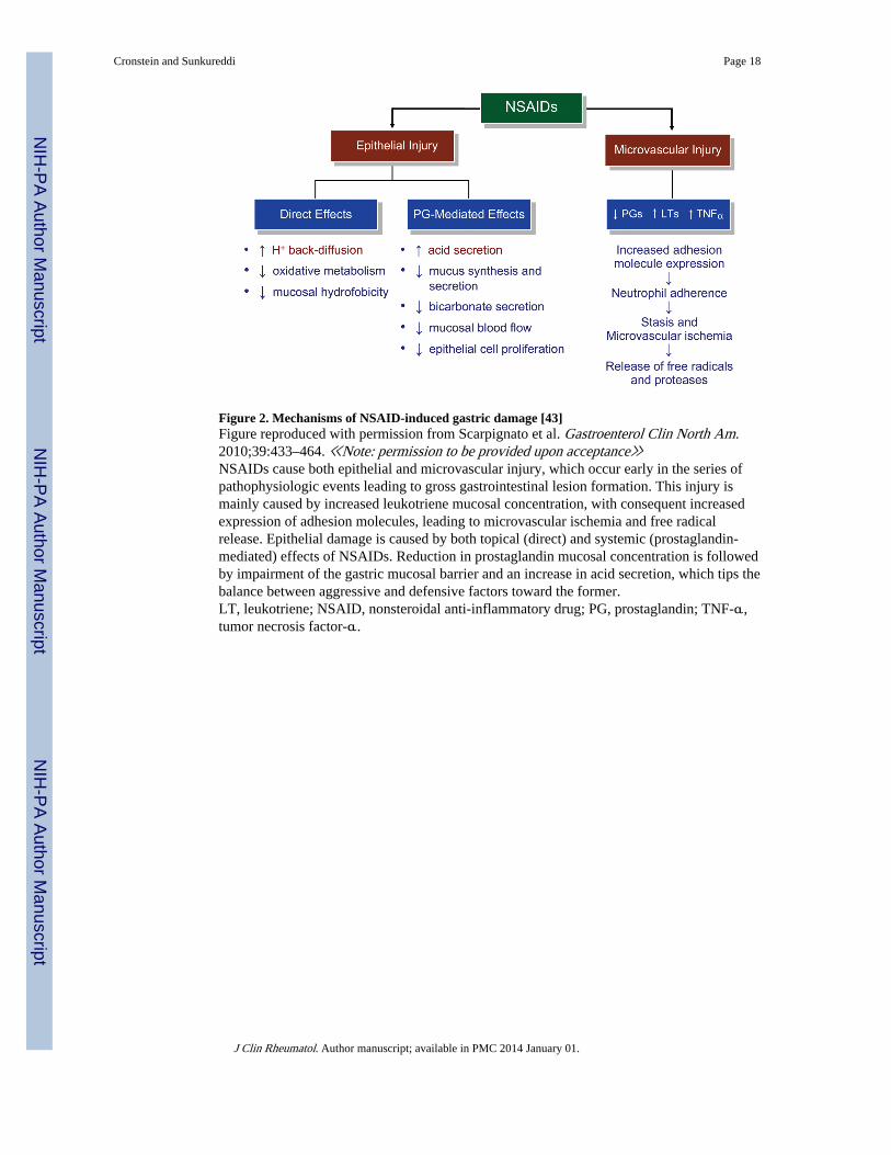
Figure 2. Mechanisms of NSAID-induced gastric damage [43]Figure reproduced with permission from Scarpignato et al. Gastroenterol Clin North Am.2010;39:433–464. ≪Note: permission to be provided upon acceptance≫NSAIDs cause both epithelial and microvascular injury, which occur early in the series ofpathophysiologic events leading to gross gastrointestinal lesion formation. This injury ismainly caused by increased leukotriene mucosal concentration, with consequent increasedexpression of adhesion molecules, leading to microvascular ischemia and free radicalrelease. Epithelial damage is caused by both topical (direct) and systemic (prostaglandin-mediated) effects of NSAIDs. Reduction in prostaglandin mucosal concentration is followedby impairment of the gastric mucosal barrier and an increase in acid secretion, which tips thebalance between aggressive and defensive factors toward the former.LT, leukotriene; NSAID, nonsteroidal anti-inflammatory drug; PG, prostaglandin; TNF-α,tumor necrosis factor-α.
Cronstein and Sunkureddi Page 18
J Clin Rheumatol. Author manuscript; available in PMC 2014 January 01.
NIH
-PA Author Manuscript
NIH
-PA Author Manuscript
NIH
-PA Author Manuscript

Figure 3. Partial molecular architecture underlying the glucocorticoid-induced antagonism ofinflammation [80]Figure reproduced with permission from Rhen et al. N Engl J Med. 2005;353:1711–1723.≪Note: permission to be provided upon acceptance≫Inflammatory pathways are characterized by positive feedback loops (ie, cytokines activateNF-κB, which in turn stimulates the synthesis of more cytokines) and by redundancy (ie,cytokines also activate c-Jun–Fos). The glucocorticoid receptor inhibits these pathways atmultiple points by directly blocking the transcription of inflammatory proteins by NF-κBand activator protein 1 and by inducing the expression of anti-inflammatory proteins such asIκB, annexin I, and MAPK phosphatase I. Red lines denote inhibition, and black arrowsactivation.COX-2 cyclooxygenase 2; cPLA2α, cytosolic phospholipase A2α; IκB, inhibitor of κB; 5-LOX, 5-lipoxygenase; MAPK, mitogen-activated protein kinase; NF-κB, nuclear factor-κB.
Cronstein and Sunkureddi Page 19
J Clin Rheumatol. Author manuscript; available in PMC 2014 January 01.
NIH
-PA Author Manuscript
NIH
-PA Author Manuscript
NIH
-PA Author Manuscript
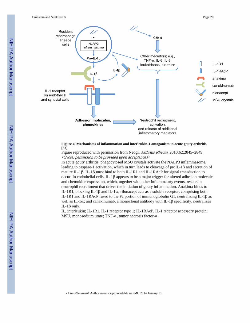
Figure 4. Mechanisms of inflammation and interleukin-1 antagonism in acute gouty arthritis[33]Figure reproduced with permission from Neogi. Arthritis Rheum. 2010;62:2845–2849.≪Note: permission to be provided upon acceptance≫In acute gouty arthritis, phagocytosed MSU crystals activate the NALP3 inflammasome,leading to caspase-1 activation, which in turn leads to cleavage of proIL-1β and secretion ofmature IL-1β. IL-1β must bind to both IL-1R1 and IL-1RAcP for signal transduction tooccur. In endothelial cells, IL-1β appears to be a major trigger for altered adhesion moleculeand chemokine expression, which, together with other inflammatory events, results inneutrophil recruitment that drives the initiation of gouty inflammation. Anakinra binds toIL-1R1, blocking IL-1β and IL-1α; rilonacept acts as a soluble receptor, comprising bothIL-1R1 and IL-1RAcP fused to the Fc portion of immunoglobulin G1, neutralizing IL-1β aswell as IL-1α; and canakinumab, a monoclonal antibody with IL-1β specificity, neutralizesIL-1β only.IL, interleukin; IL-1R1, IL-1 receptor type 1; IL-1RAcP, IL-1 receptor accessory protein;MSU, monosodium urate; TNF-α, tumor necrosis factor-α.
Cronstein and Sunkureddi Page 20
J Clin Rheumatol. Author manuscript; available in PMC 2014 January 01.
NIH
-PA Author Manuscript
NIH
-PA Author Manuscript
NIH
-PA Author Manuscript