Embed Size (px)
Citation preview
NGF signaling in PC12 cells: the cooperation of p75NTR
with TrkA is needed for the activation of both mTORC2and the PI3K signalling cascade
Sara Negrini1,2, Rosalba D’Alessandro3 and Jacopo Meldolesi1,2,*1San Raffaele Scientific Institute, DIBIT, via Olgettina 58, 20132 Milan, Italy2Vita-Salute San Raffaele University, Division of Neuroscience, via Olgettina 58, 20132 Milan, Italy3S. De Bellis Scientific Institute, Castellana Grotte, 70013 Bari, Italy
*Author for correspondence ([email protected])
Biology Open 0, 1–12doi: 10.1242/bio.20135116Received 15th April 2013Accepted 12th June 2013
SummaryPC12-27, a PC12 clone characterized by high levels of the
transcription repressor REST and by very low mTORC2
activity, had been shown to be unresponsive to NGF, possibly
because of its lack of the specific TrkA receptor. The
neurotrophin receptor repressed by high REST in PC12-27
cells, however, is shown now to be not TrkA, which is normal,
but p75NTR, whose expression is inhibited at the transcriptional
level. When treated with NGF, the PC12-27 cells lacking
p75NTR exhibited a defective TrkA autophosphorylation
restricted, however, to the TrkA(Y490) site, and an
impairment of the PI3K signaling cascade. This defect was
sustained in part by a mTORC1-dependent feed-back
inhibition that in wtPC12 cells appeared marginal.
Transfection of p75NTR to a level and surface distribution
analogous to wtPC12 did not modify various high REST-
dependent properties of PC12-27 cells such as high b-catenin,
low TSC2 and high proliferation rate. In contrast, the defective
PI3K signaling cascade and its associated mTORC2 activity
were largely rescued together with the NGF-induced neurite
outgrowth response. These changes were not due to p75NTR
alone but required its cooperation with TrkA. Our results
demonstrate that, in PC12, high REST induces alterations of
NGF signaling which, however, are indirect, dependent on the
repression of p75NTR; and that the well-known potentiation by
p75NTR of the TrkA signaling does not concern all the effects
induced by NGF but primarily the PI3K cascade and its
associated mTORC2, a complex known to play an important
role in neural cell differentiation.
� 2013. Published by The Company of Biologists Ltd. This is an
Open Access article distributed under the terms of the Creative
Commons Attribution License (http://creativecommons.org/
licenses/by/3.0), which permits unrestricted use, distribution
and reproduction in any medium provided that the original
work is properly attributed.
Key words: REST, PC12-27, ERK and PI3K cascades, mTORC2,
mTORC1-dependent feed-back inhibition
Introductionp75NTR, a receptor of the tumor necrosis factor receptor
superfamily, is known to induce different effects depending on
its interacting partners. Together with a trans-membrane protein,
sortilin, p75NTR activated by the pro-neurotrophins induces
apoptotic responses; together with LINGO-1 and Nogo-A
participates in the myelin-dependent inhibition of axonal
growth (Bandtlow and Dechant, 2004; Barker, 2004; Nykjaer
et al., 2005); together with the neurotrophin tyrosine kinase
receptors, the Trks, promotes survival, axonal growth and
differentiation of neural cells (Bandtlow and Dechant, 2004;
Barker, 2004; Nykjaer et al., 2005; Reichardt, 2006). The latter
effects, investigated primarily in the pheochromocytoma PC12
cell model treated with NGF (Greene and Tischler, 1976), were
reported to depend on the increased affinity (Barker and Murphy,
1992; Hempstead et al., 1991) and the potentiated signaling
(Barker, 2004; Reichardt, 2006) of TrkA induced by its
interaction with p75NTR. Numerous mechanisms were proposed
to account for the effects of p75NTR: increased ceramide
signaling (Brann et al., 1999) and increased L1CAM expression
(Itoh et al., 1995); activation of the PI3K signaling cascade (Roux
et al., 2001); reduced ubiquitination of TrkA (Makkerh et al.,
2005) accompanied by its increased endocytosis and retrograde
transport (Geetha et al., 2005); activation of the NF-kB and Sall2
transcription factors with ensuing changes of gene expression
(Pincheira et al., 2009); secretase-induced cleavage of p75NTR
itself, with release of its intracellular domain, first to the cell
cytoplasm and then possibly to the nucleus (Ceni et al., 2010;
Kanning et al., 2003; Parkhurst et al., 2010). Whatever the
mechanism, the cooperation of p75NTR with TrkA is now
recognized to be of great importance. Without p75NTR the
response to NGF of the TrkA-expressing cells is reduced, in some
cases strongly (Bui et al., 2002; Ceni et al., 2010; Zhang et al.,
2012).
Part of the results summarized so far were obtained by
employing PC12 cells complemented in their NGF receptors (see,
for example, Ceni et al., 2010; Ito et al., 2003; Zhang et al.,
2012). Other studies were carried out using PC12 clones
spontaneously different from the wild type PC12 (wtPC12), for
example with the PC12nnr5 clone that lacks the TrkA receptor
Research Article 1
Bio
logy
Open
by guest on December 18, 2018http://bio.biologists.org/Downloaded from
(Loeb et al., 1991) and the PC12D clone characterized by itsrapid NGF-induced neurite sprouting (Sano and Iwanaga, 1996).
In another clone, PC12-27, isolated in our laboratory, theresponse to NGF was greatly defective but was rescued by theover-expression of exogenous TrkA (Leoni et al., 1999). Based
on these findings the defective NGF-induced response of PC12-27 cells was attributed to their lack of the TrkA receptor (Leoniet al., 1999). At the moment, however, direct evidence of thislack is based only on immunocytochemistry (Schulte et al.,
2010).
In our previous studies, many differences of PC12-27 with
respect to wtPC12 cells were shown to depend on their high levelof the transcription repressor REST (RE-1 Silencer ofTranscription, also known as NRSF), a master factor of neuralcell specificity (Ballas and Mandel, 2005; Ooi and Wood, 2007).
Indeed, the level of REST in PC12-27 exceeds by 60–70-fold thevery low level typical of neurons and neural cells, includingwtPC12 (D’Alessandro et al., 2008). The consequences of the
high REST of PC12-27 cells are multiple. On the one hand, manyneural cell-specific proteins encoded by REST target genes, suchas those of regulated neurosecretion, are lacking (D’Alessandro
et al., 2008); on the other hand, PC12-27 cells exhibit acceleratedcell proliferation, controlled by a signaling loop in which RESToperates together with the GAP protein TSC2 and the co-transcription factor b-catenin (Tomasoni et al., 2011). The
activities of the two mTOR complexes were found to bedissociated: that of mTORC1 was slightly higher, that ofmTORC2 was very low (Tomasoni et al., 2011), much lower
than those of wtPC12, of other neural cells and of various typesof neurons (Carson et al., 2013; Mazei-Robison et al., 2011;Urbanska et al., 2012). Whether and to what extent these
properties of PC12-27 cells depend on their defective NGFsignaling had never been established.
Here we have employed the high REST PC12-27 cells as a
model to investigate the possible role of REST in the signaling ofthe two types of NGF receptors, TrkA and p75NTR. Our aim wasto obtain new information about the role of the two receptors, and
of their cooperation, in the expression of properties typical of thePC12 cell line. We have found that PC12-27 cells lack not theTrkA, as previously hypothesized (Leoni et al., 1999; Schulte
et al., 2010), but the p75NTR receptor. This lack induces in thecells a defect of NGF signaling which, however, is not generalbut affects especially the PI3K cascade, the activity of mTORC2and the neurite outgrowth. Our results identify new aspects of the
cooperation of TrkA and p75NTR, of great relevance for neuralcell function and differentiation.
ResultsNGF receptor expression and signaling
In the initial work, the expression and functioning of the NGFreceptors, TrkA and p75NTR, was compared in two PC12 clones,the wtPC12 and the high REST PC12-27. The levels of the
receptors in the two clones, analyzed under resting conditions(10% serum in the medium), are shown in Fig. 1A–C. In contrastto the previous hypothesis of Leoni et al. (Leoni et al., 1999),
both the mRNA and the protein of TrkA were close in the twoclones. In contrast p75NTR, which is prominent in wtPC12, wasinappreciable in PC12-27 cells at both the mRNA and the protein
level (Fig. 1A,B). These results could be due to a directrepression of p75NTR by the high REST of PC12-27 cells. Infact the gene of p75NTR includes in its promoter two copies of
RE-1, the DNA sequence specific of REST binding. In contrast,
no RE-1 sequence is present in the promoter of TrkA (Wu andXie, 2006; Bruce et al., 2004). When immunolabeled withantibodies specific for TrkA and p75NTR (Fig. 1C), the wtPC12
cells were positive both before and after detergentpermeabilization, two conditions that reveal the surface andtotal complement of the receptors, respectively. In the PC12-27cells the TrkA immunolabeling was very similar to that of
wtPC12, whereas the p75NTR was completely negative (Fig. 1C).In terms of localization, the TrkA, expressed by the cells of thetwo clones was mostly exposed at the cell surface while the
p75NTR of wtPC12 was ,40% surface-exposed and ,60%retained within the cells (Fig. 1C).
We next investigated the effects of NGF on its receptorsignaling. During preliminary studies we investigated the ERK 1
and 2 (ERK 1/2) and Akt phosphorylation effects induced in thewtPC12 and PC12-27 clones by various concentrations of NGF:2, 25 and 100 ng/ml. At the lower concentrations the effects
induced by the neurotrophin were inappreciable or small. Onlywith the highest concentration many of the differences inducedby NGF reached the level of significance. In view of these
preliminary results, the subsequent studies were most oftencarried out using NGF at 100 ng/ml, a concentration widelyemployed in recent studies (see, among others, Koch et al., 2008;
Miranda et al., 2001; Pincheira et al., 2009; Wang et al., 2013).
In a first series of phosphorylation studies the wtPC12 andPC12-27 cells were incubated in low (1%) serum medium for24 hr before treatments, and then analyzed in the same medium.
The time-course of the TrkA phosphorylation at various tyrosineresidues during the first 20 min of NGF treatment is illustratedin Fig. 2A. In the wtPC12, the Y751 site was rapidly
phosphorylated, reaching the highest level at 3 min and thendeclining to the resting level. In PC12-27 cells the Y751phosphorylation, evident at rest, failed to increase significantlyduring the stimulation (Fig. 1D,E). The phosphorylation of the
Y490 site was well appreciable only in the wt cells, withthe highest values at 5–10 min (Fig. 1D,E), whereas thephosphorylation of the Y670, Y674 and Y675, three sites of
limited importance for TrkA signaling (Bradshaw et al., 2013)that were investigated together, was similar in the two clones,with only limited changes induced by NGF stimulation
(Fig. 1D,E). In conclusion, the level of TrkA appeared similarin the two, low and high REST PC12 clones. In contrast, theNGF-induced autophosphorylation of the receptor, especially that
of the Y490 site, was defective. This might be due to the lack ofcooperation of the two NGF receptors in the PC12-27 cells.
The NGF signaling cascades: phosphorylation of ERK and Akt
We next investigated the two major signaling cascades triggered
by NGF in PC12 cells, the ERK and the PI3K cascades, analyzedby measuring the specific phosphorylation of the ERK 1/2 andAkt kinase, respectively (Fig. 2). The expression levels of ERK
1/2 were close in the wtPC12 and PC12-27 clones (Fig. 2B),however their NGF-induced phosphorylation at the T202/Y204sites exhibited different time-courses. In the wtPC12 cells the
phosphorylation, low at time 0, reached at 3 min high levels thatwere maintained for the rest of the experiments (20 min)(Fig. 2A). In PC12-27 cells, the resting level was higher than
that of wtPC12. The NGF-induced increase occurred, however itwas delayed, reaching levels similar to those of stimulatedwtPC12 only after 10 min and thereafter (Fig. 2A).
p75NTR/TrkA cooperation in NGF signaling 2
Bio
logy
Open
by guest on December 18, 2018http://bio.biologists.org/Downloaded from
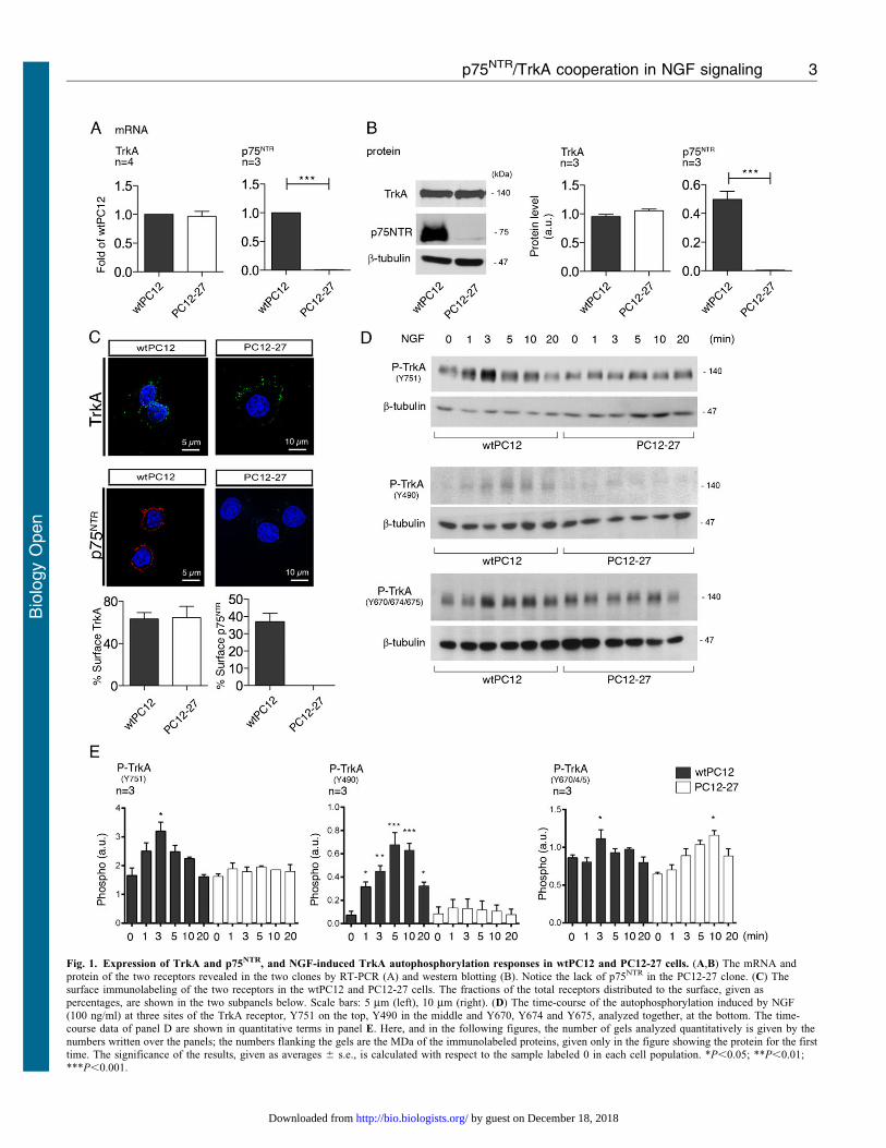
Fig. 1. Expression of TrkA and p75NTR, and NGF-induced TrkA autophosphorylation responses in wtPC12 and PC12-27 cells. (A,B) The mRNA andprotein of the two receptors revealed in the two clones by RT-PCR (A) and western blotting (B). Notice the lack of p75NTR in the PC12-27 clone. (C) Thesurface immunolabeling of the two receptors in the wtPC12 and PC12-27 cells. The fractions of the total receptors distributed to the surface, given aspercentages, are shown in the two subpanels below. Scale bars: 5 mm (left), 10 mm (right). (D) The time-course of the autophosphorylation induced by NGF(100 ng/ml) at three sites of the TrkA receptor, Y751 on the top, Y490 in the middle and Y670, Y674 and Y675, analyzed together, at the bottom. The time-course data of panel D are shown in quantitative terms in panel E. Here, and in the following figures, the number of gels analyzed quantitatively is given by the
numbers written over the panels; the numbers flanking the gels are the MDa of the immunolabeled proteins, given only in the figure showing the protein for the firsttime. The significance of the results, given as averages 6 s.e., is calculated with respect to the sample labeled 0 in each cell population. *P,0.05; **P,0.01;***P,0.001.
p75NTR/TrkA cooperation in NGF signaling 3
Bio
logy
Open
by guest on December 18, 2018http://bio.biologists.org/Downloaded from
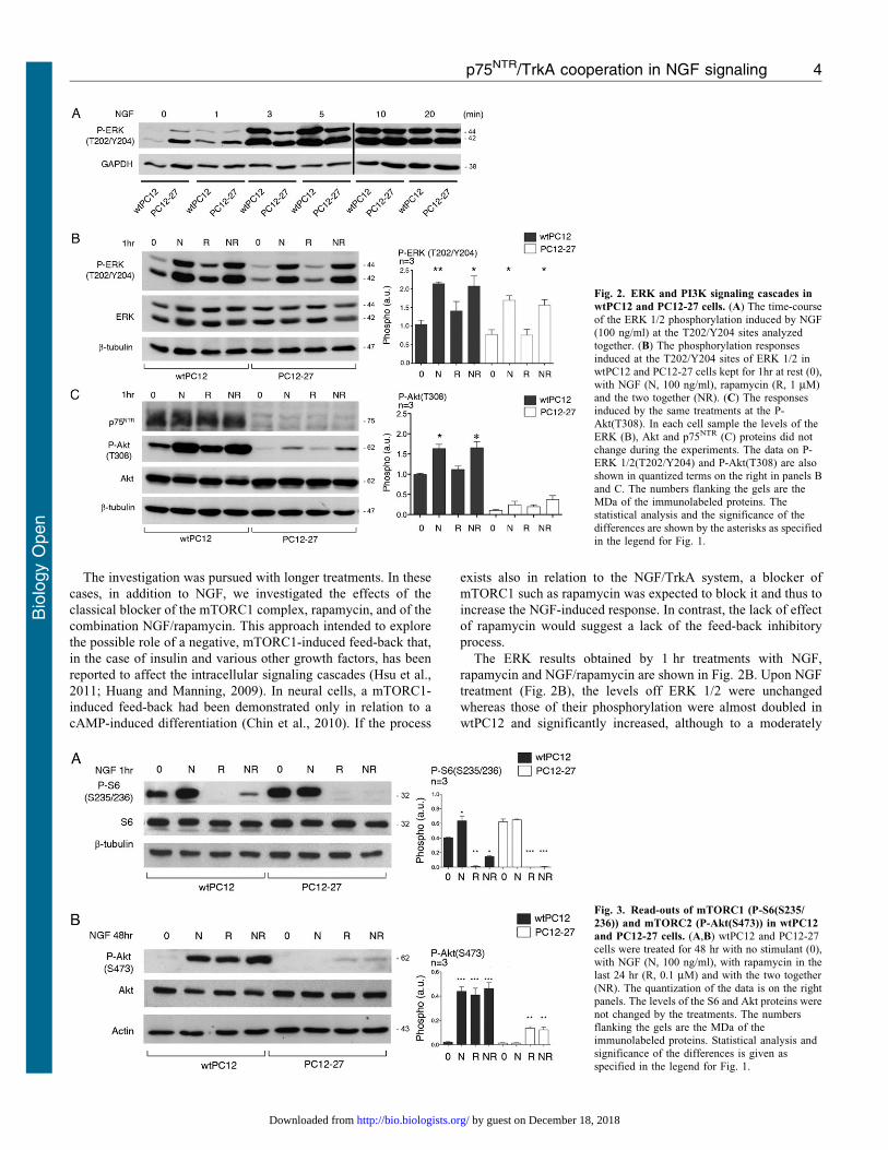
The investigation was pursued with longer treatments. In these
cases, in addition to NGF, we investigated the effects of the
classical blocker of the mTORC1 complex, rapamycin, and of the
combination NGF/rapamycin. This approach intended to explore
the possible role of a negative, mTORC1-induced feed-back that,
in the case of insulin and various other growth factors, has been
reported to affect the intracellular signaling cascades (Hsu et al.,
2011; Huang and Manning, 2009). In neural cells, a mTORC1-
induced feed-back had been demonstrated only in relation to a
cAMP-induced differentiation (Chin et al., 2010). If the process
exists also in relation to the NGF/TrkA system, a blocker of
mTORC1 such as rapamycin was expected to block it and thus to
increase the NGF-induced response. In contrast, the lack of effect
of rapamycin would suggest a lack of the feed-back inhibitory
process.
The ERK results obtained by 1 hr treatments with NGF,
rapamycin and NGF/rapamycin are shown in Fig. 2B. Upon NGF
treatment (Fig. 2B), the levels off ERK 1/2 were unchanged
whereas those of their phosphorylation were almost doubled in
wtPC12 and significantly increased, although to a moderately
Fig. 2. ERK and PI3K signaling cascades in
wtPC12 and PC12-27 cells. (A) The time-courseof the ERK 1/2 phosphorylation induced by NGF(100 ng/ml) at the T202/Y204 sites analyzedtogether. (B) The phosphorylation responses
induced at the T202/Y204 sites of ERK 1/2 inwtPC12 and PC12-27 cells kept for 1hr at rest (0),with NGF (N, 100 ng/ml), rapamycin (R, 1 mM)and the two together (NR). (C) The responsesinduced by the same treatments at the P-Akt(T308). In each cell sample the levels of the
ERK (B), Akt and p75NTR (C) proteins did notchange during the experiments. The data on P-ERK 1/2(T202/Y204) and P-Akt(T308) are alsoshown in quantized terms on the right in panels Band C. The numbers flanking the gels are theMDa of the immunolabeled proteins. Thestatistical analysis and the significance of the
differences are shown by the asterisks as specifiedin the legend for Fig. 1.
Fig. 3. Read-outs of mTORC1 (P-S6(S235/
236)) and mTORC2 (P-Akt(S473)) in wtPC12
and PC12-27 cells. (A,B) wtPC12 and PC12-27cells were treated for 48 hr with no stimulant (0),with NGF (N, 100 ng/ml), with rapamycin in thelast 24 hr (R, 0.1 mM) and with the two together(NR). The quantization of the data is on the rightpanels. The levels of the S6 and Akt proteins were
not changed by the treatments. The numbersflanking the gels are the MDa of theimmunolabeled proteins. Statistical analysis andsignificance of the differences is given asspecified in the legend for Fig. 1.
p75NTR/TrkA cooperation in NGF signaling 4
Bio
logy
Open
by guest on December 18, 2018http://bio.biologists.org/Downloaded from
lower extent, in PC12-27 cells. In contrast, both the resting andthe NGF-induced levels of ERK 1/2, phosphorylation appeared
unchanged in both clones upon treatment with rapamycin(Fig. 2B).
Also the levels of Akt were similar in the wtPC12 and PC12-27
cells, with no changes induced by the various treatments withNGF and rapamycin investigated (Fig. 2C). As far as thephosphorylations, that of Akt(T308), indicative of the PI3K
cascade, increased slowly in the wtPC12 cells, whereas in thePC12-27 cells it remained apparently unchanged during the first20 min of NGF treatment (data not shown). One hr (Fig. 2C) or
longer (up to 48 hr, data not shown) treatment with NGF inducedin wtPC12 cells significant increases of the Akt(T308)phosphorylation. Rapamycin alone, administered for 1 or 24 hr,modified neither the basal nor the NGF-induced Akt(T308)
phosphorylation of wtPC12 (Fig. 2C and data not shown). In thePC12-27 cells the basal phosphorylation of Akt(T308) was muchlower than that of wtPC12. NGF and, to a lower extent, also
rapamycin (administered alone or together for 1 (Fig. 2C) or24 hr (data not shown)) did apparently induce some increases ofP-Akt(T308) which however remained statistically non
significant (Fig. 2C).
Two targets of Akt, TSC2(S939) and GSK3b(S9), exhibitedphosphorylation patterns different from those of Akt(T308).
Specifically, the increases in the wtPC12 cells were smaller whilethose induced by rapamycin were larger than those of Akt(T308)(compare supplementary material Fig. S1 to Fig. 2C). In the
PC12-27 cells, the resting phosphorylation of TSC2(S939) andGSK3b(S9) was low. In these cells no significant increase wasinduced by NGF. In contrast, rapamycin induced considerableincreases (supplementary material Fig. S1). Taken together with
the data of Fig. 2C, the data of supplementary material Fig. S1Asuggest that some mTORC1-induced feed-back inhibition of thePI3K cascade may operate in PC12-27. In wtPC12 cells,
however, no sign of the feed-back was appreciable.
mTORC1 and mTORC2
Our previous studies had shown mTORC1 to be moderately moreactive, and mTORC2 much less active in the high REST PC12-27
cells compared to the wtPC12 cells (Tomasoni et al., 2011). Inorder to investigate the possible involvement in these differencesof the NGF receptor signaling and of its mTORC1-dependentfeed-back inhibition, we analyzed the direct read-outs of
mTORC1 and mTORC2, P-S6(S235/236) and P-Akt(S473),respectively. The latter phosphorylation, considerable only inwtPC12, was completely dissipated by an administration of the
PI3K inhibitor, wortmannin, during the last 10 min incubation(supplementary material Fig. S1B). This finding excludes the P-Akt(S473) phosphorylation to be due also to kinases independent
of the PI3K cascade, such as IKBE (Guo et al., 2011).
The expression levels of the mTORC1 target, S6, were similarin the two clones. Its basal S235/236 phosphorylation,
moderately higher in the PC12-27 cells, was increased upon 1and 48 hr treatment with NGF, however only in the wtPC12clone (Fig. 3A and data not shown). The mTORC1 blocker
rapamycin, administered alone or together with NGF for 1 to24 hr, dissipated completely the basal and largely also the NGF-stimulated phosphorylation of the mTORC1 read-out, S6(S235/
236), as expected (Fig. 3A and data not shown). We concludethat the moderate differences of mTORC1 activity betweenwtPC12 and PC12-27 cells, already reported in the two resting
clones (Tomasoni et al., 2011), were largely independent of their
different NGF signaling.
Fig. 2C had already shown the levels of Akt to be similar inthe two clones. These levels were unchanged by 24–72 hrtreatment with NGF, 24 hr treatment with rapamycin or by NGF
associated to rapamycin during the last 24 hr (Fig. 3B and datanot shown). In terms of phosphorylation, the results with thedirect mTORC2 read-out, Akt(S473) were quite different in
wtPC12 and PC12-27 cells. In the wtPC12, 1 to 24 hr treatmentwith NGF induced increases of 15–20-fold (Fig. 3B and data notshown). Treatment of wtPC12 with rapamycin induced increases
similar to those induced by NGF. The two increases, however,were not additive when the cells were exposed to the combinedNGF/rapamycin treatment (Fig. 3B and data not shown). In thePC12-27 clone, the resting phosphorylation of Akt(S473),
distinctly lower than that of wtPC12, was not changedsignificantly by NGF. In contrast rapamycin did increase the P-Akt(S473) by over 5-fold. The combination with NGF did not
change the increased P-Akt(S473) induced by rapamycin(Fig. 3B and data not shown).
In order to investigate the possible involvement of the p75NTR
receptor in the regulation of the mTORC2 activity we carried out
experiments in subclones of wtPC12 cells in which theexpression of the receptor had been downregulated by thestable transfection of a specific miRNA. Supplementary material
Fig. S2 shows results with a wtPC12 subclone with p75NTR levelreduced to approximately 20%, i.e. with a large decrease whichhowever was lower than that of PC12-27 cells, where the receptor
is inappreciable. The miRNA transfection did not modify thegeneral phenotype of the wtPC12 cells, that remained largelyspherical, different from the flat shape of the PC12-27 cells
(Tomasoni et al., 2011). Likewise, the P-Akt(T308) and the P-GSK3b(S9) were unchanged. In contrast, the Akt(S473)phosphorylation was decreased of 35%, suggesting themTORC2 activity to be reduced (supplementary material
Fig. S2). In parallel experiments, the key role of the PI3Kcascade in the control of mTORC2 was confirmed by the use ofthe PI3K inhibitor wortmannin. In both the wtPC12 and PC12-27
clones treated with NGF for 48 hr, treatment with the drug(0.3 mM) for the last 10 min attenuated considerably the P-TSC2(S939) and P-GSK3b(S9) and eliminates completely the
P-Akt(S473) phosphorylation (data not shown). Additionalsubclones, isolated in parallel to the one shown insupplementary material Fig. S2, exhibited lower downregulations
of the p75NTR and lower decreases of P-Akt(S473). Summing up,the results confirm that p75NTR has a role in the control ofmTORC2 activity, revealed by the direct read-out Akt(S473).The findings with PC12-27 cells strengthen, at the mTORC2
level, the non-significant rapamycin results of the PI3K cascade-dependent phosphorylation of Akt(T308) illustrated in Fig. 2C.In these cells the low mTORC2 activity, unaffected by NGF,
appears to be affected by the mTORC1-dependent feed-backinhibition process inhibited by the drug.
NGF signaling in wtPC12 and PC12-27 cells: role of p75NTR
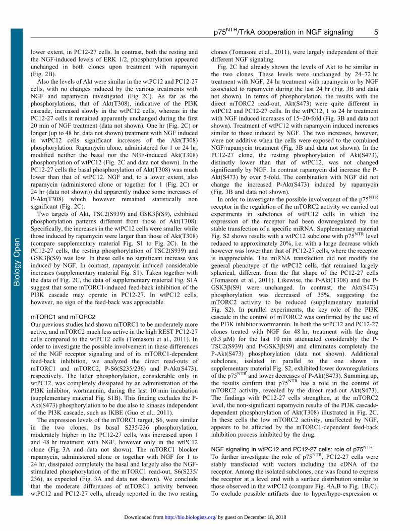
To further investigate the role of p75NTR, PC12-27 cells werestably transfected with vectors including the cDNA of thereceptor. Among the isolated subclones, one was found to express
the receptor at a level and with a surface distribution similar tothose observed in the wtPC12 (compare Fig. 4A,B to Fig. 1B,C).To exclude possible artifacts due to hyper/hypo-expression or
p75NTR/TrkA cooperation in NGF signaling 5
Bio
logy
Open
by guest on December 18, 2018http://bio.biologists.org/Downloaded from
altered distribution of the receptor, this subclone (labeled PC12-
27/p75NTR) was selected for subsequent studies, using as control
a PC12-27 subclone transfected with the empty vector (PC12-27/
Ctrl).
During the first 20 min treatment of PC12-27/p75NTR cells
with NGF, the Y751 phosphorylation of TrkA increased
markedly, similar to the wtPC12 cells, whereas that PC12-27/
Ctrl cells resembled that of the non-transfected PC12-27 cells, i.e.
it did not change significantly (compare Fig. 4C to Fig. 1D). At
the Y490 site the differences of phosphorylation observed
between the cells transfected with and without p75NTR were
even larger. In the PC12-27/Ctrl cells this phosphorylation
remained almost inappreciable, as in the non-transfected PC12-
27 cells, whereas in the PC12-27/p75NTR cells it increased
significantly and rapidly upon NGF addition, reaching a
maximum at 5–10 min, as in the wtPC12 (compare Fig. 4D to
Fig. 1D).
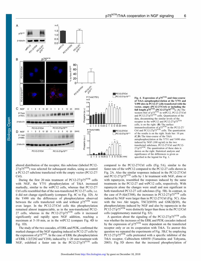
The study of the two cascades, of ERK and PI3K, confirmed the
marked changes of the NGF signaling induced in PC12-27 cells by
the expression of p75NTR. In the case of ERK the phosphorylation
of ERK 1/2(T202 and Y204), induced by 1–20 min treatment with
NGF, exhibited a faster rate in the PC12-27/p75NTR cells
compared to the PC12-27/Ctrl cells (Fig. 5A), similar to the
faster rate of the wtPC12 compared to the PC12-27 cells shown in
Fig. 2A. Also the similar responses induced in the PC12-27/Ctrl
and PC12-27/p75NTR cells by 1 hr treatment with NGF, alone or
with rapamycin, resembled the responses induced by the same
treatments in the PC12-27 and wtPC12 cells, respectively. With
rapamycin alone the changes were small and non significant in
both transfected PC12-27 cell subclones (Fig. 5B). In contrast, in
the case of P-Akt(T308), the increases in PC12-27/p75NTR cells
induced by NGF were larger than in PC12-27/Ctrl (Fig. 5C). Also
with the two Akt targets, TSC2(S939) and GSK3b(S9), the
phosphorylations induced by NGF and also by rapamycin in the
PC12-27/p75NTR were distinctly larger than those in the PC12/Ctrl
cells (supplementary material Fig. S3).
A question about the signaling of the PC12-27/p75NTR cells
was whether the increases of the ERK and PI3K cascades induced
by the expression of p75NTR were dependent on the transfected
receptor only or on its cooperation with TrkA. To answer this
question we repeated the experiments of Fig. 5B,C by employing
PC12-27/p75NTR cells pretreated with a specific inhibitor of the
TrkA receptor, Calbiochem 648450 (Yamashita and Tohyama,
2003). Fig. 5D shows that the increased phosphorylation of
Fig. 4. Expression of p75NTR and time-course
of TrkA autophosphorylation at the Y751 and
Y490 sites in PC12-27 cells transfected with the
vector, empty (PC12-27/Ctrl) or including the
full length p75NTR (PC12-27/p75NTR). (A) Thewestern blot of p75NTR in wtPC12, PC12-27/Ctrland PC12-27/p75NTR cells. Quantization of the
data, documenting the similar levels of thereceptor in the wtPC12 and PC12-27/p75NTR
cells, is on the right. (B) The surfaceimmunolocalization of p75NTR in the PC12-27/Ctrl and PC12-27p75NTR cells. The quantizationof the results is on the right. Scale bar: 10 mm.(C,D) The time-course of the TrkA
autophosphorylation at the Y751 and Y490 sitesinduced by NGF (100 ng/ml) in the twotransfected subclones, PC12-27/Ctrl and PC12-27/p75NTR. The quantization of these data isshown on the right. Statistical analysis andsignificance of the differences is given as
specified in the legend for Fig. 1.
p75NTR/TrkA cooperation in NGF signaling 6
Bio
logy
Open
by guest on December 18, 2018http://bio.biologists.org/Downloaded from
Akt(T308) induced by NGF was prevented by the pretreatment of
the cells with the drug. In order to be generated, the NGF-induced
signal requires therefore the two receptors, TrkA and p75NTR, to
be activated concomitantly. The cooperation with p75NTR,
however, does not seem to occur only with TrkA. In fact, the
responses triggered by rapamycin, alone or together with NGF,
were apparently unchanged by the pretreatment of the PC12-27/
p75NTR cells with the TrkA inhibitor (Fig. 5D). In PC12 cells the
site of action of the mTORC1-dependent feed-back inhibition,
the process blocked by rapamycin, is unknown. In other cell
types, however, this site has been proposed to coincide with the
insulin receptor substrate 1 (IRS1) (Tremblay et al., 2007). This
or another post-receptor site may therefore operate in the
cooperation of the feed-back inhibition with p75NTR. In
conclusion, the re-establishment in the PC12-27 of a TrkA/
p75NTR ratio analogous to the ratio in wtPC12 was found to
rescue the NGF signaling from a partially inactive to a fully
active state.
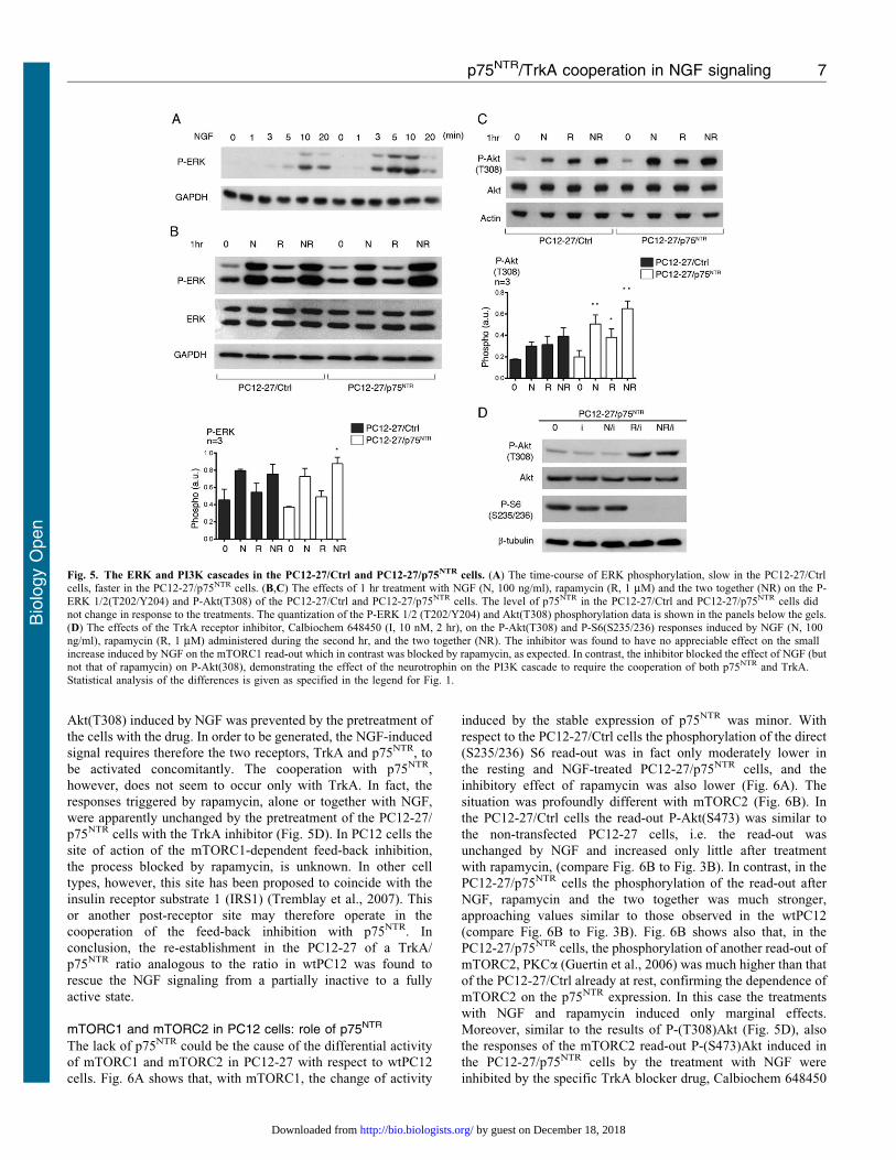
mTORC1 and mTORC2 in PC12 cells: role of p75NTR
The lack of p75NTR could be the cause of the differential activity
of mTORC1 and mTORC2 in PC12-27 with respect to wtPC12
cells. Fig. 6A shows that, with mTORC1, the change of activity
induced by the stable expression of p75NTR was minor. With
respect to the PC12-27/Ctrl cells the phosphorylation of the direct
(S235/236) S6 read-out was in fact only moderately lower in
the resting and NGF-treated PC12-27/p75NTR cells, and the
inhibitory effect of rapamycin was also lower (Fig. 6A). The
situation was profoundly different with mTORC2 (Fig. 6B). In
the PC12-27/Ctrl cells the read-out P-Akt(S473) was similar to
the non-transfected PC12-27 cells, i.e. the read-out was
unchanged by NGF and increased only little after treatment
with rapamycin, (compare Fig. 6B to Fig. 3B). In contrast, in the
PC12-27/p75NTR cells the phosphorylation of the read-out after
NGF, rapamycin and the two together was much stronger,
approaching values similar to those observed in the wtPC12
(compare Fig. 6B to Fig. 3B). Fig. 6B shows also that, in the
PC12-27/p75NTR cells, the phosphorylation of another read-out of
mTORC2, PKCa (Guertin et al., 2006) was much higher than that
of the PC12-27/Ctrl already at rest, confirming the dependence of
mTORC2 on the p75NTR expression. In this case the treatments
with NGF and rapamycin induced only marginal effects.
Moreover, similar to the results of P-(T308)Akt (Fig. 5D), also
the responses of the mTORC2 read-out P-(S473)Akt induced in
the PC12-27/p75NTR cells by the treatment with NGF were
inhibited by the specific TrkA blocker drug, Calbiochem 648450
Fig. 5. The ERK and PI3K cascades in the PC12-27/Ctrl and PC12-27/p75NTR cells. (A) The time-course of ERK phosphorylation, slow in the PC12-27/Ctrlcells, faster in the PC12-27/p75NTR cells. (B,C) The effects of 1 hr treatment with NGF (N, 100 ng/ml), rapamycin (R, 1 mM) and the two together (NR) on the P-
ERK 1/2(T202/Y204) and P-Akt(T308) of the PC12-27/Ctrl and PC12-27/p75NTR cells. The level of p75NTR in the PC12-27/Ctrl and PC12-27/p75NTR cells didnot change in response to the treatments. The quantization of the P-ERK 1/2 (T202/Y204) and Akt(T308) phosphorylation data is shown in the panels below the gels.(D) The effects of the TrkA receptor inhibitor, Calbiochem 648450 (I, 10 nM, 2 hr), on the P-Akt(T308) and P-S6(S235/236) responses induced by NGF (N, 100ng/ml), rapamycin (R, 1 mM) administered during the second hr, and the two together (NR). The inhibitor was found to have no appreciable effect on the smallincrease induced by NGF on the mTORC1 read-out which in contrast was blocked by rapamycin, as expected. In contrast, the inhibitor blocked the effect of NGF (butnot that of rapamycin) on P-Akt(308), demonstrating the effect of the neurotrophin on the PI3K cascade to require the cooperation of both p75NTR and TrkA.
Statistical analysis of the differences is given as specified in the legend for Fig. 1.
p75NTR/TrkA cooperation in NGF signaling 7
Bio
logy
Open
by guest on December 18, 2018http://bio.biologists.org/Downloaded from
(Yamashita and Tohyama, 2003). In contrast, the responses to
rapamycin, acting by removal of the mTORC1 feed-back
inhibition, were not (Fig. 6C). Together with the data of
Fig. 5D these results confirm the cooperation between the
TrkA and the p75NTR signaling to be necessary for the NGF-
induced mTORC2 activation. In contrast the cooperation appears
unnecessary when the signal is triggered not at the level of TrkA
but (by rapamycin) at a post-receptor site. Summing up, the
expression of p75NTR in the PC12-27 cells appears to modify
both the signaling of NGF and the activity of mTORC2, bringing
them to levels approaching those of wtPC12.
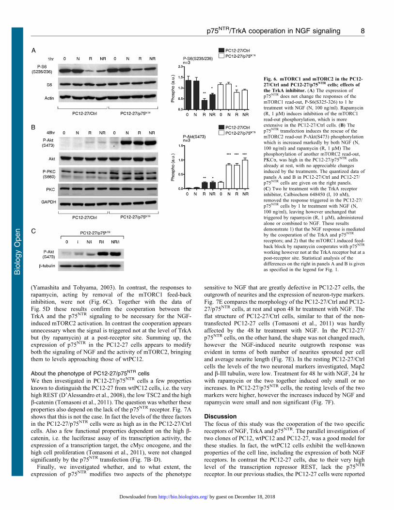
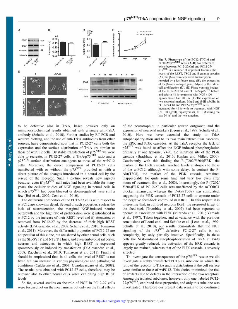
About the phenotype of PC12-27/p75NTR cells
We then investigated in PC12-27/p75NTR cells a few properties
known to distinguish the PC12-27 from wtPC12 cells, i.e. the very
high REST (D’Alessandro et al., 2008), the low TSC2 and the high
b-catenin (Tomasoni et al., 2011). The question was whether these
properties also depend on the lack of the p75NTR receptor. Fig. 7A
shows that this is not the case. In fact the levels of the three factors
in the PC12-27/p75NTR cells were as high as in the PC12-27/Ctrl
cells. Also a few functional properties dependent on the high b-
catenin, i.e. the luciferase assay of its transcription activity, the
expression of a transcription target, the cMyc oncogene, and the
high cell proliferation (Tomasoni et al., 2011), were not changed
significantly by the p75NTR transfection (Fig. 7B–D).
Finally, we investigated whether, and to what extent, the
expression of p75NTR modifies two aspects of the phenotype
sensitive to NGF that are greatly defective in PC12-27 cells, the
outgrowth of neurites and the expression of neuron-type markers.
Fig. 7E compares the morphology of the PC12-27/Ctrl and PC12-
27/p75NTR cells, at rest and upon 48 hr treatment with NGF. The
flat structure of PC12-27/Ctrl cells, similar to that of the non-
transfected PC12-27 cells (Tomasoni et al., 2011) was hardly
affected by the 48 hr treatment with NGF. In the PC12-27/
p75NTR cells, on the other hand, the shape was not changed much,
however the NGF-induced neurite outgrowth response was
evident in terms of both number of neurites sprouted per cell
and average neurite length (Fig. 7E). In the resting PC12-27/Ctrl
cells the levels of the two neuronal markers investigated, Map2
and b-III tubulin, were low. Treatment for 48 hr with NGF, 24 hr
with rapamycin or the two together induced only small or no
increases. In PC12-27/p75NTR cells, the resting levels of the two
markers were higher, however the increases induced by NGF and
rapamycin were small and non significant (Fig. 7F).
DiscussionThe focus of this study was the cooperation of the two specific
receptors of NGF, TrkA and p75NTR. The parallel investigation of
two clones of PC12, wtPC12 and PC12-27, was a good model for
these studies. In fact, the wtPC12 cells exhibit the well-known
properties of the cell line, including the expression of both NGF
receptors. In contrast the PC12-27 cells, due to their very high
level of the transcription repressor REST, lack the p75NTR
receptor. In our previous studies, the PC12-27 cells were reported
Fig. 6. mTORC1 and mTORC2 in the PC12-
27/Ctrl and PC12-27/p75NTR cells; effects of
the TrkA inhibitor. (A) The expression ofp75NTR does not change the responses of themTORC1 read-out, P-S6(S325-326) to 1 hrtreatment with NGF (N, 100 ng/ml). Rapamycin
(R, 1 mM) induces inhibition of the mTORC1read-out phosphorylation, which is moreextensive in the PC12-27/Ctrl cells. (B) Thep75NTR transfection induces the rescue of themTORC2 read-out P-Akt(S473) phosphorylationwhich is increased markedly by both NGF (N,100 ng/ml) and rapamycin (R, 1 mM) The
phosphorylation of another mTORC2 read-out,PKCa, was high in the PC12-27/p75NTR cellsalready at rest, with no appreciable changesinduced by the treatments. The quantized data ofpanels A and B in PC12-27/Ctrl and PC12-27/p75NTR cells are given on the right panels.
(C) Two hr treatment with the TrkA receptorinhibitor, Calbiochem 648450 (I, 10 nM),removed the response triggered in the PC12-27/p75NTR cells by 1 hr treatment with NGF (N,100 ng/ml), leaving however unchanged thattriggered by rapamycin (R, 1 mM), administeredalone or combined to NGF. These results
demonstrate 1) that the NGF response is mediatedby the cooperation of the TrkA and p75NTR
receptors; and 2) that the mTORC1.induced feed-back block by rapamycin cooperates with p75NTR
working however not at the TrkA receptor but at apost-receptor site. Statistical analysis of the
differences on the right in panels A and B is givenas specified in the legend for Fig. 1.
p75NTR/TrkA cooperation in NGF signaling 8
Bio
logy
Open
by guest on December 18, 2018http://bio.biologists.org/Downloaded from
to be defective also in TrkA, based however only on
immunocytochemical results obtained with a single anti-TrkA
antibody (Schulte et al., 2010). Further studies by RT-PCR and
western blotting, and the use of anti-TrkA antibodies from other
sources, have demonstrated now that in PC12-27 cells both the
expression and the surface distribution of TrkA are similar to
those of wtPC12 cells. By stable transfection of p75NTR we were
able to recreate, in PC12-27 cells, a TrkA/p75NTR ratio and a
p75NTR surface distribution analogous to those of the wtPC12
cells. Moreover, the direct comparison of PC12-27 cells
transfected with or without the p75NTR provided us with a
direct picture of the changes introduced in a neural cell by the
rescue of the receptor. Such a picture reveals new aspects
because, even if p75NTR null mice had been available for many
years, the cellular studies of NGF signaling in neural cells in
which p75NTR had been blocked or downregulated were still a
few (Bui et al., 2002; Ceni et al., 2010).
The differential properties of the PC12-27 cells with respect to
wtPC12 are known in detail. Several of such properties, such as the
lack of neurosecretion, the marginal NGF-induced neurite
outgrowth and the high rate of proliferation were i) introduced in
wtPC12 by the increase of their REST level and ii) attenuated or
removed from PC12-27 by the decrease of their high REST
activity (D’Alessandro et al., 2008; Schulte et al., 2010; Tomasoni
et al., 2011). Moreover, the differential properties of PC12-27 are
not peculiar of this clone, but are shared by other neural cells, such
as the SH-SY5Y and NT2/D1 lines, and even embryonal rat cortex
neurons and astrocytes, in which high REST is expressed
spontaneously or induced by transfection (D’Alessandro et al.,
2008; Racchetti et al., 2010; Tomasoni et al., 2011). Finally it
should be emphasized that, in all cells, the level of REST is not
fixed but can increase in various physiological and pathological
conditions (Calderone et al., 2003; Guardavaccaro et al., 2008).
The results now obtained with PC12-27 cells, therefore, may be
relevant also to other neural cells when exhibiting high REST
levels.
So far, several studies on the role of NGF in PC12-27 cells
were focused not on the mechanisms but only on the final effects
of the neurotrophin, in particular neurite outgrowth and the
expression of neuronal markers (Leoni et al., 1999; Schulte et al.,
2010). Here we have extended the study to TrkA
autophosphorylation and to its two main intracellular cascades,
the ERK and PI3K cascades. At the TrkA receptor the lack of
p75NTR was found to affect the NGF-induced phosphorylation
primarily at one tyrosine, Y490, the initiation site of the PI3K
cascade (Bradshaw et al., 2013; Kaplan and Miller, 2000).
Consistently with this finding the P-(T202/Y204)ERK, the
marker of the ERK cascade, reached levels analogous to those
of the wtPC12, although with some delay. In contrast the P-
Akt(T308), the marker of the PI3K cascade, remained
inappreciable for quite some time and very low even after
hours of treatment (Ito et al., 2003). Interestingly, the P-(T202/
Y204)ERK of PC12-27 cells was unaffected by the mTORC1
blocker rapamycin, whereas the P-Akt(T308) was stimulated,
suggesting the PI3K cascade of PC12-27 cells to operate under
the negative feed-back control of mTORC1. In this respect it is
interesting that, in cultured neurons IRS1, the proposed target of
the feed-back (Tremblay et al., 2007) had been reported to
operate in association with PI3K (Miranda et al., 2001; Yamada
et al., 1997). Taken together, and at variance with the previous
conclusions of Leoni et al. and Schulte et al. (Leoni et al., 1999;
Schulte et al., 2010), our results demonstrate that the NGF
signaling of the p75NTR-defective PC12-27 cells is not
completely, by only partially inactive. Specifically, in these
cells the NGF-induced autophosphorylation of TrkA at Y490
appears greatly reduced, the activation of the ERK cascade is
largely maintained, whereas that of the PI3K cascade is severely
affected.
To investigate the consequences of the p75NTR rescue we did
investigate a stably transfected PC12-27 subclone in which the
ratio of the receptor to TrkA and its distribution at the cell surface
were similar to those of wtPC12. This choice minimized the risk
of artifacts due to defects in the interaction of the two receptors.
Among the isolated subclones, however, only one, labeled PC12-
27/p75NTR, exhibited these properties, and only this subclone was
investigated. Therefore our present data remain to be confirmed
Fig. 7. Phenotype of the PC12-27/Ctrl and
PC12-27/p75NTR cells. (A–D) No differenceexists between PC12-27/Ctrl and PC12-27/
p75NTR in a number of important features: thelevels of the REST, TSC2 and b-catenin proteins(A); the b-catenin-dependent transcriptionrevealed by a luciferase assay (B); the expressionof the b-catenin-target gene, cMyc (C); the rate ofcell proliferation (D). (E) Phase contrast imagesof the PC12-27/Ctrl and PC12-27/p75NTR before
and after a 48 hr treatment with NGF (100ng/ml). Scale bar: 20 mm. (F) The expression oftwo neuronal markers, Map2 and b-III tubulin, inPC12-27/Ctrl and PC12-27/p75NTR cellsincubated for 48 hr with no treatment, with NGF(N, 100 ng/ml), rapamycin (R, 0.1 mM during the
last 24 hr) and the two together.
p75NTR/TrkA cooperation in NGF signaling 9
Bio
logy
Open
by guest on December 18, 2018http://bio.biologists.org/Downloaded from
in other subclones. Nevertheless, the results obtained appear
promising because a number of the NGF-induced properties
typical of wtPC12, i.e. the active PI3K cascade, neurite
outgrowth and the acquisition of neuronal markers, were
rescued in the PC12-27 by the transfected p75NTR working in
cooperation with TrkA. Other differential properties of the PC12-
27 clone, however, were not modified by the transfection of
p75NTR, suggesting their independence on the NGF signaling.
These properties include the high level of REST, which is
spontaneous in the clone; the low level of TSC2; the high level
and the transcription activity of b-catenin. High REST, low TSC2
and high b-catenin are the three coordinate factors that account
for the high proliferation rate of PC12-27 cells (Tomasoni et al.,
2011), a property that was also unaffected by the transfection of
p75NTR.
Our results provided some evidence about the links between
NGF signaling and the mTORCs. The stimulation of the PI3K
cascade by rapamycin, visible however only in PC12-27 cells,
concurs with the previous data of Chin et al. to suggest the
existence of an mTORC1-dependent feed-back inhibition of
signaling also in neural cells (Chin et al., 2010). The data on
mTORC2 were obtained by studying its direct read-outs, P-
Akt(S473) and PKCa. The transfection of p75NTR was found to
induce marked increases of the mTORC2 activity. This effect
was not a consequence of p75NTR only but required the
cooperation of TrkA, as shown by its disappearance induced by
the specific inhibitor of that receptor, Calbiochem 648450. The
activity of mTORC2, critical for both neural and non neural cells
(Oh and Jacinto, 2011), takes place under the control of two main
mechanisms, dependent one on the PI3K cascade, the other on
the TSC complex (Dalle Pezze et al., 2012; Guertin et al., 2006;
Huang et al., 2008; Shanmugasundaram et al., 2013). In the non-
transfected PC12-27 cells the TSC complex is little active due to
the reduced level of TSC2 (Tomasoni et al., 2011). The TSC
complex could therefore have a role in the low activity of
mTORC2. However, the TSC2 level did not increase in the
PC12-27/p75NTR cells. Therefore the mTORC2 rescue observed
in the latter cells might be due mostly to the PI3K cascade which
is also rescued by the transfection. These data are new. Because
of their relevance they deserve to be further investigated by
additional approaches not yet employed, including the
downregulation of specific components of the mTORC2complex such as Rictor.
mTORC2 is known to play important roles in many, andpossibly all types of cells. In non-neural cells, and especially in
their tumors, it can contribute to proliferation, migration,invasion, metastases and other functions (Kim et al., 2011;Shanmugasundaram et al., 2013; for a review, see Oh and
Jacinto, 2011). In some non-neural cells and tumors, such asmyoblasts, lymphocytes B and T and leukemias, however,
mTORC2 has been shown to induce primarily differentiation(Lazorchak and Su, 2011; Lee et al., 2012; Matheny et al., 2012;
Shu and Houghton, 2009). In the case of neural cells anmTORC2-dependent differentiation, induced via the inhibition of
FoxO3a (Wang et al., 2013), is the only effect reported so far(Carson et al., 2013; Dimitroff et al., 2012; Urbanska et al.,2012). Our results seem to add a new example along this line.
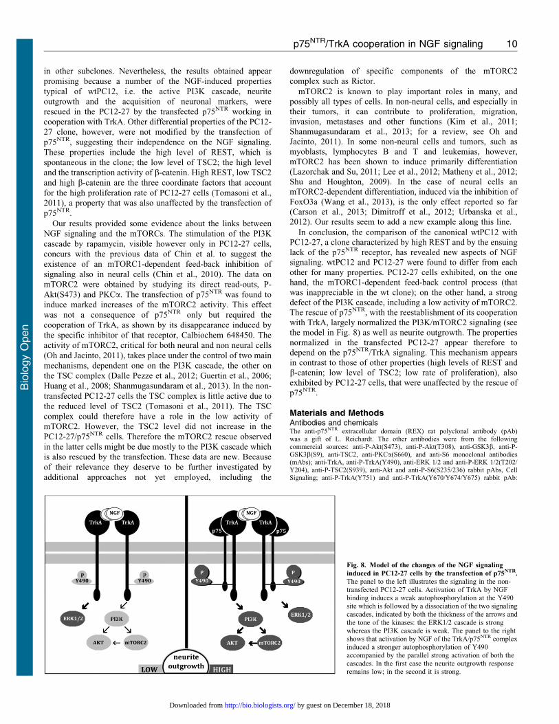
In conclusion, the comparison of the canonical wtPC12 withPC12-27, a clone characterized by high REST and by the ensuing
lack of the p75NTR receptor, has revealed new aspects of NGFsignaling. wtPC12 and PC12-27 were found to differ from each
other for many properties. PC12-27 cells exhibited, on the onehand, the mTORC1-dependent feed-back control process (that
was inappreciable in the wt clone); on the other hand, a strongdefect of the PI3K cascade, including a low activity of mTORC2.The rescue of p75NTR, with the reestablishment of its cooperation
with TrkA, largely normalized the PI3K/mTORC2 signaling (seethe model in Fig. 8) as well as neurite outgrowth. The properties
normalized in the transfected PC12-27 appear therefore todepend on the p75NTR/TrkA signaling. This mechanism appears
in contrast to those of other properties (high levels of REST andb-catenin; low level of TSC2; low rate of proliferation), also
exhibited by PC12-27 cells, that were unaffected by the rescue ofp75NTR.
Materials and MethodsAntibodies and chemicalsThe anti-p75NTR extracellular domain (REX) rat polyclonal antibody (pAb)was a gift of L. Reichardt. The other antibodies were from the followingcommercial sources: anti-P-Akt(S473), anti-P-Akt(T308), anti-GSK3b, anti-P-GSK3b(S9), anti-TSC2, anti-PKCa(S660), and anti-S6 monoclonal antibodies(mAbs); anti-TrkA, anti-P-TrkA(Y490), anti-ERK 1/2 and anti-P-ERK 1/2(T202/Y204), anti-P-TSC2(S939), anti-Akt and anti-P-S6(S235/236) rabbit pAbs, CellSignaling; anti-P-TrkA(Y751) and anti-P-TrkA(Y670/Y674/Y675) rabbit pAb:
Fig. 8. Model of the changes of the NGF signaling
induced in PC12-27 cells by the transfection of p75NTR.
The panel to the left illustrates the signaling in the non-transfected PC12-27 cells. Activation of TrkA by NGFbinding induces a weak autophosphorylation at the Y490site which is followed by a dissociation of the two signalingcascades, indicated by both the thickness of the arrows and
the tone of the kinases: the ERK1/2 cascade is strongwhereas the PI3K cascade is weak. The panel to the rightshows that activation by NGF of the TrkA/p75NTR complexinduced a stronger autophosphorylation of Y490accompanied by the parallel strong activation of both thecascades. In the first case the neurite outgrowth responseremains low; in the second it is strong.
p75NTR/TrkA cooperation in NGF signaling 10
Bio
logy
Open
by guest on December 18, 2018http://bio.biologists.org/Downloaded from
Invitrogen; anti-TrkA C20 extracellular domain rabbit pAb, anti-actin goat pAb: SantaCruz; anti-p75NTR rabbit pAb, Promega; anti-REST rabbit pAb: Upstate; anti-b-tubulin mAb and anti-actin rabbit pAb: Sigma; anti-b-catenin mAb: BD Transduction;anti-Map2 mAb, Millipore; anti-b-III tubulin mAb, Covance; FITC-conjugated andTRITC-conjugated goat anti-rabbit pAbs, and goat anti-mouse IgG subclasses:Southern Biotech.; horseradish peroxidase-conjugated goat anti-mouse and anti-rabbitpAbs: Bio-Rad. NGF was from Alomone; the BCA Protein Assay Kit from Pierce;rapamycin and the TrkA inhibitor 648450 from Calbiochem; the fluorescent DNA-binding probe DAPI, wortmannin and other chemicals, from Sigma–Aldrich.
Cell clonesThe PC12 clones, wtPC12 and PC12-27, were as described (D’Alessandro et al.,2008). Subclones were generated in this work. Cells of clones and subclones weregrown in DMEM medium supplemented with either 1% (starvation) or 10% horseserum, and incubated at 37 C.
TransfectionsStable transfections were as described (D’Alessandro et al., 2008) usinglipofectamine 2000TM (Invitrogen). PC12-27 cells were co-transfected with thepcDNA3.1Hygro(+) together with the plenty-OE-sRFP vector including the fulllength human p75NTR cDNA, to generate cells with stable p75NTR expression;wtPC12 cells received the pRRLsinPPT vector with the p75NTR miRNA, todownregulate the receptor. Both vectors were gifts of P.A. Barker (Ceni et al.,2010). The controls received the vectors only. The transfections were confirmed byqPCR and immunoblotting. For the luciferase assay, nt5iD90bcat/GFP construct(gift of A. Chenn and C.A. Walsh) or the vector backbone pEGFP-1 were used forthe stable transfection of PC12-27/Ctrl and PC12-27/p75NTR cells. Subclones weregrown in complete medium supplemented with 500 mg/ml of G418.
Cell proliferationTo investigate cell proliferation, the clones were plated at 16104/well in a 24 welldish. Medium was replaced every 48 h. Upon 3, 4 and 5 days in culture the cellswere trypsinized and counted after Trypan Blue exclusion.
q-PCRTotal RNA was extracted using RNeasy mini columns (Qiagen), followingmanufacturer’s instructions, and its concentration was determined byspectrophotometry. 1–2 mg of total RNA were used to generate cDNA templatesfor RT-PCR, using the RevertAid First Strand cDNA Synthesis kit in accordancewith the manufacturer’s protocol (from Thermo Scientific). Real-time PCR wasperformed on a LightCyclerH 480 System (FastStart DNA Master SYBR Green Iof Roche Appl. Sci.) according to a standard protocol, using 50 ng templatecDNA. All primers were used at the final concentration of 500 nM. Values werenormalized to the concentration of calmodulin mRNA. Values are expressed aseither fold of wt or PC12-27/Ctrl cells.
Western blottingTotal cell extracts were obtained by suspending cells in lysis buffer containing 1%Triton X-100, 50 mM Tris-HCl, pH 7.5, 250 mM NaCl, 5 mM EDTA, 50 mMNaF, together with protease and phosphatase inhibitors (Xu et al., 2003). Toinvestigate the time-course of TrkA phosphorylation the cells were transferredrapidly on ice and then suspended in lysis buffer containing 10 mM Tris-HClpH 7.8, 150 mM NaCl, 1 mM EDTA, 1% (v/v) Nonidet P40, 1% (w/v) sodiumdeoxycholate, with protease and phosphatase inhibitors. The lysates were clearedby centrifugation at 16.000 g for 20 min at 4 C, and the supernatants wereanalyzed (Takahashi et al., 2011). Proteins were quantified by BCA assay andappropriate amounts (most often 30 mg) were separated by SDS-PAGE. Aftertransfer to nitrocellulose filters, they were immunolabeled as described (Dignamet al., 1983). Photographic development was by chemiluminescence (ECL,Amersham Bioscience or Immobilon substrate, Millipore). Western blot bandswere quantified by the ImageJ program (rsb.info.nih.gov/ij), normalized to markersthat do not change their concentration during the experiment (b-tubulin, actin orGAPDH) immunolabeled in parallel. Data are expressed as arbitrary units (a.u.).
Luciferase assayb-Catenin transcription assay was performed using the Dual-Luciferase reporterassay kit (Promega). The 166 TOPFLASH reporter plasmid (1 mg) (gift of R.T.Moon and H.H. Das Gupta) and 100 ng of SV40-Renilla-luc were cotransfectedusing lipofectamine 2000TM, and luciferase activity was measured 24 hr later,using a luminometer (GloMax Multi Detectionm System of Promega). Data areexpressed as a.u.
Immunofluorescence and bright field microscopyThe immunofluorescence experiments were performed as described (Tomasoniet al., 2011). Specifically, cell monolayers on coverslips were fixed with 4%
formaldehyde for 10 min at room temperature and quenched in 0.1 M glycine, thenprocessed directly or permeabilized for 20 min in PBS containing 0.2% Triton X-100 and 1% bovine serum albumin, and finally immunolabeled for 1 hr with eitheranti-TrkA or anti-p75NTR pAbs, the latter against the whole receptor molecule(C14 and Promega) or against its extracellular domain (C20 and REX), diluted inPBS with 1% BSA. The bound antibodies were stained with FITC-conjugated andTRITC-conjugated goat anti-rabbit pAbs, or goat anti-mouse IgG subclasses. Insome cases nuclei were stained with DAPI. Samples were studied in a PerkinElmerUltraview ERS confocal microscope. Image deconvolution was performed in awide field microscope of the Delta Vision system.
Statistical analysesThe significance of the data was assessed using the two-tailed unpaired t-test andthe Anova test, making reference to the unstimulated samples of both the controlsand the variously stimulated cell preparations. Data shown are means 6 s.e. Thenumber of experiments is specified in the figures or figure legends. P,0.05 isconsidered significantly different. In the figures, ***P,0.001; **P,0.01;*P,0.05.
AcknowledgementsWe thank Ilaria Prada for her generous support, Enrico Ponta for hisparticipation in a few experiments, Johanna Mikulak, Davide Pozzi,Philip Barker, Moses Chao, Louis Reichardt, Luca Muzio, RandallMoon and Anjen Chenn for suggestions and gifts of antibodies,constructs and viral vectors. Supported by Telethon (grantGGGP09066 to J.M.).
Author ContributionsS.N. participated in the design and took care of the execution of theexperiments; R.D’A. was responsible for the approach and initiationof the work; J.M. participated in the design and interpretation of thedata. He also took care of preparing and editing the article.
Competing InterestsThe authors have no competing interests to declare.
ReferencesBallas, N. and Mandel, G. (2005). The many faces of REST oversee epigenetic
programming of neuronal genes. Curr. Opin. Neurobiol. 15, 500-506.
Bandtlow, C. and Dechant, G. (2004). From cell death to neuronal regeneration, effects
of the p75 neurotrophin receptor depend on interactions with partner subunits. Sci.
STKE 2004, pe24.
Barker, P. A. (2004). p75NTR is positively promiscuous: novel partners and new
insights. Neuron 42, 529-533.
Barker, P. A. and Murphy, R. A. (1992). The nerve growth factor receptor: a
multicomponent system that mediates the actions of the neurotrophin family of
proteins. Mol. Cell. Biochem. 110, 1-15.
Bradshaw, R. A., Chalkley, R. J., Biarc, J. and Burlingame, A. L. (2013). Receptortyrosine kinase signaling mechanisms: Devolving TrkA responses with
phosphoproteomics. Adv. Bio. Reg. 53, 87-96.
Brann, A. B., Scott, R., Neuberger, Y., Abulafia, D., Boldin, S., Fainzilber, M. and
Futerman, A. H. (1999). Ceramide signaling downstream of the p75 neurotrophinreceptor mediates the effects of nerve growth factor on outgrowth of cultured
hippocampal neurons. J. Neurosci. 19, 8199-8206.
Bruce, A. W., Donaldson, I. J., Wood, I. C., Yerbury, S. A., Sadowski, M. I.,
Chapman, M., Gottgens, B. and Buckley, N. J. (2004). Genome-wide analysis ofrepressor element 1 silencing transcription factor/neuron-restrictive silencing factor
(REST/NRSF) target genes. Proc. Natl. Acad. Sci. USA 101, 10458-10463.
Bui, N. T., Konig, H. G., Culmsee, C., Bauerbach, E., Poppe, M., Krieglstein, J. and
Prehn, J. H. (2002). p75 neurotrophin receptor is required for constitutive and NGF-
induced survival signalling in PC12 cells and rat hippocampal neurones.J. Neurochem. 81, 594-605.
Calderone, A., Jover, T., Noh, K. M., Tanaka, H., Yokota, H., Lin, Y., Grooms,
S. Y., Regis, R., Bennett, M. V. and Zukin, R. S. (2003). Ischemic insults derepress
the gene silencer REST in neurons destined to die. J. Neurosci. 23, 2112-2121.
Carson, R. P., Fu, C., Winzenburger, P. and Ess, K. C. (2013). Deletion of Rictor in
neural progenitor cells reveals contributions of mTORC2 signaling to tuberous
sclerosis complex. Hum. Mol. Genet. 22, 140-152.
Ceni, C., Kommaddi, R. P., Thomas, R., Vereker, E., Liu, X., McPherson, P. S.,
Ritter, B. and Barker, P. A. (2010). The p75NTR intracellular domain generated by
neurotrophin-induced receptor cleavage potentiates Trk signaling. J. Cell Sci. 123,
2299-2307.
Chin, T. Y., Kao, C. H., Wang, H. Y., Huang, W. P., Ma, K. H. and Chueh,
S. H. (2010). Inhibition of the mammalian target of rapamycin promotes cyclic AMP-
induced differentiation of NG108-15 cells. Autophagy 6, 1139-1156.
p75NTR/TrkA cooperation in NGF signaling 11
Bio
logy
Open
by guest on December 18, 2018http://bio.biologists.org/Downloaded from
D’Alessandro, R., Klajn, A., Stucchi, L., Podini, P., Malosio, M. L. and Meldolesi,J. (2008). Expression of the neurosecretory process in PC12 cells is governed byREST. J. Neurochem. 105, 1369-1383.
Dalle Pezze, P., Sonntag, A. G., Thien, A., Prentzell, M. T., Godel, M., Fischer, S.,
Neumann-Haefelin, E., Huber, T. B., Baumeister, R., Shanley, D. P. et al. (2012).A dynamic network model of mTOR signaling reveals TSC-independent mTORC2regulation. Sci. Signal. 5, ra25.
Dignam, J. D., Lebovitz, R. M. and Roeder, R. G. (1983). Accurate transcriptioninitiation by RNA polymerase II in a soluble extract from isolated mammalian nuclei.Nucleic Acids Res. 11, 1475-1489.
Dimitroff, B., Howe, K., Watson, A., Campion, B., Lee, H. G., Zhao, N., O’Connor,M. B., Neufeld, T. P. and Selleck, S. B. (2012). Diet and energy-sensing inputs affectTorC1-mediated axon misrouting but not TorC2-directed synapse growth in aDrosophila model of tuberous sclerosis. PLoS ONE 7, e30722.
Geetha, T., Jiang, J. and Wooten, M. W. (2005). Lysine 63 polyubiquitination of thenerve growth factor receptor TrkA directs internalization and signaling. Mol. Cell 20,301-312.
Greene, L. A. and Tischler, A. S. (1976). Establishment of a noradrenergic clonal lineof rat adrenal pheochromocytoma cells which respond to nerve growth factor. Proc.
Natl. Acad. Sci. USA 73, 2424-2428.Guardavaccaro, D., Frescas, D., Dorrello, N. V., Peschiaroli, A., Multani, A. S.,
Cardozo, T., Lasorella, A., Iavarone, A., Chang, S., Hernando, E. et al. (2008).Control of chromosome stability by the beta-TrCP-REST-Mad2 axis. Nature 452,365-369.
Guertin, D. A., Stevens, D. M., Thoreen, C. C., Burds, A. A., Kalaany, N. Y., Moffat,J., Brown, M., Fitzgerald, K. J. and Sabatini, D. M. (2006). Ablation in mice of themTORC components raptor, rictor, or mLST8 reveals that mTORC2 is required forsignaling to Akt-FOXO and PKCalpha, but not S6K1. Dev. Cell 11, 859-871.
Guo, J. P., Coppola, D. and Cheng, J. Q. (2011). IKBKE protein activates Aktindependent of phosphatidylinositol 3-kinase/PDK1/mTORC2 and the pleckstrinhomology domain to sustain malignant transformation. J. Biol. Chem. 286, 37389-37398.
Hempstead, B. L., Martin-Zanca, D., Kaplan, D. R., Parada, L. F. and Chao, M. V.
(1991). High-affinity NGF binding requires coexpression of the trk proto-oncogeneand the low-affinity NGF receptor. Nature 350, 678-683.
Hsu, P. P., Kang, S. A., Rameseder, J., Zhang, Y., Ottina, K. A., Lim, D., Peterson,
T. R., Choi, Y., Gray, N. S., Yaffe, M. B. et al. (2011). The mTOR-regulatedphosphoproteome reveals a mechanism of mTORC1-mediated inhibition of growthfactor signaling. Science 332, 1317-1322.
Huang, J. and Manning, B. D. (2009). A complex interplay between Akt, TSC2 and thetwo mTOR complexes. Biochem. Soc. Trans. 37, 217-222.
Huang, J., Dibble, C. C., Matsuzaki, M. and Manning, B. D. (2008). The TSC1-TSC2complex is required for proper activation of mTOR complex 2. Mol. Cell. Biol. 28,4104-4115.
Ito, H., Nomoto, H. and Furukawa, S. (2003). Growth arrest of PC12 cells by nervegrowth factor is dependent on the phosphatidylinositol 3-kinase/Akt pathway via p75neurotrophin receptor. J. Neurosci. Res. 72, 211-217.
Itoh, K., Brackenbury, R. and Akeson, R. A. (1995). Induction of L1 mRNA in PC12cells by NGF is modulated by cell–cell contact and does not require the high-affinityNGF receptor. J. Neurosci. 15, 2504-2512.
Kanning, K. C., Hudson, M., Amieux, P. S., Wiley, J. C., Bothwell, M. and
Schecterson, L. C. (2003). Proteolytic processing of the p75 neurotrophin receptorand two homologs generates C-terminal fragments with signaling capability.J. Neurosci. 23, 5425-5436.
Kaplan, D. R. and Miller, F. D. (2000). Neurotrophin signal transduction in the nervoussystem. Curr. Opin. Neurobiol. 10, 381-391.
Kim, E. K., Yun, S. J., Ha, J. M., Kim, Y. W., Jin, I. H., Yun, J., Shin, H. K., Song,
S. H., Kim, J. H., Lee, J. S. et al. (2011). Selective activation of Akt1 by mammaliantarget of rapamycin complex 2 regulates cancer cell migration, invasion, andmetastasis. Oncogene 30, 2954-2963.
Koch, A., Scherr, M., Breyer, B., Mancini, A., Kardinal, C., Battmer, K., Eder,M. and Tamura, T. (2008). Inhibition of Abl tyrosine kinase enhances nerve growthfactor-mediated signaling in Bcr-Abl transformed cells via the alteration of signalingcomplex and the receptor turnover. Oncogene 27, 4678-4689.
Lazorchak, A. S. and Su, B. (2011). Perspectives on the role of mTORC2 in Blymphocyte development, immunity and tumorigenesis. Protein and Cell 2, 523-530.
Lee, K., Nam, K. T., Cho, S. H., Gudapati, P., Hwang, Y., Park, D. S., Potter, R.,
Chen, J., Volanakis, E. and Boothby, M. (2012). Vital roles of mTOR complex 2 inNotch-driven thymocyte differentiation and leukemia. J. Exp. Med. 209, 713-728.
Leoni, C., Menegon, A., Benfenati, F., Toniolo, D., Pennuto, M. and Valtorta,
F. (1999). Neurite extension occurs in the absence of regulated exocytosis in PC12subclones. Mol. Biol. Cell 10, 2919-2931.
Loeb, D. M., Maragos, J., Martin-Zanca, D., Chao, M. V., Parada, L. F. andGreene, L. A. (1991). The trk proto-oncogene rescues NGF responsiveness in mutantNGF-nonresponsive PC12 cell lines. Cell 66, 961-966.
Makkerh, J. P., Ceni, C., Auld, D. S., Vaillancourt, F., Dorval, G. and Barker,P. A. (2005). p75 neurotrophin receptor reduces ligand-induced Trk receptor
ubiquitination and delays Trk receptor internalization and degradation. EMBO Rep. 6,936-941.
Matheny, R. W., Jr, Lynch, C. M. and Leandry, L. A. (2012). Enhanced Aktphosphorylation and myogenic differentiation in PI3K p110b-deficient myoblasts ismediated by PI3K p110a and mTORC2. Growth Factors 30, 367-384.
Mazei-Robison, M. S., Koo, J. W., Friedman, A. K., Lansink, C. S., Robison, A. J.,Vinish, M., Krishnan, V., Kim, S., Siuta, M. A., Galli, A. et al. (2011). Role formTOR signaling and neuronal activity in morphine-induced adaptations in ventraltegmental area dopamine neurons. Neuron 72, 977-990.
Miranda, C., Greco, A., Miele, C., Pierotti, M. A. and Van Obberghen, E. (2001).IRS-1 and IRS-2 are recruited by TrkA receptor and oncogenic TRK-T1. J. Cell.
Physiol. 186, 35-46.Nykjaer, A., Willnow, T. E. and Petersen, C. M. (2005). p75NTR – live or let die. Curr.
Opin. Neurobiol. 15, 49-57.Oh, W. J. and Jacinto, E. (2011). mTOR complex 2 signaling and functions. Cell Cycle
10, 2305-2316.Ooi, L. and Wood, I. C. (2007). Chromatin crosstalk in development and disease:
lessons from REST. Nat. Rev. Genet. 8, 544-554.Parkhurst, C. N., Zampieri, N. and Chao, M. V. (2010). Nuclear localization of the
p75 neurotrophin receptor intracellular domain. J. Biol. Chem. 285, 5361-5368.Pincheira, R., Baerwald, M., Dunbar, J. D. and Donner, D. B. (2009). Sall2 is a novel
p75NTR-interacting protein that links NGF signalling to cell cycle progression andneurite outgrowth. EMBO J. 28, 261-273.
Racchetti, G., Lorusso, A., Schulte, C., Gavello, D., Carabelli, V., D’Alessandro,
R. and Meldolesi, J. (2010). Rapid neurite outgrowth in neurosecretory cells andneurons is sustained by the exocytosis of a cytoplasmic organelle, the enlargeosome.J. Cell Sci. 123, 165-170.
Reichardt, L. F. (2006). Neurotrophin-regulated signalling pathways. Philos. Trans.
R. Soc. B 361, 1545-1564.Roux, P. P., Bhakar, A. L., Kennedy, T. E. and Barker, P. A. (2001). The p75
neurotrophin receptor activates Akt (protein kinase B) through a phosphatidylinositol3-kinase-dependent pathway. J. Biol. Chem. 276, 23097-23104.
Sano, M. and Iwanaga, M. (1996). Re-examination of the local control by nerve growthfactor of the outgrowth of neurites in PC12D cells. Brain Res. 730, 212-222.
Schulte, C., Racchetti, G., D’Alessandro, R. and Meldolesi, J. (2010). A new form ofneurite outgrowth sustained by the exocytosis of enlargeosomes expressed under thecontrol of REST. Traffic 11, 1304-1314.
Shanmugasundaram, K., Block, K., Nayak, B. K., Livi, C. B., Venkatachalam,
M. A. and Sudarshan, S. (2013). PI3K regulation of the SKP-2/p27 axis throughmTORC2. Oncogene 32, 2027-2036.
Shu, L. and Houghton, P. J. (2009). The mTORC2 complex regulates terminaldifferentiation of C2C12 myoblasts. Mol. Cell. Biol. 29, 4691-4700.
Takahashi, Y., Shimokawa, N., Esmaeili-Mahani, S., Morita, A., Masuda, H.,
Iwasaki, T., Tamura, J., Haglund, K. and Koibuchi, N. (2011). Ligand-induceddownregulation of TrkA is partly regulated through ubiquitination by Cbl. FEBS Lett.
585, 1741-1747.Tomasoni, R., Negrini, S., Fiordaliso, S., Klajn, A., Tkatch, T., Mondino, A.,
Meldolesi, J. and D’Alessandro, R. (2011). A signaling loop of REST, TSC2 and b-catenin governs proliferation and function of PC12 neural cells. J. Cell Sci. 124, 3174-3186.
Tremblay, F., Brule, S., Hee Um, S., Li, Y., Masuda, K., Roden, M., Sun, X. J.,
Krebs, M., Polakiewicz, R. D., Thomas, G. et al. (2007). Identification of IRS-1Ser-1101 as a target of S6K1 in nutrient- and obesity-induced insulin resistance. Proc.
Natl. Acad. Sci. USA 104, 14056-14061.Urbanska, M., Gozdz, A., Swiech, L. J. and Jaworski, J. (2012). Mammalian target of
rapamycin complex 1 (mTORC1) and 2 (mTORC2) control the dendritic arbormorphology of hippocampal neurons. J. Biol. Chem. 287, 30240-30256.
Wang, H., Duan, X., Ren, Y., Liu, Y., Huang, M., Liu, P., Wang, R., Gao, G., Zhou,
L., Feng, Z. et al. (2013). FoxO3a negatively regulates nerve growth factor-inducedneuronal differentiation through inhibiting the expression of neurochondrin in PC12cells. Mol. Neurobiol. 47, 24-36.
Wu, J. and Xie, X. (2006). Comparative sequence analysis reveals an intricate networkamong REST, CREB and miRNA in mediating neuronal gene expression. Genome
Biol. 7, R85.Xu, Y. X., Hirose, Y., Zhou, X. Z., Lu, K. P. and Manley, J. L. (2003). Pin1 modulates
the structure and function of human RNA polymerase II. Genes Dev. 17, 2765-2776.Yamada, M., Ohnishi, H., Sano, S., Nakatani, A., Ikeuchi, T. and Hatanaka,
H. (1997). Insulin receptor substrate (IRS)-1 and IRS-2 are tyrosine-phosphorylatedand associated with phosphatidylinositol 3-kinase in response to brain-derivedneurotrophic factor in cultured cerebral cortical neurons. J. Biol. Chem. 272, 30334-30339.
Yamashita, T. and Tohyama, M. (2003). The p75 receptor acts as a displacementfactor that releases Rho from Rho-GDI. Nat. Neurosci. 6, 461-467.
Zhang, C., Helmsing, S., Zagrebelsky, M., Schirrmann, T., Marschall, A. L.,
Schungel, M., Korte, M., Hust, M. and Dubel, S. (2012). Suppression of p75neurotrophin receptor surface expression with intrabodies influences Bcl-xL mRNAexpression and neurite outgrowth in PC12 cells. PLoS ONE 7, e30684.
p75NTR/TrkA cooperation in NGF signaling 12
Bio
logy
Open
by guest on December 18, 2018http://bio.biologists.org/Downloaded from