Embed Size (px)
Citation preview

Newcastle University e-prints
Date deposited: 13th
April 2012
Version of file: Published
Peer Review Status: Peer reviewed
Citation for item:
Nakjang S, Ndeh DA, Wipat A, Bolam DN, Hirt RP. A novel extracellular metallopeptidase domain
shared by animal host-associated mutualistic and pathogenic microbes. PLoS One 2012, 7(1), e30287.
Further information on publisher website:
http://www.plosone.org
Publisher’s copyright statement:
© 2012 Nakjang et al. This is an open-access article distributed under the terms of the Creative Commons
Attribution License, which permits unrestricted use, distribution, and reproduction in any medium,
provided the original author and source are credited.
The definitive version of this article is available at:
http://dx.doi.org/10.1371/journal.pone.0030287
Always use the definitive version when citing.
Use Policy:
The full-text may be used and/or reproduced and given to third parties in any format or medium,
without prior permission or charge, for personal research or study, educational, or not for profit
purposes provided that:
• A full bibliographic reference is made to the original source
• A link is made to the metadata record in Newcastle E-prints
• The full text is not changed in any way.
The full-text must not be sold in any format or medium without the formal permission of the
copyright holders.
Robinson Library, University of Newcastle upon Tyne, Newcastle upon Tyne.
NE1 7RU. Tel. 0191 222 6000

A Novel Extracellular Metallopeptidase Domain Sharedby Animal Host-Associated Mutualistic and PathogenicMicrobes
Sirintra Nakjang1, Didier A. Ndeh1, Anil Wipat1,2, David N. Bolam1, Robert P. Hirt1*
1 Institute for Cell and Molecular Biosciences, Newcastle University, Newcastle upon Tyne, United Kingdom, 2 School of Computing Science, Newcastle University,
Newcastle upon Tyne, United Kingdom
Abstract
The mucosal microbiota is recognised as an important factor for our health, with many disease states linked to imbalancesin the normal community structure. Hence, there is considerable interest in identifying the molecular basis of human-microbe interactions. In this work we investigated the capacity of microbes to thrive on mucosal surfaces, either asmutualists, commensals or pathogens, using comparative genomics to identify co-occurring molecular traits. We identified anovel domain we named M60-like/PF13402 (new Pfam entry PF13402), which was detected mainly among proteins fromanimal host mucosa-associated prokaryotic and eukaryotic microbes ranging from mutualists to pathogens. Lateral genetransfers between distantly related microbes explained their shared M60-like/PF13402 domain. The novel domain ischaracterised by a zinc-metallopeptidase-like motif and is distantly related to known viral enhancin zinc-metallopeptidases.Signal peptides and/or cell surface anchoring features were detected in most microbial M60-like/PF13402 domain-containing proteins, indicating that these proteins target an extracellular substrate. A significant subset of these putativepeptidases was further characterised by the presence of associated domains belonging to carbohydrate-binding modulefamily 5/12, 32 and 51 and other glycan-binding domains, suggesting that these novel proteases are targeted to complexglycoproteins such as mucins. An in vitro mucinase assay demonstrated degradation of mammalian mucins by arecombinant form of an M60-like/PF13402-containing protein from the gut mutualist Bacteroides thetaiotaomicron. Thisstudy reveals that M60-like domains are peptidases targeting host glycoproteins. These peptidases likely play an importantrole in successful colonisation of both vertebrate mucosal surfaces and the invertebrate digestive tract by both mutualisticand pathogenic microbes. Moreover, 141 entries across various peptidase families described in the MEROPS database werealso identified with carbohydrate-binding modules defining a new functional context for these glycan-binding domains andproviding opportunities to engineer proteases targeting specific glycoproteins for both biomedical and industrialapplications.
Citation: Nakjang S, Ndeh DA, Wipat A, Bolam DN, Hirt RP (2012) A Novel Extracellular Metallopeptidase Domain Shared by Animal Host-Associated Mutualisticand Pathogenic Microbes. PLoS ONE 7(1): e30287. doi:10.1371/journal.pone.0030287
Editor: Eugene A. Permyakov, Russian Academy of Sciences - Institute for Biological Instrumentation, Russian Federation
Received October 20, 2011; Accepted December 16, 2011; Published January 27, 2012
Copyright: ß 2012 Nakjang et al. This is an open-access article distributed under the terms of the Creative Commons Attribution License, which permitsunrestricted use, distribution, and reproduction in any medium, provided the original author and source are credited.
Funding: SN’s PhD project was supported by the Faculty of Medical Sciences and the School of Computing Science at Newcastle University and an OverseasResearch Students Awards Scheme. DN’s PhD project is funded by the Commonwealth Scholarship Commission and the Department for InternationalDevelopment. The funders had no role in study design, data collection and analysis, decision to publish, or preparation of the manuscript.
Competing Interests: The authors have declared that no competing interests exist.
* E-mail: [email protected]
Introduction
The cells of our resident microbiota are estimated to outnumber
human cells by factor of 10 and to encode in toto a significantly
more extensive proteomes than the human genome [1]. This vast
microbial proteome can be considered as an extension of our own
as microorganisms are known to mediate numerous metabolic
capabilities not carried out by mammalian cells and influence
important aspects of human development, immunity and nutrition
[1,2]. The symbiotic relationship between humans and their
microbiota ranges from mutualism through commensalism to
parasitism, which can be considered to form a continuum rather
than discretely defined phenotypes [3]. Despite the importance of
the human microbiota to health there are currently significant
gaps in our understanding of the molecular basis of host-microbe
interactions, in particular for mutualistic outcomes. Hence there is
currently tremendous interest in investigating the proteome
complement of the human mucosal microbiota, as the mucosal
surfaces are the dominant interface for host-microbe interactions,
with microbial cell surface and secreted proteins likely representing
key players mediating interactions for both mutualistic and
pathogenic outcomes [4,5,6]. The mucus gel, the defining feature
of mucosal surfaces, acts as an important defensive layer protecting
the underlying epithelial cells from chemical, physical and
microbial attacks [7,8,9]. Indeed, many pathogens produce
adhesins that binds to, and enzymes that degrade, mucins, the
major component of mucus to enable access to the underlying cells
and tissue [7,10]. In addition a small fraction of gut mutualists are
also known to degrade mucins, which represent an important
source of nutrients for these microbes and contributes to the
overall mucosa homeostasis [8,9]. Mucins are a family of high
molecular weight glycoproteins composed of a linear peptide
backbone heavily decorated with long oligosaccharide side chains
[7,9]. These sugar chains are usually O-linked and can make up
PLoS ONE | www.plosone.org 1 January 2012 | Volume 7 | Issue 1 | e30287

50–80% of the mucin by weight. Degradation of mucins thus
requires the concerted action of both glycosidases and peptidases
[9,10] but nothing is currently known about such peptidases
among mutualists [9].
Unique or enriched proteins/protein domains encoded by
microorganisms sharing a given phenotype/trait, including the
capacity to thrive in a given habitat, can be revealed through
comparative genomics [11,12,13,14], with such genotypic features
being thought to correspond to specific adaptations of the
autochthonous microbes for their habitat(s) of predilection. The
availability of vast, and rapidly expanding, genome sequence
databases enables comparative genomics to be performed over a
wide range of organisms across the three domains of cellular life,
encompassing a broad diversity of habitats [15]. Current
sequencing technologies are also enabling metagenomic studies
of microbial communities from various habitats providing
additional opportunities to generate more comprehensive under-
standing of the molecular basis of host-microbes associations
[1,2,16,17].
The elucidation of the genomes of two important human
mucosal pathogens, the microbial eukaryotes Entamoeba histolytica, a
pathogen of the gastrointestinal tract (GIT) [18,19], and
Trichomonas vaginalis a pathogen of the urogenital tract (UGT)
[20,21], identified several genes and gene families encoding
putative enzymes and surface proteins shared with other mucosal
microbes through lateral gene transfers (LGT), including patho-
genic and mutualistic Bacteria [22,23,24]. One family of T.
vaginalis candidate surface proteins, with two members recently
shown to be expressed on the cell surface of analysed clinical
isolates [25], showed significant sequence similarities [23] to an E.
histolytica immuno-dominant surface protein [26]. However, little is
currently known about the function of these surface proteins from
E. histolytica or T. vaginalis. The E. histolytica protein contains a
domain with similarity to carbohydrate-binding module (CBM)
from family 32 and was recently shown to accumulate at the
surface of the parasite uropods [27] and might be involved in
phagocytosis [28,29]. CBMs are discrete folded domains that bind
complex glycans and are normally found as ancillary modules in
carbohydrate-active enzymes [30,31]. They are organised into
sequence-based families and display specificity for a wide range of
mainly polymeric saccharide ligands [32]. While ligand specificity
within a family is often not conserved it can be indicative of the
likely activity of an uncharacterised CBM sequence belonging to
that family.
Here we present the in silico characterisation of a novel protein
domain shared between the E. histolytica and T. vaginalis surface
proteins in relation to their taxonomic distribution, structural
organisation and potential functions. Our analyses demonstrated
that the novel domain (Pfam entry PF13402, named M60-like/
PF13402) defines a new sub-family of extracellular zinc (Zn)-
metallopeptidases that are conserved amongst a range of host-
associated bacterial and eukaryotic microbes including mutualists
and pathogens of invertebrates and vertebrates. The great
majority of microbial M60-like/PF13402 containing proteins
possess surface anchoring motifs and the putative peptidase
domain was often associated with sequences that have been
implicated in complex glycan recognition such as CBMs.
Biochemical analyses demonstrated that an M60-like/PF13402
protein from Bacteroides thetaiotaomicron, a prominent member of our
indigenous gut microbiota, displayed metal and a catalytic
glutamate residue dependent proteolytic activity against mamma-
lian mucins, identifying the first peptidase with mucinase activity
from a human mutualist. These data strongly support the
hypothesis that M60-like/PF13402 containing proteins play
important roles in colonisation of the invertebrate digestive tract
and vertebrate mucosal surfaces by a broad diversity of mutualistic
and pathogenic microbes.
Results
Identification of a new protein domain and profileconstructionBased on BlastP top hits we annotated a protein family of T.
vaginalis as candidate surface immuno-dominant proteins [20,23]
sharing sequence features with a immuno-dominant surface
protein from E. histolytica [26]. The next most significant hits
included proteins from bacteria known to be able to thrive on
mammalian mucosal surfaces including, Mycoplasma penetrans (a
Tenericutes) a human mucosal pathogen that can infect the UGT
and respiratory tract (RT) [33] and Clostridium perfringens (a
Firmicute) that can infect the GIT of various mammals [34].
Related proteins encoded by mammalian genomes were also
identified (data not shown). The residues 100–500 of the N-
terminus of one member of T. vaginalis candidate surface protein
family (NCBI GI:123449825, XP_001313628, 1247 residues) were
co-aligned with sub-regions of related sequences in PSI-Blast
searches (Figure S1). However, no known functional features were
detected in the corresponding segment when scanning the T.
vaginalis query sequence against an integrated database of protein
domains and functional sites InterPro [35]. The absence of
recognised features on the broadly conserved regions suggested the
discovery of a new protein domain. Hence a more specific PSI-
Blast search, restricted to the first 500 residues of the same query
sequence, was performed against the RefSeq protein database and
identified 552 proteins (e-value #1.00E-4) from 333 different
species/strains (Table S1). The hit list was characterised by a
highly patchy taxonomic distribution including eukaryotes,
bacteria and baculoviruses (Table S1). For the 333 resulting taxa,
67% of them had one protein sequence matching the query
sequence. Another 30% had two to four proteins, each with one
hit from the query sequence. Two microbial species living on
mammalian mucosa, T. vaginalis and Bacteroides caccae were
endowed with the largest hit list with 26 and 16 distinct annotated
proteins, respectively. A multiple sequence alignment was
generated with the sequences from the PSI-Blast hit list in order
to investigate the features of the conserved domain. Maximising
conservation levels of aligned sites (see Methods) and removing
redundant and partial sequences over the conserved segment
resulted in an alignment of 206 columns across 27 sequences
(Figure S2). This alignment was submitted to the Pfam database
and was confirmed to represent a novel domain. Following
curation at the Pfam database the original alignment was extended
to a broader range of sites (387 columns) and sequences (68 entries)
(Figure 1, Figure S3) and used to generate a HMM profile defining
the Pfam entry PF13402.
The PF13402 domain is related to M60-enhancin Zn-metallopeptidasesThe newly generated profile was used to search the RefSeq
protein database (retrieved date: 20th January 2010) with
HMMER identifying 523 significant hits (e-value #1.00E-5)
derived from 322 taxa, including a subset of those hit by the
PSI-Blast search and six additional entries (Table S2). These
included members of seven major bacterial and eukaryotic taxa
and baculoviruses (Table S2), in line with the initial PSI-Blast
searches (Table S1). The great majority of these proteins, 489
entries (93%), possess the HEXXH motif (Table S2) that was
aligned to each other in a global alignment (Figure 1). This motif is
Novel Microbial Extracellular Metallopeptidases
PLoS ONE | www.plosone.org 2 January 2012 | Volume 7 | Issue 1 | e30287

characteristic of a broad range of functionally characterised Zn-
metallopeptidases (where it is called the zincin motif) where the
two histidine residues are ligands of a catalytic Zn++ and the
glutamate residue represents the single catalytic amino acid
residue [36]. An additional conserved glutamate was also aligned
across the related sequences defining the pattern
HEXXHX(8,28)E (Figure 1, Table S2). This motif is suggestive
of a gluzincin-like family of Zn-metallopeptidases, where the
second conserved glutamate potentially acts as a third proteous
Zn++ ligand [36].
Consistent with this hypothesis entries positive for the PF13402
profile were also positive for a pattern characteristic of some Zn-
metallopeptidases (PROSITE entry PS00142, 94 entries) or hit by
the M60-enhancin domain (HMMER search with Pfam entry
PF03272, 111 entries) (Table S2) with enhancin being a Zn-
metallopeptidase that is a well established baculovirus virulence
factor [37,38]. However the majority (72%) of the 523 entries
positive for the PF13402 profile were not positive for either of
these two sequence features despite possessing the pattern
HEXXHX(8,28)E. These observations prompted us to search
the MEROPS peptidase database ([39] - retrieved date: 2nd May
2010) with the PF13402 profile. Significant hits (with a
conservative cut-off e-value #1.00E-5) included 38 entries and
these are all members of the family M60-enhancin peptidase, with
the three most significant hits being from Bacillus cereus (two entries)
and Akkermansia muciniphila, a mucin degrading bacterium from the
human gut [40] (Table S3). The Zn-metallopeptidase domain of
the M60-enhancin annotated peptidases in MEROPS overlapped
with the new domain and included the shared HEXXHX(8,28)E
pattern. Of these 38 MEROPS entries, 35 were identified as
having the M60-enhancin/PF03272 domain by InterProScan
(Table S3). The three most significant hits (lowest e-values) for the
Figure 1. Multiple sequence alignment of proteins with the M60-like/PF13402 domain. Six selected proteins from the PF13402 seedalignment are indicated with their abbreviated species names (first three letters of genus and species name) followed by the position of the PF13402domain. Three sequences (Vibcho, Bacthe, Enthis) were added and aligned to the PF13402 seed alignment using the MUSCLE profile alignmentoption in SEAVIEW. Full taxa names and GI and RefSeq accession numbers are: Vibrio cholerae: GI:297579165, ZP_06941093.1; Escherichia coli:GI:91212381, YP_542367.1; Bacillus thuringiensis: GI:228930091, ZP_04093101.1; Photorhabdus asymbiotica: GI:253989814, YP_003041170.1;Bacteroides thetaiotaomicron: GI:29349652, NP_813155.1 (BT4244); Trichomonas vaginalis: GI:123975108, XP_001330197.1; Entamoeba histolytica:GI:67478183, XP_654508.1; Homo sapiens: GI:293651621. NP_001123498.2 and Akkermansia muciniphila: GI:187736004, YP_001878116.1). The colourcoding of amino acids indicate residues with similar physicochemical properties as defined in the alignment editor SEAVIEW. The minimal zincinmetallopeptidase HEXXH motif and the additional conserved glutamate (E), defining the gluzincins-like motif HEXXHX(8,24)E, are boxed.doi:10.1371/journal.pone.0030287.g001
Novel Microbial Extracellular Metallopeptidases
PLoS ONE | www.plosone.org 3 January 2012 | Volume 7 | Issue 1 | e30287
PF13402 profile corresponded to the three entries negative for the
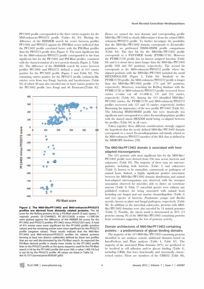
M60-enhancin/PF03272 profile (Tables S2, S3). Plotting the
difference of the HMMER search bit scores between profiles
PF13402 and PF03272 against the PSI-Blast scores indicated that
the PF13402 profile correlated better with the PSI-Blast profiles
than the PF03272 profile does (Figure 2). The most significant hits
for the M60-enhancin/PF03272 profile corresponded to the least
significant hits for the PF13402 and PSI-Blast profiles, consistent
with the characterisation of a new protein domain (Figure 2, Table
S2). The difference of the HMMER search bit scores between
profiles PF13402 and PF03272 defined a total of 415 entries
positive for the PF13402 profile (Figure 2 and Table S2). The
remaining entries positive for the PF03272 profile (enhancin-like
entries) were from two Fungi, bacteria and baculoviruses (Table
S2) of which 48 taxa also encoded one or more entries positive for
the PF13402 profile (two Fungi and 46 Firmicutes)(Table S2).
Hence we named the new domain and corresponding profile
M60-like/PF13402 to clearly differentiate it from the related M60-
enhancin/PF03272 profile. To further investigate the possibility
that the M60-like/PF13402 domain corresponds to Zn-metallo-
peptidases we performed HMM-HMM profile comparisons
(Table S4). The first hit for the M60-like/PF13402 profile
corresponds to a PANTHER family (PTHR15730). However
the PTHR15730 profile has no known assigned function (Table
S4) and it is about three times longer than the M60-like/PF13402
profile (928 and 307 positions, respectively). The second hit
corresponds to the M60-enhancin/PF03272 profile where the
aligned positions with the M60-like/PF13402 included the motif
HEXXHX(8,28)E (Figure 3, Table S4). Similarly to the
PTHR15730 profile, the M60-enhancin/PF03272 profile is longer
than the M60-like/PF13402 profile (775 and 307 positions,
respectively). Moreover, searching the RefSeq database with the
PTHR15730 or M60-enhancin/PF03272 profile recovered fewer
entries (e-value cut off #1.00E-4; 127 and 212 entries,
respectively) (Table S5). Among the 415 identified M60-like/
PF13402 entries, the PTHR15730 and M60-enhancin/PF03272
profiles recovered only 127 and 35 entries, respectively, further
illustrating the importance of the new profile PF13402 (Table S5).
The following HMM-HMM profile hits were drastically less
significant and corresponded to other Zn-metallopeptidase profiles
with the shared zincin HEXXH motif being co-aligned between
the profiles (Table S4) in all cases.
Taken together these different considerations strongly support
the hypothesis that the newly defined M60-like/PF13402 domain
corresponds to a novel Zn-metallopeptidase sub-family related to
the M60-enhancin/PF03272 member of the MA clan as defined in
the MEROPS database [39].
The M60-like/PF13402 domain is associated with host-adapted microorganismsThe 415 proteins with most significant hits for the M60-like/
PF13402 profile were derived from 256 taxa across bacteria and
eukaryotes (Table S5). The majority of these taxa are microor-
ganisms, including both bacteria (Table 1) and eukaryotes
(Table 2), known to be mutualists, commensals or pathogens of
animal hosts. Indeed, a highly significant positive association
between the M60-like/PF13402 domain distribution and animal
host-adapted microorganisms was observed; with the strongest
association observed for microbes able to thrive on vertebrate
mucosa (Table 3). Only 17 microbial species were without any
published evidence for being associated with animal hosts
including one fungus and one marine choanoflagellate (Table 2)
and two species of bacteria, Pseudomonas syringae and Bacillus
mycoides, known as plant and fungal pathogens, respectively (Table
S6). In addition to the microbial eukaryotes, proteins with M60-
like/PF13402 domains were also encoded by 14 animal genomes
(Table 2). Notably, the zincin motif is deteriorated in 36% (13
proteins among 36) of the M60-like/PF13402 containing proteins
from vertebrates suggesting the loss of protease activity.
Domain architectures of M60-like/PF13402-containingproteins – a predominance of glycan binding domainsThe majority of the 415 M60-like/PF13402-containing proteins
identified in our analyses contain additional domains based on
InterProScan and Pfam analyses (Table 4, Table S7). The
majority of the associated Pfam domains (92%) are predicted to
be involved in cell adhesion and/or glycan binding (Table 4),
including CBMs that have functionally and structurally charac-
terised entries. These are members of the CBM32 (Table S8),
Figure 2. The M60-like/PF13402 and M60-enhancin/PF03272profiles are derived from distantly related proteins. The bitscore for the RefSeq proteins hit by a PSI-Blast search (X-axis) (query: T.vaginalis protein GI:123449825, XP_001313628; e-value #1.00E-04)were plotted against the difference of the HMMER bit scores for thePF13402 and PF03272 profiles (PF13402 minus PF03272)(Y-axis). A totalof 415 entries were more significant for the PF13402 profile (positivevalues) and the remaining entries were more significant for the PF03272profile (negative values). These results indicate that the M60-like/PF13402 and M60-enhancin/PF03272 profiles hit related proteinsforming at least two distinct subfamilies defined by these two profilesthat are not well discriminated by the PSI-Blast search. As expected thePSI-Blast derived profile is clearly more similar to the PF13402 profilethan to the PF03272 profile as the query sequence used for the PSI-Blastsearch is hit by the PF13402 profile (bit-score 260, e-value 1.40E-74) butis not hit by the PF03272 profile. All values are listed in Table S2.doi:10.1371/journal.pone.0030287.g002
Novel Microbial Extracellular Metallopeptidases
PLoS ONE | www.plosone.org 4 January 2012 | Volume 7 | Issue 1 | e30287

CBM5 and CBM12 (CBM5_12; Table S9) and CBM51 (Table
S10) families respectively, as defined in the CAZy database [32]
(Table 4). Members of CBM32 commonly target galactose
configured animal and plant glycans and are found in a broad
diversity of structural architectures [32,41]; CBM5_12, typically
found in chitinases [32], are thought to bind exclusively to chitin, a
crystalline polysaccharide found in arthropods and other inverte-
brates, Fungi and some protists; whereas CBM51 family members
are known to target galactose and blood group A/B-antigens
[32,42]. Closer inspection of the microbial distribution of the
M60-like/PF13402 containing proteins linked to either CBM32
and CBM5_12 sequences revealed a distinct correlation between
the CBM family and the cognate organism’s predicted niche.
Proteins with the CBM32 were predominantly associated with
microbes known to colonise vertebrate mucosal surfaces (Table
S8), whereas entries with the CBM5_12 were correlated with a
capacity to thrive in the digestive tract of invertebrates, with
several species being able to thrive in both insects and mammals
(Table S9). One protein with CBM5_12 was derived from the
fungal pathogen Bacillus mycoides (Table S9). The genomes from
three Bacillus cereus strains and two B. thuringiensis strains encodedtwo to three M60-like/PF13402 containing proteins with one
possessing a CBM32 and the other a CBM5_12 (Table S11).
Interestingly B. cereus strains are known to be able to infect
mammals and/or insects [43]. The 19 entries possessing CBM51
were all from the genus Clostridium (nine strains of C. perfringens, C.
bartlettii DSM 16795 and Clostridium sp. 7_2_43FAA) (Table S10).
A total of 16 CBM51 containing proteins also possessed a CBM32
(Table S10). In addition to known CBM families, the recently
identified BACON domain [44] was identified among 22
Bacteroides proteins (Table S12). We also identified PA14-like [45]
and CBM32-like domains in proteins from T. vaginalis using HMM
profile-profile searches (Figure S4). Both the BACON and PA14
domains are thought to be involved in glycan binding [44,45]. The
structural organisation of selected M60-like/PF13402 containing
proteins is illustrated in Figure 4.
The unexpected M60-like/PF13402-CBM combinations we
observed led us to ask how commonly CBMs are linked to
peptidases by searching the MEROPS database for annotated
peptidases possessing CBM5_12, CBM32 or CBM51. Using
HMMER searches with a conservative cut off value (e-value
#1.00E-5) we identified 141 MEROPS entries positive for
CBM32 and/or CBM5_12. None were positive for CBM51. A
total of 110 proteins from 16 peptidase families were positive for
the CBM32 domain (Table 5), whereas 31 proteins from nine
peptidase families were positive for the CBM5_12 domain
(Table 6), indicating that these CBMs are widely distributed
across annotated peptidases. One MEROPS entry from Vibrio
campbellii (MER166461, ZP_02194874) was positive for both
CBM32 and CBM5_12 and is a member of the Zn-metallopepti-
dase family M64.
In contrast to the M60-like/PF13402 containing proteins the
domain composition of M60-enhancin/PF03272 containing
proteins was much less diverse (five additional domains) and
shared with the former CBM5_12 and fibronectin type III
domains (compare Table 4 and Table S13).
Microbial proteins with the M60-like/PF13402 domainpossess features of extracellular proteinsMost of the 415 M60-like/PF13402-containing proteins
(76%) were predicted to possess a signal peptide (SP), one or
more transmembrane domains (TMDs) or a bacterial lipopro-
tein motif (Table S5). These features suggest M60-like/
PF13402-containing proteins are extracytoplasmic, either se-
creted or anchored at the surface of microbial cells and could
therefore act on extracellular targets. In contrast, no extracel-
lular-associated sequence features were detected in the 14 M60-
Figure 3. Profile-profile alignment of M60-like/PF13402 and M60-enhancin/PF03272. The alignment was derived from an HHpred searchagainst all databases using the M60-like/PF13402 seed alignment as query (Table S4). The alignment corresponds to the second most significant hitto the enhancin/PF03272 profile (e-value 2.2E-37, score 307.8 and Probability 100%). The shown lines consists of ‘SS_pred’ lines representingsequence secondary structures predicted by PSIPRED, ‘consensus’ lines showing the consensus sequences of the PF13402 domain (shown with thetop sequence) and the corresponding PF03272 domain. Amino acid residues are marked in capital letters when occurring with a frequency $60%and lower cases when $40% in the respective seed alignments. A tilda indicates an un-conserved column. The line in between the two consensussequences shows the match quality and is defined as follows: ‘ = ’ very bad match, ‘2’ bad, ‘.’ neutral, ‘+’ good match and ‘|’ very good match. Thewell-conserved zincin HEXXH motif and an additional glutamate (E) residue part of a potential gluzincin are boxed. The upper and lower case lettersin ‘SS_pred’ lines for secondary structure predictions show high and low probability respectively where H=helix, E = strand, and C= coil.doi:10.1371/journal.pone.0030287.g003
Novel Microbial Extracellular Metallopeptidases
PLoS ONE | www.plosone.org 5 January 2012 | Volume 7 | Issue 1 | e30287

like/PF13402-containing proteins from animals or the M60-
like/PF13402-containing proteins from plant pathogens (six
Pseudomonas syringae strains) (Table S5).
Similarly, the majority of the 141 non-M60-like/PF13402
MEROPS entries (72%) positive for CBM32 and/or CBM5_12
were predicted to possess a SP and/or one or more (range 1–12)
Table 1. Short list of bacterial taxa associated with animals encoding proteins with the PF13402 domain.
Taxa
Habitat - isolation
source
Disease
via/in
PF13402
domain
per taxa
HEXXH
motif TMD SP or LP
Firmicutes
Bacillus anthracis* Soil/IG/mammals GIT, RT 1–2(12) Yes 2 Yes
Bacillus cereus* Soil/IG/GIT GIT, RT 1–5(69) +(68) +(3) +(60)
Bacillus thuringiensis* Soil/IG IG 1–4(35) Yes +(2) +(28)
Clostridium bartlettii DSM 16795 GIT - 2 Yes 2 Yes
Clostridium difficile QCD-32g58 GIT GIT 1 Yes 2 2
Clostridium perfringens* Soil/IG/GIT GIT 1–4(16) +(16) 2 +(14)
Listeria grayi DSM 20601 GIT - 1 Yes 2 Yes
Mollicutes bacterium D7 GIT 1 Yes 2 Yes
Paenibacillus larvae BRL-230010 IG IG 1 Yes 2 Yes
Tenericutes
Mycoplasma penetrans HF-2 UGT, RT UGT, RT 3 +(2) Yes 2
Bacteroidetes
Bacteroides caccae ATCC 43185 GIT Opportunist 16 Yes +(1) +(14)
Bacteroides fragilis* GIT Opportunist 1(3) Yes 2 Yes
Bacteroides thetaiotaomicron VPI-5482 GIT Opportunist 4 +(3) +(1) +(3)
Prevotella melaninogenica ATCC 25845 Oral cavity Oral cavity 1 Yes 2 Yes
Sphingobacterium spiritivorum ATCC 33861 Aquatic, soil, plants,human blood and urine
RT 6(12) Yes 2 Yes
Actinobacteria
Brachybacterium faecium DSM 4810 Poultry GIT - 1 Yes 2 Yes
Eggerthella lenta DSM 2243 GIT Bacteremia (rare) 1 Yes 2 Yes
Proteobacteria
Escherichia coli* GIT GIT, UGT 1(27) Yes 2 +(22)
Escherichia fergusonii ATCC 35469 GIT UGT, wound 1 Yes 2 Yes
Grimontia hollisae CIP 101886 GIT GIT 2 Yes 2 2
Photorhabdus asymbiotica ATCC 43949 IG Human wound 1 Yes 2 2
Pseudomonas aeruginosa* Soil/humans Opportunist 7 Yes 2 Yes
Salmonella enterica subsp. Arizona serovar62:z4,z23:– str. RSK2980
GIT GIT 1 Yes 2 Yes
Shewanella pealeana ATCC 700345 Aquatic, animal hosts, squid - 2 Yes 2 Yes
Shigella sp. D9 GIT GIT 1 Yes 2 2
Vibrio cholerae* Aquatic, animal hosts GIT 1–2(25) Yes 2 +(22)
Vibrio parahaemolyticus 16 Aquatic, animal hosts GIT 1–2(12) Yes 2 +(9)
Vibrio vulnificus* Aquatic, animal hosts GIT 1–2(3) Yes 2 +(2)
Yersinia enterocolitica 8081 Animal hosts GIT 1 Yes 2 Yes
Verrucomicrobia
Akkermansia muciniphila ATCC BAA-835 GIT - 4 Yes +(1) +(2)
Verrucomicrobium spinosum DSM 4136 Soil and GIT - 1 Yes 2 Yes
Only taxa for which there is evidence for long term or transient interactions with animal hosts are listed here. The presence of the HEXXH zincin motif, transmembranedomain (TMD) and N-terminal signal peptide (SP) or lipoprotein (LP) surface anchoring features are indicated. For each species and protein features, numbers inbrackets are the total number of protein sequences that have a given feature. ‘Yes’ means all the M60-like protein sequences have a given feature. ‘*’ denotes specieswith more than one strains having the PF13402-containing proteins. ‘2’ is used if no sequence contains a given feature. Mucosal surfaces defined in the text areindicated: GIT, UGT and RT. IG refers to invertebrate gut (including insects, nematodes or annelids). Table S5 lists all species and strains, sequence accessions andfeatures.doi:10.1371/journal.pone.0030287.t001
Novel Microbial Extracellular Metallopeptidases
PLoS ONE | www.plosone.org 6 January 2012 | Volume 7 | Issue 1 | e30287

TMD suggesting these peptidases also target extracellular
glycoproteins (Table S14).
Evidence for metal-dependent mucinase activity for oneM60-like/PF13402-containing protein from a human gutmutualistThe predicted peptidase and glycan binding activities, cellular
location and taxonomic distribution of a number of M60-like/
PF13402 containing proteins suggest their target substrates are
host glycoproteins such as mucins. In addition, a previous study
has shown that genes encoding two of the three M60-like/
PF13402 domain containing proteins with the gluzincin motif
from the human gut bacterium Bacteroides thetaiotaomicron (BT3015/
NP_811927.1 and BT4244/NP_813155.1 in Table S5) are up-
regulated in response to host O-glycan mucins, both in vitro and in
vivo [46].
To experimentally test the hypothesis that some M60-like/
PF13402 containing proteins degrade mucins we expressed and
purified full-length BT4244 and constructs lacking either its N-
terminal putative carbohydrate binding domains BACON and
CBM32 or C-terminal M60-like/PF13402 peptidase domain and
assessed their ability to degrade mucins using a gel based assay
(Figure 5). The data show that the full-length recombinant protein
comprising the two putative N-terminal carbohydrate binding and
Table 2. Eukaryotic taxa encoding proteins with the PF13402 domain.
Taxa
Habitat - isolation
source
Disease
via/in
PF13402
domain per taxa
HEXXH
motif TMD SP
Apicomplexan
Cryptosporidium muris RN66 GIT GIT 1 Yes 2 2
Cryptosporidium parvum Iowa II GIT GIT 1 Yes Yes 2
Pararasalidea
Trichomonas vaginalis G3 UGT UGT 25 +(11) +(6) +(4)
Amoebozoa
Entamoeba dispar SAW760 GIT - 1 Yes 2 +
Entamoeba histolytica HM-1:IMSS GIT GIT 1 Yes 2 +
Choanoflagellida
Monosiga brevicollis MX1 Aquatic - 1 Yes 2 2
Fungi
Aspergillus flavus NRRL3357 Soil, decayingplant and animals
Plant, RT 1 Yes 2 +
Aspergillus oryzae RIB40 Used in fermentedfood production
- 1 Yes 2 +
Metazoa
Cephalochordates
Branchiostoma floridae - - 2 +(1) 2 2
Bony fish
Danio rerio - - 4 +(3) 2 2
Amphibians
Xenopus laevis (African clawed fog) - - 1 Yes 2 2
Birds
Taeniopygia guttata (Zebra finch) - - 2 +(1) 2 2
Mammals
Homo sapiens - - 5 +(3) 2 2
Pan troglodytes (chimpanzee) - - 2 +(1) 2 2
Pongo abelii (orangutan) - - 1 2 2 2
Macaca mulatta (Rhesus monkey) - - 1 Yes 2 +
Bos taurus - - 2 +(1) 2 2
Equus caballus - - 2 +(1) 2 2
Canis familiaris - - 3 +(2) 2 2
Rattus norvegicus - - 5 +(3) 2 2
Mus musculus - - 3 +(2) 2 2
Ornithorhynchus anatinus - - 3 Yes 2 2
The higher taxa are indicated and for Metazoans the major sub-taxa are also listed. The presence of the HEXXH zincin motif, transmembrane domain (TMD) and N-terminal signal peptide (SP) are indicated. For each species and feature, numbers in brackets are the total number of protein sequences that have a given feature. ‘Yes’means all the M60-like protein sequences have a given feature. ‘2’ is used if no sequence contains a given feature. Mucosal surfaces defined in the text are indicated:GIT, UGT and RT. Table S5 lists all species and strains, sequence accessions and features.doi:10.1371/journal.pone.0030287.t002
Novel Microbial Extracellular Metallopeptidases
PLoS ONE | www.plosone.org 7 January 2012 | Volume 7 | Issue 1 | e30287

the M60-like/PF13402 domains, or a C-terminal fragment
composed of the predicted M60-like/PF13402 peptidase domain
only, both generated significant clearing of the bovine submax-
illary gland mucins from the gel, indicative of degradation of the
mucin peptide backbone (Figure 5). In contrast, no mucin
degradation was observed in the sample containing an N-terminal
segment encompassing the BACON-CBM32 domains only
(Figure 5). Addition of EDTA to the full-length enzyme and
peptidase domain reactions inhibited the observed shift in banding
pattern, as expected if a metal was required for a proteolytic
activity (Figure 5). Furthermore a conservative mutation of the
predicted catalytic glutamic acid residue (E575D of the zincin
motif HEXXH) dramatically reduced the mucinase activity
(Figure 5). These functional data clearly support the hypothesis
derived from our bioinformatics analyses that the novel M60-like/
PF13402 containing proteins represent host glycoprotein degrad-
ing Zn-metallopeptidases.
Discussion
A comprehensive understanding of the molecular basis of
mammalian host-microbe associations requires the knowledge of
the specific families of microbial proteins involved in interactions
with host mucosal surfaces. While many proteins from microbial
pathogens involved in adhesion to host tissues or degradation of
host proteins (virulence factors) have been identified there is a
paucity of data on the molecular basis of non-pathogenic
mutualistic interactions between host and microbes, despite the
importance of our microbiota in maintaining human health.
Comparative genomics can provide useful insight into structural
and taxonomic or habitat contextualisations generating valuable
hypotheses for the functions of uncharacterised proteins. In this
study we employed in silico investigations and an in vitro mucinase
assay to generate data, which together strongly support the
hypothesis that we identified a novel type of Zn-metallopeptidases
important for animal host-microbes interactions ranging from
mutualistic to pathogenic outcomes.
The M60-like/PF13402 domain define novel Zn-metallopeptidasesThe presence of the extended consensus HEXXHX(8,28)E,
suggested that the M60-like/PF13402 domain containing proteins
could be considered as gluzincin metallopeptidases. Known
Table 3. The PF13402 domain is positively associated with microorganisms interacting with animal host or vertebrate mucosa.
Isolation sources or habitat
for microorganisms
Number of microorganisms
PF13402 positive
Number of microorganisms
PF13402 negative p-values*
Animal host* 55 327 5.9E206
Non-animal host 17 333
Mucosa* 43 154 1.7E208
Non-mucosa 17 303
*Significance of the positive association for the M60-like/PF13402 domain distribution with microorganisms living in animal host or vertebrate mucosa was calculatedwith a hypergeometric test – see Methods. The taxa and their habitat annotation used for the test are listed in Table S16.doi:10.1371/journal.pone.0030287.t003
Table 4. Pfam domain composition for the 415 M60-like/PF13402-containing proteins.
Pfam accession Domain description Number of domains Number of proteins
PF00754a,b Coagulation factor 5/8 type, C-terminal – CBM32 109 78
PF00041a Fibronectin, type III 40 30
PF02839a,b Carbohydrate-binding family V/XII – CBM5_12 28 24
PF13004a,b BACON 39 22
PF08305a,b Glycosyl hydrolase family 98, putative carbohydrate-binding module – CBM51 46 18
PF01011 Pyrrolo-quinoline quinone repeat 16 16
PF07523a Bacterial Ig-like 23 15
PF07554a,b Uncharacterised sugar-binding 6 6
PF00746 Surface protein from Gram-positive cocci, anchoring region 5 5
PF00652a,b Ricin B lectin 4 4
PF00404a Dockerin type 1 2 2
PF05738a Collagen-binding surface protein Can-like, B region 2 2
PF00395 S-layer homology region 3 1
PF01416 tRNA pseudoouridine synthase 2 1
PF02368a Bacteria Ig-like, group 2 2 1
The CBMs, numbering according to the CAZy database discussed in the text, are indicated for the description of the domains PF00754, PF02839 and PF08305. Theentries are ranked according to the number of different proteins possessing a given domain.aDomains potentially involved in adhesion activities.bDomains potentially involved in glycan binding.doi:10.1371/journal.pone.0030287.t004
Novel Microbial Extracellular Metallopeptidases
PLoS ONE | www.plosone.org 8 January 2012 | Volume 7 | Issue 1 | e30287

bacterial and mammalian gluzincins have an insertion between the
second H and second E, ranging from 24 to 64 amino acids [36].
However, although none of the consensus sequences for known
gluzincins [36] correspond to the consensus region found among
the M60-like/PF13402-posessing proteins, a minority of the M60-
like/PF13402 containing proteins (23%–94 among 415) were
positive for an extended pattern characteristic of known Zn-
metallopeptidases (PROSITE entry: PS00142). In addition, the
profiles PF13402 and PF03272 were clearly related with proteins
positive for both profiles and the two profiles significantly hitting
each other in profile-profile comparisons. Enhancins (defining the
M60-enhancin/PF03272 domain) are insect mucin degrading Zn-
metallopeptidases (Clan MA, subclan MA(E), family M60) first
described in baculoviruses where they act as virulence factors
[37,38]. More recently a protein with an M60-enhancin/PF03272
domain from the insect pathogen B. thuringiensis (RefSeq accession:
ZP_04115705.1 in Table S1) was also shown to degrade insect
mucins defining a new bacterial virulence factor [47]. In insects
the peritrophic membrane can be considered as analogous to the
mammalian intestinal mucus, but unlike vertebrate mucus,
peritrophic membranes are chitin rich matrices [48]. In verte-
brates, mucus layers form an important physical surface barrier
facing the external environment in the GIT, RT and UGT [8,49].
Both vertebrate and invertebrate protective barriers play impor-
tant roles in defending the digestive tract from microbial infections
as well as promoting digestion processes [48,49]. Therefore, in
order for a microbe to colonise or break through these protective
barriers, physical interactions and enzymes capable of processing
these protective matrices, or cellular processes such as flagella
mediated directed movements, are required [7,8,10]. For some
mammalian mutualists the mucus represent an important source
of food, especially when there are little exogenous nutrients
available, as recently demonstrated for the prominent distal gut
bacterium B. thetaiotaomicron [46]. Consistent with the M60-like/
PF13402 domain being related to the enhancin Zn-metallopepti-
dases, we show here that recombinant versions of the lipoprotein
BT4244 from the mutualist B. thetaiotaomicron displayed mucin
degrading activity in vitro and this process was inhibited by the
addition of EDTA, a metal chelator known to deactivate Zn-
metallopeptidases [50] or by mutating the candidate catalytic
glutamate residue of the zincins motif [51]. In addition, a previous
study showed that the expression level of the BT4244 gene
increased significantly when B. thetaiotaomicron cells were exposed to
mammalian O-glycan mucins in vitro and in vivo and that the gene
belonged to a co-regulated polysaccharide utilisation locus
(PUL#78 – spanning BT4240-50) [46]. PUL#78 also contains
two glycoside hydrolase (GH) genes, a GH2 (BT4241) and GH109
(BT4243), two families that display activities consistent with mucin
degradation [32,46,52]. Based on the results of our mucinase assay
and gene content and expression data of the PUL#78 we
speculate that BT4244 cleaves the peptide backbone of colonic
mucins in vivo and contributes to host glycan foraging and niche
Figure 4. Structural organisation of selected M60-like/PF13402 containing proteins. Selected proteins possessing the PF13402 domainfrom a diverse taxonomic spread and cellular organisation (Gram-positive and Gram-negative bacteria and eukaryotes) with and without CBM32,CBM5_12 and/or CBM51 are depicted to illustrate their structural diversity. The putative glycan-binding BACON domain found among someBacteroides spp. proteins and divergent PA14-like and GBD-like domains found among T. vaginalis proteins are also illustrated (see Figure S4). Specienames with accession numbers, protein length and key structural features are shown. The cartoons are drawn to the same scale and all sequences arealigned to their N-terminus. See also Figure 6 for additional structural configurations. For the Clostridium and Bacillus sequences there is no evidencefor a cell surface anchoring sequence features indicated by ‘‘?-anchor’’.doi:10.1371/journal.pone.0030287.g004
Novel Microbial Extracellular Metallopeptidases
PLoS ONE | www.plosone.org 9 January 2012 | Volume 7 | Issue 1 | e30287
adaptation by B. thetaiotaomicron. This represent the first peptidase
identified for a bacterial mutualist that can target mucins [9], with
proteolytic degradation of mucins thought to be important for the
regulating the homeostasis and physicochemical properties of the
colonic mucus and contributing to its degradation along with
bacterial glycosidases [8,9]. This process benefits the energy
balance of both the bacteria and the mammalian host as short fatty
acids generated by bacterial mucin fermentation are metabolised
by the colonic epithelial cells [9].
Bacterial M60-like/PF13402 domain containing proteinsare encoded by the disposable pan-genomeAlthough proteins with an M60-like/PF13402 domain were
encoded by the genomes of many different bacterial species, not all
sequenced strains of a particular species (or species of a given
genus) contained a copy of this gene suggesting it is part of the
disposable pan-genome that contributes to specific niche adapta-
tion, including pathogenesis [6,16]. For example, several animal
bacterial pathogens such as Vibrio spp. (range: 0–2 proteins per
genome), including the important human pathogen V. cholera
(Table S15), contain an M60-like/PF13402 domain gene,
annotated as lipoprotein AcfD, whereas non-virulent strains of
V. cholera do not [53]. The V. cholera AcfD gene is part of four genes
defined as accessory colonisation factors (AcfA-D) required for
efficient human intestinal colonisation [53,54]. Interestingly there
is evidence that the V. cholera AcfB-C proteins mediate host-specific
chemotaxis towards mammalian mucus [53]. In line with the
mucinase data for the BT4244 protein, we hypothesise that the
AcfD lipoprotein is degrading human mucins, possibly in concert
with an additional secreted Zn-metallopeptidase TagA [51],
contributing to the pathogen’s capacity to penetrate the mucus
layer, a trait of virulent bacterial strains [53]. In fresh water and
other aquatic environments the AcfA-D proteins could also
contribute to the colonisation of fish mucosal surfaces, as Vibrio
cholera and other Vibrio species are often associated with these
vertebrates [55], and some species/strains can be pathogenic to
both human and fish [56].
Similar to Vibrio spp., not all sequenced genomes from Bacillus
spp. (range: 0–5), Bacteroides spp. (range: 0–16 proteins), Clostridium
spp. (range: 0–4 proteins), Escherichia spp. (range: 0–1 proteins) and
Yersinia spp. (range: 0–1 proteins) appear to encode proteins with
the M60-like/PF13402 domain (Table S15). Among 32 Escherichia
spp. possessing proteins with the M60-like/PF13402 domain 19
were defined as pathogenic strains that cause infection in various
mucosal niches including GIT, UGT and RT of both mammals
and birds [57] (Table S15). The other 13 E. coli strains encoding
M60-like/PF13402 domains are defined as non-pathogenic
members of the intestinal microbiota or one lab strain (Table
S15). For the remaining 32 Escherichia species or strains there was
no evidence for genes encoding any M60-like/PF13402 containing
proteins (Table S15). These taxa included in particular all of the E.
coli O157 strains, which are well known zoonotic pathogens that
can lead to severe human illnesses [58]. Similarly, different
Bacteroides spp. are thought to be adapted to different niches or
food sources within the mammalian GIT with only a few species
known to be able to graze host derived mucin glycans [59,60]. The
patchy distribution of M60-like/PF13402 containing proteins
Table 5. Annotated MEROPS peptidases families with CBM32.
Peptidase family Bacteria Eukaryotes Total
M04 1 - 1
M06 4 - 4
M12A - 1 1
M12B - 6 6
M14B - 65 65
M14X - 2 2
M20A 2 - 2
M23B 3 1 4
M36 3 - 3
M60 1 - 1
M64 1* - 1
S01A - 4 4
S08A 7 1 8
S45 4 - 4
S63 - 3 3
T06 - 1 1
Total 26 84 110
The number of peptidases for a given family possessing at least one CBM32 isindicated.*The unique entry with both CBM32 and CBM5_12 (Table 6).doi:10.1371/journal.pone.0030287.t005
Table 6. Annotated MEROPS peptidases families with CBM5_12.
Peptidase family Archaea Actinobacteria Firmicutes Proteobacteria Total
M04 - 2 - - 2
M06 - - 2 - 2
M28A - 1 - - 1
M60 - - 1 - 1
M64 - - - 1* 1
M66 - - - 1 1
S01A - 9 - 5 14
S08A 2 - 1 5 8
S53 - 1 - - 1
Total 2 13 4 12 31
The Number of peptidases for a given family possessing at least one CBM5_12 is indicated.*The unique entry with both CBM5_12 and CBM32 (Table 5).doi:10.1371/journal.pone.0030287.t006
Novel Microbial Extracellular Metallopeptidases
PLoS ONE | www.plosone.org 10 January 2012 | Volume 7 | Issue 1 | e30287

among Bacteroides species (Table S15), suggests the presence of this
gene could contribute to niche specialisation by providing mucin
degrading capability, a view supported by the mucinase activity
displayed by BT4244 from B. thetaiotaomicron, a known mucin
degrader.
Pathogenic microbial eukaryotes also encode M60-like/PF13402 containing proteinsIn addition to bacterial pathogens M60-like/PF13402 domains
were also identified among proteins from pathogenic microbial
eukaryotes including the extracellular parasites T. vaginalis and E.
histolytica and the intracellular parasites Cryptosporidium parvum and C.
muris. Entamoeba and Cryptosporidium species target the digestive tract
of humans and other vertebrates [61] while T. vaginalis is a human
sexually transmitted pathogen affecting both the male and female
UGT [62]. The immuno-dominant surface antigen from E.
histolytica is known as a GPI-anchored protein against which most
patients with liver abscess are known to generate an immunoglob-
ulin response [26]. Proteomics data indicated that this E. histolytica
surface protein can be found in the parasite phagosomes and
uropodes [27,29] and it was suggested that it might be involved in
phagocytosing apoptotic human cells [28]. The presence of a
galactose-binding domain like sequence (GBD, related to CBM32)
suggests that this domain could mediate binding to glycan chains in
secreted and cell surface human glycoproteins such as mucins. The
M60-like/PF13402 domain could be driving proteolysis of human
glycoproteins representing a possible source of nutrients for the
parasite and/or contribute to processing human proteins involved
in innate and adaptive immune defences for the benefit of the
microorganism. A related protein is also encoded by the genome of
the commensal E. dispar [63]. As homologues exist in both a
pathogen and a commensal, the M60-like/PF13402 containing
protein might therefore not represent a virulence factor in E.
histolytica as such but could contribute to the amoeba fitness on
mucosal surfaces. As for the E. histolytica surface protein there is
evidence for surface expression of two T. vaginalis M60-like/
PF13402 containing proteins (GI:123975108 XP_001330197.1 and
GI:123449825 XP_001313628.1 in Table S1) [25]. In contrast to
the E. histolytica GPI-anchored proteins these T. vaginalis proteins
possess a TMD. As T. vaginalis is also phagocytic [64], and mediates
endocytosis [24], the M60-like/PF13402 containing proteins could
be involved in nutrient binding, uptake and processing through
these internalisation routes.
The specificity of the M60-like proteins is driven by theirassociated carbohydrate-binding modulesA recurrent structural feature among many of the 415 identified
M60-like/PF13402 containing proteins was the co-occurrence of
CBMs and other glycan binding domains. Additional potential
glycan-binding domains included the BACON (identified among
Bacteroides spp.) [44] and PA14-like domains [45,65] (identified
among Trichomonas and Entamoeba). A total of 103 M60-like/
PF13402 containing proteins possessed CBM32, CBM5_12 and/
or CBM51 among 66 microbial species or strains that are known
in their majority to colonise animal hosts. Some of these species
are well known members of the human GIT microbiota including
Figure 5. In vitro mucin degradation assay. (A) The structural organisation of the B. thetaiotaomicron BT4244 protein (GI: 29349652; RefSeq:NP_813155.1). The entire wild type (W) protein (minus signal peptide) encoded by the BT4244 gene is contrasted to a mutant (M) (E575D) and twotruncated constructs covering either the PF13402 domain (P) or the BACON-CBM32 domains (BC) only. (B) 5 mg of purified recombinant proteins wereseparated by SDS-PAGE and stained with Coomasie blue for detection. The predicted protein sizes in kDa are: M and W: 95.1, P: 67.3 and BC: 27.8. (C)Bovine submaxillary gland mucins were incubated with 5 mg of purified recombinant proteins W, M, P or BC in the absence (2) or presence (+) of50 mM EDTA. Following incubation the mucin samples were separated on an agarose gel prior to detection on blots with wheat germ agglutininlectin.doi:10.1371/journal.pone.0030287.g005
Novel Microbial Extracellular Metallopeptidases
PLoS ONE | www.plosone.org 11 January 2012 | Volume 7 | Issue 1 | e30287

Bacteroides thetaiotaomicron, B. fragilis and B. caccae [2] or human
pathogens including Clostridium difficile [66]. Others are thought to
be both ‘‘free-living’’ (they can be isolated from the environment)
and can be pathogenic when in contact with mammalian mucosal
surfaces and/or the digestive tracts of insects. These species
include Paenibacillus larvae [67], Yersinia enterocolitica [68], Clostridium
perfringens [66], C. botulinum [69] and Bacillus cereus and B.
thuringiensis [43]. The M60-like/PF13402-CBM32 proteins are
from microbial species able to colonise mammalian mucosal
surfaces including the GIT and the UGT ranging from mutualists
to pathogens. CBM32s are known as components of enzymes
involved in the processing of complex galactose configured glycans
from predominantly animal sources [41]. The B. thetaiotaomicron
M60-like/PF13402 containing protein BT4244 possesses a
CBM32 and a BACON domain. These putative glycan-binding
domains could contribute to mucin recognition at the surface of
the bacterium by targeting the Gal and GalNAc containing O-
glycan side chains and presenting the polypeptide to the M60-like/
PF13402 Zn-metallopeptidase domain. The presence in many
M60-like proteins of multiple CBMs from different families and/or
in combination with other candidate binding domains (e.g.
BACON-CBM32, CBM32-CBM51, PA14-like-GBD and
CBM32-Fibronectin type III domain) suggests multivalent recog-
nition of a complex ligand driving high specificity and avidity,
consistent with targeting of host glycoproteins. CBMs from family
5 and 12 target chitin and were detected as components of M60-
like/PF13402-containing proteins from insect pathogens such as
Paenibacillus larvae and Bacillus thuringiensis as well as the fungal
pathogen Bacillus mycoides. [70,71]. The presence of a C-terminal
CBM5_12 in the M60-like/PF13402 containing proteins from
insect pathogens suggests that the target of these glycan binding
domains is the chitin-rich peritrophic membrane in the insect gut.
Attachment via the CBM5_12 could facilitate degradation of the
protein component through the activity of the M60-like/PF13402
peptidase domain. Intriguingly, P. larvae is a causative agent for
American foulbrood disease of honeybee larvae with the spores
germinating in the gut prior to causing disease [67] and
metallopeptidases were reported to be involved in P. larvae
pathogenicity [72]. Thus the M60-like/PF13402-CBM5_12
proteins represent an attractive candidate virulence factor similar
to the related baculovirus [37,38] and bacterial enhancin Zn-
metallopeptidases [47]. Most taxa encoded proteins combining the
M60-like/PF13402 domain with either CBM32 or CBM5_12 but
five Bacillus spp. (three strains of B. cereus and two of B. thuringiensis)
encoded two to three proteins, each with one of these two domains
combination (Table S11). Strains of B. cereus are known to cause
disease in both mammals and insects and it would be interesting to
test if these M60-like/PF13402 protein variants are differentially
expressed in insect (proteins with CBM5_12) versus mammals
(proteins with CBM32) hosts, possibly contributing to this B. cereus
host promiscuity [73].
The association of CBMs with the M60-like/PF13402 peptidase
domains and analysis of the MEROPS database clearly indicate a
novel functional context for CBMs, which are classically associated
with carbohydrate active enzymes [30,31,32]. In the context of
host-microbe interactions the presence of CBMs linked to
extracellular peptidases likely contributes to the ability of microbes
to attach to, degrade and metabolise host glycoproteins including
the abundant mucins. Interestingly, while the CBM domains are
found at either the C-terminal or N-terminal side of the M60-like/
PF13402 domain, the relative position of the protease domain
when attached to the surface is often conserved, suggesting this
configuration is functionally important (Figure 4). However some
M60-like/PF13402 domain containing proteins, such as several
entries from Clostridium spp. (Figures 4 and 5), possess CBMs on
both sides of the protease domain indicating a variety of
configurations is possible. The combination of protease-
CBM5_12 domain architecture is also observed in 13 enhancins
from some Clostridium and Bacillus taxa (Table S13).
A complex evolutionary history for the M60-like/PF13402domainThe broad and patchy taxonomic distributions of genes
encoding proteins with the M60-like/PF13402 domain also
suggest that gene sharing through LGT took place between
distantly related taxa, including between bacteria and eukaryotes.
Phylogenetic analyses of a representative selection of M60-like/
PF13402 domain sequences strongly supports this hypothesis with
in particular robust cases of gene sharing between the microbial
eukaryotes Trichomonas and Entamoeba and the bacteria Mycoplasmaand Clostridium respectively (Figure 6). Notably the majority of
Clostridium species form a distinct clan (as defined in [74]),
including C. perfringens, clearly indicating alternative origins for the
M60-like/PF13402 domain among this genus. The strong bias for
M60-like/PF13402 domains among microbial taxa able to
colonise animal hosts, suggests that an important fraction, if not
most, of these LGTs took place in the context of animal hosts
where microorganisms density can be extremely high, as in the
human distal colon, and where LGT is known to play important
roles in shaping the gene complement of the microbial community
[60]. A striking case involves four independent LGT events in the
mucin degrading Verrucomicrobia, Akkermansia muciniphila, three
from the distantly related Bacteroidetes donors (clan B in Figure 6)
that share the same niche as A. muciniphila and a fourth LGT from
an undefined source (within clan A in Figure 6). The potential
initial source gene(s) for the microbial species is difficult to establish
with the current taxonomic sampling and phylogenetic resolution.
It could be one or more eukaryotes as a broad range of these
encode M60-like/PF13402 containing proteins, indeed several
bacterial sequences are part of clan A where the majority of
eukaryotes, including animals sequences, are clustering (Figure 6).
An animal to bacteria gene transfer was recently suggested for
genes encoding two other distinct types of Zn-metallopeptidases
identified in the human associated Bacteroidetes species Bacteroides
fragilis [75] and Tannerella forsythia [76]. In the case of the two
fungal M60-like/PF13402 containing proteins the phylogeny
supports an LGT event from a bacterial donor to the Aspergillus
lineage (only one fungal species is shown in Figure 6), possibly
reflecting the adaptations of these Fungi to thrive on decaying
plant and animal material. The identified LGT of genes encoding
M60-like/PF13402 Zn-metallopeptidases involving mutualists,
commensals and pathogens further highlights the overlap between
the gene complements of microorganisms generating contrasting
symbiotic outcomes with their animal hosts [4,6,24]. Interestingly
a restricted set of taxa (two Fungi and 46 Firmicutes, Table S2),
which represent a subset of the taxa encoding M60-like/PF13402
containing proteins, encoded one (or more) protein possessing the
M60-like/PF13402 domain and another protein possessing the
M60-enhancin/PF03272 domain with the bacteria taxa all
capable to infect insects or other invertebrates (Table S2).
Analysing the relationship of protein sequence members of these
two protein families with CLANS (see Methods section) showed
that M60-enhancin/PF03272 and M60-like/PF13402 proteins
clustered in different groups further supporting our finding that
the M60-like/PF13402 domain form a novel protein family
(Figure S6). Interestingly, the CLANS result also suggests that the
M60-enhancin/PF03272 and M60-like/PF13402 containing pro-
teins from Bacillus species are more similar to each others than to
Novel Microbial Extracellular Metallopeptidases
PLoS ONE | www.plosone.org 12 January 2012 | Volume 7 | Issue 1 | e30287

other related proteins from other organisms (Figure S6). This
suggests that the PF03272 domain was derived from a gene
duplication of a ‘‘primordial’’ PF13402 domain. One possible
scenario underlying the functional relevance of such a gene
duplication event, followed by important differentiation of the
paralogues, could be a response to the selection pressure induced
by insect host peptidase inhibitors on bacterial peptidases
representing virulence factors [77]. A phylogenetic analysis of
the same dataset used to investigate M60-like/PF13402 domain
relationships (Figure 6) complemented with selected M60-
enhancin/PF03272 sequences could neither reject or provide
support for this hypothesis due to lack of resolution as indicated by
poor bootstrap support values (Figure S7). These evolutionary
considerations along with the identified taxonomic distribution of
M60-enhancin/PF03272 domains (Table S13) also suggest that
the baculoviruses obtained their enhancin genes from a bacterium
sharing the same insect host and subsequently diverged dramat-
ically from their bacterial enhancin homologues.
In summary the novel type of Zn-metallopeptidase we identified
across evolutionarily distantly related bacteria and microbial
eukaryotes, that are found on a broad range of animal hosts,
further illustrates the importance of peptidases in host-microbe
interactions. This discovery will be of benefit in guiding
investigations of the molecular basis of host-microbe interactions
in the context of both mutualistic and pathogenic outcomes
involving bacteria and microbial eukaryotes in vertebrates and
invertebrates. It will be of particular interest to identify the range,
and properties, of the host proteins that the novel microbial
peptidases can target. The possibility of peptidases representing
functional partners of other hydrolysing enzymes, such as GH
(indicated by the Bacteroides PULs encoding both activities), to
process mammalian mucins is of particular interest for the study of
the role of mutualists in the homeostasis of our mucosal surfaces.
The novel yet common domain combinations we identified
involving peptidases and CBMs offer interesting new insights into
substrate recognition by peptidases, which in turn could provide
Figure 6. Protein maximum likelihood bootstrap consensus tree for selected M60-like/PF13402 domains. The shown maximumlikelihood tree (Log likelihood: 218778.5) was generated as described in the Methods section using an alignment of 57 sequences and 175 residuesdrawn from an M60-like/PF13402 domain alignment, providing an evolutionary framework for the gene segments encoding these domains. For eachsequence the corresponding species name is indicated along with the NCBI GI number and high-ranking taxa - squares are for eukaryotes: black-Metazoa, blue-microbial eukaryotes; circles are for Bacteria: yellow-Bacteroidetes, orange-Firmicutes, green-Proteobacteria (all gamma-proteobacteria), pink-Verrucomicrobia, violet-Tenericutes, cyan-Actinobacteria. Bootstrap support values ($60%) are indicated below the branches.The scale bar represents the estimated number of changes per site. The domain organisation of the corresponding complete proteins is shown onthe right hand side (see also Figure 4 for additional domain configurations). The sequence corresponding to the BT4244 protein used for themucinase assay (Figure 5) is indicated by a *. The two major clans supported by a bootstrap value of 76% are indicated as clan A and B.doi:10.1371/journal.pone.0030287.g006
Novel Microbial Extracellular Metallopeptidases
PLoS ONE | www.plosone.org 13 January 2012 | Volume 7 | Issue 1 | e30287

exciting opportunities to engineer peptidases targeted to specific
glycoproteins for both biomedical and industrial applications.
Methods
Sequence similarity search and HMM profiles generationPSI-Blast was used at the NCBI Blast server [78] (search date:
20th January 2010) to identify related proteins to the T. vaginalis
entries annotated as immuno-dominant antigen-like protein using
as query the protein GI:123449825 XP_001313628 (positions 1–
500). An initial PSI-Blast search with the entire GI:123449825
XP_001313628 sequence identifying the first ,500 residues as
being shared across a broad range of taxa (Figure S1), hence we
used positions 1–500 to perform a most specific PSI-Blast profile
search. It was performed with a standard initial BlastP search
followed by one iteration step for the profile-based search. A multi-
sequence alignment with the sequences derived from the PSI-Blast
hit list was downloaded using the alignment retrieval option. This
alignment was used to generate a profile using HMMER [79]
defining the newly identified domain M60-like/PF13402. The
following five steps were performed to generate our initial
alignment: (1) The segment of the alignment corresponding to
positions 193–378 (inclusive) of the T. vaginalis sequence
XP_001313628 (the query sequence used for the aforementioned
PSI-Blast) was identified as the most conserved across the aligned
sequences by visual inspection and was retrieved using the masking
option of SEAVIEW4.0 [80]. (2) Sequences with high level of
identity ($80%), considered as redundant, were removed leaving
92 sequences (the shortest entries across the aligned segment were
removed). (3) These 92 sequences were re-aligned with MUSCLE
using default settings within SEAVIEW4.0. (4) To minimise
alignment length and optimise hypothesis of site homology indels
larger than 2 residues (that complicate alignments) and present in
less than 50% of the sequences were deleted. (5) Steps 3–4 were
repeated and reduced the alignment to 208 aligned positions and
27 sequences (Figure S2). HMMER [79] was then used to
generate and calibrate an HMM profile for the M60-like domain
with the ‘hmmbulid’ commands with default settings. Following
submission of the M60-like domain alignment to Pfam and Pfam
curation, M60-like/PF13402 seed alignment and profile were
generated and made available to us (Figure S3).
The HMM profile of the CMB32 domain (PF00754), CBM5_12
(PF02839), CBM51 (PF08305) and BACON (PF13004) were retrieved
from the Pfam database.
Detection of known protein structural featuresSignalP 3.0 [81], TMHMM 2.0 [82] and PHOBIUS (that
combines SP and TMD detections) [83], were employed to
detection extracellular targeting N-terminal SP and TMD. Other
characterised protein domains/motifs were searched using Inter-
ProScan version 4.3 [35]. The default parameters were used for
every tool and where relevant the appropriate taxonomic option
selected.
Protein profile HMM searchesHMMER 3.0 was used to searchM60-like/PF13402 HMMprofile
against proteins in RefSeq database (data obtained on 21st January
2010, containing 9,662,677 protein sequences). The HMM profiles
for specific domains (M60-like/PF13402, CBM32, CBM5_12,
CBM51 and BACON) were used to search (i) the annotated peptidase
library retrieved from the MEROPS database [39](release 9.1, data
obtained 2nd May 2010) and (ii) the 415 entries positive for the M60-
like/PF13402 domain. The ‘hmmsearch’ command was used to
search a given profile against the different protein sets.
Protein profile-profile searchesTo perform HMM-HMM profile comparison between the
M60-like/PF13402 profile against other known profiles, the
HHPred server running with HHSearch version 1.6.0.0 [84] was
used to search all the available databases. HHPred was also used
to identify divergent versions of PA14 and CBM32 domains from
T. vaginalis M60-like/PF13402 containing proteins (Figure S4).
Association of the M60-like/PF13402 domain to microbialhabitatTo investigate the significance of the association between the
presence of M60-like/PF13402 domain (genotype) and host
associated or mucosal-related lifestyles (phenotype/trait) of
microorganisms, we calculated the probability of the co-occur-
rence between the genotypic and phenotypic features according to
hypergeometric distribution function [12]:
p(i§m N,M,n)~X
n
i~m
M
i
� �
M{n
n{1
� �
N
n
� �
�
�
�
�
�
�
�
�
�
Of the total number of microorganisms with completed genome
sequences in the RefSeq database at the time, 455 (N) have habitat
information that can be used to determine whether an organism is
able to thrive on or penetrate through vertebrate mucosa surfaces
(Table S16). The number of these microorganism with an M60-
like/PF13402 domain annotated was 55 (n). The number of
microorganisms known to thrive on or infect host through mucosal
surfaces was 197 (M). Of these 197 taxa, 43 (m) taxa possess at
least one M60-like/PF13402 domain. As a result, the probability
(p-value) of observing the association of the M60-like/PF13402
domain and the ability of microbe to thrive on mucosal surface can
be calculated.
To determine the type of this association (either positive or
negative), the mean value (m) of hypergeometric distribution was
used [12]:
m~n �M=N
Where n, M, N and m can be referred from the previous equation,
for m.m corresponding to positive associations and for m,m
corresponding to negative associations.
Protein family visualisation with CLANSWe used CLANS [85], a graph-based protein sequence
similarity visualisation software, to investigate relationships
between M60-like/PF13402 and M60-enhancins/PF03272 con-
taining proteins. The software clusters set of protein sequences
based on their BlastP p-values of the high-scoring segment pair
alignments. All 693 sequences identified with HMMER searches
using the M60-like/PF13402 and M60-enhancins/PF03272
profiles (all entries are listed in Table S5) with the default settings,
were included into the CLANS analysis. Nodes or entries that
have no similarity to other entries based on BlastP cutoff e-value
1.00E-5 were removed from the graph.
Recombinant protein expression of BT4244 and in vitromucinase assayThe gene encoding full length BT4244 protein lacking its N-
terminal lipoprotein signal sequence was amplified from B.thetaiotaomicron VPI-5482 genomic DNA and cloned into pRSETA
Novel Microbial Extracellular Metallopeptidases
PLoS ONE | www.plosone.org 14 January 2012 | Volume 7 | Issue 1 | e30287

(Invitrogen) on BamHI/EcoRI generating the construct pRSETA-
BT4244. Truncated constructs were generated that encoded either
the C-terminal M60-like/PF13402 peptidase domain only or the
N-terminal BACON and CBM32 domains only (Figure 4). Site-
directed mutagenesis of the BT4244 catalytic glutamic residue
(conservative mutation E575D, e.g. [51]) was carried out using the
QuikChange protocol (Stratagene) according to the manufactur-
er’s instructions with the construct pRSETA-BT4244 as template
DNA. All constructs and the E575D mutant were confirmed by
sequencing. The primers used for PCR amplifications and the
mutagenesis are listed in Table S17. Recombinant proteins with
an N-terminal His-tag were expressed in BL21 (Novagen) and
purified in a single step by metal affinity chromatography using
Talon resin (Clontech) as described previously [86]. Purified
proteins were dialysed overnight against phosphate buffered saline
pH 7.3 (OXOID, Dulbeco ‘A’ PBS) prior to the mucinase assay.
Mucins from bovine submaxillary glands Type I-S (Sigma, UK)
were used as substrate for the mucinase assays in the absence or
presence of 50 mM EDTA (e.g. [51]). Following incubations at
37uC for 48 hours the mucins were run on a 1% (w/v) agarose gel
(SeaKem agarose, Melford ltd., UK)+0.1% (w/v) SDS in a Biorad
minigel system and then transferred onto PVDF membranes by
blotting. Biotin conjugated lectin from Triticum vulgaris (wheat germ
agglutinin lyophilized powder, Sigma, UK) in combination with
ExtrAvidinH–Peroxidase buffered aqueous solution (Sigma, UK)
was used to detect mucin on blots.
Phylogenetic analysesA broad protein alignment of theM60-like/PF13402 domain was
generated using the PF13402 seed alignment as reference using the
‘‘Profile alignment’’ function from CLUSTAL within SEAVIEW
[80]. A subset of 57 sequences was eventually selected to reduce the
complexity of the dataset and optimising sequence and taxonomic
diversity. Maximum likelihood trees were computed with PhyML
from within SEAVIEW with the LG model and a gamma shape
parameter to correct for site rate heterogeneity (4 discrete rates) and
using both NNI and SPR for the tree search operations. 100
bootstrap pseudo replicates were generated to calculate branch
support values. The tool iTOL [87] was used to map on the inferred
phylogenetic tree the structural organisation of the analysed
proteins. An additional phylogenetic analysis was performed
including selected enhancin proteins sequences (M60-enhancin/
PF03272 domain) (Figure S5, Figure S7)
Supporting Information
Figure S1 PSI-Blast output figure obtained from the
NCBI Blast server. The query sequence was from Trichomonas
vaginalis (GI:123449825, XP_001313628, the complete sequence)
and two iterations (e-value #1.00E-05). File format: tif.
(TIF)
Figure S2 Initial protein alignment corresponding to
the new M60-like domain, 27 sequences with 206 aligned
sites. The specie names are abbreviated with the first three letters
of the genus and species names along the corresponding protein
sequence GenBank GI number. The query sequence used for the
PSI-Blast is shown on the top with the RefSeq accession number.
Format: text file with sequences in fasta format to be open in a
sequence alignment editor such as SEAVIEW.
(FAS)
Figure S3 Pfam seed alignment for the new M60-like
domain, accession PF13402, consisting of 68 sequenceswith 387 aligned sites. The UniProt accession number for each
sequence is provided along with the position of the start and end of
the aligned segments. Format: text file with sequences in fasta
format to be open in a sequence alignment editor such as
SEAVIEW.
(FAS)
Figure S4 Figure illustrating the PA14-like and Galac-
tose binding domain-like (GBD) domain identified inTrichomonas vaginalis M60-like/PF13402 containing
proteins.
(TIF)
Figure S5 M60-like/PF13402 alignment of 57 PF13402and 4 PF03272 sequences and 175 sites used to infer the
phylogeny shown in Figure 6 (57 PF13402 sequencesonly) or Figure S7 (all 61 sequences). Format: text file with
sequences in fasta format to be open in a sequence alignment
editor such as SEAVIEW.
(FAS)
Figure S6 Two-dimensional graph layout from the
CLANS clustering results obtained from the full-length
sequences for the M60-enhancin/PF03272 or M60-like/PF13402 containing proteins. Each protein sequence is shown
by a black dot. Lines connecting dots indicate sequence similarity
generated from BlastP: red lines edges represent sequence
similarity with Blast e-value,1E-100, whereas grey lines represent
Blast e-value from 1E-5 to 1E-100. Entries that have more
significant hits with the M60-enhancin/PF03272 profile are
encircled in green. All entries outside the green circle have more
significant hit on the M60-like/PF13402 profile. Selected clusters
are labeled with their taxonomic composition. Notably the two
families of Bacillus entries are clustering at the vicinity of each
other. Format: tif.
(TIF)
Figure S7 Protein maximum likelihood bootstrap con-sensus tree for selected M60-like/PF13402 and M60-
enhancin/PF03272 domains. The shown maximum likeli-
hood tree (Log likelihood: 219083.4) was generated as described
in the Methods section using an alignment of 57 sequences and
175 residues drawn from an M60-like/PF13402 (see Figure 6)
domain alignment complemented with four M60-enhancin/
PF03272 sequences (boxed), providing an the evolutionary
framework for the gene segments encoding these domains. For
each sequence the corresponding abbreviated species name is
indicated along with the NCBI GI number. Format: tif.
(TIF)
Table S1 PSI-Blast Taxonomic report derived from theNCBI Blast server. The query sequence was from Trichomonas
vaginalis (GI:123449825, XP_001313628, residues 1–500), 2
iterations (e-value #1.00E-04). After seven iterations no new
sequences were recovered and these additional entries were all
recovered by the HMMER searches using PF13402, PF03272 and
PTHR15730 profiles, see Table S5 for the complete list of entries.
Format: html file to be opened with a web browser providing links
to original RefSeq entries at the NCBI.
(HTML)
Table S2 Table listing all 523 RefSeq protein entries
positive for the PF13402 profile (HMMER search, e-value #1.00E-05) contrasting the PSI-Blast values with
HMMER scores for the PF13402 and PF03272 profiles.The sequences for the gluzincin and zincin motif are also shown.
Format: Excel file with five worksheets.
(XLSX)
Novel Microbial Extracellular Metallopeptidases
PLoS ONE | www.plosone.org 15 January 2012 | Volume 7 | Issue 1 | e30287
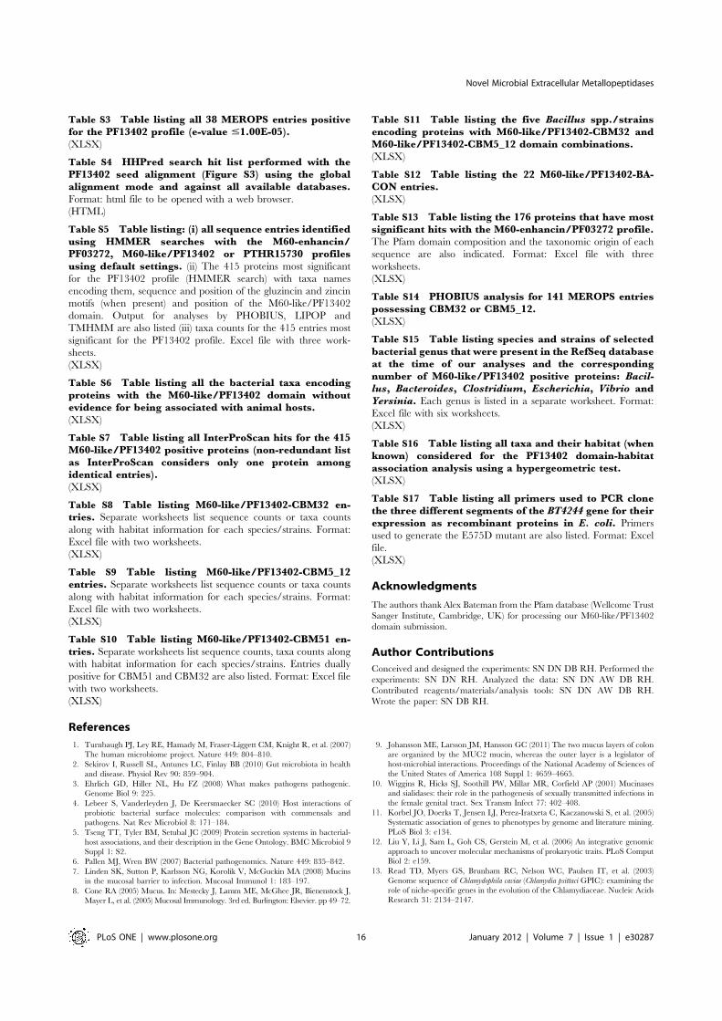
Table S3 Table listing all 38 MEROPS entries positivefor the PF13402 profile (e-value #1.00E-05).(XLSX)
Table S4 HHPred search hit list performed with thePF13402 seed alignment (Figure S3) using the globalalignment mode and against all available databases.Format: html file to be opened with a web browser.
(HTML)
Table S5 Table listing: (i) all sequence entries identifiedusing HMMER searches with the M60-enhancin/PF03272, M60-like/PF13402 or PTHR15730 profilesusing default settings. (ii) The 415 proteins most significant
for the PF13402 profile (HMMER search) with taxa names
encoding them, sequence and position of the gluzincin and zincin
motifs (when present) and position of the M60-like/PF13402
domain. Output for analyses by PHOBIUS, LIPOP and
TMHMM are also listed (iii) taxa counts for the 415 entries most
significant for the PF13402 profile. Excel file with three work-
sheets.
(XLSX)
Table S6 Table listing all the bacterial taxa encodingproteins with the M60-like/PF13402 domain withoutevidence for being associated with animal hosts.(XLSX)
Table S7 Table listing all InterProScan hits for the 415M60-like/PF13402 positive proteins (non-redundant listas InterProScan considers only one protein amongidentical entries).(XLSX)
Table S8 Table listing M60-like/PF13402-CBM32 en-tries. Separate worksheets list sequence counts or taxa counts
along with habitat information for each species/strains. Format:
Excel file with two worksheets.
(XLSX)
Table S9 Table listing M60-like/PF13402-CBM5_12entries. Separate worksheets list sequence counts or taxa counts
along with habitat information for each species/strains. Format:
Excel file with two worksheets.
(XLSX)
Table S10 Table listing M60-like/PF13402-CBM51 en-tries. Separate worksheets list sequence counts, taxa counts alongwith habitat information for each species/strains. Entries dually
positive for CBM51 and CBM32 are also listed. Format: Excel file
with two worksheets.
(XLSX)
Table S11 Table listing the five Bacillus spp./strainsencoding proteins with M60-like/PF13402-CBM32 andM60-like/PF13402-CBM5_12 domain combinations.(XLSX)
Table S12 Table listing the 22 M60-like/PF13402-BA-CON entries.(XLSX)
Table S13 Table listing the 176 proteins that have mostsignificant hits with the M60-enhancin/PF03272 profile.The Pfam domain composition and the taxonomic origin of each
sequence are also indicated. Format: Excel file with three
worksheets.
(XLSX)
Table S14 PHOBIUS analysis for 141 MEROPS entriespossessing CBM32 or CBM5_12.(XLSX)
Table S15 Table listing species and strains of selectedbacterial genus that were present in the RefSeq databaseat the time of our analyses and the correspondingnumber of M60-like/PF13402 positive proteins: Bacil-
lus, Bacteroides, Clostridium, Escherichia, Vibrio andYersinia. Each genus is listed in a separate worksheet. Format:
Excel file with six worksheets.
(XLSX)
Table S16 Table listing all taxa and their habitat (whenknown) considered for the PF13402 domain-habitatassociation analysis using a hypergeometric test.(XLSX)
Table S17 Table listing all primers used to PCR clonethe three different segments of the BT4244 gene for theirexpression as recombinant proteins in E. coli. Primers
used to generate the E575D mutant are also listed. Format: Excel
file.
(XLSX)
Acknowledgments
The authors thank Alex Bateman from the Pfam database (Wellcome Trust
Sanger Institute, Cambridge, UK) for processing our M60-like/PF13402
domain submission.
Author Contributions
Conceived and designed the experiments: SN DN DB RH. Performed the
experiments: SN DN RH. Analyzed the data: SN DN AW DB RH.
Contributed reagents/materials/analysis tools: SN DN AW DB RH.
Wrote the paper: SN DB RH.
References
1. Turnbaugh PJ, Ley RE, Hamady M, Fraser-Liggett CM, Knight R, et al. (2007)
The human microbiome project. Nature 449: 804–810.
2. Sekirov I, Russell SL, Antunes LC, Finlay BB (2010) Gut microbiota in health
and disease. Physiol Rev 90: 859–904.
3. Ehrlich GD, Hiller NL, Hu FZ (2008) What makes pathogens pathogenic.
Genome Biol 9: 225.
4. Lebeer S, Vanderleyden J, De Keersmaecker SC (2010) Host interactions of
probiotic bacterial surface molecules: comparison with commensals and
pathogens. Nat Rev Microbiol 8: 171–184.
5. Tseng TT, Tyler BM, Setubal JC (2009) Protein secretion systems in bacterial-
host associations, and their description in the Gene Ontology. BMC Microbiol 9
Suppl 1: S2.
6. Pallen MJ, Wren BW (2007) Bacterial pathogenomics. Nature 449: 835–842.
7. Linden SK, Sutton P, Karlsson NG, Korolik V, McGuckin MA (2008) Mucins
in the mucosal barrier to infection. Mucosal Immunol 1: 183–197.
8. Cone RA (2005) Mucus. In: Mestecky J, Lamm ME, McGhee JR, Bienenstock J,
Mayer L, et al. (2005) Mucosal Immunology. 3rd ed. Burlington: Elsevier. pp 49–72.
9. Johansson ME, Larsson JM, Hansson GC (2011) The two mucus layers of colon
are organized by the MUC2 mucin, whereas the outer layer is a legislator of
host-microbial interactions. Proceedings of the National Academy of Sciences of
the United States of America 108 Suppl 1: 4659–4665.
10. Wiggins R, Hicks SJ, Soothill PW, Millar MR, Corfield AP (2001) Mucinases
and sialidases: their role in the pathogenesis of sexually transmitted infections in
the female genital tract. Sex Transm Infect 77: 402–408.
11. Korbel JO, Doerks T, Jensen LJ, Perez-Iratxeta C, Kaczanowski S, et al. (2005)
Systematic association of genes to phenotypes by genome and literature mining.
PLoS Biol 3: e134.
12. Liu Y, Li J, Sam L, Goh CS, Gerstein M, et al. (2006) An integrative genomic
approach to uncover molecular mechanisms of prokaryotic traits. PLoS Comput
Biol 2: e159.
13. Read TD, Myers GS, Brunham RC, Nelson WC, Paulsen IT, et al. (2003)
Genome sequence of Chlamydophila caviae (Chlamydia psittaci GPIC): examining the
role of niche-specific genes in the evolution of the Chlamydiaceae. Nucleic Acids
Research 31: 2134–2147.
Novel Microbial Extracellular Metallopeptidases
PLoS ONE | www.plosone.org 16 January 2012 | Volume 7 | Issue 1 | e30287

14. O’Sullivan O, O’Callaghan J, Sangrador-Vegas A, McAuliffe O, Slattery L,et al. (2009) Comparative genomics of lactic acid bacteria reveals a niche-specificgene set. BMC Microbiology 9: 50.
15. Liolios K, Chen IM, Mavromatis K, Tavernarakis N, Hugenholtz P, et al. (2010)The Genomes On Line Database (GOLD) in 2009: status of genomic andmetagenomic projects and their associated metadata. Nucleic Acids Res 38:D346–354.
16. Medini D, Serruto D, Parkhill J, Relman DA, Donati C, et al. (2008)Microbiology in the post-genomic era. Nat Rev Microbiol 6: 419–430.
17. Ellrott K, Jaroszewski L, Li W, Wooley JC, Godzik A (2010) Expansion of theprotein repertoire in newly explored environments: human gut microbiomespecific protein families. PLoS Comput Biol 6: e1000798.
18. Clark CG, Alsmark UC, Tazreiter M, Saito-Nakano Y, Ali V, et al. (2007)Structure and content of the Entamoeba histolytica genome. Adv Parasitol 65:51–190.
19. Loftus B, Anderson I, Davies R, Alsmark UC, Samuelson J, et al. (2005) Thegenome of the protist parasite Entamoeba histolytica. Nature 433: 865–868.
20. Carlton JM, Hirt RP, Silva JC, Delcher AL, Schatz M, et al. (2007) Draftgenome sequence of the sexually transmitted pathogen Trichomonas vaginalis.Science 315: 207–212.
21. Carlton JM, Malik SB, Sullivan SA, Sicheritz-Ponten T, Tang P, et al. (2010)The genome of Trichomonas vaginalis. In: Clark CG, Johnson PJ, Adam RD, eds.Anaerobic Parasitic Protozoa: Genomics and Molecular Biology: CaisterAcademic Press. pp 45–77.
22. Alsmark UC, Sicheritz-Ponten T, Foster PG, Hirt RP, Embley TM (2009)Horizontal gene transfer in eukaryotic parasites: a case study of Entamoebahistolytica and Trichomonas vaginalis. Methods Mol Biol 532: 489–500.
23. Hirt RP, Noel CJ, Sicheritz-Ponten T, Tachezy J, Fiori PL (2007) Trichomonasvaginalis surface proteins: a view from the genome. Trends Parasitol 23: 540–547.
24. Noel CJ, Diaz N, Sicheritz-Ponten T, Safarikova L, Tachezy J, et al. (2010)Trichomonas vaginalis vast BspA-like gene family: evidence for functional diversityfrom structural organisation and transcriptomics. BMC Genomics 11: 99.
25. de Miguel N, Lustig G, Twu O, Chattopadhyay A, Wohlschlegel JA, et al. (2010)Proteome analysis of the surface of Trichomonas vaginalis reveals novel proteinsand strain-dependent differential expression. Mol Cell Proteomics 9: 1554–1566.
26. Edman U, Meraz MA, Rausser S, Agabian N (1990) Characterization of animmuno-dominant variable surface antigen from pathogenic and nonpathogenicEntamoeba histolytica. Journal of Experimental Medicine.
27. Marquay Markiewicz J, Syan S, Hon CC, Weber C, Faust D, et al. (2011) AProteomic and Cellular Analysis of Uropods in the Pathogen Entamoeba histolytica.PLoS Negl Trop Dis 5: e1002.
28. Marion S, Guillen N (2006) Genomic and proteomic approaches highlightphagocytosis of living and apoptotic human cells by the parasite Entamoebahistolytica. International Journal for Parasitology 36: 131–139.
29. Okada M, Huston CD, Mann BJ, Petri WA, Jr., Kita K, et al. (2005) Proteomicanalysis of phagocytosis in the enteric protozoan parasite Entamoeba histolytica.Eukaryot Cell 4: 827–831.
30. Boraston AB, Bolam DN, Gilbert HJ, Davies GJ (2004) Carbohydrate-bindingmodules: fine-tuning polysaccharide recognition. Biochem J 382: 769–781.
31. Guillen D, Sanchez S, Rodriguez-Sanoja R (2010) Carbohydrate-bindingdomains: multiplicity of biological roles. Appl Microbiol Biotechnol 85:1241–1249.
32. Cantarel BL, Coutinho PM, Rancurel C, Bernard T, Lombard V, et al. (2009)The Carbohydrate-Active EnZymes database (CAZy): an expert resource forGlycogenomics. Nucleic Acids Res 37: D233–238.
33. Sasaki Y, Ishikawa J, Yamashita A, Oshima K, Kenri T, et al. (2002) Thecomplete genomic sequence of Mycoplasma penetrans, an intracellular bacterialpathogen in humans. Nucleic Acids Res 30: 5293–5300.
34. Brynestad S, Granum PE (2002) Clostridium perfringens and foodborne infections.Int J Food Microbiol 74: 195–202.
35. Hunter S, Apweiler R, Attwood TK, Bairoch A, Bateman A, et al. (2009)InterPro: the integrative protein signature database. Nucleic Acids Res 37:D211–215.
36. Hooper NM (1994) Families of zinc metalloproteases. FEBS letters 354: 1–6.
37. Lepore LS, Roelvink PR, Granados RR (1996) Enhancin, the granulosis virusprotein that facilitates nucleopolyhedrovirus (NPV) infections, is a metallopro-tease. J Invertebr Pathol 68: 131–140.
38. Wang P, Granados RR (1997) An intestinal mucin is the target substrate for abaculovirus enhancin. Proc Natl Acad Sci USA 94: 6977–6982.
39. Rawlings ND, Barrett AJ, Bateman A (2010) MEROPS: the peptidase database.Nucleic Acids Res 38: D227–233.
40. Derrien M, Collado MC, Ben-Amor K, Salminen S, de Vos WM (2008) TheMucin degrader Akkermansia muciniphila is an abundant resident of the humanintestinal tract. Appl Environ Microbiol 74: 1646–1648.
41. Abbott DW, Eirın-Lopez JM, Boraston AB (2008) Insight into ligand diversityand novel biological roles for family 32 carbohydrate-binding modules. Mol BiolEvol 25: 155–167.
42. Gregg KJ, Finn R, Abbott DW, Boraston AB (2008) Divergent modes of glycanrecognition by a new family of carbohydrate-binding modules. J Biol Chem 283:12604–12613.
43. Swiecicka I (2008) Natural occurrence of Bacillus thuringiensis and Bacilluscereus in eukaryotic organisms: a case for symbiosis. Biocontrol Science andTechnology 18: 221–239.
44. Mello LV, Chen X, Rigden DJ (2010) Mining metagenomic data for novel
domains: BACON, a new carbohydrate-binding module. FEBS Lett 584:
2421–2426.
45. Rigden DJ, Mello LV, Galperin MY (2004) The PA14 domain, a conserved all-
beta domain in bacterial toxins, enzymes, adhesins and signaling molecules.
Trends Biochem Sci 29: 335–339.
46. Martens EC, Chiang HC, Gordon JI (2008) Mucosal glycan foraging enhances
fitness and transmission of a saccharolytic human gut bacterial symbiont. Cell
Host Microbe 4: 447–457.
47. Fang S, Wang L, Guo W, Zhang X, Peng D, et al. (2009) Bacillus thuringiensis belprotein enhances the toxicity of Cry1Ac protein to Helicoverpa armigera larvae by
degrading insect intestinal mucin. Appl Environ Microbiol 75: 5237–5243.
48. Wang P, Granados RR (2001) Molecular structure of the peritrophic membrane
(PM): identification of potential PM target sites for insect control. Arch Insect
Biochem Physiol 47: 110–118.
49. Pearson JP, Brownlee IA (2005) Structure and function of mucosal surfaces. In:
Nataro JP, Cohen PS, Mobley HLT, Weiser JN, eds. Colonization of mucosal
surfaces. Washington DC: ASM Press. pp 3–16.
50. Auld DS (1995) Removal and replacement of metal ions in metallopeptidases.
Methods Enzymol 248: 228–242.
51. Szabady RL, Yanta JH, Halladin DK, Schofield MJ, Welch RA (2011) TagA is a
secreted protease of Vibrio cholerae that specifically cleaves mucin glycoproteins.
Microbiology 157: 516–525.
52. Brockhausen I, Schachter H, Stanley P (2009) O-GalNAc Glycans. In: Varki A,
Cummings RD, Esko JD, Freeze HH, Stanley P, et al. (2009) Essentials of
Glycobiology. 2nd ed. Cold Spring Harbor: Cold Spring Harbor Laboratory
Press. pp 115–128.
53. Peterson KM (2002) Expression of Vibrio cholerae virulence genes in response to
environmental signals. Curr Issues Intest Microbiol 3: 29–38.
54. Parsot C, Taxman E, Mekalanos JJ (1991) ToxR regulates the production of
lipoproteins and the expression of serum resistance in Vibrio cholerae. Proc Natl
Acad Sci U S A 88: 1641–1645.
55. Senderovich Y, Izhaki I, Halpern M (2010) Fish as reservoirs and vectors of
Vibrio cholerae. PLoS One 5: e8607.
56. Hazen TH, Pan L, Gu JD, Sobecky PA (2010) The contribution of mobile
genetic elements to the evolution and ecology of Vibrios. FEMS Microbiol Ecol
74: 485–499.
57. Kaper JB, Nataro JP, Mobley HL (2004) Pathogenic Escherichia coli. Nat Rev
Microbiol 2: 123–140.
58. Money P, Kelly AF, Gould SW, Denholm-Price J, Threlfall EJ, et al. (2010)
Cattle, weather and water: mapping Escherichia coli O157:H7 infections in
humans in England and Scotland. Environ Microbiol 12: 2633–2644.
59. Sonnenburg ED, Zheng H, Joglekar P, Higginbottom SK, Firbank SJ, et al.
(2010) Specificity of polysaccharide use in intestinal bacteroides species
determines diet-induced microbiota alterations. Cell 141: 1241–1252.
60. Xu J, Mahowald MA, Ley RE, Lozupone CA, Hamady M, et al. (2007)
Evolution of symbiotic bacteria in the distal human intestine. PLoS Biol 5: e156.
61. Farthing MJ (2006) Treatment options for the eradication of intestinal protozoa.
Nat Clin Pract Gastroenterol Hepatol 3: 436–445.
62. Hobbs MM, Sena AC, Swygard H, Schwekke JR (2007) Trichomonas vaginalis and
trichomononisasis. In: Holmes K, Sparling S, Stamm W, Piot P, Wasserheit J, et
al. (2007) Sexually transmitted diseases. 4th ed McGraw-Hill Professional. pp
771–794.
63. Davis PH, Chen M, Zhang X, Clark CG, Townsend RR, et al. (2009) Proteomic
comparison of Entamoeba histolytica and Entamoeba dispar and the role of E.histolytica alcohol dehydrogenase 3 in virulence. PLoS Negl Trop Dis 3: e415.
64. Pereira-Neves A, Benchimol M (2007) Phagocytosis by Trichomonas vaginalis: new
insights. Biol Cell 99: 87–101.
65. de Groot PW, Klis FM (2008) The conserved PA14 domain of cell wall-
associated fungal adhesins governs their glycan-binding specificity. Mol
Microbiol 68: 535–537.
66. Songer JG (2010) Clostridia as agents of zoonotic disease. Vet Microbiol 140:
399–404.
67. Qin X, Evans JD, Aronstein KA, Murray KD, Weinstock GM (2006) Genome
sequences of the honey bee pathogens Paenibacillus larvae and Ascosphaera apis.
Insect Mol Biol 15: 715–718.
68. Heermann R, Fuchs TM (2008) Comparative analysis of the Photorhabdusluminescens and the Yersinia enterocolitica genomes: uncovering candidate genes
involved in insect pathogenicity. BMC Genomics 9: 40.
69. Zhang JC, Sun L, Nie QH (2010) Botulism, where are we now? Clin Toxicol
(Phila) 48: 867–879.
70. Brun E, Moriaud F, Gans P, Blackledge MJ (1997) Solution structure of the
cellulose-binding domain of the Endoglucanase Z secreted by Erwinia
chrysanthemi. Biochemistry.
71. Ikegami T, Okada T, Hashimoto M, Seino S (2000) Solution structure of the
chitin-binding domain of Bacillus circulans WL-12 chitinase A1. Journal of
Biological Chemistry.
72. Antunez K, Anido M, Schlapp G, Evans JD, Zunino P (2009) Characterization
of secreted proteases of Paenibacillus larvae, potential virulence factors involved in
honeybee larval infection. J Invertebr Pathol 102: 129–132.
73. Stenfors Arnesen LP, Fagerlund A, Granum PE (2008) From soil to gut: Bacilluscereus and its food poisoning toxins. FEMS Microbiol Rev 32: 579–606.
Novel Microbial Extracellular Metallopeptidases
PLoS ONE | www.plosone.org 17 January 2012 | Volume 7 | Issue 1 | e30287

74. Wilkinson M, McInerney JO, Hirt RP, Foster PG, Embley TM (2007) Of cladesand clans: terms for phylogenetic relationships in unrooted trees. Trends inecology & evolution 22: 114–115.
75. Goulas T, Arolas JL, Gomis-Ruth FX (2011) Structure, function and latencyregulation of a bacterial enterotoxin potentially derived from a mammalianadamalysin/ADAM xenolog. Proc Natl Acad Sci U S A 108: 1856–1861.
76. Cerda-Costa N, Guevara T, Karim AY, Ksiazek M, Nguyen KA, et al. (2011)The structure of the catalytic domain of Tannerella forsythia karilysin reveals it is abacterial xenologue of animal matrix metalloproteinases. Mol Microbiol 79:119–132.
77. Vilcinskas A (2010) Coevolution between pathogen-derived proteinases andproteinase inhibitors of host insects. Virulence 1: 206–214.
78. Altschul SF, Madden TL, Schaffer AA, Zhang J, Zhang Z, et al. (1997) GappedBLAST and PSI-BLAST: a new generation of protein database searchprograms. Nucleic Acids Res 25: 3389–3402.
79. Finn RD, Mistry J, Tate J, Coggill P, Heger A, et al. (2010) The Pfam proteinfamilies database. Nucleic Acids Res 38: D211–222.
80. Gouy M, Guindon S, Gascuel O (2010) SeaView version 4: A multiplatformgraphical user interface for sequence alignment and phylogenetic tree building.Mol Biol Evol 27: 221–224.
81. Dyrlovbendtsen J (2004) Improved Prediction of Signal Peptides: SignalP 3.0.
Journal of Molecular Biology 340: 783–795.
82. Krogh A, Larsson B, von Heijne G, Sonnhammer ELL (2001) Predicting
transmembrane protein topology with a hidden markov model: application to
complete genomes. Journal of Molecular Biology 305: 567–580.
83. Kall L, Krogh A, Sonnhammer EL (2005) An HMM posterior decoder for
sequence feature prediction that includes homology information. Bioinformatics
21 Suppl 1: i251–257.
84. Soding J, Biegert A, Lupas AN (2005) The HHpred interactive server for protein
homology detection and structure prediction. Nucleic Acids Research 33:
W244–248.
85. Frickey T, Lupas A (2004) CLANS: a Java application for visualizing protein
families based on pairwise similarity. Bioinformatics 20: 3702–3704.
86. Bolam DN, Xie H, Pell G, Hogg D, Galbraith G, et al. (2004) X4 modules
represent a new family of carbohydrate-binding modules that display novel
properties. J Biol Chem 279: 22953–22963.
87. Letunic I, Bork P (2011) Interactive Tree Of Life v2: online annotation and
display of phylogenetic trees made easy. Nucleic acids research.
Novel Microbial Extracellular Metallopeptidases
PLoS ONE | www.plosone.org 18 January 2012 | Volume 7 | Issue 1 | e30287


![Supplemental Table S1 Summary of mapped RNA reads ENSRNOG00000005341 Upp2 uridine phosphorylase 2 [Source:RGDSymbol;Acc:1308188]2.96 ENSRNOG00000054959 Mmp11 matrix metallopeptidase](https://img.pdfslide.us/doc/110x75/61178deb9ad86a53044a91b6/supplemental-table-s1-summary-of-mapped-rna-reads-ensrnog00000005341-upp2-uridine.jpg)


![Mutualistic Growth of the SulfateReducer Desulfovibrio ...digital.csic.es/bitstream/10261/79980/1/Mutualistic growth of the... · Meyerhof–Parnas pathway [9, 10], the degradation](https://img.pdfslide.us/doc/110x75/5e68148dbef0cd325b1073c5/mutualistic-growth-of-the-sulfatereducer-desulfovibrio-growth-of-the-meyerhofaparnas.jpg)