Embed Size (px)
Citation preview
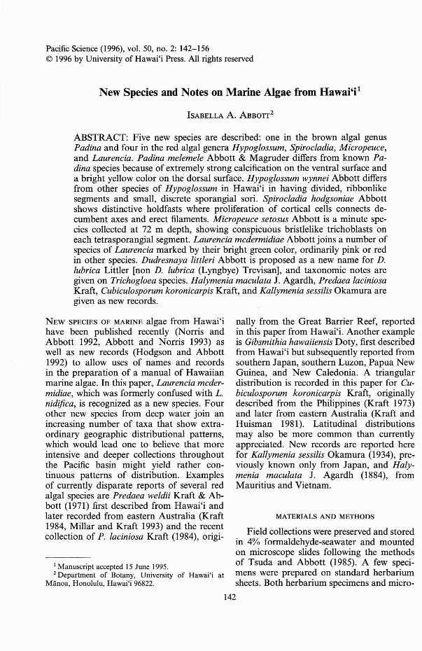
Pacific Science (1996), vol. 50, no. 2: 142-156© 1996 by University of Hawai'i Press. All rights reserved
New Species and Notes on Marine Algae from Hawai'i1
ISABELLA A. ABBorr2
ABSTRACT: Five new species are described: one in the brown algal genusPadina and four in the red algal genera Hypoglossum, Spirocladia, Micropeuce,and Laurencia. Padina melemele Abbott & Magruder differs from known Padina species because of extremely strong calcification on the ventral surface anda bright yellow color on the dorsal surface. Hypoglossum wynnei Abbott differsfrom other species of Hypoglossum in Hawai'i in having divided, ribbonlikesegments and small, discrete sporangial sori. Spirocladia hodgsoniae Abbottshows distinctive holdfasts where proliferation of cortical cells connects decumbentaxes and erect filaments. Micropeuce setosus Abbott is a minute species collected at 72 m depth, showing conspicuous bristlelike trichoblasts oneach tetrasporangial segment. Laurencia mcdermidiae Abbott joins a number ofspecies of Laurencia marked by their bright green color, ordinarily pink or redin other species. Dudresnaya littleri Abbott is proposed as a new name for D.lubrica Littler [non D. lubrica (Lyngbye) Trevisan], and taxonomic notes aregiven on Trichogloea species. Halymenia maculata J. Agardh, Predaea laciniosaKraft, Cubiculosporum koronicarpis Kraft, and Kallymenia sessilis Okamura aregiven as new records.
NEW SPECIES OF MARINE algae from Hawai'ihave been published recently (Norris andAbbott 1992, Abbott and Norris 1993) aswell as new records (Hodgson and Abbott1992) to allow uses of names and recordsin the preparation of a manual of Hawaiianmarine algae. In this paper, Laurencia mcdermidiae, which was formerly confused with L.nidifica, is recognized as a new species. Fourother new species from deep water join anincreasing number of taxa that show extraordinary geographic distributional patterns,which would lead one to believe that moreintensive and deeper collections throughoutthe Pacific basin might yield rather continuous patterns of distribution. Examplesof currently disparate reports of several redalgal species are Predaea weldii Kraft & Abbott (1971) first described from Hawai'i andlater recorded from eastern Australia (Kraft1984, Millar and Kraft 1993) and the recentcollection of P. laciniosa Kraft (1984), origi-
1 Manuscript accepted 15 June 1995.2 Department of Botany, University of Hawai'i at
Manoa, Honolulu, Hawai'i 96822.
nally from the Great Barrier Reef, reportedin this paper from Hawai'i. Another exampleis Gibsmithia hawaiiensis Doty, first describedfrom Hawai'i but subsequently reported fromsouthern Japan, southern Luzon, Papua NewGuinea, and New Caledonia. A triangulardistribution is recorded in this paper for Cubiculosporum koronicarpis Kraft, originallydescribed from the Philippines (Kraft 1973)and later from eastern Australia (Kraft andHuisman 1981). Latitudinal distributionsmay also be more common than currentlyappreciated. New records are reported herefor Kallymenia sessilis Okamura (1934), previously known only from Japan, and Halymenia maculata J. Agardh (1884), fromMauritius and Vietnam.
MATERIALS AND METHODS
Field collections were preserved and storedin 4% formaldehyde-seawater and mountedon microscope slides following the methodsof Tsuda and Abbott (1985). A few specimens were prepared on standard herbariumsheets. Both herbarium specimens and micro-
142
New Marine Algae from Hawai'i-ABBOlT
scope slides labeled IA followed by a number are in collections of I. Abbott at theUniversity of Hawai'i with the intention oftransferring them to the B. P. Bishop Museum (BISH) and other herbaria as the studyof them is completed. Slides labeled HMA orWHM are in BISH. Holotypes are now in theBishop Museum.
DESCRIPTIONS OF NEW TAXA
PHAEOPHYTA
Order DICTYOTALES
Family DICTYOTACEAE
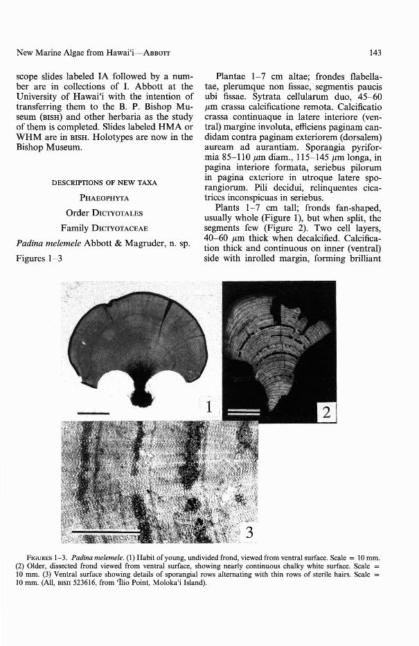
Padina melemele Abbott & Magruder, n. sp.
Figures 1-3
• yiF:!I,-.
143
Plantae 1-7 cm altae; frondes flabellatae, plerumque non fissae, segmentis paucisubi fissae. Sytrata cellularum duo, 45-60J.Lm crassa calcificatione remota. Calcificatiocrassa continuaque in 1atere interiore (ventral) margine invo1uta, efficiens paginam candidam contra paginam exteriorem (dorsalem)auream ad aurantiam. Sporangia pyriformia 85-110 J.Lm diam., 115-145 J.Lm longa, inpagina interiore formata, seriebus pilorumin pagina exteriore in utroque latere sporangiorum. Pili decidui, relinquentes cicatrices inconspicuas in seriebus.
Plants 1-7 cm tall; fronds fan-shaped,usually whole (Figure 1), but when split, thesegments few (Figure 2). Two cell layers,40-60 J.Lm thick when decalcified. Calcification thick and continuous on inner (ventral)side with inrolled margin, forming brilliant
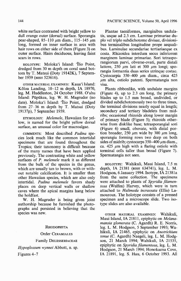
FIGURES 1-3. Padina me/erne/e. (I) Habit of young, undivided frond, viewed from ventral surface. Scale = lO mm.(2) Older, dissected frond viewed from ventral surface, showing nearly continuous chalky white surface. Scale =lO mrn. (3) Ventral surface showing details of sporangial rows alternating with thin rows of sterile hairs. Scale =10 mrn. (All, BISH 523616, from 'Ilio Point, Moloka'i Island).
144
white surface contrasted with bright yellow todull orange outer (dorsal) surface. Sporangiapear-shaped, 85-110 J1.m diam., 115-145 J1.mlong, formed on inner surface in arcs withhair rows on either side of them (Figure 3) onouter surface. Hairs deciduous, leaving faintscars in rows.
HOLOTYPE: Moloka'i Island: 'Ilio Point,dredged from 30 m depth on coral sand bottom by T. Matsui (Doty 19142K), 7 September 1959 (BISH 523616).
OTHER MATERIAL EXAMINED: Kaua'i Island:Koloa Landing, 10-12 m depth, IA 18970,leg. M. Huddleston, 24 October 1988. O'ahuIsland: Piipukea, leg. W. H. Magruder (nodate). Moloka'i Island: 'Tho Point, dredgedfrom 27-36 m depth by T. Matsui (Doty19137p), 7 September 1959.
ETYMOLOGY: Me/eme/e, Hawaiian for yellow, is named for the bright yellow dorsalsurface, an unusual color for macroalgae.
COMMENTS: Most described Padina species look much like the common intertidalspecimens that are found throughout theTropics; their taxonomy is difficult becauseof the many names that have been appliedpreviously. The contrasting white and yellowsurfaces of P. me/eme/e mark it as differentfrom the bulk of the species in the genus,which are usually tan to brown, with or without notable calcification. It is smaller thanother Hawaiian species, which are also onlyintertidal. Padina me/eme/e favors shadyplaces on deep vertical walls or shallowcaves where the apical margins hang belowthe holdfast.
W. H. Magruder is being given jointauthorship because he furnished the photographs and persisted in believing that thespecies was new.
RHODOPHYTA
Order CERAMIALES
Family DELESSERIACEAE
Hypog/ossum wynnei Abbott, n. sp.
Figures 4-7
PACIFIC SCIENCE, Volume 50, Apri11996
Plantae taeniformes, marginibus undulatis, usque ad 2.3 cm. Laminae primariae duplo vel triplo subdichotome divisae, divisionibus terminalibus longitudine prope aequalibus. Laminulae secundariae tertiariaeque excosta. Rhizoidea interdum secus inferioremmarginem laminae primariae. Sori tetrasporangiorum parvi, obverse-ovati, parte distalilatiore, 250 J1.m lati et 300 J1.m longi; sporangia formantia duas series utrinque costae.Cystocarpia 350-400 J1.m diam., circa 425J1.m alta, ostiolo patenti. Spermatangia nonvisa.
Plants ribbonlike, with undulate margins(Figure 4), up to 2.3 cm long, the primaryblades up to 1.5 mm broad, undivided, ordivided subdichotomously two to three times,the terminal divisions nearly equal in length;secondary and tertiary bladelets from midribs; occasional rhizoids along lower marginof primary blade (Figure 5); rhizoids otherwise from disklike base; tetrasporangial sori(Figure 6) small, obovate, with distal portion broader, 250 J1.m wide by 300 J1.m long,sporangia forming rows two deep on bothsides ofmidrib; cystocarps 350-400 J1.m diam.,ca. 425 J1.m high with a flaring ostiole withterminal cells taller than broad (Figure 7).Spermatangia not seen.
HOLOTYPE: Wahikuli, Maui Island, 7.5 mdepth, IA 21381 (BISH 634741), leg. L. M.Hodgson, 6 January 1994. Isotype, IA 21381afrom the same collection. The specimenswere attached to plants of Spyridia fi/amentosa (Wulfen) Harvey, which were in turnattached to Halimeda incrassata (Ellis) Lamouroux. The holotype consists of a pressedspecimen and a microscope slide. Two isotype slides are also available.
OTHER MATERIAL EXAMINED: Wahikuli,Maui Island, IA 21811, epiphytic on Me/anamansia g/omerata (C. Agardh) R. E. Norris,leg. L. M. Hodgson, 5 September 1993; Wahikuli, IA 21465, epiphytic on Anotrichiumtenue (c. Agardh) Naegeli, leg. L. M. Hodgson, 21 March 1994; Wahikuli, IA 21535,epiphytic on Spyridia fi/amentosa, leg. L. M.Hodgson, 21 March 1994; Honokaena Cove,IA 21891, leg. S. Hau, 6 October 1993. All
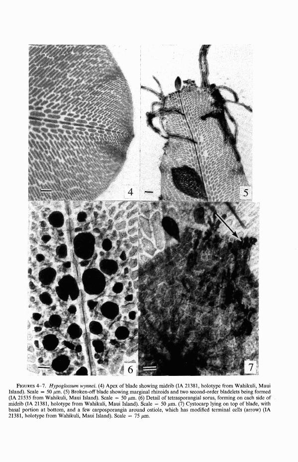
FIGURES 4-7. Hypoglossum wynnei. (4) Apex of blade showing midrib (IA 21381, holotype from Wahikuli, MauiIsland). Scale = 50 11m. (5) Broken-off blade showing marginal rhizoids and two second-order bladelets being formed(IA 21535 from Wahikuli, Maui Island). Scale = 50 11m. (6) Detail of tetrasporangial sorus, forming on each side ofmidrib (lA 21381, holotype from Wahikuli, Maui Island). Scale = 50 11m. (7) Cystocarp lying on top of blade, withbasal portion at bottom, and a few carposporangia around ostiole, which has modified terminal cells (arrow) (IA21381, holotype from Wahikuli, Maui Island). Scale = 75 11m.
146
specimens were collected subtidally at 5 to12 m depth.
ETYMOLOGY: This species is named forProfessor Michael J. Wynne, University ofMichigan, who is the leading student of thegenus Hypoglossum, and who has helped mein understanding the numerous HawaiianspeCImens.
COMMENTS: Hypoglossum wynnei, in termsof apical organization, is a member of theType I, hypoglossoides group of species inwhich all cells of the second-order cell rowsbear third-order rows (Wynne 1988). It canbe separated from other species of the genusin Hawai'i by its branched, broad ribbonlikethallus segments; the remaining species areessentially simple, slender, undivided blades,except for H. caloglossoides Wynne & Kraft,which is distinctive because of the catenate arrangement of blades. It differs fromother tropical species, such as H. anomalumWynne, in lacking branches that emerge between the midrib and margin, and from H.barbatum Okamura in lacking an attenuateapex and elongate tetrasporangial sori. Fromthe simple blades of H. simulans Wynne,Price & Ballantine, H. wynnei differs bylacking the longitudinal rows of inner cellsthat in surface view parallel the midrib(Wynne et al. 1989). Finally, the spatulateto obovate blades of H. minimum Yamada(Yoshida and Mikami 1986) are unlike thoseof H. wynnei.
Family RHODOMELACEAE, Lophothalia group
Spirocladia hodgsoniae Abbott, n. sp.
Figures 8-12
Plante dense cespitosae, usque ad 2 cm,basi contortuplicata ramis decumbentibus eterectis circa 130 /lm, hapteris I-pluribusordinatis corticatis usque ad 600 /lm diam.affixae. Ab infimo visae, cellulae hapterieffusae et furcatae, parietibus incrassatis peragratis rhizoideis internis ex cellulis inferioribus pericentralibus formatis. Bases ramorum erectorum cortice similari etsi limitatoincrassatae; bases continguae plurium ramorum erectorum connexae, et auctae paucis
PACIFIC SCIENCE, Volume 50, April 1996
rhizoideis uni-vel multicellularibus in axibus decumbentibus plerumque hic illic factis.Axes erecti ex textura basali formati, raroramosi, vel ramosi dichotome propwe extremitates basales. Ceterae proprietates vegetativae et structurae reproductivae similesceteris speciebus Spirocladiae.
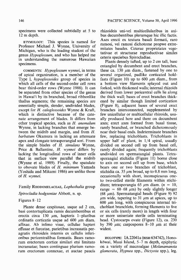
Plants densely tufted, up to 2 cm tall, baseentangled by decumbent and erect branches,these ca. 130 /lm diam., fastened by one toseveral organized, padlike corticated holdfasts (Figure 10) up to 600 /lm diam., froma bottom view filaments splayed out andforked, with thickened walls; internal rhizoidsderived from lower pericentral cells lie alongthe thick wall; bases of erect branches thickened by similar though limited cortication(Figure 8), adjacent bases of several erectbranches interconnected, supplemented byfew unicellular or multicellular rhizoids, usually produced here and there on decumbentaxes; erect axes formed from basal tissue,rarely branched, or branched dichotomouslynear their basal ends. Indeterminate branchesfew, replacing trichoblasts. Trichoblasts inupper half of plant, in 1/4 spiral, usuallydivided on second cell up from basal cell,rarely divided again; frequently trichoblastsundivided on spermatangial plants. Tetrasporangial stichidia (Figure II) borne closeto axis on second cell up from base, whichbears one or two branches of trichoblast,stichidia ca. 35 /lm broad, up to 0.8 mm long,occasionally with short, inconspicuous oneto two-celled sterile filaments within stichidium; tetrasporangia 65 /lm diam. (n = 10,range = 60-68 /lm) by only slightly longer(68 /lm). Spermatangial heads (Figure 9) 92/lm wide, tapering to 31 /lm at apices, up to600 /lm long, with conspicuous internal trichoblast branchlets, forming filaments to fiveor six cells (rarely more) in length with fouror more uniseriate sterile cells terminatinghead. Cystocarps ovate (Figure 12), ca. 230by 390 /lm; carpospores 8-10 /lm at theirwidest.
HOLOTYPE: IA 22041 a (BISH 634742), Honokawai, Maui Island, 5-7 m depth, epiphyticon a variety of macroalgae (Melanamansiaglomerata, Hypnea spp., Dictyota spp.), leg.
New Marine Algae from Hawai'i-ABBOTI
FIGURES 8-12. Spirocladia hodgsoniae. (8) Cortications (arrow) on lower erect axes. Scale = 50 /-1m. (9)Spermatangial head with two trichoblast filaments (arrows), characteristic of the genus. Scale = 100 /-1m. (10)Holdfast of solid mass of corticated basal cells, characteristic of this species. Scale = 50 /-1m. (II) Stichidiaof tetrasporangia, forming on trichoblasts. Scale = 150/-1m. (12) Oval cystocarps with simple ostioles. Scale =150 /-1m. (All from holotype or isotype slides, IA 22041[BISH 634742) from Honokowai, Maui Island).
L. M. Hodgson, 22 October 1994. Isotypesunder the same number, from the same collection. The holotype is represented by several specimens on a herbarium sheet, witha spermatangial slide from the same collection. Isotype slides contain spermatangial,
147
cystocarpic, or tetrasporangial plants, eitherseparated or on the same slides. Isotypes arein BISH, UC, MELU, NSW, SAP. Dried herbarium material will be distributed also.
OTHER MATERIAL EXAMINED: O'ahu Island:Wai'alae Beach Park, intertidal on rock inturf, IA 21399, leg. C. M. Smith, 13 February 1994; Makaha, subtidal, 7-15 m depthon Spyridia jilamentosa, IA 18846, leg. L.M. Hodgson, 5 August 1988; same place,depth, collector, date, epiphytic on Dictyotasp., IA 18886. Maui Island: Puamana, east ofLahaina on rock, 8-10 m depth, IA 21851,leg. L. M. Hodgson, 9 October 1993; offMala wharf, 7-9 m depth on eroded coral,IA 21393, leg. L. M. Hodgson, 13 February1994; Honokowai, 5-7 m depth, on varietyof algae growing on Halimeda incrassata,leg. L. M. Hodgson, 22 October 1994 (including type material); Honokeana Cove,10-12 m depth, on Halimeda incrassata, IA21903, IA 21906-21909, leg. S. Hau, 6 October 1993. Except for the O'ahu collection,which was intertidal, all collections were subtidal from 5 to 10m depth.
ETYMOLOGY: Hodgsoniae is named for thecollector of the type material and my colleague, Lynn M. Hodgson, whose carefulcollections along the West Maui coast haveyielded many interesting records and substantially increased our knowledge of theshallow and deeper water algae there.
COMMENTS: The occurrence of sterile filaments (modified trichoblasts) in the spermatangial heads and, to a lesser degree, theiroccasional presence in tetrasporangial stichidia is a generic-level characteristic of thesesmall, superficially Polysiphonia-like plants.The vegetative structure and the basal portions of S. hodgsoniae, however, are verydifferent from other species of the genus:S. barodensis B0rgesen (1933) (the type), S.minor Nasr (1939) from the Red Sea, andS. loochooensis (Yendo) Yoshida (1989) fromthe Ryukyu Islands. Both S. barodensis andS. loochooensis are heavily corticated throughout the plants, whereas S. minor is corticatedtoward the base, and, as described above, S.hodgsoniae is particularly corticated at the
148
bases of erect branches and in the regionsof attachment. None of these species has theclumping of cells, cortications, and branchingat the base of the plant as does S. hodgsoniae.
Spirocladia barodensis was previously reported from Hawai'i by Hollenberg (1968). Itis larger and nearly completely corticated.
Family RHODOMELACEAE
Tribe LOPHOTHALIEAE
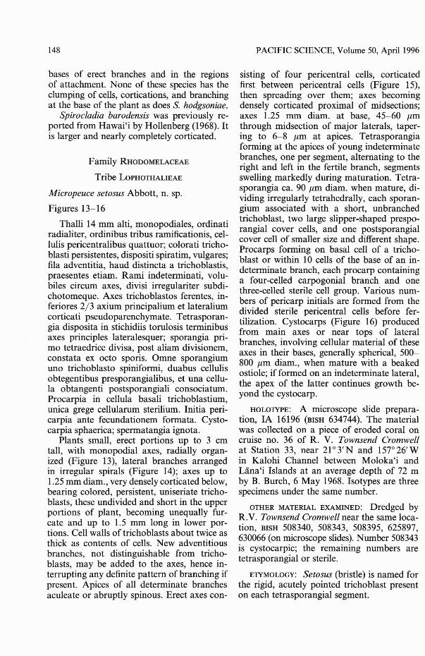
Micropeuce setosus Abbott, n. sp.
Figures 13-16
Thalli 14 rom alti, monopodiales, ordinatiradialiter, ordinibus tribus ramificationis, cellulis pericentralibus quattuor; colorati trichoblasti persistentes, dispositi spiratim, vulgares;fila adventitia, haud distincta a trichoblastis,praesentes etiam. Rami indeterminati, volubiles circum axes, divisi irregulariter subdichotomeque. Axes trichoblastos ferentes, inferiores 2/3 axium principalium et lateraliumcorticati pseudoparenchymate. Tetrasporangia disposita in stichidiis torulosis terminibusaxes principles lateralesquer; sporangia primo tetraedrice divisa, post aliam divisionem,constata ex octo sporis. Omne sporangiumuno trichoblasto spiniformi, duabus cellulisobtegentibus presporangialibus, et una cellula obtangenti postsporangiali consociatum.Procarpia in cellula basali trichoblastium,unica grege cellularum sterilium. Initia pericarpia ante fecundationem formata. Cystocarpia sphaerica; spermatangia ignota.
Plants small, erect portions up to 3 emtall, with monopodial axes, radially organized (Figure 13), lateral branches arrangedin irregular spirals (Figure 14); axes up to1.25 mm diam., very densely corticated below,bearing colored, persistent, uniseriate trichoblasts, these undivided and short in the upperportions of plant, becoming unequally furcate and up to 1.5 rom long in lower portions. Cell walls of trichoblasts about twice asthick as contents of cells. New adventitiousbranches, not distinguishable from trichoblasts, may be added to the axes, hence interrupting any definite pattern of branching ifpresent. Apices of all determinate branchesaculeate or abruptly spinous. Erect axes con-
PACIFIC SCIENCE, Volume 50, April 1996
sisting of four pericentral cells, corticatedfirst between pericentral cells (Figure 15),then spreading over them; axes becomingdensely corticated proximal of midsections;axes 1.25 mm diam. at base, 45-60 Ilmthrough midsection of major laterals, tapering to 6-8 Ilm at apices. Tetrasporangiaforming at the apices of young indeterminatebranches, one per segment, alternating to theright and left in the fertile branch, segmentsswelling markedly during maturation. Tetrasporangia ca. 90 Ilm diam. when mature, dividing irregularly tetrahedrally, each sporangium associated with a short, unbranchedtrichoblast, two large slipper-shaped presporangial cover cells, and one postsporangialcover cell of smaller size and different shape.Procarps forming on basal cell of a trichoblast or within 10 cells of the base of an indeterminate branch, each procarp containinga four-celled carpogonial branch and onethree-celled sterile cell group. Various numbers of pericarp initials are formed from thedivided sterile pericentral cells before fertilization. Cystocarps (Figure 16) producedfrom main axes or near tops of lateralbranches, involving cellular material of theseaxes in their bases, generally spherical, 500800 Ilm diam., when mature with a beakedostiole; if formed on an indeterminate lateral,the apex of the latter continues growth beyond the cystocarp.
HOLOTYPE: A microscope slide preparation, IA 16196 (BISH 634744). The materialwas collected on a piece of eroded coral oncruise no. 36 of R. V. Townsend Cromwellat Station 33, near 21 0 3' Nand 1570 26' Win Kalohi Channel between Moloka'i andUina'i Islands at an average depth of 72 mby B. Burch, 6 May 1968. Isotypes are threespecimens under the same number.
OTHER MATERIAL EXAMINED: Dredged byR.V. Townsend Cromwell near the same location, BISH 508340, 508343, 508395, 625897,630066 (on microscope slides). Number 508343is cystocarpic; the remaining numbers aretetrasporangial or sterile.
ETYMOLOGY: Setosus (bristle) is named forthe rigid, acutely pointed trichoblast presenton each tetrasporangial segment.
FIGURES 13-16. Micropeuce setosus. (13) Distal portion of plant showing central axis and (left) terminal portionof indeterminate branch transformed into fertile branch (IA 16196, dredged by R.V. Townsend Cromwell). Scale =I mm. (14) Detail of fertile branch showing spinelike trichoblast associated with each tetrasporangial segment(lA 16196, dredged by R.V. Townsend Cromwell). Scale = I mrn. (IS) Cortication between pericentral cells(arrow), spreading over them as axis matures. Scale = 25 jJ.m. (16) Lateral view of globose cystocarp (ostiole out ofview), attached by very short stalk to main axis. Scale = 500 jJ.m.
ISO
COMMENTS: Micropeuce contains six poorlyknown species, four from Australia and oneeach from the general Caribbean area andthe Gulf of California, all of which aremuch larger than the Hawaiian species. Theanatomy of the 12 small plants available ofM. setosus shows that the species is closelyrelated to Lophothalia verticil/ata (Harvey)Kiitzing (1849) in many features, as interpreted by Parsons (1975). Two features, themodification of terminal branches into stichidia of tetrasporangia and the persistent, colored trichoblasts, are considered to be important similarities. The two genera may differin details of reproduction, which could notbe completely studied because of the poorlypreserved material. The genus Micropeuceas outlined by Kylin (1956:511) has five pericentral cells, whereas M. setosus has four,but the latter number is accommodated withinthe tribe Lophothalieae as circumscribed byParsons (1975:685). From the best-describedspecies of Micropeuce, M mucronata (Harvey) Kylin by Jo1y and Oliveira (1966) asHeterodasya sertularioides, M. setosus isdistinguished by being smaller, with fourinstead of five pericentra1 cells, and withsimpler, mostly unbranched trichob1asts. Evaluation of these features is hampered by therarity of collections.
Micropeuce has not previously been reported from the North Pacific.
Family RHODOMELACEAE, Laurencia group
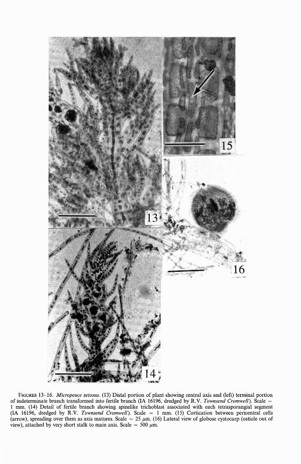
Laurencia mcdermidiae Abbott, n. sp.
Figure 17
Plantae 5-6 cm altae, virides vividae, colorem retinentes exsiccatae, caespitosae, frondibus unibus ad aliquot superantibus ceteras. Axes teretes, 0.7-1.2 mm diam., ramosiradialiter; ramuli termina1es vel simplicesvel fascicu1is brevibus ramuncolorum. Cellulae corticales subquadratae in sectione transversa, non protrudentes, 20-22 f.lm diam.;"corpora ceraqsi" praesentia. Cellulae medullae grandes parieytibus crassis, autem noncrassitudinibus lenticularibus.
Plants 5-6 cm tall (Figure 17), brightgreen except for reddish bases of erect fronds,
PACIFIC SCIENCE, Volume 50, April 1996
FIGURE 17. Laurencia mcdermidiae, habit of plant.Holotype, IA 21388, Makapu'u Point, O'ahu Island.Scale = I em.
retaining color when dried, growing in clumpswith several fronds overtopping others. Axesterete, 0.7-1.2 mm diam., densely radiallybranched, the terminal branchlets either simple or with short clusters of ultimate branchlets; in transverse section, cortical cells withsecondary pit connections, subquadrate, notprojecting, 20-22 f.lm diam., corps en ceriseone to two (mostly two) per cortical cell.Mature medullary cells with very thick walls,as much as 1/5 the diameter of the cell, butwith no lenticular thickenings. Tetrasporangia parallel type, each sporangium ca. 70 f.lmdiam. No gametophytes seen.
HOLOTYPE: IA 21388 (BISH 634743), Makapu'u Point, O'ahu Island, leg. K. Beach,7 February 1994. Isotypes: two sheets underthe same number.
OTHER MATERIAL EXAMINED: O'ahu Island:IA 293, 295, near Halona blowhole, leg. I.Abbott, 19 December 1943; IA 827, Diamond Head, leg. D. P. Abbott, 8 July 1945;IA 21325, Makaha, leg. K. Beach, 3 June1993. Maui Island: IA 14596, Hokii'ula,near Hana, 26 August 1976.
New Marine Algae from Hawai'i-ABBoIT
Previous reports of this species (some encompassing the description of L. nidifica J.Agardh) were made by Saito (1969), Abbott(1984), and as Laurencia species "green" byMcDermid (1988: 244, figs. 34, 35) and Smith(1992). Besides O'ahu and Maui Islands,the distribution also includes the islands ofKaua'i, Moloka'i, and Lana'i (McDermid1988).
ETYMOLOGY: This species is named forKarla J. McDermid, who recently reviewedand identified Hawaiian species of Laurencia.
COMMENTS: This species was previouslypartially described and well illustrated asLaurencia "green" by McDermid (1988: 244245, figs. 34-35). It was McDermid whopointed out the differences shown by thistaxon from L. nidifica, with which it hadbeen confused. McDermid (1988) found thatthe name L. nidifica should be applied to thereddish to straw-colored plants that showedlenticular thickenings (resembling a thickened U) in cells of the medulla, whereasthe green ones (which she called Laurencia"green") differed in lacking these thickenings. Moreover, alternate-opposite branchlets were formed more densely (Figure 17)in Laurencia "green," whereas branching wasmore open and mostly alternate in L. nidifica (see McDermid 1988: figs. 18 and 20, thelatter an illustration of the type specimenin the Agardh herbarium). McDermid (1988:244) also stated that biochemical compoundsin these two taxa were different.
Subsequently, other researchers have published on "green Laurencias," including GilRodriguez and Haroun (1992), who compared seven "green" species (actually six,because L. nidifica is not green). Most ofthese taxa, including L. viridis Gil-Rodriguez& Haroun, have features resembling L.mcdermidiae. From L. viridis, L. mcdermidiaediffers principally in the branching patternand the very thick walls of the maturemedullary cells. In an illustration from GilRodriguez and Haroun (1992, fig. 1b), theplants show paniculate branching at the topof straight, naked axes about 1/2 the totallength of the axis. In L. mcdermidiae, lateralbranches are given off the central axis within
151
2 mm of the fleshy base and are radiallybranched throughout the upward course ofthe axis (ca. 5-6 cm); each lateral is rebranched once to three times, each order being shorter than the previous one. The totallength of the lateral might be 3.5 cm, with asecond-order spread of 0.5-1.0 cm, thus avery different habit from L. viridis.
MISCELLANEOUS NOTES
Dudresnaya littleri Abbott, n. name
Dudresnaya lubrica M. M. Littler, Br. Phycol. J. 9: 149-156, figs. 1-20, 1974, a laterhomonym of D. lubrica (Lyngbye) Trevisan, Saggio monogr. alghe Coccotalle,p. 105, 1848 (basionym Gigartina lubricaLyngbye, Tentamen, p. 45, pI. 12a, 1819).
Dudresnaya littleri is not a common spe-cies in Hawai'i (its type locality). To myknowledge, it has only been collected oncesince its original collection at P6ka'i Bay, onthe west coast of O'ahu Island. Nevertheless,it can be easily separated from the morecommon D. hawaiiensis R. K. S. Lee becauseof its more slender branches and shorterstature.
Trichogloea lubrica J. Agardh
Figures 18-21, 23
Trichogloea lubrica J. Agardh, Sp., gen., ord.algarum 3(1): 514, 1876. Synonyms: Trichogloea subnuda Howe, J. Wash. Acad.Sci. 24: 33-34, fig. 1, 1934. Nemalion ramulosum of Tilden in [Tilden's] AmericanAlgae, Century V, no. 419, 1900. Liagoralubrica Harvey, Friendly Island Algae, no.46, nomen nudum.
Trichogloea lubrica takes on many branching patterns, and the sizes of branches varyfrom 2 to 8 mm diam.; hence if identification is being made on habit, it is easy to be inerror. Indeed, it would be hazardous to useone plant form to exemplify any of the species of Trichogloea. Figures 18-19 illustratetype material (possibly isotypes) of T lubrica,from the same collection from Tonga (theFriendly Islands). Figures 20-21 show two
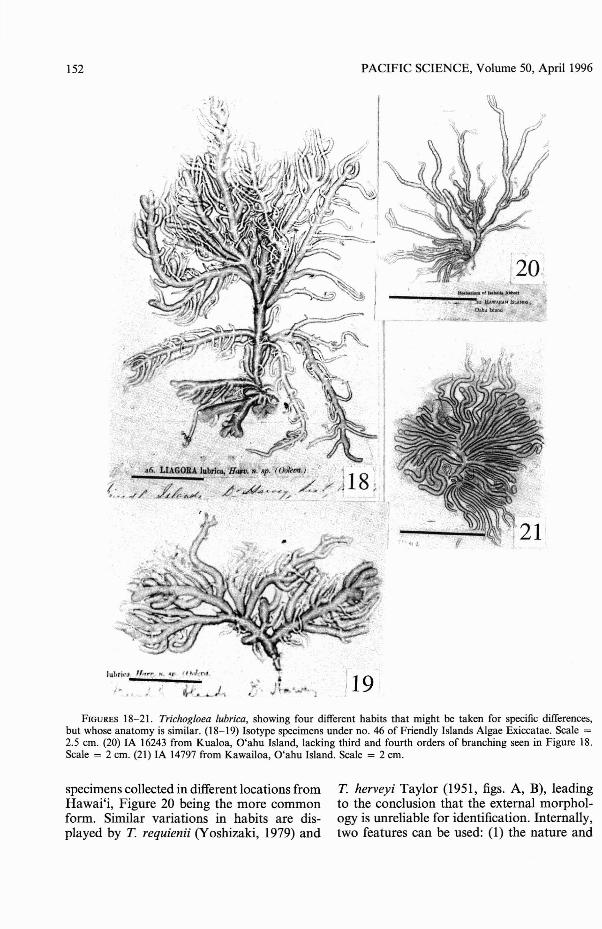
152
Juhrkl:'. -
J~-
PACIFIC SCIENCE, Volume 50, April 1996
----~~':~.,;*:.::;~~:;::,~H:=~TUNNOihU1s1I11d"ij;
)19'FIGURES 18-21. Trichogloea lubrica, showing four different habits that might be taken for specific differences,
but whose anatomy is similar. (18-19) Isotype specimens under no. 46 of Friendly Islands Algae Exiccatae. Scale =2.5 em. (20) IA 16243 from Kualoa, O'ahu Island, lacking third and fourth orders of branching seen in Figure 18.Scale = 2 em. (21) IA 14797 from Kawailoa, O'ahu Island. Scale = 2 em.
specimens collected in different locations fromHawai'i, Figure 20 being the more commonform. Similar variations in habits are displayed by T. requienii (Yoshizaki, 1979) and
T. herveyi Taylor (1951, figs. A, B), leadingto the conclusion that the external morphology is unreliable for identification. Internally,two features can be used: (l) the nature and
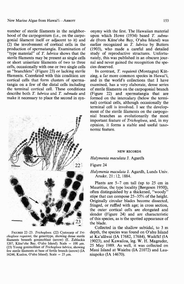
New Marine Algae from Hawai'i-ABBOTI
number of sterile filaments in the neighborhood of the carpogonium (i.e., on the carpogonial filament itself or adjacent to it) and(2) the involvement of cortical cells in theproduction of spermatangia. Examination of"type material" of T lubrica shows that thesterile filaments may be present as single cellsor short uniseriate filaments of two to threecells, occasionally with one or two single cellsas "brancWets" (Figure 23) or lacking sterilefilaments. Correlated with this condition arecortical cells that form clusters of spermatangia on a few of the distal cells includingthe terminal cortical cell. These conditionsdescribe both T lubrica and T subnuda andmake it necessary to place the second in syn-
FIGURES 22-23. Trichogloea. (22) Cystocarp of Trichogloea requienii, the generitype, showing dense sterilefilaments beneath gonimoblast (arrow) (E. Zablackis1207, Kane'ohe Bay, O'ahu Island). Scale = 100 /lm.(23) Young gonimoblast of Trichogloea lubrica, showingfew sterile filaments at base of fertile branch (arrow) (IA16246, Kualoa, O'ahu Island). Scale = 25/lm.
153
onymy with the first. The Hawaiian materialupon which Howe (1934) based T subnuda (from Kane'ohe Bay, O'ahu Island) wasearlier recognized as T lubrica by Butters(1903), who made a careful and detailedstudy of reproductive structures. Unfortunately, this was published in an obscure journal and never gained the recognition the species deserved.
In contrast, T requienii (Montagne) Kiitzing, a far more common species in Hawai'i,and in the world's collections that I haveexamined, has a very elaborate, dense seriesof sterile filaments on the carpogonial branch(Figure 22) and spermatangia that areformed on the intercalary (below the terminal) cortical cells, although occasionally theterminal cell is involved. I see the development of the sterile filaments on the carpogonial branches as evolutionarily the mostimportant feature of Trichogloea, and, in myopinion, it forms a stable and useful taxonomic feature.
NEW RECORDS
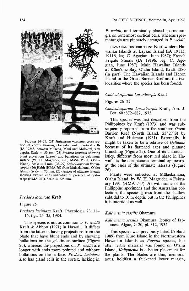
Halymenia maculata J. Agardh
Figure 24
Halymenia maculata J. Agardh, Lunds Univ.Arsskr. 21 : 12, 1884.
Plants are 5-7 cm tall (up to 25 cm inMauritius, the type locality [Bargesen 1950)),often distinguished by a thickened, "woody"stipe that can compose 25-35% of the height.Originally circular blades become dissected,fringed, or ruffled with age; in cross section,the outer cortical cells are elongated andslender (Figure 24) and are characteristicof this species, as is the spotted appearance ofthe blade.
Collected in the shallow subtidal, to 3 mdepth, the species was found on O'ahu Islandat Ka'alawai (IA 17682, 17684), Waikik:"i (IA19023), and Kawailoa, leg. W. H. Magruder,25 May 1989. As well, it was collected onMaui Island at Waiehu (IA 21072) and Launiupoko (IA 14670).
154
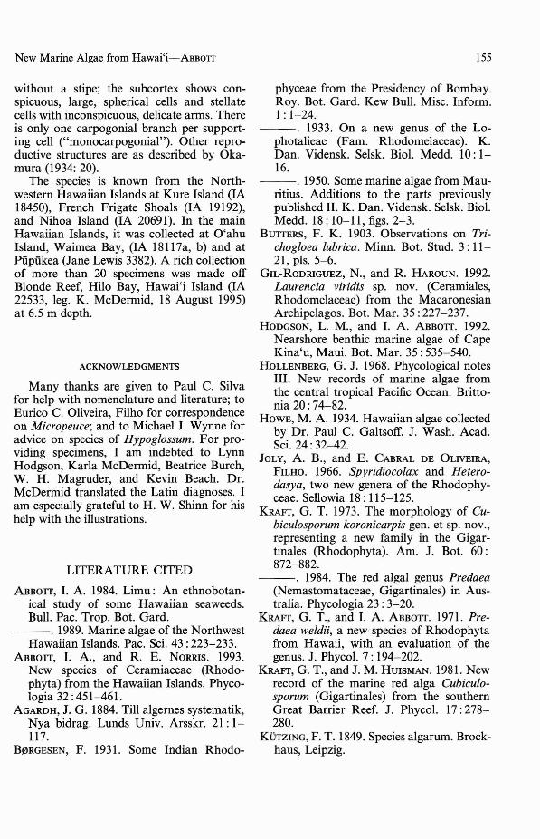
FIGURES 24-27. (24) Halymenia maculata, cross section of cortex showing elongated outer cortical cells(IA 19245, between Makena, Maui and Molokini, 5 mdepth). Scale = 50 Ilm. (25) Predaea laciniosa showingblunt projections (arrow) and bullations on gelatinoussurface (W. H. Magruder, s.n., Ma'ili Point, O'ahuIsland). Scale = 5 mm. (26-27) Cubiculosporum koronicarpus. (26) Habit (HMA 767 from Malaekahana, O'ahuIsland). Scale = 75 mm. (27) Apices of ultimate lateralsshowing swollen ends indicative of presence of cystocarps (HMA 767). Scale = 225 mm.
Predaea laciniosa Kraft
Figure 25
Predaea laciniosa Kraft, Phycologia 23 : IllS, figs. 25-35, 1984.
This species is not as common as P. weldiiKraft & Abbott (1971) in Hawai'i. It differsfrom the latter in having projections from theblade that have blunt ends and by showingbullations on the gelatinous surface (Figure25), whereas the projections on P. weldii arelonger with ends more pointed and withoutbullations on the surface. Predaea laciniosaalso has gland cells in the cortex, lacking in
PACIFIC SCIENCE, Volume 50, April 1996
P. weldii, and terminally placed spermatangia on outermost cortical cells, whereas spermatangia are pinnately arranged in P. weldii.
HAWAllAN DISTRIBUTION: Northwestern Hawaiian Islands at Laysan Island (IA 19115,19116, leg. C. Agegian, June 1987); FrenchFrigate Shoals (IA 19196, leg. C. Agegian, June 1987). Main Hawaiian Islandsat Kane'ohe Bay, O'ahu Island, Kraft 1280(in part). The Hawaiian Islands and HeronIsland in the Great Barrier Reef are the twolocalities where the species has been found.
Cubiculosporum koronicarpis Kraft
Figures 26-27
Cubiculosporum koronicarpis Kraft, Am. J.Bot. 60: 872-882, 1973.
This species was first described from thePhilippines by Kraft (1973) and was subsequently reported from the southern GreatBarrier Reef (North Island, 23° 27' S) byKraft and Huisman (1981). Externally, itmight be taken to be a relative of Gelidiumbecause of its flattened axes and pinnatebranching (Figure 25). One of its characteristics, different from most red algae in Hawai'i, is the conspicuous terminal cystocarpsat the ends of the ultimate laterals (Figure26).
Plants were collected at Malaekahana,O'ahu Island, by W. H. Magruder, 4 February 1991 (HMA 747). As with some of thePhilippine specimens and the Australian collection, the species grows from the shallowsubtidal to 10m depth, but in the Philippinesit is intertidal as well.
Kallymenia sessilis Okamura
Kallymenia sessilis Okamura, leones of Japanese Algae, 7: 20, pI. 312, 1934.
This species was previously listed (Abbott1989) from Kure Island in the NorthwesternHawaiian Islands as Pugetia species, butafter fertile material was found on O'ahuIsland, Kallymenia is a better placement forthe plants. The blades are thin, membranous, holdfast a thickened lower margin,
New Marine Algae from Hawai'i-ABBOTI
without a stipe; the subcortex shows conspicuous, large, spherical cells and stellatecells with inconspicuous, delicate anns. Thereis only one carpogonial branch per supporting cell ("monocarpogonial"). Other reproductive structures are as described by Okamura (1934: 20).
The species is known from the Northwestern Hawaiian Islands at Kure Island (IA18450), French Frigate Shoals (IA 19192),and Nihoa Island (IA 20691). In the mainHawaiian Islands, it was collected at O'ahuIsland, Waimea Bay, (IA 18117a, b) and atPupiikea (Jane Lewis 3382). A rich collectionof more than 20 specimens was made offBlonde Reef, Hilo Bay, Hawai'i Island (IA22533, leg. K. McDennid, 18 August 1995)at 6.5 m depth.
ACKNOWLEDGMENTS
Many thanks are given to Paul C. Silvafor help with nomenclature and literature; toEurico C. Oliveira, Filho for correspondenceon Micropeuce; and to Michael J. Wynne foradvice on species of Hypoglossum. For providing specimens, I am indebted to LynnHodgson, Karla McDermid, Beatrice Burch,W. H. Magruder, and Kevin Beach. Dr.McDennid translated the Latin diagnoses. Iam especially grateful to H. W. Shinn for hishelp with the illustrations.
LITERATURE CITED
ABBOTT, I. A. 1984. Limu: An ethnobotanical study of some Hawaiian seaweeds.Bull. Pac. Trop. Bot. Gard.
---.1989. Marine algae of the NorthwestHawaiian Islands. Pac. Sci. 43: 223-233.
ABBOTT, I. A, and R. E. NORRIS. 1993.New species of Ceramiaceae (Rhodophyta) from the Hawaiian Islands. Phycologia 32: 451-461.
AGARDH, J. G. 1884. Till algernes systematik,Nya bidrag. Lunds Univ. Arsskr. 21: 1117.
B0RGESEN, F. 1931. Some Indian Rhodo-
155
phyceae from the Presidency of Bombay.Roy. Bot. Gard. Kew Bull. Misc. Infonn.I: 1-24.
---. 1933. On a new genus of the Lophotalieae (Fam. Rhodomelaceae). K.Dan. Vidensk. Selsk. BioI. Medd. 10: 116.
---.1950. Some marine algae from Mauritius. Additions to the parts previouslypublished II. K. Dan. Vidensk. Selsk. BioI.Medd. 18: 10-11, figs. 2-3.
BUTTERS, F. K. 1903. Observations on Trichogloea lubrica. Minn. Bot. Stud. 3: 1121, pIs. 5-6.
GIL-RODRIGUEZ, N., and R. HAROUN. 1992.Laurencia viridis sp. nov. (Ceramiales,Rhodomelaceae) from the MacaronesianArchipelagos. Bot. Mar. 35: 227-237.
HODGSON, L. M., and I. A ABBOTT. 1992.Nearshore benthic marine algae of CapeKina'u, Maui. Bot. Mar. 35: 535-540.
HOLLENBERG, G. J. 1968. Phycological notesIII. New records of marine algae fromthe central tropical Pacific Ocean. Brittonia 20: 74-82.
HOWE, M. A 1934. Hawaiian algae collectedby Dr. Paul C. Galtsoff. J. Wash. Acad.Sci. 24: 32-42.
JOLY, A B., and E. CABRAL DE OLIVEIRA,FILHO. 1966. Spyridiocolax and Heterodasya, two new genera of the Rhodophyceae. Sellowia 18: 115-125.
KRAFT, G. T. 1973. The morphology of Cubiculosporum koronicarpis gen. et sp. nov.,representing a new family in the Gigartinales (Rhodophyta). Am. J. Bot. 60:872-882.
---. 1984. The red algal genus Predaea(Nemastomataceae, Gigartinales) in Australia. Phycologia 23: 3-20.
KRAFT, G. T., and I. A ABBOTT. 1971. Predaea weldii, a new'species of Rhodophytafrom Hawaii, with an evaluation of thegenus. J. PhycoI. 7: 194-202.
KRAFT, G. T., and J. M. HUISMAN. 1981. Newrecord of the marine red alga Cubiculosporum (Gigartinales) from the southernGreat Barrier Reef. J. PhycoI. 17: 278280.
KOTzING, F. T. 1849. Species algarum. Brockhaus, Leipzig.
156
KYLIN, H. 1956. Die Gattungen der Rhodophyceen. C. W. K. G1eerups, Lund.
McDERMID, K. J. 1988. Laurencia from theHawaiian Islands. Pages 231-247 in I. A.Abbott (ed.), Taxonomy of economic seaweeds, vol. 2. California Sea Grant Publication No. T-CSGCP-018, University ofCalifornia, La Jolla, California.
MILLAR, A. J. K., and G. T. KRAFT. 1993.Catalogue of marine and freshwater redalgae (Rhodophyta) of New South Wales,including Lord Howe Island, south-western Pacific. Aust. Syst. Bot. 6 : 1-90.
NASR, A. H. 1939. On a new species of theRhodomelaceae from Egypt. Rev. Algol.11 : 332-337.
NORRIS, R. E., and I. A. ABBOlT. 1992. Newtaxa of Ceramieae (Rhodophyta) fromHawai'i. Pac. Sci. 46: 453-465.
OKAMURA, K. 1934. leones of Japanese Algae7(3): 20, pI. 312.
PARSONS, M. J. 1975. Morphology and taxonomy of the Dasyaceae and the Lophotha1ieae (Rhodome1aceae) of the Rhodophyta. Aust. J. Bot. 23: 549-713.
SAITO, Y. 1969. The algal genus Laurenciafrom the Hawaiian Islands, the PhilippineIslands and adjacent areas. Pac. Sci. 23:148-160.
SMITII, C. M. 1992. Diversity in intertida'lhabitats: An assessment of the marine algae of select high islands in the HawaiianArchipelago. Pac. Sci. 46: 466-479.
PACIFIC SCIENCE, Volume 50, Apri11996
TAYLOR, W. R. 1951. Structure and taxonomic status of Trichogloea herveyi. Hydrobiologia 3: 113-121.
TSUDA, R. T., and I. A. ABBOlT. 1985. Collection, handling, preservation, and logistics. Pages 67-86 in M. M. Littler andD. S. Littler (eds.), Handbook of phycological methods ecological field methods:Macroa1gae. Cambridge Univ. Press.
WYNNE, M. J. 1988. A reassessment of theHypoglossum group (De1esseriaceae, Rhodophyta), with a critique of its genera.He1gol. Meeresunters. 42: 511-534.
WYNNE, M. J., I. R. PRICE, and D. L. BALLANTINE. 1989. Distinctions between Hypoglossum barbatum Okamura, H. minimum Yamada and H. simulans sp. nov.(Delesseriaceae, Rhodophyta). Phycologia28: 28-38.
YOSHIDA, T. 1989. Notes on Spirocladia 100
chooensis (Yendo) Yoshida, comb. nov.(Rhodomelaceae, Rhodophyta). Jpn. J.Phycol. 37: 271-273.
YOSHIDA, T., and H. MIKAMI. 1986. Observations on morphology of Hypoglossumminimum Yamada and H. geminatum Okamura (Delesseriaceae, Rhodophyta). Jpn.J. Phycol. 34: 177-181.
YOSHIZAKI, M. 1979. Morphology and taxonomy of the Japanese representatives ofNemaliales (3) Thallus structure and reproductive organs of Trichogloea requienii. J. Jpn. Bot. 54: 225-233.