Embed Size (px)
Citation preview
New Life for an Old Drug: the Anthelmintic Drug NiclosamideInhibits Pseudomonas aeruginosa Quorum Sensing
Francesco Imperi,a Francesco Massai,b Cejoice Ramachandran Pillai,b Francesca Longo,b Elisabetta Zennaro,b Giordano Rampioni,b
Paolo Visca,b Livia Leonib
Department of Biology and Biotechnology Charles Darwin, Sapienza University of Rome, Rome, Italya; Department of Biology, University Roma Tre, Rome, Italyb
The need for novel antibacterial strategies and the awareness of the importance of quorum sensing (QS) in bacterial infectionshave stimulated research aimed at identifying QS inhibitors (QSIs). However, clinical application of QSIs identified so far is stilldistant, likely due to their unsuitability for use in humans. A promising way to overcome this problem is searching for anti-QSside activity among the thousands of drugs approved for clinical use in the treatment of different diseases. Here, we applied thisstrategy to the search for QSIs, by screening a library of FDA-approved compounds for their ability to inhibit the QS response inthe Gram-negative pathogen Pseudomonas aeruginosa. We found that the anthelmintic drug niclosamide strongly inhibits the P.aeruginosa QS response and production of acyl-homoserine lactone QS signal molecules. Microarray analysis showed thatniclosamide affects the transcription of about 250 genes, with a high degree of target specificity toward the QS-dependent regu-lon. Phenotypic assays demonstrated that niclosamide suppresses surface motility and production of the secreted virulence fac-tors elastase, pyocyanin, and rhamnolipids, and it reduces biofilm formation. In accordance with the strong antivirulence activ-ity disclosed in vitro, niclosamide prevented P. aeruginosa pathogenicity in an insect model of acute infection. Besides thefinding that an FDA-approved drug has a promising antivirulence activity against one of the most antibiotic-resistant bacterialpathogens, this work provides a proof of concept that a lateral anti-QS activity can be detected among drugs already used in hu-mans, validating a new approach to identify QSIs that could easily move into clinical applications.
The introduction of antibiotics into clinical practice at the mid-dle of the 20th century was a milestone in the history of med-
icine. However, the original expectation that all bacterial infec-tions could be defeated one day by antibiotics was soondiminished by the emergence of antibiotic-resistant strains,prompting the still-ongoing race for the discovery of new antibac-terial agents. While the treatment of infections sustained by anti-biotic-resistant bacteria has high socio-economic costs and repre-sents a major health problem worldwide, the pharmaceuticalindustry has dramatically reduced investments in antibiotics re-search. As traditional antibiotic research appears to be helpless incoping with the emergence of antibiotic-resistant strains, novelscientifically sound, and cost-effective approaches should be un-dertaken in order to identify new drugs (1).
Selective optimization of side activities of drug molecules (theSOSA approach) is a smart strategy for the identification of newpotential drugs (2). A limited number of highly diverse drugswhose use in humans has already been approved are screened forside activities against unrelated diseases. Once a hit compound hasbeen found, it can be either tested directly in clinical studies orused as the lead for drug optimization programs. This strategy hasa high probability of yielding safe and bioavailable drug-like com-pounds, and it is thus expected to reduce the time and cost gener-ally associated with standard drug discovery processes (2–4).
An innovative strategy to combat bacterial infections relies onspecific inhibition of bacterial virulence, hence the ability to causedisease rather than bacterial growth (5). The use of “antivirulencedrugs” could have the advantage of reducing bacterial adaptabilityto the host environment, facilitating the host immune system tocombat the infection and reducing the strong selective pressureexerted by conventional antibiotics (6), although this is not yetsupported by direct clinical evidence.
In many bacteria, pathogenicity is controlled and coordinated
by an intercellular communication process named quorum sens-ing (QS). QS is based on the synthesis and secretion of a signalmolecule that binds to a cognate receptor. The signal-activatedreceptor controls the expression of target genes. Since the produc-tion of the signal molecule is proportional to bacterial growth, QScoordinates gene expression in response to a bacterial populationdensity (7). So far, QS is considered one of the most promisingtargets for antivirulence therapies (6, 8, 9).
In this study, the SOSA approach has been applied to the iden-tification of antivirulence drugs targeting bacterial QS, using Pseu-domonas aeruginosa as the model organism. P. aeruginosa is one ofthe most dreaded Gram-negative pathogens in developed coun-tries, being responsible for both community- and hospital-ac-quired infections. In addition, P. aeruginosa chronic lung infec-tion is the major cause of death in cystic fibrosis (CF) patients, agenetic disease affecting about 1/3,000 newborns in the Caucasianpopulation (10–13). Besides being intrinsically resistant to severalantibiotics, P. aeruginosa can easily acquire new resistance deter-minants, and indeed the emergence of pan-resistant strains hasalready been documented (14). For these reasons, P. aeruginosainfections are generally characterized by high morbidity and mor-tality rates (13, 15).
Received 19 September 2012 Returned for modification 27 October 2012Accepted 7 December 2012
Published ahead of print 17 December 2012
Address correspondence to Livia Leoni, [email protected].
Supplemental material for this article may be found at http://dx.doi.org/10.1128/AAC.01952-12.
Copyright © 2013, American Society for Microbiology. All Rights Reserved.
doi:10.1128/AAC.01952-12
996 aac.asm.org Antimicrobial Agents and Chemotherapy p. 996–1005 February 2013 Volume 57 Number 2
on July 4, 2018 by guesthttp://aac.asm
.org/D
ownloaded from
The pathogenic potential of P. aeruginosa relies on the coordi-nated expression of a large array of virulence factors (16), themajority of which are positively controlled by QS (17). The P.aeruginosa QS network consists of three different QS systems,based on the production of specific signal molecules: N-3-oxodo-decanoyl-homoserine lactone (3OC12-HSL), N-butanoyl-homo-serine lactone (C4-HSL), and 2-heptyl-3-hydroxy-4-quinolone(PQS). P. aeruginosa QS is hierarchically organized, since 3OC12-HSL is required for optimal production of the other QS signals(17).
QS controls the expression of nearly 10% of the P. aeruginosagenome, including genes for biofilm formation, secreted virulencefactors, and immune-modulatory and proinflammatory agents(17). QS signal molecules can be detected in clinical samples,proof that QS is active during P. aeruginosa infections. Moreover,QS-defective mutants show strongly impaired virulence in severalanimal models of infection, corroborating the importance of QSfor P. aeruginosa pathogenicity and its suitability as a target for thedevelopment of anti-Pseudomonas drugs (18, 19).
We recently developed a convenient system for the identifica-tion of compounds affecting the P. aeruginosa 3OC12-HSL-basedQS system at multiple levels: (i) expression/activity of the signalreceptor, (ii) expression/activity of the signal synthase, and (iii)activity/availability of the signal molecule (20). Here, the screen-ing of a library of about 1,000 compounds with known pharma-cological activities has validated this system.
Seven hit compounds were identified. Among these, we fo-cused our investigation on the anthelmintic drug niclosamide,which showed high inhibitory activity against P. aeruginosa QSand virulence both in vitro and in vivo. To the best of our knowl-edge, this is the first demonstration that the SOSA approach can besuccessfully applied to the search for anti-QS drugs.
MATERIALS AND METHODSBacterial strains, growth conditions, and chemicals. Wild-type P.aeruginosa PA14 (21), the PA14 lasI mutant (22), and the 3OC12-HSLreporter strain PA14-R3 (20) were routinely grown in Luria-Bertani (LB)broth (23) supplemented with 50 mM 3-(N-morpholino)propanesulfo-nic acid (MOPS; pH 7.0), unless otherwise stated. AB minimal mediumsupplemented with 0.02% (wt/vol) glucose as carbon source was used inthe biofilm assay (24). Synthetic acyl-HSLs were purchased from the Uni-versity of Nottingham, United Kingdom. Stock solutions (25 mM) wereprepared in ethyl acetate acidified with 0.1% (vol/vol) acetic acid. Niclos-amide was purchased from Sigma-Aldrich and resuspended in dimethylsulfoxide (DMSO) at a 10 mM final concentration. Niclosamide ethanol-amine salt was purchased from Chemos and resuspended in DMSO at 1 Mand then diluted in water to a 10 mM final concentration.
QSI screening assay. P. aeruginosa PA14 and the 3OC12-HSL reporterstrain PA14-R3 were grown overnight at 37°C on LB agar plates. Bacteriawere scraped from plate surfaces and diluted in LB to an absorbance at 600nm (A600) of 0.045 and 0.015 for PA14-R3 and PA14, respectively (3/1reporter/wild type ratio) (20). Aliquots of 200 �l of the coculture weregrown at 37°C in 96-well microtiter plates in the presence of 10-fold di-lutions of each compound from the Prestwick Chemical Library (finalconcentrations of 100, 10, and 1 �g/ml). The A600 and light counts persecond (LCPS) were measured at 4 h of growth in a Wallac 1420 Victor3Vmultilabel plate reader (PerkinElmer). Six wells containing the same co-culture grown in the absence of any added compound were used as con-trols in each microtiter plate.
Quantification of QS signal molecules. Levels of QS signal moleculesin P. aeruginosa culture supernatants were determined at different timesduring bacterial growth by using the previously described reporter strains
specific for 3OC12-HSL (20), C4-HSL (25), and PQS (26), according to arecently developed protocol (20). Briefly, 10 �l of culture supernatant (orappropriate dilutions) was added to 190 �l of LB inoculated with eachreporter strain (final A600, 0.045) in 96-well microtiter plates. Microtiterplates were incubated at 37°C with gentle shaking, and the A600 and LCPSwere measured after 4 h of growth. Dedicated calibration curves weregenerated by growing each reporter strain in the presence of increasingconcentrations of the corresponding synthetic signal molecule, and thesecurves were used to calculate the concentration of the different QS signalmolecules in each culture supernatant.
Transcriptome analysis. P. aeruginosa PA14 was inoculated at an A600
of 0.01 into 20 ml of LB supplemented with 50 mM MOPS (pH 7.0), withor without 20 �M niclosamide. The cultures were grown at 37°C withvigorous shaking until they reached an A600 of 2.5, and then 1 ml of cellswas harvested by centrifugation and resuspended in 2 ml of RNAProtectbacteria reagent (Qiagen). Transcriptome analysis was performed by us-ing high-density oligonucleotide microarrays as previously described(27), with minor modifications. Briefly, total RNA was purified by usingRNeasy minicolumns (Qiagen), including the on-column DNase I diges-tion described by the manufacturer. In addition, the eluted RNA sampleswere incubated for 1 h at 37°C with Turbo DNase (0.2 U per �g of RNA;Ambion) and SUPERase-In (0.4 U per �g of RNA; Ambion). DNase I wasremoved by using the RNeasy MinElute cleanup kit (Qiagen) according tothe manufacturer’s instructions. RNA integrity was monitored by agarosegel electrophoresis of glyoxylated samples and use of an RNA 6000 NanoLabChip in an Agilent 2100 bioanalyzer (Agilent Technologies); the RNAintegrity number (RIN) was �9.7. Next, 10 �g of total RNA was used withrandom primers and Superscript II reverse transcriptase (Life Technolo-gies) to perform cDNA synthesis. cDNA fragmentation, labeling, hybrid-ization, staining, and washing steps were performed according to themanufacturer’s protocol for the Affymetrix P. aeruginosa GeneChip ar-rays. Finally, the arrays were scanned with the Affymetrix GeneChip scan-ner 3000. Processing of the P. aeruginosa GeneChip (Affymetrix) was per-formed at the Genopolis Consortium for Functional Genomics(University of Milan-Bicocca, Milan, Italy). Under each condition, cul-tures were grown in triplicate, and RNAs from these cultures were pooledbefore proceeding to cDNA synthesis. In addition, biological replicates foreach condition were performed on a separate day and run on a differentmicroarray chip. The P value threshold was �0.05, and the cutoff for foldchanges in gene expression was �2.
Assays for production of secreted virulence factors. Pyocyanin wasextracted with 3 ml of chloroform from 5-ml cell-free supernatants of P.aeruginosa cultures grown at 37°C for 10 h in LB supplemented withdifferent concentrations of niclosamide and then reextracted into 1 ml of0.2 N HCl. The A520 of the resulting solution was measured to determinethe amount of extracted pyocyanin (28). Elastase activity was determinedin 100 �l of the same cell-free supernatants by the elastin-Congo redmethod as described previously (29).
Rhamnolipids in cell-free supernatants of P. aeruginosa culturesgrown for 24 h in LB at 37°C were determined by the orcinol method asdescribed previously (30), using defined concentrations of rhamnose as astandard. The rhamnolipid concentration was calculated based on theassumption that 1 �g of rhamnose corresponds to 2.5 �g of rhamnolipids(30).
Bacterial motility and biofilm assays. Swimming, swarming, andtwitching motilities were assessed as described previously (31). PA14 culturesgrown in LB for 14 h were either directly transferred to swimming (0.1%[wt/vol] tryptone, 0.05% [wt/vol] yeast extract, 0.5% [wt/vol] NaCl, 0.3%[wt/vol] bacteriological agar) and twitching (1.0% [wt/vol] tryptone,0.5% [wt/vol] yeast extract, 0.5% [wt/vol] NaCl, 1.0% [wt/vol] bacterio-logical agar) plates by using a sterile toothpick or diluted in fresh LBmedium to an A600 of 0.1 and then spotted (2 �l) onto swarming plates(0.8% [wt/vol] nutrient broth N.2, 0.5% [wt/vol] glucose, 0.5% [wt/vol]bacteriological agar). Plates were supplemented or not with increasingconcentrations of niclosamide. After 16 h of growth at 37°C, swimming
Niclosamide Inhibits P. aeruginosa Pathogenicity
February 2013 Volume 57 Number 2 aac.asm.org 997
on July 4, 2018 by guesthttp://aac.asm
.org/D
ownloaded from
and swarming motilities were directly observed at the air-agar interface,while twitching motility was measured at the agar-plastic interface afterremoval of the agar layer and staining with crystal violet (31).
Biofilm formation was assessed using the microtiter plate biofilm assay(32). Bacterial cells were grown in LB for 14 h, washed twice with ABmedium, and resuspended in AB medium supplemented with 0.02% (wt/vol) glucose at an A600 of 0.1 in the presence or absence of increasingniclosamide concentrations. Aliquots of 100 �l were transferred to a ster-ile 96-well polystyrene microtiter plate (3 wells per sample) and incubatedat 30°C for 24 h. Planktonic cells (80 �l) were transferred to a sterilemicrotiter plate for A600 measurements in a Wallac 1420 Victor3V multi-label plate reader, while the attached cells were gently washed three timeswith sterile phosphate-buffered saline, air dried, and stained with 1%(wt/vol) crystal violet. After washing the wells four times with distilledwater, the surface-associated dye was solubilized with 200 �l of ethanol.The A600 of the dye solutions was measured in a Wallac 1420 Victor3Vmultilabel plate reader.
Galleria mellonella killing assay. The G. mellonella killing assay wasperformed as previously described (33), with minor modifications.Briefly, G. mellonella caterpillars in the final instar larval stage (averageweight, 490 � 90 mg) were infected with a lethal inoculum of P. aerugi-nosa (adjusted to about 10 bacterial cells in 10 �l of saline) containing ornot niclosamide ethanolamine salt at 750 �M. Although P. aeruginosacells were incubated in the presence of niclosamide for less than 5 minbefore injection, preliminary assays showed that 750 �M niclosamidetreatment in vitro (for up to 90 min) does not significantly affect P. aerugi-nosa PA14 cell viability (data not shown). G. mellonella larvae were incu-bated at 28°C in petri dishes (five larvae per dish) and monitored for aweek. Larvae were considered dead when they did not respond to gentleprodding. At least 30 larvae were inoculated per condition in three inde-pendent experiments. To rule out any growth-inhibitory effect of niclos-amide due to conversion into a toxic compound(s) in the larval hemo-lymph, additional larvae were inoculated with 10 �l of 750 �Mniclosamide or saline as a control. Five-microliter aliquots of the larvalhemolymph recovered 2, 4, or 6 h after treatment were spotted on Pseu-domonas isolation agar plates previously inoculated with P. aeruginosaPA14 cells to produce a lawn of confluent growth. The appearance ofgrowth inhibition halos was checked after 14 h of incubation at 37°C. Inthis experiment, no inhibition halos were detected.
Statistical analysis. Statistical analysis was performed with the soft-ware GraphPad Instat, using one-way analysis of variance (ANOVA) fol-lowed by Tukey-Kramer multiple comparison tests. Survival curves forthe G. mellonella killing assay were generated by the Kaplan-Meiermethod and analyzed by the log-rank test. Differences having a P valueof �0.05 were considered statistically significant.
RESULTSIdentification of FDA-approved compounds that inhibit P.aeruginosa QS. We recently developed a novel screening systemfor the identification of P. aeruginosa QSI. This system is based onthe cocultivation of a biosensor strain for 3OC12-HSL detection,PA14-R3, and a wild-type P. aeruginosa PA14 strain. The 3OC12-HSL signal synthesized by the wild-type PA14 induces biolumi-nescence emission by the biosensor (20). The addition of a mole-cule with inhibitory activity toward any process related to the3OC12-HSL-dependent QS system, including 3OC12-HSL synthe-sis, transport, and perception, reduces the luminescence emittedby the biosensor with respect to a control coculture without anycompound added (20).
The PA14/PA14-R3 cocultivation system was used to screen acommercial library of marketed drugs from Prestwick Chemicals(www.prestwickchemical.fr). This library contained 1,120 chem-ical compounds with known biological activities, selected for theirhigh chemical and pharmacological diversities, as well as for
known bioavailability and safety information for humans. Eachdrug was tested at three different concentrations (100, 10, and 1�g/ml) in duplicate. Criteria used for the selection of hit com-pounds were (i) �50% inhibition of bioluminescence emissionand (ii) �20% reduction of growth with respect to the untreatedcontrols. The latter criterion was aimed at avoiding any unspecificeffect of impaired growth on the QS response.
The screening assay allowed the identification of seven putativeQSIs that reproducibly inhibited the QS response of the PA14/PA14-R3 cocultivation system, without affecting bacterial growthat the highest concentration tested. The seven hits were furthertested in triplicate at 100, 80, 60, 40, 20, 10, 5, and 2.5 �g/ml finalconcentrations, showing a half-maximal inhibitory concentration(IC50) in the range of 3 to 77 �g/ml (corresponding to 10 to 150�M) (Table 1). Four of the identified compounds were antibiot-ics, in agreement with the well-known negative effect of subinhibi-tory concentrations of antibiotics on the P. aeruginosa QS re-sponse (34, 35). The remaining three compounds correspondedto a quaternary ammonium salt, an anticancer drug, and a teni-acide for the treatment of tapeworm infections (Table 1). Amongnonantibiotic drugs, the teniacide niclosamide showed the highestanti-QS activity (lowest IC50) (Table 1) and was therefore selectedfor further investigations.
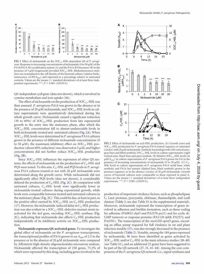
Niclosamide inhibits the 3OC12-HSL-dependent QS systemof P. aeruginosa. To verify the result of the screening assay, niclos-amide was purchased from an alternative supplier (Sigma-Al-drich) and retested in the PA14/PA14-R3 cocultivation system. Asexpected, a strong inhibition of the 3OC12-HSL-dependent QSresponse was observed, with an IC50 even lower than that calcu-lated for the compound from the Prestwick library (Fig. 1A). No-tably, niclosamide was also able to inhibit luminescence emissionby the PA14-R3 reporter strain grown in the presence of exoge-nously added synthetic 3OC12-HSL (3 �M final concentration)(Fig. 1B). This result suggests that the QS-inhibitory activity ofniclosamide relies on its ability to hamper the response of P.aeruginosa to the signal molecule rather than to inhibit its synthe-sis. The possibility that the observed QS-inhibitory activity ofniclosamide was due to unspecific inhibition of either biolumines-cence-generating enzymes or bacterial transcription was ruled outby the observation that niclosamide had no effect on the biolumi-nescence emitted by a P. aeruginosa strain in which biolumines-cence genes were under the control of the promoter region of the
TABLE 1 Hit compounds identified by screening the PrestwickChemical Library with the PA14/PA14-R3 QSI screening system
Prestwickcode Compound name
IC50
(�M)a Property
01D11 Niclosamide 10 Anthelmintic02H11 Gentamicin 20 Aminoglycoside antibiotic05G06 Mitoxantrone
dihydrochloride150 Antineoplastic agent
07E06 Rifampin 50 Antibiotic of the rifamycin group07H08 Dirithromycin 50 Macrolide glycopeptide antibiotic13C08 Sanguinarine 60 Quaternary ammonium salt of the
benzylisoquinoline alkaloidsgroup
14G10 Rifabutin 10 Antibiotic of the rifamycin groupa The IC50 values were determined using the PA14/PA14-R3 coculture grown for 4 h at37°C in the presence of 100, 80, 60, 40, 20, 10, 5 and 2.5 �g/ml of each compound.
Imperi et al.
998 aac.asm.org Antimicrobial Agents and Chemotherapy
on July 4, 2018 by guesthttp://aac.asm
.org/D
ownloaded from
QS-independent cysB gene (data not shown), which is involved incysteine metabolism and iron uptake (36).
The effect of niclosamide on the production of 3OC12-HSL wasthen assessed. P. aeruginosa PA14 was grown in the absence or inthe presence of 20 �M niclosamide, and 3OC12-HSL levels in cul-ture supernatants were quantitatively determined during thewhole growth curve. Niclosamide caused a significant reduction(30 to 60%) of 3OC12-HSL production from late exponentialgrowth to the entry into the stationary phase, after which the3OC12-HSL concentration fell to almost-undetectable levels inboth niclosamide-treated and -untreated cultures (Fig. 2A). When3OC12-HSL levels were determined in P. aeruginosa PA14 culturesgrown in the presence of different niclosamide concentrations (0to 50 �M), the maximum inhibitory effect on 3OC12-HSL pro-duction (about 60% reduction) was observed at 5 �M, and higherconcentrations did not further reduce 3OC12-HSL production(Fig. 2B).
Since 3OC12-HSL influences the expression of other QS sys-tems, the effects of niclosamide on the production of C4-HSL andPQS were tested. To this aim, C4-HSL and PQS levels in P. aerugi-nosa PA14 cultures treated or not with 20 �M niclosamide weredetermined along the growth curve. While niclosamide did notsignificantly affect PQS levels (data not shown), it considerablydelayed the production of C4-HSL (Fig. 2C). By comparison withuntreated cultures, C4-HSL levels were significantly lower inniclosamide-treated cultures during exponential growth, whilelevels were comparable between treated and untreated cultures inthe stationary phase (Fig. 2C). This could be due, at least in part, tothe positive effect exerted by 3OC12-HSL on C4-HSL production(17). However, the niclosamide-induced delay in C4-HSL produc-tion was also evident in a 3OC12-HSL-defective mutant strain in-activated for the lasI gene, encoding 3OC12-HSL synthase (Fig.2C), indicating that niclosamide also affects C4-HSL productionindependently of its inhibitory activity on 3OC12-HSL produc-tion.
Niclosamide represses QS-activated genes. To investigate theglobal effect of niclosamide on the P. aeruginosa transcriptome,the transcriptional profiles of PA14 grown to an A600 of 2.5 in thepresence or in the absence of 20 �M niclosamide were comparedby Affymetrix high-density oligonucleotides microarray analysis.Niclosamide affected the transcription of 258 genes, 73.2% ofwhich were repressed by this drug, including genes involved in the
production of important virulence factors, such as phospholipaseC, LasA protease, pyocyanin, chitinase, rhamnolipids, and LasBelastase (Table 2; see also Table S1 in the supplemental material).Moreover, niclosamide repressed the transcription of genes in-volved in adhesion and biofilm formation, such as those codingfor adhesins (PA0852-cbpD and PA2570-pa1L) and for cyclic di-GMP turnover or response proteins (PA1120-tpbB, PA2572, andPA4781). The transcription of the mexGHI-opmD genes, encod-ing an efflux pump required for full virulence in rat and plantinfection models (37), was also strongly decreased in the presenceof niclosamide (Table 2). Notably, among the 189 genes repressedby niclosamide, 96 have been identified as genes activated by3OC12-HSL and/or C4-HSL in the main reference studies (38–40)(see Table S1), and an additional 25 genes have been suggested tobe part of the QS network (27, 31, 41–44). Among the core com-ponents of the P. aeruginosa QS network (i.e., signal synthases and
FIG 1 Effect of niclosamide on the 3OC12-HSL-dependent QS of P. aerugi-nosa. Response to increasing concentrations of niclosamide (0 to 50 �M) of thePA14/PA14-R3 cocultivation system (A) and the PA14-R3 biosensor (B) in thepresence of 3 �M exogenously provided 3OC12-HSL. Bioluminescence emis-sion was normalized to the cell density of the bacterial culture (relative biolu-minescence, LCPS/A600) and expressed as a percentage relative to untreatedcontrols. Values are the means (� standard deviations) of at least three inde-pendent experiments. ***, P � 0.001 (ANOVA).
FIG 2 Effect of niclosamide on acyl-HSL production. (A) Growth curve and3OC12-HSL production by P. aeruginosa PA14 treated (squares) or untreated(circles) with 20 �M niclosamide. Symbols: bacterial growth (left vertical axis),gray lines and filled symbols; 3OC12-HSL levels in culture supernatants (rightvertical axis), black lines and open symbols. (B) Relative 3OC12-HSL levels (in�M/A600) in culture supernatants of P. aeruginosa PA14 grown for 8 h in thepresence of increasing concentrations of niclosamide (0 to 50 �M). (C) C4-HSL levels in culture supernatants of P. aeruginosa PA14 (solid lines, whitesymbols) and PA14 lasI mutant (dashed lines, black symbols) grown in thepresence (squares) or in the absence (circles) of 20 �M niclosamide. Growthcurves of bacterial cultures were comparable to those reported in panel A.Values are the means (� standard deviations) of at least three independentexperiments. ***, P � 0.001 (ANOVA).
Niclosamide Inhibits P. aeruginosa Pathogenicity
February 2013 Volume 57 Number 2 aac.asm.org 999
on July 4, 2018 by guesthttp://aac.asm
.org/D
ownloaded from
signal receptor genes), only the C4-HSL receptor gene rhlR wassignificantly repressed by niclosamide (Table 2). Similar resultshave been reported for other QSIs, such as furanone C-30 (38),iberin (45), and ajoene (46). In total, 121 out of the 189 genesrepressed by niclosamide (64%) can be classified as QS regulated.
Niclosamide displayed a positive effect on the transcription of69 genes, including two genes involved in type VI secretion path-ways (PA0070-tagQ1 and PA0085-hcp1) (Table 2). These genesare the only virulence-related determinants whose transcription isinduced by niclosamide. Only four of the niclosamide-activatedgenes were previously reported to be repressed by 3OC12-HSLand/or C4-HSL (38–40) (see Table S2 in the supplementalmaterial), suggesting that the majority of the genes induced byniclosamide are affected via a QS-independent pathway(s). Thisobservation, together with the finding that 36% of the niclos-amide-repressed genes have never been reported to be QS con-trolled, suggests that this drug may have additional cellular targetsbesides the QS network. Notably, a total of 16 putative or con-firmed transcriptional regulators were identified among the genesrepressed or activated by niclosamide (see Tables S1 and S2). Be-sides rhlR, niclosamide decreased the transcription of pprB, whichencodes a transcriptional activator associated with biofilm forma-tion (47). Conversely, it positively affected the transcription ofcifR (Table 2), which encodes the transcriptional repressor of theCif toxin, responsible for apical membrane downregulation of the
cystic fibrosis transmembrane conductance regulator (CFTR) inepithelial cells (48). The niclosamide-affected transcriptional fac-tors may act as ancillary regulators, increasing the number ofgenes whose expression is altered by this drug beyond the QSregulon. However, a complete understanding of the niclosamideimpact on P. aeruginosa physiology is partially hampered by thehigh percentage of niclosamide-controlled genes (�41%) codingfor proteins still classified as hypothetical (see Tables S1 and S2 inthe supplemental material).
Niclosamide strongly reduces the virulence potential of P.aeruginosa in vitro. In order to validate the transcriptomic data atthe phenotypic level, we assessed the effect of niclosamide on theproduction of a set of QS-regulated virulence traits. In particular,we focused on (i) the LasB elastase, which is directly regulated bythe 3OC12-HSL receptor LasR at the transcriptional level (49), (ii)pyocyanin and rhamnolipids, which are regulated by a number ofdifferent regulatory pathways and extracellular signals (50, 51),and (iii) multifactorial phenotypes, such as motility and biofilm,which are crucial for the establishment and persistence of P.aeruginosa infections (52–54).
In accordance with microarray analysis, niclosamide had amarked inhibitory effect on the levels of QS-regulated secretedvirulence factors of P. aeruginosa PA14 (Fig. 3). Production levelsof both pyocyanin and elastase were dramatically reduced (85 to90%) by 5 to 10 �M niclosamide. Likewise, the amount of rham-
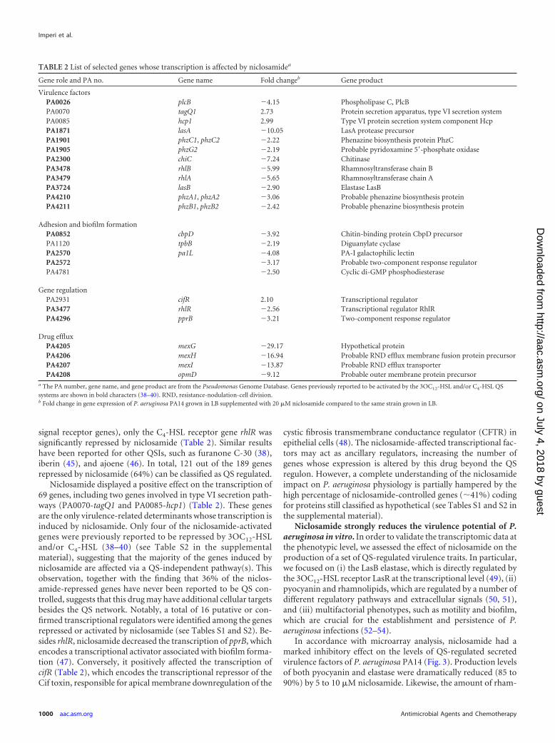
TABLE 2 List of selected genes whose transcription is affected by niclosamidea
Gene role and PA no. Gene name Fold changeb Gene product
Virulence factorsPA0026 plcB �4.15 Phospholipase C, PlcBPA0070 tagQ1 2.73 Protein secretion apparatus, type VI secretion systemPA0085 hcp1 2.99 Type VI protein secretion system component HcpPA1871 lasA �10.05 LasA protease precursorPA1901 phzC1, phzC2 �2.22 Phenazine biosynthesis protein PhzCPA1905 phzG2 �2.19 Probable pyridoxamine 5=-phosphate oxidasePA2300 chiC �7.24 ChitinasePA3478 rhlB �5.99 Rhamnosyltransferase chain BPA3479 rhlA �5.65 Rhamnosyltransferase chain APA3724 lasB �2.90 Elastase LasBPA4210 phzA1, phzA2 �3.06 Probable phenazine biosynthesis proteinPA4211 phzB1, phzB2 �2.42 Probable phenazine biosynthesis protein
Adhesion and biofilm formationPA0852 cbpD �3.92 Chitin-binding protein CbpD precursorPA1120 tpbB �2.19 Diguanylate cyclasePA2570 pa1L �4.08 PA-I galactophilic lectinPA2572 �3.17 Probable two-component response regulatorPA4781 �2.50 Cyclic di-GMP phosphodiesterase
Gene regulationPA2931 cifR 2.10 Transcriptional regulatorPA3477 rhlR �2.56 Transcriptional regulator RhlRPA4296 pprB �3.21 Two-component response regulator
Drug effluxPA4205 mexG �29.17 Hypothetical proteinPA4206 mexH �16.94 Probable RND efflux membrane fusion protein precursorPA4207 mexI �13.87 Probable RND efflux transporterPA4208 opmD �9.12 Probable outer membrane protein precursor
a The PA number, gene name, and gene product are from the Pseudomonas Genome Database. Genes previously reported to be activated by the 3OC12-HSL and/or C4-HSL QSsystems are shown in bold characters (38–40). RND, resistance-nodulation-cell division.b Fold change in gene expression of P. aeruginosa PA14 grown in LB supplemented with 20 �M niclosamide compared to the same strain grown in LB.
Imperi et al.
1000 aac.asm.org Antimicrobial Agents and Chemotherapy
on July 4, 2018 by guesthttp://aac.asm
.org/D
ownloaded from
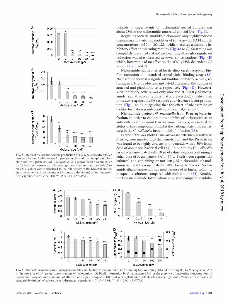
nolipids in supernatants of niclosamide-treated cultures wasabout 25% of the niclosamide-untreated control level (Fig. 3).
Regarding bacterial motility, niclosamide only slightly reducedswimming and twitching motilities of P. aeruginosa PA14 at highconcentrations (�50 to 100 �M), while it exerted a dramatic in-hibitory effect on swarming motility (Fig. 4A to C). Swarming wascompletely prevented at 4 �M niclosamide, although a significantreduction was also observed at lower concentrations (Fig. 4B)which, however, had no effect on the 3OC12-HSL-dependent QSsystem (Fig. 1 and 2).
Niclosamide was also tested for its effect on P. aeruginosa bio-film formation in a standard crystal violet binding assay (32).Niclosamide showed a significant biofilm-inhibitory activity, re-sulting in a 2-fold reduction and 3-fold increase in the number ofattached and planktonic cells, respectively (Fig. 4D). However,such inhibitory activity was only observed at �200 �M niclos-amide, i.e., at concentrations that are exceedingly higher thanthose active against the QS response and virulence factor produc-tion (Fig. 1 to 3), suggesting that the effect of niclosamide onbiofilm formation is independent of its anti-QS activity.
Niclosamide protects G. mellonella from P. aeruginosa in-fection. In order to explore the suitability of niclosamide as anantivirulence drug against P. aeruginosa infections, we assessed theability of this compound to inhibit the pathogenicity of P. aerugi-nosa in the G. mellonella insect model of infection (33).
Larvae of the wax moth G. mellonella are extremely sensitive toP. aeruginosa injected into the hemolymph, and the PA14 strainwas found to be highly virulent in this model, with a 50% lethaldose of about one bacterial cell (33). In our study, G. mellonellalarvae were inoculated with 10 �l of saline solution containing alethal dose of P. aeruginosa PA14 (10 � 4 cells from exponentialcultures) and containing or not 750 �M niclosamide ethanol-amine salt and then incubated at 28°C for up to 1 week. Niclos-amide ethanolamine salt was used because of its higher solubilityin aqueous solutions compared with niclosamide (55). Notably,the two niclosamide formulations displayed comparable inhibi-
FIG 3 Effects of niclosamide on the production of QS-regulated extracellularvirulence factors. LasB elastase (A), pyocyanin (B), and rhamnolipids (C) lev-els in culture supernatants of P. aeruginosa PA14 grown for 10 h (A and B) orfor 24 h (C) in the presence of increasing concentrations of niclosamide (0 to50 �M). Values were normalized to the cell density of the bacterial culture(relative values) and are the means (� standard deviations) of four indepen-dent experiments. **, P � 0.01; ***, P � 0.001 (ANOVA).
FIG 4 Effects of niclosamide on P. aeruginosa motility and biofilm formation. (A to C) Swimming (A), swarming (B), and twitching (C) by P. aeruginosa PA14in the presence of increasing concentrations of niclosamide. (D) Biofilm formation by P. aeruginosa PA14 in the presence of increasing concentrations ofniclosamide, assessed as the amount of attached cells (gray histograms, left axis) versus planktonic cells (black squares, right axis). Values are the means (�standard deviations) of at least three independent experiments. **, P � 0.01; ***, P � 0.001 (ANOVA).
Niclosamide Inhibits P. aeruginosa Pathogenicity
February 2013 Volume 57 Number 2 aac.asm.org 1001
on July 4, 2018 by guesthttp://aac.asm
.org/D
ownloaded from
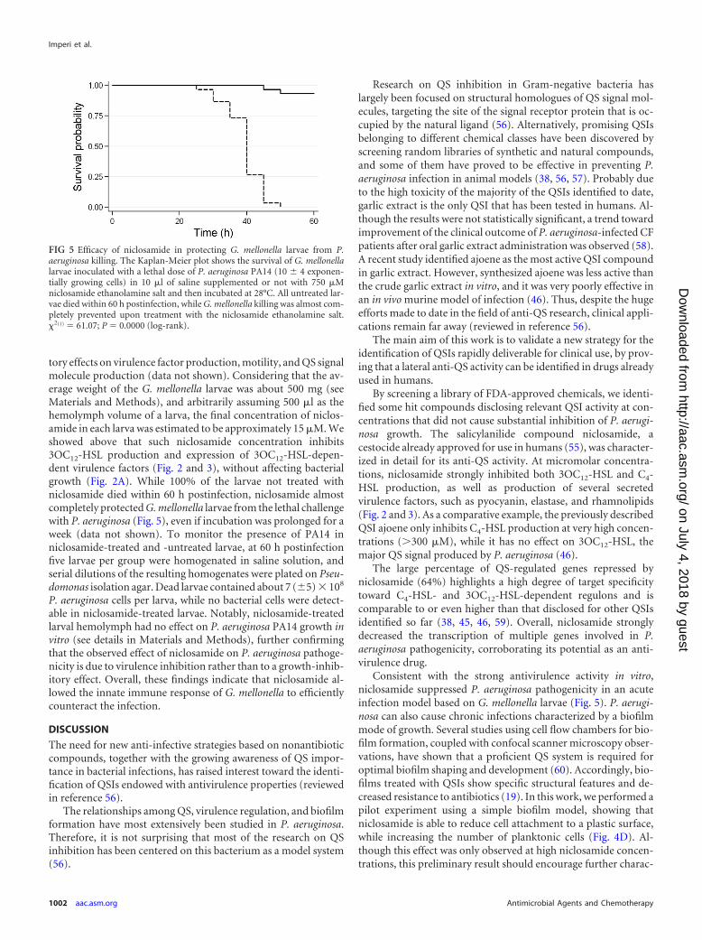
tory effects on virulence factor production, motility, and QS signalmolecule production (data not shown). Considering that the av-erage weight of the G. mellonella larvae was about 500 mg (seeMaterials and Methods), and arbitrarily assuming 500 �l as thehemolymph volume of a larva, the final concentration of niclos-amide in each larva was estimated to be approximately 15 �M. Weshowed above that such niclosamide concentration inhibits3OC12-HSL production and expression of 3OC12-HSL-depen-dent virulence factors (Fig. 2 and 3), without affecting bacterialgrowth (Fig. 2A). While 100% of the larvae not treated withniclosamide died within 60 h postinfection, niclosamide almostcompletely protected G. mellonella larvae from the lethal challengewith P. aeruginosa (Fig. 5), even if incubation was prolonged for aweek (data not shown). To monitor the presence of PA14 inniclosamide-treated and -untreated larvae, at 60 h postinfectionfive larvae per group were homogenated in saline solution, andserial dilutions of the resulting homogenates were plated on Pseu-domonas isolation agar. Dead larvae contained about 7 (�5) � 108
P. aeruginosa cells per larva, while no bacterial cells were detect-able in niclosamide-treated larvae. Notably, niclosamide-treatedlarval hemolymph had no effect on P. aeruginosa PA14 growth invitro (see details in Materials and Methods), further confirmingthat the observed effect of niclosamide on P. aeruginosa pathoge-nicity is due to virulence inhibition rather than to a growth-inhib-itory effect. Overall, these findings indicate that niclosamide al-lowed the innate immune response of G. mellonella to efficientlycounteract the infection.
DISCUSSION
The need for new anti-infective strategies based on nonantibioticcompounds, together with the growing awareness of QS impor-tance in bacterial infections, has raised interest toward the identi-fication of QSIs endowed with antivirulence properties (reviewedin reference 56).
The relationships among QS, virulence regulation, and biofilmformation have most extensively been studied in P. aeruginosa.Therefore, it is not surprising that most of the research on QSinhibition has been centered on this bacterium as a model system(56).
Research on QS inhibition in Gram-negative bacteria haslargely been focused on structural homologues of QS signal mol-ecules, targeting the site of the signal receptor protein that is oc-cupied by the natural ligand (56). Alternatively, promising QSIsbelonging to different chemical classes have been discovered byscreening random libraries of synthetic and natural compounds,and some of them have proved to be effective in preventing P.aeruginosa infection in animal models (38, 56, 57). Probably dueto the high toxicity of the majority of the QSIs identified to date,garlic extract is the only QSI that has been tested in humans. Al-though the results were not statistically significant, a trend towardimprovement of the clinical outcome of P. aeruginosa-infected CFpatients after oral garlic extract administration was observed (58).A recent study identified ajoene as the most active QSI compoundin garlic extract. However, synthesized ajoene was less active thanthe crude garlic extract in vitro, and it was very poorly effective inan in vivo murine model of infection (46). Thus, despite the hugeefforts made to date in the field of anti-QS research, clinical appli-cations remain far away (reviewed in reference 56).
The main aim of this work is to validate a new strategy for theidentification of QSIs rapidly deliverable for clinical use, by prov-ing that a lateral anti-QS activity can be identified in drugs alreadyused in humans.
By screening a library of FDA-approved chemicals, we identi-fied some hit compounds disclosing relevant QSI activity at con-centrations that did not cause substantial inhibition of P. aerugi-nosa growth. The salicylanilide compound niclosamide, acestocide already approved for use in humans (55), was character-ized in detail for its anti-QS activity. At micromolar concentra-tions, niclosamide strongly inhibited both 3OC12-HSL and C4-HSL production, as well as production of several secretedvirulence factors, such as pyocyanin, elastase, and rhamnolipids(Fig. 2 and 3). As a comparative example, the previously describedQSI ajoene only inhibits C4-HSL production at very high concen-trations (�300 �M), while it has no effect on 3OC12-HSL, themajor QS signal produced by P. aeruginosa (46).
The large percentage of QS-regulated genes repressed byniclosamide (64%) highlights a high degree of target specificitytoward C4-HSL- and 3OC12-HSL-dependent regulons and iscomparable to or even higher than that disclosed for other QSIsidentified so far (38, 45, 46, 59). Overall, niclosamide stronglydecreased the transcription of multiple genes involved in P.aeruginosa pathogenicity, corroborating its potential as an anti-virulence drug.
Consistent with the strong antivirulence activity in vitro,niclosamide suppressed P. aeruginosa pathogenicity in an acuteinfection model based on G. mellonella larvae (Fig. 5). P. aerugi-nosa can also cause chronic infections characterized by a biofilmmode of growth. Several studies using cell flow chambers for bio-film formation, coupled with confocal scanner microscopy obser-vations, have shown that a proficient QS system is required foroptimal biofilm shaping and development (60). Accordingly, bio-films treated with QSIs show specific structural features and de-creased resistance to antibiotics (19). In this work, we performed apilot experiment using a simple biofilm model, showing thatniclosamide is able to reduce cell attachment to a plastic surface,while increasing the number of planktonic cells (Fig. 4D). Al-though this effect was only observed at high niclosamide concen-trations, this preliminary result should encourage further charac-
FIG 5 Efficacy of niclosamide in protecting G. mellonella larvae from P.aeruginosa killing. The Kaplan-Meier plot shows the survival of G. mellonellalarvae inoculated with a lethal dose of P. aeruginosa PA14 (10 � 4 exponen-tially growing cells) in 10 �l of saline supplemented or not with 750 �Mniclosamide ethanolamine salt and then incubated at 28°C. All untreated lar-vae died within 60 h postinfection, while G. mellonella killing was almost com-pletely prevented upon treatment with the niclosamide ethanolamine salt.�2(1) 61.07; P 0.0000 (log-rank).
Imperi et al.
1002 aac.asm.org Antimicrobial Agents and Chemotherapy
on July 4, 2018 by guesthttp://aac.asm
.org/D
ownloaded from
terization of the effect of niclosamide on biofilm development andresistance to antibiotics by using advanced biofilm models.
Concerning future developments of niclosamide as an anti-P.aeruginosa drug, there are some issues that need to be addressed.First of all, the effect of niclosamide on a wide panel of clinical P.aeruginosa strains isolated from different infection sites should beassessed, including CF patient chronically infected lungs. More-over, even if niclosamide is currently used as an anthelmintic drugto treat intestinal infections and displays overall low toxicity (55),it is poorly soluble in water, shows low intestinal absorption, andonce in the bloodstream, it is quickly cleared via the urinary tractor by enzymatic modification in the liver (55). Although thesefeatures could represent drawbacks to the systemic administrationof niclosamide, they could be advantageous in the local treatmentof wound infections, burns, otitis, gastrointestinal infections, andother external P. aeruginosa infections. It is also worth mentioningthat the toxicity of inhaled niclosamide powder is quite low formammals (55), opening new perspectives for aerosol treatment ofP. aeruginosa lung infections. Additional studies in differentmammalian models of both acute and chronic infections are re-quired to assess the suitability of niclosamide as an anti-P. aerugi-nosa drug, prior to the move into clinical trials. However, in ac-cordance with the SOSA approach (2), niclosamide could also beused as a promising scaffold for the design of structural analoguesendowed with improved activity and pharmacokinetic properties.The hypothesis that some niclosamide derivatives may retain an-ti-QS activity is strengthened by our preliminary observation thatother compounds belonging to the same structural class of niclos-amides, i.e., the salicylanilides rafoxanide and oxyclozanide,showed in vitro the same anti-QS activity as niclosamide (data notshown).
The development of niclosamide-based QSIs could be pursuedeither by screening random chemical modifications introducedwithin the salicylanilide structure or by rational drug design. Thelatter approach requires detailed information about the anti-QSmechanism of action of niclosamide. Unfortunately, despiteniclosamide’s use since the 1960s, its mechanism of action re-mains elusive. The anthelmintic activity of niclosamide seems torely on its ability to uncouple mitochondrial oxidative phosphor-ylation (61). More recently, it has been found to inhibit prolifer-ation of some tumor cells by hampering different regulatory path-ways, without relevant effects on normal nontumor cells (62–64).Notably, niclosamide has also been reported to act as an antimy-cobacterial agent (64), plausibly disrupting Mycobacterium mem-brane potential and pH homeostasis (65). Although the charac-terization of the molecular targets of niclosamide was not the aimof this study, some of our observations might be the first stepstoward the comprehension of the niclosamide mechanism of ac-tion in P. aeruginosa. First, niclosamide is likely to target the3OC12-HSL reception process rather than signal biosynthesis (Fig.1). Second, the maximum inhibitory effect disclosed for niclos-amide on 3OC12-HSL production (60%) is reached at 5 �M, andincreases in concentration did not reduce 3OC12-HSL production(Fig. 2), ruling out the possibility that niclosamide competes with3OC12-HSL for receptor binding. Third, microarray analysisshowed that niclosamide also affects the expression of genes notcontrolled by QS, including some putative or confirmed tran-scriptional regulators. Fourth, niclosamide represses C4-HSL pro-duction both dependently and independently from its action on3OC12-HSL (Fig. 2). Finally, the repressive effect disclosed by
niclosamide is higher on swarming motility and on the produc-tion of virulence factors than on 3OC12-HSL production (Fig. 2, 3,and 4). Although this could be due to the combined effect of thisQSI on 3OC12-HSL and C4-HSL production, it cannot be ex-cluded that niclosamide also influences some QS-independentcellular process involved in virulence gene regulation.
Putting together our observations and data from the availableliterature on the cellular processes affected by niclosamide inother organisms, a very preliminary hypothesis about the mecha-nism of action of niclosamide in P. aeruginosa is that this moleculetargets some regulatory pathway(s) responsive to the energetic/metabolic status of the cell and that is required for full activity ofthe QS signaling network. Studies on the mechanism of action ofniclosamide in P. aeruginosa are therefore in progress in our lab-oratory.
In conclusion, the major outcome of this study was the identi-fication of a strong anti-QS activity in a compound already ap-proved for use in humans. Our findings provide a new promisingdrug candidate against P. aeruginosa and a proof of concept thatFDA-approved drugs may be endowed with antivirulence prop-erties that are worthy of exploration.
ACKNOWLEDGMENTS
We heartily thank Paolo Landini (University of Milan, Milan, Italy) forencouraging this work, for providing us with the Prestwick library, and forprecious advice.
This work was supported by a grant from the Ministry of Universityand Research of Italy PRIN-2008 (232P4H_003) and by grants from theItalian Cystic Fibrosis Research Foundation (Projects FFC 14/2010 andFFC 13/2011).
REFERENCES1. Gilbert N. 2010. Universities shun Europe’s drug initiative. Nature 466:
306 –307.2. Wermuth CG. 2006. Selective optimization of side activities: the SOSA
approach. Drug Discov. Today 11:160 –164.3. Ejim L, Farha MA, Falconer SB, Wildenhain J, Coombes BK, Tyers M,
Brown ED, Wright GD. 2011. Combinations of antibiotics and nonan-tibiotic drugs enhance antimicrobial efficacy. Nat. Chem. Biol. 7:348 –350.
4. Antoniani D, Bocci P, Maciag A, Raffaelli N, Landini P. 2010. Moni-toring of diguanylate cyclase activity and of cyclic-di-GMP biosynthesis bywhole-cell assays suitable for high-throughput screening of biofilm inhib-itors. Appl. Microbiol. Biotechnol. 85:1095–1104.
5. Cegelski L, Marshall GR, Eldridge GR, Hultgren SJ. 2008. Thebiology and future prospects of antivirulence therapies. Nat. Rev. Mi-crobiol. 6:17–27.
6. Rasko DA, Sperandio V. 2010. Anti-virulence strategies to combat bac-teria-mediated disease. Nat. Rev. Drug Discov. 9:117–128.
7. Atkinson S, Williams P. 2009. Quorum sensing and social networking inthe microbial world. J. R. Soc. Interface 6:959 –978.
8. Njoroge J, Sperandio V. 2009. Jamming bacterial communication: newapproaches for the treatment of infectious diseases. EMBO Mol. Med.1:201–210.
9. Amara N, Krom BP, Kaufmann GF, Meijler MM. 2011. Macromolecularinhibition of quorum sensing: enzymes, antibodies, and beyond. Chem.Rev. 111:195–208.
10. Talbot GH, Bradley J, Edwards JE, Jr, Gilbert D, Scheld M, Bartlett JG;Antimicrobial Availability Task Force of the Infectious Diseases Society ofAmerica. 2006. Bad bugs need drugs: an update on the development pipe-line from the Antimicrobial Availability Task Force of the Infectious Dis-eases Society of America. Clin. Infect. Dis. 42:657– 668.
11. Orsi GB, Raponi M, Franchi C, Rocco M, Mancini C, Venditti M. 2005.Surveillance and infection control in an intensive care unit. Infect. ControlHosp. Epidemiol. 26:321–325.
12. Driscoll JA, Brody SL, Kollef MH. 2007. The epidemiology, pathogenesisand treatment of Pseudomonas aeruginosa infections. Drugs 67:351–368.
13. Rosenthal VD, Bijie H, Maki DG, Mehta Y, Apisarnthanarak A, Me-
Niclosamide Inhibits P. aeruginosa Pathogenicity
February 2013 Volume 57 Number 2 aac.asm.org 1003
on July 4, 2018 by guesthttp://aac.asm
.org/D
ownloaded from
deiros EA, Leblebicioglu H, Fisher D, & Aacute;lvarez-Moreno C,Khader IA, Del RocíO González Martínez M, Cuellar le, Navoa-Ng JA,Abouqal R, Guanche Garcell H, Mitrev Z, Pirez GarcíA MC, Hamdi A,Dueñas L, Cancel E, Gurskis V, Rasslan O, Ahmed A, Kanj SS, UgaldeOC, Mapp T, Raka L, Yuet Meng C, Thu le, Ghazal TAS, Gikas A,Narváez LP, MejíA N, Hadjieva N, Gamar Elanbya MO, Guzmán SirittME, Jayatilleke K; INICC members. 2012. International NosocomialInfection Control Consortium (INICC) report, data summary of 36 coun-tries, for 2004 –2009. Am. J. Infect. Control 40:396 – 407.
14. Page MG, Heim J. 2009. Prospects for the next anti-Pseudomonas drug.Curr. Opin. Pharmacol. 9:558 –565.
15. Breidenstein EB, de la Fuente-Núñez C, Hancock RE. 2011. Pseudomo-nas aeruginosa: all roads lead to resistance. Trends Microbiol. 19:419 – 426.
16. Lee DG, Urbach JM, Wu G, Liberati NT, Feinbaum RL, Miyata S,Diggins LT, He J, Saucier M, Déziel E, Friedman L, Li L, Grills G,Montgomery K, Kucherlapati R, Rahme LG, Ausubel FM. 2006.Genomic analysis reveals that Pseudomonas aeruginosa virulence is com-binatorial. Genome Biol. 7:R90. doi:10.1186/gb-2006-7-10-r90.
17. Williams P, Cámara M. 2009. Quorum sensing and environmental ad-aptation in Pseudomonas aeruginosa: a tale of regulatory networks andmultifunctional signal molecules. Curr. Opin. Microbiol. 12:182–191.
18. Winstanley C, Fothergill JL. 2009. The role of quorum sensing in chroniccystic fibrosis Pseudomonas aeruginosa infections. FEMS Microbiol. Lett.290:1–9.
19. Bjarnsholt T, Tolker-Nielsen T, Høiby N, Givskov M. 2010. Interferenceof Pseudomonas aeruginosa signalling and biofilm formation for infectioncontrol. Expert Rev. Mol. Med. 12:e11.
20. Massai F, Imperi F, Quattrucci S, Zennaro E, Visca P, Leoni L. 2011. Amultitask biosensor for micro-volumetric detection of N-3-oxo-dodecanoyl-homoserine lactone quorum sensing signal. Biosens. Bioelec-tron. 26:3444 –3449.
21. Rahme LG, Stevens EJ, Wolfort SF, Shao J, Tompkins RG, Ausubel FM.1995. Common virulence factors for bacterial pathogenicity in plants andanimals. Science 268:1899 –1902.
22. Liberati NT, Urbach JM, Miyata S, Lee DG, Drenkard E, Wu G,Villanueva J, Wei T, Ausubel FM. 2006. An ordered, nonredundantlibrary of Pseudomonas aeruginosa strain PA14 transposon insertion mu-tants. Proc. Natl. Acad. Sci. U. S. A. 103:2833–2838.
23. Sambrook J, Fritsch EF, Maniatis T. 1989. Molecular cloning: a labora-tory manual, 2nd ed. Cold Spring Harbor Laboratory Press, New York,NY.
24. Clark JMB, Clark DJ, Maaløe O. 1967. DNA replication and the divisioncycle in Escherichia coli. J. Mol. Biol. 23:99 –112.
25. Duan K, Surette MG. 2007. Environmental regulation of Pseudomonasaeruginosa PAO1 Las and Rhl quorum-sensing systems. J. Bacteriol. 189:4827– 4836.
26. Fletcher MP, Diggle SP, Crusz SA, Chhabra SR, Cámara M, Williams P.2007. A dual biosensor for 2-alkyl-4-quinolone quorum-sensing signalmolecules. Environ. Microbiol. 9:2683–2693.
27. Rampioni G, Schuster M, Greenberg EP, Bertani I, Grasso M, VenturiV, Zennaro E, Leoni L. 2007. RsaL provides quorum sensing homeostasisand functions as a global regulator of gene expression in Pseudomonasaeruginosa. Mol. Microbiol. 66:1557–1565.
28. Essar DW, Eberly L, Hadero A, Crawford IP. 1990. Identification andcharacterization of genes for a second anthranilate synthase in Pseudomo-nas aeruginosa: interchangeability of the two anthranilate synthases andevolutionary implications. J. Bacteriol. 172:884 –900.
29. Ohman DE, Burns RP, Iglewski BH. 1980. Corneal infections in micewith toxin A and elastase mutants of Pseudomonas aeruginosa. J. Infect.Dis. 142:547–555.
30. Wilhelm S, Gdynia A, Tielen P, Rosenau F, Jaeger KE. 2007. Theautotransporter esterase EstA of Pseudomonas aeruginosa is required forrhamnolipid production, cell motility, and biofilm formation. J. Bacteriol.189:6695– 6703.
31. Rampioni G, Schuster M, Greenberg EP, Zennaro E, Leoni L. 2009.Contribution of the RsaL global regulator to Pseudomonas aeruginosa vir-ulence and biofilm formation. FEMS Microbiol. Lett. 301:210 –217.
32. Merritt JH, Kadouri DE, O’Toole GA. 2005. Growing and analyzingstatic biofilms. Curr. Protoc. Microbiol. Chapter 1:Unit 1B.1.
33. Jander G, Rahme LG, Ausubel FM. 2000. Positive correlation betweenvirulence of Pseudomonas aeruginosa mutants in mice and insects. J. Bac-teriol. 182:3843–3845.
34. Hoffmann N, Lee B, Hentzer M, Rasmussen TB, Song Z, Johansen HK,
Givskov M, Høiby N. 2007. Azithromycin blocks quorum sensing andalginate polymer formation and increases the sensitivity to serum andstationary-growth-phase killing of Pseudomonas aeruginosa and attenu-ates chronic P. aeruginosa lung infection in Cftr�/� mice. Antimicrob.Agents Chemother. 51:3677–3687.
35. Babic F, Venturi V, Maravic-Vlahovicek G. 2010. Tobramycin at subin-hibitory concentration inhibits the RhlI/R quorum sensing system in aPseudomonas aeruginosa environmental isolate. BMC Infect. Dis. 10:148.doi:10.1186/1471-2334-10-148.
36. Imperi F, Tiburzi F, Fimia GM, Visca P. 2010. Transcriptional control ofthe pvdS iron starvation sigma factor gene by the master regulator of sulfurmetabolism CysB in Pseudomonas aeruginosa. Environ. Microbiol. 12:1630 –1642.
37. Aendekerk S, Diggle SP, Song Z, Høiby N, Cornelis P, Williams P,Cámara M. 2005. The MexGHI-OpmD multidrug efflux pump controlsgrowth, antibiotic susceptibility and virulence in Pseudomonas aeruginosavia 4-quinolone-dependent cell-to-cell communication. Microbiology151:1113–1125.
38. Hentzer M, Wu H, Andersen JB, Riedel K, Rasmussen TB, Bagge N,Kumar N, Schembri MA, Song Z, Kristoffersen P, Manefield M, Coste-rton JW, Molin S, Eberl L, Steinberg P, Kjelleberg S, Høiby N, GivskovM. 2003. Attenuation of Pseudomonas aeruginosa virulence by quorumsensing inhibitors. EMBO J. 22:3803–3815.
39. Schuster M, Lohstroh CP, Ogi T, Greemberg EP. 2003. Identification,timing and signal specifity of Pseudomonas aeruginosa quorum-controlledgenes: a transcriptome analysis. J. Bacteriol. 185:2066 –2079.
40. Wagner VE, Bushnell D, Passador L, Brooks AI, Iglewsky BH. 2003.Microarray analysis of Pseudomonas aeruginosa quorum-sensing regu-lons: effects of growth phase and environment. J. Bacteriol. 185:2080 –2095.
41. Déziel E, Gopalan S, Tampakaki AP, Lépine F, Padfield KE, Saucier M,Xiao G, Rahme LG. 2005. The contribution of MvfR to Pseudomonasaeruginosa pathogenesis and quorum sensing circuitry regulation: multi-ple quorum sensing-regulated genes are modulated without affectinglasRI, rhlRI or the production of N-acyl-L-homoserine lactones. Mol. Mi-crobiol. 55:998 –1014.
42. Bredenbruch F, Geffers R, Nimtz M, Buer J, Häussler S. 2006. ThePseudomonas aeruginosa quinolone signal (PQS) has an iron-chelatingactivity. Environ. Microbiol. 8:1318 –1329.
43. Lequette Y, Lee JH, Ledgham F, Lazdunski A, Greenberg EP. 2006. Adistinct QscR regulon in the Pseudomonas aeruginosa quorum-sensingcircuit. J. Bacteriol. 188:3365–3370.
44. Rampioni G, Pustelny C, Fletcher MP, Wright VJ, Bruce M, RumbaughKP, Heeb S, Cámara M, Williams P. 2010. Transcriptomic analysisreveals a global alkyl-quinolone-independent regulatory role for PqsE infacilitating the environmental adaptation of Pseudomonas aeruginosa toplant and animal hosts. Environ. Microbiol. 12:1659 –1673.
45. Jakobsen TH, Bragason SK, Phipps RK, Christensen LD, van GennipM, Alhede M, Skindersoe M, Larsen TO, Høiby N, Bjarnsholt T,Givskov M. 2012. Food as a source for quorum sensing inhibitors: iberinfrom horseradish revealed as a quorum sensing inhibitor of Pseudomonasaeruginosa. Appl. Environ. Microbiol. 78:2410 –2421.
46. Jakobsen TH, van Gennip M, Phipps RK, Shanmugham MS, Chris-tensen LD, Alhede M, Skindersoe ME, Rasmussen TB, Friedrich K,Uthe F, Jensen PØ Moser C, Nielsen KF, Eberl L, Larsen TO, Tanner D,Høiby N, Bjarnsholt T, Givskov M. 2012. Ajoene, a sulfur-rich moleculefrom garlic, inhibits genes controlled by quorum sensing. Antimicrob.Agents Chemother. 56:2314 –2325.
47. Giraud C, Bernard CS, Calderon V, Yang L, Filloux A, Molin S, FichantG, Bordi C, de Bentzmann S. 2011. The PprA-PprB two-componentsystem activates CupE, the first non-archetypal Pseudomonas aeruginosachaperone-usher pathway system assembling fimbriae. Environ. Micro-biol. 13:666 – 683.
48. MacEachran DP, Stanton BA, O’Toole GA. 2008. Cif is negatively reg-ulated by the TetR family repressor CifR. Infect. Immun. 76:3197–3206.
49. Anderson RM, Zimprich CA, Rust L. 1999. A second operator is involvedin Pseudomonas aeruginosa elastase (lasB) activation. J. Bacteriol. 181:6264 – 6270.
50. Lau GW, Hassett DJ, Ran H, Kong F. 2004. The role of pyocyanin inPseudomonas aeruginosa infection. Trends Mol. Med. 10:599 – 606.
51. Reis RS, Pereira AG, Neves BC, Freire DM. 2011. Gene regulation ofrhamnolipid production in Pseudomonas aeruginosa: a review. Bioresour.Technol. 102:6377– 6384.
Imperi et al.
1004 aac.asm.org Antimicrobial Agents and Chemotherapy
on July 4, 2018 by guesthttp://aac.asm
.org/D
ownloaded from
52. Parsek MR, Singh PK. 2003. Bacterial biofilms: an emerging link todisease pathogenesis. Annu. Rev. Microbiol. 57:677–701.
53. Zolfaghar I, Evans DJ, Fleiszig SM. 2003. Twitching motility contributesto the role of pili in corneal infection caused by Pseudomonas aeruginosa.Infect. Immun. 71:5389 –5393.
54. Arora SK, Neely AN, Blair B, Lory S, Ramphal R. 2005. Role of motilityand flagellin glycosylation in the pathogenesis of Pseudomonas aeruginosaburn wound infections. Infect. Immun. 73:4395– 4398.
55. Andrews P, Thyssen J, Lorke D. 1982. The biology and toxicology ofmolluscicides, Bayluscide. Pharmacol. Ther. 19:245–295.
56. Galloway WR, Hodgkinson JT, Bowden S, Welch M, Spring DR. 2012.Applications of small molecule activators and inhibitors of quorum sens-ing in Gram-negative bacteria. Trends Microbiol. 20:449 – 458.
57. Rasmussen TB, Bjarnsholt T, Skindersoe ME, Hentzer M, KristoffersenP, Köte M, Nielsen J, Eberl L, Givskov M. 2005. Screening for quorum-sensing inhibitors (QSI) by use of a novel genetic system, the QSI selector.J. Bacteriol. 187:1799 –1814.
58. Smyth AR, Cifelli PM, Ortori CA, Righetti K, Lewis S, Erskine P,Holland ED, Givskov M, Williams P, Cámara M, Barrett DA, Knox A.2010. Garlic as an inhibitor of Pseudomonas aeruginosa quorum sensing incystic fibrosis: a pilot randomized controlled trial. Pediatr. Pulmonol. 45:356 –362.
59. Müh U, Hare BJ, Duerkop BA, Schuster M, Hanzelka BL, Heim R,
Olson ER, Greenberg EP. 2006. A structurally unrelated mimic of aPseudomonas aeruginosa acyl-homoserine lactone quorum-sensing signal.Proc. Natl. Acad. Sci. U. S. A. 103:16948 –16952.
60. Kirisits MJ, Parsek MR. 2006. Does Pseudomonas aeruginosa use inter-cellular signalling to build biofilm communities? Cell. Microbiol. 8:1841–1849.
61. Weinbach EC, Garbus J. 1969. Mechanism of action of reagents thatuncouple oxidative phosphorylation. Nature 221:1016 –1018.
62. Jin Y, Lu Z, Ding K, Li J, Du X, Chen C, Sun X, Wu Y, Zhou J, Pan J.2010. Antineoplastic mechanisms of niclosamide in acute myelogenousleukemia stem cells: inactivation of the NF-B pathway and generation ofreactive oxygen species. Cancer Res. 70:2516 –2527.
63. Osada T, Chen M, Yang XY, Spasojevic I, Vandeusen JB, Hsu D, ClaryBM, Clay TM, Chen W, Morse MA, Lyerly HK. 2011. Antihelminthcompound niclosamide downregulates Wnt signaling and elicits antitu-mor responses in tumors with activating APC mutations. Cancer Res.71:4172– 4182.
64. Sun Z, Zhang Y. 1999. Antituberculosis activity of certain antifungal andantihelmintic drugs. Tuber. Lung. Dis. 79:319 –320.
65. de Carvalho LP, Darby CM, Rhee KY, Nathan C. 2011. Nitazoxanidedisrupts membrane potential and intrabacterial pH homeostasis of Myco-bacterium tuberculosis. ACS Med. Chem. Lett. 2:849 – 854.
Niclosamide Inhibits P. aeruginosa Pathogenicity
February 2013 Volume 57 Number 2 aac.asm.org 1005
on July 4, 2018 by guesthttp://aac.asm
.org/D
ownloaded from