Embed Size (px)
Citation preview
www.chembiochem.org
Accepted Article
A Journal of
Title: The Anthelmintic Drug Niclosamide and its Analogues Activatethe Parkinson's Disease Associated Protein Kinase PINK1
Authors: Erica Barini, Ageo Miccoli, Federico Tinarelli, KatieMulholand, Hachemi Kadri, Farhat Khanim, Laste Stojanovski,Kevin D Read, Kerry Burness, Julian J Blow, YoucefMehellou, and Miratul Muqit
This manuscript has been accepted after peer review and appears as anAccepted Article online prior to editing, proofing, and formal publicationof the final Version of Record (VoR). This work is currently citable byusing the Digital Object Identifier (DOI) given below. The VoR will bepublished online in Early View as soon as possible and may be differentto this Accepted Article as a result of editing. Readers should obtainthe VoR from the journal website shown below when it is publishedto ensure accuracy of information. The authors are responsible for thecontent of this Accepted Article.
To be cited as: ChemBioChem 10.1002/cbic.201700500
Link to VoR: http://dx.doi.org/10.1002/cbic.201700500
COMMUNICATION
For internal use, please do not delete. Submitted_Manuscript
The Anthelmintic Drug Niclosamide and its Analogues Activate
the Parkinson’s Disease Associated Protein Kinase PINK1
Erica Barini,[a] Ageo Miccoli,[b] Federico Tinarelli,[c] Katie Mulholland,[a] Hachemi Kadri,[b] Farhat
Khanim,[d] Laste Stojanovski,[e] Kevin D. Read,[e] Kerry Burness,[f] Julian J. Blow,[c] Youcef
Mehellou,*[b] Miratul M. K. Muqit*[a,g]
Abstract: Mutations in PINK1, which impair its catalytic kinase activity,
are causal for autosomal recessive early onset Parkinson’s disease
(PD). Various studies have indicated that the activation of PINK1
could be a useful strategy in treating neurodegenerative diseases
such as PD. Herein, we show that the anthelmintic drug niclosamide
and its analogues are capable of activating PINK1 in cells via
reversible impairment of the mitochondrial membrane potential. Using
these compounds, we demonstrate for the first time that the PINK1
pathway is active and detectable in primary neurons. Our findings
suggest that niclosamide and its analogues are robust compounds to
study the PINK1 pathway and may hold promise as a therapeutic
strategy in Parkinson’s and related disorders.
Loss-of-function mutations in the genes encoding the PTEN
(phosphatase and tensin homologue deleted on chromosome
10)-induced kinase 1 (PINK1) and the E3 ubiquitin ligase Parkin
lead to autosomal recessive early-onset PD.[1] PINK1 is a
serine/threonine protein kinase that possesses an N-terminal
mitochondrial targeting sequence, a transmembrane domain, and
three insertional loops within its catalytic kinase domain.[2] A large
body of cell biological and biochemical analyses has linked PINK1
to the regulation of mitochondrial homoeostasis.[3] Indeed, it is
now understood that upon mitochondrial membrane
depolarization, PINK1 becomes activated and consequently
phosphorylates Parkin and ubiquitin at a conserved serine 65
(Ser65) residue. This stimulates Parkin recruitment to the
mitochondria whereupon it becomes maximally active and
ubiquitylates multiple substrates on the outer mitochondrial
membrane to trigger degradation of damaged mitochondria via
autophagy (mitophagy).[4]
The majority of PD-related PINK1 mutations abrogate its kinase
activity[5] and prevent the initiation of mitophagy in cells upon
mitochondrial damage leading to the accumulation of reactive
oxygen species and premature neuronal loss.[6] This underlines
the kinase activity of PINK1 as being critical to the prevention of
neurodegeneration. Such hypothesis has been verified in
Drosophila models of PINK1 in which kinase-inactive versions of
PINK1 failed to rescue neurodegeneration compared to the wild-
type gene.[7] This important finding highlighted the activation of
PINK1 as a promising strategy for inducing and maintaining
neuroprotective effects.
To date, a series of agents have been reported to efficiently
activate PINK1 in various immortalized human cell lines. These
could be divided into two groups; compounds that act directly as
PINK1 ATP neosubstrates[8] and indirect PINK1 activators that
cause the loss of the mitochondrial membrane potential (∆m)[9].
Undoubtedly, the latter class of compounds, which include the
proton ionophore, carbonyl cyanide m-chlorophenyl hydrazone
(CCCP), the potassium uniporter valinomycin, or a combination of
Antimycin A and Oligomycin A (O/A) have attracted more interest
in the study of PINK1-signalling. Despite the promise of these
agents in activating PINK1, their cellular toxicity has limited their
translation to activating PINK1 in vivo. Hence, the elaboration of
novel and safe (direct or indirect) activators of PINK1 is of great
biological and therapeutic interest.
As indirect PINK1 activation can be triggered by the uncoupling of
the mitochondria,[9] we focused our search of small molecule
PINK1 activators on niclosamide (Figure 1A), an anthelminthic
drug that was previously reported for its potential in treating
myeloma through the uncoupling of oxidative phosphorylation in
the mitochondria.[10] Given that niclosamide has been used for a
long time as a safe anthelminthic drug[11] and studied in vivo with
no apparent severe side effects,[12] we were encouraged to
explore the activation of PINK1 by this clinical agent. For this,
untagged Parkin was expressed in both wild-type and PINK1
knockout HeLa cells generated by CRISPR/Cas9 technology.[13]
The cells were treated with different concentrations of
niclosamide (0.2-20 µM) for 40 min, DMSO or 10 µM Antimycin A
/1 µM Oligomycin (A/O) for 3 h. The cell lysates were
[a] Dr. E. Barini, K. Mulholland, Dr. M. M. K. Muqit
MRC Protein Phosphorylation and Ubiquitylation Unit
University of Dundee
Dow Street, Dundee DD1 5EH (UK)
E-mail: [email protected]
[b] A. Miccoli, Dr. H. Kadri, Dr. Y. Mehellou
School of Pharmacy and Pharmaceutical Sciences
Cardiff University
King Edward VII Avenue, Cardiff CF10 3NB (UK)
E-mail: [email protected]
[c] Dr. F. Tinarelli, Prof. J. J. Blow
Centre for Gene Regulation & Expression
University of Dundee
Dow Street, Dundee DD1 5EH (UK)
[d] Dr. F. Khanim
School of Biosciences
University of Birmingham
Edgbaston, Birmingham B15 2TT (UK)
[e] Dr. L. Stojanovski, Dr. K. D. Read
Drug Discovery Unit
University of Dundee
Dow Street, Dundee DD1 5EH (UK)
[f] K. Burness
MRC PPU Reagents and Services
University of Dundee
Dow Street, Dundee DD1 5EH (UK)
[g] Dr. M. M. K. Muqit
School of Medicine
University of Dundee
Dow Street, Dundee DD1 9SY (UK)
Supporting information for this article is given via a link at the end of
the document.((Please delete this text if not appropriate))
10.1002/cbic.201700500
Acc
epte
d M
anus
crip
t
ChemBioChem
This article is protected by copyright. All rights reserved.
COMMUNICATION
For internal use, please do not delete. Submitted_Manuscript
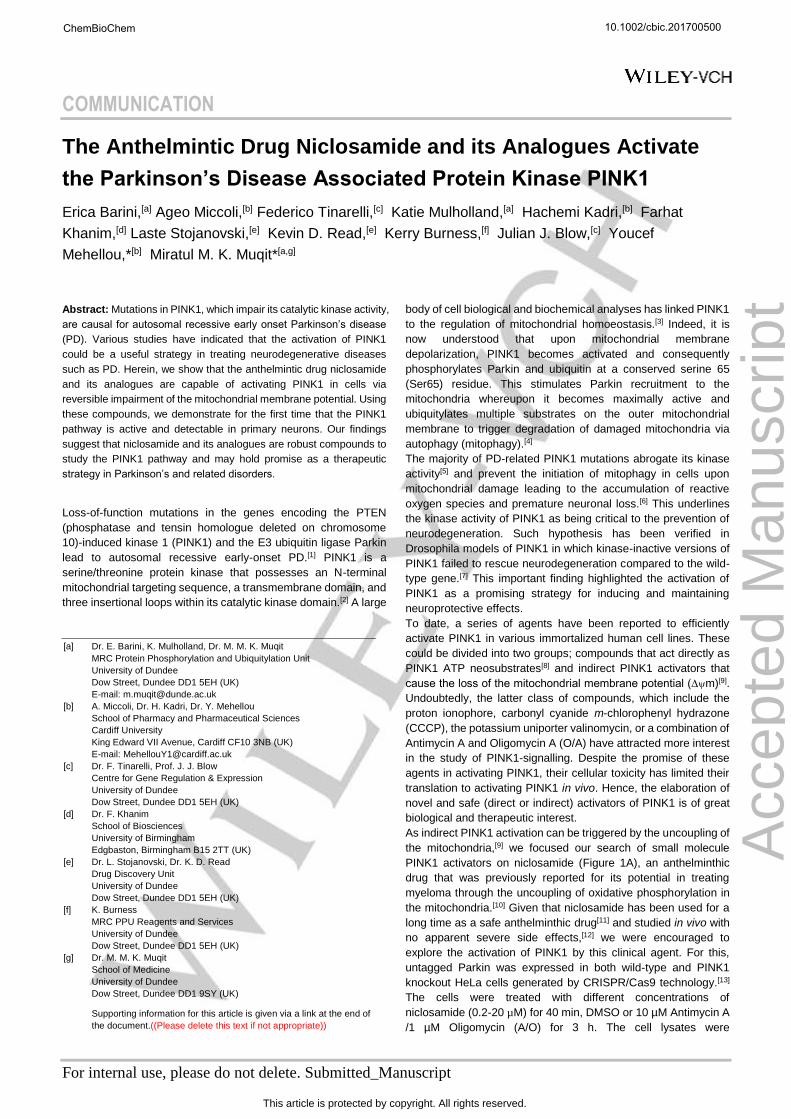
immunoblotted with an anti-phospho-Parkin Ser65 antibody to
monitor PINK1 activity.[4c] Niclosamide has been shown to
uncouple the mitochondria preventing the creation of adenosine
triphosphate (ATP).[12b] To monitor for the ability of niclosamide
and A/O to induce mitochondrial uncoupling, we probed for the
cleavage of the mitochondrial protein, optic atrophy protein 1
(OPA1), that is catalyzed by the zinc metalloprotease, OMA 1,
upon mitochondrial membrane depolarization in cells.[14] We
observed mild activation of PINK1 as judged by Parkin Ser65
phosphorylation at 0.2 μM and more striking activation at 2 μM or
higher concentrations of niclosamide comparable to that induced
by A/O treatment at 3 h (Figures 1B). This was associated with
ubiquitylation of the mitochondrial Fe/S domain containing protein,
CISD1, that is a readout of Parkin ubiquitin E3 ligase activity
(Figure 1B).[15]
Figure 1. Niclosamide activates PINK1 in HeLa cells. A. Chemical structure
of niclosamide. B. Niclosamide dose-response analysis. Wildtype (WT) and
PINK1 knockout (PINK1 KO) HeLa cells transfected with Parkin were stimulated
with either a combination of A/O for 3 h or with different concentrations (0.2, 0.8,
2, 8, 20 µM) of niclosamide (Niclo) for 40 min. Parkin Ser65 phosphorylation
(pS65Parkin), Parkin, Full length OPA1 (F/L), Cleaved OPA1, ubiquitylated
CISD1 (CISD1-Ub) and CISD1 were detected by immunoblotting. GAPDH was
used as a loading control.
Importantly, the ability of niclosamide as well as A/O to induce
Parkin Ser65 phosphorylation and CISD1 ubiquitylation was
abolished in PINK1 knockout cells (Figure 1B). However, their
ability to induce uncoupling was not affected as judged by
cleavage of OPA1 (Figure 1B). Under similar transfection and cell
conditions we next undertook a time-course analysis of Parkin
Ser65 phosphorylation and CISD1 ubiquitylation in the presence
of 20 μM niclosamide. We observed robust niclosamide induced
Parkin Ser65 phosphorylation after 20 min of treatment
(Supplementary Figures 1) associated with ubiquitylation of
CISD1 (Supplementary Figure 1A). In vitro kinase assays of
PINK1 in the presence or absence of niclosamide showed no
evidence for direct activation of PINK1 by the compound (data not
shown).
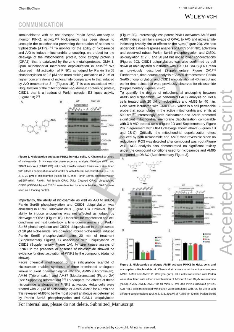
Facile chemical modification of the salicynalide scaffold of
niclosamide enabled synthesis of three brominated analogues
known to exert pharmacological efficacy, AM85 (Dibromsalan),
AM86 (Tribromsalan) and AM87 (Metabromsalan) (Figure 2A)
(see Supporting Information).[16] To compare the effects of these
niclosamide analogues on PINK1 activation, HeLa cells were
treated with 20 µM of niclosamide or AM85-AM87 for 40 min and
this revealed AM85 to be the most potent analogue as determined
by Parkin Ser65 phosphorylation and CISD1 ubiquitylation
(Figure 2B). Interestingly less potent PINK1 activators AM86 and
AM87 induced similar cleavage of OPA1 to A/O and niclosamide
indicating broadly similar effects on the ∆m (Figure 2B). We next
undertook a dose-response analysis of AM85 on PINK1 activation
and observed robust Parkin Ser65 phosphorylation and CISD1
ubiquitylation at 2, 8 and 20 µM but not at lower concentrations
(Figures 2C). CISD1 ubiquitylation, was also confirmed by pull
down of ubiquitylated substrates with HALO-UBAUBQLN1 resin
as previously described (Supplementary Figure 2A).[4a]
Furthermore, time-course analysis of AM85 demonstrated Parkin
Ser65 phosphorylation and CISD1 ubiquitylation at 40 min but not
earlier time points that were previously observed for niclosamide
(Supplementary Figures 2B-C).
To quantify the degree of mitochondrial uncoupling between
AM85 and niclosamide, we performed FACS analysis on HeLa
cells treated with 20 µM of niclosamide and AM85 for 40 min.
Cells were incubated with CMX ROS, which is a cell permeable
probe that accumulates in the active mitochondria and emits at
599 nm.[17] Interestingly, both niclosamide and AM85 promoted
significant mitochondrial membrane depolarization comparable
with 3 h A/O-treated cells (Figure 2D and Supplementary Figure
2d) in agreement with OPA1 cleavage shown above (Figures 1B
and 2B-C). Critically, the mitochondrial depolarization effect
induced by both niclosamide and AM85 was reversible since no
reduction in ROS was detected after compound wash-out (Figure
2e). FACS analysis also demonstrated no significant toxicity
under the compound conditions used for niclosamide and AM85
compared to DMSO (Supplementary Figure 3).
Figure 2. Niclosamide analogue AM85 activate PINK1 in HeLa cells and
uncouples mitochondria. A. Chemical structures of niclosamide analogues
AM85, AM86 and AM87. B. Wildtype (WT) HeLa cells transfected with Parkin
were stimulated with either a combination of A/O for 3 h or 10 M niclosamide
(Niclo), AM85, AM86, AM87 for 40 mins. C. WT and PINK1 knockout (PINK1
KO) HeLa cells transfected with Parkin were stimulated with A/O for 3 h or with
different concentrations (0.2, 0.8, 2, 8, 20 M) of AM85 for 40 min. Parkin Ser65
10.1002/cbic.201700500
Acc
epte
d M
anus
crip
t
ChemBioChem
This article is protected by copyright. All rights reserved.
COMMUNICATION
For internal use, please do not delete. Submitted_Manuscript
phosphorylation (pS65Parkin), Parkin, Full length OPA1 (F/L), Cleaved OPA1,
ubiquitylated CISD1 (CISD1-Ub) and CISD1 were detected by immunoblotting.
GAPDH was used as a loading control. D. Histograms of CMXRos Fluorescence
Intensity (arbitrary units, a.u.) for Hela cells treated on site for 3h with A/O, green
or with 1h of niclosamide (Niclo, red) and AM85 (blue). Values are normalized
to the vehicle, DMSO (black). E. Quantification of CMXRos Fluorescence
Intensity (arbitrary units, a.u.) for Hela cells subjected to drug wash out, after
treatment with A/O (green), niclosamide (Niclo, red), AM85 (blue), normalized
to the vehicle, DMSO (black). Bars represent the average ratio ± SEM of three
independent experiments. **p < 0.01, one-way ANOVA followed by Bonferroni
post-test correction.
We next determined the ability of niclosamide and AM85 to
activate PINK1 in cells of pathophysiological relevance to PD. To
date no studies have assessed PINK1 activity in primary neurons
under conditions where the PINK1 and Parkin are expressed at
endogenous levels. Marked neuronal loss and Lewy body
accumulation occurs in the frontal cortex particularly the anterior
cingulate gyrus in advancing Parkinson’s disease.[18] Therefore,
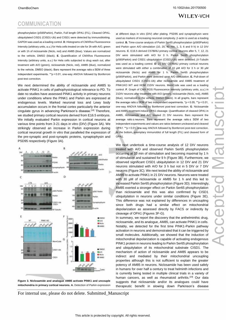
we studied primary cortical neurons derived from E16.5 embryos.
We initially evaluated Parkin expression in cortical neurons at
various time points from 3-21 days in vitro (DIV) (Figure 3A). We
strikingly observed an increase in Parkin expression during
cortical neuronal growth in vitro that paralleled the expression of
the pre-synaptic and post-synaptic proteins, synaptophysin and
PSD95 respectively (Figure 3A).
Figure 3. Niclosamide and analogue AM85 activate PINK1 and uncouple
mitochondria in primary cortical neurons. A. Detection of Parkin expression
at different days in vitro (DIV) after plating. PSD95 and synaptophysin were
used as markers of increasing neuronal complexity. -actin is used as a loading
control. B. Time-course analysis of Parkin Ser65 phosphorylation (pS65Parkin)
and Parkin upon A/O stimulation (10, 20, 40 min, 1, 3, 6 and 9 h) in 12 DIV
neurons. C. E16.5 derived C57Bl/6J primary cortical neurons after 5, 7, 12, 21
DIV were stimulated with A/O for 3 h. Parkin Ser65 phosphorylation
(pS65Parkin) and CISD1 ubiquitylation (CISD1-Ub) were detected. I-Tubulin
was used as a loading control. D. 21 DIV C57Bl/6J primary cortical neurons
were stimulated with either a combination of 10 µM A/O for 3 h or 30 µM
niclosamide (Niclo) and AM85 for 1 h. Parkin Ser65 phosphorylation
(pS65Parkin), and Parkin were detected upon A/O stimulation. E. Pull-down of
ubiquitylated CISD1 (CISD1-Ub) after niclosamide and AM85 treatment of
PINK1KO WT and HOM 21DIV neurons. MemCode was used as a loading
control. F. Graph of CMX ROS Fluorescence Intensity (arbitrary units, a.u.) in
21DIV neurons after treatment with A/O (green), niclosamide (Niclo, red), AM85
(blue), normalized to the vehicle, DMSO (black). In all graphs, bars represent
the average ratio ± SEM of two independent experiments. *p < 0.05, **p < 0.01,
one-way ANOVA followed by Bonferroni post-test correction. G. Niclosamide
and AM85 treatment induce OPA1 cleavage. Quantification of cleaved OPA1 in
AM85, niclosamide and A/O treated 21 DIV neurons. Bars represent the
average ratio ± neurons. Bars represent the average ratio ± SEM of two
independent experiments and values are ratios between uncleaved and cleaved
OPA1. **p < 0.01, one-way ANOVA followed by Bonferroni post-test correction.
At the bottom, exemplary immunoblot of full length (F/L) and cleaved form of
OPA1.
We next undertook a time-course analysis of 12 DIV neurons
treated with A/O and observed Parkin Ser65 phosphorylation
occurring at 10 min of stimulation and becoming maximal by 1 h
of stimulation and sustained for 9 h (Figure 3B). Furthermore, we
observed significant CISD1 ubiquitylation in 12 DIV and 21 DIV
neurons stimulated with A/O for 3 h but not in 5 DIV or 7 DIV
neurons (Figure 3C). We next tested the ability of niclosamide and
AM85 to activate PINK1 in 21 DIV neurons. Neurons were treated
with 30 µM of niclosamide or AM85 for 1 h and this led to
increased Parkin Ser65 phosphorylation (Figure 3D). Interestingly,
AM85 exerted a stronger effect on Parkin Ser65 phosphorylation
than niclosamide and this was also confirmed by CISD1
ubiquitylation in neurons under similar conditions (Figure 3E).
This difference was not explained by differences in uncoupling
since both drugs had a similar effect on mitochondrial
depolarization as assessed directly by FACS or indirectly by
cleavage of OPA1 (Figures 3F-G).
In summary, we report the discovery that the anthelminthic drug,
niclosamide, and its analogue, AM85, can activate PINK1 in cells.
Notably, we detected for the first time PINK1-Parkin pathway
activation in neurons and demonstrated that it can be triggered by
small molecules. Additionally, we showed that the induction of
mitochondrial depolarization is capable of activating endogenous
PINK1 protein in neurons leading to Parkin Ser65 phosphorylation
and ubiquitylation of its mitochondrial substrate CISD1. The
mechanism of action of niclosamide and AM85 appears to be
indirect and mediated by their mitochondrial uncoupling
properties although this is not sufficient to explain the greater
potency of AM85 in neurons. Niclosamide has been used safely
in humans for over half a century to treat helminth infections and
is currently being tested in multiple clinical trials in a variety of
human cancers, as well as rheumatoid arthritis.[19] Our data
suggests that niclosamide and/or its analogues could have
therapeutic benefit in slowing down Parkinson’s disease
10.1002/cbic.201700500
Acc
epte
d M
anus
crip
t
ChemBioChem
This article is protected by copyright. All rights reserved.
COMMUNICATION
For internal use, please do not delete. Submitted_Manuscript
progression through the activation of PINK1. Further in vivo
studies in appropriate PD models are warranted to test this
hypothesis.
Experimental Section
Immunoblotting and immunoprecipitation
Tissues, primary cortical neurons or Hela cells were sonicated in lysis
buffer containing 50 mM Tris–HCl (pH 7.5), 1 mM EDTA, 1 mM EGTA, 1%
(w/v) Triton, 1 mM sodium orthovanadate, 10 mM sodium
glycerophosphate, 50 mM sodium fluoride, 10 mM sodium pyrophosphate,
0.25 M sucrose, 1 mM benzamidine, 0.1 mM PMSF and protease inhibitor
cocktail (Roche). Following the sonication, lysates were incubated for 30
min on ice. Samples were spun at 20,800 x g in an Eppendorf 5417R
centrifuge for 30 min. Supernatants were collected and protein
concentration was determined using the Bradford kit (Pierce). Samples
were subjected to SDS/PAGE (4–12% gels) and were transferred on to
Protran 0.2 NC nitrocellulose membranes (Amersham). Membranes were
blocked for 1 h at room temperature with 5% nonfat milk or bovine serum
albumin (BSA) in TBST [Tris-buffered saline (50 mM Tris/HCl and 150 mM
NaCl, pH 7.5) containing 0.1% Tween-20 in phosphate buffered saline
(PBS) pH 7.4 and probed with the indicated antibodies overnight at 4 °C.
Detection was performed using HRP-conjugated secondary antibodies
and enhanced chemiluminescence reagent.
Ubiquitin enrichment
For ubiquitylated protein capture, 400 μg of extract was used for pull down
with HALO‐UBAUBQLN1 resin as described previously5. Halo-tagged
UBDs were incubated with 200 μl of the HaloLink resin (Promega) in
binding buffer (50 mM Tris-HCl [pH 7.5], 150 mM NaCl, 0.05% NP-40)
overnight at 4 °C. 20 μl of Halo Tube beads were added to neuronal or
tissue lysates and were incubated for 4 h at 4 °C. The beads were washed
three times with lysis buffer containing 0.25 M NaCl and eluted by
resuspending in 20 μl of 1× LDS sample buffer with 1mM DTT.
Flow Cytometry Analysis of mitochondrial membrane potential
Hela Cells were incubated with 20 μM niclosamide (manufacturer), AM85
(manufacturer) for 40 min before trypsinization and collection.
Oligomycin(manufacturer) and Antimycin (manufacturer) 20 μM were used
as positive control and incubated for 3 h before samples harvest. DMSO
(Sigma Aldrich) at the same concentration was used as control. After 10
min from start of drug treatment, cells were treated with 100 nM Mito
Tracker CMXRos (Cell Signaling Technology) for 30 min directly on wells.
Drug Washout was performed incubating trypsinized floating cells with 100
nM Mito Tracker CMXRos (Cell Signaling Technology) for 30 min at 37 °C
in drug absence. All harvested cells were incubated for 5 min on ice after
CMXRos incubation and then centrifuged and washed two times with a 1%
BSA/PBS solution. Finally, cells were treated with DAPI (1:200) solution (1
mg/ml DAPI, 50 μg/mL RNaseA in 1% BSA/PBS) and transferred to FACS
tubes for analysis. Samples were acquired using a BD FACS Canto and
the results analysed using FlowJo software.
Statistical analysis
Statistical analysis of groups with normal distributions was performed
using One-way ANOVA or two-way ANOVA followed by Holm-Sidak or
Bonferroni post-test. Differences among groups were considered
statistically significant when p < 0.05. Data throughout the text are reported
as average values ± SEM unless otherwise specified.
Acknowledgements
We thank Axel Knebel (Dundee) for providing TUBE proteins for
ubiquitin capture, Andrew Waddell (Dundee) for expressing
recombinant human PINK1 and Richard Youle (NIH) for HeLa
PINK1 knockout cell lines. We also thank the MRC Genotyping
and Tissue Culture teams for technical support. We also thank
Thomas McWilliams (Dundee) for useful discussions. J. B and F.
T. are funded by Wellcome Trust (WT096598MA). YM is funded
by a Medical Research Council grant (MC_PC_16041). M.M.K.M.
is funded by a Wellcome Trust Senior Research Fellowship in
Clinical Science (101022/Z/13/Z). This work was supported by the
Medical Research Council; the Wellcome Trust; Parkinson’s UK;
the Michael J. Fox Foundation for Parkinson’s disease research;
and the EMBO YIP programme.
Keywords: PINK1 • Parkin • Mitochondria • Niclosamide •
Parkinson’s disease
[1] aE. M. Valente, P. M. Abou-Sleiman, V. Caputo, M. M. Muqit,
K. Harvey, S. Gispert, Z. Ali, D. Del Turco, A. R. Bentivoglio,
D. G. Healy, A. Albanese, R. Nussbaum, R. Gonzalez-
Maldonado, T. Deller, S. Salvi, P. Cortelli, W. P. Gilks, D. S.
Latchman, R. J. Harvey, B. Dallapiccola, G. Auburger, N. W.
Wood, Science 2004, 304(5674), 1158-1160; bT. Kitada, S.
Asakawa, N. Hattori, H. Matsumine, Y. Yamamura, S.
Minoshima, M. Yokochi, Y. Mizuno, N. Shimizu, Nature 1998,
392(6676), 605-608.
[2] A. Kazlauskaite, M. M. Muqit, Febs J 2015, 282(2), 215-223.
[3] T. G. McWilliams, M. M. Muqit, Curr Opin Cell Biol 2017, 45,
83-91.
[4] aL. A. Kane, M. Lazarou, A. I. Fogel, Y. Li, K. Yamano, S. A.
Sarraf, S. Banerjee, R. J. Youle, J Cell Biol 2014, 205(2), 143-
153; bA. Kazlauskaite, C. Kondapalli, R. Gourlay, D. G.
Campbell, M. S. Ritorto, K. Hofmann, D. R. Alessi, A. Knebel,
M. Trost, M. M. Muqit, Biochem J 2014, 460(1), 127-139; cA.
Kazlauskaite, R. J. Martinez-Torres, S. Wilkie, A. Kumar, J.
Peltier, A. Gonzalez, C. Johnson, J. Zhang, A. G. Hope, M.
Peggie, M. Trost, D. M. van Aalten, D. R. Alessi, A. R. Prescott,
A. Knebel, H. Walden, M. M. Muqit, EMBO Rep 2015, 16(8),
939-954; dF. Koyano, K. Okatsu, H. Kosako, Y. Tamura, E. Go,
M. Kimura, Y. Kimura, H. Tsuchiya, H. Yoshihara, T.
Hirokawa, T. Endo, E. A. Fon, J. F. Trempe, Y. Saeki, K.
Tanaka, N. Matsuda, Nature 2014, 510(7503), 162-166.
[5] H. I. Woodroof, J. H. Pogson, M. Begley, L. C. Cantley, M.
Deak, D. G. Campbell, D. M. van Aalten, A. J. Whitworth, D.
R. Alessi, M. M. Muqit, Open Biol 2011, 1(3), 110012.
[6] Z. Yue, L. Friedman, M. Komatsu, K. Tanaka, Biochim
Biophys Acta 2009, 1793(9), 1496-1507.
[7] Y. Kim, J. Park, S. Kim, S. Song, S. K. Kwon, S. H. Lee, T.
Kitada, J. M. Kim, J. Chung, Biochem Biophys Res Commun
2008, 377(3), 975-980.
[8] aN. T. Hertz, A. Berthet, M. L. Sos, K. S. Thorn, A. L.
Burlingame, K. Nakamura, K. M. Shokat, Cell 2013, 154(4),
737-747; bL. Osgerby, Y. C. Lai, P. J. Thornton, J. Amalfitano,
C. S. Le Duff, I. Jabeen, H. Kadri, A. Miccoli, J. H. Tucker, M.
M. Muqit, Y. Mehellou, J Med Chem 2017, 21(10).
[9] C. Kondapalli, A. Kazlauskaite, N. Zhang, H. I. Woodroof, D.
G. Campbell, R. Gourlay, L. Burchell, H. Walden, T. J.
Macartney, M. Deak, A. Knebel, D. R. Alessi, M. M. Muqit,
Open Biol 2012, 2(5), 120080.
[10] F. L. Khanim, B. A. Merrick, H. V. Giles, M. Jankute, J. B.
Jackson, L. J. Giles, J. Birtwistle, C. M. Bunce, M. T. Drayson,
Blood Cancer J 2011, 1(10), e39.
[11] aM. Katz, Drugs 1977, 13(2), 124-136; bD. O’fori-Adjei, A. N.
O. Dodoo, A. Appiah-Danquah, M. Couper, International
Journal of Risk and Safety in Medicine 2008, 20, 113-122.
[12] aT. Ye, Y. Xiong, Y. Yan, Y. Xia, X. Song, L. Liu, D. Li, N.
Wang, L. Zhang, Y. Zhu, J. Zeng, Y. Wei, L. Yu, PLoS One
2014, 9(1), e85887; bH. Tao, Y. Zhang, X. Zeng, G. I. Shulman,
10.1002/cbic.201700500
Acc
epte
d M
anus
crip
t
ChemBioChem
This article is protected by copyright. All rights reserved.
COMMUNICATION
For internal use, please do not delete. Submitted_Manuscript
S. Jin, Nat Med 2014, 20(11), 1263-1269; cK. Satoh, L. Zhang,
Y. Zhang, R. Chelluri, M. Boufraqech, N. Nilubol, D. Patel, M.
Shen, E. Kebebew, Clin Cancer Res 2016, 22(14), 3458-3466.
[13] D. P. Narendra, C. Wang, R. J. Youle, J. E. Walker, Hum Mol
Genet 2013, 22(13), 2572-2589.
[14] K. Zhang, H. Li, Z. Song, EMBO Rep 2014, 15(5), 576-585.
[15] S. A. Sarraf, M. Raman, V. Guarani-Pereira, M. E. Sowa, E. L.
Huttlin, S. P. Gygi, J. W. Harper, Nature 2013, 496(7445), 372-
376.
[16] aR. A. Coburn, A. J. Batista, R. T. Evans, R. J. Genco, J Med
Chem 1981, 24(10), 1245-1249; bH. M. Norbury, R. H. Waring,
Xenobiotica 1981, 11(8), 511-518; cH. M. Norbury, R. H.
Waring, Xenobiotica 1981, 11(7), 501-508.
[17] C. Cottet-Rousselle, X. Ronot, X. Leverve, J. F. Mayol,
Cytometry A 2011, 79(6), 405-425.
[18] C. Buddhala, S. K. Loftin, B. M. Kuley, N. J. Cairns, M. C.
Campbell, J. S. Perlmutter, P. T. Kotzbauer, Ann Clin Transl
Neurol 2015, 2(10), 949-959.
[19] W. Chen, R. A. Mook, Jr., R. T. Premont, J. Wang, Cell Signal
2017, 4(17), 30101-30108.
10.1002/cbic.201700500
Acc
epte
d M
anus
crip
t
ChemBioChem
This article is protected by copyright. All rights reserved.
COMMUNICATION
For internal use, please do not delete. Submitted_Manuscript
Entry for the Table of Contents
COMMUNICATION
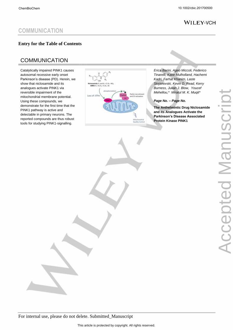
Catalytically impaired PINK1 causes
autosomal recessive early onset
Parkinson’s disease (PD). Herein, we
show that niclosamide and its
analogues activate PINK1 via
reversible impairment of the
mitochondrial membrane potential.
Using these compounds, we
demonstrate for the first time that the
PINK1 pathway is active and
detectable in primary neurons. The
reported compounds are thus robust
tools for studying PINK1-signalling.
Erica Barini, Ageo Miccoli, Federico
Tinarelli, Katie Mulholland, Hachemi
Kadri, Farhat Khanim, Laste
Stojanovski, Kevin D. Read, Kerry
Burness, Julian J. Blow, Youcef
Mehellou,* Miratul M. K. Muqit*
Page No. – Page No.
The Anthelmintic Drug Niclosamide
and its Analogues Activate the
Parkinson’s Disease Associated
Protein Kinase PINK1
10.1002/cbic.201700500
Acc
epte
d M
anus
crip
t
ChemBioChem
This article is protected by copyright. All rights reserved.