Embed Size (px)
Citation preview
RESEARCH ARTICLE Open Access
New insights from Opisthorchis felineusgenome: update on genomics of theepidemiologically important liver flukesNikita I. Ershov1*, Viatcheslav A. Mordvinov1*, Egor B. Prokhortchouk3,7*, Mariya Y. Pakharukova1,2,Konstantin V. Gunbin1, Kirill Ustyantsev1, Mikhail A. Genaev1, Alexander G. Blinov1, Alexander Mazur3,Eugenia Boulygina6, Svetlana Tsygankova6, Ekaterina Khrameeva7, Nikolay Chekanov3, Guangyi Fan4,5†, An Xiao4†,He Zhang4, Xun Xu4, Huanming Yang4, Victor Solovyev8, Simon Ming-Yuen Lee5†, Xin Liu4†,Dmitry A. Afonnikov1,2 and Konstantin G. Skryabin3,6
Abstract
Background: The three epidemiologically important Opisthorchiidae liver flukes Opisthorchis felineus, O. viverrini,and Clonorchis sinensis, are believed to harbour similar potencies to provoke hepatobiliary diseases in their definitivehosts, although their populations have substantially different ecogeographical aspects including habitat, preferredhosts, population structure. Lack of O. felineus genomic data is an obstacle to the development of comparativemolecular biological approaches necessary to obtain new knowledge about the biology of Opisthorchiidaetrematodes, to identify essential pathways linked to parasite-host interaction, to predict genes that contribute toliver fluke pathogenesis and for the effective prevention and control of the disease.
Results: Here we present the first draft genome assembly of O. felineus and its gene repertoire accompanied by acomparative analysis with that of O. viverrini and Clonorchis sinensis. We observed both noticeably high heterozygosityof the sequenced individual and substantial genetic diversity in a pooled sample. This indicates that potency of O.felineus population for rapid adaptive response to control and preventive measures of opisthorchiasis is higher than inO. viverrini and C. sinensis. We also have found that all three species are characterized by more intensive involvement oftrans-splicing in RNA processing compared to other trematodes.
Conclusion: All revealed peculiarities of structural organization of genomes are of extreme importance for a properdescription of genes and their products in these parasitic species. This should be taken into account both in academicand applied research of epidemiologically important liver flukes. Further comparative genomics studies of liver flukesand non-carcinogenic flatworms allow for generation of well-grounded hypotheses on the mechanisms underlyingdevelopment of cholangiocarcinoma associated with opisthorchiasis and clonorchiasis as well as species-specificmechanisms of these diseases.
Keywords: Opisthorchiidae, Opisthorchis felineus, Genome, Trans-splicing, Microintrons, Liver flukes, Transcriptome,Metacercariae
© The Author(s). 2019 Open Access This article is distributed under the terms of the Creative Commons Attribution 4.0International License (http://creativecommons.org/licenses/by/4.0/), which permits unrestricted use, distribution, andreproduction in any medium, provided you give appropriate credit to the original author(s) and the source, provide a link tothe Creative Commons license, and indicate if changes were made. The Creative Commons Public Domain Dedication waiver(http://creativecommons.org/publicdomain/zero/1.0/) applies to the data made available in this article, unless otherwise stated.
* Correspondence: [email protected]; [email protected];[email protected]†Guangyi Fan, An Xiao, Simon Ming-Yuen Lee and Xin Liu contributedequally to this work.1Institute of Cytology and Genetics SB RAS, 10 Lavrentiev Ave, Novosibirsk630090, Russia3Russian Federal Research Center for Biotechnology, 33/2 Leninsky prospect,Moscow 119071, RussiaFull list of author information is available at the end of the article
Ershov et al. BMC Genomics (2019) 20:399 https://doi.org/10.1186/s12864-019-5752-8
BackgroundOpisthorchis felineus (Rivolta, 1884) is a member of thetriad of epidemiologically important fish-borne livertrematodes, which also includes O. viverrini (Poirier,1886) and Clonorchis sinensis (Loos, 1907). These liverflukes are known to cause serious human diseases affect-ing bile ducts and the gall bladder. Liver fluke infection isrecognized as the major risk factor of cholangiocarcinoma[1–3]. An estimated 12.5, 67.3 and 601 million people arecurrently at risk for infection with O. felineus, O. viverriniand C. sinensis, respectively [4]. According to the Foodand Agriculture Organization and World HealthOrganization [5, 6], these liver flukes are the 8th inthe overall global list of 24 food-borne parasites.The liver flukes O. felineus, O. viverrini, and C. sinensis
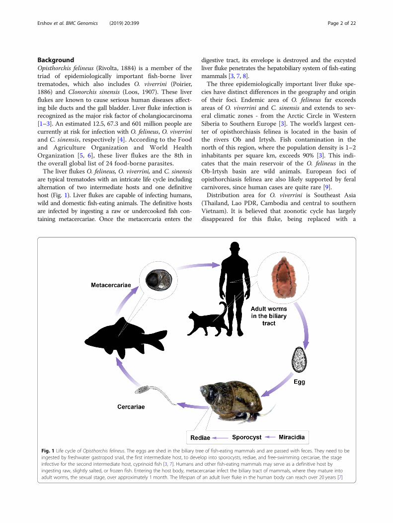
are typical trematodes with an intricate life cycle includingalternation of two intermediate hosts and one definitivehost (Fig. 1). Liver flukes are capable of infecting humans,wild and domestic fish-eating animals. The definitive hostsare infected by ingesting a raw or undercooked fish con-taining metacercariae. Once the metacercaria enters the
digestive tract, its envelope is destroyed and the excystedliver fluke penetrates the hepatobiliary system of fish-eatingmammals [3, 7, 8].The three epidemiologically important liver fluke spe-
cies have distinct differences in the geography and originof their foci. Endemic area of O. felineus far exceedsareas of O. viverrini and C. sinensis and extends to sev-eral climatic zones - from the Arctic Circle in WesternSiberia to Southern Europe [3]. The world’s largest cen-ter of opisthorchiasis felinea is located in the basin ofthe rivers Ob and Irtysh. Fish contamination in thenorth of this region, where the population density is 1–2inhabitants per square km, exceeds 90% [3]. This indi-cates that the main reservoir of the O. felineus in theOb-Irtysh basin are wild animals. European foci ofopisthorchiasis felinea are also likely supported by feralcarnivores, since human cases are quite rare [9].Distribution area for O. viverrini is Southeast Asia
(Thailand, Lao PDR, Cambodia and central to southernVietnam). It is believed that zoonotic cycle has largelydisappeared for this fluke, being replaced with a
Fig. 1 Life cycle of Opisthorchis felineus. The eggs are shed in the biliary tree of fish-eating mammals and are passed with feces. They need to beingested by freshwater gastropod snail, the first intermediate host, to develop into sporocysts, rediae, and free-swimming cercariae, the stageinfective for the second intermediate host, cyprinoid fish [3, 7]. Humans and other fish-eating mammals may serve as a definitive host byingesting raw, slightly salted, or frozen fish. Entering the host body, metacercariae infect the biliary tract of mammals, where they mature intoadult worms, the sexual stage, over approximately 1 month. The lifespan of an adult liver fluke in the human body can reach over 20 years [7]
Ershov et al. BMC Genomics (2019) 20:399 Page 2 of 22
predominantly anthropogenic cycle [9]. C. sinensis is en-demic to East Asia (China, Korea, Russian Far East andJapan) and northern Vietnam [4, 10] and holds an inter-mediate position, with a number of significant nativeand domestic reservoir animal hosts but also with highlevels of human fecal contamination of the environmentplaying a significant role in the transmission cycle [10].Correspondingly, O. felineus, O. viverrini, and C. sinensisalso display certain differences in the range of their pri-mary and secondary hosts [7–10].Epidemiologically important liver flukes differ also in
population structure. Analysis of mitochondrial and nu-clear genetic markers revealed that population structureis absent in O. felineus across Eastern Europe, NorthernAsia (Siberia) and Central Asia (Northern Kazakhstan)[11]. In contrast, population genetic differentiation existsin O. viverrini [12]. Genetic diversity of C. sinensis is notas pronounced as it is for O. viverrini, nevertheless geo-graphic variation in C. sinensis was detected [13].Recently it has been shown that O. viverrini differs fromO. felineus and C. sinensis in chromosome number.Karyotypes of O. felineus and C. sinensis (Russian isolate)consist of two pairs of largemeta- and submetacentricsand five pairs of small chromosomes (2n = 14). However,the karyotype of O. viverrini is 2n = 12 [14].Thus, these liver flukes are attractive research objects
from the standpoint of comparative genomics allowingfor better insight into the mechanisms underlying theevolution and adaptation of trematodes. Taking into ac-count the importance of opisthorchiasis and clonorchiasisfor the population health in endemic regions, the genomicstudies of these infectious agents can give a clue to solvingmany applied problems and are a priority direction in themodern molecular biology.O. viverrini and C. sinensis but not O. felineus have
been recently characterized at the level of genome [15–17].The results have significantly enriched our understandingof the molecular processes that ensure the vital activity ofthese parasites in the bile duct, and expanded the know-ledge about liver fluke-associated carcinogenesis. However,the genomics of O. felineus is poorly investigated and thishinders a deep understanding of the biology of this parasiteand the progress in comparative genomics of opistorchiids.To address this knowledge gap, we have sequenced the O.felineus genome and used the de novo assembled draft gen-ome to gain new insights into genetic features of the liverflukes. Here we present the first version of O. felineus draftgenome assembly and the accompanying transcriptome as-sembly. We also provide O. felineus genome annotationand describe the results of the first comparative analysis ofO. felineus, O. viverrini and C. sinensis genomics and tran-scriptomics, including taxa-specific features of RNA pro-cessing. Although the coding regions of the genes arehighly homologous to each other; however, analysis of the
genome-wide synteny between O. felineus, O. viverrini andC. sinensis demonstrates a considerable variation in theliver fluke genomes. The majority of genes in adult wormsdemonstrate similar level of mRNA expression amongthese species. We also found that trans-splicing potentiallyplays an important role in RNA processing of these threeliver flukes.
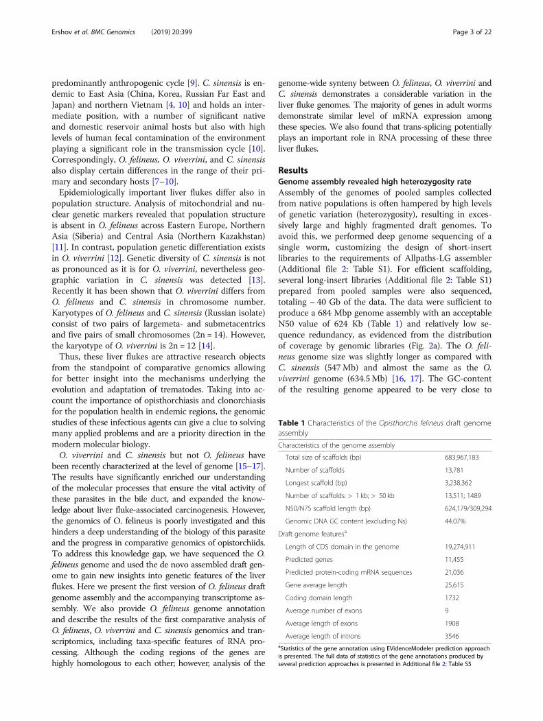
ResultsGenome assembly revealed high heterozygosity rateAssembly of the genomes of pooled samples collectedfrom native populations is often hampered by high levelsof genetic variation (heterozygosity), resulting in exces-sively large and highly fragmented draft genomes. Toavoid this, we performed deep genome sequencing of asingle worm, customizing the design of short-insertlibraries to the requirements of Allpaths-LG assembler(Additional file 2: Table S1). For efficient scaffolding,several long-insert libraries (Additional file 2: Table S1)prepared from pooled samples were also sequenced,totaling ~ 40 Gb of the data. The data were sufficient toproduce a 684 Mbp genome assembly with an acceptableN50 value of 624 Kb (Table 1) and relatively low se-quence redundancy, as evidenced from the distributionof coverage by genomic libraries (Fig. 2a). The O. feli-neus genome size was slightly longer as compared withС. sinensis (547Mb) and almost the same as the O.viverrini genome (634.5Mb) [16, 17]. The GC-contentof the resulting genome appeared to be very close to
Table 1 Characteristics of the Opisthorchis felineus draft genomeassembly
Characteristics of the genome assembly
Total size of scaffolds (bp) 683,967,183
Number of scaffolds 13,781
Longest scaffold (bp) 3,238,362
Number of scaffolds: > 1 kb; > 50 kb 13,511; 1489
N50/N75 scaffold length (bp) 624,179/309,294
Genomic DNA GC content (excluding Ns) 44.07%
Draft genome featuresa
Length of CDS domain in the genome 19,274,911
Predicted genes 11,455
Predicted protein-coding mRNA sequences 21,036
Gene average length 25,615
Coding domain length 1732
Average number of exons 9
Average length of exons 1908
Average length of introns 3546aStatistics of the gene annotation using EVidenceModeler prediction approachis presented. The full data of statistics of the gene annotations produced byseveral prediction approaches is presented in Additional file 2: Table S5
Ershov et al. BMC Genomics (2019) 20:399 Page 3 of 22
those of С. sinensis and O. viverrini. In total, 11,455protein-encoding genes (Table 1) were predicted fromthe genome based on transcriptomic evidence from pre-viously published [18] and new (Additional file 2: TableS1) RNA-seq data and sequence similarity toprotein-encoding genes of C. sinensis and O. viverrini.The estimated total number of genes, the proportion ofcoding regions (2.8%), the mean total gene length(25,615 bp), intron length (3546 bp) and the mean num-ber of exons per gene [9] were similar to those of C.sinensis and O. viverrini [16].One of the main factors that hampered the contiguity
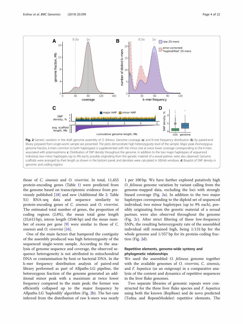
of the assembly produced was high heterozygosity of thesequenced single-worm sample. According to the ana-lysis of genome sequence and coverage, the observed se-quence heterogeneity is not attributed to mitochondrialDNA or contamination by host or bacterial DNA. In thek-mer frequency distribution analysis of paired-endlibrary performed as part of Allpaths-LG pipeline, theheterozygous fraction of the genome generated an add-itional minor peak with a maximum at twice lowerfrequency compared to the main peak; the former wasefficiently collapsed up to the major frequency byAllpaths-LG ‘haploidify’ algorithm (Fig. 2b). The het-rateinferred from the distribution of raw k-mers was nearly
1 per 100 bp. We have further explored putatively highO. felineus genome variation by variant calling from thegenome-mapped data, excluding the loci with stronglybiased coverage (Fig. 2a). In addition to the two majorhaplotypes corresponding to the diploid set of sequencedindividual, two minor haplotypes (up to 9% each), pos-sibly originating from the genetic material of a sexualpartner, were also observed throughout the genome(Fig. 2c). After strict filtering of these low-frequencySNPs, the resulting heterozygosity rate of the assembledindividual still remained high, being 1/131 bp for thewhole genome and 1/357 bp for its protein-coding frac-tion (Fig. 2d).
Repetitive elements, genome-wide synteny andphylogenetic relationshipsWe used the assembled O. felineus genome togetherwith the available genomes of O. viverrini, C. sinensis,and F. hepatica (as an outgroup) in a comparative ana-lysis of the content and dynamics of repetitive sequencesin the liver fluke genomes.Two separate libraries of genomic repeats were con-
structed for the three liver fluke species and F. hepaticausing both the known (RepBase) and de novo predicted(Tedna and RepeatModeler) repetitive elements. The
A
C D
B
Fig. 2 Genetic variation in the draft genome assembly of O. felineus. Genome coverage (a) and K-mer frequency distribution (b) by paired-endlibrary prepared from single-worm sample are presented. The plots demonstrate high heterozygosity level of the sample. Major peak (homozygousgenome fraction, k-mers common to both haplotypes) is supplemented with the minor one at twice lower coverage corresponding to the k-mersassociated with polymorphisms. c. Distribution of SNP density throughout the genome. In addition to the two major haplotypes of sequencedindividual, two minor haplotypes (up to 9% each), possibly originating from the genetic material of a sexual partner, were also observed. Genomicscaffolds were arranged by their length as shown in the bottom panel, and densities were calculated in 500-kb windows. d. Boxplot of SNP density ingenomic and coding regions
Ershov et al. BMC Genomics (2019) 20:399 Page 4 of 22
analysis showed an extremely low overlap between theTedna and RepeatModeler repeat libraries (< 0.1%) foreach of the genomes, demonstrating the advantage ofusing both methods. The repeats accounted for 30.3,30.9, 29.6, and 55.3% of O. felineus, O. viverrini, C. sinen-sis, and F. hepatica genomes (Additional file 2: TablesS3.1–3.4), respectively. The total numbers obtained forthe O. viverrini and C. sinensis genomes are consistentwith those obtained in previous studies (30.6% for O.viverrini [16] and 32% for C. sinensis [17], but with alarger share of annotated elements (9.3 and 14.3%, re-spectively) and different ratios of repeat superfamilies(Additional file 2: Table S3.3–3.4). While the F. hepaticagenome was earlier reported to be 32% repetitive [19],we found at least 54% of the genome masked for anno-tated transposons only, not taking into account the tan-dem, satellite, and other simple repeats. This feature ofF. hepatica genome can partly explain its considerablylarger size as compared with other studied trematodes(Additional file 2: Table S3.1).The majority of repeats (90.2%) in the O. felineus gen-
ome are retrotransposons, with 17.9% of LTR, 72.3% ofLINE, and 0.4% of SINE elements, while the remaining9.8% are formed by cut-and-paste DNA transposons(Additional file 2: Table S3.2). The overall repeat land-scape of O. felineus genome is similar to those of theother three trematodes in question (Additional file 2:Table S3.1-ST3.4; Additional file 1: Figure S4). Diver-gence of transposable element copies from their consen-sus is correlated with the age of their activity. Moresimilar copies (low distance from the consensus) are in-dicative of recent activity of an element and vice versa[20]. We found that the majority of the transposableelement copies identified in the studied opisthorchiid ge-nomes have a similar distance from their correspondingconsensuses (approximately 20%) (Additional file 1:Figure S4), indicating the same time of the last trans-position burst in these genomes.We conducted genome-wide synteny comparisons be-
tween O. felineus, O. viverrini, C. sinensis, and Schistosomamansoni. The genomic sequences of these flukes werecompared in a pairwise manner using MUMmer (seeMethods). The large (> 100 kb) scaffolds of O. felineus, O.viverrini, and C. sinensis at the level of amino acid se-quences display nearly the same level of differences ascompared to the first chromosome of S. mansoni genome.The amino acid identity and similarity in the alignmentwas approximately 70 and 80%, respectively (Additionalfile 2: Table S4). The identity and similarity parameters forO. felineus are somewhat higher as compared with theother two opisthorchiids.A comparison of three pairs of liver fluke genomes at
a nucleotide level without filtering repeats demonstratesthat the pair O. felineus–C. sinensis has the highest
similarity as compared with the remaining two pairs (Add-itional file 2: Table S4). The largest number of aligned scaf-folds for both the reference (O. felineus) and query (C.sinensis) has been detected for this genome pair as well as alarge share of aligned nucleotides, accounting for 40 to 50%of the total length of the reference and query, respectively.In addition, the average length of aligned fragments for thispair is the longest (~ 1600 bp versus ~ 1200 bp for theremaining pairs) and the alignments display a higher levelof nucleotide identity (84.2 versus 83.5%). The compari-son of the O. viverrini and C. sinensis both to eachother and to S. mansoni chromosome 1 fits well thedata obtained by Young et al. [16].Similar results were obtained when comparing the
liver fluke genomes at the level of amino acid sequences(Additional file 2: Table S4). Comparison of the aminoacid sequence of the pair O. felineus–C. sinensis showsthe largest number of aligned scaffolds, longest homolo-gous regions, and highest sequence similarity as comparedwith the other genome pairs. Analysis of the genomicsequences with masked repeats suggests an analogous in-ference as well as analysis of the characteristics of syntenicblocks in the three genomes by the SyMap software [21]using both unmasked and masked sequences (Additionalfile 2: Table S4).We additionally analyzed synteny between three
Opistorchiidae genomic sequences using OrthoClustersoftware (see Methods). Results demonstrate (Additionalfile 2: Table S4) that the synteny conservation between O.felineus and C. sinensis is higher (0.363) than that for O.viverrini and C. sinensis (0.215) or O. felineus and O. viver-rini (0.256). Other parameters of OrthoCluster syntenicblock comparison demonstrates also that the genomicstructure O. felineus and C. sinensis is more similar(Additional file 2: Table S4). This is in accordancewith genomic sequence alignment results (Additionalfile 1: Figure S3).The phylogenetic relationships between three liver
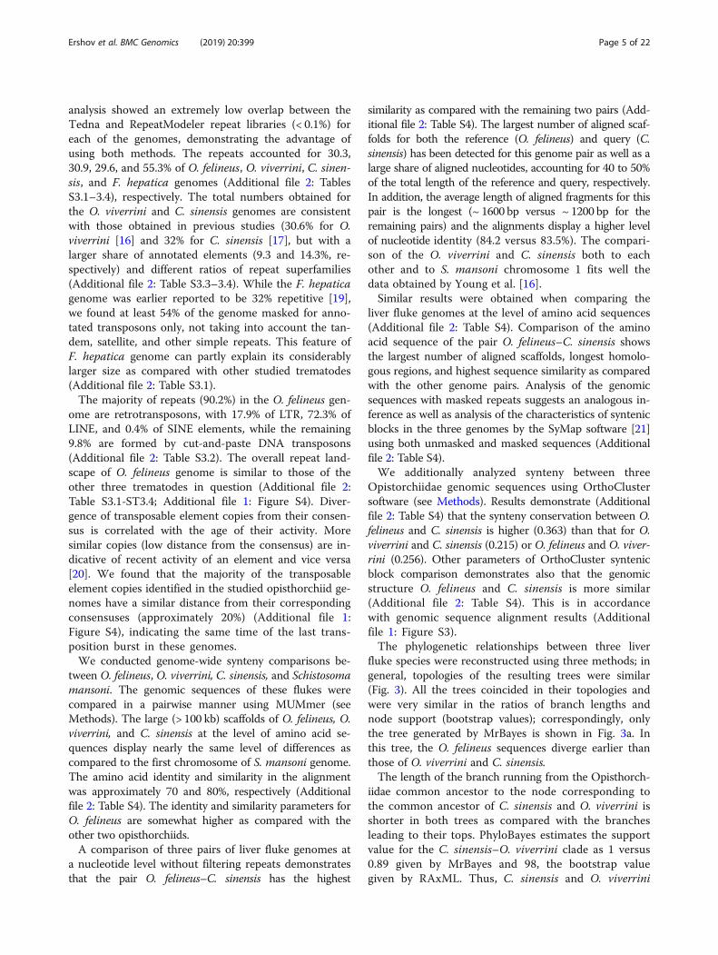
fluke species were reconstructed using three methods; ingeneral, topologies of the resulting trees were similar(Fig. 3). All the trees coincided in their topologies andwere very similar in the ratios of branch lengths andnode support (bootstrap values); correspondingly, onlythe tree generated by MrBayes is shown in Fig. 3a. Inthis tree, the O. felineus sequences diverge earlier thanthose of O. viverrini and C. sinensis.The length of the branch running from the Opisthorch-
iidae common ancestor to the node corresponding tothe common ancestor of C. sinensis and O. viverrini isshorter in both trees as compared with the branchesleading to their tops. PhyloBayes estimates the supportvalue for the C. sinensis–O. viverrini clade as 1 versus0.89 given by MrBayes and 98, the bootstrap valuegiven by RAxML. Thus, C. sinensis and O. viverrini
Ershov et al. BMC Genomics (2019) 20:399 Page 5 of 22
diverged almost immediately after O. felineus was sepa-rated from the common ancestor of these three liverfluke species.
Analysis of pre-mRNA processing revealed many trans-spliced genesWe used a combination of several gene finding ap-proaches to refine a reliable annotation of protein-codinggenes in O. felineus genome (Additional file 1: Figure S2;Additional file 2: Table S5). The RNA-seq data for two lifestages (metacercaria and adult) allowed for a total of11,455 protein-coding genes and 21,036 their mRNAproducts to be identified (Additional file 2: Table S2). Thenumber of found genes is less than that predicted in C.sinensis and O. viverrini genomes [15, 16]. Nevertheless,this difference is not much informative, since it was at-tributed mainly to the strictness of filtering invalid orinsufficiently supported gene models, the initial numberof which was quite large (Additional file 2: Table S5). Infact, the comparative evaluation of the three genomeannotations using BUSCO software (Additional file 2:Table S10) as well as the results of the orthology infer-ence (Fig. 7a, discussed further) showed that the ap-plied filters did not hamper the completeness of O.felineus annotation.When analyzing the RNA-seq data, we found that
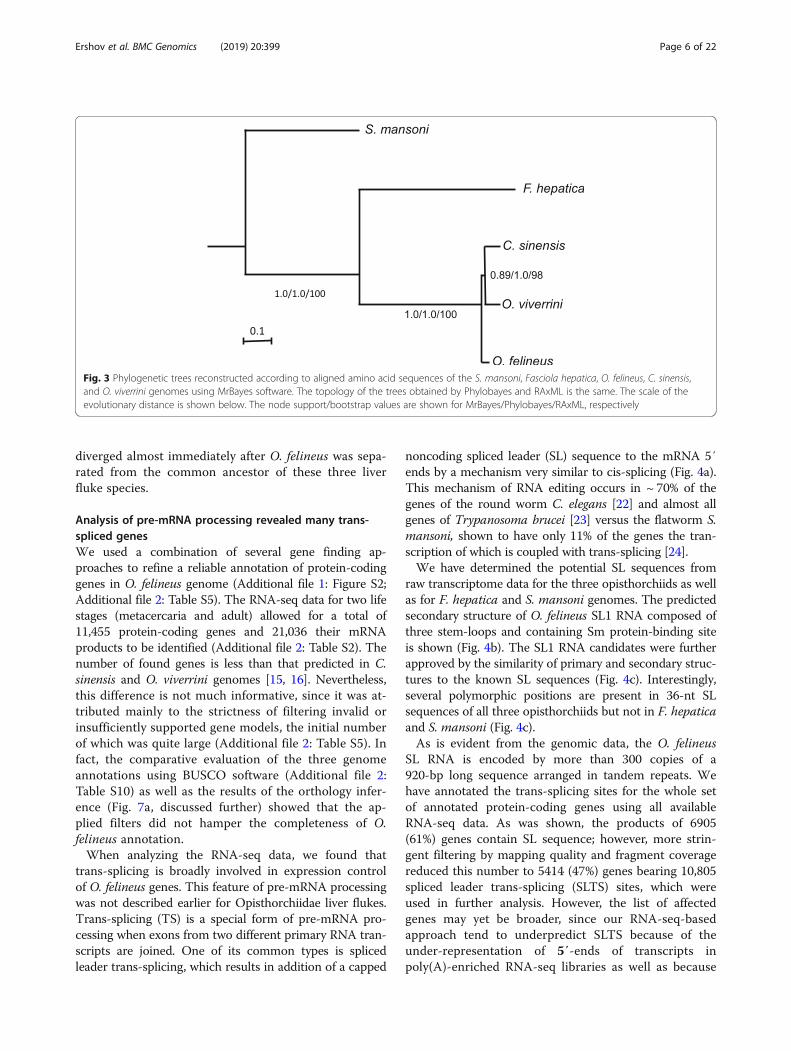
trans-splicing is broadly involved in expression controlof O. felineus genes. This feature of pre-mRNA processingwas not described earlier for Opisthorchiidae liver flukes.Trans-splicing (TS) is a special form of pre-mRNA pro-cessing when exons from two different primary RNA tran-scripts are joined. One of its common types is splicedleader trans-splicing, which results in addition of a capped
noncoding spliced leader (SL) sequence to the mRNA 5′ends by a mechanism very similar to cis-splicing (Fig. 4a).This mechanism of RNA editing occurs in ~ 70% of thegenes of the round worm C. elegans [22] and almost allgenes of Trypanosoma brucei [23] versus the flatworm S.mansoni, shown to have only 11% of the genes the tran-scription of which is coupled with trans-splicing [24].We have determined the potential SL sequences from
raw transcriptome data for the three opisthorchiids as wellas for F. hepatica and S. mansoni genomes. The predictedsecondary structure of O. felineus SL1 RNA composed ofthree stem-loops and containing Sm protein-binding siteis shown (Fig. 4b). The SL1 RNA candidates were furtherapproved by the similarity of primary and secondary struc-tures to the known SL sequences (Fig. 4c). Interestingly,several polymorphic positions are present in 36-nt SLsequences of all three opisthorchiids but not in F. hepaticaand S. mansoni (Fig. 4c).As is evident from the genomic data, the O. felineus
SL RNA is encoded by more than 300 copies of a920-bp long sequence arranged in tandem repeats. Wehave annotated the trans-splicing sites for the whole setof annotated protein-coding genes using all availableRNA-seq data. As was shown, the products of 6905(61%) genes contain SL sequence; however, more strin-gent filtering by mapping quality and fragment coveragereduced this number to 5414 (47%) genes bearing 10,805spliced leader trans-splicing (SLTS) sites, which wereused in further analysis. However, the list of affectedgenes may yet be broader, since our RNA-seq-basedapproach tend to underpredict SLTS because of theunder-representation of 5′-ends of transcripts inpoly(A)-enriched RNA-seq libraries as well as because
Fig. 3 Phylogenetic trees reconstructed according to aligned amino acid sequences of the S. mansoni, Fasciola hepatica, O. felineus, C. sinensis,and O. viverrini genomes using MrBayes software. The topology of the trees obtained by Phylobayes and RAxML is the same. The scale of theevolutionary distance is shown below. The node support/bootstrap values are shown for MrBayes/Phylobayes/RAxML, respectively
Ershov et al. BMC Genomics (2019) 20:399 Page 6 of 22
of the skipping too short SL sequences in homologysearch. Thus, we have for the first time demonstratedthat the products of almost half O. felineus genescontain an SL sequence. This suggests an importantrole of trans-splicing in the processing of RNA inliver flukes.
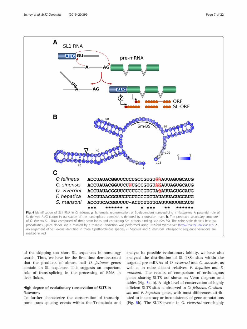
High degree of evolutionary conservation of SLTS inflatwormsTo further characterize the conservation of transcrip-tome trans-splicing events within the Trematoda and
analyze its possible evolutionary lability, we have alsoanalyzed the distribution of SL-TSSs sites within thetargeted pre-mRNAs of O. viverrini and C. sinensis, aswell as in more distant relatives, F. hepatica and S.mansoni. The results of comparison of orthologousgenes sharing SLTS are shown as Venn diagram andtables (Fig. 5a, b). A high level of conservation of highlyefficient SLTS sites is observed in O. felineus, C. sinen-sis, and F. hepatica genes, with most differences attrib-uted to inaccuracy or inconsistency of gene annotations(Fig. 5b). The SLTS events in O. viverrini were highly
A
B
C
Fig. 4 Identification of SL1 RNA in O. felineus. a. Schematic representation of SL-dependent trans-splicing in flatworms. A potential role ofSL-derived AUG codon in translation of the trans-spliced transcript is denoted by a question mark. b. The predicted secondary structureof O. felineus SL1 RNA composed of three stem-loops and containing Sm protein-binding site (Sm-BS). The color scale depicts base-pairprobabilities. Splice donor site is marked by a triangle. Prediction was performed using RNAfold WebServer (http://rna.tbi.univie.ac.at/). c.An alignment of SL1 exons identified in three Opisthorchiidae species, F. hepatica and S. mansoni. Intraspecific sequence variations aremarked in red
Ershov et al. BMC Genomics (2019) 20:399 Page 7 of 22
underestimated owing to an insufficient depth of avail-able RNA-seq data. However, only 15% of the geneswere found to be trans-spliced in S. mansoni, which isconsistent with the previous studies [24]. Thus, thetrans-splicing machinery of schistosomes has consider-ably diverged from the remaining studied species, as isevident from the primary SL-RNA structure as well asthe conservation and overall occurrence rate of SLTSevents.
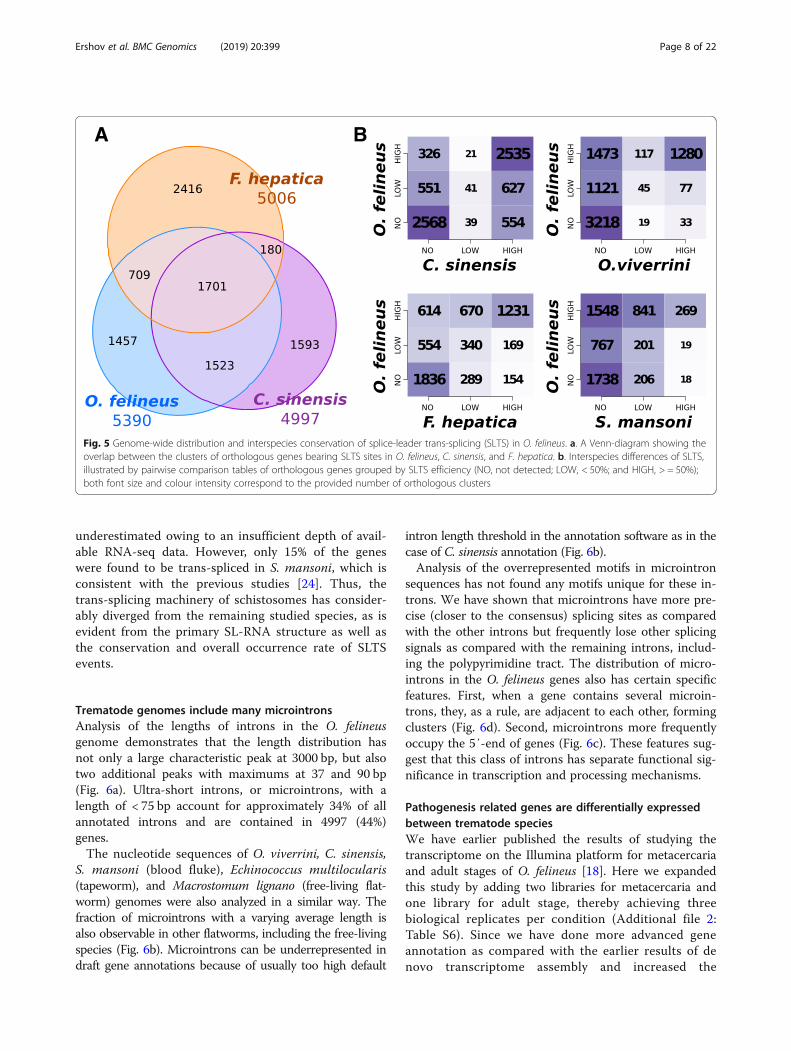
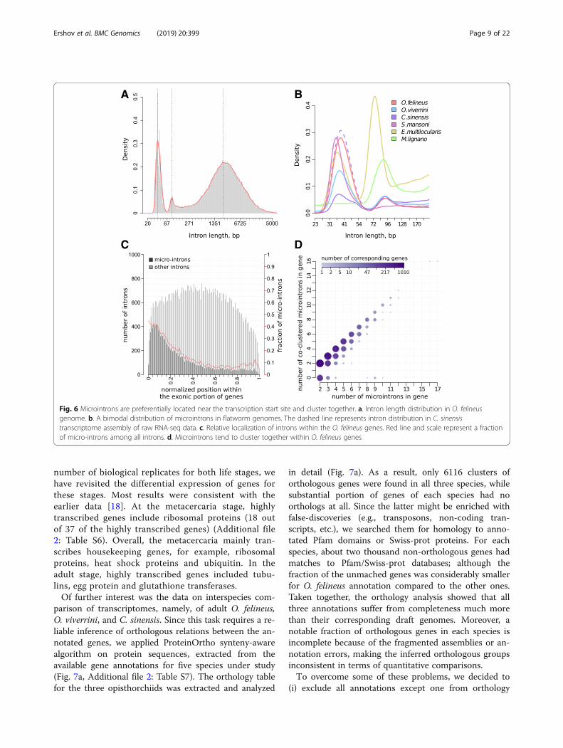
Trematode genomes include many microintronsAnalysis of the lengths of introns in the O. felineusgenome demonstrates that the length distribution hasnot only a large characteristic peak at 3000 bp, but alsotwo additional peaks with maximums at 37 and 90 bp(Fig. 6a). Ultra-short introns, or microintrons, with alength of < 75 bp account for approximately 34% of allannotated introns and are contained in 4997 (44%)genes.The nucleotide sequences of O. viverrini, C. sinensis,
S. mansoni (blood fluke), Echinococcus multilocularis(tapeworm), and Macrostomum lignano (free-living flat-worm) genomes were also analyzed in a similar way. Thefraction of microintrons with a varying average length isalso observable in other flatworms, including the free-livingspecies (Fig. 6b). Microintrons can be underrepresented indraft gene annotations because of usually too high default
intron length threshold in the annotation software as in thecase of C. sinensis annotation (Fig. 6b).Analysis of the overrepresented motifs in microintron
sequences has not found any motifs unique for these in-trons. We have shown that microintrons have more pre-cise (closer to the consensus) splicing sites as comparedwith the other introns but frequently lose other splicingsignals as compared with the remaining introns, includ-ing the polypyrimidine tract. The distribution of micro-introns in the O. felineus genes also has certain specificfeatures. First, when a gene contains several microin-trons, they, as a rule, are adjacent to each other, formingclusters (Fig. 6d). Second, microintrons more frequentlyoccupy the 5′-end of genes (Fig. 6c). These features sug-gest that this class of introns has separate functional sig-nificance in transcription and processing mechanisms.
Pathogenesis related genes are differentially expressedbetween trematode speciesWe have earlier published the results of studying thetranscriptome on the Illumina platform for metacercariaand adult stages of O. felineus [18]. Here we expandedthis study by adding two libraries for metacercaria andone library for adult stage, thereby achieving threebiological replicates per condition (Additional file 2:Table S6). Since we have done more advanced geneannotation as compared with the earlier results of denovo transcriptome assembly and increased the
A B
Fig. 5 Genome-wide distribution and interspecies conservation of splice-leader trans-splicing (SLTS) in O. felineus. a. A Venn-diagram showing theoverlap between the clusters of orthologous genes bearing SLTS sites in O. felineus, C. sinensis, and F. hepatica. b. Interspecies differences of SLTS,illustrated by pairwise comparison tables of orthologous genes grouped by SLTS efficiency (NO, not detected; LOW, < 50%; and HIGH, > = 50%);both font size and colour intensity correspond to the provided number of orthologous clusters
Ershov et al. BMC Genomics (2019) 20:399 Page 8 of 22
number of biological replicates for both life stages, wehave revisited the differential expression of genes forthese stages. Most results were consistent with theearlier data [18]. At the metacercaria stage, highlytranscribed genes include ribosomal proteins (18 outof 37 of the highly transcribed genes) (Additional file2: Table S6). Overall, the metacercaria mainly tran-scribes housekeeping genes, for example, ribosomalproteins, heat shock proteins and ubiquitin. In theadult stage, highly transcribed genes included tubu-lins, egg protein and glutathione transferases.Of further interest was the data on interspecies com-
parison of transcriptomes, namely, of adult O. felineus,O. viverrini, and C. sinensis. Since this task requires a re-liable inference of orthologous relations between the an-notated genes, we applied ProteinOrtho synteny-awarealgorithm on protein sequences, extracted from theavailable gene annotations for five species under study(Fig. 7a, Additional file 2: Table S7). The orthology tablefor the three opisthorchiids was extracted and analyzed
in detail (Fig. 7a). As a result, only 6116 clusters oforthologous genes were found in all three species, whilesubstantial portion of genes of each species had noorthologs at all. Since the latter might be enriched withfalse-discoveries (e.g., transposons, non-coding tran-scripts, etc.), we searched them for homology to anno-tated Pfam domains or Swiss-prot proteins. For eachspecies, about two thousand non-orthologous genes hadmatches to Pfam/Swiss-prot databases; although thefraction of the unmached genes was considerably smallerfor O. felineus annotation compared to the other ones.Taken together, the orthology analysis showed that allthree annotations suffer from completeness much morethan their corresponding draft genomes. Moreover, anotable fraction of orthologous genes in each species isincomplete because of the fragmented assemblies or an-notation errors, making the inferred orthologous groupsinconsistent in terms of quantitative comparisons.To overcome some of these problems, we decided to
(i) exclude all annotations except one from orthology
A B
C D
Fig. 6 Microintrons are preferentially located near the transcription start site and cluster together. a. Intron length distribution in O. felineusgenome. b. A bimodal distribution of microintrons in flatworm genomes. The dashed line represents intron distribution in C. sinensistranscriptome assembly of raw RNA-seq data. c. Relative localization of introns within the O. felineus genes. Red line and scale represent a fractionof micro-introns among all introns. d. Microintrons tend to cluster together within O. felineus genes
Ershov et al. BMC Genomics (2019) 20:399 Page 9 of 22
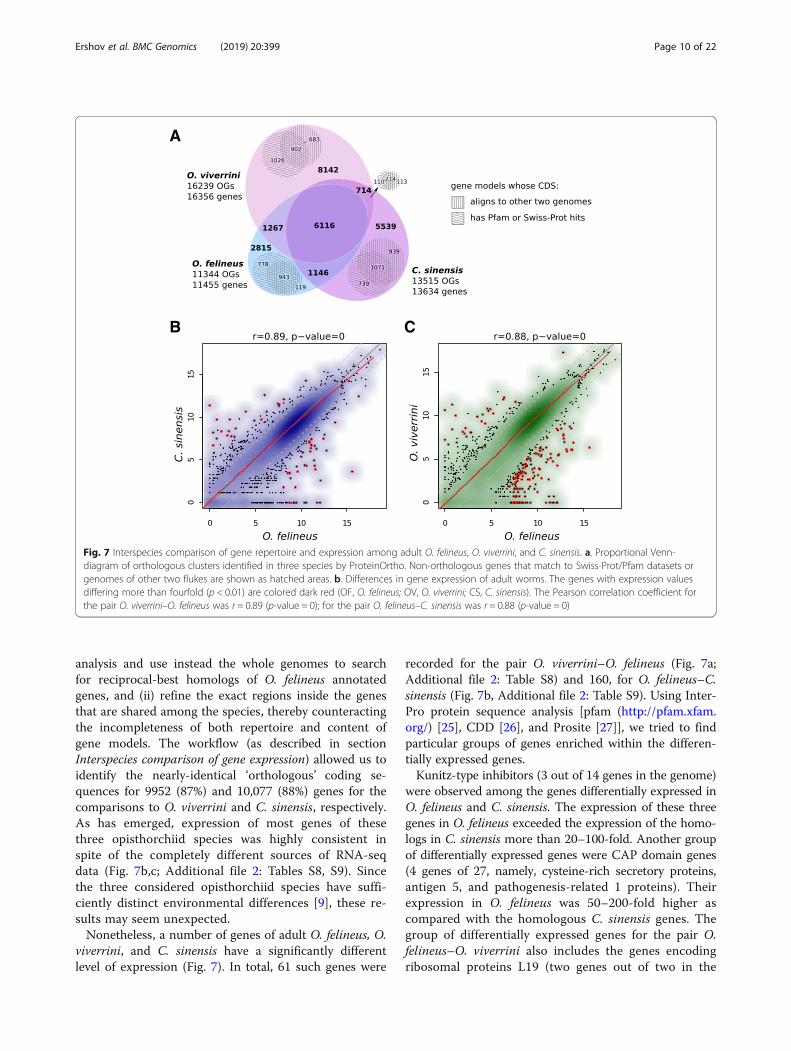
analysis and use instead the whole genomes to searchfor reciprocal-best homologs of O. felineus annotatedgenes, and (ii) refine the exact regions inside the genesthat are shared among the species, thereby counteractingthe incompleteness of both repertoire and content ofgene models. The workflow (as described in sectionInterspecies comparison of gene expression) allowed us toidentify the nearly-identical ‘orthologous’ coding se-quences for 9952 (87%) and 10,077 (88%) genes for thecomparisons to O. viverrini and C. sinensis, respectively.As has emerged, expression of most genes of thesethree opisthorchiid species was highly consistent inspite of the completely different sources of RNA-seqdata (Fig. 7b,c; Additional file 2: Tables S8, S9). Sincethe three considered opisthorchiid species have suffi-ciently distinct environmental differences [9], these re-sults may seem unexpected.Nonetheless, a number of genes of adult O. felineus, O.
viverrini, and C. sinensis have a significantly differentlevel of expression (Fig. 7). In total, 61 such genes were
recorded for the pair O. viverrini–O. felineus (Fig. 7a;Additional file 2: Table S8) and 160, for O. felineus–C.sinensis (Fig. 7b, Additional file 2: Table S9). Using Inter-Pro protein sequence analysis [pfam (http://pfam.xfam.org/) [25], CDD [26], and Prosite [27]], we tried to findparticular groups of genes enriched within the differen-tially expressed genes.Kunitz-type inhibitors (3 out of 14 genes in the genome)
were observed among the genes differentially expressed inO. felineus and C. sinensis. The expression of these threegenes in O. felineus exceeded the expression of the homo-logs in C. sinensis more than 20–100-fold. Another groupof differentially expressed genes were CAP domain genes(4 genes of 27, namely, cysteine-rich secretory proteins,antigen 5, and pathogenesis-related 1 proteins). Theirexpression in O. felineus was 50–200-fold higher ascompared with the homologous C. sinensis genes. Thegroup of differentially expressed genes for the pair O.felineus–O. viverrini also includes the genes encodingribosomal proteins L19 (two genes out of two in the
A
CB
Fig. 7 Interspecies comparison of gene repertoire and expression among adult O. felineus, O. viverrini, and C. sinensis. a. Proportional Venn-diagram of orthologous clusters identified in three species by ProteinOrtho. Non-orthologous genes that match to Swiss-Prot/Pfam datasets orgenomes of other two flukes are shown as hatched areas. b. Differences in gene expression of adult worms. The genes with expression valuesdiffering more than fourfold (p < 0.01) are colored dark red (OF, O. felineus; OV, O. viverrini; CS, C. sinensis). The Pearson correlation coefficient forthe pair O. viverrini–O. felineus was r = 0.89 (p-value = 0); for the pair O. felineus–C. sinensis was r = 0.88 (p-value = 0)
Ershov et al. BMC Genomics (2019) 20:399 Page 10 of 22
genome), papain family cysteine proteases (2 genes outof 25) and glyceraldehyde 3-phosphate dehydrogenase(one out of three).Interestingly, products of majority of the differentially
expressed genes found in our study contain domainscharacteristic for helminth-secreted proteins. In par-ticular, CAP protein family (cysteine-rich secretory pro-teins, antigen 5, and pathogenesis-related 1 proteins),papain family cysteine proteases and Kunitz-type inhib-itors are among the most represented compounds ofsecretome across 44 helminth species [28]. The propertiesof several gene families selected by their relevance in thepathogenesis of opisthorchiasis are discussed below.
Detoxification network genes were found in the genomeThe detoxification system of the liver fluke is of specialinterest, since its components are promising pharmaco-logical targets [29–32]. Detoxification system is essentialfor both the adaptation to host environment and survivalof the parasite [30, 31]. In addition, parasite’s detoxifica-tion system is most likely responsible for synthesis ofparasite-specific genotoxic metabolites of cholesterol, re-cently discovered in O. felineus and other carcinogenictrematodes, S. haematobium and O. viverrini [33, 34].The proteins putatively involved in oxidation and re-
duction of substrates form the group of enzymes preva-lently implementing phase I metabolism of exogenoussubstrates and are the most important component ofdetoxification system in all organisms. CytochromesP450 (CYPs) are among these proteins. CYPs family inparasitic and free-living flatworms is drastically different:the free-living species have dozens of diverged CYPgenes (39 CYPs in Schmidtea mediterranea), whereasthe parasitic species (Schistosomatidae, Opisthorchiidae,Taeniidae, and Fasciolidae) most likely have only onecytochrome P450 [30, 35]. O. felineus CYP is involved inthe metabolism of exogenous substrates, is important forsurvival of adult individuals, and represents a promisingtarget for anthelminthic therapy [30, 32]. Differentialexpression of this gene was consistent with the earlierdata [30]. Furthemore, O. felineus detoxification phase Iis also represented by the genes encoding aldo-ketoreductases, aldehyde dehydrogenases, and alcohol dehy-drogenases (Additional file 2: Table S6). Expression of allaldehyde dehydrogenases was higher in adult stage, thanin metacercariae; while the expression of aldo-ketoreductases was almost on the same level in both stages.Interestingly, the liver flukes lack the group of enzymeswith a monooxygenase activity (analogous to CYPs),namely, flavin monooxygenases (Pfam00743). Moreover,we failed to find any flavin monooxygenase sequences inany parasitic flatworm genomes.Phase II enzymes are represented, in particular, by
glutathione peroxidase (GPx) and glutathione-S-transferase
(GST). The O. felineus genome contains six GST genes,which display the highest expression among all detoxifica-tion genes. The 28 kDa GST sigma gene (CRM22_011285in Additional file 1: Figure S5) is especially active; itsexpression in the adult worm is by two–three orders ofmagnitude higher as compared with the other detoxifica-tion genes.UGTs, common phase II xenobiotic metabolism en-
zymes in vertebrates, enhance hydrophilicity and avail-ability of substrates to efflux transporters. The UGTsuperfamily comprises two families (UGT1 and UGT2)and over 20 isozymes. UGTs play a significant role indrug resistance of helminths [29]. Currently, 34 UGTgenes are known in the nematode Haemonchus contortusgenome [36] and 72 UGT genes, in C. elegans genome[37]. We failed to find any UGT genes in the O. felineusgenome as well as in the genomes of other Opisthorchii-dae and Schistosomatidae. In addition, we did not findany arylamine N-acetyltransferases (PF00797), which arealso common phase II enzymes in vertebrates. Thus, thedetoxification phase II of parasitic flatworms has somefeatures distinguishing it from the corresponding systemsof the other organisms, including the host.The detoxification phase III is represented by mem-
brane efflux transporters, protecting organisms from ex-ternal toxic compounds, including drugs. Five distinctfamilies of the efflux transporters are recognized. Themost frequently studied are ABC transporters, for ex-ample, ABCB1 (also known as P-glycoprotein or multi-drug resistance protein 1, MDR1), ABCC1 (multidrugresistance associated protein, MRP1), and ABCG2 (alsoknown as breast cancer resistance) [38, 39]. The O.felineus genome contains 23 genes encoding ABC trans-porters. Four of ABC genes are homologous to humanP-glycoprotein (Additional file 1: Figure S5). Differentialexpression of these genes was consistent with the earlierdata [40].
Dozens of NPC2 genes were revealed in OpisthorchiidaegenomesOne of the specific features of genomes of closely relatedspecies is the number of copies of orthologous genes.The NPC2 genes (Niemann–Pick disease type C2) en-code the proteins that are potentially involved in intra-cellular and extracellular transport of sterols. We havediscovered 48 NPC2 genes in the O. felineus genome.Most eukaryotes have only one NPC2 gene. Earlier, 25genes coding for NPC2-like proteins have been detectedin the O. viverrini and C. sinensis genomes [16]. Thefunction of these proteins in helminths is vague. Thereis evidence suggesting the chemosensory role for theseproteins in Arthropodae [41]. Such diversity of theseproteins in the liver flukes might be determined by thelife activities of these helminths, i.e., their function may
Ershov et al. BMC Genomics (2019) 20:399 Page 11 of 22
putatively consist in binding and transporting the hostlipids and sterols for the adult helminth metabolism.However, comparative transcriptomic analysis of the O.felineus metacercariae and adult has shown that 43 ofthe observed 48 NPC2 proteins are differentiallyexpressed in metacercariae rather than in adult worm
(Additional file 2: Table S6). Expression of only twoNPC2-like genes is specific of the adult worm stage.Thus, the NPC2-like proteins in general are not cruciallyimportant for the life activities of an adult individual andpotentially are not associated with the lipid binding andtransport in an adult individual living in the bile. The
A
B
C D
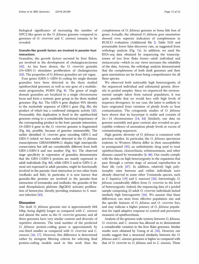
Fig. 8 Granulin-like genes in Opisthorchiidae genomes. a. Granulin-like genes are co-localized to one locus in three opisthorchiid genomes.Alignment (b) and cladogram (c) of granulin-like proteins are presented. d. Heatmap of granulin-like gene expression in three Opisthorchiidae species
Ershov et al. BMC Genomics (2019) 20:399 Page 12 of 22
biological significance of increasing the number ofNPC2-like genes in the O. felineus genome compared togenomes of O. viverrini and C. sinensis remains to berevealed.
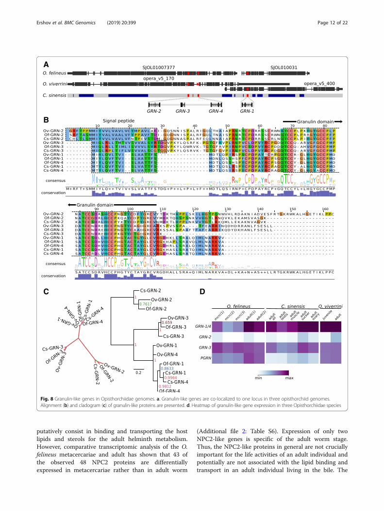
Granulin-like growth factors are involved in parasite–hostinteractionsGranulins, the growth factors secreted by liver flukes,are involved in the development of cholangiocarcinoma[42]. As has been shown, the O. viverrini granulin(Ov-GRN-1) stimulates proliferation of cholangiocytes[42]. The properties of O. felineus granulins are yet vague.Four genes (GRN-1–GRN-4) coding for single domain
granulins have been detected in the three studiedopisthorchiid genomes as well as one gene of a multido-main progranulin, PGRN (Fig. 8). The genes of singledomain granulins are localized to a single chromosomelocus and form a syntenic gene group in the three studiedgenomes (Fig. 8a). The GRN-4 gene displays 95% identityto the nucleotide sequence of GRN-1 gene (Fig. 8b), theproduct of which has a confirmed mitogenic activity [42].Presumably, this duplication is fixed in the opisthorchiidgenomes owing to a considerable functional importance ofthe corresponding product in the host–parasite interaction.Ov-GRN-4 gene in the O. viverrini genome is fragmented(Fig. 8a), possibly, because of genome misassembly. Theearlier identified O. viverrini gene encoding GRN-2 andGRN-3 (which we have earlier detected in the O. felineustranscriptome; GBJA01006896.1) display high interspecificconservation but still are considerably different from bothGRN-1/GRN-4 and one another (Fig. 8c). The observedstage specificity in expression of granulins demonstratesthat the GRN-1/GRN-4 proteins are mainly expressed inadult individuals (Fig. 8d), while GRN-2 and/or GRN-3, al-most not expressed in adult parasites, might be functionallyinvolved in the parasite–host interaction in two other hosts(mollusks and fish). In particular, it is now known thatgranulin-like proteins are involved in the parasite–hostinteraction of trematodes and mollusks: the granulin of thesnail Biomphalaria glabrata (BgGRN) activates prolifera-tion of hemocytes, thereby providing resistance to S. man-soni infection [43].
DiscussionThe draft O. felineus genome size is approximately 684Mbp, being slightly longer as compared with С. sinensisand almost the same as the O. viverrini genome; and allthree genomes have very similar content and diversity ofrepetitive elements. The total number of the predictedO. felineus protein-coding genes is approximately byone-third smaller as compared with O. viverrini and C.sinensis [16, 17]. However, this difference is determinedrather by stringent filtering criteria for selecting finalprotein-coding models used in this work than the
completeness of O. felineus genome or bona fide loss ofgenes. Actually, the obtained O. felineus gene annotationshowed even superior indicators of completeness inBUSCO evaluation (Additional file 2: Table S10) andpresumably lower false-discovery rate, as suggested fromorthology analysis (Fig. 7a). In addition, we used theRNA-seq data obtained by sequencing the transcrip-tomes of two liver fluke forms—adult individual andmetacercaria—which in our views increases the reliabilityof the data. Anyway, the orthology analysis demonstratedthat the completeness of both draft genomes and theirgene annotations are far from being comprehensive for allthree species.We observed both noticeably high heterozygosity of
the sequenced individual and substantial genetic diver-sity in pooled samples. Since we sequenced the environ-mental sample taken from natural population, it wasquite possible that we would face with high levels ofsequence divergence. In our case, the latter is unlikely tohave originated from variation of ploidy levels or hostcontamination. The cytogenetic studies of O. felineushave shown that its karyotype is stable and consists of2n = 14 chromosomes [14, 44] Similarly, our data ongenome assembly and gene content also revealed no per-ceptible evidence of anomalous ploidy levels or excess ofcontaminating sequences.High genetic diversity of O. felineus is consistent with
previous studies. In particular, the O. felineus specimensendemic to Western Siberia differ in their susceptibilityto praziquantel [45], an anthelmintic drug used to treatopisthorchiasis, clonorchiasis, schistosomiasis, and otherdiseases caused by trematodes [4, 46]. Our results complywith the data on high heterozygosity in the organisms thatpass through a certain stage of asexual reproduction intheir life cycle [47]. In addition, relatively high poly-morphic rates between and within individuals werealready observed in some other Trematoda species, suchas F. hepatica [19] and S. mansoni [24]. Interestingly, O.felineus considerably differs from O. viverrini in the levelof heterozygosity. Indeed, the sequencing data of a pooledsample comprising 25 adult O. viverrini individuals lackedsimilarly high heterogeneity [16]. We assume that thesedifferences can stem from effective population size andthe specific features of O. felineus and O. viverrini foci,and may indicate a higher potency of O. felineus popula-tion for rapid adaptive response to control and preventivemeasures of opisthorchiasis.Analysis of the genome-wide synteny between O. felineus,
O. viverrini, and C. sinensis has allowed us to demonstratea considerable variation in the liver fluke genomes. Similarresults were obtained by Young et al. [16]. However, ourresults suggest that a structural similarity between the O.felineus and C. sinensis genomes is higher as compared withthat of O. viverrini to O. felineus and to C. sinensis. These
Ershov et al. BMC Genomics (2019) 20:399 Page 13 of 22
data match well the results of karyotyping: O. felineus andC. sinensis have seven pairs of chromosomes versus O.viverrini, carrying six chromosome pairs [14, 44].The phylogenetic relationships between the genera
Opisthorchis and Clonorchis is controversal [48]. Kanget al. [49] used the ITS1 sequences from O. felineus, O.viverrini and C. sinensis, and demonstrated that C. sinensisis sister group to O. viverrini and O. felineus. Analysis ofthe phylogenetic relationship between ITS2 and mitochon-drial cox1 showed that O. felineus was more closely relatedto C. sinensis than to O. viverrini [50]. Similar relationshipwas demonstrated using cox1 sequences by Saijuntha et al.[51], cox1, nad1 and paramyosin gene (Pm-int9) sequencesby Pitaksakulrat et al. [52]. The same topology of phylogen-etic tree between O. felineus, C. sinensis and O. viverrini ob-tained by analysis of concatenated amino acid sequencesfrom mitochondrial protein-coding genes by Wang et al.[53]. and Liu et al. [54].In our work analysis of 1563 protein families suggested
the phylogenetic tree topology with C. sinensis and O.viverrini species grouping together and O. felineus repre-senting a sister group. Our results are in agreement withthe phylogenetic tree estimated using paramyosin gene(Pm-int9) [55], mitochondrial amino acid sequences ana-lyzed by Cai et al. [56], ribosomal proteins [18].Taken together, the results of analysis of the synteny
between three opisthorchiid species and of their phylo-genetic relationships demonstrate that O. felineus and C.sinensis are closely related and do not support separationof C. sinensis from the genus Opisthorchis. Presumably, C.sinensis occupies an intermediate position between O. feli-neus and O. viverrini. The geographic vector of distribu-tion of opisthorchiasis and clonorchiasis foci also favorsthis hypothesis. The opisthorchiasis caused by O. felineushas been recorded in many European countries; however,the most intense foci of this disease are in North Asia,namely, in Russia and Kazakhstan [3]. Clonorchiasis ismainly spread in the Russian Far East, China, Korea,Japan, and to a lesser degree in Laos and Vietnam [10].The main foci of O. viverrini opisthorchiasis are in theSoutheast Asia, namely, in Thailand, Laos, Vietnam, andCambodia [8].We have found that all three studied opisthorchiidae
species are characterized by much more extensive in-volvement of trans-splicing in RNA processing com-pared to the most well-studied trematode, Schistosomamansoni [24]. Since trans-splicing was found to occurwith high efficiency in these species, the affected 5` endsof many transcripts tend to generate spurious alignmentsto the reference genome, leading to incorrect predictionof gene structure and even the corresponding structure ofencoded protein as a result of the standard genome anno-tation workflow. Thereafter, accounting properly thetrans-splicing events in transcriptomes along with another
important feature of gene organization in flatworms,microintrons, resulted in a much more reliable annotationof genes and their protein products in O. felineus, e.g. theresolution of polycistronic gene products.The bimodality of intron length distributions has been
observed also in many eukaryotic species. Two classes ofintrons, with a peak of short introns or microintronsand a much flatter peak of longer introns ranging up tothousands of base pairs, are present in humans, Arabi-dopsis thaliana, Drosophila melanogaster, and C. elegans[57]. Some Ciliates (Paramecium) contain microintronswith a length divisible particular, 33–35, 47–51, and 78–80 bp [58]. Two peaks of short introns has been earlierdescribed in parasitc flatworms, including Cestoda [59]and Monogenea species [60]. However, we have foundthat this feature is of the same relevance for opisthorch-iidae species. The peculiarities of microintrons revealedin the current study suggest that the apartness of thisfraction of introns is maintained by their functionalsignificance in transcription and processing mecha-nisms. For example, their clustering at the 5′ ends ofpre-mRNAs may be driven by a preference of an in-tron definition mechanism over exon definition.There are some difficulties in comparative analysis of
gene content and expression between three liver flukes,since the existing genome assemblies are draft, and thecorresponding gene annotations are far from beingcomplete and compatible. However, we have made an ef-fort to generate a congruent set of orthologous expres-sion units based on O. felineus annotation only. Whileexpression levels of a number of genes showed remark-able differences between the species, the overall expres-sion profiles were highly consistent across the comparedspecies, suggesting a high similarity of all biologicalpathways in adult liver flukes that colonize the bile ductsof mammals.
ConclusionsLack of O. felineus genomic data is an obstacle to thedevelopment of comparative molecular biological ap-proaches necessary to obtain new knowledge about thebiology of epidemiologically important Opisthorchiidaetrematodes, to identify essential pathways linked toparasite-host interaction, to predict genes that contrib-ute to liver fluke pathogenesis and for the effective pre-vention and control of the disease. Here we present thefirst draft genome assembly of O. felineus and its generepertoire accompanied by a comparative analysis withthat of O. viverrini and Clonorchis sinensis.This study contribute to comparative genomics of flat-
worms, molecular mechanisms of RNA processing inhelminths and evolutionary history of Opisthorchiidaetrematodes. The availability of O. felineus genome weprovide, and comparative transcriptomics data will help
Ershov et al. BMC Genomics (2019) 20:399 Page 14 of 22
support the development of novel drugs and vaccines forthe treatment and prevention of liver fluke infection.
MethodsEthical statementAll of the procedures were in compliance with The Codeof Ethics of the World Medical Association (Declarationof Helsinki) for animal experiments http://ec.europa.eu/environment/chemicals/lab_animals/legislation_en.htm. The animals were kept and treated according tothe protocols approved by the Committee on the Eth-ics of Animal Experiments with the Institute ofCytology and Genetics (Permit Number 7 of November19, 2011).
Study designTwo thousands of O. felineus metacercariae were collectedfrom naturally infected fish (Leuciscus idus) caught in theOb River near Novosibirsk (Western Siberia) and rou-tinely extracted [45, 61]. Territories where sample collec-tion (fishing) took place were neither conservation areasnor private, nor otherwise protected; hence, no fishingpermits were required. The fish species collected are notconsidered endangered or rare, and the fishing methodscomplied with the Federal Law N166-F3 of 20.12.2004(ed. 18.07.2011), “Fishing and conservation of waterbio-resources”. Syrian hamsters (Mesocricetus auratus)were purchased from the Animal Breeding Facility withthe Institute of Cytology and Genetics, Siberian Branch,Russian Academy of Sciences. Five hamsters aged 6 to 8weeks were orally infected with 75 O. felineus metacercar-iae. Euthanasia was performed using carbon dioxide, andall efforts were made to minimize suffering. The animalswere maintained in their home cage while CO2 wasinduced at a flow 10% per minute. Adult flukes wererecovered from the hepatobiliary tract of hamsters threemonths after the infection, pooled and thoroughly washedwith saline.
List of data used for the interspecies analysisTo analyze transcriptomic data Sequence Read Archives(SRA) were taken from NCBI https://www.ncbi.nlm.nih.gov/sra in the research: ERR604978-ERR604981,SRR189060 for C. sinensis; SRR497632, SRR497633 for Oviverrini; ERR576952, ERR576954, ERR576956, ERR576958,ERR576968 for F hepatica; ERR1328228-ERR1328233,ERR1328243, ERR1328260-ERR1328267 for S mansoni.Genome assemblies and gene annotations were takenfrom WormBase ParaSite (version WBPS6) [62, 63]:PRJNA222628 (O. viverrini) [16], PRJDA72781 (C. sinen-sis) [17], PRJEA36577 (S. mansoni) [24, 64], PRJEB122 (E.multilocularis) [59], PRJNA284736 (M. lignano) [65], F.hepatica (PRJEB6687) [19].
Genome sequencing and data processingGenomic DNA (gDNA) was isolated from using protein-ase K digestion following phenol–chloroform extraction.For short insert libraries, the gDNA isolated from a ran-domly chosen single adult O. felineus worm was fragmen-ted in a Covaris S2 (Covaris™, United States) sonicator toan average fragment size of 200–500 bp and the sizes se-lected on Pepin Prep (Sage Science, Inc., United States) forthe insert were 180 and 270 bp. Construction of genome li-braries and all subsequent manipulations were performedusing paired-end library preparation kit in accordance withthe manufacturer’s protocols (Illumina, United States).Genome libraries were suitable for paired-end reading.Sequencing was performed on Illumina Genome AnalyzerII (GAII) and HiSeq1500 (Illumina, United States) plat-forms. For mate-pair libraries, the gDNA was isolated fromthe pool of 50 randomly chosen worms, and Nextera MatePair Sample Preparation Kit (Illumina, United States) wasused according to manufacturer’s protocols. Three differentinsert size libraries of 3, 5, 8, and 10 kb were sequenced onIllumina HiSeq 1500 (Illumina, United States).TRIzol® reagent was used for total RNA extraction
from the pooled adult (randomly chosen 50 adultworms) and the pooled metacercaria (1000 worms) ho-mogenized tissue using standard protocol (Invitrogen,United States). The RNA concentration in samples wasmeasured using BioAnalyzer 2100 (RNA 6000 Nano Kit;Agilent, US). The mRNA-seq libraries (cDNA libraries)were prepared with mRNA-Seq Sample Preparation kit(Illumina) according to the manufacturer’s instructions.The cDNA libraries were sequenced in an IlluminaGenome Analyzer II (GAII) DNA Sequencing Platform(Illumina, United States).
Genome assemblyRaw sequencing data were preprocessed (Additional file 1:Figure S1) by removing low-quality base-calls and adaptersequences and potential PCR duplicates using cutadaptv1.18 [66]. Mate-pair libraries were also filtered on asubstantial read pair overlap determined by pearread-merging statistical algorithm [67]. The resultingreads were initially assembled using Allpaths-LG [68]software in a with ‘haploidify’ mode to overcome ahigh level of genome heterozygosity evident fromk-mer frequency distribution (Fig. 2). Additional scaf-folding with jumping libraries was performed usingthe BESST [69] program and the remaining gaps werepartially filled by applying SOAPdenovo GapClosertool [70]. A small fraction of short redundant contigsshowing > 95% homology and reduced read coveragewas filtered out from the assembly. Finally, three iter-ations of iCORN2 algorithm from PAGIT toolkit [71]were performed to correct 1–3 bp errors in the as-sembly. Final assembly was evaluated using REAPR
Ershov et al. BMC Genomics (2019) 20:399 Page 15 of 22
([72], Additional file 2: Table S11) and BUSCO ([73],Additional file 2: Table S10) tools.
Variant callingVariant calling was performed using the GATK pipeline.Two libraries (both prepared from the same single individ-ual worm) were used as technical replicates. Initial callingwas done by HaplotypeCaller module in -ERC(GVCF)mode, and the results were subjected to GenotypeGVCFsmodule for joint genotyping. Indels were hard-filtered ac-cording to the GATK best practices recommendations.Hard filtering of the resulting SNPs was performed usingthe stricter criteria QD < 5.0, FS > 40.0, MQ< 35.0,MQRankSum < − 2.5, SOR > 2.5, ReadPosRankSum < −4.0, and DP < 150 or DP < 90 (depending on the libraryanalyzed). An intersection of the two technically repeatedvariant annotations was finally taken (resulting in3,723,880 SNPs and 302,377 indels). Further analysis ofthe heterozygosity rate was limited to genomic intervalswith fragment coverage lying between 0.75 and 1.25 of themedian coverage in order to reduce the impact of poten-tial assembly and mapping errors (collapsed or duplicatedsequences). Heterozygous variants falling into these inter-vals were counted and divided by the total length of theintervals.
Genome syntenyThe examined genomes were compared using thescheme implemented by Young et al. [16]. Genomic scaf-folds were aligned using the nucmer (nucleotide similar-ity) or promer (amino acid similarity) tools with theprogram MUMmer v. 3.23 [74] to assess genome-widesimilarities.The genomic sequences of O. felineus, O. viverrini, C.
sinensis, and S. mansoni were compared in a pairwisemanner. The sequences were aligned with the help ofMUMmer v. 3.23 [74]. First, the O. felineus, O. viverrini,and C. sinensis scaffolds with a length over 1000 bp werealigned at the level of amino acid sequences by the pro-mer program (parameters: –maxgap = 500 –c 20; uniquepairwise alignments were filtered with the delta-filterprogram with parameters –r –q) to S. mansoni chromo-some 1 (Smp.Chr_1), used as a reference. The syntenicblocks were identified in the constructed alignmentswith SyMap v. 4.2 [21]. Second, the O. viverrini and C.sinensis scaffolds were aligned at the amino acid se-quence level (the promer program with the above listedparameters) to SJOL01009849 sequence of O. felineus,which was the longest one (3238 362 bp). Third, the O.viverrini and C. sinensis scaffold sequences with a lengthover 1000 bp were aligned to the O. felineus scaffoldsusing the nucmer program (parameters: –maxgap = 500 –c100, delta filter –r –q). Then 10 O. felineus scaffolds thathad the largest coverage by O. viverrini and C. sinensis
sequences according to the nucleotide alignment wereselected. These sequences were analyzed using the SyMapprogram together with the homologous opisthorchiidscaffolds. The analysis was conducted for both the se-quences with unmasked repeats and the genome sequenceswith repeats masked with N symbols.Additionally we used OrthoCluster software to calculate
syntenic blocks using order and direction between sets oforthologous protein coding genes [75]. Unlike SyMap, thismethod does not require sequence alignment. The ortho-logous relationship between genes calculated as describedbelow (see Clusters of orthologous genes section). TheWeb-interface was used to run the OrthoCluster withdefault parameters (http://genome.sfu.ca/cgi-bin/orthoclusterdb/runortho.cgi).
Repeat sequence analysisTwo methods were applied to de novo identify repetitiveelements in both raw sequencing data and already as-sembled scaffolds/contigs. Tedna 1.2.1 [76] was used toassemble transposable element models directly from therepeated fraction of raw Illumina paired-end sequencingreads for each of the genomes. The raw sequencing datafor O. viverrini (AN: SRR1821044), C. sinensis (AN:SRR096372), and F. hepatica (AN: ERR576947) wereobtained from the Sequence Read Archive database(SRA, NCBI). RepeatModeler 1.0.8 [77] was applied tomine the repeat models from genomic assemblies. Theidentified repeats from both de novo libraries were auto-matically annotated using the RepeatClassifier perl scriptfrom the RepeatModeler package, which utilizes BLASTNand TBLASTX, against the repeats contained in theRepBase [78] database v. 20,150,807. Tandem and simplerepeats, rRNA. and repeat models not affiliated with anysuperfamily of interspersed repeats represented in RepBase(DNA transposons (DNA), LINE, LTR, Rolling Circle (RC),and SINE) were filtered out and withdrawn from subse-quent analyses.The repeat sequences were clustered by similarity
using cd-hit [79] with a clustering threshold of 90%. Theproduced joint libraries for each of the opisthorchiidspecies were combined into one opisthorchiid-specificlibrary, which, in turn, was combined with the generalRepeatMasker library v. 20,150,807 [77, 78]. The repeatsfrom the generated custom mixed library were mapped onthe genome assembly for each of the opisthorchiid ge-nomes using RepeatMasker v. 4.0.5 [77]. The de novo re-peat libraries for F. hepatica were also merged with thegeneral RepeatMasker library, combined, and used to mapthe repeats on F. hepatica genome assembly only.The dynamics of transposable elements in opisthorch-
iid genomes were estimated by taking CpG adjustedKimura 2P distance to adjust for multiple substitutionsof repeats masked by RepeatMasker to their consensus
Ershov et al. BMC Genomics (2019) 20:399 Page 16 of 22
sequences from the library using perl scripts (calcDiver-genceFromAlign.pl and createRepeatLandscape.pl) avail-able from the RepeatMasker package [77].
Gene predictionThe protein-coding genes in the O. felineus genomewere predicted using a combination of ab initio genefinding algorithms, O. felineus transcriptome assemblies,and cross-species alignments of transcript and proteinsequences from other trematodes (Additional file 1:Figure S2). Where appropriate, the allowed minimumintron length was set to 20 bp to account for highlyoccurring microintrons.For ab initio gene finding, Fgenesh++ [80, 81] and
Augustus [82] HMM-based approaches were used.Fgenesh++ included the training on a set of highly hom-ologous cross-species alignments of known proteins, re-finement of potential splice sites from alignments ofknown transcripts, prediction of genes with supportfrom homology to known proteins (NCBI NR database),and ab initio gene prediction in the remaining genomeregions. Augustus [82] gene prediction was performed intwo ways. Purely ab initio gene prediction was doneusing the precomputed set of S. mansoni parameters. Al-ternatively, BRAKER1 [83] pipeline was trained againstthe mapped O. felineus RNA-seq data to provide the pa-rameters for further prediction by Augustus.
Additionally, core eukaryotic gene model set [84] in O.felineus genome was also predicted using CEGMApackage.De novo and genome-guided assemblies of the O.
felineus transcriptome were built from RNA-seq datausing Trinity [85] and cufflinks [86] software. The as-sembled transcripts were subsequently passed to thePASA [87] comprehensive pipeline to obtain experimen-tally supported transcript models. Additionally, align-ments of proteins from C. sinensis, O. viverrini, and S.mansoni were constructed from exonerate [88] output.All resulting prediction (Additional file 2: Table S2)
statistics on the predictions of individual gene findingapproaches used) were combined into the consensusgene models using EvidenceModeler [87] and furtherupdated with alternatively spliced variants using the it-erative runs of PASA pipeline. Transfer RNAs were alsopredicted by the tRNAscan-SE [89] tool.In order to explore the properties of transcriptomes in
C. sinensis and O. viverrini, that may be biased in theavailable gene annotations (e.g. distribution of intronlengths), we also performed genome-guided transcriptomeassembly for these species using cufflinks [86] software.
Functional annotation of genes and gene setsThe protein-coding genes were annotated by runningInterProScan [25] against all natively supported
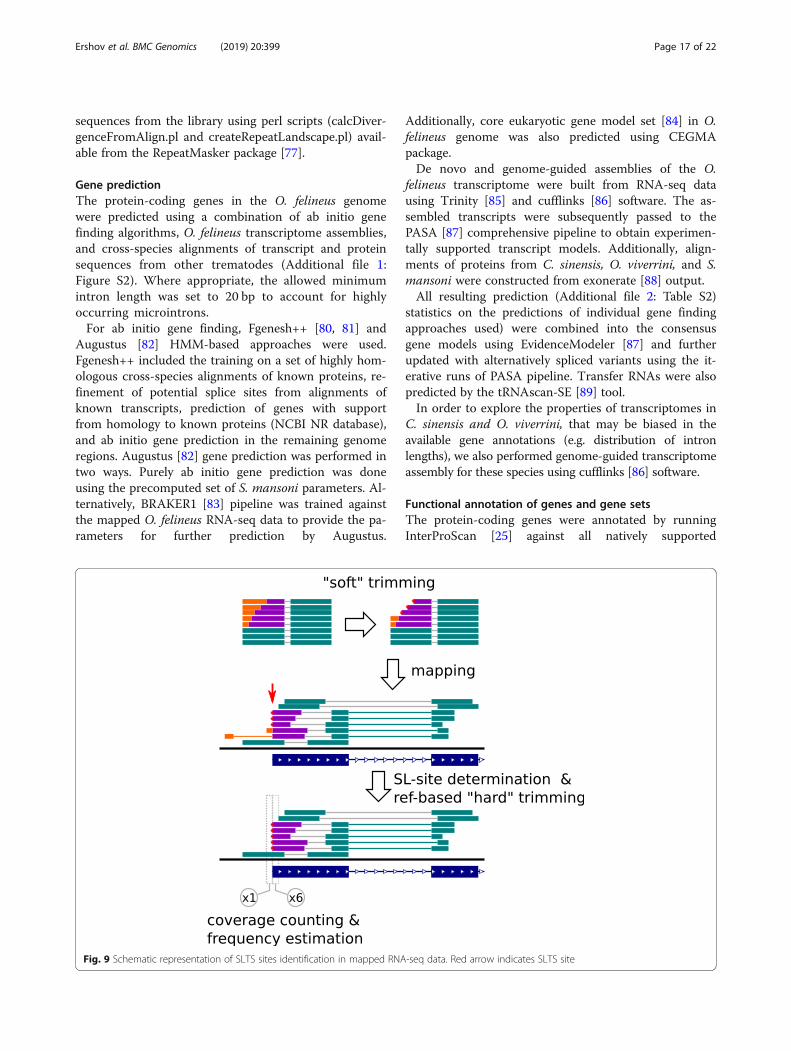
Fig. 9 Schematic representation of SLTS sites identification in mapped RNA-seq data. Red arrow indicates SLTS site
Ershov et al. BMC Genomics (2019) 20:399 Page 17 of 22
databases (Pfam 29.0, SUPERFAMILY 1.75, SMART 7.1,and PRINTS 42.0; E-value <1E-05, and using the Trino-tate pipeline (Additional file 1: Figure S2). Additionally,a homology-based assignment of KEGG pathway identi-fiers to O. felineus protein products was done usingKOBAS and KAAS web-services. Gene set enrichmentanalyses (GSEA) were performed with the use of GOstat[90] R package.
Trans-splicing analysisThe SL RNA sequence was determined from the overrep-resented 5-prime sequences of Trinity de novo assembledtranscripts and further verified by overrepresentation ofits forward strand in both reads of paired-end RNA-seqlibraries as well as by homology to the known flatwormSL RNAs. To determine the sites of trans-splicing in thetarget transcripts, a fraction of SL RNA containing readswas extracted from RNA-seq data using cutadapt [66],requiring the presence of at least 5 bp of SL sequence.After trimming out SL, the reads were mapped to thereference genome by Tophat v2.1.1 [91]. The splice sites,targeted by SL RNA, that were supported by at least threeuniquely mapped reads, were regarded as valid.To reduce a potential bias in further estimates of spli-
cing efficiency, all RNA-seq reads were subject to thesecond round of “hard” SL trimming: up to 1-bp matchto SL sequence was cut out but only if it was supportedby a valid site of SL splicing determined before (Fig. 9).The efficiency (frequency) of SL splicing was defined as1 minus ratio of read coverage at position upstream ofthe SL site to that at position downstream of the SL site.
Interstage and interspecies comparison of geneexpressionThe available and newly obtained RNA-seq data waspreprocessed by cutadapt v1.18 [66] in order to removelow-quality base-calls, adapters and SL-RNA sequences,and aligned using Tophat v2.1.1 [91] with parametersmodified to allow the minimum intron size of 20.The differential expression between O. felineus meta-
cercariae and adult worms (three biological replicates ofpooled individuals per stage) was analyzed using full O.felineus gene annotation and default DESeq2 [92] work-flow without any restriction on fold-change and with con-trolling 5% false-discovery rate by Benjamini–Hochberg(BH) algorithm.A comparative analysis of gene expression across three
opisthorchiid species is complicated by pronounced in-consistencies between their existing gene annotationsbased on draft genome assemblies. To solve this problem,we identified the gene fragments exactly common for O. feli-neus and two other opisthorchiids. The predicted cDNAs ofO. felineus were aligned to the O. viverrini and C. sinensisgenomes using Spaln v2.3.0 [93] splice-aware aligner
(parameters: -Q7 -Tschimans -yX -yZ2 -yB1 -M1 -LS -S1).Similarly, the best found alignments were mapped backto the O. felineus genome. Finally, only reciprocal-bestpairs that cover each other by > 90% were retained.Viverrini and C.The obtained annotations of ‘orthologous’ coding
regions of genes detected for each species were used toprepare gene-wise count tables from RNA-seq data onadult stages. Raw counts were normalized to the actuallength of the region in the corresponding species to ex-clude the bias arising from slight interspecies fluctua-tions (< 10%) of the latter. RLE (Relative Log Expression,DESeq2) method was used to normalize the data tosequencing depth. DESeq2 [92] R package was utilizedto extract the genes with high interspecies differences inexpression, since it allowed for taking into account bothRNA-seq underdetection bias (e.g. excessively high dif-ferences of genes with low read coverage) and intraspe-cific variation of expression (using the data on three O.felineus biological replicates). A threshold-based Waldtest was used for statistical testing of more than fourfolddifference between the species, with controlling 1%false-discovery rate by BH algorithm.
Clusters of orthologous genesThe protein sequences of four trematodes, namely, O.viverrini, C. sinensis, F. hepatica, and S. mansoni, wereused to identify orthologous clusters. Proteinortho v.5.15 [94] with ‘-synteny’ ans ‘-dups = 3’ options set wasapplied to determine the orthologous clusters among afull set of annotated protein isoforms. O. felineus, F.hepatica and S. mansoni gene models suggested variousmRNA splicing variants and, correspondingly, severalamino acid sequences for some genes. The producedmRNA-level orthology tables were subsequently mergedinto gene-level orthologies. This procedure was found to beslightly more sensitive compared to the simple approach ofselecting the longest amino acid sequence per gene.
Phylogeny reconstructionPhylogenetic trees were constructed based on the aminoacid sequences, represented by one sequence for eachorganism in the clusters of orthologous groups; 1563groups we used for analysis. Amino acid sequences werealigned in each group of orthologous genes using AQUAv. 1.1 [95] with default parameters. The evolution modelwas constructed and the alignments were partitionedinto uniform blocks with the PartitionFinder2 program[96]. The phylogenetic tree was constructed usingMrBayes v. 3.2.6 [97], RAxML v. 8.2.0 [98], and Phylo-Bayes v. 4.1 [99]. Parameters for MrBayes were set asfollows: four MCMC chains 500,000 iterations each,convergence was analyzed by Tracer 1.6 software [100].RAxML used with 1000 rapid bootstrap inferences and
Ershov et al. BMC Genomics (2019) 20:399 Page 18 of 22
LGF model (selected as best). PhyloBayes using CATmodel (options -dgam 6 –cat) was run with two MCMCchains (options -nchain 2 30 0.1100). PhyloBayes resultspresented on a 50% majority-rule consensus tree calcu-lated with SumTrees v3.3.1 [101].
Additional files
Additional file 1: Figure S1. Genome assembly pipeline. Figure S2.Gene annotation pipeline. Figure S3. Genome-wide synteny between O.felineus, O. viverrini and С. sinensis. Figure S4. Repetitive elements in O.felineus, C. sinensis, O. viverrini and F. hepatica genomes. Figure S5.Stage-specific expression of detoxification genes estimated on O. felineustranscriptome data. (PDF 438 kb)
Additional file 2: Table S1. Sequenced libraries of O. felineus. Table S2.Functionally annotated genes of O. felineus. Table S3. F. hepaticatransposable elements and their classification. 3.2. O. felineus transposableelements and their classification. 3.3. O. viverrini transposable elementsand their classification. 3.4. C. sinensis transposable elements and theirclassification. Table S4. Genome-wide synteny between O. felineus, O. viverriniand С. sinensis. Table S5. Summary statistics of the gene annotationsproduced by a number of prediction approaches used for theannotation of O. felineus genome assembly. Table S6. Differentialgene expression in O. felineus metacercariae and adult stage. Data arepresented as FPKM values (Fragments Per Kilobase Of Exon Per MillionFragments Mapped) with p-value adjusted < 0.05. Table S7. Orthologousgenes detected by ProteinOrtho software with the aid of syntenic information.Table S8. Differentially expressed genes in adult O. felineus and O.viverrini (expression values differing more than fourfold). Table S9.Differentially expressed genes in adult O. felineus and C. sinensis (expressionvalues differing more than fourfold). Table S10. Statistics on completenessof genome annotations, as reported by BUSCO software using Eukaryota(EU) & Metazoa (MZ) sets of orthologs. Table S11. Evaluation report of O.felineus genome assembly produced by REAPR software. (XLSX 4591 kb)
AbbreviationsORF: Open reading frame; SF: Supplementary Figs.; SL: Spliced leader;SLTS: Spliced leader trans-splicing; ST: Supplementary tables
AcknowledgementsAuthors thank Prof. Nikolay A. Kolchanov (Institute of Cytology and GeneticsSB RAS, Novosibirsk, Russia) and Prof. Ludmila M. Ogorodova (Siberian StateMedical University, Tomsk, Russia) who provided general support for theresearch. Authors also thank Dr. Alexey V. Katokhin (Institute of Cytology andGenetics SB RAS, Novosibirsk, Russia) for the animal work and valuableadvices; Dr. Vladimir Ivanisenko and Dr. Olga Saik (both from Institute ofCytology and Genetics SB RAS, Novosibirsk, Russia) for the expertise thatgreatly assisted the manuscript. Authors acknowledge the support ofmembers of the TOPIC (Tomsk OPIsthorchiasis Consortium). The SiberianBranch of the Russian Academy of Sciences (SB RAS) Siberian SupercomputerCenter, Novosibirsk State University High Performance Computing Clusterand ICG Bioinformatics Shared Access Center are gratefully acknowledgedfor providing supercomputer facilities.
FundingThis research was supported in part by the Russian Science Foundation,http://rscf.ru/en/ [No. 18–15-00098 (MYP, VAM: detoxification system);18–74-00101 (NIE: comparative functional genomics and transcriptomics ofopisthorchids)]. The work (genome synteny and phylogeny) was also supportedby the state project of the Institute of Cytology and Genetics, the SiberianBranch of the Russian Academy of Sciences, http://www.bionet.nsc.ru/en [No.0324–2019-0040 (DAA, MAG, KVG)]. The funders had no role in this study suchas study design, data collection, or decision to publish.
Availability of data and materialsSequence data were submitted to publicly available NCBI databases(Bioproject accession number PRJNA413383). The raw reads were submitted
to the Sequence Read Archive (SRA) under accession numbers SRR6232231-SRR6232242.
Authors’ contributionsNIE, MYP, VAM are major contributors in interpretation of the data andwriting the manuscript; KS and VAM constructed an idea for the research;EBP, AM, EB, ST, EK, NC performed DNA and mRNA extraction, constructionof libraries; GF, AX, HZ, XX, HY, SMYL, XL performed sequencing; NIE and VSperformed genome assembly and gene prediction; NIE performed SNPcalling and functional annotation, SL splicing, transcriptomic analysis,differentially expressed genes; DAA, MAG, KVG performed synteny andphylogeny analysis and interpretation; KU, AGB analyzed the repetitive DNAin genomes. All authors read and approved the final manuscript.
Ethics approval and consent to participateThe sampling (fishing) areas were neither conservation areas, nor private, norotherwise protected; hence, no fishing permits were required. The collectedfish species are not endangered or rare, and fishing was in full compliancewith the Federal Law no. 166-F3 of December 20, 2004 (ed. July 18, 2011)“Fishing and conservation of water bio-resources”.All procedures met EU Directive 2010/63/EU for animal experiments. Theanimals were kept and treated according to the protocols approved by theCommittee on the Ethics of Animal Experiments with the Institute ofCytology and Genetics (Permit Number 7 of November 19, 2011).
Consent for publicationNot applicable
Competing interestsThe authors declare that they have no competing interests.
Publisher’s NoteSpringer Nature remains neutral with regard to jurisdictional claims inpublished maps and institutional affiliations.
Author details1Institute of Cytology and Genetics SB RAS, 10 Lavrentiev Ave, Novosibirsk630090, Russia. 2Novosibirsk State University, 2 Pirogova Str, Novosibirsk630090, Russia. 3Russian Federal Research Center for Biotechnology, 33/2Leninsky prospect, Moscow 119071, Russia. 4BGI-Shenzhen, 11 BeishanIndustrial Zone, Yantian District, Shenzhen 518083, China. 5State KeyLaboratory of Quality Research in Chinese Medicine, Institute of ChineseMedical Sciences, University of Macau, Macao, China. 6Federal ResearchCenter Kurchatov Institute, Moscow, Russia. 7ZAO Genoanalytica, 1 LeninskieGory street, Moscow 119234, Russia. 8Softberry Inc., 116 Radio Circle, Suite400, Mount Kisco, NY 10549, USA.
Received: 25 October 2018 Accepted: 29 April 2019
References1. Choi BI, Han JK, Hong ST, Lee KH. Clonorchiasis and cholangiocarcinoma:
etiologic relationship and imaging diagnosis. Clin Microbiol Rev. 2004;17:540–52. https://doi.org/10.1128/cmr.17.3.540-552.2004.
2. Fried B, Reddy A, Mayer D. Helminths in human carcinogenesis. Cancer Lett.2011;305:239–49. https://doi.org/10.1016/j.canlet.2010.07.008.
3. Pakharukova MY, Mordvinov VA. The liver fluke Opisthorchis felineus:biology, epidemiology and carcinogenic potential. Trans R Soc Trop MedHyg. 2016;110:28–36. https://doi.org/10.1093/trstmh/trv085.
4. Keiser J, Utzinger J. Emerging foodborne trematodiasis. Emerg Infect Dis.2005;11:1507–14. https://doi.org/10.3201/eid1110.050614.
5. Food and agriculture Organization of the United Nations/World HealthOrganization FAO/WHO. “Top ten” list of food-borne parasites released.2014. http://www.fao.org/news/story/en/item/237323/icode/ Accessed 31Mar 2018.
6. Multicriteria-based ranking for risk management of food-borne parasites.Microbiological risk assessment series 23. Food and Agriculture Organizationof the United Nations Headquarters. 2014. http://www.waterpathogens.org/book/liver-flukes. Accessed 31 Mar 2018.
7. Beer SA. Biology of the agent of opisthorchiasis. Moscow: KMK ScientificPress Ltd; 2005.
Ershov et al. BMC Genomics (2019) 20:399 Page 19 of 22
8. Sithithaworn P, Yongvanit P, Duenngai K, Kiatsopit N, Pairojkul C. Roles ofliver fluke infection as risk factor for cholangiocarcinoma. J HepatobiliaryPancreat Sci. 2014;21:301–8. https://doi.org/10.1002/jhbp.62.
9. Petney TN, Andrews RH, Saijuntha W, Wenz-Mücke A, Sithithaworn P. Thezoonotic, fish-borne liver flukes Clonorchis sinensis, Opisthorchis felineus andOpisthorchis viverrini. Int J Parasitol. 2013;43:1031–46. https://doi.org/10.1016/j.ijpara.2013.07.007.
10. Lun ZR, Gasser RB, Lai DH, Li AX, Zhu XQ, Yu XB, et al. Clonorchiasis: a keyfoodborne zoonosis in China. Lancet Infect Dis. 2005;5:31–41. https://doi.org/10.1016/s1473-3099(04)01252-6.
11. Brusentsov II, Katokhin AV, Brusentsova IV, Shekhovtsov SV, Borovikov SN,Goncharenko GG, et al. Low genetic diversity in wide-spread Eurasian liverfluke Opisthorchis felineus suggests special demographic history of thistrematode species. PLoS One. 2013;8:e62453. https://doi.org/10.1371/journal.pone.0062453.
12. Sun J, Huang Y, Huang H, Liang P, Wang X, Mao Q, et al. Low divergence ofClonorchis sinensis in China based on multilocus analysis. PLoS One. 2013;8(6):e67006. https://doi.org/10.1371/journal.pone.0067006.
13. Laoprom N, Sithithaworn P, Andrews RH, Ando K, Laha T, Klinbunga S, et al.Population genetic structuring in Opisthorchis viverrini over various spatialscales in Thailand and Lao PDR. PLoS Negl Trop Dis. 2012;6:e1906.https://doi.org/10.1371/journal.pntd.0001906.
14. Zadesenets KS, Katokhin AV, Mordvinov VA, Rubtsov NB. Comparativecytogenetics of opisthorchid species (Trematoda, Opisthorchiidae). ParasitolInt. 2012;61:87–9. https://doi.org/10.1016/j.parint.2011.07.006.
15. Wang X, Chen W, Huang Y, Sun J, Men J, Liu H, et al. The draft genome ofthe carcinogenic human liver fluke Clonorchis sinensis. Genome Biol. 2011;12:R107. https://doi.org/10.1186/gb-2011-12-10-r107.
16. Young ND, Nagarajan N, Lin SJ, Korhonen PK, Jex AR, Hall RS, et al. TheOpisthorchis viverrini genome provides insights into life in the bile duct.Nat Commun. 2014;5:4378. https://doi.org/10.1038/ncomms5378.
17. Huang Y, Chen W, Wang X, Liu H, Chen Y, Guo L, et al. The carcinogenicliver fluke, Clonorchis sinensis: new assembly, reannotation and analysis ofthe genome and characterization of tissue transcriptomes. PLoS One. 2013;8:e54732. https://doi.org/10.1371/journal.pone.0054732.
18. Pomaznoy MY, Logacheva MD, Young ND, Penin AA, Ershov NI, KatokhinAV, et al. Whole transcriptome profiling of adult and infective stages of thetrematode Opisthorchis felineus. Parasitol Int. 2016;65:12–9. https://doi.org/10.1016/j.parint.2015.09.002.
19. Cwiklinski K, Dalton JP, Dufresne PJ, La Course J, Williams DJ,Hodgkinson J, et al. The Fasciola hepatica genome: gene duplicationand polymorphism reveals adaptation to the host environment and thecapacity for rapid evolution. Genome Biol. 2015;16:71. https://doi.org/10.1186/s13059-015-0632-2.
20. Chalopin D, Naville M, Plard F, Galiana D, Volff JN. Comparative analysisof transposable elements highlights mobilome diversity and evolutionin vertebrates. Genome Biol Evol. 2015;7:567–80. https://doi.org/10.1093/gbe/evv005.
21. Soderlund C, Bomhoff M, Nelson W. SyMAP v3.4: a turnkey synteny systemwith application to plant genomes. Nucleic Acids Res. 2011;39:e68.https://doi.org/10.1093/nar/gkr123.
22. Allen MA, Hillier LW, Waterston RH, Blumenthal T. A global analysis of C.elegans trans-splicing. Genome Res. 2011;21:255–64. https://doi.org/10.1101/gr.113811.110.
23. Lei Q, Li C, Zuo Z, Huang C, Cheng H, Zhou R. Evolutionary insights intoRNA trans-splicing in vertebrates. Genome Biol Evol. 2016;8:562–77.https://doi.org/10.1093/gbe/evw025.
24. Protasio AV, Tsai IJ, Babbage A, Nichol S, Hunt M, Aslett MA, et al. Asystematically improved high quality genome and transcriptome of thehuman blood fluke Schistosoma mansoni. PLoS Negl Trop Dis. 2012;6:e1455.https://doi.org/10.1371/journal.pntd.0001455.
25. Jones P, Binns D, Chang HY, Fraser M, Li W, McAnulla C, et al. InterProScan5: genome-scale protein function classification. Bioinformatics. 2014;30:1236–40. https://doi.org/10.1093/bioinformatics/btu031.
26. Marchler-Bauer A, Bo Y, Han L, He J, Lanczycki CJ, Lu S, et al. CDD/SPARCLE: functional classification of proteins via subfamily domainarchitectures. Nucleic Acids Res. 2017;45:D200–3. https://doi.org/10.1093/nar/gkw1129.
27. Sigrist CJA, de Castro E, Cerutti L, Cuche BA, Hulo N, Bridge A, BougueleretL, Xenarios I. New and continuing developments at PROSITE. Nucleic AcidsRes. 2013;41:D344–7. https://doi.org/10.1093/nar/gks1067.
28. Cuesta-Astroz Y, Oliveira FS, Nahum LA, Oliveira G. Helminth secretomesreflect different lifestyles and parasitized hosts. Int J Parasitol. 2017;47:529–44. https://doi.org/10.1016/j.ijpara.2017.01.007.
29. Matouskova P, Vokřál I, Lamka J, Skálová L. The role of xenobiotic-metabolizingenzymes in anthelmintic deactivation and resistance in helminths. TrendsParasitol. 2016;32:481–91. https://doi.org/10.1016/j.pt.2016.02.004.
30. Pakharukova MY, Vavilin VA, Sripa B, Laha T, Brindley PJ, Mordvinov VA.Functional analysis of the unique cytochrome P450 of the liver flukeOpisthorchis felineus. PLoS Negl Trop Dis. 2015;9:e0004258. https://doi.org/10.1371/journal.pntd.0004258.
31. Ziniel PD, Karumudi B, Barnard AH, Fisher EM, Thatcher GR, Podust LM, et al.The Schistosoma mansoni cytochrome P450 (CYP3050A1) is essential forworm survival and egg development. PLoS Negl Trop Dis. 2015;9:e0004279.https://doi.org/10.1371/journal.pntd.0004279.
32. Mordvinov VA, Shilov AG, Pakharukova MY. Anthelmintic activity ofcytochrome P450 inhibitors miconazole and clotrimazole: in-vitro effect onthe liver fluke Opisthorchis felineus. Int J Antimicrob Agents. 2017;507:97–100. https://doi.org/10.1016/j.ijantimicag.2017.01.037.
33. Gouveia MJ, Pakharukova MY, Laha T, Sripa B, Maksimova GA, Rinaldi G, etal. Infection with Opisthorchis felineus induces intraepithelial neoplasia of thebiliary tract in a rodent model. Carcinogenesis. 2017;38:929–37. https://doi.org/10.1093/carcin/bgx042.
34. Gouveia MJ, Santos J, Brindley PJ, Rinaldi G, Lopes C, Santos LL, et al.Estrogen-like metabolites and DNA-adducts in urogenital schistosomiasis-associated bladder cancer. Cancer Lett. 2015;359:226–32. https://doi.org/10.1016/j.canlet.2015.01.018.
35. Pakharukova MY, Ershov NI, Vorontsova EV, Katokhin AV, Merkulova TI,Mordvinov VA. Cytochrome P450 in fluke Opisthorchis felineus: identificationand characterization. Mol Biochem Parasitol. 2012;181:190–4. https://doi.org/10.1016/j.molbiopara.2011.11.005.
36. Laing R, Kikuchi T, Martinelli A, Tsai IJ, Beech RN, Redman E, et al. Thegenome and transcriptome of Haemonchus contortus, a key model parasitefor drug and vaccine discovery. Genome Biol. 2013;14:R88. https://doi.org/10.1186/gb-2013-14-8-r88.
37. Lindblom TH, Dodd AK. Xenobiotic detoxification in the nematodeCaenorhabditis elegans. J Exp Zool. 2006;305A:720–9. https://doi.org/10.1002/jez.a.324.
38. Saier MH Jr, Reddy VS, Tamang DG, Västermark A. The transporterclassification database. Nucleic Acids Res. 2014;42:D251–8. https://doi.org/10.1093/nar/gkt1097.
39. Wong K, Ma J, Rothnie A, Biggin PC, Kerr ID. Towards understandingpromiscuity in multidrug efflux pumps. Trends Biochem Sci. 2014;39:8–16.https://doi.org/10.1016/j.tibs.2013.11.002.
40. Mordvinov VA, Ershov NI, Pirozhkova DS, Pakharukov YV, PakharukovaMY. ABC transporters in the liver fluke Opisthorchis felineus. MolBiochem Parasitol. 2017;216:60–8. https://doi.org/10.1016/j.molbiopara.2017.07.001.
41. Pelosi P, Zhu J, Knoll W. Odorant-binding proteins as sensing elements forodour monitoring. Sensors (Basel). 2018;18. https://doi.org/10.3390/s18103248.
42. Smout MJ, Sotillo J, Laha T, Papatpremsiri A, Rinaldi G, Pimenta RN, et al.Carcinogenic parasite secretes growth factor that accelerates woundhealing and potentially promotes neoplasia. PLoS Pathog. 2015;11:e1005209.https://doi.org/10.1371/journal.ppat.1005209.
43. Pila EA, Gordy MA, Phillips VK, Kabore AL, Rudko SP, Hanington PC.Endogenous growth factor stimulation of hemocyte proliferationinduces resistance to Schistosoma mansoni challenge in the snail host.Proc Natl Acad Sci U S A. 2016;113:5305–10. https://doi.org/10.1073/pnas.1521239113.
44. Zadesenets KS, Karamysheva TV, Katokhin AV, Mordvinov VA, Rubtsov NB.Distribution of repetitive DNA sequences in chromosomes of fiveopisthorchid species (Trematoda, Opisthorchiidae). Parasitol Int. 2012;61:84–6. https://doi.org/10.1016/j.parint.2011.06.027.
45. Pakharukova MY, Shilov AG, Pirozhkova DS, Katokhin AV, Mordvinov VA. Thefirst comprehensive study of praziquantel effects in vivo and in vitro onEuropean liver fluke Opisthorchis felineus (Trematoda). Int J AntimicrobAgents. 2015;46:94–100. https://doi.org/10.1016/j.ijantimicag.2015.02.012.
46. Mordvinov VA, Furman DP. The Digenea parasite Opisthorchis felineus: atarget for the discovery and development of novel drugs. Infect DisordDrug Targets. 2010;10:385–401.
47. Paland S, Lynch M. Transitions to asexuality result in excess amino acidsubstitutions. Science. 2006;311:990–2. https://doi.org/10.1126/science.1118152.
Ershov et al. BMC Genomics (2019) 20:399 Page 20 of 22
48. Petney TN, Andrews RH, Saijuntha W, Tesana S, Prasopdee S, Kiatsopit N, etal. Taxonomy, ecology and population genetics of Opisthorchis viverrini andits intermediate hosts. Adv Parasitol. 2018;101:1–39.
49. Kang S, Sultana T, Loktev VB, Wongratanacheewin S, Sohn WM, Eom KS, etal. Molecular identification and phylogenetic analysis of nuclear rDNAsequences among three opisthorchid liver fluke species (Opisthorchiidae:Trematoda). Parasitol Int. 2008;57:191–7.
50. Katokhin AV, Shekhovtsov SV, Konkow S, Yurlova NI, Serbina EA, VodianitskaiSN, et al. Assessment of the genetic distinctions of Opisthorchis felineusfrom O. viverrini and Clonorchis sinensis by ITS2 and CO1 sequences. DoklBiochem Biophys. 2008;421:214–7.
51. Saijuntha W, Sithithaworn P, Wongkham S, Laha T, Chilton NB, PetneyTN, et al. Mitochondrial DNA sequence variation among geographicalisolates of Opisthorchis viverrini in Thailand and Lao PDR, andphylogenetic relationships with other trematodes. Parasitology. 2008;135(12):1479–86.
52. Pitaksakulrat O, Webster BL, Webster JP, Laha T, Saijuntha W, Lamberton PH,et al. Phylogenetic relationships within the Opisthorchis viverrini speciescomplex with specific analysis of O. viverrini sensu lato from Sakon Nakhon,Thailand by mitochondrial and nuclear DNA sequencing. Infect Genet Evol.2018;62:86–94.
53. Wang D, Young ND, Koehler AV, Tan P, Sohn WM, Korhonen PK, Gasser RB.Mitochondrial genomic comparison of Clonorchis sinensis from South Koreawith other isolates of this species. Infect Genet Evol. 2017;51:160–6.
54. Liu GH, Gasser RB, Young ND, Song HQ, Ai L, Zhu XQ. Completemitochondrial genomes of the ‘intermediate form’of Fasciola and Fasciolagigantica, and their comparison with F. hepatica. Parasit Vectors. 2014;7(1):150.
55. Shekhovtsov SV, Katokhin AV, Romanov KV, Besprozvannykh VV, Fedorov KP,Yurlova NI, et al. A novel nuclear marker, pm-int9, for phylogenetic studiesof Opisthorchis felineus, Opisthorchis viverrini, and Clonorchis sinensis(Opisthorchiidae, Trematoda). Parasitol Res. 2009;106:293–7.
56. Cai XQ, Liu GH, Song HQ, Wu CY, Zou FC, Yan HK, et al. Sequences andgene organization of the mitochondrial genomes of the liver flukesOpisthorchis viverrini and Clonorchis sinensis (Trematoda). Parasitol Res.2012;110(1):235–43.
57. Yu J, Yang Z, Kibukawa M, Paddock M, Passey DA, Wong GK. Minimalintrons are not "junk". Genome Res. 2002;12:1185–9. https://doi.org/10.1101/gr.207102.
58. Bondarenko VS, Gelfand MS. Evolution of the exon-intron structure in ciliategenomes. PLoS One. 2016;11:e0161476. https://doi.org/10.1371/journal.pone.0161476.
59. Tsai IJ, Zarowiecki M, Holroyd N, Garciarrubio A, Sánchez-Flores A, Brooks KL,et al. The genomes of four tapeworm species reveal adaptations toparasitism. Nature. 2013;496:57–63. https://doi.org/10.1038/nature12031.
60. Hahn C, Fromm B, Bachmann L. Comparative genomics of flatworms(Platyhelminthes) reveals shared genomic features of ecto- andendoparastic neodermata. Genome Biol Evol. 2014;6:1105–17.https://doi.org/10.1093/gbe/evu078.
61. Maksimova GA, Pakharukova MY, Kashina EV, Zhukova NA, Kovner AV, LvovaMN, et al. Effect of Opisthorchis felineus infection and dimethylnitrosamineadministration on the induction of cholangiocarcinoma in Syrian hamsters.Parasitol Int. 2017;66:458–63.
62. WormBase ParaSite. WBPS6 release. https://parasite.wormbase.org/index.html. Accessed 31 Mar 2016.
63. Howe KL, Bolt BJ, Shafie M, Kersey P, Berriman M. WormBase ParaSite – acomprehensive resource for helminth genomics. Mol Biochem Parasitol.2017;215:2–10. https://doi.org/10.1016/j.molbiopara.2016.11.005.
64. Berriman M, Haas BJ, LoVerde PT, Wilson RA, Dillon GP, Cerqueira GC, et al.The genome of the blood fluke Schistosoma mansoni. Nature. 2009;460:352–8.https://doi.org/10.1038/nature08160.
65. Wasik K, Gurtowski J, Zhou X, Ramos OM, Delás MJ, Battistoni G, et al.Genome and transcriptome of the regeneration-competent flatworm,Macrostomum lignano. Proc Natl Acad Sci U S A. 2015;112:12462–7. https://doi.org/10.1073/pnas.1516718112.
66. Martin M. Cutadapt removes adapter sequences from high-throughputsequencing reads. EMBnet J. 2011;17:10–2. https://doi.org/10.14806/ej.17.1.200.
67. Zhang J, Kobert K, Flouri T, Stamatakis A. PEAR: a fast and accurate Illuminapaired-end reAd mergeR. Bioinformatics. 2014;30:614–20. https://doi.org/10.1093/bioinformatics/btt593.
68. Gnerre S, Maccallum I, Przybylski D, Ribeiro FJ, Burton JN, Walker BJ, et al.High-quality draft assemblies of mammalian genomes from massively
parallel sequence data. Proc Natl Acad Sci U S A. 2011;108:1513–8. https://doi.org/10.1073/pnas.1017351108.
69. Sahlin K, Vezzi F, Nystedt B, Lundeberg J, Arvestad L. BESST-efficientscaffolding of large fragmented assemblies. BMC Bioinformatics. 2014;15:281. https://doi.org/10.1186/1471-2105-15-281.
70. Li R, Fan W, Tian G, Zhu H, He L, Cai J, et al. The sequence and de novoassembly of the giant panda genome. Nature. 2010;463:311–7. https://doi.org/10.1038/nature08696.
71. Swain MT, Tsai IJ, Assefa SA, Newbold C, Berriman M, Otto TD. A post-assembly genome-improvement toolkit (PAGIT) to obtain annotatedgenomes from contigs. Nat Protoc. 2012;7:1260–84. https://doi.org/10.1038/nprot.2012.068.
72. Hunt M, Kikuchi T, Sanders M, Newbold C, Berriman M, Otto TD. REAPR: auniversal tool for genome assembly evaluation. Genome Biol. 2013;14:R47.https://doi.org/10.1186/gb-2013-14-5-r47.
73. Simão FA, Waterhouse RM, Ioannidis P, Kriventseva EV, Zdobnov EM.BUSCO: assessing genome assembly and annotation completeness withsingle-copy orthologs. Bioinformatics. 2015;31:3210–2. https://doi.org/10.1093/bioinformatics/btv351.
74. Kurtz S, Phillippy A, Delcher AL, Smoot M, Shumway M, Antonescu C, et al.Versatile and open software for comparing large genomes. Genome Biol.2004;5:R12. https://doi.org/10.1186/gb-2004-5-2-r12.
75. Ng MP, Vergara IA, Frech C, Chen Q, Zeng X, Pei J, et al. OrthoClusterDB: anonline platform for synteny blocks. BMC bioinformatics. 2009;10:192.
76. Zytnicki M, Akhunov E, Quesneville H. Tedna: a transposable element denovo assembler. Bioinformatics. 2014;30:2656–8. https://doi.org/10.1093/bioinformatics/btu365.
77. Smit AFA, Hubley R. RepeatModeler Open-1.0. 2008-2015. 2015.http://www.repeatmasker.org. Accessed 31 Mar 2018.
78. Jurka J, Kapitonov VV, Pavlicek A, Klonowski P, Kohany O, Walichiewicz J.Repbase update, a database of eukaryotic repetitive elements. CytogenetGenome Res. 2005;110:462–7. https://doi.org/10.1186/s13100-015-0041-9.
79. Fu L, Niu B, Zhu Z, Wu S, Li W. CD-HIT: accelerated for clustering the next-generation sequencing data. Bioinformatics. 2012;28:3150–2. https://doi.org/10.1093/bioinformatics/bts565.
80. Salamov A, Solovyev V. Ab initio gene finding in Drosophila genomic DNA.Genome Res. 2000;10:516–22. https://doi.org/10.1101/gr.10.4.516.
81. Solovyev V, Kosarev P, Seledsov I, Vorobyev D. Automatic annotation ofeukaryotic genes, pseudogenes and promoters. Genome Biol. 2006;7 Suppl1:S10.1-12. https://doi.org/10.1186/gb-2006-7-s1-s10.
82. Stanke M, Morgenstern B. AUGUSTUS: a web server for gene prediction ineukaryotes that allows user-defined constraints. Nucleic Acids Res. 2005;33:W465–7. https://doi.org/10.1093/nar/gki458.
83. Hoff KJ, Lange S, Lomsadze A, Borodovsky M, Stanke M. BRAKER1:unsupervised RNA-Seq-based genome annotation with GeneMark-ET andAUGUSTUS. Bioinformatics. 2016;32:767–9. https://doi.org/10.1093/bioinformatics/btv661.
84. Parra G, Bradnam K, Korf I. CEGMA: a pipeline to accurately annotate coregenes in eukaryotic genomes. Bioinformatics. 2007;23:1061–7.https://doi.org/10.1093/bioinformatics/btm071.
85. Haas BJ, Papanicolaou A, Yassour M, Grabherr M, Blood PD. Bowden, et al.De novo transcript sequence reconstruction from RNA-Seq: referencegeneration and analysis with trinity. Nat Protoc. 2013;8:1494–512.https://doi.org/10.1038/nprot.2013.084.
86. Pollier J, Rombauts S, Goossens A. Analysis of RNA-Seq data with TopHatand cufflinks for genome-wide expression analysis of jasmonate-treatedplants and plant cultures. Methods Mol Biol. 2013;1011:305–15.https://doi.org/10.1007/978-1-62703-414-2_24.
87. Haas BJ, Salzberg SL, Zhu W, Pertea M, Allen JE, Orvis J, et al. Automatedeukaryotic gene structure annotation using EVidenceModeler and theprogram to assemble spliced alignments. Genome Biol. 2008;9:R7.https://doi.org/10.1186/gb-2008-9-1-r7.
88. Slater GS, Birney E. Automated generation of heuristics for biologicalsequence comparison. BMC Bioinformatics. 2005;6:31. https://doi.org/10.1186/1471-2105-6-31.
89. Lowe TM, Eddy SR. tRNAscan-SE: a program for improved detection oftransfer RNA genes in genomic sequence. Nucleic Acids Res. 1997;25:955–64.
90. Beissbarth T, Speed TP. GOstat: find statistically overrepresented geneontologies within a group of genes. Bioinformatics. 2004;20:1464–5.https://doi.org/10.1093/bioinformatics/bth088.
Ershov et al. BMC Genomics (2019) 20:399 Page 21 of 22
91. Kim D, Pertea G, Trapnell C, Pimentel H, Kelley R, Salzberg SL. TopHat2:accurate alignment of transcriptomes in the presence of insertions,deletions and gene fusions. Genome Biol. 2013;14:R36. https://doi.org/10.1186/gb-2013-14-4-r36.
92. Love MI, Huber W, Anders S. Moderated estimation of fold change anddispersion for RNA-seq data with DESeq2. Genome Biol. 2014;15:550.https://doi.org/10.1186/s13059-014-0550-8.
93. Iwata H, Gotoh O. Benchmarking spliced alignment programs includingSpaln2, an extended version of Spaln that incorporates additional species-specific features. Nucleic Acids Res. 2012;40:e161. https://doi.org/10.1093/nar/gks708.
94. Lechner M, Findeiß S, Steiner L, Marz M, Stadler PF, Prohaska SJ.Proteinortho: detection of (co-) orthologs in large-scale analysis. BMCbioinformatics. 2011;12:124. https://doi.org/10.1186/1471-2105-12-124.
95. Muller J, Creevey CJ, Thompson JD, Arendt D, Bork P. AQUA: automatedquality improvement for multiple sequence alignments. Bioinformatics.2009;26:263–5. https://doi.org/10.1093/bioinformatics/btp651.
96. Lanfear R, Frandsen PB, Wright AM, Senfeld T, Calcott B. PartitionFinder 2:new methods for selecting partitioned models of evolution for molecularand morphological phylogenetic analyses. Mol Biol Evol. 2017;34:772–3.https://doi.org/10.1093/molbev/msw260.
97. Ronquist F, Teslenko M, Van Der Mark P, Ayres DL, Darling A, Höhna S, et al.MrBayes 3.2: efficient Bayesian phylogenetic inference and model choiceacross a large model space. Syst Biol. 2012;61:539–42.
98. Stamatakis A. RAxML version 8: a tool for phylogenetic analysis and post-analysis of large phylogenies. Bioinformatics. 2014;309:1312–3.
99. Lartillot N, Rodrigue N, Stubbs D, Richer J. PhyloBayes MPI: phylogeneticreconstruction with infinite mixtures of profiles in a parallel environment.Syst Biol. 2013;62:611–5.
100. Rambaut A, Drummond AJ, Xie D, Baele G, Suchard MA. Posterior summarizationin Bayesian phylogenetics using tracer 1.7. Syst Biol. 2018;67:901–4.
101. Sukumaran J, Holder MT. DendroPy: a Python library for phylogeneticcomputing. Bioinformatics. 2010;26:1569–71.
Ershov et al. BMC Genomics (2019) 20:399 Page 22 of 22