Embed Size (px)
Citation preview
UNIVERSIDAD AUTÓNOMA DE MADRID FACULTAD DE CIENCIAS
DEPARTAMENTO DE QUÍMICA-FÍSICA APLICADA Sección Departamental de Ciencias de la Alimentación
Instituto de Investigación en Ciencias de la Alimentación (CIAL)
New hypocholesterolemic ingredients obtained from edible mushrooms
Nuevos ingredientes alimentarios hipocolesterolemicos obtenidos a partir de hongos comestibles
Memoria presentada por Alicia Gil Ramírez
Para optar al grado de Doctor en Biología y Ciencias de la Alimentación
Mención Internacional
Trabajo realizado bajo la dirección de:
Dra. Cristina Soler Rivas Dr. Francisco R. Marín Martín
(Universidad Autónoma de Madrid)
DÑA. CRISTINA SOLER RIVAS Y D. FRANCISCO R. MARÍN MARTÍN, AMBOS PROFESORES TITULARES DE LA UNIVERSIDAD AUTÓNOMA DE MADRID, CERTIFICAN,
Que el trabajo recogido en este documento titulado “New hypocholesterolemic ingredients obtained from edible mushrooms/ Nuevos ingredientes alimentarios hipocolesterolemicos obtenidos a partir de hongos comestibles”, y que constituye la memoria presentada por Dña. Alicia Gil Ramírez para optar al grado de Doctor en Biología y Ciencias de la Alimentación, ha sido realizado bajo su dirección en el Instituto de Investigación en Ciencias de las Alimentación (CIAL) y la Universidad Autónoma de Madrid.
Y para que así conste firman el presente informe en Madrid a 15 de Mayo de 2015. Fdo. Dña. Cristina Soler Rivas Fdo. D. Francisco R. Marín Martín

A todos aquellos que conocen mis despertares… To all those who know my awakenings…
…y en concreto a vosotros: papá, mamá y hermana. …specially to you: dad, mom and sister.

Agradecimientos
En primer lugar me gustaría agradecer a los Dres. Soler-Rivas, Marín Martín, como directores de esta tesis, y al Prof. Reglero, como investigador principal del proyecto en el que se enmarca esta tesis, el haberme dado la oportunidad de iniciarme en este apasionante mundo de la investigación. Gracias de todo corazón.
A mis directores de tesis. Gracias por haberme aceptado como vuestra alumna. Formáis una buena pareja científica en la que el sosiego y el ímpetu, viniendo por distintos raíles, conviven juntos.
Cristina, nunca pensé que un cuerpo tan chiquitito pudiera albergar tanto, humana y profesionalmente. A pesar de las listas de “cosas por hacer en tus ratos libres” que (aún no sé cómo) no mermaba nunca, sabes que repetiría contigo una y mil veces. No olvidaré nuestras aventuras con y sin pipeta en mano, así como tu compromiso, alegría y entusiasmo. Ha sido un placer “jefa”. Y sí, lo recuerdo, te debo un barril de cerveza, o dos. Gracias por haberme tratado con cariño y, sobretodo, haberme considerado una igual. CUAK.
Chesco, tú fuiste el primero de todos que apostó por mí y te estaré eternamente agradecida, gracias a ti empecé a pensar que valía para esto. Aún recuerdo la lucha y correos electrónicos mandados para que nos hicieran entender por qué un 7,14 no era un 2 en la escala del 0-4, mi gratitud por acompañarme en esa y tantas otras “guerras burocráticas”. Ejemplo de “pasito a pasito” pero lo que bien hecho está, bien parece. Gracias por intentar siempre facilitar mi tarea y sobre todo por aceptar aquello de “dos mujeres contra un hombre” que a veces te ha dejado en desventaja.
A las Dras. Tiziana Fornani, Susana Santoyo, Laura Jaime, Mónica Rodríguez y Diana Martín así como a los Dres. Alejandro Ruiz, Luis Vázquez, Carlos Torres y Marín Prodanov, gracias por vuestras manos amigas tendidas, dedicación y plena disponibilidad no sólo en cuanto a temas científicos se refiere. He aprendido de todos vosotros.

A ti en especial Dra. Montserrat González, sin ti el camino burocrático hubiera sido mucho más arduo o imposible. Si me he sentido miembro del departamento, ha sido en gran parte por ti. Gracias por ser cariñosa, amable e involucrarte siempre con una sonrisa en el rostro.
No me gustaría dejar pasar la oportunidad de agradecer al personal del Instituto de Investigación en Ciencias de la Alimentación (CIAL) el haberme hecho sentir parte de un todo. En especial, mi agradecimiento a Mª Victoria Moreno por su esfuerzo diario para hacer que este complejo engranaje no se oxide.
Gracias a las Dras. Carlota Largo y María Tabernero como al Dr. Victor Caz, miembros del Departamento de Cirugía Experimental del Instituto de Investigación del Hospital La Paz (IdiPaz), porque vuestra profesionalidad y compromiso ha hecho posible el trabajo aquí presentado.
Por supuesto, mi más sincero agradecimiento al personal del Instituto Madrileño de Estudios Avanzados (IMDEA)-Alimentación por su ayuda y disponibilidad, en especial a Dr. Roberto Martín y Dra. Arantxa Rodríguez.
Gracias a mis compañeros de laboratorio porque con vosotros he crecido y de todos he aprendido algo. Nunca me olvidaré de los buenos momentos y de lo que os caracteriza a cada uno, Inés, Gonzalo, Victor, Guzmán, Dani, Óscar, Erika, Ana, Mayka, Alexis, Juanan y David. Quien sabe, ¡quizá volvamos a vernos con la bata puesta!
Aunque ya haya pasado mucho tiempo, yo me sigo acordando de esos días Dr. José Mendiola. Tu sensibilidad te hace especial, como compañero y docente. Gracias también por seguir “sacándome las castaña del fuego” de vez en cuando. Extiendo este agradecimiento al resto de componentes del grupo “Foodomics”, en especial a la Dra. Elena Ibáñez porque en “su laboratorio”, empezó esta aventura.
Mi más sinceros agradecimiento a todas las personas que han embellecido mis estancias en Praga y Wageningen. Daniele, Yanko, Mariña, Mary, Roman, Coen, Gracián, Eliz, Marketa, Ondrej, Aran…gracias por vuestro ánimo y comprensión, sobre todo a estos últimos involucrados en la etapa final de la tesis. Specially, thanks to all the people who made my Prague

and Wageningen´s stays more beautiful. Daniele, Yanko , Mariña, Mary, Roman, Coen, Gracián, Eliz, Marketa, Ondrej, Aran ... thank you for your encourage and understanding, particularly the latter, needed at the final stage of the thesis. Hard is life! But…i think…may be….we will see yesterday.
Aunque lo importante son las personas, no quiero dejar de agradecer a las instituciones sin las que este trabajo habría sido imposible: al Ministerio de Economía y Competitividad, por la financiación de estos años de estudios mediante la concesión de la beca de “Formación de Personal Investigador (PI); al Hospital Universitario de La Paz, y en particular al Departamento de Cirugía Experimental, por las facilidades experimentales en los estudios con animales. Al IMDEA-Alimentación, por la posibilidad de usar sus instalaciones y servicios en los estudios moleculares; al CIAL por permitirme el uso de los laboratorios y de diversos equipos como liofilizadores, extractores de fluidos comprimidos, etc y, cómo no, al alma mater, la Universidad Autónoma de Madrid, quien en gran medida soporta materialmente estas infraestructuras, quien me permitió dar los primeros pasos gracias a una ayuda para inicio de estudios de postgrado pero, sobre todo, porque en su seno una ve fácil saltar a la máxima del sapere aude.
Y ahora apuntando directamente al corazón…María y Laila, ¡no os imagináis cómo os he echado de menos!, el laboratorio sin vosotras…”una oficina, un teléfono ardiendo en la cabina”… ¡ay! que me voy por Sabina... Muchas gracias por vuestro apoyo, vuestras risas con y sin lágrimas y vuestras lágrimas junto a las mías. Mary, no puedo evitar sonreír cuando recuerdo la historia de “María, Marimuthu y el zumo de naranja incontenido”… ¡qué recuerdos! Laila, gracias por esos momentos antológicos que tantas risas nos ha provocado… ¿pero en qué momento he decidido hacer esto?... Sin vosotras no hubiera sido lo mismo. ¡GRACIAS!
Dr. Palanisamy, thank you so much for a great atmosphere in the laboratory. I will never forget your daily smile and your positive outlook.
A ti, Dra. Ana María Sánchez, mi compañera más fiel en mi última etapa. Tú, sin saberlo, me has dado la fuerza y el positivismo que necesitaba a mi vuelta de Praga. Gracias de todo corazón.

Dra. Marta Corzo, gracias por las confidencias con cafeína y las risas con lúpulo entre capítulo y capítulo. Aquí o allí pero siempre en contacto, ¿vale?
Elena y Bea, ¡amigas mías! El mejor descubrimiento no científico que me ha dado el CIAL. Gracias por los años juntas y ya de paso, os agradezco los que quedan por venir. A pesar de que un océano nos separe…te sieto a un “click” de Skype, Elena. Bea, “menudo carácter” dicen…pero a mí me encanta, “las cosas claras y el chocolate espeso”. ¡Ya sabéis dónde encontrarme! GRACIAS.
¿Cómo me voy a olvidar de vosotras? ¡Ay mis “TOFOS”! Cada una de vosotras sois un tesoro de valor incalculable. Gracias por enseñarme valores tan diversos y sobretodo, diferentes caras de un mismo prisma. Con o sin queso… ha sido un placer compartir estos años con vosotras compañeras.
Gracias a mi familia, “los Ramírez” y “los Gil”, a los que están y los que no están. Sois excepcionales. Por haberme hecho libre e independiente, a vosotros papá y mamá, ¡GRACIAS! Mi madre, mi primera instructora en el arte de la precisión con nuestras “tardes de recortar”, aun cuando las tijeras eran demasiado grandes para mis manitas. Me enseñaste a pipetear con precisión y meticulosidad sin saberlo. Mi padre, hombre capaz de pronunciar la palabra “cianofícea” de diez maneras diferentes y quedarse “tan pichi”… me enseñaste que se termina pronunciando bien aunque sea con un poco más de esfuerzo. Hermana… Sta. Paula Gil Ramírez, tú acabando tu TFG… yo acabando la tesis… mil gracias por obligarme a recordar continuamente lo que yo era y sentía ocho años atrás. ¡Os quiero! Ojalá algún día pueda devolveros todo lo que me habéis dado para empezar, seguir y finalizar este trabajo.
A ti Nico, mi compañero de vida. Eternamente agradecida por dejarme ver el mundo desde tus ojos (todo reluce mucho más) y por enseñarme que aunque la desviación estándar sea grande no tiene porqué ser malo, el cómo lo interpretes es esencial. Respira, adáptate (no te enfades) y sigue… lo eres todo cariño. Mejor que nadie sabes lo duro que ha sido esta última etapa, parte del trabajo es tuyo. ¡GRACIAS!
Al futuro….porque a base de reveses o caricias, seguirá haciéndome crecer.

“Ves cosas y dices, -¿Por qué? Pero yo sueño cosas que nunca fueron y digo,
-¿Por qué no?” George Beranrd Shaw

Table of contents/ Tabla de contenidos Abbreviations 1 Summary/Resumen 3 General introduction 13 Objectives/Objetivos 79 Preliminary studies 89 Chapter 1. Effect of mushrooms polysacharides on cholesterol metabolism 109
Preface 111 Manuscript 1. Pressurized water extraction of β-glucan enriched
fractions with bile acids-binding capacities obtained from edible mushrooms. 117
Manuscript 2. Modulation of cholesterol-related gene expression by dietary fiber fractions from edible mushrooms. 139
Chapter 2. Influence of fungal sterols on cholesterol metabolism 171 Preface 173 Manuscript 1. Sterols enriched fractions obtained from Agaricus
bisporus fruiting bodies and by-products by compressed fluid technologies (PLE and SFE). 179
Manuscript 2. Effect of ergosterol-enriched extracts obtained from Agaricus bisporus on cholesterol absorption using an in vitro digestion model 201
Manuscript 3. Modulation of cholesterol-related gene expression by ergosterol and ergosterol-enriched extracts obtained from Agaricus bisporus 223
Chapter 3. Inhibition of pancreatic lipase activity by fungal extracts 257 Preface 259 Manuscript 1. Testing edible mushrooms to inhibit the
pancreatic lipase activity by an in vitro digestion model 263 Additional non-published results 279

Chapter 4. Effect of fungal compounds with HMGCR inhibitory activity on the cholesterol metabolism 281
Preface 283 Manuscript 1. Screening of edible mushrooms and extraction by
pressurized water (PWE) of 3-hydroxy-3-methyl-glutaryl CoA reductase inhibitors 289
Manuscript 2. Study on the 3-hydroxy-3-methyl-glutaryl CoA reductase inhibitory properties of Agaricus bisporus and extraction of bioactive fractions using pressurized solvent technologies (ASE and SFE) 307
Manuscript 3. Water-soluble polysaccharides from Pleurotus ostreatus with HMGCR (3-hydroxy-3-methyl-glutaryl-CoA-reductase) inhibitory activity 331
Manuscript 4. Water-soluble compounds from Lentinula edodes influencing the HMGCoA-reductase activity and the expression of genes involved in the cholesterol metabolism 355
Chapter 5. Influence of food products functionalized with fungal extracts on cholesterol metabolism 383
Preface 385 Manuscript 1. The cholesterol-lowering effects of food products
supplemented with specific fungal extracts are independent of Niemann-Pick C1-like 1 protein and ABC sterol transporters gene expression in mice fed an hypercholesterolemic diet 389
Conclussions 411 About the author 425
1
Abbreviations ASE Accelerated solvent extraction. ATTC American type culture collection. Caco2 Human colorectal adenocarcinoma
cell line. cDNA Complementary deoxyribonucleic
acid. CHD Coronary heart disease. CoA Coenzyme A. CVD Cardiovascular disease. DF Dietary fiber. DMEM Dulbecco’s modified eagle’s
medium. DMM Dietary mixed micelle. FT-IR Fourier transform infrared
spectroscopy. GC/MS/FID
Gas chromatography with flame ionization and mass spectrophotometer detector.
GRAS Generally recognized as safe. HDL High density lipoprotein. HepG2 Human hepatocellular liver
carcinoma cell line. HG-AAS
Hydride generation atomic absorption spectrometric.
HMGCR 3-Hydroxy-3-methyl-glutaryl CoA reductase.
HPLC-MS/MS High performance liquid chromatography-tandem mass spectrometry.
HSQC Heteronuclear single quantum coherence.
IMBC Intermicellar bile salt concentration. LDA Low density array. LDL Low density lipoprotein. MWCO Molecular weight cut off. NMR Nuclear magnetic resonance. PDA Photodiode array. PL Pancreatic lipase. PLE/WE Pressurized liquid/water extraction. PSC Polysaccharides. qPCR Quantitative polymerase chain
reaction. SCFA Short chain fatty acids. SFE Supercritical fluid extraction. SWE Subcritical water extraction. TEER Transepithelial electrical resistance. TG Tryglycerides VLDL Very low density lipoprotein

Summary/Resumen

Summary/Resumen
5
Summary
The aim of this PhD. thesis was to evaluate the potential of edible mushrooms as novel sources of natural hypocholesterolemic compounds to develop specific food products with cholesterol lowering properties. However, cholesterol levels are strictly regulated to maintain its homeostasis therefore, if it is not absorbed with the diet, the cholesterol biosynthetic pathway is enhanced and vice versa. Nowadays, the commonly prescribed therapeutic treatments for hypocholesterolemic patients are targeted toward the reduction of both cholesterol intestinal absorption and/or its endogenous biosynthesis. But, when hypercholesterolemia is still moderate the consumption of food products with cholesterol-lowering capacities are more desirable than drugs. The marketed food supplemented with hypocholesterolemic compounds are only inhibiting mechanisms for cholesterol absorption. Consequently, in this work experiments were conducted to design a specific food supplemented with fungal extracts able of modulating cholesterol levels by both strategies as pharmaceutical drugs.
Previous reports suggested that some of the fungal hypocholesterolemic compounds exerted their activity via different mechanisms i.e. inhibiting the pancreatic lipase (PL) during digestion process or limiting the activity of the 3-hydroxy-3-methyl-glutaryl CoA reductase (HMGCR), the key enzyme in the cholesterol biosynthetic pathway (due to the presence of lovastatin) etc. However, when several mushroom extracts were tested using an in vitro digestion model that mimics the in vivo physiological conditions in gut, no interesting PL inhibition was noticed in strains that showed certain inhibitory capacity with in vitro enzymatic tests and therefore no further studies were performed.
On the other hand, lovastatin was not detected in extracts of mushrooms showing high HMGCR inhibitory activity thus, further identification of the responsible compounds was carried out. Specific water soluble polysaccharides with different structures depending on the mushroom specie were pointed as HMGCR inhibitors. They could be extracted from mushrooms with traditional and advanced technologies (such as pressurized liquid extraction, PLE or supercritical fluid extraction, SFE) and depending on the mushroom specie, their fragments could also retain

Summary/Resumen
6
their inhibitory activity at least until molecular weights of 1 KDa. When digested (in vitro) and applied to cell cultures resembling the enterocytic barrier (Caco2), they were detected at the basolateral compartment indicating that they were partly bioavailable and when the bioavailable fraction was applied to hepatic cell cultures (HepG2), they were able of modulating the expression of genes related to the cholesterol metabolism. However, the transcriptomic response was not directed toward a specific metabolic pathway suggesting that the changes observed in mRNAs profiles might be an indirect result of post-transcriptional events then, in vivo experiments were carried out using Lentinula edodes extracts.
Other compounds investigated for their potentially as hypocholesterolemic molecules were the fungal dietary fibers (DF). DF-fractions obtained by classical and advanced methods such as pressurized water extraction (PWE) containing mainly β-glucans and lower levels of chitins and α-glucans. They were acting as scavengers of bile acids during an in vitro digestion model with only a slightly lower effectivity than β-glucans extracted from cereals. When applied to Caco2 cells they modulated a few cholesterol-related genes but differently depending on the studied mushroom specie. DF-fraction from Pleurotus ostreatus was selected and further tested to elucidate in vivo hypocholesterolemic influence.
Fungal sterols were also studied in detail because of their structural similarity with plant sterols (phytosterols/phytostanols). They could be extracted from mushroom fruiting bodies and from their by-products by PLE and SFE although sterol yields were higher in the latter. Obtained fractions contained mainly ergosterol although other derivatives were also found in quantities specie-dependent. Ergosterol and particularly SFE extracts obtained from Agaricus bisporus displaced cholesterol from dietary mixed micelles (DMM) more effectively than β-sitosterol using an in vitro digestion model where DMMs were isolated. The mixture of ergosterol-enriched extracts with fungal β-glucans reduced even more the presence of cholesterol in DMMs. When applied to Caco2 cell cultures they modified the transcriptional pattern of genes related to the cholesterol metabolism and also later on, the pattern of HepG2 cells. SFE-extracts were further used for animal trials.

Summary/Resumen
7
In vivo studies using normo- and hypercholesterolemic mice models were carried out following different experimental settings depending on the mushroom extract investigated. None of the tested fungal extracts were able to lower significantly cholesterol levels in plasma and only some of them reduced triglycerides levels in liver. However, fungal sterols down-regulated genes involved in the cholesterol homeostasis (such as Srebf2 and Nr1h4 (FXR)) and the other mentioned extracts also stimulated transcriptional profiles similar to simvastatin or ezetimibe (two hypocholesterolemic drugs). Therefore, the three extracts were separately or pooled together into a high-lipid containing food matrix (simulating unhealthy dietary habits) and given in higher doses to mice during 4 weeks together with a high-cholesterol diet. All the extracts lowered cholesterol levels in serum particularly the β-glucan extracts, but the modulated transcriptomic response was different than the one noticed by direct administration of the extracts. This and other observations suggested that the hypocholesterolemic effect of mushrooms extracts could be due to post-transcriptional changes being the observed modulations result of indirect effects. Moreover, the supplemented food including the mixture of the extracts showed similar hypocholesterolemic activities than the separate extracts indicating no positive synergies.

Summary/Resumen
9
Resumen
El objetivo de esta tesis ha sido la evaluación de hongos comestibles, como nueva fuente de compuestos naturales hipocolesterolemicos, para el desarrollo de productos alimenticios específicos con propiedades reductoras de los niveles de colesterol. Sin embargo, con el fin de mantener la homeostasis del colesterol los niveles del mismo se encuentran estrictamente regulados, de tal manera que si el colesterol no es absorbido con la dieta, la vía de síntesis endógena se ve reforzada y viceversa. Hoy en día, los tratamientos terapéuticos comúnmente prescritos, a pacientes que sufren de hipocolesterolemia, están dirigidos hacia la reducción de la absorción intestinal de colesterol y/o su biosíntesis endógena. Sin embargo, en casos de hipercolesterolemia moderada, el consumo de alimentos beneficiosos para la salud y con capacidad de reducir los niveles de colesterol podría ser más adecuado que el uso de fármacos. La comercialización de productos alimenticios suplementados con compuestos hipocolesterolemicos se centran en la inhibición de los mecanismos de absorción del colesterol. En consecuencia, en este trabajo se detallan los experimentos llevados a cabo para diseñar un alimento suplementado con extractos naturales de origen fúngico, capaces de modular ambas estrategias, tal y como actúan las drogas farmacéuticas.
Informes anteriores al presente trabajo sugirieron que los compuestos fúngicos hipocolesterolemicos pueden ejercer dicha actividad por diversos mecanismos de acción, mediante la inhibición de la lipasa pancreática (PL) durante el proceso de digestión o limitando la actividad de la 3-hidroxi-3-metil-glutaril CoA reductasa (HMGCR), enzima clave de la biosíntesis de colesterol (debido a la posible presencia de lovastatina), etc…Sin embargo, la capacidad inhibidora de la PL que mostraron determinadas cepas de hongos tras su evaluación con test químicos in vitro, no mostraron ningún efecto sobre la actividad de la PL una vez sometidos a un modelo de digestión in vitro que simula las condiciones fisiológicas in vivo del intestino. Por tanto, no se realizaron estudios adicionales.
De manera adicional, no se detectó la presencia de lovastatina en aquellos extractos de hongos con capacidad inhibidora de la actividad HMGCR; por tanto, se llevó a cabo la

Summary/Resumen
10
identificación de los compuestos responsables de dicha actividad inhibitoria; señalándose como inhibidores de la HMGCR a polisacáridos solubles en agua, estructuralmente dependientes de la especie de hongo. La extracción de dichos polisacáridos se puede realizar mediante el uso de tecnologías tradicionales y avanzadas (como puede ser la extracción con líquidos presurizados, PLE o la extracción con fluidos supercríticos, SFE) y, dependiendo de la especie de hongo, distintos tipos de fragmentos conservan su actividad inhibidora, al menos hasta pesos moleculares de 1KDa. Un una vez digeridos (in vitro) y aplicados a cultivos celulares que asemejan a la barrera entérica (Caco2), se detectaron dichas estructuras en el compartimento basolateral indicando que, al menos parcialmente, son biodisponibles. Posteriormente, células hepáticas (HepG2) tratadas con dicha fracción biodisponible mostraron cierta modulación en la expresión de genes relacionados con el metabolismo del colesterol. Sin embargo, la respuesta transcriptómica no se centró de forma específica en una vía metabólica en concreto, sugiriendo que los cambios observados en los perfiles de ARNm pueden ser resultado indirecto de eventos post-transcripcionales. Por consiguiente, se evaluó el efecto de los extractos obtenidos a partir de L. edodes en modelos experimentales in vivo.
Las fibras dietéticas (DF) de origen fúngico resultan un grupo ampliamente investigado debido a su potencial hipocolesterolemico. Las fracciones DF obtenidas mediante el uso de métodos de extracción tradicionales y avanzados, como extracción con agua presurizada (PWE), contienen principalmente β-glucanos y, en menor proporción, quitinas y α-glucanos. Dichas fracciones actuaron como captadores de ácidos biliares tras ser sometidos a un modelo de digestión in vitro con tan sólo una ligera reducción respecto a la efectividad mostrada por β-glucanos extraídos de cereales. Los extractos aplicados a las células Caco2 modularon ciertos genes relacionados con el metabolismo del colesterol aunque este efecto resultó ser diferente dependiendo de la especie estudiada. La fracción DF de P. ostreatus se seleccionó y posteriormente se evaluó su influencia hipocolesterolemica en condiciones in vivo.
A su vez, se llevó a cabo un estudio en detalle de los esteroles de origen fúngico debido a su similitud estructural con los esteroles vegetales (fitosteroles/fitostanoles). Dichos compuestos se pudieron extraer tanto del cuerpo fructífero así como de los productos de desecho de los

Summary/Resumen
11
hongos mediante el empleo de tecnologías de extracción como PLE y SFE, aunque esta última técnica permitió elevados rendimientos en cuanto a porcentaje de esteroles referidos a extracto. Las fracciones obtenidas contuvieron principalmente ergosterol seguido de derivados del mismo en cantidades que dependieron de la especie de partida. Las micelas mixtas de digestión (DMM) aisladas del resto de componentes de la digestión in vitro de extractos obtenidos por tecnología SFE (así como ergosterol), mostraron capacidad de desplazamiento el colesterol de dichas micelas de una forma más efectiva que el β-sitosterol. La mezcla de extractos enriquecidos en ergosterol con β-glucans de origen fúngico redujo aún más la presencia de colesterol en las DMM. Los extractos enriquecidos en esteroles fúngicos demostraron capacidad moduladora en el patrón de trascripción de genes relacionados con el metabolismo del colesterol en células Caco2, extendiendo su efecto en células hepáticas (HepG2). Las fracciones obtenidas mediante el uso de tecnología SFE fueron seleccionadas para los ensayos con animales.
Posteriormente, se llevaron a cabo estudios in vivo con modelos de ratones normo- e
hipercolesterolemicos, siguiendo diferentes parámetros experimentales en función del extracto de hongo. Ninguno de los extractos de hongos analizados fueron capaces de disminuir los niveles de colesterol en el plasma de manera significativa y sólo algunos de ellos redujeron los niveles de triglicéridos en el hígado. Sin embargo, los esteroles de origen fúngico fueron capaces de disminuir la trascripción de genes involucrados en la homeostasis del colesterol (como Srebf2 y Nr1h4 (FXR)); en cuanto a los otros extractos mencionados dieron lugar a perfiles de transcripción similares a aquellos obtenidos tras la administración de simvastatina y ezetimibe (dos fármacos hipocolesterolemicos). Por lo tanto, los tres extractos por separado y la mezcla de los mismos se adicionaron a una matriz alimentaria de alto contenido lipídico (simulando hábitos alimentarios poco saludables) y se administraron a los ratones en dosis superiores a las utilizadas en los ensayos in vivo, anteriormente mencionados, durante 4 semanas junto con una dieta alta en colesterol. Todos los extractos y en especial los extractos de β-glucanos, redujeron significativamente los niveles de colesterol en suero sin embargo, la modulación en la respuesta transcriptómica fue diferente a la obtenida tras la aplicación directa de éstos. A partir de éstas, y de otras observaciones detalladas en la presente memoria, se sugirió que el efecto

Summary/Resumen
12
hipocolesterolemico de los extractos de hongos podría deberse a cambios post-transcripcionales, siendo las modulaciones génicas observadas resultado de efectos indirectos. Sumando a esto, el producto alimenticio suplementado con la mezcla de los tres extractos mostró efectos hipocolesterolémicos similares a cada una de las fracciones de manera individual, indicando la ausencia de sinergias positivas.

General Introduction

General Introduction
15
Although cardiovascular diseases (CVDs) incidence is decreasing over the last decades due to medical advances and advise, they are still the second leading cause of premature death in Western world after cancer (Figure 1) [1, 2].
CVD risks are influenced by genetic factors such as specific tendencies to obesity, hypertension, etc., gender or age however, many risk factors are also modulated by life style habits such as smoking, sedentary/sporting or in/adequate diets [3] and this is the reason that CVDs are considered as multifactorial diseases.
Figure 1. Mortality trends of vascular disease of males and females in a range of age 35-69 years (Spain).
Source: www.mortality-trends.org.
The decreasing CVD tendency might be partially due to the combined interest of physicians, nutritionists and food scientists. Already for many years, health authorities are reinforcing efforts to inform and advice people about what CVD are, their main symptoms, their health consequences and the way to decrease the risk of suffering them [2, 4]. Moreover, food scientists are focusing their studies on the precise cause and influence of those CVD risks in a `CVD-high risk population´ in order to maintain a healthy life, avoiding critical problems such as

General Introduction
16
heart attacks or coronary thrombosis. In these terms, one of the most interesting scientific area is the development of food supplemented by bioactive compounds from natural sources providing health benefits such as hypocholesterolemic effects, despite its own nutritional value. In fact, the food industry is exploring the possibility of increasing components in the diet with cholesterol-lowering effects and nowadays there are already marketed products with EFSA, FDA, FOSHU etc. approved health claims [5-7].
All the marketed products claiming a reduction in the cholesterol levels in serum are able to perform their beneficial effect by reducing cholesterol absorption. However, it has been shown that in subjects who were administered some of them (depending on genetic polymorphisms), the cholesterol biosynthetic pathway was stimulated compared with control subjects [4]. Thus, in order to design a novel hypocholesterolemic food, it could be convenient to combine inhibitors of the cholesterol absorption with inhibitors of the cholesterol synthesis to increase the product efficiency.
I. Metabolism of cholesterol
Cholesterol is a lipid-like molecule present in all vertebrates. The relative amphiphilic character of this sterol makes it an essential compound for the biological membranes. Cholesterol, together with phospholipids, modulate the fluidity of the membrane influencing transport through membranes, permeability, and configuration of membrane proteins or enzyme activities. Furthermore, cholesterol is involved in many metabolic pathways since it is a precursor of a wide range of biological molecules such as bile acids (i.e. cholic acid), steroids hormones (i.e. testosterone) and lipophilic vitamins (i.e. vitamin D3) [8]. This sterol is synthetized mainly in the liver, besides other organs such as adrenals glands, intestine or ovaries. But, it can also be incorporated from the diet after the digestion process.
Well-balanced mechanisms of cholesterol synthesis, bile acids catabolism, cholesterol intake and excretion through feces will maintain healthy stable values of cholesterol in serum

General Introduction
17
(homeostasis). Until few years ago, liver was considered the main control center of cholesterol homeostasis however, more recent studies pointed intestine as a tissue highly involved in the regulation of plasma cholesterol levels and homeostasis [9-11].
I.1. Exogenous cholesterol absorption The exogenous cholesterol is coming from 3 different sources: diet, bile and intestinal
epithelial sloughing. In diets of people from industrialized areas the average daily intake is approximately 300 – 500 mg. Bile provides 800 – 1200 mg cholesterol per day to the intraluminal pool. The turnover of intestinal mucosal epithelium is the third source of intraluminal cholesterol, and it is estimated to contribute with 300 mg cholesterol per day [12, 13].
I.1.1. Molecular events occurring during cholesterol intake Although food digestion in humans starts in mouth with the mechanical chewing and
starch degradation by salivary enzymes (mastication process), fatty contain remains undigested until it reaches the stomach (only in case of babies, lingual lipase plays an important role in the oral lipid degradation). Gastric digestion is mainly oriented toward protein degradation however, some lipid-degraded enzymes are also active at this step. Afterwards, the main lipid degradation takes place at the beginning of the intestine where cholesterol and rest of lipid molecules are micellated prior to their assimilation [14].
So, hydrophobic molecules i.e cholesterol once they arrive to the intestine, and in order to be available for the enterocyte brush border before the absorption step, they should firstly undergo the a few transitions such as emulsification and solubilization.
I.1.1.1. Partial fat digestion and emulsification in the stomach At the stomach, the presence of gastric acids reduce the pH until approx. 2 generating
a special environment where some lipolytic enzymes such as lingual and gastric lipases, are activated. These enzymes are capable of breaking down some ester linkages of tri-, di- or monoacylglycerols. The generated free fatty acids in the presence of co-lipases produce an optimal emulsification of fat-like compounds and the rest of partially degraded proteins and carbohydrates becoming the “gastric bolus”. However, the activity of those enzymes is low

General Introduction
18
therefore, most of lipid digestion process take place in the intestine. The gastric bolus is delivered by peristaltic movements to the first part of the gut, the duodenum.
I.1.1.2. Micellar solubilization (micellar structure formation) in the duodenum Further on along the intestinal track, the presence of lipids in the duodenum (the small
intestine area nearest to the stomach) stimulates the secretion of taurocholic and deoxycholic salts (bile acids), phosphatidylcholine and cholesterol from the gall bladder and lipases/co-lipases from from the pancreas. The real fat digestion take place in duodenal lumen where cholesterol and lipid compounds from diet and desquamated cells together with the secreted fluids, form small emulsified particles. The hydrolytic activity of pancreatic lipase, phospholipase A2 and cholesterol esterase transform the emulsified particles in a series of colloidal structures including emulsion droplets, vesicles, micelles and dietary mixed micelles (DMM) [15]. Lipophilic compounds are absorbed by intestinal cells (enterocytes) only if they are inside or forming part of the latter structures. The micellated cholesterol molecule should pass the mucoid barrier of enterocytes and enter by protein binding inside the cell. Then, it will be further transformed, processed and immediately transported to lymph.
I.1.2. Molecular events occurring during cholesterol absorption Most of the micellated lipid-like compounds are incorporated into the organism through
the second part of small intestine (jejunum), except bile acids that can be absorbed also along ileum´s area [16]. Once DMMs diffused across the unstirred mucous layer of enterocyte brush border-membrane, the absorption process of the lipid-like molecules will take place.
I.1.2.1. Lipid uptake through enterocyte membrane (Figure 2) Each compound integrated in the DMMs i.e. fatty acid, cholesterol, lysophosphatidic acid
(LPA) acid or monoacylglycerols (MGA), request a particular transport mechanism through the membrane. For instance, part of the bile acids are absorbed by passive diffusion when reaching the upper intestine but, most of them (95%) are incorporated through the ileal enterocytes by an apical sodium-dependent bile acid transporter (ABTS) [16, 17]. LPA as well as MGA can cross

General Introduction
19
the membrane
by
Figure 2. Fat digestion and absorption pathway. BA: bile acids, CL: cholesterol, TAG: triacylglycerol, PL: phospholipids, FA: fatty acids, MAG: monoacylglycerols, LPA: lysophosphatidic acid, CE: cholesterol esters, CoA: coenzyme A, acylCoA: acyl coenzyme A, PA: phosphatidic acid, DAG:
diacylglycerol, ABCG5, ABCG8 and ABCA1: ABC membrane transporters, NPC1L1: Niemann-Pick C1-like protein, SOAT: Sterol-O acetyltransferase, MTTP: microsomal triglyceride transfer protein large subunit, DAGT: diacylglycerol O-acyltransferase, ApoB48: apolipoprotein B
and ApoA1: apolipoprotein A-I.

General Introduction
20
passive diffusion but, fatty acids need the aminotransferase FABPpm (plasma membrane fatty acid binding protein) and several scavenging membrane-receptors such as SR-B1 and CD36 to enter into the cytoplasm [16, 18].
SR-B1 scavenger receptor (encoded by SCARB1 gen in humans) is mainly located at both apical and basolateral membranes [11, 19] of adrenals glands, hepatocytes and enterocytes. This receptor is involved in the regulation of the endocrine metabolism, vitamin absorption or bile secretion. It plays a role in the cholesterol transport through membranes as a receptor of HDL-cholesterol but not in the small intestine absorption context [19, 20]. SR-B1 transport allows a passive bi-directional cholesterol flux depending of concentration gradients [21] pointing SR-B1 as an important modulator of reverse cholesterol transport (RCT) (described later) [19]. However, although SR-B1 contribute to enterocytic lumen cholesterol absorption, recent studies demonstrated that Niemann-Pick C1-like protein (NPC1L1) is the main sterol transporter from the intestinal lumen to the enterocyte cytoplasm, being imperative for non-esterified cholesterol absorption [22].
NPC1L1 is involved in this transmembrane sterol efflux due to a sterol-sensing domain (SSD) and it is co-localized at the cellular and intracellular vesicular membranes [23]. The distribution of non-esterified cholesterol determines the main location of NPC1L1 protein, at low intracellular amounts, NPC1L1 protein will be mostly dispose in the brush-border enterocyte membrane and it will be translocate inside the cell at high levels of non-esterified cholesterol [24]. In human, NPC1L1 genes are not only expressed in enterocytes of small intestine but also in liver where they are expressed in large amounts. According to Dikkers and Tietge (2010) [19] suggestions, human hepatocytic NPC1L1 protein is located at the canalicular membrane facilitating the uptake of newly secreted biliary cholesterol and therefore, showing a similar role that intestinal NPC1L1. Transcriptional regulation of NPC1L1 is not yet elucidated but, it seems to be influenced by sterol regulatory element-binding protein or SREBP2 (encoded by SREBF2 gene in humans) that are sensors activating different answers depending on the intracellular amounts of cholesterol. At low cholesterol levels, SCAP (integral membrane protein) goes along with SREBP2 from endoplasmic reticulum (ER) to Golgi body (GB) for subsequent processing and

General Introduction
21
activation. On contrary, at high or enough cholesterol levels SCAP-SREBP2 complex is retained by INSIG proteins (Insulin induced gene 1 protein located in ER membrane) to avoid SREBP2 maturation impairing its transcription [25]. Moreover, other reports pointed PPARδ (peroxisome-proliferator-activated receptor δ) as another NPC1L1 modulator since down-regulation of the cholesterol transporter has been induced by PPARδ activation in mice [26].
Intracellular non-esterified cholesterol concentrations could also be modulated by 3 ATP-binding cassette (ABC) transporters, ABCG5/ABCG8 and ABCA1, located respectively at apical and basolateral enterocyte sides. ABCG5 and ABCG8 proteins, expressed in liver and small intestine, are involved in the reverse cholesterol transport (RCT) of sterols, from intracellular environment to lumen. Independent expression of both genes is necessary for the proper function of this heterodimer [27]. Over-expression of ABCG5/8 heterodimer increases non-esterified cholesterol excretion to the lumen reducing its internal concentration and consequently inducing activation of cholesterol synthesis rate [28]. In small intestine, the ABCG5/8 gene expression seems to be regulated by a LXR-dependent member of nuclear receptor family named RXR (retinoid X receptor) [27-29] while in liver, the heterodimer is directly modulated by LXR [30]. Apparently, the latter receptor along with PPARδ are also modulators of the ABCA1 expression [15, 27]. ABCA1 is a transport protein directly involved in excretion of exceeding non-esterified cholesterol into HDL. LXR agonist administration or high cholesterol concentrations in the cytosol stimulated a direct effect on the transcriptional modulation of these transport proteins although its specific regulatory mechanisms remains still unclear. A protein-protein interaction with another transcription factor affecting the transcription rate of the ABC proteins have been hypothesized [31].
I.1.2.2. Intracellular cholesterol transformation and enterocyte secretion Once non-esterified cholesterol reaches the cytoplasm and after esterification by
transferase proteins, more than a half of these molecules are assembled in pre-chylomicrons and further transformed into chylomicrons through an ER and GB biosynthetic pathway. Then, the formed structures are secreted from the basolateral membrane of enterocytes into the lymph system and blood stream.

General Introduction
22
ACAT vs SOAT controversy (AvS)
Due to confusions noticed in several publications, it is worthy to define the
specific role of two widely mentioned enzymes involved in the transferring of acyl groups within the cholesterol metabolism. Acetyl-Coenzime A transferase (ACAT) and Sterol O-acyltransferase (SOAT) are two enzymes belonging to the acyltransferases family however, they do not catalyze the same reaction (Table 1). These enzymes are encoded by genes located in different chromosomes.
Table 1. Chemical reaction as well as metabolism pathway step and cellular gene location of ACAT and SOAT enzymes involved in cholesterol metabolism.
ACAT/SOAT are involucrate in
Cholesterol
synthesis Cholesterol absorption
Reaction Pre-
HMGCR Post-
HMGCR Small
intestine Cellular gene
location
ACAT
yes no no Mitochondrion
SOAT
no yes yes
Endoplasmic
reticle
ACAT isoforms (ACAT1/ACAT2) are responsible of the reversible formation of
acetoacetyl-CoA from two molecules of acetyl-CoA. The ACAT1 and ACAT2 genes are respectively located in chromosome 11 (11q22.3) and chromosome 6 (6q25.3) [32, 33]. However, SOAT isoforms (SOAT1/SOAT2) catalyze the formation of fatty acid-cholesterol esters from cholesterol and acyl-CoA molecules and are encoded by two genes located respectively in loci 1q25.2 and 12q13.13 [34, 35].
Therefore, in those mentioned works where ACAT is wrongly referred as SOAT, a personal advice (AvS superscript mark) was added pointing attention where required.
General Introduction
23
ACAT2(AvS) isoform is an integral membrane protein mainly expressed in small intestine and liver responsible of cholesterol esterification. ACAT2(AvS) decreased cytoplasmic amounts of non-esterified cholesterol promoting its integration into the ER pre-chylomicrons and modulating the cholesterol transmembrane absorption rate from intestinal lumen [36]. It also plays an important role maintaining the dynamic equilibrium (homeostasis) between free-cholesterol and esterified-cholesterol [37]. More than 50% of sterols esterification within the enterocytes is carried out by ACAT2(AvS) with a higher affinity for cholesterol esterification rather than other non-cholesterol sterols.
The internal ER triglycerides re-assemblage is carried out by several enzymes such as lysophosphatidate acyltransferase (AGPAT), phosphatidate phosphatase (LPAP), 2-acylglycerol O-acyltransferase 2 (MGAT2) and diacylglycerol O-acyltransferase 1 (DGAT1). Then, esterified-cholesterol is also packed by the microsomal triglyceride transfer protein (MTTP) and the apolipoprotein B-48 (an isoform derived from APOB gene characteristic of enterocytes). MTTP and APOB-48 proteins constitute an active heterodimeric complex joined by ionic interactions with a particular feedback assembly and secretion system i.e. the longer the APOB-48 subunit is, the lower joining capacity with MTTP is noticed and less APOB-48 is secreted. Therefore, the APOB48-MTTP binding process plays an important role in lipoprotein biogenesis [38-40].
The combined regulatory effect of NPC1L1, ABCA1, ABCG5/8 and ACAT2(AvS) activities play a critical role in modulating the amount of esterified cholesterol that will be integrated in the prechylomicrons with the assistance of the apolipoporotein B48 (ApoB48), the microsomal triglyceride transfer protein (MTTP) and the diacylglicerol-o-acyltransferase (DGAT1/2) [41].
Once prechylomicrom structure is assembled, it is further transformed into chylomicron in the GB and excreted by exocytosis into the lymph system through enterocyte basolateral membrane. On the other hand, the non-esterified cholesterol remaining in the cytoplasm could bind to APOA1 protein for a further transport to the lymphatic vessels leading to nascent HDL lipoproteins. Thus, HDL as well as chylomicroms are released free into the blood stream and transported to the liver and peripheral organs such as adrenal glands [26].

General Introduction
24
I.2. Endogenous cholesterol synthesis Total blood stream cholesterol levels are not only modulated by exogenous cholesterol
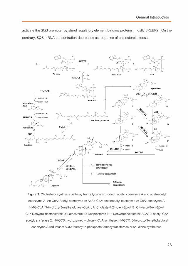
absorption but also by cholesterol endogenous synthesis. Several structures are involucrated in de novo cholesterol synthesis i.e. enterocytes, adrenal glands, ovaries or testicles but, mostly it is generated by hepatic cells. In fact, liver´s main role is the production of the bile salts from cholesterol as constitutive compounds of biliary fluids needed for the digestion processes while the cholesterol synthetized in adrenals glands or intestine is used respectively as hormone precursor and cholesterolemia modulator [26]. Cholesterol biosynthesis is carry out by a combination of mevanolate and steroid biosynthetic pathways. After more than two tens of chemical changes, acetyl-coenzyme A (Acetyl-CoA) (considered as initial precursor) promotes the synthesis of a widely range of compounds, including cholesterol, by i.e. oxidation, reduction, decarboxylation or transfer molecules reactions (Figure 3) [42].
I.2.1. Molecular events occurring during cholesterol synthesis The 3-hydroxy-3-methylglutaryl coenzyme A reductase (HMGCR) is considered the key
enzyme of cholesterol synthesis although the activities of many enzymes involved in the biosynthetic pathway such as ACAT2, hydroxymethylglutaryl-CoA synthase (HMGCS), Delta24-sterol reductase (DHCR24), farnesyl-diphosphate farnesyltransferase (FDFT1/SQS) or 7-dehydrocholesterol reductase (DHCR7), are susceptible of modulation. Recently, Gill et al (2011) suggested that squalene monoxygenase (SQLE) might be the second critical modulatory point despite its lower specificity within the cholesterol metabolism compared to HMGCR (after in vitro experiments) [43]. In fact, several natural compounds with cholesterol lowering effects such as resveratrol and gallocatechins from red wine [44], or theasinensin A [45] from green tea were described as SQLE inhibitors.
Nowadays, the SQS is also gaining attention as a potential stop-point of cholesterol synthesis since it is involved in the transformation of farnesyl pyrophosphate into squalene, being the first specific reaction at the branching point between sterol and non-sterol biosynthesis. SQS transcriptional product and protein are modulated by cholesterol since low levels of this sterol
General Introduction
25
activate the SQS promoter by sterol regulatory element binding proteins (mostly SREBP2). On the contrary, SQS mRNA concentration decreases as response of cholesterol excess.
Figure 3. Cholesterol synthesis pathway from glycolysis product acetyl coenzyme A and acetoacetyl coenzyme A. Ac-CoA: Acetyl coenzyme A; AcAc-CoA: Acetoacetyl coenzyme A; CoA: coenzyme A; HMG-CoA: 3-Hydroxy-3-methylglutaryl-CoA; ; A: Cholesta-7,24-dien-3β-ol; B: Cholesta-8-en-3β-ol;
C: 7-Dehydro-desmosterol; D: Lathosterol; E: Desmosterol; F: 7-Dehydrocholesterol; ACAT2: acetyl-CoA acetyltransferase 2; HMGCS: hydroxymethylglutaryl-CoA synthase; HMGCR: 3-hydroxy-3-methylglutaryl
coenzyme A reductase; SQS: farnesyl-diphosphate farnesyltransferase or squalene synthetase;
SQS
OH
OH CH3O
O-
HMGCR
2NADPH + 2H+
2NADP+ + CoA
OH OH
OH CH3O
Mevanolate
Acid
Mevanolate
2NADPH + 2H+
2NADP+ + CoAHMGCR
B
D
O
O
N
NN
OH O P
O
OH
OH
P
OH
O
O
P O
OH
ONHNH
SCH3
CH3
CH3
OH
OO
O
2x
ACAT2O
O
N
NN
OH O P
O
OH
OH
P
OH
O
O
P O
OH
ONHNH
S
CH3
CH3
OH
OO
O
CH3
O
+
O
O
N
NN
OH O P
O
OH
OH
P
OH
O
O
P O
OH
ONHNH
SH
CH3
CH3
OH
OO
Ac-CoA AcAc-CoA CoAH2O
CoAHMGCS
HMG-CoA
O
O
N
NN
OH O P
O
OH
OH
P
OH
O
O
P O
OH
ONHNH
S
CH3
CH3
OH
OO
OOH
O
CH3 OH
CH3
CH3
CH3 CH3 CH3
CH3 CH3 CH3
Squalene
CH3
H
OH
H
CH3
CH3
CH3
CH3
H
H
CH3
H
OH
H
CH3
CH3
CH3
CH3
H
H
A
CH3
H
OH
H
CH3
CH3
CH3
CH3
H
H
Zymosterol
CH3
H
OH
H
CH3
CH3
CH3
CH3
H
H
CH3
H
OH
H
CH3
CH3
CH3
CH3
H
H
H
CH3
H
OH
H
CH3
CH3
CH3
CH3
H
H
H
CH3
H
OH
H
CH3
CH3
CH3
CH3
H
H
CH3
H
OH
H
CH3
CH3
CH3
CH3
H
H
H
C
DHCR24
DHCR7
NADPH + H+
NADP+
NADPH + H+
NADP+
CH3
CH3 CH3
CH3 CH3 CH3CH3
CH3
O
SQLE
Squalene 2,3-epoxide
CH3
CH3
CH3
H
O
H H
CH3
CH3
H
H
R
O
Cholesterol
Oxysterol
SOAT
STEROL
STERASE
Bile acids
biosynthesis
Steroid hormone
biosynthesis
Steroid degradation
DHCR24CDI
CDI
LO
EF
General Introduction
26
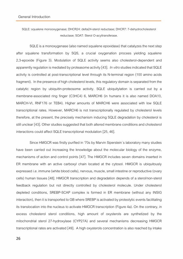
SQLE: squalene monooxygenase; DHCR24: delta24-sterol reductase; DHCR7: 7-dehydrocholesterol reductase; SOAT: Sterol O-acyltransferase.
SQLE is a monoxygenase (also named squalene epoxidase) that catalyzes the next step after squalene transformation by SQS, a crucial oxygenation procees yielding squalene 2,3-epoxide (Figure 3). Modulation of SQLE activity seems also cholesterol-dependent and apparently regulation is mediated by proteasome activity [43]. In vitro studies indicated that SQLE activity is controlled at post-transcriptional level through its N-terminal region (100 amino acids fragment). In the presence of high cholesterol levels, this regulatory domain is separated from the catalytic region by ubiquitin-proteosome activity. SQLE ubiquitylation is carried out by a membrane-associated ring finger (C3HC4) 6, MARCH6 (in humans it is also named DOA10, MARCH-VI, RNF176 or TEB4). Higher amounts of MARCH6 were associated with low SQLE transcriptional rates. However, MARCH6 is not transcriptionally regulated by cholesterol levels therefore, at the present, the precisely mechanism inducing SQLE degradation by cholesterol is still unclear [43]. Other studies suggested that both altered membrane conditions and cholesterol interactions could affect SQLE transcriptional modulation [25, 46].
Since HMGCR was firstly purified in ‘70s by Marvin Siperstein´s laboratory many studies have been carried out increasing the knowledge about the molecular biology of the enzyme, mechanisms of action and control points [47]. The HMGCR includes seven domains inserted in ER membrane with an active carboxyl chain located at the cytosol. HMGCR is ubiquitously expressed i.e. immune (white blood cells), nervous, muscle, small intestine or reproductive (ovary cells) human tissues [48]. HMGCR transcription and degradation depends of a sterol/non-sterol feedback regulation but not directly controlled by cholesterol molecule. Under cholesterol depleted conditions, SREBP-SCAP complex is formed in ER membrane (without any INSIG interaction), then it is transported to GB where SREBP is activated by proteolytic events facilitating its translocation into the nucleus to activate HMGCR transcription (Figure 4a). On the contrary, in excess cholesterol sterol conditions, high amount of oxysterols are synthetized by the mitochondrial sterol 27-hydroxylase (CYP27A) and several mechanisms decreasing HMGCR transcriptional rates are activated [49]. A high oxysterols concentration is also reached by intake

General Introduction
27
of cholesterol-enriched food stored for a long time or submitted to heat treatments [50]. The HMGCR regulatory mechanisms could be classified in INSIG-dependent (modulating at transcriptional and post-transcriptional levels), or INSIG-independent.
INSIG-dependent HMGCR regulation mechanisms (figure 4b):
- INSIG can disrupt SREBP activation by binding to SCAP when high sterol concentrations are noticed in the cytosol. Once INSIG-SCAP complex is formed, SCAP is structurally altered impairing the SREBP recognition and stopping the assembly of SREBP-SCAP complex for his further transport from ER to AB. In consequence, SREBP is not translocated to the nucleus and HMGCR transcription is inactivated [11, 47].
- Under similar conditions, INSIG can also binds to the N-terminal region of HMGCR and conjugate it with ubiquitin. Ubiquitation is carried out by gp78 (membrane-bound ubiquitin E3 ligase) assisted by Ubc7 (an E2 ubiquitin conjugating enzyme) providing active ubiquitins and a few other enzymes. Then, ubiquitinated HMGCR is rejected to cytosol and subsequently proteasome-degraded with the participation of p97/VCP (ATPase associate to membrane). However, the presence of high levels of sterols is not mandatory for the ubiquitination process but its stimulate HMGCR degradation by enhancing INSIG-HMCR bindings [47].
INSIG-independent HMGCR regulation mechanisms (figure 4b):
- HMGCR activity could be also modulated in situations of cellular stress (low ATP levels) by AMPkinase (AMP-activated protein kinase). In this case, HMGCR is inactivated by a serine phosphorylation due to the AMPkinase activity. It is a reversible reaction and HMGCR can also be activated by a protein phosphatase 2A (PP2A) [47, 51].
- Non-sterol isoprenoids might also modulate HMGCR translation by a mechanism still unclear Burg et al. (2011) [47].

General Introduction
28
I.3. Molecular events occurring during cholesterol excretion For several decades, classical reverse cholesterol transport (RCT) have been considered
the main mechanism to eliminate cholesterol however, recent studies suggest an alternative pathway, the so called transintestinal cholesterol excretion (TICE) [11].
RCT is a derivative branch of the hepatobiliary pathway. Lipoproteins such as HDL or LDL makes available cholesterol for hepatic absorption as esterified or non-esterified molecules. Esterified cholesterol is transformed by the hepatic cholesteryl ester hydrolase (NCEH1) into the non-esterified form after hydrolysis of the ester linkage. Thus, the generated forms are directly excreted through the ABCG8/5 heterodimer protein or transformed into bile salts [52]. CYP7A1 (cholesterol 7alpha-monooxygenase) is the enzyme responsible for cholesterol transformation into primary bile salts (cholic and chenodeoxycholic acid) within the neutral bile acids pathway in liver. Synthetized bile salts are secreted to bile canaliculus by the bile salt export pump (BSEP) or the multidrug resistance-associated protein 2 (MRP2) and become part of bile fluids. Recent in vivo studies have suggested the involvement of other cholesterol transporters such as NPC1L1 in RCT. According to Temel et al (2007)[53] overexpression of NPC1L1 in transgenic mice resulted in a 10-20 fold decrease in biliary cholesterol concentration. Dikkers and Tietge (2010) [19] noticed a 90% decrease in biliary cholesterol in knockout NPC2 subjects and in both cases no quantitative changes in bile acids or phospholipids bile content were observed. The role of NPC1L1 in biliary cholesterol excretion is not yet elucidated but, it might involve adjustments in cholesterol balance to avoid excessive loss of the metabolite through the intestinal track.
TICE have been suggested as an alternative cholesterol excretion mechanism where the sterol is directly eliminated from blood through the intestinal mucosa and excreted via feces [11]. The hypothesis was drawn after the unexpected results obtained by several authors that noticed an unwarranted balance between cholesterol inputs and outputs in mouse models. They showed a higher amount of fecal cholesterol than the sum of dietary intake and biliary secretion [54] or an unaltered cholesterol excretion rate in knockout NPC1L1 mice with a decreasing of 90% biliary excretion [55].

General Introduction
29
Figure 4a. Molecular regulatory pathway of HMGCR transcription under low intracellular cholesterol levels.

General Introduction 3
0
Figure 4b. Molecular regulatory pathway of HMGCR transcription under high intracellular cholesterol levels.

General Introduction
31
These results questioned the complete RTC classical concept [56] as well as the contribution of biliary or non-biliary cholesterol to its RCT excretion [57]. Although the involvement of several membrane transports in TICE have been studied (i.e. SR-B1 [58], NPC1L1 [59], ApoA1 [60], LDLR or ABCG5/8 [61]), it is still unknown whether TICE is carried out through the basolateral or apical transporters or whether HDLs are involved [62] therefore, the mechanism is not yet elucidated. There is scientific controversy about the importance of TICE, some authors suggest that it could be only a compensatory cholesterol excretion mechanism in case of biliary cholesterol depletion but, other authors pointed it as the main mechanism in cholesterol excretion. Van der Velde et al (2007, 2010) estimated TICE contribution as 70 and 30% of total cholesterol excretion respectively in mice and humans [54, 62].
I.4. Maintenance of cholesterol transport To ensure a dynamic blood-tissue cholesterol transport and to avoid related diseases,
rates of cholesterol absorption, synthesis and excretion should be balanced. Thus, an effective communication mainly between liver and small intestine (also adrenals glands) is necessary. HDL, IDL, LDL and VLDL are the connecting structures responsible for transporting of cholesterol molecules through the blood stream from one tissue to another until they are detected by cellular membrane receptors such as SR-B1 for HDL or LDLr (LDL-receptor) for the latter two.
After the enterocyte absorption process, the non-esterified cholesterol eliminated by ABCA1 through the basolateral membrane is bound to apoA-1 generating nascent-HDL and the esterified cholesterol is similarly assembled with apoB-48 in ER synthetized prechylomicrons further transformed into chylomicrons in GA and excreted by exocytosis to intracellular space. Therefore, both structures become cholesterol transporters and they distribute it via the blood stream to the rest of the organism.
Nascent HDL is transformed into mature HDL by accumulation of non-esterified cholesterol molecules secreted by hepatocyte and enterocyte ABCA1 protein (also by ABCG1 transporter in gland adrenal cells). Once mature HDLs are formed, some cholesterol molecules are esterified by action of lecithin—cholesterol acyltransferase (LCAT) along it blood transport.

General Introduction
32
Esterified and non-esterified cholesterol is detected by the SR-B1 located in the basolateral membrane of cells from liver, small intestine or gland adrenals allowing the incorporation of esterified cholesterol inside the cell. Intracellular esterified cholesterol is transformed into non-esterified cholesterol (SOAT action) and HDL is turned into LDL. The non-esterified cholesterol can be used for bile salts synthesis and their further intestinal secretion by BSEP or to be directly excreted via ABCA1 (basolateral membrane) and ABCG5/ABCG8 (apical membrane) activity. In turn, ejected non-esterified cholesterol again could be attached to nascent HDL to create mature HDL and continue with cholesterol transport.
LDL molecules are recognized by liver, intestine and gland adrenal LDLR as esterified cholesterol suppliers. LDL particles can be generated not only by VLDL transformation (by SR-B1 activity) but also by addition of esterified cholesterol to apoB-100 (apolipoprotein isoform characteristic of hepatocytes) leading very low density lipoproteins (VLDL). These structures are secreted to blood stream by hepatocytes. VLDL lipids are used by the muscle and peripheral tissues as they pass through the blood stream generating IDL and LDL by lipoprotein lipases activity (LPL). Moreover, esterified cholesterol of chylomicron structures is also recognized by LDLR providing to the hepatocyte those cholesterol molecules assembled in enterocytic ER after digestion process.
The difficult cholesterol biosynthesis enginery, the complexity of ABCA1, ABCG5/8, SR-B1 and LDLR activities and the multifactorial regulation system make the control of cholesterol metabolism a large challenge for the scientific community. Particularly because some of the involved compounds are also intermediates of other metabolic pathways i.e LXR modulates ABCG5/8 activity but also DIO1, a selenoprotein involved in the thyroid hormone metabolism. However, these facts are also making it a flexible system that could be modulate from different critical points.

General Introduction
33
II. Strategies to lower serum cholesterol
Moderate to severe hypercholesterolemia is usually treated with several drugs acting as inhibitors of endogenous cholesterol biosynthesis or impairing exogenous cholesterol absorption. These pharmacological compounds lower cholesterol levels in serum following different mechanisms of actions.
Similarly, many natural compounds are able of performing the same effect than those drugs although for some, more studies are needed because so far the experiments were only carried out using in vivo test. Drugs and natural extracts might follow several potential cholesterol-lowering strategies.
II.1. Inhibitors of the pancreatic lipase At the present, the pancreatic lipase (PL) inhibitor more frequently consumed is
tetrahydrolipstatin (commercially named orlistat), a natural compound from Streptomyces toxytricini. Orlistat is acting by binding to a serine located in the active site preventing the lipase activity. It is widely accepted by physitians but induced unpleasant gastrointestinal side-effects [63].
Many natural extracts from plant, microorganism and marine algae showed PL inhibitory activity such as alcoholic fractions from Cudrania tricuspidata [64], Dioscorea niponica, Nelumbo nucifera [65], Hygrocybe conica, Laetiporus sulphureus, Tylopilus felleus or Caulerpa taxifolia but the responsible compounds still remain undetermined. However, catequines, saponines, triterpenoids, flavonoids, carnosic acid, manno-oligosacharides, ε-polylysine, crocin, caffeine, vibrolactones, lipistatin or flavan dimers from different sources were pointed as PL inhibitors [63].
II.2. Cholesterol and bile acids scavengers Bile acid scavengers reduce cholesterol absorption via interruption of the enterohepatic
circulation of bile acids and results in a secondary increase in the hepatic LDL receptor activity. A few synthetic compounds are used as bile acids scavengers such as cholestyramine. Cholestyramine is an ion exchange resin that could reduce total cholesterol and LDL-cholesterol

General Introduction
34
in a 9-25% and 15-33% by impairing total or partial bile acid absorption [66]. Treatment with this drug induced similar cholesterol-lowering effects than other molecules such as statins, fibers or ezetimibe (described below) and when combined with other drugs the reduction is enhanced because of synergistic effects [67, 68] [69].. For example, cholestyramine-statins treatments improve blood cholesterol levels an additional 20% more than only statins [66].
Dietary fibers from several sources are also described as cholesterol bile acid binders but they will be further described elsewhere (paragraph III.2).
II.3. Displacers or cholesterol from DMMs Nowadays, plant sterols (phytosterols and phytostanols) are considered the most potent
cholesterol competitors for their inclusion into DMMs since no chemically synthetized compound have been designed so far to act at this strategic step. They will be described more in detail further on paragraph III.1. In fact, any lipid compound that has to be integrated in the DMMs such as phosphatidylcholine, tocopherols, bile acids etc. will modify the DMM composition modulating its hydrophobicity and therefore the amount of cholesterol that will be included per DMM [70].
II.4. Inhibitiors of NPC1L1 and ABC transporters Another cholesterol lowering strategy is by impairing its transport through the plasmatic
membranes using several NPC1L1 and ABC transporters inhibitors. Stimulation or silencing of NPC1L1 gene expression respectively facilitated or inhibited the free cholesterol uptake [24] in a mechanism that seems to involve a deficient ACAT2(AvS) activity [23, 71],and apparently, NPC1L1 is the molecular target of ezetimibe (a non-acylated β-lactam family member [72]). In vitro and in vivo studies indicated that NPC1L1 and ezetimibe are involved in the same pathway [73-76]. However, there is certain controversy about NPC1L1 inhibition mechanism, some hypothesis suggested that ezetimibe inhibition action is due to the blocking of NPC1L1 movement from cytosol to brush border membrane and vice versa [22]. Other publications indicated that ezetimibe undergoes glucuronidation to a single metabolite with higher affinity to brush border membrane NPC1L1 impairing the correct cholesterol absorption and enterohepatic recirculation [76]. However, ezetimibe was able of decreasing hepatic oxysterols levels, which are endogenous

General Introduction
35
agonists of liver X receptor (LXR), reducing hepatic lipogenic gene expression of a few enzymes such as stearoyl-CoA desaturase-1 (SCD1) [77].
Others compounds were described as potential NPC1L1 inhibitior such as spiroimidazolidinone derivates (they showed high NPC1L1 binding affinity) [78], novel amino β-lactams derivates [79] or curcuminoids polyphenols (acting by indirect influence on SREBP-1) [39].
Atorvastatine, a synthetic statin with high HMGCR inhibitory capacity, was also able of decreasing ABCG5/8 mRNA levels in enterocytes of hyperlipidemic animals. Similar effect was reported for an endotoxine in murine liver [80]. Posttranscriptional regulations were also described for spironolactones or polyphenols from Aronia Melanocarpa [80, 81]. In the latter case, specific miRNAs could be the responsible compounds.
II.5. Inhibitors of cholesterol transport and assembling in the endoplasmic reticle Some compounds from natural sources have been reported as ACAT(AvS) inhibitors
reducing cholesterol esterification rates such as alkamides from Piper nigrum [82], shikonin derivatives from Lithospermum erythrorhizon [83], an isoprenyl flavonoid identified as grabol from licorice roots [84], ursolic acid (via (PPAR)-α activation) [85] or flucoxanthin from marine plants [86].
However, until now, only drugs obtained by chemical synthesis are been tested in clinical trials such as certain xanthone sulfamides [77], efluzimibe [78] or avasimibe. Recent studies indicated that avasimibe was not only involved in ACAT(AvS) inhibition but also in the blocking of APOB-48 secretion (in HepG2 cells) [87]. Several compounds are also described as apoB-48 blockers such as atorvastatin [88], adrenocorticotropic hormones [66] [89] [69] etc.
Other cholesterol regulating procedures included MTTP inhibition. Apparently, MTTP was the target of lomitapide [39]. This compound impaired TG, phospholipids and cholesteryl esters transferring from ER to the nascent APOB, leading a lower loading of this lipoprotein,

General Introduction
36
inhibition of VLDLs assembly and chylomicrons secretion [67] resulting in a fat and fat-soluble vitamins malabsorption [39, 68].
II.6. Inhibitors of enzymes involved in cholesterol biosynthesis From several decades, statins were considered the most effective compounds for
HMGCR inhibition and considered safe and ideal as primary treatment for most of hypercholesterolemic patients. Many natural and synthetic statins are nowadays prescribed such as lovastatin, simvastatin, pravastatin, fluvastatin, atorvastatin, pitavastatin, rosuvastatin, etc. They all act as substrate competitors showing different binding affinities for the enzyme but always higher than the 3-hydroxy-3methylglutryl-Coenzime A (HMGcoA) [90].
On the other hand, HMGCR inhibition is also mediated by an AMP kinase via phosphorylation and green and black tea polyphenols induced a direct increase of HMGCR phosphorylation possibly via AMP kinases phosphorylation. Their precise mechanism of action is still unclear but, it seems to involve activation of regulatory factors such as PPAR [91, 92].
Only a few compounds are still nowadays pointed as potential inhibitors of the SQS such as resveratrol, quinuclidine, 4, 1-benzoxazepine-3-acetic acid derivatives such as TAK-475 (or Lapaquistat) or morpholine derivates. The SQS inhibition by two 2-biphenylmorpholine derivatives showed multiple consequences in lipid metabolism i.e. inhibition of triglyceride biosynthesis, increasing of LDLR gene expression and LDL uptake or lower apoB synthesis rate. Thus, modulation of this enzyme could also affect cholesterol metabolism at several levels [44-46, 78, 82, 93].
24(S), 25-epoxycholesterol was also pointed as inhibitor of the DHCR24 activity. This enzyme catalyze the transformation of desmosterol into cholesterol. The inhibitor did not modify DHCR24 protein levels, but increased desmosterol accumulation decreasing cholesterol levels in in vitro studies due to its structural similarity with desmosterol [94].

General Introduction
37
III. Marketed supplemented foods containing hypocholesterolemic compounds
People with incipient hypercholesterolemia can prevent, delay or enhance the pharmaceutical treatment by taking a few functional foods already available at the supermarkets. At the present, many supplements are indicated by herbalists because they might help against CVD such as garlic extracts, soy preparations, ω3 oils, etc. However, only two types of compounds are nowadays authorized by the competent institutions from most of the industrialized countries to bear the health claim ̈ hypocholesterolemic properties¨ in their labels and they are marketed under many different brands by the food industry: plant sterols and polysaccharides (β-glucans and chitins derivatives).
III.1. Phytosterols This group of compounds gained much attention in the last decade since, in 1999 a
sitostanol-containing margarine was launched in the market [95, 96]. A few years later, other sterols such as esterified phytosterols and not free sterols were utilized to functionalize foods because the esterification increases their solubility in fat and improved their bioavailability [97, 98].
Although more than 40 plant sterols and derivatives have been identified (Figure 5), only stigmasterol, campesterol and β-sitosterol are present in most of vegetables (0.1-0.5% w/w). Brassicasterol is also present in large quantities but only in Brassica sp. Other derivatives such as stanols (campestanol, sitostanol etc.) are also present in lower amounts although after some culinary treatments their composition increase [99]. The amount of phytosterols and derivatives depend on the type of plant being those rich in oils and lipidic compounds the food with higher sterol contents (Table 2) [100-102].

General Introduction
38
Figure 5. Structural similarities of cholesterol, β-sitosterol and fungal sterols molecules.
Table 2. Plant sterols composition (mg/g edible portion) obtained from several food sources.
Plant-derived
food Campesterol β-sitosterol Stigmasterol Campestanol Sitostanol
Sacha inchi Oil 0.42 1.47 0.48 nd nd
Soybean Oil 0.42 1.47 0.48 nd nd
Seed 0.07 0.17 0.08 0.1 x 10⁻² 0.7 x 10⁻²
Corn Oil 2.20 4.45 0.61 nd nd Carrot Vegetable 0.02 0.10 0.03 nd 0.6 x 10⁻³ Peanut Oil 0.10 0.82 0.07 nd nd
nd: non-detected compounds.
III.1.1. Physiological changes modulated by plants sterols Cholesterol absorption from dietary and biliary sources is significantly reduced in the
presence of plant sterols. Their mechanism of action is related to their structural similarity since
they appear to compete with dietary cholesterol absorption displacing it from the dietary mixed

General Introduction
39
micelles formed during intestinal digestion and then, the unabsorbed cholesterol is excreted in
the feces [15]. According to both static and dynamic studies, phytosterols become more efficiently
incorporated into micelles in the intestinal lumen, displace the cholesterol and lead to its
precipitation with other non-solubilized phytosterols [12, 103-105] but, the specific mechanisms
remains still unclear.
Cholesterol solubilization in DMMs was lowered by β-sitosterol because both compounds competed for the DMMs binding sites specific for steroid interactions [106]. When comparing between phytosterols (β-sitosterol and sitostanol) micelles of different size and composition were obtained depending on the sterol solubilized [103, 105].
Furthermore, sterols are also described as promotors of cholesterol co-crystallization at gastric duodenal levels and compounds stimulating their competition for their transfer through the brush border membrane and within chylomicron assembly[103]. Their effect on the SOAT activity have also been studied although it remains partially unclear. Some reports indicated that cholesterol enterocytic esterification by ACAT(AvS) decreased by sterols competition although the enzyme showed lower esterification efficiency for plant sterol than cholesterol. Other reports suggested plant sterols influence on MTTP and APOB48 lipoprotein. APOB48 inhibitory effect was noticed by stigmanterol, campesterol and β-sitosterol in cell cultures but not in an animal model. More recent publications suggested other mechanism of actions involving modulations at the molecular level.
III.1.2. Molecular events modulated by plants sterols Transcription of some ABC transporters (ABCG5/8 etc.) was induced by LXR factor in
enterocytes (but not in hepatocytes) [103] although, there is a controversy about it influence on other transporters (ABCA1). Oxysterols such as 22(R)-hydroxycholesterol, 24(S),25-epoxycholesterol or 27-hydroxycholesterol etc., are considered as endogenous natural LXR agonist however, plant sterols derivatives showed higher LXR agonist activity. Brasicasterols from unicellular algae and Brassiceas sp (i.e. rapeseed) induced large variations in the gene expression of ABC transporters due to their ability to act as the LXR factor in mice. Moreover,

General Introduction
40
sitostanol (in mice), sitosterol (in Caco2 cells) and a few 4-desmethylsterol derivatives were also able of inducing ABCA1 up-regulation using their LXR agonist activity [9, 26, 103, 107]. However, no direct effects of phytoesterols was noticed on the transcriptional levels of the transporters because in ABCA1 and ABCG5/8-deficient mice decreasing of cholesterol intestinal absorption was also noticed indicating that ABC transporters were not their direct targets.
Other reports indicated that phytosterol/stanols modulated HMGCR expression via ACAT(AvS) inhibition. Apparently, lower ACAT(AvS) activity led to higher free cholesterol amounts, inhibiting the cholesterol biosynthetic pathway and HMGCR expression beside others. It also reduced chylomicron assembling, and promoted the back efflux of non-esterified sterols to the lumen [26, 103, 108]. However, their influence on SOAT seemed to be by chemical inhibition more than by molecular modulation.
Several in vitro studies pointed out that SREBP-2, NPC1L1 and SR-B1 gene expression is modulated by plant sterols such as stigmasterol and β-sitosterol toward the reduction of cholesterol absorption. Surprisingly, HepG2 cells treated with these sterols showed simultaneous down-regulation of NPC1L1 and SR-B1 when a opposite effect on this two molecules could be expected . Studies using homozygous and heterozygous knockout mice (NPC1L1-/- and NPC1L1+/-
) showed a lower cholesterol absorption in homozygous animals however, heterozygous mice showed higher HMGCR mRNA levels in gut and liver tissues than wild-type animals without changes in ABC transporters expression rates. Authors explained their results by compensatory effects: difficulties for cholesterol absorption were compensated by a stimulation of endogenous cholesterol synthesis to maintain physiological plasma levels [23, 73]. Moreover, when the knockout mice were treated with plant sterols or ezetimibe, the wild-type and heterozygous animals showed a reduction in cholesterol and TG levels. Absorption of campesterol and β-sitosterol was reduced in NPC1L1+/- and almost absent in NPC1L1-/- mice indicating that certain modulation of NPC1L1 took place [23, 73].
β-Sitosterol addition to Caco2 cultures induced down-regulation of HMGCR expression although this effect was not noticed in mice. Only sitosterolemic individuals showed reduction in

General Introduction
41
HMGCR activity in ileum so apparently only large amounts of plan sterols or long-term 2% (w/w) plant sterol administration can induce such effect [26, 109].
Other reports indicated that phytosterols might modulate the expression of other closely related genes such as those encoding the hepatic cholesterelogenic farnesyl phosphate synthase (FFPS), liver CYP7A1 (cytochrome P450 family 7 subfamily A polypeptide 1) or anexin 2-Caveolin 1 (ANXA2-CAV1) protein complex [26].
III.2. β-Glucans More recently, β-glucans containing foods such as oat or barley products have also been
launched in the market claiming up to 15% cholesterol reduction after regular consumption [4]. Many epidemiological studies have demonstrated a reduced CVD risk for consumers of high amounts of dietary fiber. The (13),(14)-β-glucans are believed to perform many biological functions and one of them is the ability to lower serum cholesterol (LDL) levels [110].
Cereals are the main source of β-glucans and other dietary fibers with hypocholesterolemic properties (Table 3) being oat and barley glucans those pointed as more effective in lowering cholesterol and glucose levels or increasing immunity against infection.
Table 3. β-Glucan composition (mg/100mg dry weight) obtained from several food sources [111].
Cereal Total β-glucan Soluble β-glucan
Oats 10.37 1.73-5.7
Barley 10.87-19.7 1.87-5.37
Wheat 8.48-16.19 0.19-0.67
Rye 7.45 2.26
Until a few years ago, the term dietary fiber included a wide range of polysaccharides that could not be digested by human track becoming substrates for microbiota fermentation. Nowadays, a precise definition is more complicated because of the multiple different structures and biological activities ascribed.

General Introduction
42
III.2.1. Physiological changes provoked by dietary fibers or β-glucans Apparently the cholesterol-lowering mechanisms of dietary fibers are dependent of their
solubility and can be distinguished as water-insoluble (WIF) or water-soluble (WSF) fibers (although some authors divide them depending on the extraction solvent utilized i.e. alkalis-soluble etc.). The mechanism of actions proposed for WSF are [112, 113]:
- WSF might provoke changes in the properties of digestion fluids i.e. high viscosity. Higher water binding capacity on the chime might lead to a bad diffusion of cholesterol and bile acids for their proper incorporation into the DMMs and further absorption/reabsorption.
- WSF presence during digestion might decrease the glycemic response stimulating hepatocytic cholesterol synthesis.
- Products generated from WSF after colonic fermentation by microbiota such as propionate etc. (generally called short chain fatty acids, SCFAs) might act as hypocholesterolemic compounds by modulating cholesterol-related gene expression.
The cholesterol reducing properties of WIF appeared to be more related to their ability to act as bile acid scavengers following a similar mechanism as previously described for cholestyramine by direct specific bindings when passing the small intestine [112]. This mechanism impair bile acids re-absorption stimulating the hepatic synthesis of new bile acids from cholesterol reducing its blood concentration [15, 89, 114].
Although the above described mechanisms have been propose to explain hypocholesterolemic fiber effects, it is still hard to define them properly due to wide variety of fibers from different natural sources and their different extraction and study protocoles [115]. Moreover, despite the indicated properties, their hypocholesterolemic effects (in particular for β-glucans) might be due to a combination of several mechanisms including potential modulation of cholesterol-related gene expresions [116].

General Introduction
43
III.2.2. Molecular events modulated by β-glucans At the present, not many investigations have been carried out concerning the molecular
effect of dietary fibers. High viscosity oat or barley β-glucan extracts demonstrated their ability to down-regulate SREBF2 gene expression in intestinal cells (NCl-H716) [116] but no with in vivo testing. However, according to Hu et al (2008) corn bran dietary fiber up-regulated the expression of other genes such as FXR in ileum intestinal cells or PPAR in liver [117].
Hepatic HMGCR up-regulation was observed after administration of hydroxylpropylmethycellulose or inulin-oligofructose (1:1) to animal models [118, 119] however barley β-glucans administration induced no changes on HMGCR expression [113]. Jones (2008) noticed hepatic SREBF2 up-regulation when soluble dietary fiber (guar gum, a galactomannan) was administrated to pigs. In consequence, LDLr expression was enhanced and reduced the LDLs from the blood stream. Hepatic ABCG5/8 gene expression was also enhanced by guar gum consumption leading a higher cholesterol efflux from the liver to intestinal lumen [120].
Other studies indicated that the scavenging of bile acids by fiber (β-glucans) activated CYP7A1 to convert cholesterol into new bile acids [4, 117] provoking an hepatic cholesterol decrease that up-regulated LDLr expression and reduced cholesterol blood levels. Due to these changes the biosynthetic pathway was also activated via HMGCR up-regulation to compensate the lack of hepatic cholesterol [4, 113].
III.3. Chitosan Chitosan, a de-N-acetylated derivative of chitin, is a heteropolysaccharide (considered
as DF) consisting of linear β-(14)-linked glucosamine and N-acetyl-D-glucosamine units and it is included in the list of approved health claims according to FOSHU regulations with the claim ¨helps lower cholesterol level¨. At the present (2011) in Japan, there are more than one hundred marketed products including powdered or soft drinks containing chitosan. This compound is obtained from chitins extracted from crustacean shell wastes, insect skeletons, mushrooms and other fungi or bacteria [121]. A particular enterprise is conducting double-blind studies with animals and it is scheduled to begin with human clinical tests next year to submit a specific

General Introduction
44
preparation for FOSHU approval. The preparation includes a mushroom-derived chitosan combined with β-glucans.
The hypocholesterolemic action of chitosan might be due to its ability to decrease cholesterol absorption and interfere with bile acid absorption following a mechanism similar to those explained for the other above mentioned β-glucans [122] since both the water-soluble and water-insoluble chitosan fractions were able to lower cholesterol in a clinical trial with 60 elderly hyperlipidemic patients. Total cholesterol significantly declined by 7.5% in the water-soluble and by 8.9% in the water-insoluble chitosan groups over 8 weeks [123]. Other studies carried out on rat groups fed on a diet rich in cholesterol found that chitosan hydrolysates with different molecular weights and viscosity were also effective and that the lower the molecular weight, the better its cholesterol-lowering potential [124, 125].
IV. Evidences of the hypocholesterolemic properties of mushrooms Edible mushrooms should also contain compounds able to lower cholesterol levels in
serum since already for years, many works have been published pointing some mushrooms species as source of hypocholesterolemic compounds. However, many of them did not identified the responsible molecules due to most of the studies were animal or clinical tests using the complete fruiting body (sporophore) or certain specific extracts or fractions.
IV.1. Evidences supported by animal trials In vivo studies in Wistar rats, Syrian hamsters and Chinchilla rabbits demonstrated the
ability of oyster mushrooms (Pleurotus ostreatus) to lower the cholesterol levels. Rats with hereditary enhanced sensitivity to alimentary cholesterol were fed with 4% fruiting bodies in a diet containing 1% cholesterol. Mushroom consumption prevented serum cholesterol increase after 4 weeks and after 7 weeks the cholesterolemia was lowered by 40% compared with control animals. This effect was due to a decrease in the concentrations of very low density lipoproteins (VLDL) and LDL [126] and to an induced delay in cholesterol absorption [127, 128]. A 50% increase in

General Introduction
45
the HDL and activation of antioxidative enzymes was also observed [129]. Similar results were observed using Chinchilla rabbits (fed with 10% P. ostreatus) where a decrease in total serum cholesterol was also detected primarily affected by the reduction (70%) of the VLDL-cholesterol. An increase by a factor of 3 in the HDL was also observed [130]. However, the hypocholesterolemic effect was dependent on the amount of dietary oyster mushroom administrated, 1% was ineffective while doses of 5% induced a significantly reduction in normal rats [131].The ethanol extracts obtained from the same mushroom were less efficient with increasing concentrations of ethanol than the whole body or its water extract. The latter and the extracts including 30 and 60% ethanol were also able to reduce the cholesterol and triacylglycerol levels in hamsters serum and liver (reduction of 34% cholesterol and 48% triacylglycerols). The consumption of the water and 30% ethanol extracts reduced the VLDL fraction [127]. Another extract obtained from its mycelium with dichloromethane and 95% ethanol lowered cholesterol levels in liver of normal rats [132] but, a β-glucan fraction extracted from the fruiting bodies was unable to affect cholesterols levels in both serum and liver [129].
Supplementation of a high-fat-diet given to hyperlipidemic rats with Pleurotus
citrinopileatus fruiting bodies, a hot-water extract and two specific fractions obtained by eluting
from a silica gel column with ethyl acetate and methanol at different doses showed that serum
triglycerides and total cholesterol levels were significantly lower and HLD significantly higher in
the groups supplemented with the highest dosage of the two eluted fractions as compared with
the control groups with no mushroom addition. The major constituents of the eluted fractions were
ergosterol in one of them and nicotinic acid in the other [133].
Other species from the same genera such as Pleurotus eryngii reduced the plasma total
cholesterol, triglyceride, LDL, total lipid, phospholipids and LDL/HDL ratio by 24, 46.3, 62.5, 24.6,
19.2 and 57.1% respectively and showed no adverse effects when they were added as
supplement (5%) to hypercholesterolemic Sprague-Dawley albino rats. Feeding mushrooms
increased total lipid and cholesterol excretion in feces [134]. Its water extracts showed

General Introduction
46
hypolipidemic and hypocholesterolemic effects in fat-loaded mice. The low fat absorption
provoked was due to its lipase inhibitory activity. Apparently, the water extract might prevent the
interactions between lipid emulsions and pancreatic lipase [135]. The remarkably reduction of
lipid levels, total cholesterol, total triglyceride and LDL-cholesterol and increase in HDL-
cholesterol was because of its water-soluble polysaccharides [136].
Ganoderma lucidum is considered by the Asiatic culture as a medicinal mushroom
because of its many different beneficial properties. Several of these properties have been
confirmed by scientific studies [137, 138] including its hypocholesterolemic effects in hamsters
and minipigs [139]. The organic fractions containing oxygenated lanosterol derivatives inhibited
cholesterol synthesis in T9A4 hepatocytes. In hamsters, 5% supplementation did not affect LDL
but decreased 9.8% total cholesterol, 11.2% HDL and had effects on several faecal neutral sterols
and bile acids. It also reduced hepatic microsomal ex-vivo HMGCoA reducase activity but not its
gene expression [140]. In minipigs, 2.5% supplementation decreased 20% total cholesterol, 27%
LDL and 18% HDL-cholesterol and increased faecal cholestanol and coprostanol and decreased
cholate. Results also indicated that G. lucidum reduced LDL cholesterol in vivo through various
mechanisms [139].
Similar studies with the white button mushroom (Agaricus bisporus) indicated that they
were able to lowers blood glucose and cholesterol levels in diabetic and hypercholesterolemic
Sprague-Dawley rats fed with hypercholesterolemic diets for 3 or 4 weeks [141].
Hypercholesterolemic rats significantly decreased their levels of plasma total cholesterol and LDL
(22.8 and 33.1% respect.) and their hepatic levels of cholesterol and triglycerides (36.2 and 20.8%
respect.) increasing significantly their plasma HDL concentrations. Apparently, these effects
might have been induced by the presence of A. bisporus fibers because when rats were fed with
these compounds, the serum total cholesterol, VLDL, IDL (intermediate density lipoprotein) and

General Introduction
47
LDL were lower than control rats while the HDL were lower. These observations could be caused
by the overexpression of the hepatic LDL receptor since its mRNA level was significantly higher
than in the control rats [142].
After these results, the fiber fraction from other edible mushrooms was also investigated.
Rats were fed with the fiber fraction from Grifola frondosa (maitake), Lentinula edodes (Shiitake)
and Flammulina velutipes (enokitake mushrooms) for 4 weeks and compared with cellulose as
control. The total cholesterol in serum was significantly lower in rats fed with G. frondosa and F.
velutipes than in those rats fed with cellulose. The VLDL, IDL and LDL levels were also lower when
the rats ate any of the three mushrooms whereas only the HDL of the F. velutipes group was
significantly lower than the others. The LDL receptor mRNA levels were only significantly higher in
the F. velutipes group, thus, F. velutipes lowered cholesterol in serum by enhancement of hepatic
LDL receptor mRNA while the other mushroom fibers enhanced the faecal cholesterol excretion
[143]. Hericium erinaceus (hot water and ethanol extracts) also induced transcriptional changes
but affecting to the PPARα gene, this modulation influenced others such as ACAT, ApoA1, LPL or
SREBP1 in mice fed a high-fat diet [144]. Other authors indicated that this mushroom was also
inducing the up-regulation of many genes involved in the fatty acid transport but repressed
SREBP1 and ACAT [144]. Moreover, G. frondosa and two other mushroom species such as
P. eryngii, and particularly Hypsizygus marmoreus also showed antiatherosclerotic effects in
atherosclerosis-susceptible C57BL/6J, apolipoprotein E–deficient (apoE−/−) mice [145]. The
effect of G. frondosa and H. marmoreus together with P. ostreatus were also investigated in
feeding experiments using normal mice where a DNA microarray analysis of liver indicated
differences of gene expression patterns among mushrooms. CTP1A and FABP families were
upregulated in the P. ostreatus-fed group, which were considered to promote lipid transport and
β-oxidation. In the G. frondosa-fed group, not only the gene involved in signal transduction of

General Introduction
48
innate immunity via TLR3 and interferon but also virus resistance genes, such as MX1, RSAD2
and OAS1 were upregulated [146].
Auricularia auricular (tree-ear mushroom) and Tremella fuciformis (white jelly-leaf
mushroom) fruiting bodies were also capable of reducing total cholesterol in serum (5% fed
Sprague-Dawley rats) after 4 weeks (17 and 19% reduction respect.) by decreasing the
LDL-cholesterol levels but with no significant differences in the HDL levels nor total lipids or
cholesterol in liver [147]. However, the hypocholesterolemic properties of the mushrooms were
effective when the animals consumed the fruiting bodies in regular basis and for long terms
because when the cholesterol levels were determined 3 or 6 h after in taking, although the
glycemic levels were decreased no statistical differences were observed in cholesterol levels in
Wistar rats administrated with water or methanol extracts obtained from Lentinula lepideus,
Calvatia cyathiformis and Ganoderma applantum [148].
IV.2. Evidences supported by clinical trials In a clinical investigation carried out on 17 diabetic subjects eating Pleurotus ostreatus
for 24 days, it was concluded that mushrooms significantly reduced blood glucose, blood pressure, triglycerides and cholesterol of diabetic subject without any deleterious effect on liver and kidney. The mushroom diet modified particularly the total cholesterol leaving unaffected the HDL levels [149]. A similar trial was performed by Schneider et al. (2011) but using healthy volunteers [150]. They drank an oyster mushroom soup as part of a diet followed by 20 subjects (9 male, 11 female 20-34 years old) during 21 days. After the treatment, the volunteers showed a significant decrease in triacylglycerol concentrations and oxidized-LDL and a tendency in lowering total cholesterol values with no effect on HDL or LDL levels. They attributed the beneficial effects to the presence of linoleic acid, ergosterol and ergosta-derivatives showing antioxidant activities.
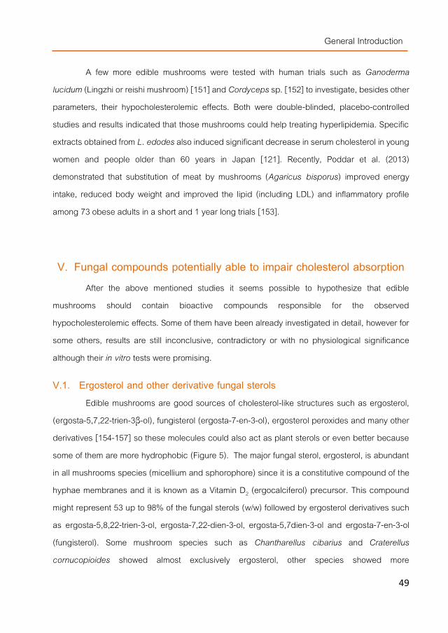
General Introduction
49
A few more edible mushrooms were tested with human trials such as Ganoderma lucidum (Lingzhi or reishi mushroom) [151] and Cordyceps sp. [152] to investigate, besides other parameters, their hypocholesterolemic effects. Both were double-blinded, placebo-controlled studies and results indicated that those mushrooms could help treating hyperlipidemia. Specific extracts obtained from L. edodes also induced significant decrease in serum cholesterol in young women and people older than 60 years in Japan [121]. Recently, Poddar et al. (2013) demonstrated that substitution of meat by mushrooms (Agaricus bisporus) improved energy intake, reduced body weight and improved the lipid (including LDL) and inflammatory profile among 73 obese adults in a short and 1 year long trials [153].
V. Fungal compounds potentially able to impair cholesterol absorption After the above mentioned studies it seems possible to hypothesize that edible
mushrooms should contain bioactive compounds responsible for the observed hypocholesterolemic effects. Some of them have been already investigated in detail, however for some others, results are still inconclusive, contradictory or with no physiological significance although their in vitro tests were promising.
V.1. Ergosterol and other derivative fungal sterols Edible mushrooms are good sources of cholesterol-like structures such as ergosterol,
(ergosta-5,7,22-trien-3β-ol), fungisterol (ergosta-7-en-3-ol), ergosterol peroxides and many other derivatives [154-157] so these molecules could also act as plant sterols or even better because some of them are more hydrophobic (Figure 5). The major fungal sterol, ergosterol, is abundant in all mushrooms species (micellium and sphorophore) since it is a constitutive compound of the hyphae membranes and it is known as a Vitamin D2 (ergocalciferol) precursor. This compound might represent 53 up to 98% of the fungal sterols (w/w) followed by ergosterol derivatives such as ergosta-5,8,22-trien-3-ol, ergosta-7,22-dien-3-ol, ergosta-5,7dien-3-ol and ergosta-7-en-3-ol (fungisterol). Some mushroom species such as Chantharellus cibarius and Craterellus cornucopioides showed almost exclusively ergosterol, other species showed more
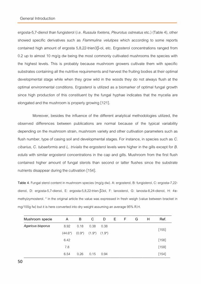
General Introduction
50
ergosta-5,7-dienol than fungisterol (i.e. Russula foetens, Pleurotus ostreatus etc.) (Table 4), other showed specific derivatives such as Flammulina velutipes which according to some reports contained high amount of ergosta 5,8,22-trien3β-ol, etc. Ergosterol concentrations ranged from 0.2 up to almost 10 mg/g dw being the most commonly cultivated mushrooms the species with the highest levels. This is probably because mushroom growers cultivate them with specific substrates containing all the nutritive requirements and harvest the fruiting bodies at their optimal developmental stage while when they grow wild in the woods they do not always flush at the optimal environmental conditions. Ergosterol is utilized as a biomarker of optimal fungal growth since high production of this constituent by the fungal hyphae indicates that the mycelia are elongated and the mushroom is properly growing [121].
Moreover, besides the influence of the different analytical methodologies utilized, the observed differences between publications are normal because of the typical variability depending on the mushroom strain, mushroom variety and other cultivation parameters such as flush number, type of casing soil and developmental stages. For instance, in species such as C. cibarius, C. tubaeformis and L. trivialis the ergosterol levels were higher in the gills except for B. edulis with similar ergosterol concentrations in the cap and gills. Mushroom from the first flush contained higher amount of fungal sterols than second or latter flushes since the substrate nutrients disappear during the cultivation [154].
Table 4. Fungal sterol content in mushroom species (mg/g dw). A: ergosterol, B: fungisterol, C: ergosta-7,22-
dienol, D: ergosta-5,7-dienol, E: ergosta-5,8,22-trien-β3ol, F: lanosterol, G: lanosta-8,24-dienol, H: 4α-
methylzymosterol. * in the original article the value was expressed in fresh weigh (value between bracket in
mg/100g fw) but it is here converted into dry weight assuming an average 95% R.H.
Mushroom specie A B C D E F G H Ref. Agaricus bisporus 8.92
(44.6*) 0.18 (0.9*)
0.38 (1.9*)
0.38 (1.9*)
[155]
6.42 [158] 7.8 [159]
6.54 0.26 0.15 0.94 [154]

General Introduction
51
Mushroom specie A B C D E F G H Ref. Amanita caesarea 8.25 1.75 [160] Boletus edulis
4.89 [154] 38.4
(192.2*) 3.28
(16.4*) 2.24
(11.2*) 2.5
(12.5*) [155]
4.00 [158] Calocybe gambosa 3.61 [158] Clitocybe maxima 6.42 [161] Clitocybe nebularis 6.38 1.04 [156] Catathelasma ventricosum
6.51 [161]
Cantharellus cibarius
3.04 [154] 4.94
(24.7*) 0.04 (0.2*)
0.08 (0.4*)
0.08 (0.4*)
[155]
0.23 [158] Cantharellus tubaeformis
1.73 [156] 3.36
(16.8*) 0.24 (1.2*)
0.36 (1.8*)
0.9 (4.5*)
[155]
3.77 [154] Craterellus cornucopioides
3.27 [161] 0.44 [158]
Coprinus atramentarius 2.45 7.55 [160] Flamulina velutipes 0.68 [159]
4.53 1.41 1.05 3.01 [160] Ganoderma lucidum 0.705 [162] Ganoderma sinense 0.0801 [162] Grifola frondosa
0.32 [158] 1.85
(9.25*) 0.204 (1.02*)
0.104 (0.52*)
0.01 (0.05*)
0.01
(0.05*) 0.011 (0.56*)
0.004 (0.02*)
[163]
Lactarius sangruifluus
3.6 (18*)
0.26 (1.32*)
0.19 (0.97*)
0.008 (0.04*)
0.008 (0.04*)
0.102 (0.51*)
0.008 (0.04*)
[163]

General Introduction
52
Mushroom specie A B C D E F G H Ref. Lactarius semisanguifluus
2.12 (10.58*)
0.26 (1.3*)
0.174 (0.87*)
0.012 (0.06*)
0.014 (0.07*)
0.11 (0.57*)
0.01 (0.05*)
[163]
Lactarius trivialis 2.96 [154] Lampteromyces japonicus
4.56 5.44 [160]
Laccaria amethystea 6.37 [161] Lentinula edodes 21.58
(107.9*) 1.82 (9.1*)
1.7 (8.5*)
1.4 (7.0*)
[155]
3.64 [158] 6.05 [159] 6.79 0.63 0.17 0.28 [154]
Leucopaxillus giganteus 8.59 0.73 [160] Hydnum repandum 6.28 0.85 [156] Hygrophorus marzuolus 6.81 [158] Hypsizygus marmoreus 10.56
(52.8*) [145]
Hygrocybe punicea 7.88 2.12 [160] Pleurotus ostreatus 12.14
(60.7*) 1.24 (6.2*)
0.64 (4.7*)
1.18 (5.9*)
[155]
3.31 [158] 4.4 [159] 6.74 0.18 0.15 0.83 [154] Pleurotus eryngii 9.1
(45.5*) [145]
Pleurotus cystidus
4.35 [159]
9.86 (49.3*)
[145]
Russula cyanoxantha 6.32 1.28 [156] Russula xerampelina 6.55 1.12 [156]

General Introduction
53
Mushroom specie A B C D E F G H Ref. Russula delica
2.5 (12.51*)
0.38 (1.90*)
0.266 (1.33*)
0.034 (0.17*)
0.048 (0.24*)
0.12 (0.61*)
0.014 (0.07*)
[163]
Russula foetens 7.09 2.91 [160] Russula senecis 8.45 [160] Russula nigricans 8.38 1.62 [160] Suillus granulatus 7.02 0.8 [156] Suillus luteus 6.62 0.83 [156] Suillus bellinii 2.46
(12.31*) 0.298 (1.49*)
0.14 (0.70*)
0.076 (0.38*)
0.094 (0.47*)
0.054 (0.27*)
0.01 (0.05*)
[163]
Stropharia rugoso-annulata
7.89 [161]
Empty boxes indicate non-detected compound.
Many of these compounds showed interesting bioactive properties such as antioxidant, antimicrobial or antitumor activities [157]. But, two of their biological activities can be particularly interesting because of their relation with the cholesterol metabolism such that the ability of ergosterol derivatives (ergost-22-ene-1,3-diol, ergosta-5,7-dien-3b-ol, (22E)-ergosta-1,4,6,22-tetraen-3-one, etc.) that were pointed as a potent agonists for LXR able to induce the expression of ABC transporters [107]. Moreover, ergosterol was a competitive inhibitor (showing Ki values = 36.8 µM) of the enzyme that catalyzes the reduction of the double bond at C-24 in the cholesterol-biosynthesis pathway (DHCR24, delta24-sterol reductase). Ergosterol together with other sterols such as stigmasterol and brassicasterol were efficient because of its double bond at C-22 in the side chain of their structure [164].
V.2. Dietary fibers (soluble polysaccharides, β-glucans and chitins) Edible mushrooms also contain interesting polysaccharides such as β-glucans (Figure
6). Their molecular structure is different than those present in cereals but they share many biological activities such as antioxidant, antitumoral and immunomodulatory activities including the capability of lowering serum cholesterol levels [110, 147, 165] and those are the reason for the detailed studies carried out on many particular fungal β-glucans such as lentinan from shiitake

General Introduction
54
mushrooms (Lentinula edodes), schizophyllan from Schizophyllum commune, grifolan from G. frondosa, and other from Agaricus blazei, G. lucidum, P. ostreatus as well as the (13)-β-glucans and glucuronoxylomannans from Auricularia polytricha and Tremella fuciformis [166, 167]. Mushroom β-glucans, despite characteristic residues and special branches, share a similar structure including a main chain of (13)-β-linked glucoses to which some side chains were connected by (16)-β-glicosidic linkages randomly or following a pattern with a DB range (degree of branching) approx. of 0.2-0.3. Obviously, the tertiary structure varies with the linkage degree because β-glucans with few or no β-(16)-linkages mainly have a single helix structure while those with higher degrees of β-(16)-glycosidic bonds form a triple helix [168]. Other reports indicated that triple helix structure together with the molecular mass positively affected the biological activity of the β-glucans [169]. However, the more polysaccharides are studied the least correlation between structure and functiona can be drawn [15].
Figure 6. Typical fungal β-glucans molecular structures (including chitin).
(1→3)-β-D-glucan
O
OH
OH
OH
OR
1 2
3
4
5
6O
OH
OH
OHR
O
O
O
1 2
3
4
5
6
(1→3),(1→6)-β-D-glucan
6
1 2
3
4
5
6
O
OH
OH
1 2
3
4
5
6
1 2
3
4
5 O
R
O
O O
OH
OH
ROH
O
O
OH
OH
O
OH
OH
1 2
3
4
5
6
O
OH
1 2
3
4
5 O
OH R
O
O
OH
OH
R
O
OH
OH
(1→6)-β-D-glucan
6
6
6
1 2
3
4
5
6
1 2
3
4
5 O
OH
O
O O
OH
OH
ROH
1 2
3
4
5 O
OH
R
O
O
OH
OH
O
OH
OH
(1→6),(1→3)-β-D-glucan
6
1 2
3
4
5 O
OH
OH
O
NH
CH3
O
R
OO
OHR
OH
NH
CH3O
1
23
4
5
6
N-acetylglucosamine
Chitin

General Introduction
55
The main problem with finding the correlations between structures and functions within the β-glucans are the different extraction procedures followed to obtain polysaccharides enriched extracts. For instance, traditional β-glucan extraction protocols included: hot water extraction, ethanol precipitation, alkali/acid treatments, enzymatic degradations, washing with Sevag or other reagents etc., yielding fractions with completely different composition difficult to characterize or quantify [170-172]. However, several publications indicated that β-glucan contents ranged from 0.14 to 0.53 mg/100 mg in similar strains such as Pleurotus pulmonarius, P. ostreatus, P. eryngii, B. edulis, A. aegerita and L. edodes. Other more recent publications estimated the total β-glucan levels between 2.6 - 13.4 mg/100mg dw for A. bisporus, F. Velutipes, L. edodes, P. ostreatus and P. eryngii using other analytical determinations [169, 173]. An example of these wide quantitative differences can be noticed in the Table 5.
A particular type of β-glucans which is lately getting interest is chitin and chitins derivatives such as the previously mentioned chitosans and their degradation products (low molecular weight polymers generated from chitin/chitosan hydrolysis (LMWC) or chitooligosaccharides). Chitin is a water-insoluble β-(14)-glucan of N-acetylglucosamine monomers and chitosan can be obtained from chitin by de-N-acetylation, both are considered as dietary fiber [122]. The immunomodulatory and antitumor properties seemed to be more related to the water soluble fraction including the oligomers and low molecular weight polymers generated from chitin hydrolysis (LMWC) [174] while their effect as prebiotic and hypocholesterolemic compounds might be due to their water-insoluble β-glucans (chitins and protein-bound glucans) [165] although as previously indicated for other type of β-glucans, results are largely different depending on report [124]. Some indicated that chitin itself was unable of lowering cholesterol levels in serum if it is not transformed into chitosan or degraded in smaller products [122]. β-Glucan enriched fractions obtained from P. ostreatus reduced total cholesterol levels of Winstar mice that were administrated simultaneously with a hypercholesterolemic diet for 4 weeks [175]. Thus, perhaps, the presence of other polysacharides (which are not chitins, could be proteoglucans) might enhance their hypocholesterolemic effect or stimulated certain synergy between fungal polysaccharides.

General Introduction
56
Chitin concentrations were more similar between different publications than those values described for β-glucans if the same or similar analytical methods were utilized (Table 5). Many authors determined chitin by the amount of N-glucosamine residues present in the samples [176] while others quantify them by the amount of non-protein nitrogen content detected with Keldar [173, 177, 178].
Table 5. Fungal β-glucan content in mushroom species (mg/100mg dw) according to different authors indicating those that were quantified as chitins (mg/100 mg dw). Empty boxes indicate non-detected compound.
Mushroom specie
β-glucan Chitin
mg/100mg dw Ref. mg/100mg dw Ref.
Agaricus bisporus 58.2 [179] 4.69 [169]
8.68-6.17 [176]
Agaricus blazei (or A. brasiliensis)
22.8 [179]
Pleurotus ostreatus 0.38-0.24 [170] 0.76 [169]
5.46-2.42 [176]
Pleurotus eryngii 0.38-0.29 [170] 3.16 [169]
Lentinula edodes 0.2 [170] 1.87 [169]
8.07-5.36 [176]
Flammulina veluttipes 3.2 [173] 9.83 [169]
Ganoderma lucidum 1.2 [179]
Grifola frondosa 33.5 [173]
Pholiota nameko 32.8 [173]
Phellinus linteus 21.8 [179]
Pleurotus pulmonarius 0.53 [170]
Panellus serotinus 19.6 [173]
Hypsizygus marmoreus 13.7 [173]
Pleurotus cornucopiae 7.5 [173]
Armillaria mellea 5.8 [173]

General Introduction
57
V.3. Inhibitors of the Pancreatic Lipase Inhibiting the pancreatic lipase (PL) during digestion can be considered as an indirect
mechanism of lowering cholesterol because if this enzyme cannot generate properly the DMMs, less lipids are absorbed as well as less cholesterol since this molecule should also be included in the micelles for enterocyte absorption.
A few different PL inhibitors were isolated from edible fungi, two of them were β-lactones with unusual configurations named percyquinin (obtained from Stereum complicatum) and vibralactone (Boreostereum vibrans) with similar IC50 (0.4 μg/mL) (Figure 7). Several publications and particularly Slanc et al [177, 180, 181] screened more than 60 edible and non-edible fungi species and found that some of them exhibited PL inhibitory activities ranging from 1% till 97% depending on the specie considered. However, only the report from Mizutani et al [135] managed to record PL inhibitory activity in vivo (for P. eryngii).
Figure 7. Vibralactone and percyquinin structures.
Other authors pointed to other compounds such as chitin oligosaccharides derivatives (and chitosan) as molecules involved not only in a direct inhibition of pancreatic lipase during the digestion process but also as modulators of the PPAR gene expression [182].
VI. Fungal compounds potentially able to inhibit cholesterol metabolism Actually, no functional food able to inhibit the cholesterol synthesis have been designed,
there are only drugs prescribed for people with cholesterol levels in serum reaching pathological
O
O
H
CH3
CH3
OH
O
O
CH3
CH3
OH

General Introduction
58
levels. However, it seems that edible mushrooms might contain several types of compounds able to perform such a task via different mechanisms.
VI.1. Inhibitors of the HMGCR and other enzymes involved in the cholesterol synthesis
According to several authors, oyster mushrooms species such as P. ostreatus, P. sapidus, P. eryngii and P. cornucopiae, contain mevinolin (lovastatin) (Figure 8) [183-185]. In lower quantities, statins were also described in other species such as A. bisporus, B. edulis, Clitocybe maxima, Hipsizigus marmoreus etc. [136, 185]. The statin levels were high in the spores and widely distributed through the complete fruiting body and they were present during all the developmental stages [183]. Statins or vastatins were in fact isolated for the first time from fungi by the pharmaceutical industry. However, other authors found no statins [139, 150] but other compounds such as lanosteroids, ganoderols etc. in G. lucidum able to reduce the mRNA expression of the HMGCoA reducase.
Figure 8. Mevinolin structure.
Inhibitors of this key enzyme (i.e. synthetic statins) are well tolerated, having little collateral effects. However, when this enzyme is blocked, other important metabolic pathways are affected such as ubiquinone, dolichol and isoprenylated protein synthesis [186, 187]. Thus, new hypocholesterolemic drugs are at the present being developed to inhibit other enzymes down-stream in the cholesterol biosynthetic pathway such as the squalene synthase. This enzyme also named farnesyl-diphosphate:farnesyl-diphosphate farnesyl transferase catalyzes the reductive dimerization of two molecules of farnesyl pyrophosphate to form squalene at the final branch point

General Introduction
59
of the cholesterol biosynthetic pathway (Figure 3). Inhibition of squalene synthase could therefore avoid any potential adverse effect associated with reduced synthesis of isoprenylated metabolites by the inhibitors of HMG-CoA reductase.
Only a few inhibitors of this enzyme have been studied in detail, one is the synthetic RPR 107393 compound (3-hydroxy-3-[4-(quinolin-6-yl)phenyl]-1-azabicyclo [2-2-2]octane dihydrochloride) [188] but others are called zaragozic acids and they are obtained from the liquid broth of certain ascomicetes able to form macroscopic fruiting bodies [189]. These types of compounds might also be found in the basidiomycota family where most of the macrofungi are included (together with the ascomycota).
Moreover and as previously indicated, ergosterol was able to inhibit another enzyme related with the cholesterol metabolic pathway such as the C24-reductase, the enzyme that catalyzes the reduction of the double bond at C-24 (Figure 3) [164].
Bobek et al., (1998) also suggested that the reduction in the cholesterol biosynthesis found in rats was not only due to the suppression of the hepatic HMGCoA reducase activity but also by accelerating the cholesterol catabolism by up-regulating hepatic cholesterol 7α-hydroxylase [128, 131].
VI.2. Modulators of the phospholipid metabolism Lentinula edodes was able to induce hypocholesterolemia and reduce the triacylglycerol
levels in rats due to its content in eritadenine (2(R), 3(R),-dihydroxy-4-(9-adenyl)-butyric acid, lentinacin or lentysine) a specific compound with S-adenosylhomocysteine hydrolase (SAHH) inhibitory activity able to modify the hepatic phospholipid metabolism (Figure 9) [190].
Figure 9. Eritadenine molecule.

General Introduction
60
Rats supplemented with eritadenite significantly decreased the phosphatidylcholine: phosphatidylethanolamine ratio in liver microsomes and the S-adenosylmethionine:S-adenosylhomocysteine ratio in the liver in addition to the plasma cholesterol concentration suggesting that the hypocholesterolemic action of this compound might be associated with a modification of hepatic phospholipid metabolism [191]. Moreover, eritadenine also decreased in a dose-dependent manner levels of VLDL, LDL and HDL, increased the hepatic S-adenosylhomocysteine (SAH) level and decreased the ratio of S-adenosylmethionine (SAM) to SAH indicating that the hypocholesterolemic action of eritadenine might be elicited through an alteration of the hepatic phospholipid metabolism that resulted from an inhibition of phosphatidylethanolamine N-methylalion due to a decreased SMA/SAH ratio in the liver [191].
VI.3. Modulators of the thyroid hormones metabolism Another important factor in the cholesterol regulation is via thyroid. Triiodothyronine (T3)
is a thyroid hormone directly involved in the regulation of LDLR gene expression. A 20% of this hormone is secreted directly by thyroid but the major amount is produced from thyroxine (T4) (produced exclusively in the thyroid) by 5´-deiodination in peripheral tissues. This reaction is catalyzed by the selenoprotein, type-I 5´-iodothyronine deiodinasein (DIO1 and 2). Other related selenoprotein is the glutathione peroxidase (GSH-Px) since the reduced expression of GSH-Px has been shown to increase cell-mediated oxidation of LDL [192].
Mushrooms contain selenium and can be easily Se-fortified by addition of sodium selenite to their cultivation substrates. This supplementation can modify their biological properties [193]. Moreover, selenium supplementation combined with statins therapy was proved to be significantly beneficial to lipid therapy. The observed upregulation of LDLR expression by selenium supplementation might have offered more receptor sites for LDL binding and its clearance from the circulation. One ppm Se supplementation on rats decreased the total cholesterol and LDL levels, increased T3 levels due to increased DIO2 expression, decreased the apoB and HMG-CoA reductase mRNAs expression [192].Thus, it could be assumed that if an statin-producer mushroom strains could be Se-fortified during its cultivation, it might enhance their

General Introduction
61
hypocholesterolemic effect. However, up to the date, no studies about this posibility have been carried out yet.
VII. The use of pressurized technologies to obtain bioactive fungal extracts
In the past, most of the potential bioactive compounds described for edible mushrooms were obtained by traditional extraction systems or simple solid-liquid extraction using cold or hot water or others such as chloroform, hexane, methanol, ethanol or their mixtures. However, the use of the organic solvents to extract non-polar compounds can be toxic for humans and dangerous for the environment. Moreover, the extraction conditions and solvent removing from obtained fraction could be laborious and expensive. In consequence, environmentally friendly technologies have been optimized to extract bioactive fractions from mushrooms and many other sources ensuring that there are safer for human consumption (food grade labeling) than most of the standardized methods.
VII.1. Pressurized liquid extraction Pressurized solvent extraction (PLE) is an extraction technology utilized in the food
industry to obtain bioactive fractions from solid or semi-solid material using nontoxic and/or GRAS (generally recognized as safe) solvents such as ethanol, limonene, lactic acid, glycerine or their mixtures. A particular PLE method named SWE (subcritical water extraction) is also utilized for the extraction of bioactive compounds using water as solvent under specific combinations of high pressures and temperatures below supercritical conditions [194]. Despite the obvious ecological interest of PLE technology, it is applied in the food industry because of the technological advantages described bellow [195-198]:
- The use of elevated temperatures (50 °C to 200 °C) increases the kinetics of extraction process and consequently decreases the extraction time making possible a high solubility and diffusion rates.
- Solvent polarity can be modulated since temperature is involved in its dielectric constant. At higher temperatures the dielectric constant decreases, consequently the solvent

General Introduction
62
polarity is reduced. Thus, higher temperatures could improve the extraction capacities of non-toxic solvents.
- The high pressures applied (such as 10.34 MPa) allows a quick extraction vessel filling and force liquid into the solid matrix.
- High pressures-temperatures combinations enables maintenance of the solvent liquid state preserving it extraction properties. Thus, this technology allows a fast extraction of a wide variety of compounds from
different nature by the use of less amounts of solvent and obtaining higher yield rates in comparison with traditional solid-liquid extractions. Moreover, PLE is automated and the sample remains in an oxygen and light free environment avoiding oxidation and polymerization processes (Figure 10).
Figure 10. Example of pressurized liquid extraction (ASE) equipment.
At the present, only a few studies using PLE technology to obtaining fractions enriched in potential bioactive compunds from mushrooms have been reported. PLE was used to obtain certain fatty acids from Cordyceps spp. [199]. or polysaccharides from several fungal species [200-203]. High content β-glucans extracts could be obtained from mushrooms such as G. lucidum and B. edulis using water as extraction solvent and 200 ºC, 5 cycles of 5 min each at 10.3 MPa. The crude polysaccharide (PSC) fractions, isolated from the PWE extracts contained

General Introduction
63
mainly β-glucans (including N-glucosamine-containing compounds deriving from chitin hydrolysis), β-glucans and other PSCs (hetero-/proteo-glucans) depending on the extraction temperature and mushroom strain considered [201, 204]. The same solvent submitted to 10.1 MPa for 70 min at 28 °C was also used to isolate α-(14) and β-(16) glucans mixture from L. edodes [203].
VII.2. Supercritical fluid extraction Supercritical fluids extraction (SFE) is another pressurized extraction technology also
utilized for the extraction or removal of specific fractions of interest for the food industry [205] (Figures 11,12). It is based on the transition that suffer a substance when it is submitted to a temperature and pressure above its critical point. At this state, its liquid and gas status are identical since it is turned into a supercritical fluid (SF). Thus, a supercritical fluid maintain both liquid and gas specific properties such as liquid density and gas viscosity [196].
The advantages of this technology are many such as high selectivity (higher even than PLE), shorter extraction times of than those neccesary for solid-liquid extractions and the use of non-toxic organic solvents [196]. Many compounds are brought to the supercritical state in order to extract specific compounds from food matrix such as methane or nitrogen but, carbone dioxide (CO2) is more frequently used because it is considered more safe and cheap despite nonexplosive, nontoxic, inexpressive and ease to be removed from the obtained fraction [206]. Moreover, CO2 is considered GRAS by FDA (Food and Drug Administration) and EFSA (European Food Safety Authority) and with the ability to extract lipophilic compounds, although for polar molecules is not so effective. The CO2 extraction capacity can be increased to improve the poor solubility of polar compounds in this fluid by combine this solvent with small quantities of others. Then, the selectivity of the extraction process is also improved with the named co-solvent effect [207]. Specific percentages of hexane, methanol, isopropanol, acetonitrile or ethanol are widely used as co-solvents/modifiers in combination with CO2 but, the latter solvent is specifically recommended by many authors for its lower toxicity, high CO2miscibility [208] and low environmental impact [209]

General Introduction
64
Studies using SF for the extraction of specific fungal metabolites are limited. This advanced technology was utilized to obtain triterpenoids from G. lucidum, carboxylic and fatty acids from Agaricus spp., antioxidant and antimicrobial compounds from the fruiting bodies of L. edodes and A. brasiliensis, ergothineine and polyphenols from P. ostreatus, antitumoral fractions from Cordyceps sinensis mycelia and immune-modulators polysaccharides from G. lucidum [206, 210-216].
Figure 11. Example of supercritical fluid extraction equipment.
Figure 12. SFE equipment details. CO2 tank and extraction cell even collecting cells (separators) details from left to right.

General Introduction
65
VIII. References [1]. Araújo, F., et al., Trends in cardiovascular diseases and cancer mortality in 45 countries from five continents
(1980–2010). European Journal of Preventive Cardiology, 2013. [2]. Uemura, K. and Z. Pisa, Trend in cardiovascular disease mortality in industrialized countries since 1950. World
Health Statistics Quarterly, 1988. 41: p. 155-178. [3]. Graham, I., European guidelines on cardiovascular disease prevention in clinical practice: Executive summary.
Atherosclerosis, 2007. 194(1): p. 1-45. [4]. Chen, J. and X.-F. Huang, The effects of diets enriched in beta-glucans on blood lipoprotein concentrations.
Journal of Clinical Lipidology, 2009. 3(3): p. 154-158. [5]. EFSA Panel on Dietetic Products, N.a.A.N., Scientific Opinion on the substantiation of a health claim related to 3
g/day plant sterols/stanols and lowering blood LDL-cholesterol and reduced risk of (coronary) heart disease pursuant to Article 19 of Regulation (EC) No 1924/2006. EFSA Journal 2012. 10(5): p. 2693-2708.
[6]. EFSA Panel on Dietetic Products, N.a.A.N., Scientific Opinion on the substantiation of a health claim related to oat beta glucan and lowering blood cholesterol and reduced risk of (coronary) heart disease pursuant to Article 14 of Regulation (EC) No 1924/2006. EFSA Journal 2010. 8(12): p. 1885-1900.
[7]. EFSA Panel on Dietetic Products, N.a.A.N., Scientific Opinion on the substantiation of a health claim related to barley beta-glucans and lowering of blood cholesterol and reduced risk of (coronary) heart disease pursuant to Article 14 of Regulation (EC). No 1924/2006. EFSA Journal 2011. 9(12): p. 2471-2484.
[8]. Rozner, S. and N. Garti, The activity and absorption relationship of cholesterol and phytosterols. Colloids and Surfaces A: Physicochemical and Engineering Aspects, 2006. 282–283(0): p. 435-456.
[9]. Sudhop, T., D. Lütjohann, and K. von Bergmann, Sterol transporters: targets of natural sterols and new lipid lowering drugs. Pharmacology & Therapeutics, 2005. 105(3): p. 333-341.
[10]. Kruit, J.K., et al., Emerging roles of the intestine in control of cholesterol metabolism. World Journal of Gastroenterology, 2006. 12(40): p. 6429-6439.
[11]. van der Wulp, M.Y.M., H.J. Verkade, and A.K. Groen, Regulation of cholesterol homeostasis. Molecular and Cellular Endocrinology, 2013. 368(1–2): p. 1-16.
[12]. Wilson, M.D. and L.L. Rudel, Review of cholesterol absorption with emphasis on dietary and biliary cholesterol. Journal of Lipid Research, 1994. 35(6): p. 943-55.
[13]. Ginter PhD, D.E., Cardiovascular disease prevention in eastern Europe. Nutrition, 1998. 14(5): p. 452-457. [14]. Porter, C.J.H., et al., Enhancing intestinal drug solubilisation using lipid-based delivery systems. Advanced Drug
Delivery Reviews, 2008. 60(6): p. 673-691. [15]. Gil-Ramírez, A. and C. Soler-Rivas, The use of edible mushroom extracts as bioactive ingredients to design novel
functional foods with hypocholesterolemic activities, in Mushrooms: Cultivation, Antioxidant Properties and Health Benefits, G. Pesti, Editor. 2014, Nova Science Publishers: New York. p. 43-73.
[16]. Chiang, J.Y., Bile acids: regulation of synthesis. J Lipid Res, 2009. 50(10): p. 1955-66.

General Introduction
66
[17]. Geyer, J., T. Wilke, and E. Petzinger, The solute carrier family SLC10: more than a family of bile acid transporters regarding function and phylogenetic relationships. Naunyn Schmiedebergs Arch Pharmacol, 2006. 372(6): p. 413-31.
[18]. Silverstein, R.L. and M. Febbraio, CD36, a scavenger receptor involved in immunity, metabolism, angiogenesis, and behavior. Sci Signal, 2009. 2(72).
[19]. Dikkers, A. and U.J. Tietge, Biliary cholesterol secretion: more than a simple ABC. World Journal of Gastroenterology, 2010. 16: p. 5936-5945.
[20]. van Bennekum, A., et al., Class B scavenger receptor-mediated intestinal absorption of dietary beta-carotene and cholesterol. Biochemistry, 2005. 44(11): p. 4517-25.
[21]. Turley, S.D. and J.M. Dietschy, Sterol absorption by the small intestine. Curr Opin Lipidol, 2003. 14(3): p. 233-40. [22]. Jia, L., J.L. Betters, and L. Yu, Niemann-pick C1-like 1 (NPC1L1) protein in intestinal and hepatic cholesterol
transport. Annu Rev Physiol, 2011. 73: p. 239-59. [23]. Davis, H.R., Jr., et al., Niemann-Pick C1 Like 1 (NPC1L1) is the intestinal phytosterol and cholesterol transporter
and a key modulator of whole-body cholesterol homeostasis. J Biol Chem, 2004. 279(32): p. 33586-92. [24]. Yu, L., et al., Cholesterol-regulated translocation of NPC1L1 to the cell surface facilitates free cholesterol uptake.
J Biol Chem, 2006. 281(10): p. 6616-24. [25]. Sharpe, L.J., et al., The UPS and downs of cholesterol homeostasis. Trends in Biochemical Sciences, 2014.
39(11): p. 527-535. [26]. Calpe-Berdiel, L., J.C. Escolà-Gil, and F. Blanco-Vaca, New insights into the molecular actions of plant sterols
and stanols in cholesterol metabolism. Atherosclerosis, 2009. 203(1): p. 18-31. [27]. Kusuhara, H. and Y. Sugiyama, ATP-binding cassette, subfamily G (ABCG family). Pflugers Arch, 2007. 453(5):
p. 735-44. [28]. Yu, L., et al., Overexpression of ABCG5 and ABCG8 promotes biliary cholesterol secretion and reduces fractional
absorption of dietary cholesterol. J Clin Invest, 2002. 110(5): p. 671-80. [29]. Berge, K.E., et al., Accumulation of dietary cholesterol in sitosterolemia caused by mutations in adjacent ABC
transporters. Science, 2000. 290(5497): p. 1771-1775. [30]. Cummins, C.I. and D.J. Hangelsdorf, Liver X receptors and cholesterol homeostasis: spotlight on the adrenal
gland. Biocheistry Society Transcations, 2006. 34: p. 1110-1113. [31]. Repa, J.J., et al., Regulation of ATP-binding cassette sterol transporters ABCG5 and ABCG8 by the liver X
receptors alpha and beta. J Biol Chem, 2002. 277(21): p. 18793-800. [32]. Acetyl-CoA Acetyltransferase 1 in The GeneCards Human. Weizmann Institute of Science, Rehovot, Israel.
http://www.genecards.org/cgi-bin/carddisp.pl?gene=ACAT1 16/10/2014. [33]. Acetyl-CoA Acetyltransferase 2, The GeneCards Human. Weizmann Institute of Science, Rehovot, Israel.
http://www.genecards.org/cgi-bin/carddisp.pl?gene=ACAT2 16/10/2014. [34]. Sterol O-Acyltransferase 1, The GeneCards Human. Weizmann Institute of Science, Rehovot, Israel.
http://www.genecards.org/cgi-bin/carddisp.pl?gene=SOAT1 16/10/2014

General Introduction
67
[35]. Sterol O-Acyltransferase 2, The GeneCards Human. Weizmann Institute of Science, Rehovot, Israel. http://www.genecards.org/cgi-bin/carddisp.pl?gene=SOAT2 16/10/2014
[36]. Chang, T.-Y., et al., Acyl-coenzyme A:cholesterol acyltransferases. 2009. 297(1): p. E1-E9. [37]. Tabas, I., Cholesterol in health and disease. Journal of Clinical Investigation, 2002. 110(5): p. 583-590. [38]. Hussain, M.M., J. Shi, and P. Dreizen, Microsomal triglyceride transfer protein and its role in apoB-lipoprotein
assembly. Journal of Lipid Research, 2003. 44(1): p. 22-32. [39]. Sahebkar, A., G.T. Chew, and G.F. Watts, Recent advances in pharmacotherapy for hypertriglyceridemia.
Progress in Lipid Research, 2014(0). [40]. Mahmood Hussain, M., A proposed model for the assembly of chylomicrons. Atherosclerosis, 2000. 148(1): p. 1-
15. [41]. Lammert, F. and D.Q.H. Wang, New insights into the genetic regulation of intestinal cholesterol absorption.
Gastroenterology, 2005. 129(2): p. 718-734. [42]. Plana, N., Peroxidacion lipidica y factores de riesgo cardiovascular, in Departamento de Medicina y Cirujia de la
Facultad de Medicina de la Universidad de Roivira I Virgili. 1993, Universidad Roivira y Vigili: Reus. [43]. Gill, S., et al., Cholesterol-dependent degradation of squalene monooxygenase, a control point in cholesterol
synthesis beyond HMG-CoA reductase. Cell Metabolism, 2011. 13(3): p. 260-273. [44]. Laden, B.P. and T.D. Porter, Resveratrol inhibits human squalene monooxygenase. Nutrition Research, 2001.
21(5): p. 747-753. [45]. Abe, I., et al., Green tea polyphenols: Novel and potent inhibitors of squalene epoxidase. Biochemical and
Biophysical Research Communications, 2000. 268(3): p. 767-771. [46]. Hulce, J.J., et al., Proteome-wide mapping of cholesterol-interacting proteins in mammalian cells. Nat Meth, 2013.
10(3): p. 259-264. [47]. Burg, J.S. and P.J. Espenshade, Regulation of HMG-CoA reductase in mammals and yeast. Progress in Lipid
Research, 2011. 50(4): p. 403-410. [48]. 3-Hydroxy-3-Methylglutaryl-CoA Reductase, The GeneCards Human. Weizmann Institute of Science, Rehovot,
Israel. http://www.genecards.org/cgi-bin/carddisp.pl?gene=HMGCR. 15/10/2014. [49]. Ikonen, E., Mechanisms for cellular cholesterol transport: Defects and human disease. Vol. 86. 2006. 1237-1261.
[50]. Derewiaka, D. and E. Molińska, Cholesterol transformations during heat treatment. Food Chemistry, 2015. 171(0): p. 233-240.
[51]. Lee, J.P., et al., The TRC8 ubiquitin ligaseis sterol regulated and Iinteracts with lipid and protein biosynthetic pathways. Molecular Cancer Research, 2010. 8(1): p. 93-106.
[52]. Norlin, M. and K. Wikvall, Enzymes in the conversion of cholesterol into bile acids. Curr Mol Med, 2007. 7(2): p. 199-218.
[53]. Temel, R.E., et al., Hepatic Niemann-Pick C1–like 1 regulates biliary cholesterol concentration and is a target of ezetimibe. The Journal of Clinical Investigation, 2007. 117(7): p. 1968-1978.
General Introduction
68
[54]. van der Velde, A.E., et al., Direct intestinal cholesterol secretion contributes significantly to total fecal neutral sterol excretion in mice. Gastroenterology, 2007. 133(3): p. 967-975.
[55]. Tang, W., et al., Ezetimibe restores biliary cholesterol excretion in mice expressing Niemann–Pick C1-Like 1 only in liver. Biochimica et Biophysica Acta (BBA) - Molecular and Cell Biology of Lipids, 2011. 1811(9): p. 549-555.
[56]. Brufau, G., A.K. Groen, and F. Kuipers, Reverse cholesterol transport revisited: Contribution of biliary versus intestinal cholesterol excretion. Arteriosclerosis, Thrombosis, and Vascular Biology, 2011. 31(8): p. 1726-1733.
[57]. Temel, R.E. and J. Mark Brown, A new framework for reverse cholesterol transport: Non-biliary contributions to reverse cholesterol transport. World Journal of Gastroenterology, 2010. 16(47): p. 5946-5952.
[58]. Bura, K.S., et al., Intestinal SR-BI does not impact cholesterol absorption or transintestinal cholesterol efflux in mice. Journal of Lipid Research, 2013. 54(6): p. 1567-1577.
[59]. de Boer, J.F., et al., Inhibition of NPC1L1 increases transintestinal cholesterol excretion (tice) dependent on Abcg5/g8 but independent of plasma Apob-containing lipoproteins. Atherosclerosis, 2014. 235(2): p. e47.
[60]. Danielsen, E.M., et al., Apolipoprotein A-1 (apoA-1) deposition in, and release from, the enterocyte brush border: A possible role in transintestinal cholesterol efflux (TICE)? Biochimica et Biophysica Acta (BBA) - Biomembranes, 2012. 1818(3): p. 530-536.
[61]. Vrins, C.L.J., From blood to gut: Direct secretion of cholesterol via transintestinal cholesterol efflux. World Journal of Gastroenterology, 2010. 16(47): p. 5953-5957.
[62]. van der Velde, A.E., G. Brufau, and A.K. Groen, Transintestinal cholesterol efflux. Current Opinion in Lipidology, 2010. 21(3): p. 167-171 10.1097/MOL.0b013e3283395e45.
[63]. Yun, J.W., Possible anti-obesity therapeutics from nature – A review. Phytochemistry, 2010. 71(14–15): p. 1625-1641.
[64]. Jeong, J.Y., et al., Optimization of pancreatic lipase inhibition by Cudrania tricuspidata fruits using response surface methodology. Bioorganic & Medicinal Chemistry Letters, 2014. 24(10): p. 2329-2333.
[65]. Liu, S., et al., Inhibition of pancreatic lipase, α-glucosidase, α-amylase, and hypolipidemic effects of the total flavonoids from Nelumbo nucifera leaves. Journal of Ethnopharmacology, 2013. 149(1): p. 263-269.
[66]. Xu, N., U. Ekstrom, and P. Nilsson-Ehle, ACTH decreases the expression and secretion of apolipoprotein B in HepG2 cell cultures. J Biol Chem, 2001. 276(42): p. 38680-4.
[67]. Cuchel, M., et al., Inhibition of microsomal triglyceride transfer protein in familial hypercholesterolemia. N Engl J Med, 2007. 356(2): p. 148-56.
[68]. Chen, H.C. and R.V. Farese, Jr., Inhibition of triglyceride synthesis as a treatment strategy for obesity: lessons from DGAT1-deficient mice. Arterioscler Thromb Vasc Biol, 2005. 25(3): p. 482-6.
[69]. Karimian Pour, N. and K. Adeli, Insulin silences Apolipoprotein B mRNA translation by inducing intracellular traffic into cytoplasmic RNA granules. Biochemistry, 2011. 50(32): p. 6942-6950.
[70]. Cohen, D.E. and M.C. Carey, Rapid (1 hour) high-performance gel-filtration chromatography resolves coexisting simple micelles, mixed micelles and vesicles in bile. Journal of Lipid Research, 1990. 31(11): p. 2103-2112.

General Introduction
69
[71]. Temel, R.E., et al., Intestinal cholesterol absorption is substantially reduced in mice deficient in both ABCA1 and ACAT2. J Lipid Res, 2005. 46(11): p. 2423-31.
[72]. Kluge, A.F. and R.C. Petter, Acylating drugs: redesigning natural covalent inhibitors. Current Opinion in Chemical Biology, 2010. 14(3): p. 421-427.
[73]. Altmann, S.W., et al., Niemann-Pick C1 Like 1 protein is critical for intestinal cholesterol absorption. Science, 2004. 303(5661): p. 1201-1204.
[74]. Garcia-Calvo, M., et al., The target of ezetimibe is Niemann-Pick C1-Like 1 (NPC1L1). Proc Natl Acad Sci U S A, 2005. 102(23): p. 8132-7.
[75]. Ge, L., et al., The cholesterol absorption inhibitor Ezetimibe acts by blocking the sterol-induced internalization of NPC1L1. Cell Metabolism, 2008. 7(6): p. 508-519.
[76]. Davis, H.R. and E.P. Veltri, Zetia: inhibition of Niemann-Pick C1 Like 1 (NPC1L1) to reduce intestinal cholesterol absorption and treat hyperlipidemia. J Atheroscler Thromb, 2007. 14(3): p. 99-108.
[77]. Sugizaki, T., et al., The Niemann-Pick C1 like 1 (NPC1L1) inhibitor ezetimibe improves metabolic disease via decreased liver X receptor (LXR) activity in liver of obese male mice. Endocrinology, 2014. 155(8): p. 2810-9.
[78]. Arya, N., et al., Some molecular targets for antihyperlipidemic drug research. European Journal of Medicinal Chemistry, 2014. 85(0): p. 535-568.
[79]. Dražić, T., et al., Novel amino-β-lactam derivatives as potent cholesterol absorption inhibitors. European Journal of Medicinal Chemistry, 2014. 87(0): p. 722-734.
[80]. Yu, X.-H., et al., ABCG5/ABCG8 in cholesterol excretion and atherosclerosis. Clinica Chimica Acta, 2014. 428(0): p. 82-88.
[81]. Dieter, M.Z., et al., Expression and regulation of the sterol half-transporter genes ABCG5 and ABCG8 in rats. Comparative Biochemistry and Physiology Part C: Toxicology & Pharmacology, 2004. 139(4): p. 209-218.
[82]. Kourounakis, A.P., A.N. Matralis, and A. Nikitakis, Design of more potent squalene synthase inhibitors with multiple activities. Bioorganic & Medicinal Chemistry, 2010. 18(21): p. 7402-7412.
[83]. An, S., et al., Human ACAT inhibitory effects of shikonin derivatives from Lithospermum erythrorhizon. Bioorganic & Medicinal Chemistry Letters, 2007. 17(4): p. 1112-1116.
[84]. Choi, J.H., et al., Glabrol, an acyl-coenzyme A: Cholesterol acyltransferase inhibitor from licorice roots. Journal of Ethnopharmacology, 2007. 110(3): p. 563-566.
[85]. Jia, Y., et al., Ursolic acid is a PPAR-α agonist that regulates hepatic lipid metabolism. Bioorganic and Medicinal Chemistry Letters, 2011. 21(19): p. 5876-5880.
[86]. Woo, M.N., et al., Fucoxanthin supplementation improves plasma and hepatic lipid metabolism and blood glucose concentration in high-fat fed C57BL/6N mice. Chemico-Biological Interactions, 2010. 186(3): p. 316-322.
[87]. Taghibiglou, C., et al., Intracellular mechanisms mediating the inhibition of apoB-containing lipoprotein synthesis and secretion in HepG2 cells by avasimibe (CI-1011), a novel acyl-coenzyme A: cholesterol acyltransferase (ACAT) inhibitor. Biochemical Pharmacology, 2002. 63(3): p. 349-360.

General Introduction
70
[88]. Hogue, J.-C., et al., Differential effect of fenofibrate and atorvastatin on in vivo kinetics of apolipoproteins B-100 and B-48 in subjects with type 2 diabetes mellitus with marked hypertriglyceridemia. Metabolism, 2008. 57(2): p. 246-254.
[89]. Skoog, M., et al., ACTH reduces the rise in ApoB-48 levels after fat intake. Atherosclerosis, 2007. 191(2): p. 433-439.
[90]. Nieto-Ramirez, I.J., et al. (2013) Las estatinas: química, técnicas analíticas, biosíntesis y farmacocinética. Vitae 20, 49-63.
[91]. Singh, D.K., S. Banerjee, and T.D. Porter, Green and black tea extracts inhibit HMG-CoA reductase and activate AMP kinase to decrease cholesterol synthesis in hepatoma cells. The Journal of Nutritional Biochemistry, 2009. 20(10): p. 816-822.
[92]. Zang, M., et al., Polyphenols stimulate AMP-activated protein kinase, lower lipids, and inhibit accelerated atherosclerosis in diabetic LDL receptor-deficient mice. Diabetes, 2006. 55(8): p. 2180-91.
[93]. Tavridou, A., et al., Pharmacological characterization in vitro of EP2306 and EP2302, potent inhibitors of squalene synthase and lipid biosynthesis. European Journal of Pharmacology, 2006. 535(1–3): p. 34-42.
[94]. Zerenturk, E.J., et al., The endogenous regulator 24(S),25-epoxycholesterol inhibits cholesterol synthesis at DHCR24 (Seladin-1). Biochimica et Biophysica Acta (BBA) - Molecular and Cell Biology of Lipids, 2012. 1821(9): p. 1269-1277.
[95]. Ling, W.H. and P.J.H. Jones, Dietary phytosterols: A review of metabolism, benefits and side effects. Life Sciences, 1995. 57(3): p. 195-206.
[96]. Thurnham, D.I., Functional foods: cholesterol-lowering benefits of plant sterols. Br J Nutr, 1999. 82(4): p. 255-6. [97]. Miettinen, T.A., et al., Reduction of serum cholesterol with sitostanol-ester margarine in a mildly
hypercholesterolemic population. New England Journal of Medicine, 1995. 333(20): p. 1308-1312. [98]. Heinemann, T., O. Leiss, and K. von Bergmann, Effect of low-dose sitostanol on serum cholesterol in patients with
hypercholesterolemia. Atherosclerosis, 1986. 61(3): p. 219-223. [99]. Wong, A., Chemical and microbiological considerations of phytosterols and their relative efficacies in functional
foods for the lowering of serum cholesterol levels in humans: A review. Journal of Functional Foods, 2014. 6(0): p. 60-72.
[100]. Jonnala, R.S., N.T. Dunford, and K.E. Dashiell, Tocopherol, phytosterol and phospholipid compositions of new high oleic peanut cultivars. Journal of Food Composition and Analysis, 2006. 19(6–7): p. 601-605.
[101]. Martins, C.M., et al., Common sources and composition of phytosterols and their estimated intake by the population in the city of São Paulo, Brazil. Nutrition, 2013. 29(6): p. 865-871.
[102]. Chirinos, R., et al., Sacha inchi (Plukenetia volubilis): A seed source of polyunsaturated fatty acids, tocopherols, phytosterols, phenolic compounds and antioxidant capacity. Food Chemistry, 2013. 141(3): p. 1732-1739.
[103]. Trautwein, E.A., et al., Proposed mechanisms of cholesterol-lowering action of plant sterols. European Journal of Lipid Science and Technology, 2003. 105(3-4): p. 171-185.

General Introduction
71
[104]. Mel’nikov, S.M., J.W.M. Seijen ten Hoorn, and A.P.A.M. Eijkelenboom, Effect of phytosterols and phytostanols on the solubilization of cholesterol by dietary mixed micelles: an in vitro study. Chemistry and Physics of Lipids, 2004. 127(2): p. 121-141.
[105]. Matsuoka, K., et al., Competitive solubilization of cholesterol and β-sitosterol with changing biliary lipid compositions in model intestinal solution. Chemistry and Physics of Lipids, 2012. 165(1): p. 7-14.
[106]. Matsuoka, K., et al., NMR study on solubilization of sterols and aromatic compounds in sodium taurodeoxycholate micelles. Bulletin of the Chemical Society of Japan, 2007. 80(12): p. 2334-2341.
[107]. Kaneko, E., et al., Induction of intestinal ATP-binding cassette transporters by a phytosterol-derived liver X receptor agonist. J Biol Chem, 2003. 278(38): p. 36091-8.
[108]. García-Llatas, G. and M.T. Rodríguez-Estrada, Current and new insights on phytosterol oxides in plant sterol-enriched food. Chemistry and Physics of Lipids, 2011. 164(6): p. 607-624.
[109]. Nguyen, L.B., et al., Deficient ileal 3-hydroxy-3-methylglutaryl coenzyme A reductase activity in sitosterolemia: Sitosterol is not a feedback inhibitor of intestinal cholesterol biosynthesis. Metabolism, 1994. 43(7): p. 855-859.
[110]. Schepetkin, I.A. and M.T. Quinn, Botanical polysaccharides: Macrophage immunomodulation and therapeutic potential. International Immunopharmacology, 2006. 6(3): p. 317-333.
[111]. Havrlentová, M., et al., Cereal β-glucans and their significance for the preparation of functional foods - a review. Czech Journal of Food Sciences, 2011. 29(1): p. 1-14.
[112]. Jeon, Y.-J., F. Shahidi, and S.-K. Kim, Preparation of chitin and chitosa oligomers and their applications in physiological functional foods. Food Reviews International, 2000. 16(2): p. 159-176.
[113]. Kaczmarczyk, M.M., M.J. Miller, and G.G. Freund, The health benefits of dietary fiber: Beyond the usual suspects of type 2 diabetes mellitus, cardiovascular disease and colon cancer. Metabolism, 2012. 61(8): p. 1058-1066.
[114]. Zacherl, C., P. Eisner, and K.-H. Engel, In vitro model to correlate viscosity and bile acid-binding capacity of digested water-soluble and insoluble dietary fibres. Food Chemistry, 2011. 126(2): p. 423-428.
[115]. Gohil, D.D. and S.S. Lele, Insoluble vegetable fiber as a potential functional ingredient: in vitro studies on hypoglycemic and hypocholesterolemic effect. American Journal of Food Science and Technology, 2014. 2(2): p. 48-52.
[116]. Drozdowski, L.A., et al., β-Glucan extracts inhibit the in vitro intestinal uptake of long-chain fatty acids and cholesterol and down-regulate genes involved in lipogenesis and lipid transport in rats. The Journal of Nutritional Biochemistry, 2010. 21(8): p. 695-701.
[117]. Hu, Y.-B., Z. Wang, and S.-Y. Xu, Corn bran dietary fibre modified by xylanase improves the mRNA expression of genes involved in lipids metabolism in rats. Food Chemistry, 2008. 109(3): p. 499-505.
[118]. Parnell, J.A. and R.A. Reimer, Effect of prebiotic fibre supplementation on hepatic gene expression and serum lipids: a dose–response study in JCR:LA-cp rats. The British journal of nutrition, 2010. 103(11): p. 10.1017/S0007114509993539.

General Introduction
72
[119]. Bartley, G.E., et al., Hypocholesterolemic effects of hydroxypropyl methylcellulose are mediated by altered gene expression in hepatic bile and cholesterol pathways of male hamsters. The Journal of Nutrition, 2010. 140(7): p. 1255-1260.
[120]. Jones, P.J.H., Dietary agents that target gastrointestinal and hepatic handling of bile acids and cholesterol. Journal of Clinical Lipidology, 2008. 2(2): p. S4-S10.
[121]. Cheung, P.C.K., Mushrooms as functional foods, P.C. Cheung, Editor. 2008, John Wiley & Sons, Inc., publications: New Jersey.
[122]. Harish Prashanth, K.V. and R.N. Tharanathan, Chitin/chitosan: modifications and their unlimited application potential—an overview. Trends in Food Science & Technology, 2007. 18(3): p. 117-131.
[123]. Liao, F.-H., et al., Chitosan supplementation lowers serum lipids and maintains normal calcium, magnesium, and iron status in hyperlipidemic patients. Nutrition Research, 2007. 27(3): p. 146-151.
[124]. Sugano, M., et al., Hypocholesterolemic action of chitosans with different viscosity in rats. Lipids, 1988. 23(3): p. 187-191.
[125]. Ylitalo, R., et al., Cholesterol-lowering properties and safety of chitosan. Arzneimittel-Forschung, 2002. 52(1): p. 1-7.
[126]. Bobek, P., et al., Cholesterol-lowering effect of the mushroom Pleurotus ostreatus in hereditary hypercholesterolemic rats. Annals of Nutrition and Metabolism, 1991. 35(4): p. 191-195.
[127]. Bobek, P., L. Kuniak, and L. Ozdín, The mushroom Pleurotus ostreatus reduces secretion and accelerates the fractional turnover rate of Very-Low-Density Lipoproteins in the rat. Annals of Nutrition and Metabolism, 1993. 37(3): p. 142-145.
[128]. Bobek, P., L. Ozdin, and L. Kuniak, Mechanism of hypocholesterolemic effect of oyster mushroom (Pleurotus ostreatus) in rats: reduction of cholesterol absorption and increase of plasma cholesterol removal. Zeitschrift für Ernährungswissenschaft, 1994. 33(1): p. 44-50.
[129]. Bobek, P., Ĺ. Ozdín, and Ĺ. Kuniak, Effect of oyster mushroom and isolated β-glucan on lipid peroxidation and on the activities of antioxidative enzymes in rats fed the cholesterol diet. The Journal of Nutritional Biochemistry, 1997. 8(8): p. 469-471.
[130]. Bobek, P. and Š. Galbavý, Hypocholesterolemic and antiatherogenic effect of oyster mushroom (Pleurotus ostreatus) in rabbits. Food / Nahrung, 1999. 43(5): p. 339-342.
[131]. Bobek PhD, P., L. Ozdín PhD, and Š. Galbavý Md, Dose- and time-dependent hypocholesterolemic effect of Oyster Mushroom (Pleurotus ostreatus) in rats. Nutrition, 1998. 14(3): p. 282-286.
[132]. Opletal, L., et al., Evidence for the anti-hyperlipidaemic activity of the edible fungus Pleurotus ostreatus. British Journal of Biomedical Science, 1997. 54(4): p. 240-243.
[133]. Hu, S.H., et al., Antihyperlipidemic and antioxidant effects of extracts from Pleurotus citrinopileatus. Journal of Agricultural and Food Chemistry, 2006. 54(6): p. 2103-2110.
[134]. Alam, N., et al., Dietary effect of Pleurotus eryngii on biochemical function and histology in hypercholesterolemic rats. Saudi Journal of Biological Sciences, 2011. 18(4): p. 403-409.

General Introduction
73
[135]. Mizutani, T., et al., Hypolipidemic effect of Pleurotus eryngii extract in fat-loaded mice. Journal of Nutritional Science and Vitaminology, 2010. 56(1): p. 48-53.
[136]. Chen, S.-Y., et al., Contents of lovastatin, γ-aminobutyric acid and ergothioneine in mushroom fruiting bodies and mycelia. LWT - Food Science and Technology, 2012. 47(2): p. 274-278.
[137]. Kabir, Y. and S. Kimura, Dietary mushrooms reduce blood pressure in spontaneously hypertensive rats (SHR). Journal of Nutritional Science and Vitaminology, 1989. 35(1): p. 91-94.
[138]. R. Russell M, P., Ganoderma – A therapeutic fungal biofactory. Phytochemistry, 2006. 67(18): p. 1985-2001. [139]. Berger, A., et al., Cholesterol-lowering properties of Ganoderma lucidum in vitro, ex vivo, and in hamsters and
minipigs. Lipids in Health and Disease, 2004. 3(1): p. 2. [140]. Seto, S., et al., Novel hypoglycemic effects of Ganoderma lucidum water-extract in obese/diabetic (+db/+db)
mice. Phytomedicine, 2009. 16: p. 426 - 436. [141]. Jeong, S.C., et al., White button mushroom (Agaricus bisporus) lowers blood glucose and cholesterol levels in
diabetic and hypercholesterolemic rats. Nutrition Research, 2010. 30(1): p. 49-56. [142]. Fukushima, M., et al., Hepatic LDL receptor mRNA in rats is increased by dietary mushroom (Agaricus bisporus)
fiber and sugar beet fiber. The Journal of Nutrition, 2000. 130(9): p. 2151-2156. [143]. Fukushima, M., et al., Cholesterol-lowering effects of maitake (Grifola frondosa) fiber, shiitake (Lentinus edodes)
fiber, and enokitake (Flammulina velutipes) fiber in rats. Experimental Biology and Medicine, 2001. 226(8): p. 758-765.
[144]. Hiwatashi, K., et al., Yamabushitake mushroom (Hericium erinaceus) improved lipid metabolism in mice fed a high-fat diet. Bioscience, Biotechnology, and Biochemistry, 2010. 74(7): p. 1447-1451.
[145]. Mori, K., et al., Antiatherosclerotic effect of the edible mushrooms Pleurotus eryngii (Eringi), Grifola frondosa (Maitake), and Hypsizygus marmoreus (Bunashimeji) in apolipoprotein E–deficient mice. Nutrition Research, 2008. 28(5): p. 335-342.
[146]. Sato, M., et al., Profiling of hepatic gene expression of mice fed with edible Japanese mushrooms by DNA microarray analysis: comparison among Pleurotus ostreatus, Grifola frondosa, and Hypsizigus marmoreus. Journal of Agricultural and Food Chemistry, 2011. 59(19): p. 10723-10731.
[147]. Cheung, P.C.K., The hypocholesterolemic effect of two edible mushrooms: Auricularia auricula (tree-ear) and Tremella fuciformis (white jelly-leaf) in hypercholesterolemic rats1. Nutrition Research, 1996. 16(10): p. 1721-1725.
[148]. Tamez de la O, E.J., et al., Hypoglycemic and hypocholesterolemic effects of aqueous and methanolic extracts of Lentinus lepideus, Calvatia cyathiformis and Ganoderma applanatum, from Northeastern Mexico, in Wistar rats. Journal of Medicinal Plants Research, 2013. 7: p. 661-668.
[149]. Khatun, K., et al., Oyster mushroom reduced blood glucose and cholesterol in diabetic subjects. Mymensingh medical journal : MMJ, 2007. 16(1): p. 94-99.
[150]. Schneider, I., et al., Lipid lowering effects of oyster mushroom (Pleurotus ostreatus) in humans. Journal of Functional Foods, 2011. 3(1): p. 17-24.
General Introduction
74
[151]. Wachtel-Galor, S., B. Tomlinson, and I.F.F. Benzie, Ganoderma lucidum (‘Lingzhi’), a Chinese medicinal mushroom: biomarker responses in a controlled human supplementation study. British Journal of Nutrition, 2004. 91(02): p. 263-269.
[152]. Shao, G., Treatment of hyperlipidemia with cultivated Cordyceps--a double-blind, randomized placebo control trial. Zhong xi yi jie he za zhi = Chinese journal of modern developments in traditional medicine / Zhongguo Zhong xi yi jie he yan jiu hui (chou), Zhong yi yan jiu yuan, zhu ban, 1985. 5(11): p. 652-4, 642.
[153]. Poddar, K.H., et al., Positive effect of mushrooms substituted for meat on body weight, body composition, and health parameters. A 1-year randomized clinical trial. Appetite, 2013. 71(0): p. 379-387.
[154]. Mattila, P., et al., Sterol and vitamin D-2 contents in some wild and cultivated mushrooms. Food Chemistry, 2002. 76(3): p. 293-298.
[155]. Teichmann, A., et al., Sterol and vitamin D-2 concentrations in cultivated and wild grown mushrooms: Effects of UV irradiation. Lwt-Food Science and Technology, 2007. 40(5): p. 815-822.
[156]. Kalač, P., Chemical composition and nutritional value of European species of wild growing mushrooms: A review. Food Chemistry, 2009. 113(1): p. 9-16.
[157]. Kobori, M., et al., Ergosterol peroxide from an edible mushroom suppresses inflammatory responses in RAW264.7 macrophages and growth of HT29 colon adenocarcinoma cells. British Journal of Pharmacology, 2007. 150(2): p. 209-219.
[158]. Villares, A., et al., Storage temperature and UV-irradiation influence on the ergosterol content in edible mushrooms. Food Chemistry, 2014. 147(0): p. 252-256.
[159]. Jasinghe, V.J. and C.O. Perera, Distribution of ergosterol in different tissues of mushrooms and its effect on the conversion of ergosterol to vitamin D2 by UV irradiation. Food Chemistry, 2005. 92(3): p. 541-546.
[160]. Yokokawa, H. and T. Mitsuhashi, The sterol composition of mushrooms. Phytochemistry, 1981. 20(6): p. 1349-1351.
[161]. Liu, Y.-T., et al., Chemical composition of five wild edible mushrooms collected from Southwest China and their antihyperglycemic and antioxidant activity. Food and Chemical Toxicology, 2012. 50(5): p. 1238-1244.
[162]. Lv, G.-p., et al., Comparison of sterols and fatty acids in two species of Ganoderma. Chemistry Central Journal, 2012. 6(1): p. 10.
[163]. Kalogeropoulos, N., et al., Bioactive microconstituents and antioxidant properties of wild edible mushrooms from the island of Lesvos, Greece. Food and Chemical Toxicology, 2013. 55(0): p. 378-385.
[164]. Fernández, C., et al., Inhibition of cholesterol biosynthesis by Delta22-unsaturated phytosterols via competitive inhibition of sterol Delta24-reductase in mammalian cells. Biochem. J., 2002. 366(1): p. 109-119.
[165]. Aida, F.M.N.A., et al., Mushroom as a potential source of prebiotics: a review. Trends in Food Science & Technology, 2009. 20(11–12): p. 567-575.
[166]. Gonzaga, M.L.C., et al., Isolation and characterization of polysaccharides from Agaricus blazei Murill. Carbohydrate Polymers, 2005. 60(1): p. 43-49.

General Introduction
75
[167]. Zhang, M., P.C.K. Cheung, and L. Zhang, Evaluation of mushroom dietary fiber (nonstarch polysaccharides) from sclerotia of Pleurotus tuber-regium (Fries) singer as a potential antitumor agent. Journal of Agricultural and Food Chemistry, 2001. 49(10): p. 5059-5062.
[168]. Synytsya, A. and M. Novák, Structural diversity of fungal glucans. Carbohydrate Polymers, 2013. 92(1): p. 792-809.
[169]. Nitschke, J., et al., A new colorimetric method to quantify β-1,3-1,6-glucans in comparison with total β-1,3-glucans in edible mushrooms. Food Chemistry, 2011. 127(2): p. 791-796.
[170]. Manzi, P. and L. Pizzoferrato, Beta-glucans in edible mushrooms. Food Chemistry, 2000. 68(3): p. 315-318. [171]. Manzi, P., A. Aguzzi, and L. Pizzoferrato, Nutritional value of mushrooms widely consumed in Italy. Food
Chemistry, 2001. 73(3): p. 321-325. [172]. Manzi, P., et al., Commercial mushrooms: nutritional quality and effect of cooking. Food Chemistry, 2004. 84(2):
p. 201-206. [173]. Lee, J.-S., et al., Immunomodulatory effect of mushrooms on cytotoxic activity and cytokine production of intestinal
lamina propria leukocytes does not necessarily depend on β-glucan contents. Food Chemistry, 2011. 126(4): p. 1521-1526.
[174]. Rop, O., J. Mlcek, and T. Jurikova, Beta-glucans in higher fungi and their health effects. Nutr Rev, 2009. 67(11): p. 624-31.
[175]. Caz, V., C. Soler-Rivas, and M. Tabernero. Oyster mushroom (Pleurotus ostreatus) decreases blood cholesterol levels in diet-induced hypercholesterolemic mice. in 20th Congress of Nutrition. 2013. Granada, Spain.
[176]. Vetter, J., Chitin content of cultivated mushrooms Agaricus bisporus, Pleurotus ostreatus and Lentinula edodes. Food Chemistry, 2007. 102(1): p. 6-9.
[177]. Liu, D.-Z., et al., Vibralactone: A lipase inhibitor with an unusual fused β-Lactone produced by cultures of the Basidiomycete Boreostereum vibrans. Organic Letters, 2006. 8(25): p. 5749-5752.
[178]. Cheung, Y.-C., et al., Molecular properties and antioxidant activities of polysaccharide–protein complexes from selected mushrooms by ultrasound-assisted extraction. Process Biochemistry, 2012. 47(5): p. 892-895.
[179]. Kozarski, M., et al., Antioxidative and immunomodulating activities of polysaccharide extracts of the medicinal mushrooms Agaricus bisporus, Agaricus brasiliensis, Ganoderma lucidum and Phellinus linteus. Food Chemistry, 2011. 129(4): p. 1667-1675.
[180]. Birari, R.B. and K.K. Bhutani, Pancreatic lipase inhibitors from natural sources: unexplored potential. Drug Discovery Today, 2007. 12(19–20): p. 879-889.
[181]. Slanc, P., et al., Screening of wood damaging fungi and macrofungi for inhibitors of pancreatic lipase. Phytotherapy Research, 2004. 18(9): p. 758-762.
[182]. Kumar, S.G., et al., Plasma proteome analysis for anti-obesity and anti-diabetic potentials of chitosan oligosaccharides in ob/ob mice. PROTEOMICS, 2009. 9(8): p. 2149-2162.
[183]. Gunde-Cimerman, N., A. Plemenitas, and A. Cimerman, Pleurotus fungi produce mevinolin, an inhibitor of HMGCoA reductase. FEMS Microbiology Letters, 1993. 113(3): p. 333-337.

General Introduction
76
[184]. Gunde-Cimerman, N., A. Plemenitaš, and A. Cimerman, A hydroxymethylglutaryl-CoA reductase inhibitor synthesized by yeasts. FEMS Microbiology Letters, 1995. 132(1–2): p. 39-43.
[185]. Gunde-Cimerman, N., et al., Screening fungi for the production of an inhibitor of HMG CoA reductase: Production of mevinolin by the fungi of the genus Pleurotus. FEMS Microbiology Letters, 1993. 111(2-3): p. 203-206.
[186]. Folkers, K., et al., Lovastatin decreases coenzyme Q levels in humans. Proceedings of the National Academy of Sciences of the United States of America, 1990. 87(22): p. 8931-8934.
[187]. Willis, R.A., et al., Lovastatin decreases coenzyme Q levels in rats. Proceedings of the National Academy of Sciences of the United States of America, 1990. 87(22): p. 8928-8930.
[188]. Amin, D., et al., RPR 107393, a potent squalene synthase inhibitor and orally effective cholesterol-lowering agent: Comparison with inhibitors of HMG-CoA reductase. Journal of Pharmacology and Experimental Therapeutics, 1997. 281(2): p. 746-752.
[189]. Hosoya, T., et al., Zaragozic acids production from discomycetes. Mycoscience, 1997. 38(3): p. 305-311. [190]. Yamada, T., et al., Structure and function of eritadenine and its 3-deaza analogues: Potent inhibitors of S-
adenosylhomocysteine hydrolase and hypocholesterolemic agents. Biochemical Pharmacology, 2007. 73(7): p. 981-989.
[191]. Sugiyama, K., T. Akachi, and A. Yamakawa, Eritadenine-induced alteration of hepatic phospholipid metabolism in relation to its hypocholesterolemic action in rats. The Journal of Nutritional Biochemistry, 1995. 6(2): p. 80-87.
[192]. Dhingra, S. and M.P. Bansal, Modulation of hypercholesterolemia-induced alterations in apolipoprotein B and HMG-CoA reductase expression by selenium supplementation. Chemico-Biological Interactions, 2006. 161(1): p. 49-56.
[193]. Spolar, M.R., et al., Selenium-enriched Agaricus bisporus mushrooms suppress 7,12-dimethlybenz[a]anthracene bioactivation in mammary tissue. Cancer Letters, 1999. 138(1–2): p. 145-150.
[194]. Martinez Aguda, R., Modeling the solubility of sclareol in organic solvents using solubility parameters, in Chemical engineering. 2007, Faculty of North Carolina State University Raleigh, North Carolina
[195]. Dunford, N.T., S. Irmak, and R. Jonnala, Pressurised solvent extraction of policosanol from wheat straw, germ and bran. Food Chemistry, 2010. 119(3): p. 1246-1249.
[196]. Joana Gil-Chávez, G., et al., Technologies for extraction and production of bioactive compounds to be used as nutraceuticals and food ingredients: An Overview. Comprehensive Reviews in Food Science and Food Safety, 2013. 12(1): p. 5-23.
[197]. Abboud, J.L.M. and R. Notari, Critical compilation of scales of solvent parameters. Part I. Pure, non-hydrogen bond donor solvents, in Pure and Applied Chemistry. 1999. p. 645.
[198]. Miron, T.L., et al., Chemical composition of bioactive pressurized extracts of Romanian aromatic plants. Journal of Chromatography A, 2011. 1218(30): p. 4918-4927.
[199]. Yang, F.Q., et al., Analysis of sterols and fatty acids in natural and cultured Cordyceps by one-step derivatization followed with gas chromatography–mass spectrometry. Journal of Pharmaceutical and Biomedical Analysis, 2009. 49(5): p. 1172-1178.
General Introduction
77
[200]. Fan, X., et al., Optimization of technology for pressurized solvent extraction of lentinan by response surface method. Journal of Chinese Institute of Food Science and Technology, 2012. 12(2): p. 98-104.
[201]. Aldars-Garcia, L., M. Palanisamy, and A. Gil-Ramírez. Extracción acelerada con disolventes (ASE) de polisacáridos de Boletus edulis. in VI Reunión de Expertos en Tecnología de Fluidos Comprimidos (FLUCOMP). 2013. Madrid, Spain.
[202]. Di, X., et al., Fingerprint profiling of acid hydrolyzates of polysaccharides extracted from the fruiting bodies and spores of Lingzhi by high-performance thin-layer chromatography. Journal of Chromatography A, 2003. 1018(1): p. 85-95.
[203]. Lo, T.C.T., et al., Pressurized water extraction of polysaccharides as secondary metabolites from Lentinula edodes. Journal of Agricultural and Food Chemistry, 2007. 55(10): p. 4196-4201.
[204]. Palanisamy, M., A. Gil-Ramírez, and L. Aldars-Garcia. Pressurized water extraction of bioactive b-glucans from the medicinal mushroom Ganoderma lucidum. in XVI Jornadas Nacionales de Nutrición Práctica. 2012. Madrid, Spain.
[205]. Mendiola, J.A., et al., Use of compressed fluids for sample preparation: Food applications. Journal of Chromatography A, 2007. 1152(1–2): p. 234-246.
[206]. Wang, B.J., et al., Free radical scavenging and apoptotic effects of Cordyceps sinensis fractionated by supercritical carbon dioxide. Food and Chemical Toxicology, 2005. 43(4): p. 543-552.
[207]. Daintree, L.S., A. Kordikowski, and P. York, Separation processes for organic molecules using SCF Technologies. Advanced Drug Delivery Reviews, 2008. 60(3): p. 351-372.
[208]. Liza, M.S., et al., Supercritical carbon dioxide extraction of bioactive flavonoid from Strobilanthes crispus (Pecah Kaca). Food and Bioproducts Processing, 2010. 88(2–3): p. 319-326.
[209]. Wang, L. and C.L. Weller, Recent advances in extraction of nutraceuticals from plants. Trends in Food Science & Technology, 2006. 17(6): p. 300-312.
[210]. Huang, S.-q. and Z.-x. Ning, Extraction of polysaccharide from Ganoderma lucidum and its immune enhancement activity. International Journal of Biological Macromolecules, 2010. 47(3): p. 336-341.
[211]. Hsu, R.C., B.H. Lin, and C.W. Chen, The study of supercritical carbon dioxide extraction for Ganoderma Lucidum. Industrial & Engineering Chemistry Research, 2001. 40(20): p. 4478-4481.
[212]. Ibrahim Abdullah, M., J.C. Young, and D.E. Games, Supercritical fluid extraction of carboxylic and fatty acids from Agaricus spp. mushrooms. Journal of Agricultural and Food Chemistry, 1994. 42(3): p. 718-722.
[213]. Kitzberger, C.S.G., et al., Antioxidant and antimicrobial activities of shiitake (Lentinula edodes) extracts obtained by organic solvents and supercritical fluids. Journal of Food Engineering, 2007. 80(2): p. 631-638.
[214]. Kitzberger, C.S.G., et al., Supercritical fluid extraction of shiitake oil: Curve modeling and extract composition. Journal of Food Engineering, 2009. 90(1): p. 35-43.
[215]. Bhattacharya, M., P.P. Srivastav, and H.N. Mishra, Optimization of process variables for supercritical fluid extraction of ergothioneine and polyphenols from Pleurotus ostreatus and correlation to free-radical scavenging activity. The Journal of Supercritical Fluids, 2014. 95(0): p. 51-59.

General Introduction
78
[216]. Mazzutti, S., et al., Supercritical fluid extraction of Agaricus brasiliensis: Antioxidant and antimicrobial activities. The Journal of Supercritical Fluids, 2012. 70(0): p. 48-56.

Objectives/Objetivos

Objectives/Objetivos
81
Objectives
Edible mushrooms hold an interesting potential to develop specific food products with beneficial health properties because they contain many bioactive compounds showing not only hypocholesterolemic properties but many other health beneficial activities. Several cholesterol- lowering mechanisms have been postulate for specific fungal compounds including impairing of both endogenous cholesterol biosynthesis and exogenous cholesterol absorption. Therefore, this work was aimed to study the hypocholesterolemic properties of specific extracts obtained from commonly consumed edible mushrooms by advanced technologies. Experimental assays were planned in order to point out the compounds responsible for the hypocholesterolemic activities and define their different mechanisms of action using in vitro and in vivo tests including their effect as modulators of cholesterol-related gene expression. Results are expected to indicate whether the fungal extracts could be used as ingredients to design novel hypocholesterolemic food with higher effectivity than those actually marketed.
Thus, in order to achieve this ambitious objective, the work plan is divided in several specific secondary objectives focused on the different potentially hypocholesterolemic fungal compounds such as:
1. Evaluation of mushrooms polysaccharides as compounds able of impairing exogenous cholesterol absorption: Screening of polysaccharides content in several mushroom species. Optimization of selective polysaccharide extraction methods using environmentally
friendly technologies from selected mushrooms. Determination of the bile acids scavenging properties of specific mushrooms extracts
during an in vitro digestion model. Modulatory effect of specific polysaccharide extracts on cholesterol-related gene
expression using in vitro models. Hypocholesterolemic activity and modulatory effect on the expression of genes related
to cholesterol metabolism using animal models.

Objectives/Objetivos
82
2. Evaluation of fungal sterols as compounds able of impairing exogenous cholesterol absorption: Identification and screening of the main fungal sterols present in several mushroom
species. Influence of cultivation parameters on the sterols content of several commercially
available strains. Optimization of pressurized extraction methods to obtain fungal sterols-enriched
fractions. Evaluation of the ability of fungal sterols as cholesterol displacers from dietary mixed
micelles during an in vitro digestion model. In vitro studies of fungal sterols as modulators of cholesterol-related gene expression. Hypocholesterolemic activity and modulatory effect on the expression of genes related
to cholesterol metabolism using animal models.
3. Evaluation of mushroom extracts as pancreatic lipase inhibitors influencing diet lipid absorption: Screening of several mushroom species with pancreatic lipase inhibitory activity. Testing extraction processes to obtain lipase inhibitory extracts from mushrooms. Study of the lipase inhibitory activities of specific mushroom extracts using an in vitro
digestion model.
4. Evaluation of mushroom extracts with HMGCR inhibitory activity able of impairing endogenous cholesterol biosynthesis: Screening of the HMGCR inhibitory activities of mushrooms species and commercial
varieties. Selection of the optimal extraction method to obtain HMGCR inhibitors-enriched
fractions. Isolation and identification of the compounds responsible for HMGCR inhibition.

Objectives/Objetivos
83
Bioavailability study and influence on the cholesterol-related gene expression of mushrooms extracts with HMGCR inhibitory properties using in vitro models.
Hypocholesterolemic activity and modulatory effect on the expression of genes related to cholesterol metabolism using animal models.
5. Evaluation of the hypocholesterolemic effect of a food matrix supplemented with specific mushroom extracts using animal trials: Determination of the blood cholesterol lowering capacity of several supplemented food
products. Study of cholesterol-related gene modulation of supplemented food products.

Objectives/Objetivos
85
Objetivos
Los hongos comestibles constituyen una fuente potencialmente interesante para el desarrollo de productos alimenticios de uso específico para la salud debido a su amplio contenido en compuestos bioactivos, responsables no solo de sus propiedades hipocolesterolemicas sino de otros efectos beneficiosos. Son varios los mecanismos de reducción de colesterol que han sido vinculados a determinados compuesto fúngicos, incluyendo estrategias tanto de inhibición de síntesis endógena como de absorción exógena del mismo. Por tanto, este trabajo tuvo como objetivo estudiar las propiedades hipocolesterolemicas de extractos específicos obtenidos mediante el uso de tecnologías avanzadas a partir de hongos comestibles más comúnmente consumidos. Los ensayos experimentales se planearon con el fin de indicar los compuestos fúngicos responsables de las actividades hipocolesterolemicas y definir sus mecanismos de acción mediante el uso de ensayos in vitro e in vivo, así como su capacidad reguladora en la expresión de genes relacionados con el metabolismo del colesterol. Se espera que los resultados obtenidos indiquen si dichos extractos fúngicos pueden ser considerados como ingredientes para el diseño de nuevas matrices alimentarias con mayor capacidad hipocolesterolemica que aquellas disponibles actualmente en el mercado.
Por lo tanto, con el fin de lograr este objetivo tan ambicioso, el plan de trabajo se divide en varios objetivos secundarios específicamente centrados en diferentes compuestos fúngicos considerados potencialmente hipocolesterolemiantes, descritos a continuación:
1. Evaluación de polisacáridos fúngicos como compuestos capaces de impedir la absorción exógena de colesterol: Análisis del contenido de polisacáridos en varias especies de hongos. Optimización del proceso de extracción de polisacáridos fúngicos de varios hogos
seleccionados mediante el uso de tecnologías respetuosas con el medio ambiente. Determinación de la capacidad de captura de ácidos biliares por parte de extractos
fúngicos específicos tras su aplicación a un modelo de digestión in vitro.

Objectives/Objetivos
86
Efecto modulador de polisacáridos específicos presentes en extractos fúngicos sobre la expresión de genes relacionados con el metabolismo del colesterol en modelos in vitro.
Actividad hipocolesterolemica y efecto modulador en la expresión de genes relacionados con el metabolismo del colesterol mediante el uso de modelos animales.
2. Evaluación de polisacáridos fúngicos como compuestos capaces de impedir la absorción de colesterol exógeno. Identificación y análisis de los principales esteroles fúngicos de varias especies de
hongos. Influencia de los parámetros de cultivo en el contenido de esteroles de diversas cepas
de hongos comercialmente disponibles. Optimización de un método de extracción presurizada para la obtención de fracciones
enriquecidas en esteroles fúngicos. Evaluación de la habilidad de desplazamiento del colesterol de las micelas mixtas de
digestión por parte de esteroles fúngicos siguiendo un modelo de digestión in vitro. Estudio in vitro de esteroles fúngicos como moduladores de la expresión de genes
relacionados con el metabolismo del colesterol. Actividad hipocolesterolemica y efecto modulador en la expresión de genes
relacionados con el metabolismo del colesterol mediante el uso de modelos animales.
3. Evaluación de la influencia de extractos fúngicos en la absorción de lípidos dietéticos por su potencial inhibidor de la lipasa pancreática: Análisis de diversas especies de hongos con actividad inhibidora de la lipasa
pancreática. Evaluación de varios procesos de extracción para la obtención de fracciones fúngicas
inhibidoras de la lipasa pancreática. Estudio de la actividad inhibidora de la lipasa pancreática de extractos fúngicos
seleccionados mediante su aplicación a un modelo de digestión in vitro.

Objectives/Objetivos
87
4. Evaluación de extractos fúngicos con actividad inhibidora de la HMGCR capaces de impedir la biosíntesis endógena de colesterol: Evaluación de la actividad inhibidora de la HMGCR de una amplia variedad de
especies de hongos y variedades comerciales. Selección del método óptimo de extracción para la obtención de fracciones
enriquecidas con inhibidores de la HMGCR. Aislamiento e identificación de los compuestos responsables de la inhibición de la
HMGCR. Estudio de biodisponibilidad e influencia moduladora de extractos fúngicos con
actividad inhibidora de la HMGCR, sobre la expresión de genes relacionados con el metabolismo del colesterol mediante el uso de modelos in vitro e in vivo.
Actividad hipocolesterolemica y efecto modulador en la expresión de genes
relacionados con el metabolismo del colesterol mediante el uso de modelos
animales.
5. Evaluación de la capacidad hipocolesterolemica de una matriz alimentaria suplementada con extractos fúngicos seleccionados en ensayos con modelos animales: Determinación de la eficacia de varios productos alimenticios suplementados en la
disminución de los niveles de colesterol en sangre. Estudio de la capacidad moduladora de productos alimenticios suplementados en
genes involucrados en el metabolismo del colesterol.

Preliminary Studies
Preliminary studies
91
Edible mushrooms as potential sources of new hypocholesterolemic compounds
Alicia Gil-Ramírez1, Cristina Clavijo2, Marimuthu Palanisamy1, Cristina Soler-Rivas1, Alejandro
Ruiz-Rodríguez1, Francisco R. Marín1, Guillermo Reglero1, Margarita Pérez2
1Department of Production and Characterization of Novel Foods, CIAL – Research Institute in Food Science (UAM+CSIC), C/ Nicolas Cabrera 9, Campus de Cantoblanco, Universidad Autónoma de Madrid, 28049 Madrid, Spain. 2Centro Tecnológico de Investigación del Champiñón de La Rioja (CTICH). Autol. Spain
Proceedings of 7th International Conference on Mushroom Biology and Mushroom Products
Volume 2, pages 110–119, October 2011, Arcachon, France Edited byJean-Michel Savoie, Marie Foulongne-Oriol, Michèle Largeteau and Gérard Barroso
INRA, UR1264, Mycology and Food Safety, Bordeaux, France

Preliminary studies
92
Abstract Coronary heart disease (CHD) is the leading cause of death in the Western world after
cancer according to World Health Organization. Many studies have established that high total-cholesterol levels and low-density lipoprotein (LDL) cholesterol levels are risk factors for CHD and mortality.
Many investigations have been carried out to explore the possibility of increasing components which have hypocholesterolemic effects in the diet. Two particular groups of substances phytosterols and β-glucans gained much attention in the last decade and there are already commercialized functional food products supplemented with plant sterols and/or derivatives (sterol-esters, stanols, etc) and specific polysaccharides mainly obtained from cereals which are able to inhibit the absorption of exogenous cholesterol.
Edible mushrooms are good sources of phytosterol-like structures such as ergosterol, fungisterol and other derivatives since they were present in all mushrooms because they are constitutive compounds of the hyphae membranes. On the other hand, edible mushrooms contained polysaccharides and depending on the specie, they showed high levels of β-glucans. Apart from these compounds, some mushroom species included certain molecules that were different than lovastatin (since statins were not detected) able to impair the synthesis of endogenous cholesterol by inhibiting the HMGCR (3-hydroxy-3-methyl-glutaryl CoA reductase) the key enzyme in the cholesterol metabolism.

Preliminary studies
93
Introduction Maintenance of cholesterol homeostasis is one of the major issues in the human body
since it is a key constituent of the cell membranes. Thus, if the molecule is not obtained from diet liver synthesizes it by a specific metabolic pathway. When cholesterol enters the lumen of the small intestine is coming from 3 different sources: diet, bile and intestinal epithelial sloughing. Nowadays in industrialized countries, the average daily intake is approximately 300 – 500 mg. Bile provides 800 mg – 1200 mg cholesterol per day to the intraluminal pool. The turnover of intestinal mucosal epithelium is approximately 300 mg cholesterol per day. The synthesized cholesterol can reach ca. 1000-1600 mg per day. When an excess of exogenous cholesterol is absorbed and reach the liver, it induces several regulation effects such as inhibition of cholesterol biosynthetic pathway and of the LDLR (low density lipoproteins-receptor) gene expression [1, 2].
Nutritionists have given wide range of dietary recommendations (fruit, vegetables, fish etc.) but with limited success. Only the novel foods products (yogurt, breakfast cereals etc.) offered in the market claiming hypocholesterolemic effects have got a little higher acceptance by consumers. At present there are two types of functional foods recognized by EFSA as able to reduce the risk of CVD because of their ability to reduce cholesterol absorption [3, 4]. One is those products including plant sterols (phytosterols) or derivatives (sitostanol esters etc.). Apparently, intaking of 1.5 g/day of these compounds reduced LDL-cholesterol in hypercholesterolemic patients by 10 and 15% within 3 - 4 weeks [5]. The other type of functional foods is those containing β-glucans, mainly obtained from cereal products and able to reach 15% cholesterol reduction [6].
Both functional products are able to lower cholesterol in serum by reducing its absorption. However, it has been shown that in subjects who were administered β-glucan, the cholesterol biosynthetic pathway was stimulated compared with control subjects [6]. Thus, in order to increase treatment efficiency, it could be necessary to combine inhibitors of the cholesterol absorption with inhibitors of the cholesterol synthesis.
Edible mushrooms are good sources of phytosterol-like structures such as ergosterol, fungisterol and many other derivatives. The major fungal sterol, ergosterol (9.61-1.28 mg/g dw), is

Preliminary studies
94
abundant in all mushrooms species since it is a constitutive compound of the hyphae membranes and it is known as a vitamin D2 (ergocalciferol) precursor [7-9]. These molecules might act as plant phytosterols and reduce cholesterol absorption by displacement of the molecule from the dietary mixed micelles formed during intestinal digestion.
Beside oat bran, pectin, guar gum etc. edible mushrooms also contains β-glucans such as lentinan from shiitake, schizophyllan from splitgill, grifolan from maitake mushrooms, α- and β-glucans from sun, reishi and oyster mushrooms, glucuronoxylomannans from tree-ear and white jelly-leaf mushroom etc. [10] and some of them were able to effectively lower serum cholesterol levels [11-13]. Apparently, the viscous and gel-forming properties of these compounds could lower the cholesterol absorption by inhibiting the formation of micelles in the small intestine and perhaps they might also interact with the bile acids similarly as explained for bran β-glucans, leading to an increase in fecal bile acids excretion and increasing of hepatic conversion of cholesterol into bile acids [12].
However, if cholesterol is not obtained from the diet, it enhanced the de novo synthesis in the liver. Statins or vastatins are the most potent drugs available for reducing plasma low density lipoproteins (LDL)-cholesterol concentrations [14]. According to previous reports, several oyster mushrooms strains showed lovastatin (mevinolin) a compound able to lower cholesterol levels by inhibiting the 3-hydroxy-3-methyl-glutaryl CoA reductase (HMGCR), a key-enzyme in the cholesterol metabolism [15]. However, other compounds such as lanosteroids, ganoderols etc., obtained from other mushrooms are also able to perform such an inhibition [11].
Thus, in this work a preliminary screening of these potentially active compounds is carried out as an attempt to design new functional foods based on mushrooms extracts able to effectively reduce the cholesterol levels in serum by impairing both the synthesis and the absorption of cholesterol.
Preliminary studies
95
Materials and Methods
Biological material and samples preparation Mushroom strains used in this investigation were Cantharellus cibarius (Fr.), Agaricus
bisporus L. (Imbach), Pleurotus ostreatus (Jacq.Ex Fr.) Kummer, Lentinus edodes S. (Berkeley), Boletus edulis (Bull. Ex Fr.), Amanita caesarea (Scop. Ex Fri.) Pers. Ex Schw., Lactarius deliciosus (Fr.), Lyophyllum Shimeji (Kawam.), Agrocybe aegerita (Briganti) Singer, Ganoderma lucidum (Curtis) P.Karst., Craterellus cornucopioides (L. Ex Fr.) Pers, Marasmius oreades (Bolt. Ex Fr.) Fr., Pleurotus eryngii (D.C. Ex Fr.) Quel, Lepiota procera (Scop. Ex Fr.) Singer, Agaricus blazei Murill ss. (Heinem), Amanita ponderosa Malençon & R. Heim, Grifola frondosa (Dicks.) Gray and Flammulina velutipes (Curt. Ex Fr.) Singer.
Fruiting bodies from wild mushrooms were purchased on season from the local market in Madrid (Spain) and the cultivated strains were harvested from the cultivation rooms at CTICH (Autol, Spain) facilities. Fruiting bodies were dehydrated and ground into fine powder as described by Ramirez-Anguiano et al. (2007) [16]. Dried mushroom powders were stored at -20 ºC until further use.
Reagents Ascorbic acid and KOH (potassium hydroxide) were obtained from Sigma-Aldrich.
Ethanol was purchased from Panreac (Barcelona, Spain), 2,6 di-tert-butyl-p-cresol (BHT) from Fluka (Madrid, Spain), ergosterol from Alfa Aesar GmbH & Co KG (Karlsruhe, Germany), pravastatin, lovastatin, simvastatin and atorvastatin from Cinfa (Navarra, Spain).
Determination of sterols levels in mushrooms Freeze-dried mushroom powders (0.2 g) and 0.3 g of ascorbic acid were mixed with
15 ml of 11.5% KOH in ethanol:water (55:45, v/v) and vigorously stirred for 15 min at 80 ºC. Afterwards, the mixture was cooled down and 10 ml of 0.01% (w/v) BHT in hexane were added. The mixture was shacked during 2 min and left at room temperature (5 min) until complete separation of the phases. Organic fraction was collected and 5 ml BHT solution were added to the aqueous fraction for a second extraction. Both organic phases were pooled together in a round

Preliminary studies
96
bottom flask and evaporated on a rotary vacuum extractor at 30 ºC until dryness. Dry extracts were dissolved in a ClCH3: MeOH (2:1, v/v) solution including hexadecane as internal standard and submitted to GC analysis.
Mushroom extracts were injected into a GC column (Zebron ZB-5 30m x 0.25 mm, 0.25 µm, Phenomenex, France) from a CP-3380 gas chromatography (Varian, Spain) with a flame ionization detector (FID). Split was set 1:10. The injector was set at 260 ºC, the detector at 350 ºC and the oven temperature was maintained at 60 ºC for 1 min, then increased with a rate of 40 ºC/min until a final temperature (310 ºC) that was maintained during 30 min (modification of the method proposed by Teichman et al. (2007) [8]).
A standard curve of ergosterol was used to develop and validate the GC method (linearity, LOD, LOQ, precision and reproducibility were determined using standardized protocols) and to quantify ergosterol and derivatives.
Determination of β-glucans levels in mushrooms The beta-glucan content of the selected mushroom powders (50 mg) was determined
according to the protocol described at the user´s manual of the Megazyme assay for mushroom and yeast β-glucan determination (Megazyme, Barcelona, Spain).
Determination of HMGCR inhibitors in mushrooms Mushroom powders (50 mg/ml) were dissolved in methanol, water or combined mixtures.
Suspensions were shacked in a Vortex for 1 min and centrifuged at 12000 × g for 2 min. Supernatants (10 µl) were applied into a 96 wells-plate according to the user´s manual of the HMG-CoA Reductase Assay Kit (Sigma, Madrid). Absorbance at 340 nm was monitored at 37 ºC using a microplate reader (Tecan Group Lt, Männedorf, Switzerland).
Several extraction methods were tested to isolate statins such as mixture of 200 mg mushroom powder with methanol:water (1:1, v/v), methanol or water and 20 mM phosphate buffer (pH 7.7):acetonitrile (1:1, v/v) following the procedures before described for lovastatin determination [15, 17, 18]. Depending on experiment, mushroom extracts and lovastatin solutions

Preliminary studies
97
were centrifuged (12000 × g, 2 min) and the obtained supernatants filtered through 0.45 µm filters (Millipore, Madrid, Spain). Filtrates were injected into a Sep-Pak® C18 cartridge (Waters, Milford, MA, United States) (preconditioned with acetonitrile and washed with water) and eluted using acetonitrile. Acetonitrile fraction was vacuum concentrated and injected into an HPLC system. Lovastatine and sample preparations were also treated according to Yang & Hwang (2006) [19] to transform lovastatin-lactone into its hydroxy acid form.
Mushroom extracts showing higher HMGCR inhibitory activity (20 µl) and statins were injected in an HPLC system (Agilent, Madrid, Spain) equipped with a column (Zorbax SB-C18
0.3 x 150 mm, 5 µm particle size, Agilent, Madrid, Spain) and developed using an isocratic mixture of CH3CN:0.5% CH3COOH (60:40, v/v) and a at a flow of 1 ml/min. Peaks were detected with a Diode array detector and identified comparing the spectra and retention times with those of a few statins.
The same samples and lovastatin solutions (20 µL) were also injected in an LC-MS (Agilent 6410A Triple Quadrupole LC/MS system coupled with an Agilent 1200 Series Rapid Resolution (RRLC) system) with a C18 column (ACE 3 C18-AR, 150 x 4.6 mm particle size 3 µm) and developed on isocratic conditions using CH3CN:0.5% CH3COOH (60:40, v/v) as mobile phase and a flow rate of 0.5 ml/min. The column eluent was introduced into the electrospray ionization source. The nebulizing gas flow-rate was 9 mL/min, drying gas temperature was 350 °C and the capillary voltage was 3500 V. The samples responses from the column were monitored in the positive as well as in the negative ionization mode with full scan from (m/z: 90-1200).
Results and discussion
Ergosterol-derivatives in mushrooms Ergosterol (ergosta-5,7,22-trien-3β-ol) and its derivatives are compounds structurally
similar to plant phytosterols however, only a few reports studied their influence on the cholesterol metabolism. Ergosterol was pointed as a potent agonist for liver X receptor (a factor involved in the regulation of cholesterol homeostasis) and as inducer of ABC-transporters expression (promoters of the active efflux of cholesterol and plant sterols from the enterocyte into the intestinal

Preliminary studies
98
lumen for excretion). It was also described as a potent C24-reductase inhibitor, an enzyme which catalyzes the reduction of the double bond at C-24 in the cholesterol-biosynthesis pathway [20].
Ergosterol was the major sterol found in all the analyzed samples (Figure 1) except for Ganoderma lucidum which showed similar concentrations of ergosterol and ergosta-7,22-dienol. The distribution of ergosterol derivatives was strain dependent (Table 1). Some mushroom species such as Chantharellus cibarius and Craterellus cornucopioides showed almost exclusively ergosterol, other species showed more ergosta-5,7-dienol than fungisterol (ergosta-7-enol) (Lyophyllum shimeji, Pleurotus ostreatus), other lacked one or two of the derivatives or presented similar concentrations of the three identified ergosterol-derivatives.
Figure 1. GC chromatogram of the unsaponifiable fraction of Amanita caesarea. a) ergosterol, b) ergosta-7,22-dienol, c) ergosta-5,7-dienol, d) fungisterol.
Ergosterol concentrations in the selected strains ranged from 0.7 up to 5.7 mg/g dw being Boletus edulis the specie with the highest levels and Craterellus cornucopioides the specie with the lowest concentration of total sterols. These results were in concordance with values previously reported for some of the strains for instance, B. edulis was also pointed as the mushroom with the largest ergosterol content with concentrations estimated between 9.61-4.89 mg/g depending on authors, C. cibarius showed 2.78-3.04 mg/g, Agaricus bisporus, P. ostreatus and Lentinula edodes 7.8-4.4 mg/g. Other mushroom species such as Suillus granulatus showed 7.02 mg/g ergosterol and 0.8 mg/g fungisterol (ergosta-7-enol), Russula
a
b c d

Preliminary studies
99
cyanoxantha and Clitocybe nebularis contained 1.28 and 1.04 mg/g fungisterol too [8, 9, 21, 22]. However, no previous information concerning the ergosterol content was found for other mushroom species indicated in this study such as for instance Lepiota procera, Marasmius oreades, Agrocybe aegerita, Lyophyllum shimeji etc.
Table 1.Sterols concentration (mg/g dw) in several mushroom species.
β-Glucans in mushrooms At present, fungal polysaccharides are the subject of several studies because their
specific carbohydrate composition and structure appears to confer many important biological activities as antitumor, antioxidant, antiviral, immumodulatory activities etc. [10]. Many of the polysaccharides responsible for those activities are β-glucans including hypocholesterolemic activities [6, 13, 23].
Mushroom specie Ergosta-5,7,22-trien-3-ol
(ergosterol) Ergosta-
7,22-dienol Ergosta-5,7-
dienol Ergosta-7-enol (fungisterol)
Total
Cantharellus cibarius 2,61 ND ND ND 2,61 Agaricus bisporus 3,06 0,65 0,67 0,67 5,05 Pleurotus ostreatus 3,75 0,79 0,98 0,51 6,04 Lentinula edodes 5,51 0,40 ND 0,43 6,34
Boletus edulis 5,69 1,21 0,93 0,87 8,71 Amanita caesarea 3,81 1,05 1,15 1,09 7,09
Lactarius deliciosus 1,60 0,36 ND ND 1,96 Lyophyllum shimeji 4,64 ND 1,55 0,68 6,87 Agrocybe aegerita 5,11 ND 1,01 ND 6,12
Ganoderma lucidum 0,69 0,59 0,17 0,25 1,70 Craterellus cornucopioides 0,79 ND ND ND 0,79
Marasmius oreades 3,85 0,54 0,59 0,57 5,55 Pleurotus eryngii 1,40 0,20 0,25 0,22 2,06 Lepiota procera 2,57 0,67 ND 0,64 3,88 Agaricus blazeii 1,73 1,06 0,60 0,75 4,13
Amanita ponderosa 1,65 ND 0,60 0,62 2,87 Grifola frondosa 3,24 0,57 ND 0,61 4,42
ND: Not detectable

Preliminary studies
100
β-Glucans were also quantified in the same mushroom species than above described and similarly, their β-glucan content was strain dependent (Figure 2). Some of them showed a high β-glucan concentration such as for instance G. Lucidum, P. ostreatus, L. edodes, Agrocybe aegerita and Lactarius deliciosus. However, mushrooms such as L. procera, Agaricus blazeii and A. bisporus showed low levels compared to the others.
Figure 2. β-glucan concentration in several mushroom species.
These values were higher than those previously reported by few publications from the the same research group [24-26] since the described β-glucan contents ranged from 0.14 to 0.53 mg/100 mg in similar strains such as Pleurotus pulmonarius, P. ostreatus, P. eryngii, B. edulis, A. aegerita and L. edodes. Differences between results could be due to values expression since in their first publication they express their results on ´dry matter´ basis and later for similar values they indicate that they were referred to their ´edible portion´ which for raw mushrooms could be considered as ´fresh weight´. Moreover, in their β-glucan extraction procedure fresh fruiting bodies were utilized while dry mushroom powder was used to determine the β-glucan content of the samples presented in Figure 2.
A more recent publication estimating the total β-glucan levels by a completely different method indicated concentrations between 2.6 - 13.4 mg/100mg dw for A. bisporus, F. velutipes, L. edodes, P. ostreatus and P. eryngii [27] and Lee et al. (2011) [28] ranged the β-glucan contents

Preliminary studies
101
from 3.2 mg/100mg for F. velutipes to 33.5 mg/100mg for Grifola frondosa with other intermediate values for other species including Pholliota nameko. Those values are more in concordance with the presented results.
HMGCR inhibitors in Oyster mushrooms Nowadays, there are many in vivo evidences indicating the capability of Pleurotus spp.
fruiting bodies to lower cholesterol levels in serum [29, 30]. Apparently, this activity could be partially due to the presence of lovastatin, a statin detected in mycelia culture broths as well as in mushroom fruiting bodies in all the developmental stages and tissues [15, 17, 18].
A few Pleurotus ostreatus strains were screened for HMGCR inhibitors using different extraction procedures such as a mixture of methanol:water (1:1, v/v), water or methanol with not or overnight incubation at 30 ºC and applying different extract concentrations.
According to previous publications, overnight incubation (30 ºC) of the fruiting bodies with methanol:water (1:1, v/v) was the best method to extract lovastatine from fresh Oyster mushrooms [15, 17, 18]. When the dry mushroom powders were tested, the three selected P. ostreatus strains showed significant HMGCR inhibitory activity (54.4, 37.7 and 18.8 %) and their inhibitory activity was increasing with increasing extracts concentrations (Figure 3). However, no significant differences were found when the extracts were freshly prepared and applied or after overnight incubation.
When 100 % methanol or water were tested as solvents no significant inhibitory activity was found in the methanol extracts but for some strains, the water extracts showed a remarkable HMGCR inhibitory activity (Figure 3). Extraction in 20 mM phosphate buffer (pH 7.7) showed slightly higher HMGCR values than water and its mixture with acetonitrile did not change the value indicating that water was the best solvent to extract HMGCR inhibitors.
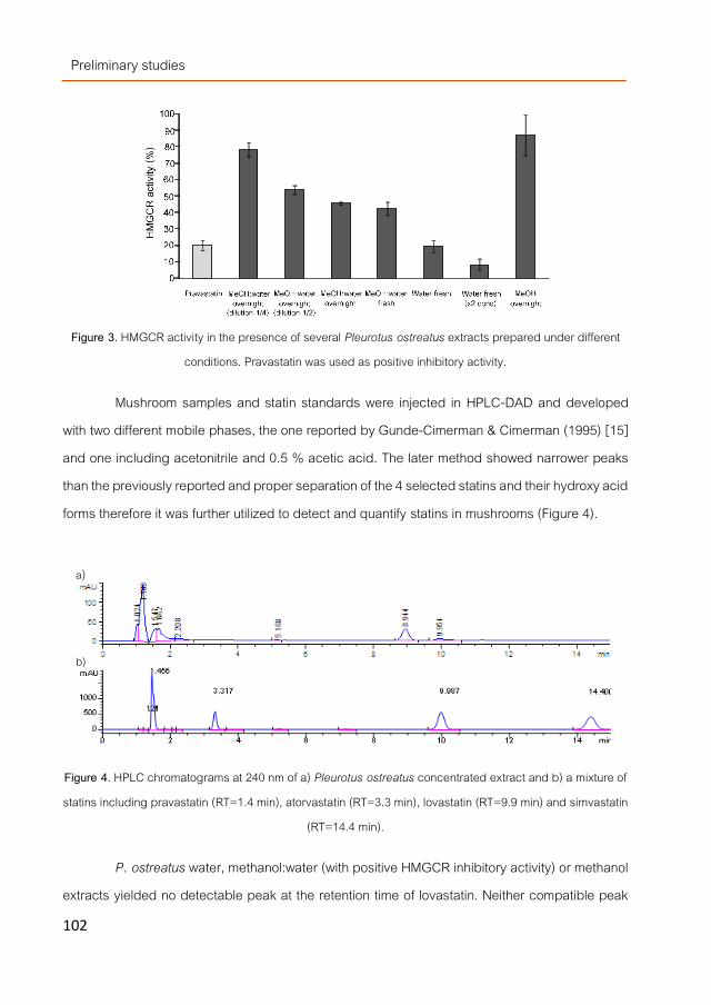
Preliminary studies
102
Figure 3. HMGCR activity in the presence of several Pleurotus ostreatus extracts prepared under different
conditions. Pravastatin was used as positive inhibitory activity.
Mushroom samples and statin standards were injected in HPLC-DAD and developed with two different mobile phases, the one reported by Gunde-Cimerman & Cimerman (1995) [15] and one including acetonitrile and 0.5 % acetic acid. The later method showed narrower peaks than the previously reported and proper separation of the 4 selected statins and their hydroxy acid forms therefore it was further utilized to detect and quantify statins in mushrooms (Figure 4).
Figure 4. HPLC chromatograms at 240 nm of a) Pleurotus ostreatus concentrated extract and b) a mixture of statins including pravastatin (RT=1.4 min), atorvastatin (RT=3.3 min), lovastatin (RT=9.9 min) and simvastatin
(RT=14.4 min).
P. ostreatus water, methanol:water (with positive HMGCR inhibitory activity) or methanol extracts yielded no detectable peak at the retention time of lovastatin. Neither compatible peak
a)
b)

Preliminary studies
103
was detected when the samples were treated to generate lovastatin-hydroxy acid form. Only when the samples were concentrated using the SPE (Solid phase extraction) column, two peaks were observed at 8.9 and 9.9 min with similar spectra than lovastatin since they both showed a maximum at 240 nm (Figure 5).
Figure 5. UV-VIS spectra of a) peak eluting at 8.9 min and b) 9.9 min from the P. ostreatus extract and c) lovastatin spectrum (9.9 min).
Nevertheless, since many different compounds could also show similar UV-VIS spectra at that maximum, a standard of lovastatin and a P. ostreatus extract were injected into a LC-MS in order to determine and compare their mass. Lovastain injected in an LC-MS showed a single peak with a retention time of 8.2 min and a single mass peak of 405.1 undoubtedly corresponding to the 404.55 gram/mol assigned by the literature (Figure 6a). However, the peak from the P. ostreatus extract eluting at similar R.T. showed a mass of 522.3 indicating that they were different compounds (Figure 6b). Moreover, none of the other detectable peaks showed a mass similar to neither lovastatin nor other statins.
a) b)
c)

Preliminary studies
104
Figure 6. UV-VIS and mass spectra of a) lovastatin (eluting at 8.2 min) and b) of P. ostreatus (peak with RT= 8.2 min).
Conclusions Mushrooms fruiting bodies or their extracts might be considered as a new source of
compounds with potential hypocholesterolemic activity because they are rich in ergosterol-derivatives, β-glucans and HMGCR inhibitors. Since not a single mushroom strain showed the highest levels of the three type of compounds, a mixture of a few of them could be used to prepare bioactive supplements to functionalize foods potentially able to reduce levels of cholesterol in serum.
Although lovastatin was not detected in oyster mushrooms, water extracts showed remarkable HMGCR inhibitory activity. Therefore, further investigations are at the present being carried out to optimize these supplements and to identify the responsible for the HMGCR inhibitory capacity observed.
a)
b)

Preliminary studies
105
Acknowledgments The research was supported by the European Union within the 7th framework programme
(7FM-PEOPLE-2009-IFF project 251285), by the ALIBIRD-CM S2009/AGR-1469 and ADER 2010-I-ID-0096 regional programs from respectively the Community of Madrid and La Rioja (Spain) and by the AGL2010-21537 national R+D program from the Spanish Ministry of Science and Innovation.

Preliminary studies
106
References [1]. Wilson, M.D. and L.L. Rudel, Review of cholesterol absorption with emphasis on dietary and biliary cholesterol.
Journal of Lipid Research, 1994. 35(6): p. 943-55. [2]. Ginter PhD, D.E., Cardiovascular disease prevention in eastern Europe. Nutrition, 1998. 14(5): p. 452-457. [3]. EFSA Panel on Dietetic Products, N.a.A.N., Scientific substantiation of a health claim related to a low fat fermented
milk product (Danacol) enriched with plant sterols/stanols and lowering/reducing blood cholesterol an reduced risk of (coronary) heart disease pursuant to Article 14 of Regulation (EC) No 1924/20061. EFSA Journal 2009. 2: p. 2.
[4]. EFSA Panel on Dietetic Products, N.a.A.N., Scientific Opinion on the substantiation of a health claim related to oat beta glucan and lowering blood cholesterol and reduced risk of (coronary) heart disease pursuant to Article 14 of Regulation (EC) No 1924/2006. EFSA Journal 2010. 8(12): p. 1885-1900.
[5]. Miettinen, T.A., et al., Reduction of serum cholesterol with sitostanol-ester margarine in a mildly hypercholesterolemic population. New England Journal of Medicine, 1995. 333(20): p. 1308-1312.
[6]. Chen, J. and X.-F. Huang, The effects of diets enriched in beta-glucans on blood lipoprotein concentrations. Journal of Clinical Lipidology, 2009. 3(3): p. 154-158.
[7]. Mattila, P., et al., Sterol and vitamin D-2 contents in some wild and cultivated mushrooms. Food Chemistry, 2002. 76(3): p. 293-298.
[8]. Teichmann, A., et al., Sterol and vitamin D-2 concentrations in cultivated and wild grown mushrooms: Effects of UV irradiation. Lwt-Food Science and Technology, 2007. 40(5): p. 815-822.
[9]. Kalač, P., Chemical composition and nutritional value of European species of wild growing mushrooms: A review. Food Chemistry, 2009. 113(1): p. 9-16.
[10]. Aida, F.M.N.A., et al., Mushroom as a potential source of prebiotics: a review. Trends in Food Science & Technology, 2009. 20(11–12): p. 567-575.
[11]. Berger, A., et al., Cholesterol-lowering properties of Ganoderma lucidum in vitro, ex vivo, and in hamsters and minipigs. Lipids in Health and Disease, 2004. 3(1): p. 2.
[12]. Cheung, P.C.K., The hypocholesterolemic effect of two edible mushrooms: Auricularia auricula (tree-ear) and Tremella fuciformis (white jelly-leaf) in hypercholesterolemic rats1. Nutrition Research, 1996. 16(10): p. 1721-1725.
[13]. Fukushima, M., et al., Cholesterol-lowering effects of maitake (Grifola frondosa) fiber, shiitake (Lentinus edodes) fiber, and enokitake (Flammulina velutipes) fiber in rats. Experimental Biology and Medicine, 2001. 226(8): p. 758-765.
[14]. Alegret, M., et al., Effect of hypolipidemic drugs on key enzyme activities related to lipid metabolism in normolipidemic rabbits. European Journal of Pharmacology, 1998. 347(2–3): p. 283-291.
[15]. Gunde-Cimerman, N. and A. Cimerman, Pleurotus fruiting bodies contain the inhibitor of 3-Hydroxy-3-Methylglutaryl-Coenzyme A reductase—Lovastatin. Experimental Mycology, 1995. 19(1): p. 1-6.
Preliminary studies
107
[16]. Ramírez-Anguiano, A.C., et al., Radical scavenging activities, endogenous oxidative enzymes and total phenols in edible mushrooms commonly consumed in Europe. Journal of the Science of Food and Agriculture, 2007. 87(12): p. 2272-2278.
[17]. Gunde-Cimerman, N., et al., Screening fungi for the production of an inhibitor of HMG CoA reductase: Production of mevinolin by the fungi of the genus Pleurotus. FEMS Microbiology Letters, 1993. 111(2-3): p. 203-206.
[18]. Gunde-Cimerman, N., A. Plemenitaš, and A. Cimerman, Pleurotus fungi produce mevinolin, an inhibitor of HMG CoA reductase. FEMS Microbiology Letters, 1993. 113(3): p. 333-337.
[19]. Yang, D.-J. and L.S. Hwang, Study on the conversion of three natural statins from lactone forms to their corresponding hydroxy acid forms and their determination in Pu-Erh tea. Journal of Chromatography A, 2006. 1119(1–2): p. 277-284.
[20]. Calpe-Berdiel, L., J.C. Escolà-Gil, and F. Blanco-Vaca, New insights into the molecular actions of plant sterols and stanols in cholesterol metabolism. Atherosclerosis, 2009. 203(1): p. 18-31.
[21]. Yokokawa, H. and T. Mitsuhashi, The sterol composition of mushrooms. Phytochemistry, 1981. 20(6): p. 1349-1351.
[22]. Jasinghe, V.J. and C.O. Perera, Distribution of ergosterol in different tissues of mushrooms and its effect on the conversion of ergosterol to vitamin D-2 by UV irradiation. Food Chemistry, 2005. 92(3): p. 541-546.
[23]. Jeong, S.C., et al., White button mushroom (Agaricus bisporus) lowers blood glucose and cholesterol levels in diabetic and hypercholesterolemic rats. Nutrition Research, 2010. 30(1): p. 49-56.
[24]. Manzi, P. and L. Pizzoferrato, Beta-glucans in edible mushrooms. Food Chemistry, 2000. 68(3): p. 315-318. [25]. Manzi, P., A. Aguzzi, and L. Pizzoferrato, Nutritional value of mushrooms widely consumed in Italy. Food
Chemistry, 2001. 73(3): p. 321-325. [26]. Manzi, P., et al., Commercial mushrooms: nutritional quality and effect of cooking. Food Chemistry, 2004. 84(2):
p. 201-206. [27]. Nitschke, J., et al., A new colorimetric method to quantify β-1,3-1,6-glucans in comparison with total β-1,3-glucans
in edible mushrooms. Food Chemistry, 2011. 127(2): p. 791-796. [28]. Lee, J.-S., et al., Immunomodulatory effect of mushrooms on cytotoxic activity and cytokine production of intestinal
lamina propria leukocytes does not necessarily depend on β-glucan contents. Food Chemistry, 2011. 126(4): p. 1521-1526.
[29]. Bobek PhD, P., L. Ozdín PhD, and Š. Galbavý Md, Dose- and time-dependent hypocholesterolemic effect of Oyster Mushroom (Pleurotus ostreatus) in rats. Nutrition, 1998. 14(3): p. 282-286.
[30]. Opletal, L., et al., Evidence for the anti-hyperlipidaemic activity of the edible fungus Pleurotus ostreatus. British Journal of Biomedical Science, 1997. 54(4): p. 240-243.

Chapter 1 Effect of mushrooms polysaccharides on cholesterol metabolism

Chapter 1- Effect of mushroom polysaccharides…
111
Preface Edible mushrooms are considered as good source of dietary fibers (DF) because their
hyphae membranes contained large amounts of non-digestible polysaccharides. They show many different structural conformations but mostly are composed by large chains of glucose joined by α- , β- or mixed α, β-glucose branches. However, those pointed by the literature as related to the hypocholesterolemic properties (not only for mushrooms but also for other food products) are constituted by D-glucose monomers linked by β-glycosidic bonds with β-branching after a few monomers. The hypocholesterolemic β-glucans from cereals showed β-(1→3),(1→4)-glucan linkages while mushrooms β-glucans combined β-(1→3)- and β-(1→3),(1→6)-linkages in different degrees depending on the mushroom specie. Apparently, their molecular size, branching degree, monomer composition or structure conformation modulated their biological properties as they enhance or impair their water-solubility. However, their precise mechanism for lowering cholesterol remains still unclear. Several hypothesis have been drawn including their ability for increasing their faecal excretion by a direct bile acid and cholesterol bindings or by their mobility impairing in a high gelled digestive bolus. Added to this, SCFA (short chain fatty acids) generated after mushroom fiber degradation by the intestinal microbiota have been pointed as HMGCR inhibitor. Moreover, modulation of lipid-related gene expression i.e SREBF1, SREBF2 or NR1H4 by β-glucans was also observed, as previously indicated (see Introduction section).
Other fungal carbohydrate structures related to hypocholesterolemic compounds are chitins (β-(1→4)-glucan linkages of N-acetylglucosamine monomers) derivative products, i.e. chitosans, molecules that showed lowering-cholesterol capabilities after in vivo animal model experiments.
In addition to commented variability in the chemical properties i.e. water solubility of mushrooms DF, one of the main reasons for the diversity in results or conclusions when correlating structures and functions of polysaccharides is partially due to the wide range of extraction methods utilized before biological analysis. Both traditional and advanced technologies, i.e. boiling water, alkali, ultrasonic- or microwave- assisted, pressurized solvent extractions etc., have been used to isolate them. Thus, this chapter describes the experimental settings followed for

Chapter 1- Preface
112
optimizing an extraction method to obtain specific β-glucan-enriched DF fractions from selected mushrooms and compare it with a more traditional extraction method. The influence of the obtained fractions as bile acid scavengers during an in vitro digestion model is also studied as well as their effect on the expression of genes related with the cholesterol metabolism using cell cultures and animal models.
Initially, a screening through several species was carried out in order to select the edible mushrooms with higher levels of total β-glucans and according to the results obtained (previously described in the preliminary studies section) a high quantitative variability was found among the tested species. Thus, P. ostreatus was selected as raw material to obtain β-glucan-enriched DF fractions because it was one of the strains with higher levels and easier to cultivate than others such as Ganoderma lucidum. However, since the biological properties described are highly dependent on the β-glucan structural conformation and each mushroom synthetize its own particular polysaccharide mixture, two other species were also selected. A. bisporus was selected because it is widely cultivated and their fruiting bodies fiber extracts were already pointed as the responsible for the hypocholesterolemic and hypoglucemic properties found in vivo and L. edodes because a specific polysaccharides structure (lentinan), different from the other two species, was also pointed as bioactive compound. Thus, in the work titled Pressurized water extraction of β-glucans enriched fractions with bile acids-binding capacities obtained from edible mushroom, the steps followed to optimize an isolation process for fungal polysaccharides are described using an advanced environmentally friendly technology (pressurized water extraction, PWE). Several extraction procedures and parameters such as temperature, static extraction time etc., were tested to detect a possible structural selectivity of fungal fiber compounds according to different extraction conditions. The three mushroom species were also submitted to a more standardized extraction (STD) procedure to compare with PWE, finding out their fiber composition as well as advantages and disadvantages of each extraction method. After DF-enriched-extracts were obtained, both STD and selected PWE fractions were submitted to an in vitro digestion process to quantify their bile acid-binding capacity. The amount of free bile acids in the digestates, when different polysaccharides of DF-fractions were present, was evaluated by HPLC. A cereal

Chapter 1- Effect of mushroom polysaccharides…
113
DF-enriched fraction and cholestyramine were used as controls to compare with the fungal DF extracts.
On the other hand, DF are by definition, those polysaccharides that cannot be digested by humans and are left for colonic microbial degradation until excreted in feces. Due to their complex and polymeric structures DF are not absorbed by enterocytes but they, together with their degradation products (fragments resulting from their partial digestion/transformation), might get in contact with intestinal cells during digestion process. The chances of interaction increases when digestion reduce their molecular size enabling their solubilization in physiological media such as the lumen, stimulating cellular responses (as observed for instance for the enterocytic cytokine-activation mechanisms induced by β-glucans triggering an immune response). Thus, in order to study whether DF-fractions from A. bisporus, L. edodes and P. ostreatus could induce an enteric response, digestates obtained after theirs in vitro digestion were administrated to Caco2 cell line. Enterocytes mRNAs were extracted 1 and 24h after the administration, transformed into cDNA and analyzed by qPCR to observe whether the DF-fractions might modulate the cholesterol-related gene expression of enterocytes. The results of the DF effects at the transcriptional level are described in the work entitled Modulation of cholesterol-related gene expression by dietary fiber fractions from edible mushrooms, and they were used to select one mushroom specie for further in vivo experiments. Animal interventions were carried out following two different experimental designs using male C57BL/6JRj mice. In the first one, hypercholesterolemia was firstly induced using diets with high cholesterol and cholic acid concentrations for 4 weeks and afterwards mice were treated for 4 more weeks with simvastatin or ezetimibe (as standardized hypocholesterolemic drugs) or a DF-fraction obtained from P. ostreatus. In the second one, the hypercholesterolemic diet was administrated simultaneously with one of the two drugs or the DF-fraction for 4 weeks. Biochemical parameters at final experimental time such as total cholesterol, HDL, LDL and triglycerides in serum and liver and sterols content in feces were determined. The mRNA expression pattern in four tissues (jejunum, ileum, cecum and liver) was also evaluated.

Chapter 1- Effect of mushroom polysaccharides…
115
WORKPLAN
In vitro digestion of PSC fractions and bile acids quantification
Proteins Total polysaccharides
Glucans Chitin/Chitooligosaccharides
Chemical analysis
P. ostreatus
A. bisporus
L. edodes
PLE
Std method
+ Cold precipitation
Hot extraction
Dialysis Process
MWCO 3.5 KDa
vs
PSC extracts
HCl
NaOH
pH=237 C
pH=6,337˚C
bile extract pancreatinbuffer pH 6.3
Simulation of gastric and intestinal digestion
Bile acids C18
isolation
HPLC bile acids analysis
In vitro and in vivo experiments of PSC-enriched fraction
Extraction and isolation procedures to obtain PSC fraction
Brush border enterocytes
In vitro gene modulation assay (Caco2) cells)
RT-PCR
Gene expression analysis
Caco2 cell culture
Sequential
Simultaneous
Animal experimental trials Mus musculus (Male C57BL/6JRj mice)
Chemical analysis
Plasma Liver Feces
Selection of mushroom specie Supplementation type
In vivo gene modulation assay cells)

Chapter 1- Effect of mushroom polysaccharides…
117
Manuscript 1
Pressurized water extraction of β-glucan enriched fractions with bile acids-binding capacities obtained from edible mushrooms
Marimuthu Palanisamy, Laila Aldars-García, Alicia Gil-Ramírez, Alejandro Ruiz-Rodríguez, Francisco R. Marín, Guillermo Reglero, and
Cristina Soler-Rivas
Department of Production and Characterization of Novel Foods, CIAL – Research Institute in Food Science (UAM+CSIC), C/ Nicolas Cabrera 9, Campus de Cantoblanco, Universidad Autónoma de Madrid, 28049 Madrid, Spain.
Biotechnology Process
Volume 30 (2), pages 391–400, March 2014 DOI: 10.1002/btpr.1865

Chapter 1- Manuscript 1
118
Abstract A pressurized water extraction (PWE) method was developed in order to extract β-
glucans with bile acids-binding capacities from cultivated mushrooms (Agaricus bisporus, Lentinula edodes, and Pleurotus ostreatus) to be used as supplements to design novel foods with hypocholesterolemic properties. Extraction yields were higher in individual than sequential extractions being the optimal extraction parameters: 200 °C, 5 cycles of 5 min each at 10.3 MPa. The crude polysaccharide (PSC) fractions, isolated from the PWE extracts contained mainly β-glucans (including chitooligosaccharides deriving from chitin hydrolysis), α-glucans, and other PSCs (hetero-/proteo-glucans) depending on the extraction temperature and mushroom strain considered. The observed bile acids-binding capacities of some extracts were similar to a β-glucan enriched fraction obtained from cereals.

Chapter 1- Effect of mushroom polysaccharides…
119
Introduction Certain polysaccharides (PSC) present in plants and particularly those from cereals have
recently gained attention because of their potential beneficial effects for human health. The (1→3),(1→4)-β-glucans are believed to perform many biological functions such as i.e. modeling of immune response [1] or reducing of cholesterol levels in serum [2].
Direct binding of bile acids and cholesterol (from ingested food) and increasing their faecal excretion has been hypothesized as a possible mechanism by which the water-insoluble PSCs lower cholesterol [3]. By binding bile acids, they prevent their reabsorption and stimulate plasma and liver cholesterol conversion to additional bile acids [4, 5]. The cholesterol-lowering effect of water-soluble dietary fibers seems to be due to several mechanisms being the increase in viscosity (water binding capacity in the 119ydra) the main effect. This leads to a reduced diffusion rate of bile acids, which cannot be reabsorbed by the body being then excreted [3].
Edible mushrooms also contain interesting PSCs [6] such as other β-glucans, which are structurally different from plants since their branches are (1→3) and (1→3),(1→6) but share biologically important properties including hypocholesterolemic effects [7], and chitin, a water-insoluble β-(1→4)-glucan of N-acetylglucosamine monomers, also considered as dietary fiber. However, not all the fungal β-glucans show all the beneficial biological activities, the 119ydrazine119latory and antitumor properties seemed to be more related to the water soluble fraction [8] including the oligomers and low molecular weight polymers generated from chitin hydrolysis (LMWC or chitooligosaccharides) [9] while their effect as prebiotic might be due to their water-insoluble β-glucans (chitins and protein-bound glucans) [10]. Obviously, their solubility in water depends, above all, on their molecular structure largely influenced by their monomer composition, type and degree of branching, conformation (single or triple helix), molecular weight, N-acetylation degree etc. [11] and, according to recent review, there is insufficient information to establish a broad generalization correlating structure with function [12].
A wide variety of PSCs with different structures and biological activities have been obtained from edible mushrooms using different extraction processes. The traditional methods include the use of boiling water, sodium hydroxide, ammonium oxalate, and more complex protocols [13], while the more advanced technologies included ultrasonic-assisted [14, 15] and

Chapter 1- Manuscript 1
120
microwave assisted [16] extractions or combinations [17], supercritical carbon dioxide [18] and pressurized solvent extraction using water [19] or ethanol [20] as extraction solvents.
The work presented in this article, describes the optimization of a specific procedure to obtain β-glucan enriched fractions from the three most commonly consumed edible mushrooms using an accelerated solvent extraction (ASE) device, which enable the use of pressurized water as extraction (PWE) solvent. Several PWE conditions and procedures were tested to investigate whether the mushrooms could be fractionated separating in one/some fraction/s those β-glucans with bile acid-binding activities to use them as food-grade supplements to design novel foods with hypocholesterolemic properties. Then, the obtained fractions were submitted to an in vitro digestion model to determine whether they are able to bind bile acids as the β-glucans from a cereal foodstuff.
Materials and Methods
Biological material Mushroom strains used in this investigation were Agaricus bisporus L. (Imbach), Lentinus
edodes S. (Berkeley), and Pleurotus ostreatus (Jacq.Ex Fr.) Kummer. Mushroom strains were cultivated at CTICH (Research Center for Mushroom Science and Technology, Autol, Spain) and harvested at the optimal developmental stage for consumption. Fruiting bodies were dehydrated and ground into fine powder as described by Ramírez-Anguiano et al. [21] and stored at -18 °C until further use.
Commercially available breakfast cereals containing oat, rice, wheat, and barley β-glucans bearing the claim containing β-glucans able to reduce cholesterol levels were obtained from a local supermarket. All the experiments were performed from the same package.
Reagents Ethylenediaminetetraacetic acid, sea sand, and methanol HPLC grade were purchased
from Panreac (Barcelona, Spain). D(1)-glucose, ferric chloride, ammonium sulfamate, sodium nitrite, 3-methyl-2-benzothiazolone 120ydrazine hydrochloride, phenol, chitin (from shrimp), glucose, pancreatin, standard bile acids, and porcine bile extract, cellulose, cholestyramine, and
Chapter 1- Effect of mushroom polysaccharides…
121
bovine serum albumin (BSA) were procured from Sigma-Aldrich. (Steinheim, Germany). All other reagents and solvents were used of analytical grade.
Pressurized water extractions to obtain PSC enriched fractions Mushroom powder (1 g) were mixed with washed sea sand (4 g) and submitted to
pressurized water extraction (PWE) at 10.68 Mpa using an ASE (Dionex Corporation, ASE 350, Sun-nyvale, CA). Extraction procedure per cycle was as follows: firstly, the sample was loaded into 11 ml extraction cells. Then, the cell was filled with solvent (MilliQ water), heat-up time applied and static extraction was carried out with all system valves closed. Afterwards, the cell was rinsed, solvent was purged from cell with N2 gas and depressurization took place. Several parameters such as solvent, static extraction time, or cycles number and temperature were changed in order to optimize the extraction method.
A sequential extraction procedure was also tested by mixing the mushroom powders (1 g) with sea sand (4 g) into the ASE extraction cells. Then, the samples were submitted to 5 extraction cycles of 5 min each at 25 °C. Afterwards, the solvent was removed and another extraction was carried out for 5 cycles of 5 min each at 50 °C. These processes were repeated at 100, 150, and 200 °C.
Obtained fractions were immediately frozen, lyophilized, and stored at -18 °C until further analysis. Extracted dry matter content was measured to calculate the extraction yields. A minimum of two fractions were collected from each type of sample.
Standardized extraction method to obtain PSC enriched fractions Mushroom powders (1 g) and powdered dehydrated cereals (1 g) were mixed with MilliQ
water (100 ml), heated at 120 °C for 20 min and cooled down to 4 °C following the method reported by Jeurink et al. [22]. The crude PSC fraction was isolated as described below.
Isolation of the PSC fractions The from the extracts obtained by both standardized and PWE methods were isolated by
adding two volumes of cold ethanol, vigorous stirring, and allowing PSC precipitation overnight at

Chapter 1- Manuscript 1
122
4 °C. The precipitated PSCs were collected by centrifugation (10,000g for 30 min at 4 °C) (Thermo Scientific Heraeus Multifuge, Fisher scientific, Madrid, Spain), suspended in Milli-Q water and dialyzed with membrane tubing (MWCO 3.5 Kda; Medicell International Ltd. London, UK) against distilled water to remove low molecular weight (LMW) compounds during 24 h at 5 °C with three or four changes of the distilled water. After the dialysis, the fraction remaining inside the membrane (MW >3.5 kDa) was lyophilized, weighed, and stored at -18 °C until further use [22]. This extract was called crude PSC fraction.
Total polysaccharide determination The PSC concentration of the obtained extracts was evaluated as their total carbohydrate
content with the modified phenol–sulfuric acid method as described by Fox and Robyt [23]. Glucose was used as standard for quantification.
Determination of β-glucans The β-glucan content of the obtained mushroom extracts (50 mg) was evaluated by a
β-glucan determination kit specific for mushrooms and yeasts (Megazyme, Barcelona, Spain) following the instructions of the user’s manual. Absorbance at 510 nm was measured using a spectrophotometer (Evolution 600, Fisher scientific, Madrid, Spain).
The procedure calculates the β-glucan concentration directly from mushroom powder by subtracting the values obtained from a first assay in which the content of total glucans (α-glucan + β-glucan), D-glucose in oligosaccharides, sucrose and free D-glucose are quantified, to a second assay in which the content of α-glucans (glycogen and starch), D-glucose in sucrose and free D-glucose are quantified. Therefore, if these assays are carried out using the isolated crude PSC fractions (obtained after dialysis), mono-, di-, and oligosaccharides as well as broken or small PSCs are lacking. Thus, the values obtained by carrying out the first assay should be mainly due to the total glucans and in the second assay to α-glucans. Therefore, in these types of samples total, α-, and β-glucans can be quantified.

Chapter 1- Effect of mushroom polysaccharides…
123
Determination of chitin and chitooligosaccharides The N-glucosamine containing β-glucans were determined in the crude PSC fractions
following the method described by Vetter [24] adjusted from a previous method [25]. This method is usually utilized to determine chitin; however, if it is applied to extracts obtained with water (or pressurized water) where chitin is not extracted, it evaluates the amount of N-glucosamine-containing residues present in the fraction determining in this case, the amount of chitooligosaccharides, also referred as LMWC (low molecular weight chitin-hydrolysis products), which are depolymerized fractions showing values of n > 20 (n=monomers number) with increased water solubility [9]. Chitin from shrimp was used as standard for quantification.
Determination of total protein The total protein concentration of the samples (2 mg/ml) was determined using the
Bradford method reagents (cat. Num. B6916, Sigma-Aldrich, Madrid, Spain) according to the Instruction Manual. BSA was used as standard for protein quantification.
In vitro digestion of the PSC fractions Mushroom PSC fractions (isolated after PWE or the standard extraction methods), cereal
PSC fractions, cellulose (negative control for bile acid-binding properties), and cholestyramine (positive control) were submitted to in vitro digestion following the procedure described by Kahlon et al. [4] with a few modifications. Briefly, the PSC extracts were digested in 1 ml of 0.01M HCl for 1 h at 37 °C with gentle agitation (Orbital incubator S150, Stuart, Stone, UK). Afterwards, the sample pH was adjusted to 6.3 with 0.1 ml of 0.1M NaOH. Then, 4 ml of bile extract (14.3 mg in Tris-maleate buffer pH 6.3) and 5 ml of pancreatin (10 mg in Tris-maleate buffer pH 6.3) were added and the mixture was incubated for 1 h at 37 °C. After digestion, the samples were filtered through filter paper and the bile acids isolated from the filtrate. A control sample was prepared following the same digestion protocol but without the addition of PSC fractions.
Quantification of bile acids by HPLC Isolation of the bile acids was carried out using C18 Seppak cartridges and resulting
extracts were injected (20 μl) in a HPLC system (Varian Pro-star 330, Madrid, Spain) equipped

Chapter 1- Manuscript 1
124
with a C18 column (Microsorb-MV 100-5 Varian, 25 cm × 4.6 mm and 5 μm particle size) and a PDA detector according to Pang et al. [26].
The chromatograms from the utilized bile extract (before and after digestion) showed four major peaks in concordance with those described by Qiao et al. [27] for pig bile extract and identified as glycoursodeoxycholic acid (RT=7.3 min) (GUDCA), glycohyodeoxycholic acid (RT=7.7 min) (GHDCA), taurochenodeoxycholic acid (RT=10.9 min) (TCDCA), and taurohyodeoxycholic acid (RT=11.8 min) (THDCA). The bile acid-binding capacity of the PSC extracts was determined by subtracting the total bile acid concentration found in the control samples (digestion with no PSC fractions) and the digested PSC-containing samples. The total bile acid concentration considered was the sum of the four major bile acids present in the utilized porcine bile extract accounting for almost 93% of the total bile acids [27].
Statistical analysis One way analysis of variance (ANOVA) was performed using a Statgraphics®Plus 3.1 for
Windows software (Statis-tical Graphics Corporation, Rockville, MD). The mean comparison test used was Fisher’s least significant differences procedure (LSD).
Results and discussion The PWE procedure was initially optimized for only one of the mushroom species
(Lentinula edodes) in order to define the parameters inducing larger variations in the extraction yields to be taking into consideration for the extractions of the other mushroom species.
Extractions of L. edodes PSC using different temperatures The temperature of the extraction water could be the most critical parameter to define
the distribution of the extracted β-glucans within the fractions obtained at different temperatures since their solubility in water depends on their molecular structure and it increases with temperature [8]. Indeed, the extraction yields of the PWE extracts and of the crude PSC fraction obtained from these extracts exponentially increased with the temperature (Table 1). However, when the results of the PWE extractions were compared with those obtained after a standard PSC extraction method, the latter yielded higher crude PSC fraction (48.6%) than the best PWE

Chapter 1- Effect of mushroom polysaccharides…
125
condition at 200 °C (28.3%) but, it contained 39% PSCs while the PWE extract (at 200 °C) almost doubled the amount of PSCs (74.2%) pointing PWE as a more selective procedure to extract PSCs.
Extractions of L. edodes PSC using different extraction times There are two parameters that can be adjusted in PWE to modify the extraction time, one
is the time that the solvent is in contact with the sample in the extraction cell and the other is the number of cycles of extraction. Therefore, temperature was fixed at 200 °C and the influence of both parameters in the extraction yields was investigated.
No significant differences were found neither in the levels of material extracted by PWE (~80% of dry weight) nor in the levels of the crude PSC fraction extracted (219–285 mg/g mushroom powder) if the extraction time of each cycle was set from 1 to 5 min (with a total of 5 cycles). However, the amount of extracted PSCs slightly increased after 4 min (from on average 187.7 mg/g after 1, 2, or 3 min up to 206.9 mg/g) pointing 5 min as the best extraction time (255.7 mg/g). Moreover, 1, 5, 10, and 15 cycles of 5 min each one at 200 °C extracted from 627.6 to almost 890 mg dry matter with a concomitant increase of the crude PSC fraction (from 215 up to 303.4 mg/g); however, no significant PSC increase was observed from 5 to 15 cycles, apparently all the extractable PSCs were already obtained after 5 cycles.
Sequential extractions of L. edodes PSC A sequential extraction of L. edodes powder was carried out by gradually increasing
temperatures from 25 to 200 °C (Figure 1). The first extraction carried out at 25 °C extracted only 5.6% of dry weight and the second extraction (at 50 °C) yielded higher amount (9.7%); however, apparently in these extractions mostly low molecular weight material was extracted since their PSC content was very low. The third extraction showed a low yield thus most of the extracted PSCs were obtained after the forth (150 °C) and particularly the fifth (200 °C) extraction where 30.3% of material was extracted containing only 5.4% of PSCs. Thus, sequential extractions did not improve the yields obtained by individual extractions and if the data from the complete set of obtained fractions were added together, by sequentially performing the extractions not all the mushroom PSCs were extracted.

Chapter 1- Manuscript 1
126
Thus, sequential extraction was discarded as a method to fractionate the β-glucans from mushrooms.
Figure 1. Extraction yields (PWE extracts and crude PSC fractions) and PSC concentration obtained after a PWE sequential extraction of Lentinula edodes (5 cycles, 5 minutes). 1st ext: extraction at 25
ºC; 2nd ext: extraction at 50 ºC; 3rd ext: extraction at 100 ºC; 4th ext: extraction at 150 ºC and 5th ext: extraction at 200 ºC.
Table 1. Extraction yields (total dry matter extracted in the PWE extracts and in the crude PSC fractions isolated from those PWE extracts) and PSC concentrations obtained after submitting Lentinula edodes powder to pressurized water extractions at different temperatures.
Extraction temperature
(°C)
PWE extract (mg/g
mushroom dw)
Extraction yield (%)
Crude PSC fraction obtained in PWE extracts
(mg/g mushroom)
Extraction yield (%)
Crude PSC fraction in
PWE extracts (%)
Total PSCs obtained in
PWE extracts (mg/g mushroom)
Extraction yield (%)
Total PSCs in the crude PSC fraction
(%)
Total PSCs in
PWE extract (%)
25 201 ± 35a 20.1 48.0 ± 20.4a 4.8 23.9 15.1 ± 1.2a 1.51 31.5 7.5
50 291 ± 48a 29.1 42.6 ± 19.0a 4.3 14.7 12.1 ± 1.8a 1.21 28.5 4.2
100 248 ± 92a 24.8 44.0 ± 14.2a 4.4 17.7 19.6 ± 4.1a 1.96 44.6 7.9
150 301 ± 48a 30.1 85.7 ± 13.7b 8.6 28.5 47.1 ± 17.8b 4.71 54.9 15.6
200 823 ± 34b 82.3 282.7 ± 73.8c 28.3 34.4 209.7 ± 28.0c 21.0 74.2 25.5
Std PSC ext. method 485.5 ± 72.8d 48.6 189.2 ± 27.5c 18.9 39.0 a,b,c,d Denotes statistically significant differences (P < 0.05) among values from the same column.
0
50
100
150
200
250
300
350
1st ext 2nd ext 3rd ext 4th ext 5th ext
mg/g
mus
hroo
m (d
w)
Sequential PWE extractions (5 cycles /5 min)
PWE extract Crude PSC fraction
Total PSCs

Chapter 1- Effect of mushroom polysaccharides…
127
Table 2. Extraction yields (total dry matter extracted in the PWE extracts and in the crude PSC fractions isolated from those PWE extracts) and PSC concentrations obtained after submitting Agaricus bisporus powder to pressurized water extractions at different temperatures.
Extraction temperature
(°C)
PWE extract (mg/g
mushroom dw)
Extraction yield (%)
Crude PSC fraction obtained in PWE extracts
(mg/g mushroom)
Extraction yield (%)
Crude PSC fraction in
PWE extracts (%)
Total PSCs obtained in
PWE extracts (mg/g mushroom)
Extraction yield (%)
Total PSCs in the crude
PSC fraction (%)
Total PSCs in
PWE extract
(%)
25 267 ± 20a 26.7 48.5 ± 12a 4.8 18.2 14.8 ± 3.3a 1.5 30.6 5.6
50 240 ± 12a 24.0 42.5 ± 4.9a 4.3 17.7 6.9 ± 3.3a 0.7 16.2 2.9
100 317 ± 21b 31.7 36.0 ± 11a 3.6 11.4 14.1 ± 2.1a 1.4 39.1 4.4
150 597 ± 19c 59.7 89.5 ± 12b 9.0 15.0 30.9 ± 3.5b 3.1 34.5 5.2
200 783 ± 88d 78.3 211 ± 38.2c 21.1 26.9 103 ± 7.1c 10.4 49.2 13.2
Std PSC ext. method 290 ± 83.4 c 29.0 83.5 ± 6.1c 8.4 28.8 a,b,c,d Denotes statistically significant differences (P < 0.05) among values from the same column.
Extractions of PSC from other mushroom species The results obtained after submitting L. edodes to PWE extractions indicated that the
highest PSC yields were obtained by carrying out individual extractions, setting 5 cycles of 5 min as extraction time and 200 °C being the extraction temperature the most crucial parameter. Therefore, PWE extractions of two other cultivated mushrooms such as Agaricus bisporus and Pleurotus ostreatus were carried out at different temperatures maintaining other parameters as described above.
The observed increase in extraction yields with the temperature for both mushroom species was similar to the one observed for L. edodes being 200 °C the optimal temperature to obtain ~78% of the dry matter for both A. bisporus (Table 2) and P. ostreatus (Table 3). Also from these two other species, a higher amount of the crude PSC fractions could be extracted using the standard PSCs extraction method; however, they contained less PSCs than those obtained using PWEs at 200 °C pointing PWE also for these mushrooms as a more specific method for fungal PSC extraction.

Chapter 1- Manuscript 1
128
Table 3. Extraction yields (total dry matter extracted in the PWE extracts and in the crude PSC fractions isolated from those PWE extracts) and PSC concentrations obtained after submitting Pleurotus ostreatus powder to pressurized water extractions at different temperatures
Extraction temperature
(°C)
PWE extract (mg/g
mushroom dw)
Extraction yield (%)
Crude PSC fraction obtained in PWE extracts
(mg/g mushroom)
Extraction yield (%)
Crude PSC fraction in
PWE extracts (%)
Total PSCs obtained in
PWE extracts (mg/g mushroom)
Extraction yield (%)
Total PSCs in the crude
PSC fraction (%)
Total PSCs in
PWE extract (%)
25 154 ± 26a 15.4 36.0 ± 5.7a 3.6 23.4 19.9 ± 4.4a 2.0 55.2 12.9
50 148 ± 36a 14.8 48.5 ± 6.4a 4.9 32.8 28.9 ± 8.7a 2.9 59.6 19.5
100 198 ± 31a 19.8 58.0 ± 17.0a 5.8 29.3 40.5 ± 12a 4.1 70.0 20.5
150 293 ± 87a 29.3 75.0 ± 26.9a 7.5 25.6 43.5 ± 9.3a 4.4 58.0 14.8
200 786 ± 272b 78.6 187 ± 78b 18.8 23.9 153 ± 30b 15.3 81.6 19.5
Std PSC ext. method 494 ± 28.3c 49.4 209 ± 58b 21.0 42.4
a,b,c Denotes statistically significant differences (P < 0.05) among values from the same column
PWE have already been utilized to extract PSC enriched fractions from mushrooms such as Ganoderma sp. [19] using a similar ASE device and conditions (120 °C, 10.7 Mpa, and 2 cycles of 5 min) because the total amount of extracted PSC was higher than those using sonication (water 100 °C for 30 min). But, no optimization of the extraction process was carried out and no more detailed information was given about the obtained yield. Moreover, the filtrate obtained from the culture broth where L. edodes mycelium was growing was also submitted to PWE in order to isolate bioactive fractions [20]. However, precipitation of the PSCs from the filtrates with ethanol was carried out before PWE was applied thus in this case, PWE was not used for extraction but to further fractionate isolated PSCs obtained by a standard method therefore, no previous information on the yields of fungal PSC extractions by PWE was found in the literature. But, PWE of β-glucans from barley using temperatures from 135 to 180 °C (15–75 min total extraction times) generated fractions with 23–54% β-glucans being the optimal condition at 157.5 °C (45 min) [28]. These values indicated that the yields obtained for fungal PSCs at 150 °C were similar to those obtained from barley. However, if higher temperatures were utilized, barley β-glucans were partially

Chapter 1- Effect of mushroom polysaccharides…
129
hydrolyzed therefore, the presence of hydrolyzed β-glucans in the obtained PWE extracts and the presence of other compounds was determined.
Composition of the PWE extracts As expected, the obtained PWE extracts showed higher amounts of proteins and
peptides in the fractions obtained at temperatures below 100 °C and higher PSC concentrations above that temperature. However, a different distribution was observed depending on the mushroom specie considered (Figure 2). The PWE extracts obtained from L. edodes and A. bisporus at 50 °C showed the largest protein concentrations while more proteins were extracted from P. ostreatus with only 25 °C. The fraction of the latter mushroom obtained at 200 °C also showed high protein levels. Since proteins precipitate at temperatures close to 100 °C these values might be due to proteins or fragments tightly bound to PSCs forming proteoglucans, glucoproteins, or other protein-glucan complexes.
A. bisporus PWE extracts contained less PSCs and β-glucans than L. edodes and P. ostreatus (Figure 2b) probably because this mushroom contained significantly lower levels of these compounds (respect. 12.30 mg β-glucans/100 mg A. bisporus, 38.18 mg/100 mg L. edodes, and 41.79 mg/100 mg P. ostreatus mushroom powders dw).
L. edodes PWE extractions carried out at low temperatures (Figure 2a) yielded larger amounts of hydrolyzed β-glucans (or β-glucan oligomers, with monomers number (n) between 2 and 10, molecular weight < 3.5 kDa) than the other two mushroom species. Their concentration was increasing with the temperature up to 100 °C, above that temperature β-glucans of higher molecular weight were extracted. At 200 °C other PSCs beside β-glucans were also extracted since the total PSC concentration was significantly higher than the amount of higher molecular weight β-glucans (> 3.5 kDa, β-glucan polymers). The distinction between β-glucan oligomers and polymers could be calculated because the β-glucan levels were determined in the complete PWE extracts and in the separated (though a dialysis membrane with a MWCO 3.5 kDa) crude PSC fractions obtained from them.

Chapter 1- Manuscript 1
130
The A. bisporus and P. ostreatus PWE extracts obtained at temperatures higher than 100 °C showed β-glucan oligomer concentrations that were absent at lower temperatures suggesting that in these cases, degradation of the β-glucan polymers might have occurred probably as effect of the pressure and temperature as occurred with barley β-glucans [28].
The P. ostreatus extracts obtained at 200 °C contained other PSC that were not β-glucans (Figure 2c) as it was previously observed for L. edodes. Thus, a more detailed study on the type of PSCs present in the PWE extracts was carried out.
Polysaccharides in the crude PSC fractions The composition of the crude PSC fractions (> 3.5 kDa) obtained from PWE extracts was
as compared with the crude fraction obtained from a standard PSC extraction method and results indicated that they contained different type of PSCs (Figure 3).
To distinguish between β-glucans derived from chitin hydrolysis (chitooligosaccharides) and other type of β-glucans, the N-glucosamine concentration was determined in the fractions. Chitins levels in the selected mushrooms were 4.6, 4.9, and 3.3 mg/100 mg for respectively L. edodes, A. bisporus and P. ostreatus mushrooms so these values were in concordance with previous determinations [24]. However, these molecules cannot be present in the PWE fractions because they are not soluble in water and they request heavy extraction methods to solubilize them [29]. Thus, the N-glucosamine levels measured in the PWE fractions corresponded to the so called chitooligosaccharides or LMWC (low molecular weight chitin derivatives obtained after its hydrolysis), which were pointed as water soluble compounds with therapeutic properties [9].
The amount of chitooligosaccharides in the crude PSC fractions obtained from the PWE extracts of L. edodes (Figure 3a) was increasing with the temperature up to 150 °C following a similar tendency than the β-glucan oligomers determined in the same PWE extracts (Figure 2a), suggesting that these fractions might be rich in monomers and oligomers considered as immunomodulators, with antimicrobial, antitumor activities etc [9]. Indeed, the crude PSC fraction from the PWE extract obtained at 50 °C showed very interesting immunomodulatory activities in macrophages that were lacking in the fraction obtained at 200 °C [30].

Chapter 1- Effect of mushroom polysaccharides…
131
Figure 2. Proteins (and peptides), polysaccharides and β-glucans (oligomers and polymers) of the PWE extracts obtained at different temperatures (5 cycles of 5 min) from a) Lentinula edodes b) Agaricus
bisporus and c) Pleurotus ostreatus.
The reason could be because in the latter fraction, the major PSCs were other β-glucans rather than those chitooligosaccharides (Figure 3a). Most of these β-glucans and a few α-glucans (glycogen and starch) were also present in the crude fraction obtained using the standard PSC extraction method. However, besides these glucans, the fraction obtained from the PWE extract
05
101520253035404550
25 50 100 150 200
Conc
entra
tion
in PW
E e
xtrac
ts (%
)
PWE temperature (ºC)
Proteins and peptidesβ-glucan oligomersTotal PSCβ-glucan polymers
0
5
10
15
20
25
30
35
40
45
50
25 50 100 150 200
Conc
entra
tion
in PW
E ex
tracts
(%
)
PWE temperature (ºC)
Proteins and peptidesβ-glucan oligomerstotal PSCβ-glucan polymers
0
5
10
15
20
25
30
35
40
45
50
25 50 100 150 200
Conc
entra
tion
in PW
E ex
tracts
(%)
PWE temperature (ºC)
Proteins and peptidesβ-glucan oligomerstotal PSCβ-glucan polymers
a)
b)
c)

Chapter 1- Manuscript 1
132
at 200 °C contained other PSCs probably with different structures since they were only detected by the total PSCs determination test, thus, they might be heteroglucans, proteoglucans, or others with specific configurations [11]. These other PSCs were extracted in larger concentrations with the increase of temperature and they were the responsible for the higher PSC extraction yield obtained in the PWE extracts at 200 °C (74.2%) as compared to the one obtained using the standard method (39%).
The PSC composition of the crude fractions obtained from PWE extracts isolated from the other two mushroom species showed higher amount of chitooligosaccharides than the fractions obtained by the standard PSC extraction method but similar content of other β-glucans and α-glucans (Figures 3b,c). The main difference between these mushrooms was that P. ostreatus contained larger amounts of other PSCs while in A. bisporus most of the extracted PSC were β-glucans as in the fraction obtained using the standard PSC extraction method. Since, the protein concentration in the P. ostreatus PWE extract at 200 °C was so high (Figure 2c) these other PSCs might be proteoglucans or other protein-PSC complexes.
Bile acid-binding capacity by crude PSC fractions Direct binding or scavenging (because of their gel forming properties) of bile acids
during digestion have been postulated as the main mechanisms of action for respectively the water-insoluble and water-soluble cereal β-glucans to reduce cholesterol from serum [3] and even some reports indicate that the cholesterol-reducing effect of water-insoluble fraction is rather low compared to the water-soluble PSCs [3]. However, since the crude PSC fractions obtained from both the PWE extracts and the fractions obtained using the standard PSC extraction method contained both water-soluble and –insoluble material (depending on the water temperature utilized for their extractions), the fractions were submitted to an in vitro digestion model and their bile acid-binding capacity determined by quantifying the bile acids, which were not retained by the PSC fractions independently whether this retention was by binding or scavenging of the bile acids. Thus, the PSCs effect will be referred as their “bile-acid binding activity” since some studies also suggested that direct binding forces between soluble PSCs and bile acids might also take place [3, 31].
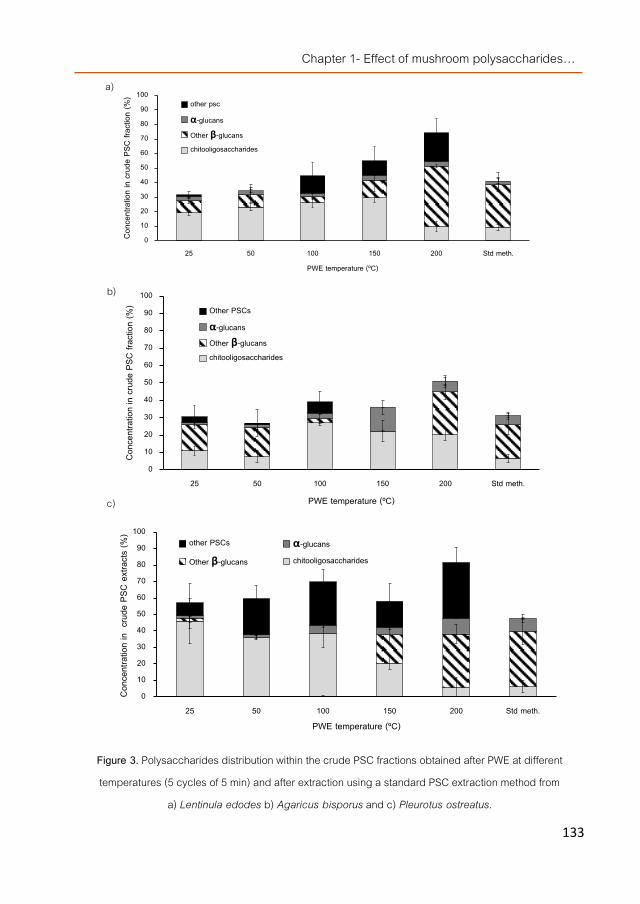
Chapter 1- Effect of mushroom polysaccharides…
133
Figure 3. Polysaccharides distribution within the crude PSC fractions obtained after PWE at different temperatures (5 cycles of 5 min) and after extraction using a standard PSC extraction method from
a) Lentinula edodes b) Agaricus bisporus and c) Pleurotus ostreatus.
0102030405060708090
100
25 50 100 150 200 Std meth.
Conc
entra
tion
in cru
de P
SC fr
actio
n (%
)
PWE temperature (ºC)
other psc
α-glucans
Other β-glucanschitooligosaccharides
0
10
20
30
40
50
60
70
80
90
100
25 50 100 150 200 Std meth.
Conc
entra
tion
in cru
de P
SC fr
actio
n (%
)
PWE temperature (ºC)
Other PSCs
α-glucansOther β-glucanschitooligosaccharides
0
10
20
30
40
50
60
70
80
90
100
25 50 100 150 200 Std meth.
Conc
entra
tion
in c
rude
PSC
extr
acts
(%)
PWE temperature (ºC)
other PSCs α-glucans
Other β-glucans chitooligosaccharides
a)
c)
b)

Chapter 1- Manuscript 1
134
The crude PSC fractions were digested in the presence of bile extracts in ratios 1:10 (bile extract: PSC extract), 1:30, 1:50, 1:75, and 1:100 but, although a slight reduction of GUDCA, GHDCA, TCDCA, and THDCA were observed as compared to the control digestion with increasing concentrations of the cereal and mushroom PSCs, they were only significant when the ratio was 1:100. Digestion of cellulose showed no reduction at any of the selected concentrations while the positive control, cholestyramine, showed a remarkable binding capacity already at ratios 1:10 (w/w).
Thus, the crude PSC fraction obtained from the cereals mixture (as positive control) and applied at ratio 1:100 was able to bind almost 40% of the four major bile acids showing no preference or binding affinity for any of the specific bile acids since their concentrations were almost equally reduced. The crude PSC fractions isolated from the PWE extracts obtained using temperatures lower than 200 °C showed none or insignificant bile acid-binding capacity; however, those obtained from the PWE extracts at 200 °C of A. bisporus and L. edodes showed similar binding capacity than the cereal. P. ostreatus showed lower binding capacity (23%) although still significantly higher than the negative control (Figure 4). When the crude PSC fractions obtained by the standard PSC extraction method were similarly digested (ratio 1:100) no significant differences were observed respect to their corresponding PWE extracts except for L. edodes. A slight reduction on the binding capacity of L. edodes and A. bisporus extracts (std. method) respect to the cereal fraction could be noticed.
Chitin and their chitooligosaccharides enhanced excretion of triglycerides in feces but showed no cholesterol-lowering properties [9] thus, the fact that the crude PSC fractions from the PWE extracts obtained at 25–150 °C showed no bile acid-binding properties was not surprising since most of the β-glucans present in these fractions were the water-soluble chitooligosaccharides deriving from chitin hydrolysis. Only chitosan (a de-N-acetylated derivative of chitin) and its generated chitooligosaccharides, showed hypocholesterolemic action [32]; however, no acid/alkaline treatments were given to the PWE extracts to induce chitin deacetylation or transformation into chitosan hydrolysis products and therefore the chitooligosaccharides detected were probably deriving only from chitin. Moreover, the fractions obtained from PWE

Chapter 1- Effect of mushroom polysaccharides…
135
extracts at 200 °C from L. edodes and P. ostreatus showing bile acid-binding activities contained very low chitooligosaccharides levels pointing to the rest of PSCs as the responsible for the observed activities.
Figure 4. Bile acid binding capacity of cellulose, cholestyramine and crude PSC fractions obtained from a cereals mixture and mushrooms by a standard PSC extraction method and by PWE at 200 ºC after an in
vitro digestion model (mixed in a ratio 1:100 bile extract: PSC fractions except for cholestyramine which was 1:10). * Denotes statistically significant differences (P < 0.05) between samples extracted by PWE and the
standard PSC extraction method.
Within these other PSCs, the fraction from P. ostreatus PWE extract at 200 °C showed similar concentration of “nonchitooligosaccharides or chitins”(or deaminated β-glucans, named in the figures as “other β-glucans”) than the fraction obtained using the standard PSC extraction method and higher amount of other PSCs (tentatively proteo-glucans) but both fractions showed the same bile acid-binding capacity pointing those deaminated β-glucans as the responsible for the activity as occurred in cereals [28]. The crude PSC fraction obtained from cereals using the standard method and used as control contained 47.9 mg β-glucans/100 mg PSC fraction, the one obtained from L. edodes PWE extract at 200 °C contained 41.7 mg deaminated β-glucans/100 mg and the one obtained using the standard extraction method 29.3 mg/100 mg PSC fraction. The bile acid-binding capacity of the latter fraction was lower than the PWE extract and the cereal
0
10
20
30
40
50
60
70
Cholestyramine Cellulose Cereals P. ostreatus L. edodes A. bisporus
Bile
acids
bind
ing ca
pacit
y (%
)
Std PSC extraction method ASE extraction method
*

Chapter 1- Manuscript 1
136
extract suggesting again that these compounds could be the responsible for the observed activities.
However, apparently not all the β-glucans from the three species were equally efficient in the binding capacity since both A. bisporus extracts (obtained from the PWE and standard method) contained respect. 24.6 and 19.21 mg/100 mg PSC fraction while L. edodes extracts almost double their β-glucan content and still both mushroom extracts showed bile acid-binding activities similar to each other and to those observed for the cereal extract.
Conclusions It can be concluded that PWE can be used to obtain β-glucans-rich fractions from edible
mushrooms with different biological activities and compositions depending on the selected temperature. If the extraction is carried out at 200 °C (5 cycles of 5 min) the obtained fractions contain higher percentage of PSC than those usually obtained by a standardized PSC extraction method facilitating their incorporation into food matrices for their technological manufacture (faster extraction, higher PSC concentration can be added with less extract, more homogeneous chemical composition etc.). Their bile acid-binding capacities were similar to the fractions obtained by the standard PSC extraction method and they were also in the range of the β-glucans obtained from a cereal foodstuff commercially available and claiming to be able of reducing cholesterol levels. Thus, the obtained mushrooms extracts will further be tested to define whether they can be used as bioactive ingredients to functionalize foods with hypocholesterolemic properties.
Acknowledgments The research was supported by the European Union within the 7th framework programme
(7FM-PEOPLE-2009-IIF project 251285), ALIBIRD-CM S2009/AGR-1469 regional program from the Community of Madrid (Spain) and AGL2010–21537 national R1D program from the Spanish Ministry of Science and Innovation. CTICH is also acknowledged for the cultivation and supplying of the mushrooms fruiting bodies.

Chapter 1- Effect of mushroom polysaccharides…
137
References [1]. Schepetkin, I.A. and M.T. Quinn, Botanical polysaccharides: Macrophage immunomodulation and therapeutic
potential. International Immunopharmacology, 2006. 6(3): p. 317-333. [2]. Drozdowski, L.A., et al., β-Glucan extracts inhibit the in vitro intestinal uptake of long-chain fatty acids and
cholesterol and down-regulate genes involved in lipogenesis and lipid transport in rats. The Journal of Nutritional Biochemistry, 2010. 21(8): p. 695-701.
[3]. Zacherl, C., P. Eisner, and K.-H. Engel, In vitro model to correlate viscosity and bile acid-binding capacity of digested water-soluble and insoluble dietary fibres. Food Chemistry, 2011. 126(2): p. 423-428.
[4]. Kahlon, T.S., M.H. Chapman, and G.E. Smith, In vitro binding of bile acids by okra, beets, asparagus, eggplant, turnips, green beans, carrots, and cauliflower. Food Chemistry, 2007. 103(2): p. 676-680.
[5]. Chen, Z.-Y., et al., Role and classification of cholesterol-lowering functional foods. Journal of Functional Foods, 2011. 3(2): p. 61-69.
[6]. Roupas, P., et al., The role of edible mushrooms in health: Evaluation of the evidence. Journal of Functional Foods, 2012. 4(4): p. 687-709.
[7]. Chen, J. and R. Seviour, Medicinal importance of fungal β-(1→3), (1→6)-glucans. Mycological Research, 2007. 111(6): p. 635-652.
[8]. Rop, O., J. Mlcek, and T. Jurikova, Beta-glucans in higher fungi and their health effects. Nutr Rev, 2009. 67(11): p. 624-31.
[9]. Harish Prashanth, K.V. and R.N. Tharanathan, Chitin/chitosan: modifications and their unlimited application potential—an overview. Trends in Food Science & Technology, 2007. 18(3): p. 117-131.
[10]. Aida, F.M.N.A., et al., Mushroom as a potential source of prebiotics: a review. Trends in Food Science & Technology, 2009. 20(11–12): p. 567-575.
[11]. Synytsya, A. and M. Novák, Structural diversity of fungal glucans. Carbohydrate Polymers, 2013. 92(1): p. 792-809.
[12]. Ramberg, J.E., E.D. Nelson, and R.A. Sinnott, Immunomodulatory dietary polysaccharides: a systematic review of the literature. Nutrition Journal, 2010. 9: p. 54-54.
[13]. Zhang, M., et al., Antitumor polysaccharides from mushrooms: a review on their isolation process, structural characteristics and antitumor activity. Trends in Food Science & Technology, 2007. 18(1): p. 4-19.
[14]. Cheung, Y.-C., et al., Molecular properties and antioxidant activities of polysaccharide–protein complexes from selected mushrooms by ultrasound-assisted extraction. Process Biochemistry, 2012. 47(5): p. 892-895.
[15]. Chen, W., et al., Optimization of ultrasonic-assisted extraction of water-soluble polysaccharides from Boletus edulis mycelia using response surface methodology. Carbohydrate Polymers, 2012. 87(1): p. 614-619.
[16]. Zeng, W.-C., et al., Characterization of antioxidant polysaccharides from Auricularia auricular using microwave-assisted extraction. Carbohydrate Polymers, 2012. 89(2): p. 694-700.

Chapter 1- Manuscript 1
138
[17]. Huang, S.-q. and Z.-x. Ning, Extraction of polysaccharide from Ganoderma lucidum and its immune enhancement activity. International Journal of Biological Macromolecules, 2010. 47(3): p. 336-341.
[18]. Wang, B.J., et al., Free radical scavenging and apoptotic effects of Cordyceps sinensis fractionated by supercritical carbon dioxide. Food and Chemical Toxicology, 2005. 43(4): p. 543-552.
[19]. Di, X., et al., Fingerprint profiling of acid hydrolyzates of polysaccharides extracted from the fruiting bodies and spores of Lingzhi by high-performance thin-layer chromatography. Journal of Chromatography A, 2003. 1018(1): p. 85-95.
[20]. Lo, T.C.T., et al., Pressurized water extraction of polysaccharides as secondary metabolites from Lentinula edodes. Journal of Agricultural and Food Chemistry, 2007. 55(10): p. 4196-4201.
[21]. Ramírez-Anguiano, A.C., et al., Radical scavenging activities, endogenous oxidative enzymes and total phenols in edible mushrooms commonly consumed in Europe. Journal of the Science of Food and Agriculture, 2007. 87(12): p. 2272-2278.
[22]. Jeurink, P.V., et al., Immunomodulatory capacity of fungal proteins on the cytokine production of human peripheral blood mononuclear cells. International Immunopharmacology, 2008. 8(8): p. 1124-1133.
[23]. Fox, J.D. and J.F. Robyt, Miniaturization of three carbohydrate analyses using a microsample plate reader. Analytical Biochemistry, 1991. 195(1): p. 93-96.
[24]. Vetter, J., Chitin content of cultivated mushrooms Agaricus bisporus, Pleurotus ostreatus and Lentinula edodes. Food Chemistry, 2007. 102(1): p. 6-9.
[25]. Smith, R.L. and E. Gilkerson, Quantitation of glycosaminoglycan hexosamine using 3-methyl-2-benzothiazolone hydrazone hydrochloride. Analytical Biochemistry, 1979. 98(2): p. 478-480.
[26]. Pang, C.-P., et al., Simultaneous determination of conjugated bile acids in human bile. Clinical Biochemistry, 1990. 23(1): p. 85-90.
[27]. Qiao, X., et al., Differentiation of various traditional Chinese medicines derived from animal bile and gallstone: Simultaneous determination of bile acids by liquid chromatography coupled with triple quadrupole mass spectrometry. Journal of Chromatography A, 2011. 1218(1): p. 107-117.
[28]. Benito-Román, Ó., E. Alonso, and M.J. Cocero, Pressurized hot water extraction of β-glucans from waxy barley. The Journal of Supercritical Fluids, 2013. 73(0): p. 120-125.
[29]. Jeon, Y.-J., F. Shahidi, and S.-K. Kim, Preparation of chitin and chitosa oligomers and their applications in physiological functional foods. Food Reviews International, 2000. 16(2): p. 159-176.
[30]. Gil-Ramírez, A., et al. Immunomodulatory properties of edible mushroom extracts obtained by pressurized solvent technologies. in 6th International Symposium of High Pressure Processes Technology. 2013. Belgrade, Serbia: Association of Chhemical Engineers of Serbia.
[31]. Kritchevsky, D., In vitro binding properties of dietary fibre. Eur J Clin Nutr, 1995. 49(3): p. S113-5. [32]. Sugano, M., et al., Hypocholesterolemic action of chitosans with different viscosity in rats. Lipids, 1988. 23(3): p.
187-191.
Chapter 1- Effect of mushroom polysaccharides…
139
Manuscript 2
Modulation of cholesterol-related gene expression by dietary fiber fractions from edible mushrooms
Víctor Caz1*, Alicia Gil-Ramírez2*, María Tabernero1, Mónica Santamaría1, Roberto Martín-Hernandez3, Francisco R. Marín2, Guillermo Reglero2,3, Carlota Largo1 and Cristina Soler-Rivas2
1Department of Experimental Surgery, Research Institute Hospital La Paz (IdiPAZ), Paseo de la Castellana 261, 28046 Madrid, Spain.
2Department of Production and Characterization of Novel Foods, CIAL – Research Institute in Food Science (UAM+CSIC), C/ Nicolas Cabrera 9, Campus de Cantoblanco, Universidad Autónoma de Madrid, 28049 Madrid, Spain. 3IMDEA Food Institute. Pabellón Central del Antiguo Hospital de Cantoblanco (Edificio nº 7), Crta. de Cantoblanco nº 8, 28049 Madrid, Spain.
*Same contribution
In preparation. May 2015

Chapter 1- Manuscript 2
140
Abstract Edible mushrooms are an interesting source of biological compounds such as dietary
fiber with different structures but similar hypocholesterolemic properties than cereal β-glucans. However, their underlying mechanisms are still poorly understood. The effect of β-glucan enriched fractions obtained from 3 mushroom species on cholesterol-related expression was studied in vitro by low density arrays (LDA) and the Pleurotus ostreatus DF-fraction selected for two animal trials in order to assess its potential palliative or preventive effect against diet-induced hypercholesterolemia. Plasma and liver lipids and fecal cholesterol of mice fed with high-cholesterol diet were examined. LDA assays were analyzed in jejunum, ileum, cecum and liver. Upon in vitro digestion, the P. ostreatus DF-fraction up-regulated FXR mRNA after 1 h and FDFT1 and NPC1L1 after 24 h in Caco2 cells, suggesting a possible cholesterol-lowering effect. In the palliative setting, the DF-fraction reduced hepatic triglyceride likely by inhibition of synthesis since mRNA Dgat1 was down-regulated. Plasma and liver biochemical data of mice fed the DF-fraction showed no changes in cholesterol and no relation with transcriptional modulation. The highest transcriptional modulation was induced in the jejunum. Simvastatin also failed reducing cholesterol once the hypercholesterolemia was induced. In the preventive setting, no changes in plasma and liver biochemical data were induced by the DF-fraction but in the liver modulated cholesterol-related genes expression similarly to simvastatin and ezetimibe. Thus, DF-fraction may be useful limiting hepatic steatosis but not diet-induced hypercholesterolemia in mice although its similar effect to hypocholesterolemic drugs on hepatic genes involved in cholesterol homeostasis warrants further dose-dependent studies.

Chapter 1- Effect of mushroom polysaccharides…
141
Introduction An extensive number of studies in human and animal models have evidenced that in
taking of dietary fibers (DF) has benefits for health maintenance and disease prevention including diabetes, cancer, and cardiovascular diseases. The term ´dietary fiber´ usually includes those carbohydrates that resist digestion and cannot be absorbed in the small intestine being partial or completely fermented in the large intestine [1].
The mechanisms involved in the hypocholesterolemic effects of dietary fiber remain still unclear. Satiation and satiety effects or direct binding of bile acids within their structure underlie the cholesterol-lowering properties of insoluble DF while soluble DF could possibly use their water binding capabilities to increase chyme viscosity reducing the diffusion rate of bile acids [2]. Both mechanisms induce significant changes on hepatic cholesterol metabolism and on the synthesis, processing (in the intravascular compartment) and catabolism of lipoproteins because they lower hepatic cholesterol pools as a result of cholesterol being diverted to bile acid synthesis and less cholesterol delivery to the liver through chylomicron remnants [3]. At the molecular level, DF from cereals were able to down-regulate the expression of several genes involved in the lipid metabolism including SREBF1 (gene encoding two sterol regulatory element binding transcription factors: SREBP-1a and SREBP-1c) and the expression of SREBF2 gene involved in the regulation of cholesterol synthesis in NCI-H716 intestinal cell line [4] while in mice, hamster or rats, only some specific DFs were able to increase the hepatic expression of the Hmgcr gene (3-hydroxy-3-methylglutaryl-CoA reductase) [5-7]. The Hmgcr up-regulation could be due to a depletion of cholesterol pools resulting from an increased excretion of bile cholesterol. The induced cholesterol reduction also led to up-regulation of the low-density lipoprotein receptor (Ldlr) synthesis and thereby accelerated the transportation of LDL-cholesterol from the blood into hepatocytes [7]. Other reports noticed decrease in the ileal level of ApoB mRNA (apolipoprotein B) after 3 weeks supplementation of rats with beet-DF plus low-fat diets [8], or up-regulation of ileal Nr1h4 mRNA (encoding the nuclear receptor FXR, farnesoid X receptor) simultaneously with its hepatic down-regulation after modified corn-DF administration [9].
Chapter 1- Manuscript 2
142
Besides legumes, fruits, vegetable and particularly cereals, edible mushrooms are also an interesting source of DFs showing similar beneficial effects including hypocholesterolemic effects [10, 11]. DF extracts obtained from edible mushrooms such as Agaricus bisporus, Lentinus edodes, Flammulina velutipes and Grifola frondosa were able to lower cholesterol levels in rats fed with a cholesterol-free diet during 4 weeks [12, 13]. The hepatic Ldlr mRNA level in the groups supplemented with F. velutipes or A. bisporus was significantly higher than the control group. Similar up-regulation was observed after supplementation with P. ostreatus fruiting bodies [14]. Thus, this molecular effect and the enhancement of fecal cholesterol excretion were pointed as the responsible mechanisms for the observed reduction of total cholesterol levels in serum. Other studies carried out using other mushroom species such as Grifola frondosa mentioned hepatic up-regulation of the Abcg5/8 genes (genes encoding cholesterol transporters: ATP-binding cassette sub-family G proteins, involved in the reverse cholesterol transport (enterocytic cholesterol efflux)) [15]. Mushrooms DF fractions are constituted mainly by water-insoluble polysaccharides (PSC), with β-glucans and chitins being the most representatives, while the level of water-soluble PSC is usually lower than 10% (depending on specie) [1].
However, the described molecular studies on edible mushroom DFs were carried out using cholesterol-free or standard diets, therefore animals showed no hypercholesterolemia or altered cholesterol-related biochemical parameters. Functional foods with hypocholesterolemic properties (such as products enriched in phytosterols, cereal β-glucans, etc.) are usually recommended by physicians/nutritionists to people with slight to moderate hypercholesterolemia, thus, if a new functional product (based on mushroom DFs) is intended to be developed, the effect of such a fraction should be tested on individuals with certain level of hypercholesterolemia. Moreover, other reports indicated that when hypercholesterolemic animals were regularly fed with whole fruiting bodies, lowering of cholesterol in serum was noticed [16].
Thus, in order to study whether the dietary fiber fraction (DF-fraction) of edible mushrooms can be used as hypocholesterolemic ingredient to design novel functional foods and study their influence at the molecular level under cholesterol-altered physiological conditions, the modulation of mRNA expression of genes related to the cholesterol metabolism was investigated

Chapter 1- Effect of mushroom polysaccharides…
143
on two models of hypercholesterolemic mice using low-density arrays (LDA). Firstly, a preliminary in vitro study was carried out using Caco2 cell cultures to select one DF-fraction out of the three most consumed mushrooms (Agaricus bisporus, Pleurotus ostreatus and Lentinus edodes) to reduce the number of mice and assays. Then, the two different in vivo experiments were carried out in order to compare the effect of a mushroom DF-fraction with two hypocholesterolemic drugs.
Materials and Methods
Raw material and preparation of the dietary fiber-enriched extracts Mushroom strains Fresh fruiting bodies of Agaricus bisporus L. (Imbach) ¨Fungisem H-
15¨, Pleurotus ostreatus (Jacq.Ex Fr.) Kummer strain ¨Gurelan H-107¨ and Lentinus edodes S. (Berkeley) from ¨Sylvan 4312¨ from the first flush were harvested, cut in slices, lyophilized and ground until a dry powder was obtained following the procedure described by [16]. Resulting mushroom powders were utilized as starting material to extract the dietary fibers (DF). Dietary fiber-enriched fractions from the three mushroom species were prepared according to the method of [17].
Standards and reagents Solvents as hexane (95%), chloroform (HPLC grade) and methanol (HLPC grade) were
obtained from LAB-SCAN (Gliwice, Polland) and absolute ethanol and sea sand from Panreac (Barcelona, Spain). Potassium hydroxide, ascorbic acid, BHT (2,6-Di-tert-butyl-p-cresol), cholesterol, cholic acid, ergosterol, congo red as well as hexadecane were purchased from Sigma-Aldrich Química (Madrid, Spain). Simvastatin was obtained from Cinfa Laboratories (Spain) and ezetimibe (Ezetrol) from Merck Sharp & Dohme (Spain). Schizophyllan was pursached from Contripo Biotech s.r.o. (Czech Republic). All other reagents and solvents were used of analytical grade.
β-glucans content of DF-fractions The β-glucan content of the obtained DF-fractions (50 mg) was evaluated in duplicate by
a β-glucan determination kit specific for mushrooms and yeasts (Megazyme, Barcelona, Spain)

Chapter 1- Manuscript 2
144
following the instructions of the user’s manual and as described in [18]. The amount of β-(13),(16)-glucans was also determined following the method of [19] using a red congo concentration of 0.08% (w/v) and a schizophyllan as standard curve and chitins were determined as described in [18].
In vitro digestion Obtained DF-fractions were submitted to an in vitro digestion according to [18]. Briefly,
the extracts were digested in 1 ml of 0.01 M HCl for 1 h at 37 ºC with gentle agitation (Orbital incubator S150, Stuart, Stone, UK). Afterwards, the sample pH was adjusted to 6.3 with 0.1 ml of 0.1 M NaOH. Then, 4 ml of bile extract (14.3 mg in Tris-maleate buffer pH 6.3) and 5 ml of pancreatin (10 mg in Tris-maleate buffer pH 6.3) were added and the mixture was incubated for 1 h at 37 ºC. After digestion, the samples were added to differentiated Caco2 cultures. A control sample was prepared following the same digestion protocol but without the addition of DF fractions.
Caco2 cell cultures Human colorectal adenocarcinoma cell line Caco2 (ATCC HTB-37) obtained from the
Americam Type Culture Collection (Rockville, MD, USA) were maintained in Dulbecco´s Modified Eagle’s Medium (DMEM) with high glucose content (4.5 g/l) and supplemented with 10% fetal bovine serum, 1% penicillin-streptomycin, and 1% non-essential amino acids at 37 ºC in at humidified atmosphere containing 5% CO2.
Firstly, the cytotoxicity was evaluated using 3-(4,5-dimethylthiazol)-2,5-diphenyl tetrazolium bromide (MTT) according the method published by Mosmann (1983) [20].
Then, cells were seeded onto a 9.5 cm2 grown area 6 well flat bottom plate (Costar, Corning, USA) at a density of 5 × 10⁵ cell per insert (3.3 × 10⁵ cell/mL). Culture medium was replaced every three days and cells were allowed to generate a monolayer for 21 days before experiments. The digested DF fractions were applied at subtoxic concentrations (4 μl/ml) and left incubating at 37 ºC and 5% CO2 for 1 h or 24 h. Afterwards, cells were separated from the

Chapter 1- Effect of mushroom polysaccharides…
145
membranes by incubation at 37 ºC during 30 min with Triton 0.1% (200 μl) and submitted to RNA extraction. Four replicates per sample were carried out.
Animals and diets Male C57BL/6JRj mice were purchased from Janvier SAS (Le Genest Saint Isle, France).
Mice were maintained four per cage in temperature-, humidity- and light-controlled conditions (24 ± 2 ºC, 40%-60% humidity, 12:12 hour light: dark cycle) and had free access to water and food. They were randomly divided into five groups, 2 control groups and 3 treated groups. Normal control group (NC, n=6) was fed a standard diet (Safe Rodent Diet A04, Augy, France) and the hypercholesterolemic control group (HC, n=8) was fed a high-cholesterol diet. The treated groups (n=8) were given high-cholesterol diet supplemented with their corresponding mushroom extract or control drug and were identified as follows: PE (Pleurotus ostreatus DF-fraction), SV (simvastatin, as a positive control for inhibition of cholesterol biosynthesis), and EZ (ezetimibe as a positive control for inhibition of cholesterol absorption). The animal intervention was carried out following two experimental settings in order to evaluate whether the P. ostreatus DF-fraction might act as a palliative (experiment 1: sequential supplementation) or as preventive agent (experiment 2: simultaneous supplementation) against diet-induced hypercholesterolemia. In the experiment 1 (sequential supplementation) mice were fed high-cholesterol diet for 4 weeks (from 5 to 9 weeks of age) followed by another 4 weeks of high-cholesterol diet plus mushroom extract (from 10 to 13 weeks of age) while in the experiment 2 (simultaneous supplementation), mice were simultaneously fed high-cholesterol diet and the mushroom extract for 4 weeks (from 10 to 13 weeks of age). The composition of diets, the dose per day of mushroom extract, drug or β-glucans, as well as the energy that received the mice daily are shown in Table 1. Body weight was registered for all mice before the experimental feeding period and at the end of the experiments. Wet mass of the liver was also registered. Animal studies were approved by our institution’s Animal Welfare and Ethics Committee and were carried out according to Spanish and European legislation (RD 53/2013, 2010/63/EU, respectively).

Chapter 1- Manuscript 2
146
Feces, plasma and liver lipid analyses, tissue collection Following the experimental feeding period, mice were sacrificed by intracardiac
exsanguination under anesthesia with 1.5% isoflurane, and plasma was collected and stored at -80 ºC before use. Jejunum, ileum, cecum, and liver samples were removed and immediately frozen in liquid nitrogen and stored at -80 ºC. Feces were collected at the beginning and at the end of the experimental feeding period, and maintained at -20 ºC until further use. Cholesterol extraction from the stored feces (300 mg) and analysis by GC-MS were carried out as described in [16]. Plasma levels of total cholesterol, triglyceride, HDL and LDL were measured in duplicate for each sample using a Covas C311 Autoanalyzer (Roche, Spain). The lipids of the liver were extracted according to the manufacturer’s protocol. The cholesterol and triglycerides in the extracts of the liver were measured in duplicate for each sample by colorimetric methods using kits (BioVision Inc., CA, USA).
Table 6. Composition of diets.
Components NC HC PE SV EZ Carbohydrates (%) 59.9 59.9 59.9 59.9 59.9 Proteins (%) 16.1 16.1 16.1 16.1 16.1 Lipids (%) 3.1 3.1 3.1 3.1 3.1 Fibres* (%) 3.9 3.9 3.9 3.9 3.9 Mineral mixture** (%) 5.1 5.1 5.1 5.1 5.1 Moisture (%) 11.9 8.9 7.4 8.9 8.9 Cholesterol (%) 0.0 2.0 2.0 2.0 2.0 Cholic acid (%) 0.0 1.0 1.0 1.0 1.0 Mushroom extract or drug (%) 0.0 0.0 1.5 0.025 0.005 Mushroom extract or drug (mg/mouse/day)
0.0 0.0 60.0 1.0 0.2
-glucans (%) nm nm 0.6 nm nm -glucans (mg/mouse/day) nm nm 24.0 nm nm Energy (Kcal/mouse/day) 13.37 14.39 14.39 14.39 14.39
NC, normal control; HC, hypercholesterolemic control; PE: Pleurotus extract; SV: simvastatin; EZ: ezetimibe; *-glucans excluded; **A04 mineral mixture; n.m.: not measured.

Chapter 1- Effect of mushroom polysaccharides…
147
Statistical analysis for biochemical data All values were expressed as mean ± SD. The SPSS software, version 15.0 (Lead
Technologies, Chicago, IL, USA), was used to determine whether the variables differed among treatment groups in the in vivo experiments. The effects of treatments were assessed by one-way ANOVA followed by Tukey’s test when the ANOVA identified significant treatment effects. Differences with P < 0.05 were considered statistically significant.
RNA extraction from cells and mice tissues and quantitative real-time PCR for LDA Total RNA from cell cultures or mice tissues was extracted by magnetic bead technology
using a pureLinkTM Total RNA kit TRIzol® Plus RNA Kit (Invitrogen, Carlsbad, CA, USA) in an iPrepTM Purification Instrument (Invitrogen) programmed with an iPrepTM total RNA card (Invitrogen) according to manufacturer instructions. The RNA concentration was determined by spectrophotometry at 260 nm and the purity of the extracted RNA was calculated from ratio of absorbance at 260:280 nm and 260:230 nm in a NanoDrop ND-1000 (NanoDrop Technologies Inc., Wilmington, DE, USA).
An amount of 400 ng of total RNA from each sample was reversed transcribed using the High Capacity cDNA Reverse Transcription Kit (Applied Biosystems, Framinghan, MA, USA). The obtained cDNA (100 μl per port) were loaded into micro-fluid cards designed for low-density array (LDA). Before sealing them, the cards were centrifuged twice on a Sorvall centrifuge at 1200 rpm for 1 min. Finally, the micro- fluid cards were run in a 7900HT Fast Real-Time PCR system (Applied Biosystems). Amplification conditions were 50 ºC for 2 min, 94.5 ºC for 10 min, followed by 40 cycles with 97 ºC with 30 s and 59.7 ºC for 1 min.
The micro-fluid card was constructed using respectively human and mouse commercial available assays (Applied Biosystems) for several genes related to the cholesterol metabolism, some internal standard and housekeeping gene that are described in detail elsewhere [21]. The cholesterol-related genes were ABCA1 (ATP-Binding Cassette, Sub-Family A, Member 1), ABCG5 and ABCG8 (ATP-binding cassette, sub-family G (WHITE), members 5 and 8), ACAT1 and ACAT2 (Acetyl-CoA acetyltransferase 1 and 2), APOB (Apolipoprotein B), DGAT1 and DGAT2

Chapter 1- Manuscript 2
148
(Diacylglycerol O-acyltransferase 1 and 2), FDFT1 (Farnesyl-diphosphate farnesyltransferase 1), HMGCR (3-hydroxy-3-methylglutaryl-CoA reductase), LDLR (Low density lipoprotein receptor), MTTP (Microsomal triglyceride transfer protein), NPC1L1 (NPC1 (Niemann-Pick disease, type C1 like 1), NR1H3 and NR1H4 (Nuclear receptor subfamily 1, group H, member 3 and 4), SOAT1 and SOAT2 (Sterol O-acyltransferase 1 and 2), SREBF1 and SREBF2 (Sterol regulatory element binding transcription factor 1 and 2). Comparative analysis of each of these genes was performed using specialized computer programs SDS2.3 and RQ 2.1 (Applied Biosystems).
Gene expression statistical data analysis The expression of each gene was measured at least in duplicate and each biological
group included a minimum of 3 replicates. In order to control for potential outliers, Grubb’s test was applied to technical replicates with a threshold of 0.05. Biological replicates were quality controlled using the Median Absolute Deviation test (MAD test). Samples of the same biological group with a MAD score higher than 3 were removed from the analysis. Genorm algorithm [22] was used to identify the most stable reference genes for normalization. Genes GUSB and HPRT1 were found to be the most stably expressed in Caco2 cell cultures and in the mouse tissue samples, genes Hprt1 and Polr2a were the most stably expressed in cecum and liver tissues, Gusb and Polr2a in jejunum and ileum tissues. Relative gene expression levels were calculated
by using the 2-ΔΔCT method [23], using these identified most stable genes as internal control genes in each group. Statistical significance of the results was assessed using a Limma test, and Benjamini-Hochberg false discovery rate procedure was subsequently applied. Adjusted p-values lower than 0.05 were considered significant. All these calculations were carried out with Real Time StarMiner 4.5® (Integromics S.L, Spain).
Results
Modulation of the mRNA expression pattern in Caco2 cell cultures Firstly, three mushroom species (A. bisporus, L. edodes, and P. ostreatus) were selected
because they showed in vitro bile acid-binding capacity and this ability could be related to hypocholesterolemic activities [18]. Then, digestates obtained after in vitro digestion of the

Chapter 1- Effect of mushroom polysaccharides…
149
mushroom DF-fractions were applied to Caco2 cell cultures to investigate whether they were able of modulating changes in the mRNA expression profile of genes related to the cholesterol metabolism. Results indicated that one hour after application, only the digestion products obtained from two mushrooms (A. bisporus and P. ostreatus) were able to induce significant overexpression of NR1H4 (gene encoding farnesoid X receptor, FXR) (Figure 1a). Moreover, the digestate of the A. bisporus DF-fraction up-regulated ACAT2 gene and down-regulated MTTP and SOAT2 genes.
a)
b)
Figure 2. Relative mRNA expression (Log10) of Caco2 cells cholesterol-related genes after a) 1 h and b) 24 h application of the digested DF-fraction obtained from A. bisporus, L. edodes or P. ostreatus. Indicated
-0.6
-0.4
-0.2
0
0.2
0.4
0.6
0.8
ACAT2 DGAT1 FDFT1 LDLR MTTP NPC1L1 NR1H4 SOAT1 SOAT2 SREBF1
Log1
0RQ
A. bisporusL. edodesP. ostreatus
-0.2
-0.1
0
0.1
0.2
0.3
0.4
ACAT2 DGAT1 FDFT1 LDLR MTTP NPC1L1 NR1H4 SOAT1 SOAT2 SREBF1
Log1
0RQ
A. bisporusL. edodesP. ostreatus

Chapter 1- Manuscript 2
150
genes are only those pointed as significant (P<0.05) compared with their controls (addition of the digestion product without DF addition). The expression of each gene was measured at least in duplicate and each
biological group included a minimum of 3 replicates.
However, when the treated cells were incubated for a longer period (24 h), overexpression of other genes were induced by the digestates obtained from the DF-fractions of L. edodes and particularly of P. ostreatus (Figure 1b) acting differently than those of A. bisporus that showed no effect. Both digestates were able to induce significant overexpression of the NPC1L1 (Niemann-Pick C1-like 1), gene directly involved in the cholesterol absorption. Digestates from the DF-fraction of L. edodes induced mRNA LDLR expression while those from P. ostreatus induced mRNA expression of other cholesterol-related genes such as DGAT1, FDFT1, SOAT1, and SREBF1 and inhibited mRNA expression of SOAT2.
The fractions of the 3 mushroom species contained similar polysaccharide compositions [18] including lower α-glucan and chitin contents than β-glucans. However, those β-glucans apparently showed different solubilities and conformations (Table 2). The DF-fraction of A. bisporus contained less β-(1→3),(1→6)-glucans than the other two DF-fractions and they were not soluble in KOH. Most of the β-glucans from the L. edodes DF-fraction showing conformation in triple helix (those with β-(1→3),(1→6) branching) were mainly soluble in alkalis while those of the P. ostreatus DF-fraction were more soluble in KOH than in NaOH.
Table 7. Total β-glucan concentrations of the DF fractions obtained from the 3 mushroom species and their distribution as β-(1→3),(1→6)-glucans (conformation in triple helix) or chitins.
DF fractions
Total β-glucans mg/100mg
dw
β-(1→3),(1→6)-glucans (mg/100mg dw)
Chitins mg/100mg
dw KOH
fraction HCl
fraction NaOH fraction Total
A. bisporus 25.87 ± 7.87 0.0 ± 0.01 1.01 ± 0.03 1.04 ± 0.06 2.05 ± 0.14 6.67 ± 2.23
L. edodes 38.41 ± 6.50 2.54 ± 0.44 0.76 ± 0.11 2.89 ± 0.17 6.19 ± 1.01 9.13 ± 1.76
P. ostreatus 39.87 ± 11.53 2.94 ± 0.54 0.00 ± 0.00 2.57 ± 0.60 5.51 ± 1.61 6.10 ± 3.56

Chapter 1- Effect of mushroom polysaccharides…
151
Effect of mushroom extract on body and liver weight and fecal cholesterol excretion Food intake was approximately 4 g/d per mouse throughout the experimental feeding
period in all groups, with no significant difference among groups (data not shown). All mice adapted immediately to the experimental diets. No differences were also noticed in initial body weight (Table 3). The normal control group gained weight over the feeding period in the sequential and simultaneous experiments. As expected, the remaining groups showed a slight tendency to lose weight since diets supplemented with cholate and cholesterol induced even higher weight loss in C57BL/6J mice [24]. No significant differences in final body weight of the treated groups were noticed compared with the hypercholesterolemic control group either in the sequential or simultaneous experiment, except for the simvastatin group where a significantly lower final body weight was recorded compared to all other groups, including the hypercholesterolemic control group. In the sequential experiment, liver weight was significantly increased in the hypercholesterolemic controls and the treated groups, except the simvastatin group, compared with the normal control group. Similar results for liver-to-body-weight ratio were observed, with a significant increase in all groups with respect to the normal control group. In the simultaneous experiment, liver weight was significantly increased in the hypercholesterolemic controls compared with the normal control group, and liver-to-body-weight ratio was significantly increased in the hypercholesterolemic controls and the PE group. The simvastatin and ezetimibe groups showed a significantly lower liver weight compared with hypercholesterolemic controls. Similar results for liver-to-body-weight ratio were observed, although only the ezetimibe group reached statistical significance.
No cholesterol was detected in feces of normal controls but hypercholesterolemic controls significantly increased their cholesterol excretion either in the sequential or simultaneous supplementation compared with normal controls. In the sequential experiment, there was no significant difference in cholesterol excretion between hypercholesterolemic controls and the treated groups, except for mice fed the P. ostreatus DF-fraction extract where a significantly lower cholesterol excretion was observed. In the simultaneous experiment, the slight higher cholesterol excretion was significant for the ezetimibe group compared with hypercholesterolemic controls

Chapter 1- Manuscript 2
152
but not for mice fed with the P. ostreatus DF-fraction. No differences were noticed in mice treated with simvastatin with respect to hypercholesterolemic controls.
Table 8. Effect of mushroom extract on body and liver weight and fecal cholesterol excretion.
Modulation of the lipid profile of plasma and liver in the animal intervention
Sequential supplementation Plasma cholesterol levels increased 3.3-fold in hypercholesterolemic controls compared
with normal controls (Table 4). In mice fed the P. ostreatus DF-fraction or treated with simvastatin, plasma cholesterol levels remained unchanged compared with those in hypercholesterolemic controls, while a reduction reaching normal levels was observed in the ezetimibe group. There was no significant difference in the plasma HDL levels between normal controls and hypercholesterolemic controls. No significant differences between mice fed the mushroom DF-fraction or treated with simvastatin and hypercholesterolemic controls were observed,
Sequential supplementation
Variable NC HC PE SV EZ Initial BW (g) 19.2 ± 0.84 20.0 ± 0.71 19.3 ± 0.54 19.3 ± 0.60 19.8 ± 0.92
Final BW (g) 25.0 ± 0.69 18.9 ± 1.07a 18.6 ± 1.18a,c 15.4 ± 1.52a,b,d 19.5 ± 0.93a
LW (g) 1.25 ± 0.15 1.6 ± 0.13a 1.83 ± 0.15a,c,d 1.46 ± 0.31 1.56 ± 0.22a
LW/BW (%) 4.99 ± 0.62 8.5 ± 0.56a 9.80 ± 0.63a,d 9.43 ± 1.34a,d 7.99 ± 1.15a
Cholesterol (mg/g feces)
nd 61.8 ± 0.47a 52.3 ± 4.62a,b 65.4 ± 7.41a 60.2 ± 14.74a
Simultaneous supplementation Initial BW (g) 23.4 ± 0.65 23.2 ± 0.50 23.1 ± 0.62 23.2 ± 0.58 23.2 ± 0.68
Final BW (g) 24.7 ± 0.54 21.8 ± 0.57a 20.2 ± 1.22a,c 14.9 ± 0.73a,b,d 20.3 ± 1.66a
LW (g) 1.27 ± 0.15 1.68 ± 0.31a 1.53 ± 0.14c 0.96 ± 0.10b 1.26 ± 0.20b
LW/BW (%) 5.14 ± 0.63 7.69 ± 1.44a 7.54 ± 0.59a,d 6.41 ± 0.44 6.18 ± 0.63b
Cholesterol (mg/g feces)
nd 58.4 ± 2.10a 64.1 ± 8.41a 59.5 ± 7.96a 65.1 ± 2.58a,b
NC, normal control; HC, hypercholesterolemic control; PE: Pleurotus extract; SV: simvastatin; EZ: ezetimibe. BW, body weight; LW, liver weight; nd, not detected. aP<0.05 vs. NC; bP<0.05 vs. HC; cP<0.05 vs. SV; dP<0.05 vs. EZ.

Chapter 1- Effect of mushroom polysaccharides…
153
although interestingly, the mushroom DF-extract slightly increased HDL levels by another 9.3% as compared with hypercholesterolemic controls. Ezetimibe induced a significant reduction. The LDL levels were markedly increased by 6.6-fold in hypercholesterolemic controls compared with normal controls but remained unchanged in mice fed the mushroom DF-fraction or treated with simvastatin with respect to hypercholesterolemic controls. Reduction was noticed with the administration of ezetimibe as compared with hypercholesterolemic controls. In addition to the plasma lipid profile, two atherogenic indexes were calculated: total cholesterol/HDL and LDL/HDL ratios. Compared with normal controls, the total cholesterol/HDL and the LDL/HDL ratios were significantly increased in hypercholesterolemic controls by 2.1- and 5.3-fold, respectively. It is noteworthy that, although slight and no significantly, that negative balance in both indexes was improved in mice fed the P. ostreatus DF-fraction compared with hypercholesterolemic controls, mainly due to the increase in HDL levels. A significant reduction in both indexes was noticed in the ezetimibe group but not in the simvastatin group. Hypercholesterolemic controls and treated groups showed a reduction in plasma triglyceride levels with respect to normal controls. Compared with hypercholesterolemic controls, treated groups showed quite similar plasma triglyceride levels.
Liver cholesterol levels were significantly increased by 4.9-fold in hypercholesterolemic controls compared with normal controls. In contrast with the lack of effect of the mushroom DF-fraction on total plasma cholesterol, a 21.4% reduction in liver cholesterol levels with respect to hypercholesterolemic controls was observed, although it did not reach statistical significance. Simvastatin had no effect while ezetimibe produced a significant reduction compared with hypercholesterolemic controls. No significant differences in liver triglyceride levels were detected between hypercholesterolemic and normal controls. Treated groups showed a significant reduction with respect to normal and hypercholesterolemic controls and mice treated with

Chapter 1- Manuscript 2
154
simvastatin or ezetimibe showed significant lower values compared with those of mice fed the P. ostreatus DF-fraction.
Table 9. Plasma and liver lipid profile in mice treated for 4 weeks with the P. ostreatus DF-fraction, simvastatin or ezetimibe simultaneously with high-cholesterol diet after 4 weeks of high-cholesterol diet alone (sequential supplementation).
Simultaneous supplementation Plasma cholesterol levels increased 1.9-fold in hypercholesterolemic controls compared
with normal controls (Table 5). There was no significant difference between the mushroom DF-fraction and hypercholesterolemic control groups. Simvastatin and ezetimibe produced a significant reduction with ezetimibe the most effective. Plasma HDL levels were increased in hypercholesterolemic controls compared with normal controls. There was no significant differences in plasma HDL levels between the mushroom DF-fraction and hypercholesterolemic control groups while treatment with simvastatin or ezetimibe induced a significant reduction.
Variable
Plasma level (mg/dl)
NC HC PE SV EZ TC 65.33 ± 26.63 214.2 ± 97.30a 207.1 ± 36.71a,d 156.2 ± 31.80a,d 59.43 ± 19.03b
HDL 59.80 ± 25.07 89.70 ± 27.97 98.06 ± 17.93d 81.90 ± 21.92 47.91 ± 10.49b
LDL 23.40 ± 11.03 155.0 ± 77.38a 152.6 ± 28.07a,d 102.5 ± 24.01a,d 23.40 ± 14.94b
TC/HDL 1.10 ± 0.07 2.31 ± 0.38a 2.12 ± 0.16a,d 1.95 ± 0.21a,d 1.22 ± 0.16b
LDL/HDL 0.31 ± 0.13 1.65 ± 0.35a 1.57 ± 0.17a,d 1.35 ± 0.22a,d 0.46 ± 0.23b
TG 72.00 ± 20.00 47.25 ± 8.41a 39.43 ± 10.05a 46.29 ± 11.04a 43.43 ± 8.30a
Liver level (mg/g liver) TC 0.22±0.09 1.09±0.23a 0.86±0.46a 1.44±0.43a,d 0.45±0.20b
TG 4.49±0.87 3.72±0.61 2.29±0.60a,b,c,d 0.78±0.18a,b 0.88±0.52a,b NC, normal control; HC, hypercholesterolemic control; PE: Pleurotus extract; SV: simvastatin; EZ: ezetimibe. TC, total cholesterol; HDL, high-density lipoprotein cholesterol; LDL, low-density lipoprotein cholesterol; TG, triglyceride. aP<0.05 vs. NC; bP<0.05 vs. HC; cP<0.05 vs. SV; dP<0.05 vs. EZ. The results are mean ± SD of six (NC group) or eight (remaining groups) mice on each diet and were analyzed by one-way
ANOVA and the Tukey’s procedure. The assays were performed in duplicate for each sample.

Chapter 1- Effect of mushroom polysaccharides…
155
A 16-fold increase in plasma LDL levels was observed in hypercholesterolemic controls compared with normal controls. Mice fed with mushroom DF-fraction showed no significant changes compared with hypercholesterolemic controls while simvastatin and ezetimibe groups showed significant reductions. The total cholesterol/HDL ratio was moderately increased (P=0.071) in hypercholesterolemic controls compared with normal controls. No significant differences in total cholesterol/HDL ratio were detected between hypercholesterolemic controls and treated groups. A 10.3-fold increase in the LDL/HDL ratio was observed in hypercholesterolemic controls compared with normal controls. Similar values were observed in treated groups except for the ezetimibe group where a significant reduction compared with hypercholesterolemic controls was observed. Plasma triglyceride levels were reduced in hypercholesterolemic controls with respect to normal controls. Compared to hypercholesterolemic controls, similar plasma triglyceride levels were observed in the treated groups.
Table 10. Plasma and liver lipid profile in mice treated for 4 weeks with the P. ostreatus DF-fraction, simvastatin or ezetimibe simultaneously with high-cholesterol diet (simultaneous supplementation).
Variable Plasma level (mg/dl)
NC HC PE SV EZ TC 120.8 ± 18.58 226.57 ± 16.64a 209.00 ± 40.33a,c,d 132.00 ± 13.37b 95.00 ± 14.14b
HDL 84.69 ± 17.11 118.11 ± 13.07a 97.50 ± 21.36c,d 64.35 ± 9.81b 64.35 ± 17.57b
LDL 10.03 ± 3.81 160.46 ± 25.42a 143.00 ± 29.46a,c,d 91.65 ± 17.30a,b,d 34.13 ± 16.11b
TC/HDL 1.45 ± 0.15 1.93 ± 0.20 2.16 ± 0.18a,d 2.10 ± 0.44a 1.58 ± 0.51
LDL/HDL 0.12 ± 0.04 1.36 ± 0.13a 1.48 ± 0.11a,d 1.47 ± 0.43a,d 0.62 ± 0.57b
TG 161.43 ±
49.42 50.29 ± 3.73a 38.00 ± 4.20a 42.00 ± 12.54a 68.50 ± 14.88a
Liver level (mg/g liver) TC 0.65 ± 0.23 0.98 ± 0.38 1.39 ± 0.21a,d 1.94 ± 0.64a,b,d 0.59 ± 0.42
TG 3.03 ± 0.85 2.35 ± 0.69 3.47 ± 0.97c,d 0.38 ± 0.18a,b 1.03 ± 0.51a,b NC, normal control; HC, hypercholesterolemic control; PE: Pleurotus extract; SV: simvastatin; EZ: ezetimibe. TC, total cholesterol; HDL, high-density lipoprotein cholesterol; LDL, low-density lipoprotein cholesterol; TG, triglyceride. aP<0.05 vs. NC; bP<0.05 vs. HC; cP<0.05 vs. SV; dP<0.05 vs. EZ. The results are mean ± SD of six (NC group) or eight (remaining groups) mice on each diet and were analyzed by
one-way ANOVA and the Tukey’s procedure. The assays were performed in duplicate for each sample.

Chapter 1- Manuscript 2
156
There was no significant difference in liver cholesterol levels between normal and hypercholesterolemic controls. Liver cholesterol levels in the simvastatin group were significantly higher than those in the hypercholesterolemic control group but no significance differences in the mushroom DF-fraction or ezetimibe groups were observed compared with hypercholesterolemic controls. No significant differences in liver triglyceride levels were detected between normal controls and hypercholesterolemic controls or between the mushroom DF-fraction group and hypercholesterolemic controls. Liver triglyceride levels in mice treated with simvastatin or ezetimibe were significantly lower than those in hypercholesterolemic controls.
Modulation of the mRNA expression pattern in the animal intervention
Sequential supplementation In the jejunum, treatment with either simvastatin or ezetimibe inhibited the expression of
Srebf1 and Srebf2 and simvastatin further inhibited other genes such as Fdft1, Hmgcr (involved in synthesis), Ldlr (involved in transport), Npc1l1 (involved in absorption) and Nr1h3 and Nr1h4 (genes encoding respect. liver X receptor (LXR) and farnesoid X receptor (FXR), two nuclear receptors involved in regulation of lipid homestasis) (Figure 2a).
In addition, administration of the P. ostreatus DF-fraction induced significant over-expression of 10 genes related to the cholesterol metabolism with the higher levels for Fdft1 (100 fold), and Abca1 and Npc1l1 (10 fold). On the contrary, it inhibited the expression of Nr1h4 and Srebf2 (regulatory genes) in a similar degree than simvastatin and ezetimibe. Slight transcriptional changes by the DF-fraction or the two drugs were observed in the ileum, not reaching statistical significance (data not shown). However, a higher transcriptional regulation was found in the cecum being the DF-fraction the most effective and simvastatin the least showing no effect, suggesting a response for the accumulation of the indigestible DF-fraction in the large intestine (Figure 2b).

Chapter 1- Effect of mushroom polysaccharides…
157
Ezetimibe induced up-regulation of Abcg5 gene and down-regulation of ApoB and Fdft1. DF-fraction induced up-regulation of 5 genes and down-regulation of 2 genes. Particularly, the expression of Abcg5, Acat1, Ldlr, and Srebf1 were increased while the expression of Hmgcr and Nr1h4 were slightly decreased. In liver, simvastatin inhibited the expression of genes such as Abcg8, Ldlr and Nr1h4, and those related to the lipid metabolism such as both diacylglycerol O-acyltransferases (Dgat1/2) (Figure 2c). Ezetimibe inhibited expression of Abcg8 and Dgat1. DF-fraction was also able of inhibiting Dgat1, Ldlr, and Nr1h4 expression in a similar degree than simvastatin and induced overexpression of Acat1, Srebf1, and Srebf2. a)
b)
Figures 2 a) and b). Legend in the next page.
-1.5
-1
-0.5
0
0.5
1
1.5
2
2.5
Abca1 Abcg5 Abcg8 Acat1 Apob Dgat1 Dgat2 Fdft1 Hmgcr Ldlr Npc1l1 Nr1h3 Nr1h4 Soat1 Soat2 Srebf1 Srebf2
Log1
0RQ
P. ostreatus DF
Simvastatine
Ezetimibe
-2
-1.5
-1
-0.5
0
0.5
1
Abca1 Abcg5 Abcg8 Acat1 Apob Dgat1 Dgat2 Fdft1 Hmgcr Ldlr Npc1l1 Nr1h3 Nr1h4 Soat1 Soat2 Srebf1 Srebf2
Log1
0RQ
P. ostreatus DFSimvastatine
Ezetimibe

Chapter 1- Manuscript 2
158
c)
Figure 3. Relative mRNA expression (Log10) of cholesterol-related genes in a) jejunum, b) cecum and c) liver of mice treated for 4 weeks with the P. ostreatus DF-fraction, simvastatin or ezetimibe simultaneously
with high-cholesterol diet after 4 weeks of high-cholesterol diet alone (sequential supplementation). Indicated genes are only those pointed as significant (P<0.05) compared with hypercholesterolemic mice at
8 week as control. The expression of each gene was measured at least in duplicate and each biological group included a minimum of 3 replicates.
Simultaneous supplementation Simvastatin induced overexpression of Abcg8 in the jejunum, while ezetimibe did not
induce statistically significant changes. P. ostreatus DF-fraction induced Fdft1 overexpression (Figure 3a) at similar levels than those for the sequential supplementation (Figure 2a) and Srebf2 overexpression. In the ileum, significant changes were observed but only when mice were treated with the control drugs since DF-fraction supplementation, as occurred in the sequential experiment, did not modify the gene expression profile of this tissue (Figure 3b). Simvastatin induced an up-regulation on Abca1, Fdft1 and Nr1h3 genes and a down-regulation on ApoB gene while ezetimibe induced an up-regulation on Dgat1 and Nr1h3 genes the latter at lower levels than simvastatin.
-1
-0.8
-0.6
-0.4
-0.2
0
0.2
0.4
0.6
0.8
1
1.2
Abca1 Abcg5 Abcg8 Acat1 Apob Dgat1 Dgat2 Fdft1 Hmgcr Ldlr Npc1l1 Nr1h3 Nr1h4 Soat1 Soat2 Srebf1 Srebf2
Log1
0RQ
P. ostreatus DF
Simvastatine
Ezetimibe

Chapter 1- Effect of mushroom polysaccharides…
159
In cecum, Soat2 expression was increased by simvastatin and the changes induced by the DF-fraction were restricted to overexpression of the Dgat1 gene (Figure 3c) but, it was up-regulated at higher levels than observed in the sequential supplementation experiment.
The hepatic expression profile was largely modulated by both DF-fraction and drugs with a clear tendency to down-regulate rather than up-regulating of cholesterol-related gene expression (Figure 3d). Simvastatin slightly increased expression levels of ApoB and decreased expression levels of 9 genes particularly Fdft1 expression. Ezetimibe increased and decreased expression levels of respectively 3 and 10 genes. P. ostreatus DF-fraction increased Acat1 and Srebf2 expression and decreased expression levels of 8 genes. Down-regulation of Abcg5, Fdft1, Ldlr, Nr1h3 and Nr1h4 was observed not only by simvastatin or ezetimibe but also by the P. ostreatus DF-fraction.
a)
b)
Figure 3 a), b) and c). Legend in next page.
00.20.40.60.8
11.21.41.61.8
Abca1 Abcg5 Abcg8 Acat1 Apob Dgat1 Dgat2 Fdft1 Hmgcr Ldlr Mttp Nr1h3 Nr1h4 Soat1 Soat2 Srebf1 Srebf2
Log1
0RQ
P. ostreatus DFSimvastatineEzetimibe
-1
-0.5
0
0.5
1
1.5
Abca1 Abcg5 Abcg8 Acat1 Apob Dgat1 Dgat2 Fdft1 Hmgcr Ldlr Mttp Nr1h3 Nr1h4 Soat1 Soat2 Srebf1 Srebf2
Log1
0R
Q
P. ostreatus DFSimvastatineEzetimibe

Chapter 1- Manuscript 2
160
c)
d)
Figure 4. Relative mRNA expression (Log10) of cholesterol-related genes in a) jejunum, b) ileum, c) cecum and d) liver of mice treated for 4 weeks with the P. ostreatus DF-fraction, simvastatin or ezetimibe
simultaneously with high-cholesterol diet (simultaneous supplementation). Indicated genes are only those pointed as significant (P<0.05) compared with hypercholesterolemic mice at 4 week as control. The
expression of each gene was measured at least in duplicate and each biological group included a minimum of 3 replicates.
Discussion
In order to select the more interesting DF-fraction regarding its potential hypocholesterolemic effect, in vitro gene expression studies were carried out previously to in vivo studies. The DF-fractions obtained from the white button (A. bisporus), oyster (P. ostreatus) and shiitake (L. edodes) mushrooms were submitted to in vitro digestion in order to mimic in vivo conditions.
0
0.2
0.4
0.6
0.8
1
1.2
Abca1 Abcg5 Abcg8 Acat1 Apob Dgat1 Dgat2 Fdft1 Hmgcr Ldlr Mttp Nr1h3 Nr1h4 Soat1 Soat2 Srebf1 Srebf2
Log1
0RQ
P. ostreatus DF
Simvastatine
Ezetimibe
-2
-1.5
-1
-0.5
0
0.5
1
Abca1 Abcg5 Abcg8 Acat1 Apob Dgat1 Dgat2 Fdft1 Hmgcr Ldlr Mttp Nr1h3 Nr1h4 Soat1 Soat2 Srebf1 Srebf2
Log1
0RQ
P. ostreatus DFSimvastatineEzetimibe
Chapter 1- Effect of mushroom polysaccharides…
161
The expression profiles were different depending on the DF-fraction assayed being P. ostreatus the one that showed larger transcriptional changes. Recent evidences suggested that DF digestion can induce conformational changes in the β-glucans structure influencing their solubility or induces partial liberation of more water-soluble oligosaccharides [18, 25] modifying their biological properties [26]. Thus, the different effects noticed within DF-fractions at transcriptional level could be due to the different solubility and conformations formed in the DF-digestates. P. ostreatus DF-fraction induced more transcriptional changes in the long term than after 1 h since the Fdft1 and Npc1l1 genes were significantly up-regulated after 24 h. These results might suggest a possible cholesterol-lowering effect because on the one hand, FXR activation lowers plasma cholesterol in mice [27] and on the other hand, the retarded activation of genes that enhance cholesterol biosynthesis (Fdft1) and cholesterol absorption (Npc1l1) suggests a compensatory response to the smaller amount of available cholesterol in the cells 24 h after application of P. ostreatus digestate. Taking into account the results together with the fact that Pleurotus is one of the genera that have been target of more studies describing its hypocholesterolemic properties, P. ostreatus DF-fraction was selected for in vivo studies. Two experimental models were used in order to assess the potential palliative (sequential supplementation) or preventive effect (simultaneous supplementation) of P. ostreatus DF.
When P. ostreatus DF-fraction was administrated to mice that were already hypercholesterolemic (sequential supplementation) no improvement in plasma and liver biochemical data concerning cholesterol was noticed indicating that the extract was not able of acting as a palliative agent only a slight reduction in fecal cholesterol mas noticed. However, increased plasma and liver cholesterol levels as a consequence of reduced fecal cholesterol excretion were also not observed. On the other hand, concomitant overexpression of Npc1l1, Abcg5 and Abcg8 in the jejunum makes difficult a clear explanation since Npc1l1 up-regulation could be associated with enhanced intestinal cholesterol absorption and therefore reduction of fecal cholesterol excretion [28] but not up-regulation of Abcg5 and Abcg8 genes, which indeed have opposite effect to Npc1l1. Abca1, Abcg5 and Abcg8 are Nr1h3 (Lxr) target genes and their expression is increased upon Nr1h3 activation [29] as observed in the jejunum of mice fed

Chapter 1- Manuscript 2
162
DF-fraction. Thus, the slightly higher overexpression of Abca1 might suggest deviation of cholesterol into HDL formation. Elevation in HDL levels occurs through an increase in liver production of ApoAI [30] and correlate with increase and HDL plasma levels in mice [31] thus apparently, the induction level was insufficient for the HDL values to reach significance.
Diet supplementation with P. ostreatus DF-fraction and control drugs decreased triglyceride in the liver but not hepatomegaly (measured as liver-to-body-weight ratio) induced by high-cholesterol diet. Parallel to that reduction, hepatic Dgat1 down-regulation in the 3 experimental groups was observed suggesting that the reduced hepatic triglyceride accumulation could be induced by inhibition of triglyceride synthesis. Other pathway reducing hepatic triglyceride is the one induced by cholic acid. In mouse models of hypertriglyceridemia cholic acid reduces hepatic expression of Srebp-1c via Fxr up-regulation and Lxr down-regulation [32]. In contrast, Nr1h4 down-regulation (gene encoding the bile acid receptor Fxr) and Srebf1 up-regulation were observed in mice fed P. ostreatus DF-fraction which rule out regulation of liver triglyceride via that pathway and support the possibility of inhibition of triglyceride synthesis. Reduction of liver triglyceride by simvastatin and ezetimibe also was not associated with that pathway since simvastatin induced Nr1h4 down-regulation and ezetimibe did not influence Nr1h4 or Srebf1 gene expression. Therefore, gene expression data suggest a common effect for P. ostreatus DF-fraction, simvastatin and ezetimibe reducing liver triglyceride by inhibition of triglyceride synthesis mediated via down-regulation of Dgat1 gene. Triglyceride accumulation in the liver caused by lifestyle-related diseases such as non-alcoholic fatty liver disease (the most common hepatic disease in developed countries) begins with steatosis and can result in more serious diseases such as non-alcoholic steatohepatitis and others. Therefore, mushroom extracts could be of interest in the development of new therapeutic approaches because of their protective effect. In this line, administration of Agaricus bisporus prevents hepatic steatosis in mice [33], and Panellus serotinus ameliorates non-alcoholic fatty liver disease in obese, diabetic mice [34, 35]. Thus, the P. ostreatus DF-fraction might similarly help preventing hepatic steatosis and lifestyle-related diseases such as non-alcoholic fatty liver disease.
Chapter 1- Effect of mushroom polysaccharides…
163
Cholesterol-lowering effect in the liver can be directly induced by inhibition of synthesis (statins), or indirectly, decreasing enterohepatic circulation of bile acids (bile acid sequestrants) which in turn results in increased bile acid synthesis and then decreased hepatic cholesterol [36]. Concerning the first case, neither P. ostreatus DF-fraction nor control drugs influenced Fdft1 or Hmgcr expression in the liver suggesting a lack of transcriptional effect on cholesterol synthesis. However, mice fed the P. ostreatus DF-fraction showed hepatic over-expression of Srebf-2 (gene encoding Srebp2) and Srebp2 over-expression induces up-regulation of Fdft1 and Hmgcr in liver of mice fed a high protein/low carbohydrate diet [37]. Differences between the diet used by Sakakura et al. and those used in the models of this work could explain that apparent controversy.
Regarding the second case, the P. ostreatus DF-fraction induced down-regulation of jejunal Nr1h4 (Fxr) expression. In this line, the classical mechanism of action for the bile acid sequestrants is mediated by blocking apical bile acid uptake in the ileum which results in down-regulation of Fxr and its target genes [38] suggesting that the uptake of bile acids in the jejunum could be similarly blocked. Further studies assessing intestinal transport of bile acids are needed to confirm this possibility. The association between reduced liver cholesterol and reduced jejunal Nr1h4 expression was more clearly noticed for ezetimibe but not for simvastatin where despite of reduced jejunal Nr1h4 a lack of effect on liver cholesterol was noticed.
P. ostreatus DF-fraction induced Srebf1 and Srebf2 up-regulation in the liver but not changes in plasma cholesterol and LDL levels, which is consistent with the effect of prebiotic fiber supplementation on lean rats [6].
In contrast with the effect of the P. ostreatus DF-fraction in the sequential supplementation, when the fraction was simultaneously administrated no significant changes in fecal cholesterol excretion was noticed. Lack of significant influence on cholesterol excretion in feces compared with the hypercholesterolemic controls was also previously reported in hypercholesterolemic rats given powder of P. ostreatus fruiting bodies [39] although it was not directly associated with jejunal regulation of genes involved in cholesterol absorption or reverse cholesterol transport. Srebf2 and Fdft1 overexpression induced by the P. ostreatus DF-fraction in

Chapter 1- Manuscript 2
164
the jejunum could be related with a possible enhancing of cholesterol synthesis in the small intestine however, no increase of cholesterol levels nor significantly increase of fecal excretion were noticed.
The hypocholesterolemic effect of edible mushrooms, particularly P. ostreatus, have been evidenced by animal trials [11, 40, 41] and some of them pointed mushroom dietary fibers from other mushrooms as the potential responsible of the observed effects [12, 13, 42] although not all [43, 44]. The fiber composition of the P. ostreatus DF-fraction used in this study was more similar to those described by [12, 13]. Rats administrated powdered P. ostreatus fruiting bodies (including similar amounts of dietary fibers than the P. ostreatus DF-fraction) following similar experimental setting than the simultaneous supplementation but for a longer period of time were able of lowering plasma and liver cholesterol after 8 [45] and 10 weeks [43]. The lower experimental time set for this experiment or different extract concentration could be the reason for the lack of hypercholesterolemic activity because in the latter reports, a higher mushroom fiber dose (5% vs. 1.5%) and normocholesterolemic condition instead of diet-induced hypercholesterolemia were utilized.
Regarding the tissue-specific transcriptional regulation, no large changes were observed except for the liver where administration of the P. ostreatus DF-fraction or the two control drugs down-regulated most of the genes. Interestingly, the P. ostreatus DF-fraction, simvastatin and ezetimibe coincided down-regulating expression of Abcg5, Fdft1, Ldlr, Nr1h3 and Nr1h4 genes in the liver. However, these changes do not mirror those observed in hepatic cholesterol or triglyceride levels. Concerning Abcg5 expression in the liver, the heterodimer conformed by Abcg5 and Abcg8 is necessary for cholesterol excretion into bile [46, 47] and their overexpression promotes this phenomena [48]. Therefore, the physiological significance of down-regulation of Abcg5 gene alone by the P. ostreatus DF-fraction is unknown. Simvastatin and ezetimibe down-regulated Srebf1 gene and its target gene Fdft1 [37] and the P. ostreatus DF-fraction down-regulated Fdft1 too but up to a lower level. These changes might be associated with reduction of hepatic cholesterol levels, although none of them induced significant changes in cholesterol levels in the liver. On the other hand, the Ldlr down-regulation observed was neither

Chapter 1- Effect of mushroom polysaccharides…
165
correlated with that lack of changes in hepatic cholesterol. However, the down-regulation of Dgat2 gene by simvastatine or ezetimibe or of Dgat1 by P. ostreatus DF-fraction suggested a possible association with the reduction of hepatic triglyceride levels observed.
Conclusions The DF-fraction induced a similar down-regulation of Dgat1 mRNA than simvastatin or
ezetimibe in liver, suggesting a possible relation between their reduced hepatic triglyceride levels and inhibition of triglyceride synthesis in the sequential supplementation. Thus, DF-fraction may be useful limiting hepatic steatosis and preventing lifestyle-related diseases such as non-alcoholic fatty liver disease.
The biochemical data recorded in plasma and liver for mice treated with the P. ostreatus DF-fraction showed no significant changes in the cholesterol levels which could be related to the ineffective modulation of the genes involved in its homeostasis. Once the hypercholesterolemia was induced in mice, administration of the DF-fraction or simvastatin was unable of revert that status. Thus, it seems that doses of fungal DF-fractions similar to those effective in normocholesterolemic models for lowering cholesterol levels are ineffective in hypercholesterolemic mice. However, a mushroom extract that can modulate the transcription of genes involved in the cholesterol homeostasis similarly to hypocholesterolemic drugs encourage further in a dose-dependent experimental setting to investigate it more in detail. Those studies are at the present being developed.
Acknowledgements The research was supported by AGL2010-21537 national R+D program from the Spanish
Ministry of Science and Innovation and ALIBIRD-CM S2009/AGR-1469 regional program from the Community of Madrid (Spain).CTICH (Centro Tecnológico de Investigación del Champiñón de La Rioja, Autol, Spain) is also acknowledged for the cultivation and supplying of the mushrooms fruiting bodies and Maria Navarro-Rubio for her technical assistance.

Chapter 1- Manuscript 2
166
References [1]. Cheung, P.C.K., Mini-review on edible mushrooms as source of dietary fiber: Preparation and health benefits.
Food Science and Human Wellness, 2013. 2(3–4): p. 162-166. [2]. Zacherl, C., P. Eisner, and K.-H. Engel, In vitro model to correlate viscosity and bile acid-binding capacity of
digested water-soluble and insoluble dietary fibres. Food Chemistry, 2011. 126(2): p. 423-428. [3]. Galisteo, M., J. Duarte, and A. Zarzuelo, Effects of dietary fibers on disturbances clustered in the metabolic
syndrome. The Journal of Nutritional Biochemistry, 2008. 19(2): p. 71-84. [4]. Drozdowski, L.A., et al., β-Glucan extracts inhibit the in vitro intestinal uptake of long-chain fatty acids and
cholesterol and down-regulate genes involved in lipogenesis and lipid transport in rats. The Journal of Nutritional Biochemistry, 2010. 21(8): p. 695-701.
[5]. Kaczmarczyk, M.M., M.J. Miller, and G.G. Freund, The health benefits of dietary fiber: Beyond the usual suspects of type 2 diabetes mellitus, cardiovascular disease and colon cancer. Metabolism, 2012. 61(8): p. 1058-1066.
[6]. Parnell, J.A. and R.A. Reimer, Effect of prebiotic fibre supplementation on hepatic gene expression and serum lipids: a dose–response study in JCR:LA-cp rats. The British journal of nutrition, 2010. 103(11): p. 10.1017/S0007114509993539.
[7]. Chen, J. and X.-F. Huang, The effects of diets enriched in beta-glucans on blood lipoprotein concentrations. Journal of Clinical Lipidology, 2009. 3(3): p. 154-158.
[8]. Mazur, A., et al., Apolipoprotein B gene expression in rat intestine The effect of dietary fiber. FEBS Letters, 1991. 284(1): p. 63-65.
[9]. Hu, Y.-B., Z. Wang, and S.-Y. Xu, Corn bran dietary fibre modified by xylanase improves the mRNA expression of genes involved in lipids metabolism in rats. Food Chemistry, 2008. 109(3): p. 499-505.
[10]. Aida, F.M.N.A., et al., Mushroom as a potential source of prebiotics: a review. Trends in Food Science & Technology, 2009. 20(11–12): p. 567-575.
[11]. Gil-Ramírez, A. and C. Soler-Rivas, The use of edible mushroom extracts as bioactive ingredients to design novel functional foods with hypocholesterolemic activities, in Mushrooms: Cultivation, Antioxidant Properties and Health Benefits, G. Pesti, Editor. 2014, Nova Science Publishers: New York. p. 43-73.
[12]. Fukushima, M., et al., Hepatic LDL receptor mRNA in rats is increased by dietary mushroom (Agaricus bisporus) fiber and sugar beet fiber. The Journal of Nutrition, 2000. 130(9): p. 2151-2156.
[13]. Fukushima, M., et al., Cholesterol-lowering effects of maitake (Grifola frondosa) fiber, shiitake (Lentinus edodes) fiber, and enokitake (Flammulina velutipes) fiber in rats. Experimental Biology and Medicine, 2001. 226(8): p. 758-765.
[14]. Sato, M., et al., Profiling of hepatic gene expression of mice fed with edible Japanese mushrooms by DNA microarray analysis: comparison among Pleurotus ostreatus, Grifola frondosa, and Hypsizigus marmoreus. Journal of Agricultural and Food Chemistry, 2011. 59(19): p. 10723-10731.

Chapter 1- Effect of mushroom polysaccharides…
167
[15]. Sato, M., et al., Effect of dietary Maitake Grifola frondosa mushrooms on plasma cholesterol and hepatic gene expression in cholesterol-fed mice. Journal of Oleo Science, 2013. 62(12): p. 1049-1058.
[16]. Gil-Ramírez, A., et al., Sterol enriched fractions obtained from Agaricus bisporus fruiting bodies and by-products by compressed fluid technologies (PLE and SFE). Innovative Food Science & Emerging Technologies, 2013. 18(0): p. 101-107.
[17]. Jeurink, P.V., et al., Immunomodulatory capacity of fungal proteins on the cytokine production of human peripheral blood mononuclear cells. International Immunopharmacology, 2008. 8(8): p. 1124-1133.
[18]. Palanisamy, M., et al., Pressurized water extraction of β-glucan enriched fractions with bile acids-binding capacities obtained from edible mushrooms. Biotechnology Progress, 2014. 30(2): p. 391-400.
[19]. Nitschke, J., et al., A new method for the quantification of chitin and chitosan in edible mushrooms. Carbohydrate Research, 2011. 346(11): p. 1307-1310.
[20]. Mosmann, T., Rapid colorimetric assay for cellular growth and survival: Application to proliferation and cytotoxicity assays. Journal of Immunological Methods, 1983. 65(1–2): p. 55-63.
[21]. Gil-Ramírez, A., et al., Modulation of cholesterol-related gene expression by ergosterol and ergosterol-enriched extracts obtained from A. bisporus. European Journal of Nutrition, 2014. Available online. DOI 10.1007/s00394-015-0918-x.
[22]. Vandesompele, J., et al., Accurate normalization of real-time quantitative RT-PCR data by geometric averaging of multiple internal control genes. Genome Biology, 2002. 3(7): p. 1-12.
[23]. Pfaffl, M.W., A new mathematical model for relative quantification in real-time RT–PCR. Nucleic Acids Research, 2001. 29(9): p. e45.
[24]. Ikemoto, S., et al., Cholate inhibits high-fat diet-induced hyperglycemia and obesity with acyl-CoA synthetase mRNA decrease. Vol. 273. 1997. E37-E45.
[25]. Harish Prashanth, K.V. and R.N. Tharanathan, Chitin/chitosan: modifications and their unlimited application potential—an overview. Trends in Food Science & Technology, 2007. 18(3): p. 117-131.
[26]. Barsanti, L., et al., Chemistry, physico-chemistry and applications linked to biological activities of b-glucans. Natural Product Reports, 2011. 28(3): p. 457-466.
[27]. Zhang, M., et al., Antitumor polysaccharides from mushrooms: a review on their isolation process, structural characteristics and antitumor activity. Trends in Food Science & Technology, 2007. 18(1): p. 4-19.
[28]. Sudhop, T., et al., Changes in cholesterol absorption and cholesterol synthesis caused by ezetimibe and/or simvastatin in men. Journal of Lipid Research, 2009. 50(10): p. 2117-2123.
[29]. Repa, J.J., et al., Regulation of ATP-binding cassette sterol transporters ABCG5 and ABCG8 by the liver X receptors alpha and beta. J Biol Chem, 2002. 277(21): p. 18793-800.
[30]. Sahoo, D., et al., ABCA1-dependent lipid efflux to apolipoprotein A-I mediates HDL particle formation and decreases VLDL secretion from murine hepatocytes. Journal of Lipid Research, 2004. 45(6): p. 1122-1131.
Chapter 1- Manuscript 2
168
[31]. Joyce, C.W., et al., The ATP binding cassette transporter A1 (ABCA1) modulates the development of aortic atherosclerosis in C57BL/6 and apoE-knockout mice. Proceedings of the National Academy of Sciences of the United States of America, 2002. 99(1): p. 407-412.
[32]. Watanabe, M., et al., Bile acids lower triglyceride levels via a pathway involving FXR, SHP, and SREBP-1c. Journal of Clinical Investigation, 2004. 113(10): p. 1408-1418.
[33]. Kanaya, N., et al., Protective effects of white button mushroom (Agaricus bisporus) against hepatic steatosis in ovariectomized mice as a model of postmenopausal women. PLoS ONE, 2011. 6(10): p. e26654.
[34]. Inoue, N., et al., Effect of Mukitake mushroom (Panellus serotinus) on the pathogenesis of lipid abnormalities in obese, diabetic ob/ob mice. Lipids in Health and Disease, 2013. 12: p. 18-18.
[35]. Inafuku, M., et al., Protective effects of fractional extracts from Panellus serotinus on non-alcoholic fatty liver disease in obese, diabetic db/db mice. British Journal of Nutrition, 2012. 107(05): p. 639-646.
[36]. Catapano, A.L., et al., ESC/EAS Guidelines for the management of dyslipidaemias. Atherosclerosis. 217(1): p. 3-46.
[37]. Sakakura, Y., et al., Sterol regulatory element-binding proteins induce an entire pathway of cholesterol synthesis. Biochemical and Biophysical Research Communications, 2001. 286(1): p. 176-183.
[38]. Dawson, P.A., T. Lan, and A. Rao, Bile acid transporters. Journal of Lipid Research, 2009. 50(12): p. 2340-2357. [39]. Alam, N., et al., Hypolipidemic activities of dietary Pleurotus ostreatus in hypercholesterolemic rats. Mycobiology,
2011. 39(1): p. 45-51. [40]. Anandhi, R., et al., Antihypercholesterolemic and antioxidative effects of an extract of the oyster mushroom,
Pleurotus ostreatus, and its major constituent, chrysin, in Triton WR-1339-induced hypercholesterolemic rats. Journal of Physiology and Biochemistry, 2013. 69(2): p. 313-323.
[41]. Ravi, B., et al., Evaluation of antidiabetic potential of oyster mushroom (Pleurotus ostreatus) in alloxan-induced diabetic mice. Immunopharmacology and Immunotoxicology, 2013. 35(1): p. 101-109.
[42]. Jeong, S.C., et al., White button mushroom (Agaricus bisporus) lowers blood glucose and cholesterol levels in diabetic and hypercholesterolemic rats. Nutrition Research, 2010. 30(1): p. 49-56.
[43]. Bobek, P., Ĺ. Ozdín, and Ĺ. Kuniak, Effect of oyster mushroom and isolated β-glucan on lipid peroxidation and on the activities of antioxidative enzymes in rats fed the cholesterol diet. The Journal of Nutritional Biochemistry, 1997. 8(8): p. 469-471.
[44]. Seto, S., et al., Novel hypoglycemic effects of Ganoderma lucidum water-extract in obese/diabetic (+db/+db) mice. Phytomedicine, 2009. 16: p. 426 - 436.
[45]. Bobek, P., et al., Oyster mushroom (Pleurotus ostreatus) decreases serum and liver cholesterol and increases cholesterol 7α-hydroxylase activity and fecal excretion of neutral sterols and bile acids in hypercholesterolemic rats. Nutrition Research, 1994. 14(11): p. 1683-1688.
[46]. Yu, L., et al., Disruption of Abcg5 and Abcg8 in mice reveals their crucial role in biliary cholesterol secretion. Proceedings of the National Academy of Sciences, 2002. 99(25): p. 16237-16242.

Chapter 1- Effect of mushroom polysaccharides…
169
[47]. Graf, G.A., et al., ABCG5 and ABCG8 are obligate heterodimers for protein trafficking and biliary cholesterol excretion. Journal of Biological Chemistry, 2003. 278(48): p. 48275-48282.
[48]. Yu, L., et al., Overexpression of ABCG5 and ABCG8 promotes biliary cholesterol secretion and reduces fractional absorption of dietary cholesterol. J Clin Invest, 2002. 110(5): p. 671-80.

Chapter 2 Influence of fungal sterols on cholesterol metabolism
CH3
CH3
CH3
CH3
OH
H
CH3
CH3
H
H
CH3
CH3
CH3
CH3
OH
H
CH3
CH3
H
H
CH3
CH3
CH3
CH3
OH
H
CH3
CH3
H
H

Chapter 2- Influence of fungal sterols…
173
Preface Ergosterol and other derivatives such as ergosta-5,8,22-trien-3-ol, fungisterol etc. are
fungal sterols with certain structural similarity to those from plants. As previously explained, several publications pointed phytosterols as compounds capable of displacing cholesterol from DMMs during digestion impairing its intestinal absorption. Other studies demonstrated the modulatory effect of dietary phytostanols and phytosterols on the mRNA expression levels of several genes involved on cholesterol metabolism in both hepatic and intestinal tissues. Therefore, the lowering-cholesterol properties described for several edible mushrooms might be due to the presence of these fungal compounds (beside the fungal β-glucans activities described in previous chapter). Thus, in this chapter, the procedures to obtain specific sterol-enriched fractions from a selected mushroom are described. The influence of those extracts on the cholesterol absorption mechanism is studied and their effect on the expression of genes related with the cholesterol metabolism is evaluated using cell cultures and animal models.
According to the results obtained in the preliminary screening previously described (preliminary studies section) and as expected, ergosterol and their derivatives were present in all the selected mushrooms, because they are compounds constitutive of their hyphal membranes. Thus, since cultivated mushrooms were easier to obtain showing less variability due to environmental conditions, Agaricus bisporus was selected as raw material to obtain sterol-enriched fractions because it showed similar sterol levels than other cultivated mushrooms and its production for commercialization is higher than any other edible specie (representing almost 70% of the total Spanish mushroom market).
Firstly, in the work titled Sterol enriched fractions obtained from Agaricus bisporus fruiting bodies and by-products by compressed fluid technologies (PLE and SFE), a screening within several A. bisporus commercial varieties was carried out in order to define the optimal starting material for the sterols extraction. However, besides genetic influences, sterol contents within the fruiting bodies are also highly dependent on environmental factors i.e. cultivation conditions. Thus, the fungal sterol concentrations were also quantified in fruiting bodies obtained from different flushes, different developmental stages and from compost beds covered with

Chapter 2- Preface
174
different casing layers. Moreover, the sterol levels were also determined within the fruiting bodies tissues including the lower part of the stipe (which is usually discarded during harvesting) to point out whether it was convenient to use only a specific part of the mushroom or the whole fruiting body. The lower part of the stipe was also studied as an attempt to investigate its further revalorization as a potential source of these compounds. Thus, the two innovative environmentally friendly technologies (SFE and PLE) were compared to select the most convenient method to obtain fractions with a high content of fungal sterols. Several extractions of ergosterol and derivatives were carried out using a range of different parameters and solvent combinations (that could be modified depending on the device) until the optimal extraction conditions were established.
Using the optimized extraction conditions (for both PLE and SFE technologies), two extracts were prepared and tested together with a commercially available ergosterol as cholesterol displacers from dietary mixed micelles (DMMs). The bioavailability of the preparations was also estimated with transport experiments using Caco2 cell cultures. The results of the latter experiments are described in detail in the work entitled Effect of ergosterol-enriched extracts obtained from Agaricus bisporus on cholesterol absorption using an in vitro digestion model. Ergosterol and the sterol-enriched extracts were mixed with several lipid-rich food matrices and submitted to an optimized in vitro digestion process that mimicked the physiological conditions of human digestion. However, after digestion a wide range of emulsified lipid structures with different sizes were produced (from oily droplets, vesicles, etc to DMMs) as occurred in vivo. Thus, in order to isolate those structures with the proper size to be assimilated by enterocytes (DMMs) an isolation procedure was optimized using chromatographic columns.
As previously mentioned, dietary fibers (DF) lower cholesterol levels partially because of their ability to scavenge bile acids during digestion process. Mushrooms contain β-glucans that are also considered as DF and can also modulate cholesterol metabolism (see chapter 1). The aim of this thesis is to investigate whether a functional food can be designed including a mixture of all the fungal extracts with potential hypocholesterolemic properties. However, if mushroom β-glucans could bind to bile acids and sterols are structurally similar, perhaps they might also be
175
scavenged by the β-glucans impairing their ability to displace cholesterol from the DMMs. Thus, the digestion of the sterol-enriched preparations was also carried out in the presence of β-glucan-enriched extracts from P. ostreatus mixed with a lipid-rich food matrix. Afterwards, the isolated DMM fractions including cholesterol and ergosterol or one of the extracts were applied to Caco2 monolayers grown on specific membranes to investigate whether they were able to pass through the cells from the apical to the basolateral compartment.
The cholesterol included in the food matrix was not only displaced from the ergosterol- containing-DMMs but its Caco2 absorption mechanism was also altered thus, a more detailed study was carried out to investigate the effect of the above described DMM fractions on the transcription levels of cholesterol-related genes. Results were presented in the work entitled Modulation of cholesterol-related gene expression by ergosterol and ergosterol-enriched extracts obtained from Agaricus bisporus. DMMs (containing ergosterol or the ergosterol-enriched extract obtained by SFE (SFE-extract)) applied to Caco2 cells induced (after 1 and 24h) up- and down-regulation of mRNAs from genes involved in the cholesterol biosynthesis, in its absorption mechanism and from regulatory genes responsible for maintaining cholesterol homeostasis. Thus, the lower compartments of the Caco2 transport assays (potentially containing those compounds able to pass through the cells) were also applied to HepG2 cell cultures and since these solutions were also able of modulating cholesterol-related gene expression, in vivo studies with animal models were carried out.
Male C57BL/6JRj mice were fed an hypercholesterolemic diet for 4 weeks and afterwards treated during 4 weeks more with simvastatin or ezetimibe (as hypocholesterolemic drugs) or administrated ergosterol, the SFE-extract or an extract containing ergosterol and β-glucans (EβG-extract) to study the possible β-glucans interferences for the reasons before explained. After the 8 weeks, biochemical parameters (hypocholesterolemia biomarkers from serum and liver and sterols fecal excretion) were determined and their mRNA expression pattern in four tissues (jejunum, ileum, cecum and liver) compared.

Chapter 2- Influence of fungal sterols…
177
Fungisem Mispaj
Commercial varieties
(spawns)
Guerlan Somycel
WORKPLAN
Agaricus bisporus Selection of starting material
Flushes and casing soils
1st flush 2nd flush
3rd flush Black/blonde
peat(%)
Developmental
stages Primordia
Inmature
Intermidiate
Mature
Flesh
Tissue distribution
Veil
Skin Stipe
Whole fruiting
body Gills
GC analysis
In vitro digestion
Mixed micelles fraction
LC
Extractions of fungal sterols
SFE
PLE
Digestion of fungal sterols enriched extracts and DMMs isolation process
vs
In vitro and in vivo experiments of DMMs fraction
RT-PCR
e
Gene expression analysis
Mus musculus (Male C57BL/6JRj mice)
Blood chemical analysis
Bioactivity test (HepG2 cells) Bioavalability test (Caco2 cells)
Brush border enterocytes
Chapter 2- Influence of fungal sterols…
179
Manuscript 1
Sterol enriched fractions obtained from Agaricus bisporus fruiting bodies and by-products by compressed fluid technologies
(PLE and SFE)
Alicia Gil-Ramírez1, Laila Aldars-García1, Marimuthu Palanisamy1, Rodica M. Jiverdeanu1, Alejandro Ruiz-Rodríguez1, Francisco R.
Marín1, Guillermo Reglero1, 2, Cristina Soler-Rivas1
1Department of Production and Characterization of Novel Foods, CIAL – Research Institute in Food Science (UAM+CSIC), C/ Nicolas Cabrera 9, Campus de Cantoblanco, Universidad Autónoma de Madrid, 28049 Madrid, Spain. 2Imdea-Food Institute. CEI UAM+CSIC. C/ Faraday 7, 28049 Madrid, Spain.
Innovative Food Science & Emerging Technologies Volume 18, pages 101–107, April 2013
DOI: 10.1016/j.ifset.2013.01.007

Chapter 2- Manuscript 1
180
Abstract Ergosterol, ergosta7,22 dienol, ergosta 5,7 dienol, fungisterol, ergosta-4,7,2-trien-3-ona
and ergosta-4,6,8(14),22tetraen-3-ona were the fungal sterols detected in Agaricus bisporus mushrooms after optimization of a sterol extraction method. Their concentration ranged from 3.1 to 11.2 mg/g dw depending on the strain, casing soil, flush number, developmental stage and sporophore tissue analyzed. Two methods were optimized to obtain sterol enriched extracts from A. bisporus fruiting bodies using pressurized liquid extraction (PLE) and supercritical fluid extraction (SFE). PLE using ethanol as solvent at 10.7 MPa (50 °C and 100 °C) after 5 cycles of 5 min extraction (mixing in the extraction cell the sample with sand in a ratio 1:4) yielded extracts with respectively 5 and 2.9% sterols. Using SFE-CO2 at 40 °C and 9 to 30 MPa fractions containing 60% of sterols were obtained. Both technologies could be also utilized to extract sterols from mushroom by-products (the lower part of the stipe) as a method for their valorization.
Industrial relevance: In this work, two environmentally friendly methods (SFE and PLE) to obtain sterol enriched fractions from Agaricus bisporus mushrooms were optimized. Extractions from both the complete fruiting body and the lower part of the stipe (usually discarded as a by-product during harvesting) yielded extracts with high ergosterol (and derivatives) content that could be used as functional ingredients to design novel foods with hypocholesterolemic properties since fungal sterols are able to reduce cholesterol levels in serum as plant phytosterols. Moreover, if the mushroom by-products are utilized as starting material, this application can be an interesting alternative method for the commercial valorization of this residue.

Chapter 2- Influence of fungal sterols…
181
Introduction Cardiovascular diseases (CVD) are one of the leading causes of death in industrialized
countries. Although, up to 80% of the cases could be prevented by changing style life, advertises of public agencies do not have the impact they should. Nowadays, only the consumption of functional foods with hypocholesterolemic properties seems to be better accepted by the CVD risk population than other changes. The functional foods, available at the markets, bearing the claim ‘able to reduce cholesterol levels in serum‘ contain mainly plant phytosterols [1, 2], or cereal β-glucans. The mechanism of action of the hypocholesterolemic β-glucans is not completely elucidated [3] while apparently the phytosterols are able to impair cholesterol absorption by displacing it from the dietary mixed micelles (DMMs) formed during digestion [4]. Phytosterols share structural similarity with cholesterol balancing their competitive equilibrium for the DMMs toward their inclusion enhancing cholesterol precipitation and excretion [5]. Other mechanisms of action mentioned for some phytosterols included the inhibition of the ACAT (acyl-CoA: cholesterol acyltransferase) a key enzyme involved in the cholesterol absorption by intestinal enterocytes and modification at the mRNA and protein expression levels of NPC1L1 and ABC transporters (proteins involved in cholesterol transport mechanisms) [6].
Edible mushrooms also contain sterols with structural similarities to phytosterols and cholesterol. Thus, these fungal molecules might also act as phytosterols displacing cholesterol from the DMMs. Ergosterol (ergosta-5,7,22-trien-3β-ol) is the major sterol of their hyphal membranes (approx. 80% of the sterols w/w) followed by other derivatives such as ergosta-5,8,22-trien-3-ol, ergosta-7,22-dien-3-ol, ergosta-5,7-dien-3-ol and ergosta-7-en-3-ol (fungisterol). Their presence and con-centration are specie dependent influenced by many other environmental factors [7-11].
Pressurized liquid extraction (PLE) and supercritical fluids extraction (SFE) are environmentally-friendly advance technologies utilized for the extraction or removal of specific fractions of interest for the food industry [12]. These technologies are nowadays encouraged because of ecological concerns and be-cause they share the advantage of using nontoxic and/or GRAS solvents such as water, ethanol or CO2. The latter can easily evaporate after the extraction

Chapter 2- Manuscript 1
182
and depressurization leaving no solvent trace in the extract. Compressed fluid technologies such as SFE with CO2 were utilized to obtain triterpenoids (with similar rings than ergosterol molecules) from reishi mushrooms (Ganoderma lucidum), carboxylic and fatty acids from Agaricus spp., antioxidant and antimicrobial compounds from the fruiting bodies of shiitake mushrooms (Lentinula edodes) and antitumoral fractions from Cordyceps sinensis mycelia [13-17]. SFE and supercritical fluid chromatography were also utilized in flour and moldy bread for ergosterol detection as indicator of fungal contamination [18]. PLE was utilized for the selective isolation of polysaccharides from several fungal species [19-22] and certain fatty acids from Cordyceps spp.[23].
In this work, ergosterol and derivatives were measured in several Agaricus bisporus varieties during different cultivation parameters, developmental stages, sporophore tissues etc. The sterols were extracted by both SFE and PLE technologies in order to compare them and define the optimal method and extraction parameters to obtain sterol-enriched fractions. Extractions were carried out using the complete fruiting body or the lower part of the stipe usually discarded as a by-product during harvesting to investigate the possible revalorization of this by-product as starting material to generate ergosterol-enriched fractions for novel functional ingredients with hypocholesterolemic properties.
Materials and Methods
Biological material Commercialized spawns of A. bisporus L. (Imbach) were cultivated under controlled
conditions (temperature, H.R. and CO2) at CTICH (Centro Tecnológico de Investigación del Champiñón de La Rioja, Autol, Spain). Fruiting bodies obtained from different flushes (1st to 3rd flush) or cultivated under different conditions (the same substrate but with 2 different casing layers) were harvested at developmental stages 2–3 according to Hammond (1979) [24] (except when the influence of the developmental stage was studied). In some cases, the different sporophore tissues, including the lower part of the stipe, were separately collected and treated as below described for fruiting bodies.
Chapter 2- Influence of fungal sterols…
183
The fruiting bodies were sliced, lyophilized and ground using a Grindomix GM200 Retsch mill, (VERDER Group, The Netherland) as described by Ramírez-Anguiano, Santoyo, Reglero, and Soler-Rivas (2007)[25]. Mushroom powders were stored at -20 °C and in darkness until further use.
Standards and reagents Solvents as hexane (95%), cyclohexane (HPLC grade), chloroform (HPLC grade) and
methanol (HLPC grade) were obtained from LAB-SCAN (Gliwice, Polland) and absolute ethanol and sea sand from Panreac (Barcelona, Spain). Potassium hydroxide, ascorbic acid and BHT (2,6-Di-tert-butyl-p-cresol) as well as hexadecane, and (22E)-ergosta-5,7,22-trien-3β were purchased from Sigma-Aldrich Química (Madrid, Spain). Ergosterol (96% purity) was obtained from Alfa Aesar GmbH & Co (Karlsruhe, Germany). CO2 was supplied by Air-Liquid España, S.A. (Madrid, Spain). All other reagents and solvents were used of analytical grade.
Pressurized liquid extraction (PLE) A. bisporus mushroom powder (1 g) was mixed with washed sea sand (4 g) and
submitted to pressurized solvent extraction using an Accelerated Solvent Extractor (ASE) (Dionex Corporation, ASE 350, USA). The sea sand was selected as an inert material to hold the sample inside the extraction cell and to improve efficiency avoiding formation of preferential flow paths. Extraction procedure (per cycle) was carried out at 10.68 MPa (1500 psi) as follows: firstly, the sample was loaded into 11 ml extraction cell, then, the cell was filled with ethanol, heated-up and static extraction was carried out during the selected minutes with all system valves closed. When a cycle was finished, the cell was rinsed, the solvent was purged out of the cell with N2 gas and the cell remained depressurized. Then, fresh solvent was again added to the extraction cell to carry out another extraction cycle until the programmed number of cycles was finished. The fractions collected after the selected cycles were pooled together in a vial as a single extract.
Several parameters such as static extraction time, number of cycles, ratio mushroom powder/sand and temperature were changed in order to optimize the extraction method to obtain fractions enriched in fungal sterols.

Chapter 2- Manuscript 1
184
After collection, the extracts were immediately placed on a rotary vacuum extractor (Ika RV10 — VWR, PA, United States), concentrated until dryness (at 30 °C) and stored at -20 °C until further analysis. Extracted dry matter content was measured to calculate the extraction yields. All the experiments were carried out in duplicate.
Supercritical fluid extractions (SFE) Supercritical fluid extractions with CO2 (pilot-plant scale) were carried out in a plant
(TharTechnology, Pittsburgh, PA, USA, model SF2000) comprising a 2 l cylinder extraction cell and two different separators each of 0.5 l capacity (S1 and S2) with independent control of temperature and pressure. The extraction vessel has a ratio of 5.5 height/diameter. A detailed explanation of the experimental device can be found elsewhere [26]. Extraction cell was filled with 80 g of mushroom powder and 900 g of washed sea sand. In order to optimize the extraction method to obtain fractions enriched in fungal sterols, parameters such as extraction pressure (30, 18 and 9 MPa) and the use of a co-solvent (ethanol, 10% w/w) were tested. Extraction temperature as well as temperature of separators 1 and 2 was set to 40 °C for all the experimental assays. Pressure of separator 1 and separator 2 using ethanol as co-solvent was maintained at 6 and 0.1 MPa respectively, however during CO2 extraction both separators were kept at 6 MPa pressure. The CO2 flow was set at 3.4 kg/h and the total extraction time was 3 h. Extracted compounds were precipitated in separator 1 in all assays. In the extractions with the co-solvent, separators were emptied every half hour. At the end of extraction process, the fractions were dragged with ethanol and immediately submitted to concentration until dryness on a rotary vacuum evaporator. Dried extract was stored at -20 °C until further analysis. Extracted dry matter content was measured to calculate the extraction yields. All the experiments were carried out in duplicate.
Fungal sterols extraction method Fungal sterols from mushrooms were extracted following the procedure described by
Mattila et al. (2002)[8] with modifications. Briefly, the mushroom samples or extracts (0.2 g) and 0.3 g of ascorbic acid were mixed with 15 ml of 11.5% KOH in methanol:water (55:45 v/v) and vigorously stirred for 15 min at 80 °C. Afterwards, the mixture was cooled down and 10 ml of 0.01%

Chapter 2- Influence of fungal sterols…
185
(w/v) BHT in hexane was added. The mixture was shacked during 2 min and left at room temperature (5 min) until complete separation of the phases. Organic fraction was collected and 5 ml BHT solution were added to the aqueous fraction for a second extraction. Both organic phases were pooled together in a round bottom flask and evaporated on a rotary vacuum extractor at 30 °C until dryness. Dry extracts were dissolved in a ClCH3:MeOH (2:1 v/v) solution including hexadecane as internal standard and submitted to GC–MS– FID analysis.
GC-MS-FID analysis The unsaponifiable fraction obtained from the mushroom extracts was injected
(2 mg/ml) into an Agilent 19091S-433 capillary column (30m×0.25mm ID and 0.25 μm phase thickness). The column was connected to a 7890A System gas chromatograph (Agilent Technologies, USA) equipped with an electronic pressure control, G4513A Auto Injector, a 5975C Triple-Axis Mass Spectrometer detector, and GC–MS Solution software. The injector was set at 260 °C, the detector at 350 °C and the oven temperature program followed the method described by Teichmann et al. (2007)[10] with modifications: 60 °C for 1 min, at a rate of 40 °C/min, to a final temperature of 310 °C, and held for 30 min. Helium was used as carrier gas at a flow of 29.4 ml/min and inlet pressure of 0.19 MPa.
Ergosterol was used as standard to develop and validate the GC method, using hexadecane (10% v/v) as internal standard. Linearity, LOD, LOQ, precision and reproducibility were determined following the ICH guidelines (1996) and IUPAC technical report of 2002 [27].
According to the GC–MS database, the identified the peaks were in concordance with previous studies [10, 28]. The detected sterols were ergosterol (ergosta-5,7,22-trien3β-ol) as the major sterol (RT=13.3 min) being 83% of the total sterol content followed by ergosta-7,22-dienol (RT=13.6 min), ergosta-5,7dienol (RT=14.0 min) and ergosta-7-enol (fungisterol) (RT=14.3 min). Two other derivatives ergosta-4,7,2-trien-3-ona (RT=14.9 min) and ergosta-4,6,8(14),22-tetraen-3-ona (RT=15.7 min) were also detected but in very low concentration.

Chapter 2- Manuscript 1
186
Statistical data analysis Significance differences between variables were determined by the Fisher test
(α =0.05) using Statgraphics Plus (Statistical Graphics Corporation, Manugistics Inc., MD, USA 1997).
Results and discussion
Selection of the starting material for sterols extractions A screening within several commercial A. bisporus strains was carried out in order to
define the optimal starting material for the sterol extractions. Similarly, the influence of other cultivation parameters (casing soil, flush number, developmental stage and tissue distribution) in their sterol content was also determined. The total fungal sterol content of A. bisporus mushrooms using the modified extraction method ranged from 3.1 to 11.2 mg/g dw thus results were in concordance with previous reports [8, 10].
Firstly, the fungal sterol content was measured in the fruiting bodies harvested from substrates inoculated with different spawn varieties (Table 1). Two of the A. bisporus strains (Fungisem H15 and Mispaj 365) showed significantly higher ergosterol levels than the others although the levels of the minor sterols were similar.
No significant differences in sterol content were found within fruiting bodies (Fungsiem H15) obtained from the first, second or third flush when the mushrooms were cultivated using as casing layer a mixture of 70% black and 30% blonde peat. However, when only black peat was utilized, lower sterol levels were observed in mushrooms from the third flush. Mushrooms from the third flush are usually of lower quality than the previous flushes because when the third flush arises almost all nutrients from the substrate have been consumed thus, their hyphae grow less, they are weaker and their levels of ergosterol are reduced (as this molecule is considered as a maker for the fungal biomass and nutrition status [29]).

Chapter 2- Influence of fungal sterols…
187
Table 1. Sterols distribution (mg/g) in different A. bisporus samples.
The sterol content was also measured during the development of the fruiting bodies (from the first flush). Results indicated that the largest sterol content occurred during the faster growth phase of the sporophore formation (Table 1). At the initial primordial stage, pin heads readjust their metabolic mechanisms in order to synthesize the fruiting bodies, then, their hyphal extension
Ergosterol Ergosta-7, 22-dienol Ergosta-5,7-dienol Fungisterol Commercial varieties (spawns)
Fungisem H5 5.61 ± 0.76 0.29 ± 0.03 0.70 ± 0.09 0.38 ± 0.04 Fungisem H15 8.65 ± 0.69 0.42 ± 0.17 1.51 ± 0.20 0.64 ± 0.11
Mispaj 365 7.40 ± 0.38 0.29 ± 0.03 1.23 ± 0.36 0.47 ± 0.10 Gurelan 60 5.94 ± 0.14 0.27 ± 0.01 0.86 ± 0.03 0.37 ± 0.03
Somycel A15 5.15 ± 0.12 0.30 ± 0.00 0.99 ± 0.02 0.50 ± 0.01
Flushes in casing soil 1 (70% black 30% blonde peat) 1st flush 4.08 ± 1.12 0.30 ± 0.02 0.58 ± 0.10 0.38 ± 0.04 2nd flush 4.20 ± 0.94 0.30 ± 0.02 0.60 ± 0.10 0.37 ± 0.04 3rd flush 3.56 ± 0.20 0.30 ± 0.01 0.50 ± 0.03 0.38 ± 0.01
Flushes in casing soil 2 (100% black peat)
1st flush 3.36 ± 0.02 0.33 ± 0.01 0.53 ± 0.03 0.43 ± 0.01 2nd flush 3.64 ± 0.12 0.33 ± 0.01 0.58 ± 0.00 0.44 ± 0.03 3rd flush 2.13 ± 1.04 0.28 ± 0.06 0.39 ± 0.14 0.34 ± 0.08
Developmental stages
Primordia (Stage 1) 4.12 ± 0.56 nd 0.81 ± 0.10 0.35 ± 0.03 Immature (stage 2-3) 6.20 ± 0.11 0.26 ± 0.01 1.00 ± 0.02 0.41 ± 0.04
Intermediate (stage 4-5) 4.51 ± 0.17 0.25 ± 0.00 0.96 ± 0.03 0.40 ± 0.00 Mature (stage 6-7) 3.87±0.23 0.24±0.01 0.81±0.06 0.38±0.02
Tissue distribution
Veil 8.72 ± 1.09 0.32 ± 0.01 0.29 ± 0.02 0.19 ± 0.01 Skin 3.66 ± 0.28 0.18 ± 0.02 0.31 ± 0.05 0.15 ± 0.02 Gills 6.04 ± 0.29 0.29 ± 0.02 0.28 ± 0.03 0.26 ± 0.02 Flesh 3.07 ± 0.12 0.25 ± 0.01 0.35 ± 0.01 0.20 ± 0.01 Stipe 3.91 ± 0.99 0.18 ± 0.04 0.15 ± 0.02 0.13 ± 0.02
Lower stipe (by-product) 4.7 ± 0.96 0.59 ± 0.01 0.6 ± 0.05 0.53 ± 0.01 Whole fruiting body 5.9 ± 0.95 0.49 ± 0.15 0.64 ± 0.14 0.44 ± 0.19
nd: not detected

Chapter 2- Manuscript 1
188
is not very significant. However immediately after, they start their exponential growth (immature stage), mushrooms almost double their size in a few hours indicating large development of hyphal membranes and therefore large ergosterol biosynthesis. Afterwards, senescence processes begins to open their caps and expose their gills therefore the hyphal as well as ergosterol synthesis stopped.
The sterol distribution within the different sporophore tissues was also evaluated (Table 1). Results indicated that ergosterol levels were higher in the veil and gills than in other cap tissues. Interestingly, the lower part of the stipe usually discarded as by-product also showed high sterol levels. The distribution of the other minor sterols was similar than for ergosterol except for the levels of ergosta-5,7 dienol that were slightly higher in skin and flesh than in the other tissues.
Thus, A. bisporus (Fungisem H15) fruiting bodies from the first flush and harvested at the stages 2–3 according to Hammond (1979)[24] were selected as starting material to obtain fungal sterol enriched fractions by PLE and SFE.
Sterols enriched fractions by Pressurized Liquid Extraction (PLE) Several parameters such as extraction temperature, static extraction time, number of
cycles or amount of sample inserted in the extraction cell (ratio mushroom:sand (w/w)) were tested in order to optimize the ASE conditions to obtain sterol-enriched extracts using ethanol as pressurized liquid (10.7 MPa).
Influence of extraction temperature Accelerated solvent extractions carried out to obtain polysaccharide enriched fractions
from A. bisporus fruiting bodies were largely influenced, beside the type of solvent, by the extraction temperature [22]. Thus, in order to investigate the influence of the temperature in the sterol extraction yields, ASE extractions were performed maintaining fixed other parameters (5 cycles of 5 min each one with a ratio 1:4 mushroom:sand (w/w)) and changing temperature from 25 °C up to 200 °C. Results indicated that the extraction yield (%) increased with the temperature from approx. 5% at 25 °C to almost 52% of the total dry matter at 200 °C (Table 2). Thus, extractions at 200 °C were less selective for total sterols since only 1% of the obtained

Chapter 2- Influence of fungal sterols…
189
fractions were sterols while at lower temperatures (25 and 50 °C) less material was obtained but they accounted for almost 5–6% of the extracted material.
Table 2. Dry matter content and total sterol concentrations in the ASE extracts obtained at different temperatures.
However, in terms of quantitative amounts of sterols obtained from the mushroom powder, increasing of temperature generated higher sterol recovery rates (mg/g sample). In the samples carried out at 25 °C four folds less sterols were extracted that the amount present in the fruiting body (7.5 mg/g) and temperatures higher than 100 °C were necessary to extract most of them. Thus, extraction temperatures of 50 °C and 100 °C were selected for further testing since fraction obtained at 50 °C showed the highest sterol concentration (6%) and at 100 °C almost all the extractable sterols present in the mushroom were obtained.
When the influence of the extraction temperature was evaluated within the individual sterols, no specific temperature could be selected to preferentially extract one of the sterol derivatives more than the others. Apparently, temperature similarly influenced the extraction of ergosterol and the other derivatives.
Influence of extraction time Total extraction time can be adjusted by modifying two parameters: by changing the time
in which the sample remains in the extraction cell (static time) or the number of extraction cycles. Both conditions were studied.
Temperature Extracted dry matter Total sterols in extract Total sterols from fruiting bodies
(ºC) mg/g % mg/g % mg/g %
25 47.75 ± 9.00 4.77 49.7 ± 22.20 4.98 1.91 ± 0.60 0.19
50 82.67 ± 11.90 8.26 59.68 ± 20.32 5.97 4.53 ± 0.50 0.45
100 194.00 ± 27.00 19.40 28.64 ± 1.31 2.86 5.39 ± 0.76 0.53
150 365.60 ± 29.01 36.56 14.80 ± 4.63 1.48 5.40 ± 1.70 0.54
200 517.80 ± 76.6 51.78 11.94 ± 1.61 1.19 6.47 ± 0.77 0.64

Chapter 2- Manuscript 1
190
Thus firstly parameters such as temperature (50 and 100 °C), ratio mushroom:sand 1:4 (w/w) and 5 cycles were set and extractions were carried out at 1, 5, 10 and 15 min per cycle (total extraction times were respectively 5, 25, 50 and 95 min). Results indicated that at 50 °C the extraction yields were higher after 5 min; further increase of the extraction time did not increase the extracted dry matter (Table 3). However, if the extractions were carried out at 100 °C shorter extraction time was required. The total sterol concentration of the obtained extracts was more influenced by the extraction temperature than by the extraction time.
Table 3. Extraction yields (%) and sterol content of extracted fractions from A. bisporus using ASE under different extractions parameters.
When results were compared in terms of sterol concentrations extracted per gram of mushroom powder, only extractions carried out at 100 °C and 1 min per cycle were able to extract all the sterols present in the mushroom (Figure 1a). In the fractions obtained from the extractions carried out during 10 or 15 min (per cycle) at 100 °C and in those carried out during 15 min at
50ºC 100ºC
Extraction yield (%)
Sterol yield (% in the extract)
Extraction yield (%)
Sterol yield (% in the extract)
Time per cycle
(5 cycles)
1 min 6.14 ± 0.55 4.99 ± 0.17 24.36 ± 8.55 2.66 ± 1.39
5 min 10.05 ± 2.18 4.99 ± 1.46 18.41 ± 4.04 2.71 ± 0.20
10 min 7.07 ± 0.54 5.53 ± 0.20 19.89 ± 4.09 2.68 ± 0.38
15 min 7.71 ± 3.13 5.56 ± 1.20 19.83 ± 0.01 2.84 ± 1.32
Number of cycles
(5 min. each one)
1 cycle 5.14 ± 0.46 4.77 ± 0.90 19.2 ± 3.00 1.34 ± 0.30
5 cycles 9.38 ± 3.13 4.99 ± 0.80 18.28 ± 3.81 2.71 ± 0.26
10 cycles 9.68 ± 0.13 2.72 ± 0.18 25.95 ± 11.32 1.63 ± 0.53
15 cycles 12.87 ± 4.75 2.86 ± 0.40 27.34 ± 6.20 1.47 ± 0.42
Ratio mushroom: sand (w/w)
1:19 4.26 ± 1.40 4.18 ± 0.74 9.72 ± 0.80 1.36 ± 0.42
1:9 4.36 ± 0.18 6.01 ± 2.04 14.6 ± 1.62 2.11 ± 0.57
1: 4 9.37 ± 2.13 6.02 ± 1.46 18.28 ± 3.85 2.71 ± 0.26

Chapter 2- Influence of fungal sterols…
191
50 °C, a slightly increase of ergosta-5,7-dienol could be noticed suggesting that long extraction times, beside sterols degradation, might also induce transformation between them.
The influence of changing the number of extraction cycles instead of the static extraction time (that was set at 5 min per cycle) was also studied at 50 and 100 °C maintaining the same total extraction times (5, 25, 50 and 90 min). Results showed that extraction yields increased with the number of cycles at both temperatures however, differences were only significant between 1 and 5 cycles at 50 °C and further increase in the number of cycles did not result in significantly higher extraction yield (Table 3). Thus, the extractions carried out during 5 cycles of 5 min yielded the ASE extracts with higher sterol concentrations reaching almost 5% of the extract at 50 °C. Extractions of 5 cycles were also pointed as the condition to extract more sterols from the mushroom at both 50 and 100 °C (Figure 1b). As observed with the increasing of the static extraction time, more extraction cycles also induced shifting of some sterols into others (at 100 °C). Particularly if the 10 cycles extractions were compared with those of 15 cycles, the total sterol concentrations were in both cases similar but a small reduction in the ergosterol content was noticed in the latter toward a slight increase of the other three identified sterol derivatives.
The sterol yields of the obtained ASE fractions collected after 50 and 95 min (total time) were different depending on the condition changed (static time or number of extraction cycles) (Table 3). Apparently, longer contact time between the sample and the solvent is a more effective extraction process than extracting using more solvent (as occurred if the number of cycles is increased).
Influence of ratio mushroom/sand The amount of sample placed into the extraction cell was another of the tested
parameters to ensure that the cell was not saturated and that the sample is homogeneously distributed forming no aggregates. Several ratios mushroom:sand (w/w) were tested fixing the total material weight inside the cell (5 g) and the rest of extraction parameters (5 extraction cycles of 5 min at 50º and 100 °C). As expected, the ASE extraction yield was increasing with the amount

Chapter 2- Manuscript 1
192
of sample utilized at both temperatures (Table 3). However, the sterol content in the extracted samples showed no significant differences in the ratios 1:9 and 1:4 mushroom:sand (w/w).
Figure 1. Sterols content (mg/g dw) of ASE extracted fractions from A. bisporus at 50 ºC (dark color) and 100 ºC (light color) changing a) extraction time per cycle, b) the number of cycles and c) mushroom:sand ratio. Full bars indicate ergosterol content, dotted bars ergosta-7,22-dienol, diamonds bars ergosta-5,7-
dienol and vertical lines fungisterol.
0
1
2
3
4
5
6
7
8
1 5 10 15
Ster
ols (m
g/g d
w)
Extraction time per cycle (min)
100ºC50ºC
a)
0
1
2
3
4
5
6
1 5 10 15
Ster
ols (m
g/g
dw)
Number of cycles
100ºC50ºC
b)
0123456789
1:19 1:9 1:4
Ster
ols (m
g/g d
w)
Mushroom:sea sand ratio
100ºC50ºCc)
Chapter 2- Influence of fungal sterols…
193
When results were expressed as amount of sterols extracted from the amount of mushroom powder loaded in the cell, no significant differences were observed if the total sterol concentrations were compared indicating that the cell was not saturated. However, extractions using a ratio 1:19 (mushroom:sand) contained higher levels of fungisterol, ergosta 5,7-dienol and ergosta-7,22-dienol than using two or four folds higher amount of sample where ergosterol was in these cases between 80 and 95% of the total sterols (Figure 1c). Differences were observed at the two selected temperatures being more significant at 100 °C. Apparently, if lower amount of sample is utilized for ASE extractions, the extraction times should be again properly adjusted because 25 min (total extraction time) provoked similar transformation effects between sterols than the one observed at longer extraction times (95 min) when the ratio 1:4 (mushroom:sand) was utilized.
Sterols enriched fractions by supercritical fluid extraction (SFE) A. bisporus fruiting bodies were also submitted to supercritical fluids extractions (SFE)
with CO2 to evaluate whether they were more effective for fungal sterol extraction than PLE. Several extraction conditions were tested such as pressure (9, 18 and 30 MPa) and use of 10% (v/v) ethanol as co-solvent. Previous studies stated that an in-crease of temperature (from 40 up to 60 °C) did not enhance the extraction yield as remarkably as it did when using PLE [30] therefore; the extraction temperature was set to 40 °C for the entire set of experiments.
Results indicated that extraction yields were more influenced by the presence of ethanol as co-solvent than by the extraction pressure (Figure 2a). The highest extraction yields were obtained using CO2 with ethanol as co-solvent. This was due to the fact that, regardless of the pressure tested, the mixture widened the extracting polarity range leading to an increase in the amount of extracted compounds. Similar extraction yields were reported in previous studies carried out using other mushroom species such as L. edodes (approx. 0.5% and 2.5% with 10% ethanol), Agaricus brasiliensis (also known as Agaricus blazei) (0.85–0.98% at 10– 30 MPa and up to 4% with 10% ethanol as co-solvent) and G. lucidum (1.3–1.6%) [13, 15, 30]. Opposite to what was expected, differences between the extraction yields resulting from different extraction pressures were not significant. This might be explained because at 9 MPa all possible extractable

Chapter 2- Manuscript 1
194
matter was already extracted. These results were also in agreement with those above mentioned reports using other mushroom species.
However, sterol concentrations were higher in the SFE extracts where only CO2 was used as extraction solvent than in those extracts obtained with CO2 modified with 10% ethanol (Figure 2b). These differences might be due to the higher extraction yield obtained with ethanol addition. When only CO2 was used, no significant differences in the sterol concentration were found. Within the extracts obtained at different pressures however, the fraction obtained at 30 MPa with CO2 and 10% ethanol showed a significantly lower sterol content than the extracts obtained at lower pressures.
The amount of sterols extracted with SFE using CO2 and ethanol as co-solvent represented the 80% of the total sterols present in A. bisporus fruiting bodies however, only 50% of the sterols were extracted when only CO2 was used.
Figure 2. a) Extraction yields and b) sterol content in the SFE extracted fractions from A. bisporus fruiting bodies.
When the influence of the extraction conditions were evaluated within the individual sterol content, the use of the ethanol as co-solvent appeared to extract a significantly higher amount of ergosta-7,22-dienol (6.57% of the total sterols) than without it (3.61%) (Figure 3). Particularly in the extractions carried out at 30 MPa, the higher levels of this sterol (9.2% of the total sterols) appeared to correlate with the lower ergosterol level (74% while on average, in the rest of the extractions was 77.8%) indicating that perhaps shifting between sterol structures might also take place with
0.0
0.5
1.0
1.5
2.0
2.5
3.0
9 MPa 18 MPa 30 MPa
SFE
yields
(%)
Cell pressure (MPa)
10% of ethanol as co-solvent
no co-solvent
a)
0
10
20
30
40
50
60
70
9 MPa 18 MPa 30 MPa
Ster
ols c
onten
t (%
)
Cell pressure (MPa)
10% of ethanol as co-solvent
no co-solvent
b)

Chapter 2- Influence of fungal sterols…
195
increasing pressures as observed in PLE extractions. Moreover, the ratio of the minor sterols within the total sterol levels seemed higher in SFE than in PLE obtained extracts.
Figure 3. Sterol content (mg/g dw) of SFE fractions extracted from A. bisporus. Dark bars indicate no co-solvent extraction and light one corresponds to an extraction with co-solvent. Full bars indicate ergosterol content, dotted bars ergosta-7,22-dienol, diamonds bars ergosta-5,7-dienol and vertical lines fungisterol.
Sterols enriched fractions from A.bisporus by-products The lower part of the stipe of the A. bisporus sporophore is usually cut and discarded
during harvesting because it is stained with the attached casing soil and depreciates the quality of the fruiting bodies during storage [31]. Then, this part is wasted and considered as a by-product. This by-product was submitted to PLE and SFE extractions to investigate whether it could be used as starting material to obtain similar sterol enriched fractions as with the fruiting bodies.
PLE extractions The A. bisporus by-product was submitted to PLE using ethanol as the pressurized
solvent and selecting the optimal extraction conditions (10.7 MPa at 50 °C, 5 cycles of 5 min, 1:4 mushroom/sand) that according to the obtained results using fruiting bodies, they were appropriated to extract high sterol enriched fractions.
0
1
2
3
4
5
6
7
8
9 MPa 18 MPa 30 MPa
Ster
ols (m
g/g d
w)
Cell pressure (MPa)
10% of ethanol as co-solventNo co-solvent

Chapter 2- Manuscript 1
196
Results indicated that the extraction yields obtained using the stipes (93.0 ± 4.2 mg/g) were similar than when the whole fruiting body was used (82.6 ± 11.9 mg/g) under the same conditions. However, the percentage of sterols in the ASE extracts obtained from the by-product was lower than when fruiting body was utilized (respectively 3.3% and 6%). The ASE extraction process using the by-product extracted approx. 47.5% of the sterols present in that part of the stipe (6.4 ± 0.5 mg/g) while submission of the fruiting body to PLE yielded 60% of the sterols from the whole sporophore. Certain selectivity within the extracted sterols was also observed depending on the selected starting material since ergosterol was almost 93% of the sterols present in the extracts obtained from the by-product while in the extracts obtained from the fruiting body it represented 85% of the measured sterols. These differences were probably due to the lower concentration of the minor sterols in the lower part of A. bisporus stipe (Table 1).
SFE extractions The A. bisporus by-product was also subjected to SFE extraction at 40 °C and 30 MPa
during 3 h using CO2 modified with ethanol (10% v/v). Results indicated that extraction yield obtained when the by-product was used as starting material was lower than when the commercialized fruiting body was utilized since 2.1% w/w dry matter was extracted from the fruiting bodies while 1.4% was extracted from the discarded stipes. However, the amount of sterols in the extracts obtained from the by-product was 37.25% while 23.6% sterols were found in the extracts obtained from the fruiting body. Moreover, the total sterol content of the SFE extract obtained from the mushroom by-product was 5.3 ± 0.4 mg sterols/g stipes, a slightly higher value than the one observed in the SFE extracts obtained from the commercial fruiting bodies (4.4 ± 0.8 mg/g) indicating that the sterol recovery rate from the stipes was higher (82.8%) than from whole sporophore (59%).
As occurred during PLE, preferential extraction of ergosterol instead of the other derivatives was observed when the starting material was the by-product (84% of the total extracted sterols) because when the whole sporophore was utilized, ergosterol was only the 74% of the total extracted sterols.

Chapter 2- Influence of fungal sterols…
197
Thus, it could be concluded that ergosterol might also be extracted from these mushroom by-products using PLE and SFE technologies making this application an interesting alternative method for the commercial valorization of this residue.
Conclusions Ergosterol and other minor sterols can be extracted from A. bisporus fruiting bodies and
by-products (such as the lower part of the stipe) using PLE and SFE technologies. SFE extractions extracted less material than PLE but the sterol concentration of the obtained fractions was higher (approx. 50% of the extract) than the PLE obtained fractions (approx. 5%) thus, SFE was a more selective method and the obtained extracts could be used, for instance, as sterol concentrates or supplements to functionalize foods. However, if the aim of the process is to extract all the sterols from the mushroom, by selecting the proper parameters, PLE is more recommended.
Acknowledgments The research was supported by the European Union within the 7th Framework Program (7FM-
PEOPLE-2009-IIF project 251285), ALIBIRD-CM S2009/AGR-1469 regional program from the Community of Madrid (Spain) and AGL2010-21537 national R+D Program from the Spanish Ministry of Science and Innovation. CTICH is also acknowledged for the cultivation and supplying of the mushrooms fruiting bodies.

Chapter 2- Manuscript 1
198
References [1]. Ling, W.H. and P.J.H. Jones, Dietary phytosterols: A review of metabolism, benefits and side effects. Life
Sciences, 1995. 57(3): p. 195-206. [2]. Thurnham, D.I., Functional foods: cholesterol-lowering benefits of plant sterols. Br J Nutr, 1999. 82(4): p. 255-6. [3]. Cheickna, D. and Z. Hui, Oat beta-glucan: its role in health promotion and prevention of diseases. Comprehensive
Reviews in Food Science and Food Safety, 2012. 11(4): p. 355-365. [4]. Jesch, E.D. and T.P. Carr, Sitosterol reduces micellar cholesterol solubility in model bile. Nutrition Research, 2006.
26(11): p. 579-584. [5]. Valenzuela B., A. and A.M. Ronco M., Fitoesteroles y fitoestanoles: aliados naturales para la proteccion de la
salud cardiovascular. Revista chilena de nutrición, 2004. 31: p. 161-169. [6]. Davis, H.R., Jr., et al., Niemann-Pick C1 Like 1 (NPC1L1) is the intestinal phytosterol and cholesterol transporter
and a key modulator of whole-body cholesterol homeostasis. J Biol Chem, 2004. 279(32): p. 33586-92.
[7]. Kalač, P., Chemical composition and nutritional value of European species of wild growing mushrooms: A review. Food Chemistry, 2009. 113(1): p. 9-16.
[8]. Mattila, P., et al., Sterol and vitamin D-2 contents in some wild and cultivated mushrooms. Food Chemistry, 2002. 76(3): p. 293-298.
[9]. Senatore, F., et al., Chemical constituents of some basidiomycetes. Journal of the Science of Food and Agriculture, 1988. 45(4): p. 337-345.
[10]. Teichmann, A., et al., Sterol and vitamin D-2 concentrations in cultivated and wild grown mushrooms: Effects of UV irradiation. Lwt-Food Science and Technology, 2007. 40(5): p. 815-822.
[11]. Yokokawa, H. and T. Mitsuhashi, The sterol composition of mushrooms. Phytochemistry, 1981. 20(6): p. 1349-1351.
[12]. Mendiola, J.A., et al., Use of compressed fluids for sample preparation: Food applications. Journal of Chromatography A, 2007. 1152(1–2): p. 234-246.
[13]. Hsu, R.C., B.H. Lin, and C.W. Chen, The study of supercritical carbon dioxide extraction for Ganoderma Lucidum. Industrial & Engineering Chemistry Research, 2001. 40(20): p. 4478-4481.
[14]. Ibrahim Abdullah, M., J.C. Young, and D.E. Games, Supercritical fluid extraction of carboxylic and fatty acids from Agaricus spp. mushrooms. Journal of Agricultural and Food Chemistry, 1994. 42(3): p. 718-722.
[15]. Kitzberger, C.S.G., et al., Antioxidant and antimicrobial activities of shiitake (Lentinula edodes) extracts obtained by organic solvents and supercritical fluids. Journal of Food Engineering, 2007. 80(2): p. 631-638.
[16]. Kitzberger, C.S.G., et al., Supercritical fluid extraction of shiitake oil: Curve modeling and extract composition. Journal of Food Engineering, 2009. 90(1): p. 35-43.
[17]. Wang, B.J., et al., Free radical scavenging and apoptotic effects of Cordyceps sinensis fractionated by supercritical carbon dioxide. Food and Chemical Toxicology, 2005. 43(4): p. 543-552.

Chapter 2- Influence of fungal sterols…
199
[18]. Young, J.C. and D.E. Games, Supercritical fluid extraction and supercritical fluid chromatography of the fungal metabolite ergosterol. Journal of Agricultural and Food Chemistry, 1993. 41(4): p. 577-581.
[19]. Di, X., et al., Fingerprint profiling of acid hydrolyzates of polysaccharides extracted from the fruiting bodies and spores of Lingzhi by high-performance thin-layer chromatography. Journal of Chromatography A, 2003. 1018(1): p. 85-95.
[20]. Fan, X., et al., Optimization of technology for pressurized solvent extraction of lentinan by response surface method. Journal of Chinese Institute of Food Science and Technology, 2012. 12(2): p. 98-104.
[21]. Lo, T.C.T., et al., Pressurized water extraction of polysaccharides as secondary metabolites from Lentinula edodes. Journal of Agricultural and Food Chemistry, 2007. 55(10): p. 4196-4201.
[22]. Gil-Ramírez, A., et al., Screening of edible mushrooms and extraction by pressurized water (PWE) of 3-hydroxy-3-methyl-glutaryl CoA reductase inhibitors. Journal of Functional Foods, 2013. 5(1): p. 244-250.
[23]. Yang, F.Q., et al., Analysis of sterols and fatty acids in natural and cultured Cordyceps by one-step derivatization followed with gas chromatography–mass spectrometry. Journal of Pharmaceutical and Biomedical Analysis, 2009. 49(5): p. 1172-1178.
[24]. Hammond, J.B.W., Changes in composition of harvested mushrooms (Agaricus bisporus). Phytochemistry, 1979. 18(3): p. 415-418.
[25]. Ramírez-Anguiano, A.C., et al., Radical scavenging activities, endogenous oxidative enzymes and total phenols in edible mushrooms commonly consumed in Europe. Journal of the Science of Food and Agriculture, 2007. 87(12): p. 2272-2278.
[26]. García-Risco, M.R., et al., Fractionation of thyme (Thymus vulgaris L.) by supercritical fluid extraction and chromatography. The Journal of Supercritical Fluids, 2011. 55(3): p. 949-954.
[27]. Thompson, M., L.R. Ellison Stephen, and R. Wood, Harmonized guidelines for single-laboratory validation of methods of analysis (IUPAC Technical Report), in Pure and Applied Chemistry. 2002. p. 835.
[28]. Jasinghe, V.J. and C.O. Perera, Distribution of ergosterol in different tissues of mushrooms and its effect on the conversion of ergosterol to vitamin D-2 by UV irradiation. Food Chemistry, 2005. 92(3): p. 541-546.
[29]. Barajas-Aceves, M., et al., Effect of pollutants on the ergosterol content as indicator of fungal biomass. Journal of Microbiological Methods, 2002. 50(3): p. 227-236.
[30]. Mazzutti, S., et al., Supercritical fluid extraction of Agaricus brasiliensis: Antioxidant and antimicrobial activities. The Journal of Supercritical Fluids, 2012. 70(0): p. 48-56.
[31]. Soler-Rivas, C., et al., Biochemical and physiological aspects of brown blotch disease of Agaricus bisporus. FEMS Microbiology Reviews, 1999. 23(5): p. 591-614.

Chapter 2- Influence of fungal sterols…
201
Manuscript 2
Effect of ergosterol-enriched extracts obtained from Agaricus bisporus on cholesterol absorption using an in vitro digestion model.
Alicia Gil-Ramírez, Alejandro Ruiz-Rodríguez, Francisco R. Marín, Guillermo Reglero, Cristina Soler-Rivas
Department of Production and Characterization of Novel Foods, CIAL – Research Institute in Food Science (UAM+CSIC), C/ Nicolas Cabrera 9, Campus de Cantoblanco, Universidad Autónoma de Madrid, 28049 Madrid, Spain.
Journal of Functional Foods
Volume 11, pages 589–597, November 2014 DOI:10.1016/j.jff.2014.08.025

Chapter 2- Manuscript 2
202
Abstract Ergosterol-enriched extracts obtained from Agaricus bisporus were incorporated into
lard, butter and white chocolate as food carriers to study their potential as ingredients with hypocholesterolemic properties to design functional foods. Lard was selected and supplemented with several concentrations of cholesterol and ergosterol, beta-sitosterol (as control) and two types of ergosterol-enriched extracts obtained by supercritical fluid extraction (SFE) and accelerated solvent extraction (ASE). The prepared food combinations were submitted to a specific in vitro digestion model, their dietary mixed micelle (DMM) fractions isolated and administrated to Caco2 cell cultures. Ergosterol was as effective as some phytosterols such as sitosterol, in the displacement of cholesterol from the DMMs when applied to the food matrix in a ratio (1:2, w/w, cholesterol:sterol). The ASE extract was not effective but addition of the SFE extract provoked a 67% reduction in the DMM cholesterol content. Lard supplementation with fungal beta-glucans able to bind bile-acids enhanced the cholesterol displacement from the DMMs induced by the sterols although it also reduced ergosterol incorporation. Fungal sterols also impaired proper cholesterol transport through Caco2 monolayers.

Chapter 2- Influence of fungal sterols…
203
Introduction At the present, two types of functional foods can be found in the market bearing the
European Food Safety Authority (EFSA) approved health claim “able to lower cholesterol levels in serum”. One contains phytosterols or derivatives and the other specific cereal β-glucans. Both types of compounds impair cholesterol absorption from the diet but through different mechanisms of action [1-3].
Although the mechanism by which phytosterols interfere with cholesterol absorption is not completely clear, they appear to modulate it at different levels. They are able to compete with the cholesterol molecules for their incorporation in the dietary mixed micelles (DMM) during digestion because of their structural similarity provoking its displacement, precipitation and excretion [4]. They were also able to compete at the epithelial brush border of the enterocytes for their transport, cause the suppression of the acyl-CoA:cholesterol acyltransferase (ACAT) activity (a key enzyme involved in the cholesterol absorption by intestinal enterocytes) and reduce chylomicron secretion (measured by apoB48) into the lymph [5, 6].
Cereal dietary fiber (β-glucans) showed cholesterol-lowering properties apparently because they were able to scavenge bile acids during digestion via direct-binding to their structure as occurred with the water-insoluble fiber or via the gel formation properties of the water-soluble polysaccharides [7]. Then, the binding of bile acids might stimulate plasma and liver cholesterol conversion to additional bile acids for digestion [8]. However, these mechanisms appear not to be the only ones involved and other possible mechanisms are being investigated [9, 10].
The white button mushroom (Agaricus bisporus) and other edible mushrooms could be novel sources of molecules such as sterols to design hypocholesterolemic functional foods [11-16]. They contain ergosterol (ergosta-5,7,22-trien-3β-ol), the major sterol of their hyphae membranes (approx. 80% of the sterols w/w), and other derivatives with chemical structures similar to cholesterol and phytosterols. Their concentration ranged from 3.1 to 11.2 mg/g dw depending on spawn varieties, developmental stage and cultivation parameters [11]. But, their

Chapter 2- Manuscript 2
204
sporophores or by-products can be submitted to traditional or more advance extraction technologies such as accelerated solvent extraction (ASE) or supercritical fluid extraction (SFE), yielding fungal extracts containing from 3 to 60% fungal sterols [11, 17]. However, the ability of ergosterol or ergosterol enriched extracts to act as plant phytosterols has not been studied in detail, neither the possibility of combining sterol-enriched with β-glucan-enriched extracts to design a more effective hypocholesterolemic functional food. The latter possibility should be carefully evaluated because addition of β-glucans could be detrimental since they might also scavenge the beneficial sterols because of their structural similarity with cholesterol.
In this work, firstly three food products were tested in order to select the most suitable matrix (depending on the sterol stability and extractability) to incorporate the ergosterol-containing extracts to study whether they were able to act as plant phytosterols as preliminary experiments before designing hypocholesterolemic functional foods. Some of the food mixtures included, beside the fungal-sterols, a β-glucan-enriched extract obtained from Pleurotus ostreatus (Oyster mushrooms) with bile-acid binding capacity [18]. Afterwards, the specific ergosterol-enriched extracts mixed with the selected cholesterol-rich food matrix (and with or without fungal β-glucans) were submitted to an in vitro digestion model to investigate whether they were able to displace cholesterol from the dietary mixed micelles. The bioavailability of the sterols integrated into the DMMs generated after digestion of the ergosterol-enriched foods was also monitorized using Caco2-transport assays.
Materials and Methods
Biological material Lyophilized mushroom powders obtained from Agaricus bisporus L. (Imbach) Fungisem
H-15 and Pleurotus ostreatus (Jacq. Ex.Fr. Kummer) Gurelan H-107 fruiting bodies from the first flush were obtained as described by [11, 12] and utilized as starting material for respectively sterols and β-glucan extractions.
Commercially available lard (Iberian pork lard (100 g fat/ 100 g food)), BHT (butylated hydroxytoluene), BHA (butylated hydroxyanisole), butter (83 g fat, 1.5 g protein,

Chapter 2- Influence of fungal sterols…
205
0.3 carbohydrates/100 g food, vitamins A (0.67 mg), D (1 µg) and B12 (0.65 µg)) and white chocolate (31.5 g fat, 7.1 g protein, 58.3 g carbohydrates, 0.25 g salt, 0.24 g calcium/100 g food) were purchased from a local supermarket and maintained at 4 °C until use. All the experiments were performed from the same lotus.
White chocolate contained sugar, powdered milk (26.4%), cocoa butter, powdered whey milk, sunflower lecithin and natural vanilla aroma.
Accelerated solvent extraction (ASE) Agaricus bisporus mushroom powder (1 g) was mixed with washed sea sand (4 g) and
submitted to pressurized solvent extraction using an Accelerated Solvent Extractor (ASE) (Dionex Corporation, ASE 350, Bannockburn, IL, USA) as described in [11]. Five extraction cycles of 5 min each were carried out using ethanol as solvent pressurized at 10.7 MPa (1500 psi) and maintained at 50 °C. After collection, the ASE extracts were immediately placed on a rotary vacuum extractor (Ika RV10 – VWR, Bridgeport, NJ, USA), concentrated until dryness at 30 °C, and stored at -20 °C until further analysis.
Supercritical fluid extractions (SFE) Agaricus bisporus mushroom powder (80 g) were mixed with washed sea sand (900 g)
and submitted to supercritical fluid extraction with CO2 in a pilot-plant (TharTechnology, Pittsburgh, PA, USA, model SF2000) as described in [19]. Extraction was carried out at 40 °C and 30 MPa for 3 h. Obtained fractions were dragged with ethanol and immediately submitted to concentration until dryness on a rotary vacuum evaporator at 30 °C. Dried SFE extract was stored at -20 °C until further analysis.
Preparation of the β-glucan-enriched fraction Pleurotus ostreatus mushroom powder (10 g) was mixed with distilled water (1 L) and
treated following the procedure of [20] to obtain a polysaccharide crude extract containing 36% (w/w) β-glucans (determined as described in [18]. The β-glucan-containing fraction was stored at -18 °C until further use.

Chapter 2- Manuscript 2
206
Sterol incorporation in the food matrices Butter, lard, and white chocolate were supplemented with cholesterol (50 mg/g) and
ergosterol (50 mg/g) and gently stirred at their melting temperature until complete incorporation of the standard compounds into the lipidic matrices. The same supplemented products were mixed with 133 mg/g of the β-glucan-enriched extract obtained from P. ostreatus.
Similarly, lard was supplemented with several concentrations of cholesterol (2.5–10%), cholesterol and ergosterol (2.5–10%) or mixed with cholesterol (2.5%) and the ergosterol-enriched extracts (SFE or ASE) added until a concentration of 5% ergosterol was reached. Depending on the assay, β-glucans were also incorporated into the supplemented lards maintaining in all cases the same sterols:β-glucans ratio.
In vitro digestion The supplemented food matrices (1 g) were mixed with 5 mL human saliva from volunteer
and 5 mL tap water in a mortar and stirred with the pestle for 2 min simulating the mastication process. Acidified water (54 g) (pH 2 adjusted with 6 M HCl) was added and solution was transferred to a thermostatic vessel at 37 °C and stirred at level 8 in a titrator device (Titrino plus Metrohm 877, Switzerland) to simulate stomach digestion by adding a 0.275 g pepsin (equiv. 0.5 g pepsin/100 mL homogenized) and incubated for 15 min at 37 °C. Intestinal digestion was started by adding 5 mM CaCl2 and 150 mM NaCl before pH adjustment up to 6, then 6 ml of a pancreatic solution (pancreatin (20 mg, 4 × USP), bile extract (633 mg) and phosphatidylcholine (228 mg) in 50 mM trizma-maleate buffer pH 7.5) was added. Afterwards, pH was adjusted and maintained up to 7.5 (with 1 M NaOH) using a viscotrode (Metrohm, Herisau, Switzerland) placed in the titrator device for 1 h, at similar temperature and stirring conditions than those of the gastric digestion.
Determination of the IMBC of the micellar phase Immediately after digestion, an aliquot was taken and filtered through a 0.45 µm filter to
obtain a minimum of 500 µl. Then, the filtrated was centrifuged using a centrifugal filter unit (10 kDa cut off, Microcom filters; Millipore, Madrid, Spain) for 30 min to separate vesicles and

Chapter 2- Influence of fungal sterols…
207
micelles from the soluble micellar phase. Afterwards, the intermicellar bile salt concentration (IMBC) was calculated using the 3-α hydroxysteroid dehydrogenase (3-α HSD) assay (Spinreact, Madrid, Spain). The filtrate (10 µl) were placed in a cuvette containing 750 µl of Thio-NAD (1 g/l), triton-100 (0.1% v/v) and sodium azide (0.2% w/v) in Goods buffer pH 4 and 250 µl of NADH (6 g/l), 3-αHSD (12 kU/l) and sodium azide (0.5% w/v) in Goods buffer pH 9.3 and its absorbance at 405 nm was measured with a spectrophotometer (Evolution 600 Thermoscientific, England) following kit manual’s instructions.
The IMBC was also determined in the micelles fraction once they were isolated following the same procedure as described for digestion samples to check their stability. Significance differences between variables were determined by the Fisher test (α=0.05) using Statgraphics Plus (Statistical Graphics Corporation, Manugistics Inc., MD, USA 1997).
Isolation of the DMM fraction Separation of dietary mixed micelles (DMM) from vesicles and other lipidic drops of
higher size and from the major water-soluble fraction was carried out on a Sepharose®4B column (Sigma-Aldrich, Madrid, Spain) using as mobile phase 0.15 M NaCl and bile salts (Sigma) at the IMBC calculated as described earlier. The solutions resulting after the in vitro digestion (5 ml) were passed through a fine paper tissue to eliminate possible clumps and applied to the column. The fractions corresponding with the DMM with higher cholesterol and lecithin levels were pooled together (12 ml) and immediately utilized for further analysis or applied to Caco-2 cells for citotoxicity or transport studies.
The cholesterol concentration of the fractions eluting from the Sepharose®4B column was determined according to the instruction manual of the enzymatic kit for cholesterol quantification (Spinreact SAU, Girona, Spain). Each fraction eluting from the column (400 µl) were mixed with the indicated reagents and the mixture absorbance was determined at 505 nm and compared with a cholesterol standard curve.
Quantification of phospholipids present in the DMM fractions was carried out using an enzymatic kit (Wako, Madrid). Samples (4 µl) were mixed at the concentrations indicated in the

Chapter 2- Manuscript 2
208
user’s manual with the reagents in wells from a 96-wells plate. Absorbance change was followed by a microplate reader (Tecan Group Lt, Switzerland) at 37 °C and lecithin concentration calculated using a provided standard solution (choline chloride).
In vitro cell transport assay in Caco2 cell cultures Human colorectal adenocarcinoma cell line Caco2 (ATCC HTB¬37) obtained from the
American Type Culture Collection (Rockville, MD, USA) were maintained in Dulbecco’s modified eagle’s medium (DMEM) with high glucose content (4.5 g/l) and supplemented with 10% fetal bovine serum (FBS), 1% penicillin–streptomycin, 1% L-glutamine, 1% hydroxyethyl piperazineethanesulfonic acid solution (HEPES) and 1% non-essential amino acids at 37 °C in at humidified atmosphere containing 5% CO2.
Firstly, the cytotoxicity was evaluated using 3-(4,5-dimethylthiazol)-2,5-diphenyl tetrazolium bromide (MTT) according the method published by [21]. For absorption assays, cells were seeded onto a 44 cm2 insert membrane with 0.4 µm pore size (Corning Incorporated Life Sciences,Tewksbury, MA, USA) at a density of 5 × 10⁵ cell per insert. Culture medium was replaced every 3 days and cells were allowed to differentiate for 21 days before experiments. The integrity of the cell layer was evaluated by measuring the transepithelial electrical resistance (TEER) (Evon, Sarasota, FL, USA) and cell differentiation was determined by measuring alkaline phosphatase activity (ALP) as described by [22] and [23]. Only inserts with values above 400 Ω and ALP values of approximately 78 mUI/mg protein were utilized. The DMM fractions were applied to Caco2 cell monolayers at subtoxic concentrations in 975 µl of incomplete medium (medium described earlier without FBS) to the apical (upper) compartment and left incubating in incomplete medium at 37 °C and 5% CO2 for 1 h. The apical and basolateral (lower) compartments and the cell monolayer obtained on the membrane were collected and their sterol concentrations were analyzed by GC–FID–MS.
Sterols quantification Sterols were extracted from the samples and quantified following the procedure
described by [19] using a 7890A System gas chromatograph (Agilent Technologies, Las Rozas,

Chapter 2- Influence of fungal sterols…
209
Madrid, Spain) equipped with an Agilent 19091S433 capillary column (30 m × 0.25 mm ID and 0.25 µm phase thickness), an electronic pressure control, G4513A autoinjector, a 5975C triple-axis mass spectrometer detector. Under the programmed conditions, cholesterol was detected after 12.6 min and ergosterol and β-sitosterol appeared after respectively 13.3 min and 14.3 min.
Results and discussion
Selection of the food matrix Three types of food matrices were tested to select the most appropriate carrier to
properly integrate the ergosterol-enriched extracts ensuring their complete incorporation in the food product. They were matrices with high lipid content due to the apolar nature of the obtained extracts and their sterols. According to GC–MS analysis, none of the food matrices contained ergosterol but cholesterol. These results were not surprising since they are animal products (butter and lard) and white chocolate, besides cocoa butter also contained animal milk derivatives. Although white chocolate was selected instead of normal chocolate to avoid large concentrations of cocoa phytosterols and flavonoids, campesterol, stigmasterol and β-sitosterol were also detected in white chocolate. However, the cholesterol levels of the three selected products were too low (≤0.6 mg/g) to be later detected in the DMM fraction isolated after digestion therefore, the food matrices were supplemented with 5% cholesterol apart from the fungal sterol supplementation (5% ergosterol, 50 mg/g food matrix).
Before sterols addition, the matrices were gently melted to avoid lipid degradations but although temperature was maintained below caseins critical point (65 °C) a white precipitated could be observed in butter indicating emulsion destabilization. Moreover, if a mild extraction procedure was followed to partially re-extract the sterols from the three matrices after their integration, sterols from the white chocolate were more easily extracted than from the other matrices suggesting that sterols were less stabilized in the cocoa butter than in the other two fats (Table 1). When the three preparations were submitted to the in vitro digestion model, both cholesterol and ergosterol could be easier extracted from the digested mixtures. Ergosterol levels were significantly higher in digested butter than white chocolate and in this one higher than lard

Chapter 2- Manuscript 2
210
but differences were not so clear for cholesterol levels. Moreover, when the β-glucan extract was added to the same supplemented foods no changes were observed before digestion but afterwards a lower recovery rate (or higher degradation, or binding of the sterols, particularly cholesterol) was observed in butter and white chocolate while no significant influence was noticed if lard was used as matrix carrier.
Table 1. Sterol recovery rate of the supplemented food matrices before and after the digestion process with or without addition of the fungal β-glucans extract.
Thus, lard was selected as food matrix for further experiments avoiding the possible phytosterol interferences that might occur if white chocolate was utilized, to ensure carrier stability during sterol supplementation avoiding the protein precipitation observed with butter and because the observed β-glucan interferences during digestion (perhaps binding of the molecules occurred that reduced their bioaccesibility/ extractability or enhanced their degradation) were lower in this matrix.
Isolation of the DMM fractions from the digested supplemented food matrices In order to study the reduction of cholesterol solubility in micelles by β-sitosterol, Jesch
and Carr (2006)[4] prepared synthetic emulsions including mixed micelles or vesicles depending on the bile salt concentration added. A digestion model, although it is still an in vitro experiment, might better reproduce the events occurring during human digestion than synthetically formulated mixtures. However, it will also generate many different lipidic structures with a wider range of size distribution and different chemical compositions including DMM, vesicles or larger oil droplets that will impair the proper evaluation of the events occurring only in those DMM with the correct size
Food matrix Cholesterol (%) Ergosterol (%) Before digestion
Lard 26.47 2.43 31.06 7.26
White chocolate 44.86 4.19 47.55 5.28 Butter 27.36 2.23 29.52 3.45
After digestion (without β-glucans)
Lard 48.15 1.82 55.01 3.30 White chocolate 61.83 7.87 66.15 5.48
Butter 62.18 6.05 79.48 4.27 After digestion
(with β-glucans)
Lard 45.18 0.97 53.68 2.86 White chocolate 52.50 0.53 59.27 7.31
Butter 52.00 3.77 57.38 4.38

Chapter 2- Influence of fungal sterols…
211
to be absorbed by the enterocytes. Nevertheless, the micellar fraction (containing the DMM) can be isolated from vesicles and larger size lipidic structures by gel filtration columns in both simple synthetic preparations and more complex physiological mixtures such as lumenal aspirates [4, 24]. Thus, a method to isolate the DMM containing fraction from the digestate obtained after the in vitro digestion was optimized.
Under the conditions described in the previous section, lard digestion (1 g) yielded a greenish-turbid emulsion where no macroscopic fat particles were visible since the pancreatic lipases (and co-lipase etc. from a pancreas extract) were in the presence of sufficient NaCl and CaCl2 to be active during the selected incubation time [25] and had the physiological concentrations of lecithin and bile acids (from a bile extract) to form lipidic droplets, vesicles and micelles [26]. However, the equilibrium of the micellated bile salts and the so called intermicellar bile acid concentration (IMBC) is critical to maintain stable the micelles avoiding their aggregation and transformation into larger vesicles during their isolation. The digestate IMBC was determined and established in 16 mM and therefore, the buffer required to perform the chromatographic separation was prepared including 16 mM bile salts and 0.15 M NaCl since under unphysiological saline conditions, the micelles can also be converted to vesicles [24]. Indeed, if the lard digestate was applied to the gel filtration column and eluted with a buffer with lower bile salt concentration (3 mM) than the determined IMBC, an eluted peak at approximately 40 mL appeared indicatingthat larger size vesicles were being formed (Figure 1a).
The elution peak of the DMM fraction resulting from the lard digestion could not be easily detected neither by monitoring its absorbance nor cholesterol content using the kit since the utilized lard contained approximately 0.6 mg/g cholesterol. Thus, the food matrix was mixed with 10% cholesterol before digestion and the chromatographic profile of the obtained digestate showed a high cholesterol-containing peak appearing after the vesicles and before the soluble components (Abs 260 nm) and coinciding with the fraction containing the larger phospholipid composition (Figure 1b) indicating that it was the DMM fraction since phospholipids can only be “solubilized” into aqueous solutions when they are micellated [27]. Moreover, the eluting profile of the digestates obtained from food preparations containing cholesterol and other sterols or extracts

Chapter 2- Manuscript 2
212
was also monitorized in case that addition of these sterols or sterol-containing extracts could modify the size of the formed micelles displacing the DMM fraction from the determined elution volume (Figure 1c). Results indicated that the DMM fractions formed after digestion of the cholesterol-enriched lard (CH-lard) supplemented with the tested standard compounds (5% ergosterol or β-sitosterol as control) eluted at similar volume than the lard containing only cholesterol (only 3 or 4 mL before or after) while a slightly broader peak was obtained after application of the digestate from the CH-lard supplemented with the SFE extract obtained from A. bisporus (an ergosterol-enriched extract, see later). Therefore, for each tested preparation the chromatographic profile was determined by measuring the cholesterol and phospholipid levels and the fractions (16 mL) containing the higher values were considered as the DMM fraction. Several parameters such as extraction temperature, static extraction time, number of cycles or amount of sample inserted in the extraction cell (ratio mushroom:sand (w/w)) were tested in order to optimize the ASE conditions to obtain sterol-enriched extracts using ethanol as pressurized liquid (10.7 MPa).
Cholesterol displacement from the DMM by ergosterol The lipidic composition of lard facilitated the integration in high levels of the liposoluble
sterols (cholesterol and fungal sterols). However, the use of sterol-enriched preparations does not guarantee that they will be fully incorporated in the micelles. Sterols will be micellated until they reach their saturating concentration and above that level the excess will precipitate (and in vivo it would be eliminated through the feces) [28]. Thus, in order to study the maximum amount of cholesterol and other sterols that can be incorporated in the DMM fraction generated by the in vitro digestion model, lard was supplemented with few sterol mixtures and submitted to digestion. Afterwards, the DMM fractions were collected and their cholesterol levels were determined by GC–MS.
Results indicated that 10 or 5% (w/w) addition of only cholesterol to lard (CH-lard) was excessive since their micelles contained similar cholesterol concentrations (Figure 2) and although significant reduction of cholesterol levels were observed when 5% ergosterol was added (CH + ERG-lard), the reduction in both cases (10 or 5% CH) was similar and in the first case the
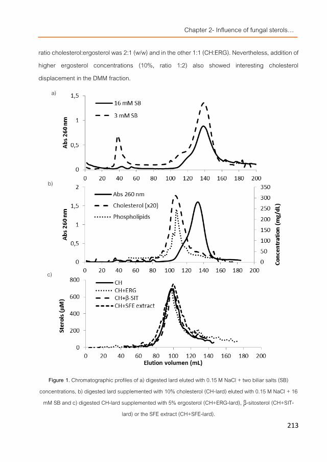
Chapter 2- Influence of fungal sterols…
213
ratio cholesterol:ergosterol was 2:1 (w/w) and in the other 1:1 (CH:ERG). Nevertheless, addition of higher ergosterol concentrations (10%, ratio 1:2) also showed interesting cholesterol displacement in the DMM fraction.
Figure 1. Chromatographic profiles of a) digested lard eluted with 0.15 M NaCl + two biliar salts (SB) concentrations, b) digested lard supplemented with 10% cholesterol (CH-lard) eluted with 0.15 M NaCl + 16
mM SB and c) digested CH-lard supplemented with 5% ergosterol (CH+ERG-lard), β-sitosterol (CH+SIT-lard) or the SFE extract (CH+SFE-lard).
a)
b)
c)

Chapter 2- Manuscript 2
214
Figure 2. Cholesterol concentration in the isolated DMM fraction after digestion of lard enriched with different
cholesterol (CH): ergosterol (ERG) ratios (w/w).
When the enriched lard was prepared with 2.5% cholesterol and supplemented with increasing concentrations of ergosterol, significantly less cholesterol was incorporated in the generated DMMs and this reduction correlated with the in-crease of ergosterol added up to 5% (ratios 1:1, 1:2 CH:ERG showed a cholesterol reduction of 32 and 49%, respectively). However, further increase in ergosterol concentration (ratios 1:3 and 1:4 CH:ERG) did not result in larger cholesterol displacement, because at these concentrations the saturation level was reached and no more ergosterol could be incorporate in the micelles. Thus, results indicated that under the experimental conditions, the optimal cholesterol concentration to supplement lard was 2.5% and ergosterol was able to displace it from the DMM but up to certain level (ratio 1:2, CH:ERG).
Cholesterol displacement from the DMM by ergosterol-enriched fractions Two ergosterol-containing extracts were evaluated as cholesterol-displacers from DMM
and compared with ergosterol and a plant phytosterol (β-sitosterol) as controls to study their potential as hypocholesterolemic extracts to design new functional foods. Both extracts were obtained from the fruiting bodies of the white button mushroom (Agaricus bisporus). The ASE extract contained 5.99% (w/w) sterols being ergosterol the major constituent (86.28%), followed by other derivatives such as ergosta-5,7-dienol (5.47%), fungisterol (4.29%) and ergosta-7,22-dienol (3.96%). However, SFE was a more selective technology to obtain high sterol-enriched fractions since 57.73% (w/w) of the obtained extract were sterols. SFE extract
0
20
40
60
80
100
120
140
0 2.5 5.0 7.5 10
Chole
stero
l (μ
g/mL)
Ergosterol (%)
10% CH5% CH2.5% CH

Chapter 2- Influence of fungal sterols…
215
composition was also a slightly different than the ASE extract because a lower ergosterol concentration (77.55%) was noticed followed by a higher level of ergosta-5,7-dienol (13.89%), fungisterol (5.09%) and ergosta-7,22-dienol (3.47%) were also detected. Other compounds were also identified such as a few esterified sterols (i.e. 9(11)-dehydroergosteryl benzoate), waxes, esterified and free fatty acids and other lipidic molecules. The ASE extract contained more phenolic compounds and less fatty acids than SFE extract.
Both extracts were mixed with CH-lard at specific concentrations in order to have a ratio 1:2 (CH:ERG) and submitted to in vitro digestion. The resulting DMM fractions were isolated and compared to control samples (Figure 3). The DMM fraction obtained after digestion of the sample including the ASE extract (CH + ASE-lard) showed the same cholesterol concentration than the DMM fraction of a control sample where only cholesterol was added. However, when the SFE extract (CH + SFE-lard) was utilized a 67% reduction was observed, a higher cholesterol displacement than ergosterol (49%) or β-sitosterol (47%). The different behavior between the two types of extracts (although they were applied at the same ergosterol concentrations) could be due to the different concentrations of ergosterol derivatives (i.e. ergosta-5,7-dienol) and/or by the presence of other compounds which might interact with positive or negative synergy.
Figure 3. Cholesterol concentration in the isolated DMM fraction after digestion of lard enriched with
cholesterol (2.5%), different ergosterol-containing extracts and ergosterol or β-sitosterol as controls (1:2, w/w, cholesterol:sterol ratio).
0
20
40
60
80
100
120
140
CH CH+ ERG CH+ SFE CH+ ASE CH+β-SITO
Chole
stero
l (μ
g/mL)

Chapter 2- Manuscript 2
216
The SFE extract contained more apolar structures than the ASE extract thus, probably they were easier included in the micelles together with the fungal sterols and apparently they influenced the micelle chemical composition as stated for some lipidic compounds [29].
On the other hand, the ASE extract might also contain other polar compounds including some β-glucans [30] and although, by using lard as food matrix they seemed not to interfere with sterols extractability during digestion, they could influence during DMM formation. Thus, the possibility of β-glucan interference was further evaluated in the DMM fractions.
Interference of β-glucans-enriched preparations in the DMM CH-lard supplemented with ergosterol (CH + ERG-lard) or SFE extract (CH + SFE-lard)
were also mixed with a β-glucan enriched fraction showing bile acid-binding activity obtained from P. ostreatus [18]. Then, the preparations were submitted to in vitro digestion and the resulting DMM fractions were isolated. Results indicated that the fungal β-glucans were able to scavenge cholesterol by themselves since when they were mixed with only CH-lard, the digestate generated DMM contained approximately threefold lower cholesterol levels (Figure 4). Previous reports also suggested that β-glucans can decrease cholesterol solubilization in the DMM besides their bile acids-binding capacity [7, 31].
Figure 4. Cholesterol concentration in the isolated DMM fraction after digestion of lard supplemented with cholesterol (2.5%), ergosterol and an ergosterol-containing extract (1:2, w/w, CH:ERG ratio) with or without
addition of a fungal β-glucan-containing extract with bile acid binding activity.
0
20
40
60
80
100
CH CH+ERG CH+SFE
Chole
stero
l (μ
g/mL)
without β-glucan extractwith β-glucan extract

Chapter 2- Influence of fungal sterols…
217
When the β-glucan extract was mixed with the CH-lards supplemented with ergosterol or the SFE extract and submitted to digestion, the generated DMM also showed significantly lower cholesterol levels than the reduction already induced by the presence of the sterol or the SFE extract. These results could suggest certain positive synergy between the effect of the
β-glucans and the fungal sterols however, if only the three samples including the β-glucans are compared, the levels of cholesterol were very similar suggesting that certain scavenging effect of the other sterols have also occurred. Moreover, almost two fold lower ergosterol was incorporated in DMM fractions generated in the presence of β-glucans thus, the polysaccharide extract was able to bind similarly to both types of sterols. It enhanced a higher displacement of cholesterol probably because it was added in lower concentration than ergosterol (ratio 1:2 CH:ERG).
Nevertheless still the cholesterol decrease in the DMMs particularly for those including β-glucans and the SFE extract was very high if compared with the control without these extracts.
Transport through Caco2 cell monolayers The DMM fractions obtained from the digestion of CH-lard with or without
supplementation of ergosterol (CH + ERG-lard) or SFE extract (CH + SFE-lard) were applied at subtoxic concentration (25 µL/mL) to Caco2 cell monolayers cultivated in specific inserts to measure transport through them. As expected, the applied DMM fraction generated from CH-lard showed higher cholesterol levels than those generated from lards containing ergosterol (Table 2). After 1 h incubation, cholesterol coming from the applied DMMs was still detected in the upper compartment. However, in the cells treated with the DMMs from the digestion of CH-lard, a reduction (78.6%) in the intracellular cholesterol was observed, followed by a release of 34.8% into the lower compartment while, in the cells treated with the DMMs from CH + ERG-lard and CH + SFE-lard higher cholesterol levels inside the Caco2 cells were noticed (almost doubled the normal cellular levels) liberating only respectively 17.6 and 30.5% of the cholesterol from applied DMMs. These different behaviors in the absence or presence of the fungal sterols indicated interferences in cholesterol transport. However, phytosterols apparently did not impair cholesterol trafficking from the plasma membrane to the endoplasmic reticulum but interfered with the uptake of micellar cholesterol [32] and ergosterol seemed to disturb that trafficking more than
Chapter 2- Manuscript 2
218
its absorption since intracellular cholesterol accumulation was observed. On the other hand, both phytosterols and ergosterol appeared to decrease levels of cholesteryl ester secreted into the basolateral compartments [32] being more effective if ergosterol was directly micellated than integrated into an extract.
Table 2. Sterol concentration of the DMM fractions applied to the apical compartment in the Caco2 transport assays.
(ND: no detected)
Ergosterol was absorbed in lower concentrations than cholesterol as occurred with phytosterols [5]. However, after (1 h) addition of the DMMs from CH + ERG-lard to the cells only 23% of the initially applied ergosterol was detected in the upper compartment and only 9.3% was found in the lower compartment rising the question whether ergosterol was being transformed into cholesterol (as suggested for other organisms [33] contributing to the observed cholesterol accumulation or into other ergosterol-derivatives that could not be detected because of the GC–MS method utilized. Thus, more detailed studies are needed (at the molecular level), in order to further clarify the ergosterol mechanism of action inside Caco2 cells, particularly because in the samples containing the SFE extract, ergosterol could not be detected in any of the compartments.
Conclusions Ergosterol and ergosterol-enriched extracts obtained from the white button mushroom
could potentially be used as hypocholesterolemic ingredients to design novel foods because when added to lipidic matrices such as lard, they were able to displace cholesterol from DMMs following a similar mechanism than the one described for phytosterols. β-Glucans from oyster
Cholesterol (μg/mL) Ergosterol (μg/mL)
CH-lard 91.06 ± 0.54 ND
CH+ERG-lard 46.06 ± 4.99 5.03 ± 1.33
CH+SFE-lard 29.83 ± 1.54 3.37 ± 2.09

Chapter 2- Influence of fungal sterols…
219
mushroom might also be added to the functionalized food because they enhance the cholesterol displacement but within controlled concentrations.
Fungal sterols seemed to hinder the bioavailability of cholesterol but their precise mechanism of action still needs to be clarified therefore, the expression of several genes related to the cholesterol absorption and metabolism is, at the present, being investigated using Caco2 cells and also HepG2. Animal studies will be also required to fully confirm the effect of ergosterol as hypocholesterolemic compound.
Acknowledgments The research was supported by AGL2010-21537 national R + D program from the Spanish
Ministry of Science and Innovation and ALIBIRD-C M S2009/AGR-1469 regional program from the Community of Madrid (Spain). CTICH is also acknowledged for the cultivation and supplying of the mushrooms fruiting bodies.

Chapter 2- Manuscript 2
220
References [1]. EFSA Panel on Dietetic Products, N.a.A.N., Scientific Opinion on the substantiation of a health claim related to oat
beta glucan and lowering blood cholesterol and reduced risk of (coronary) heart disease pursuant to Article 14 of Regulation (EC) No 1924/2006. EFSA Journal 2010. 8(12): p. 1885-1900.
[2]. EFSA Panel on Dietetic Products, N.a.A.N., Scientific Opinion on the substantiation of a health claim related to barley beta-glucans and lowering of blood cholesterol and reduced risk of (coronary) heart disease pursuant to Article 14 of Regulation (EC) No 1924/2006. EFSA Journal 2011. 9(12): p. 2471-2484.
[3]. EFSA Panel on Dietetic Products, N.a.A.N., Scientific Opinion on the substantiation of a health claim related to 3 g/day plant sterols/stanols and lowering blood LDL-cholesterol and reduced risk of (coronary) heart disease pursuant to Article 19 of Regulation (EC) No 1924/2006. EFSA Journal 2012. 10(5): p. 2693-2708.
[4]. Jesch, E.D. and T.P. Carr, Sitosterol reduces micellar cholesterol solubility in model bile. Nutrition Research, 2006. 26(11): p. 579-584.
[5]. Calpe-Berdiel, L., J.C. Escolà-Gil, and F. Blanco-Vaca, New insights into the molecular actions of plant sterols and stanols in cholesterol metabolism. Atherosclerosis, 2009. 203(1): p. 18-31.
[6]. Rozner, S. and N. Garti, The activity and absorption relationship of cholesterol and phytosterols. Colloids and Surfaces A: Physicochemical and Engineering Aspects, 2006. 282–283(0): p. 435-456.
[7]. Zacherl, C., P. Eisner, and K.-H. Engel, In vitro model to correlate viscosity and bile acid-binding capacity of digested water-soluble and insoluble dietary fibres. Food Chemistry, 2011. 126(2): p. 423-428.
[8]. Chen, Z.-Y., et al., Role and classification of cholesterol-lowering functional foods. Journal of Functional Foods, 2011. 3(2): p. 61-69.
[9]. Burton-Freeman, B., Dietary fiber and energy regulation. The Journal of Nutrition, 2000. 130(2): p. 272. [10]. van Bennekum, A.M., et al., Mechanisms of cholesterol-lowering effects of dietary insoluble fibres: relationships
with intestinal and hepatic cholesterol parameters. British Journal of Nutrition, 2005. 94(03): p. 331-337. [11]. Gil-Ramírez, A., et al., Sterol enriched fractions obtained from Agaricus bisporus fruiting bodies and by-products
by compressed fluid technologies (PLE and SFE). Innovative Food Science & Emerging Technologies, 2013. 18(0): p. 101-107.
[12]. Gil-Ramírez, A., et al., Screening of edible mushrooms and extraction by pressurized water (PWE) of 3-hydroxy-3-methyl-glutaryl CoA reductase inhibitors. Journal of Functional Foods, 2013. 5(1): p. 244-250.
[13]. Gil-Ramírez, A. and C. Soler-Rivas, The use of edible mushroom extracts as bioactive ingredients to design novel functional foods with hypocholesterolemic activities, in Mushrooms: Cultivation, Antioxidant Properties and Health Benefits, G. Pesti, Editor. 2014, Nova Science Publishers: New York. p. 43-73.
[14]. Guillamón, E., et al., Edible mushrooms: Role in the prevention of cardiovascular diseases. Fitoterapia, 2010. 81(7): p. 715-723.
[15]. Roupas, P., et al., The role of edible mushrooms in health: Evaluation of the evidence. Journal of Functional Foods, 2012. 4(4): p. 687-709.

Chapter 2- Influence of fungal sterols…
221
[16]. Schneider, I., et al., Lipid lowering effects of oyster mushroom (Pleurotus ostreatus) in humans. Journal of Functional Foods, 2011. 3(1): p. 17-24.
[17]. Hsu, R.C., B.H. Lin, and C.W. Chen, The study of supercritical carbon dioxide extraction for Ganoderma Lucidum. Industrial & Engineering Chemistry Research, 2001. 40(20): p. 4478-4481.
[18]. Palanisamy, M., et al., Pressurized water extraction of β-glucan enriched fractions with bile acids-binding capacities obtained from edible mushrooms. Biotechnology Progress, 2014. 30(2): p. 391-400.
[19]. Gil-Ramírez, A., et al., Study on the 3-hydroxy-3-methyl-glutaryl CoA reductase inhibitory properties of Agaricus bisporus and extraction of bioactive fractions using pressurised solvent technologies. Journal of the Science of Food and Agriculture, 2013. 93(11): p. 2789-2796.
[20]. Jeurink, P.V., et al., Immunomodulatory capacity of fungal proteins on the cytokine production of human peripheral blood mononuclear cells. International Immunopharmacology, 2008. 8(8): p. 1124-1133.
[21]. Mosmann, T., Rapid colorimetric assay for cellular growth and survival: Application to proliferation and cytotoxicity assays. Journal of Immunological Methods, 1983. 65(1–2): p. 55-63.
[22]. Bestwick, C.S. and L. Milne, Effects of β-carotene on antioxidant enzyme activity, intracellular reactive oxygen and membrane integrity within post confluent Caco-2 intestinal cells. Biochimica et Biophysica Acta (BBA) - General Subjects, 2000. 1474(1): p. 47-55.
[23]. Dihal, A.A., et al., Modulatory effects of quercetin on proliferation and differentiation of the human colorectal cell line Caco-2. Cancer Letters, 2006. 238(2): p. 248-259.
[24]. Yao, L., et al., Separation of micelles and vesicles within lumenal aspirates from healthy humans: solubilization of cholesterol after a meal. Journal of Lipid Research, 2002. 43(4): p. 654-660.
[25]. Palanisamy, M., et al., Testing edible mushrooms to inhibit the pancreatic lipase activity by an in vitro digestion model. International Journal of Food Science & Technology, 2012. 47(5): p. 1004-1010.
[26]. Porter, C.J.H., et al., Enhancing intestinal drug solubilisation using lipid-based delivery systems. Advanced Drug Delivery Reviews, 2008. 60(6): p. 673-691.
[27]. Cohen, D.E. and M.C. Carey, Rapid (1 hour) high-performance gel-filtration chromatography resolves coexisting simple micelles, mixed micelles and vesicles in bile. Journal of Lipid Research, 1990. 31(11): p. 2103-2112.
[28]. Mel’nikov, S.M., J.W.M. Seijen ten Hoorn, and A.P.A.M. Eijkelenboom, Effect of phytosterols and phytostanols on the solubilization of cholesterol by dietary mixed micelles: an in vitro study. Chemistry and Physics of Lipids, 2004. 127(2): p. 121-141.
[29]. Petruzzelli, M., et al., Micellar lipid composition profoundly affects LXR-dependent cholesterol transport across CaCo2 cells. FEBS Letters, 2009. 583(8): p. 1274-1280.
[30]. Lo, T.C.T., et al., Pressurized water extraction of polysaccharides as secondary metabolites from Lentinula edodes. Journal of Agricultural and Food Chemistry, 2007. 55(10): p. 4196-4201.
[31]. Chau, C.F., Y.L. Huang, and C.Y. Lin, Investigation of the cholesterol-lowering action of insoluble fibre derived from the peel of Citrus sinensis L. cv. Liucheng. Food Chemistry, 2004. 87(3): p. 361-366.

Chapter 2- Manuscript 2
222
[32]. Field, F.J., E. Born, and S.N. Mathur, Effect of micellar beta-sitosterol on cholesterol metabolism in CaCo-2 cells. Journal of Lipid Research, 1997. 38(2): p. 348-60.
[33]. Teshima, S.-i., In vivo transformation of ergosterol to cholesterol in crab, Portunus trituberculatus. NIPPON SUISAN GAKKAISHI, 1971. 37(7): p. 671-674.

Chapter 2- Influence of fungal sterols…
223
Manuscript 3
Modulation of cholesterol-related gene expression by ergosterol and ergosterol-enriched extracts obtained from Agaricus bisporus.
Alicia Gil-Ramírez1*, Víctor Caz2*, Roberto Martin-Hernandez3, Francisco R. Marín1, Carlota Largo2, Arantxa Rodríguez-Casado3,
María Tabernero2, Alejandro Ruiz-Rodríguez1, Guillermo Reglero1,3, Cristina Soler-Rivas1
1Department of Production and Characterization of Novel Foods, CIAL – Research Institute in Food Science (UAM+CSIC), C/ Nicolas Cabrera 9, Campus de Cantoblanco, Universidad Autónoma de Madrid, 28049 Madrid, Spain. 2Department of Experimental Surgery, Research Institute Hospital La Paz (IdiPAZ), Paseo de la Castellana 261, 28046 Madrid, Spain. 3IMDEA Food Institute. Pabellón Central del Antiguo Hospital de Cantoblanco (Edificio nº 7), Crta. de Cantoblanco nº 8, 28049 Madrid, Spain.
*Same contribution
European Journal of Nutrition
In Press, May 2015 DOI: 10.1007/s00394-015-0918-x
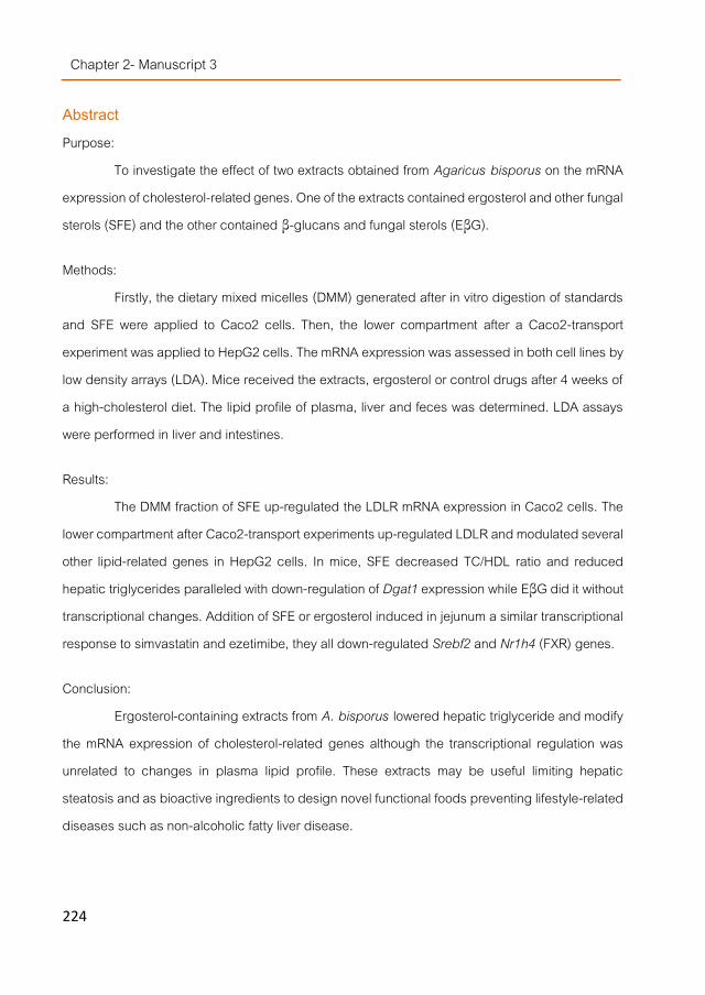
Chapter 2- Manuscript 3
224
Abstract Purpose:
To investigate the effect of two extracts obtained from Agaricus bisporus on the mRNA expression of cholesterol-related genes. One of the extracts contained ergosterol and other fungal sterols (SFE) and the other contained β-glucans and fungal sterols (EβG).
Methods: Firstly, the dietary mixed micelles (DMM) generated after in vitro digestion of standards
and SFE were applied to Caco2 cells. Then, the lower compartment after a Caco2-transport experiment was applied to HepG2 cells. The mRNA expression was assessed in both cell lines by low density arrays (LDA). Mice received the extracts, ergosterol or control drugs after 4 weeks of a high-cholesterol diet. The lipid profile of plasma, liver and feces was determined. LDA assays were performed in liver and intestines.
Results: The DMM fraction of SFE up-regulated the LDLR mRNA expression in Caco2 cells. The
lower compartment after Caco2-transport experiments up-regulated LDLR and modulated several other lipid-related genes in HepG2 cells. In mice, SFE decreased TC/HDL ratio and reduced hepatic triglycerides paralleled with down-regulation of Dgat1 expression while EβG did it without transcriptional changes. Addition of SFE or ergosterol induced in jejunum a similar transcriptional response to simvastatin and ezetimibe, they all down-regulated Srebf2 and Nr1h4 (FXR) genes.
Conclusion: Ergosterol-containing extracts from A. bisporus lowered hepatic triglyceride and modify
the mRNA expression of cholesterol-related genes although the transcriptional regulation was unrelated to changes in plasma lipid profile. These extracts may be useful limiting hepatic steatosis and as bioactive ingredients to design novel functional foods preventing lifestyle-related diseases such as non-alcoholic fatty liver disease.

Chapter 2- Influence of fungal sterols…
225
Introduction Extracts obtained from several edible mushroom species showed hypocholesterolemic
effects according to in vivo studies with animals and humans [1]. The sporophores of a commonly consumed mushroom, the white button mushroom (Agaricus bisporus), were also able to lower blood glucose and cholesterol levels in diabetic and hypercholesterolemic rats [2]. However, only a few type of molecules have been pointed as potentially responsible for these activities such as dietary fibers or β-glucans, lovastatine, eritadenine and, via an indirect mechanism, phenolic compounds or antioxidants such as ergothioneine [1].
Cholesterol is a water insoluble compound requiring emulsification and micellar solubilization during intestinal digestion before it can be absorbed by enterocytes. Uptake of this molecule from lumen is carried out by Niemann-Pick C1-like 1 (NPC1L1) protein and perhaps other complexes [3]. Depending on the cholesterol intracellular levels, it can also be transported back to intestinal lumen by the ABC transporters (ABCG5/8) located on the enterocytes brush border or incorporated into plasma HDL by ATP-binding cassette transporter A1 (ABCA1) located at the basolateral membrane. The intracellular cholesterol either absorbed or de novo synthesized can be esterified by the sterol O-acyltransferases (SOAT), packaged into apolipoproteins (APOB) and integrated into nascent chylomicrons with the assistance of the microsomal triglyceride transfer protein (MTTP) (an enzyme involved in the chylomicron assembly by transferring neutral lipids) and the acetyl-CoA acetyltransferases (ACAT, involved in the fatty acid biosynthetic pathway) in order to be transferred to lymph as chylomicrons (or packaged into APOA to release them as HDLs) [3, 4].
Fungal sterols (such as ergosterol) were able to compete with the cholesterol molecules for their incorporation in the dietary mixed micelles (DMM) during in vitro digestion because of their structural similarity [5]. This is also one of the generally accepted mechanisms of plant phytosterol for lowering cholesterol absorption in vivo [1, 2]. Moreover, supplementation of ergosterol extracts with fungal β-glucans induced an even larger cholesterol displacement from the DMM although they also partially scavenged the fungal sterols [5].

Chapter 2- Manuscript 3
226
However, phytosterols are able to influence the circulating cholesterol concentration via other mechanisms although their precise molecular targets are still not completely defined. Studies using Abca1- and Abcg5/8-deficient mice demonstrated that the phytosterol-mediated inhibition of intestinal cholesterol absorption was independent of these transporters [3]. Other reports suggested that plant sterols were able to modify cholesterol metabolism within the enterocyte (with in vivo and in vitro experiments) because of their structural similarity with sterol regulatory element-binding protein (SREBP)-signaling ligands (SREBF genes), because of their liver X receptor (LXR, NR1H3 gene) agonist activity (in particular ergost-22-ene-1,3-diol) [6] or their farnesoid X receptor (FXR, NR1H4 gene) antagonist activity (stigmasterol) [7]. FXR and LXR are nuclear receptors also involved in the regulation of cholesterol homeostasis [8]. Cholesterol analogs, e.g. phytosterols might also modulate cholesterol esterification by decreasing the SOAT activity and inhibiting the NPC1L1 transporter but, although in FHs 74 Int cells reduction of NPC1L1 mRNA expression levels was observed [9], in vivo it seems unlikely that the observed reduction was mediated via transcriptional changes [3].
The cholesterol biosynthesis is also regulated at a few key steps such as the transformation of hydroxymethylglutaryl coenzyme A into mevalonate by the hydroxymethylglutaryl coenzyme A reductase (HMGCR) and downstream at the squalene synthase (farnesyl-diphosphate farnesyltransferase, FDFT1). Moreover, esterified cholesterol loaded into LDLs can be removed from circulation by the low-density lipoprotein receptors (LDLR) located at the cell surface that mediate their endocytosis by recognizing the apoprotein B100, which is embedded in the outer phospholipid layer of LDL particles. β-Sitosterol reduced HMGCR gene expression in Caco2 cells but not in rats ileum nor mice liver and mononuclear blood cells. Moreover, phytostanols ester increased LDLR mRNA expression in human mononuclear blood cells whereas 1 - 2% phytostanol treatment did not affect Ldlr expression in hypercholesterolemic mice [3].
In this work, the modulation of mRNA levels of genes related to the cholesterol metabolism after supplementation with ergosterol and ergosterol-enriched extracts (with/without fungal β-glucans) obtained from A. bisporus is studied using cell cultures and mice fed a

Chapter 2- Influence of fungal sterols…
227
high-cholesterol diet by low density arrays (LDA). Biochemical parameters were also determined in mouse serum and liver.
Materials and Methods
Fungal material Lyophilized mushroom powder obtained from Agaricus bisporus L. (Imbach) Fungisem
H-15 fruiting bodies (from the first flush) according to Gil-Ramírez et al. (2013) [10] was utilized as starting material to obtain ergosterol-enriched fractions. Commercially available lard (Iberian pork fat, E-321, E-320), was purchased from a local supermarket. All the experiments were performed from the same lotus.
Standards and reagents Solvents as hexane (95%), chloroform and methanol were obtained from LAB-SCAN
(Gliwice, Polland) and absolute ethanol and sea sand from Panreac (Barcelona, Spain). Potassium hydroxide, ascorbic acid, BHT (2,6-Di-tert-butyl-p-cresol), cholesterol, cholic acid, ergosterol, congo red as well as hexadecane were purchased from Sigma-Aldrich Química (Madrid, Spain). Simvastatin was obtained from Cinfa Laboratories (Spain) and ezetimibe (Ezetrol) from Merck Sharp & Dohme (Spain). All other reagents and solvents were used of analytical grade.
Preparation of SFE extracts (containing sterols) Agaricus bisporus mushroom powder (80 g) were mixed with washed sea sand (900 g)
and submitted to supercritical fluid extraction (SFE) with CO2 in a pilot-plant (TharTechnology, Pittsburgh, PA, USA, model SF2000) as described in Gil-Ramírez et al. (2013) [10]. Extraction was carried out at 18 MPa and 40 ºC with a total extraction time of 3 h. Obtained fractions were immediately submitted to concentration until dryness on a rotary vacuum evaporator. Dried extract (named ´SFE extract´) was stored at -20 ºC until further analysis.
Preparation of EβG extracts (containing sterols and β-glucans) Agaricus bisporus mushroom powder (1 g) was mixed with 100 ml MilliQ water and
stirred at room temperature for 2 min. Afterwards, suspension was filtered under vacuum using Anoia filter paper (Barcelona, Spain) and a Büchner flask. Retained cake was washed with 100 ml

Chapter 2- Manuscript 3
228
distillated water and removed from the filter with the help of 10 ml water into a Falcon tube. The obtained suspension was frozen, lyophilized and the dried extract (called ´EβG extract´) stored at -20 ºC until further analysis.
In vitro digestion and isolation of the DMM Ergosterol or SFE extracts (added up to 5% ergosterol, w/w) were dissolved in lard
supplemented with/without 2.5 % (w/w) cholesterol and submitted to an in vitro digestion model as described in Gil-Ramírez et al (2014) [5]. Separation of the dietary mixed micelles (DMM) fraction from vesicles was carried out on a Sepharose®4B column (Amersham Pharmacia Biotech, madrid) [5]. The DMM fraction was immediately applied to Caco-2 cells grown on specific plates depending on the experiments.
Caco2 cell cultures and transport assay Human colorectal adenocarcinoma cell line Caco2 (ATCC HTB-37) obtained from the
Americam Type Culture Collection (Rockville, MD, USA) were maintained in Dulbecco´s Modified Eagle’s Medium (DMEM) with high glucose content (4.5 g/L) and supplemented with 10% fetal bovine serum, 1% penicillin-streptomycin, and 1% non-essential amino acids at 37 ºC in at humidified atmosphere containing 5% CO2. Firstly, the cytotoxicity was evaluated using 3-(4,5-dimethylthiazol)-2,5-diphenyl tetrazolium bromide (MTT) method [5]. Then, for the gene expression assays, cells were seeded onto a 9.5 cm2 grown area 6 well flat bottom plate (Costar, Corning, USA) at a density of 5 ×10⁵ cell per insert (3.3 ×10⁵ cell/ml). Culture medium was replaced every three days and cells were allowed to generate a monolayer for 21 days before experiments. The DMM fractions were applied at subtoxic concentrations (25 μl/ml [5]) and left incubating at 37 ºC and 5% CO2 for 1 or 24 h.
For transport assays, cells were seeded onto a 44 cm2 permeable membrane support (0.4 μm pore size, Costar, Corning, USA) at a density of 5.23 ×10⁵ cells per insert (5 ×10⁵ cell/ml). Cells were cultivated as above described and allowed to differentiate (21 days before experiments). The integrity of the cell layer was evaluated by measuring the transepithelial electrical resistance (TEER) (Evon, Sarasota, FL) and alkaline phosphatase activity (ALP) [5].

Chapter 2- Influence of fungal sterols…
229
The DMM fractions were applied to Caco2 cell monolayers. Each sample, applied at subtoxic concentration (25 μl/ml [5]), was mixed with 8.7 mL medium, applied to the apical (upper) compartment and left incubating at 37 ºC and 5% CO2 for 1 h. Afterwards, the lower compartment was collected and applied to HepG2 cell cultures. Four replicates per sample were carried out.
Treatment of HepG2 with the basolateral compartment of the Caco2 monolayers Human hepatoma HepG2 cells, obtained from American Type Culture Collection (ATCC,
Manassas, VA, USA) were cultured in Dulbecco´s modified Eagle medium (DMEM) supplemented with 10% fetal bovine serum (FBS), HEPES 1%, Tripsine - EDTA 0.5% (them all from Gibco, Grand Island, NY, USA) and 1% of antibiotic solution (containing 10,000 units/mL of penicillin base and 10,000 µg/mL of streptomycin base, Gibco). The cells were maintained under standard conditions of temperature (37 ºC), humidity (95%) and CO2 (5%).
Firstly, the cytotoxicity was evaluated as above described for the Caco2 cell cultures. Then, the HepG2 cell cultures were seeded in 6 wells plates (9.5 cm2) grown to confluence and treated with the basolateral compartment obtained after 1 h application of the DMM fractions from cholesterol-enriched lard or CH-enriched lard supplemented with ergosterol or the SFE extract (applied at subtoxic concentrations, 6.25 μl/ml) and left incubating at 37 ºC, 95% humidity and 5% CO2 during 1 or 24 h. These experiments were carried out in duplicate.
Animals and diets Male C57BL/6JRj mice (5 weeks old) were purchased from Janvier SAS (Le Genest Saint
Isle, France). The mice were maintained four per cage in temperature-, humidity- and light-controlled conditions (24 ± 2 °C, 40%-60% humidity, 12:12 hour light: dark cycle) and had free access to water and food. Mice were randomly divided into seven groups, 2 control groups and 5 treated groups. Normal control group (NC) was fed a standard diet (Safe Rodent Diet A04, Augy, France) and the hypercholesterolemic control group (HC) was fed a high-cholesterol diet that consisted of A04 supplemented with 2% cholesterol and 1% cholic acid (w/w). NC (n=6) and HC (n=8) groups were fed for 8 weeks (from 5 to 13 weeks of age) while the treated groups (n=8) received the same diet as HC for the first 4 weeks (from 5 to 9 weeks of age) and that diet together

Chapter 2- Manuscript 3
230
with their corresponding mushroom extract or control drug for the last 4 weeks of the experiments (from 10 to 13 weeks of age). The treated groups were named as follows: SFE (SFE extract, extract containing fungal sterols), EβG (EβG extract, extract containing fungal sterols and β-glucans), ERG (ergosterol-positive control), SV (simvastatin; positive control for inhibition of cholesterol biosynthesis), and EZ (ezetimibe; positive control for inhibition of cholesterol absorption). The composition of diets, the dose per day of mushroom extract or drug, total sterols, ergosterol, and β-glucans, as well as the energy that received the mice daily are shown in Table 1.
Table 2. Composition of diets.
Mushroom extracts doses were calculated taking into account the hypocholesterolemic minimal daily intake of plant sterols recommended by EFSA [11, 12]. Concerning simvastatin and ezetimibe drugs, doses were selected after reviewing literature taking into account studies which mouse models were similar to our mouse model, that is, preferably studies where C57BL/6J mouse and diet-induced hypercholesterolemia were used [13-16]. According to that works, simvastatin
Components NC HC SFE EβG ERG SV EZ Carbohydrates (%) 59.90 59.90 59.90 59.90 59.90 59.90 59.90
Proteins (%) 16.10 16.10 16.10 16.10 16.10 16.10 16.10
Lipids (%) 3.10 3.10 3.10 3.10 3.10 3.10 3.10
Fibres (%) 3.90 3.90 3.90 3.90 3.90 3.90 3.90
Mineral mixture* (%) 5.10 5.10 5.10 5.10 5.10 5.10 5.10
Moisture (%) 11.90 8.90 8.72 3.15 8.82 8.87 8.89
Cholesterol (%) 0.00 2.00 2.00 2.00 2.00 2.00 2.00
Cholic acid (%) 0.00 1.00 1.00 1.00 1.00 1.00 1.00
Mushroom extract or drug (%) 0.00 0.00 0.18 5.76 0.08 0.025 0.005
Mushroom extract or drug (mg/mouse/day) 0.00 0.00 7.20 230.40 3.20 1.00 0.20
Total sterols** (%) n.m. n.m. 0.086 0.081 0.080 n.m. n.m.
Ergosterol (%) n.m. n.m. 0.067 0.081 0.080 n.m. n.m.
β-Glucans (%) n.m. n.m. 0.00 1.676 n.m. n.m. n.m.
Total sterols** (mg/Kg/day) n.m. n.m. 173 161 160 n.m. n.m.
Ergosterol (mg/Kg/day) n.m. n.m. 134 161 160 n.m. n.m.
β-Glucans (mg/Kg/day) n.m. n.m. 0.000 3352 n.m. n.m. n.m.
Energy (Kcal/mouse/day) 13.366 14.386 14.450 15.229 14.386 14.386 14.386
NC: normal control; HC: control fed a high-cholesterol diet; SFE: SFE extract; EβG: EβG extract; ERG: ergosterol; SV: simvastatin; EZ: ezetimibe; *A04 mineral mixture; ** cholesterol excluded; n.m.: not measured.

Chapter 2- Influence of fungal sterols…
231
daily doses ranged from 0.4 to 1.6 mg/mouse, and ezetimibe ranged from 0.02 to 1.0 mg/mouse. As shown in Table 1 daily doses were used encompassed within those mentioned above. Body weight was registered for all mice before the experimental feeding period and at the end of the experiments. Wet mass of the liver was also registered. Animal studies were approved by our institution’s Animal Welfare and Ethics Committee and were carried out according to Spanish and European legislation (RD 53/2013, 2010/63/EU, respectively).
Plasma and liver lipid analyses, tissue and feces collection Following the experimental feeding period (8 weeks), mice were sacrificed by
intracardiac exsanguination under anesthesia with 1.5% isoflurane, and plasma was collected and stored at -80 °C before use. Jejunum, ileum, cecum, and liver samples were removed and immediately frozen in liquid nitrogen and stored at -80 °C. Feces were collected at the beginning, after 4 weeks of high-cholesterol diet and at the end of the experimental feeding period and maintained at -20 °C until further use. Plasma levels of total cholesterol, triglyceride, HDL-cholesterol and LDL-cholesterol were measured in duplicate for each sample at 8 weeks using a Covas C311 Autoanalyzer (Roche, Spain). Total cholesterol and triglyceride concentrations were measured in duplicate for each sample in liver extracts using colorimetric enzyme assays (Cholesterol Quantification Kit and Triglyceride Quantification Kit, respectively, BioVision Inc., CA, USA). Briefly, liver extracts for total cholesterol quantification were prepared by homogenizing a 100-mg sample of liver in 2 mL of CHCl3:isopropanol:NP-40 (7:11:0.1). The extract was centrifuged for 5 min at 15,000 × g. The supernatant was air dried at 50 °C and then placed it under vacuum for 30 min to remove any remaining solvent. Dried lipids were dissolved with 200 μl of Cholesterol Assay Buffer and stirred in a vortex until homogeneous. 5 μl of extract per assay were used and volume adjusted to 50 μl/well with Cholesterol Assay Buffer. In the assay, free cholesterol is oxidized by cholesterol dehydrogenase to generate NADH. To calculate total cholesterol concentration, absorbance at 450 nm was measured after the reaction was incubated for 30 min at 37 °C, protected from light. To determine triglycerides contents in liver tissues, 100 mg of wet liver tissues was homogenized in 1 ml of 5% NP-40. The samples were slowly heated up to 100 °C for 5 min and then cooled down to room temperature. The heating was

Chapter 2- Manuscript 3
232
repeated one more time. The tissue homogenates were centrifuged at 15,000 × g for 5 min at room temperature and the supernatants were diluted 10 fold with dH2O before the assay. In the assay, triglycerides are converted to free fatty acids and glycerol. The glycerol is then oxidized to generate a product which reacts with the probe to generate color. To calculate triglycerides concentration, absorbance at 570 nm was measured.
Sterols and β-glucan quantification Sterols (cholesterol, ergosterol and derivatives) were extracted from the ergosterol-
enriched extracts or mice feces (300 mg) and further quantified in duplicate for each sample (Table 2) by GC-MS-FID following the procedure described in Gil-Ramírez et al. (2013) [10].
The β-glucan content of both ergosterol-enriched extracts (50 mg) was evaluated in duplicate by a β-glucan determination kit specific for mushrooms and yeasts (Megazyme, Barcelona, Spain) following the instructions of the user´s manual and as described in Palanisamy et al. [17]. The amount of β-(1→3),(1→6)-glucans was also determined following the method of Nitschke et al. [18] (Table 2).
Table 3. Composition of the ergosterol-enriched extracts (% dw).
Statistical analysis for biochemical data All values were expressed as mean ± SD. The SPSS software, version 15.0 (Lead
Technologies, Chicago, IL, USA), was utilized to determine whether the variables differed among
SFE extract EβG extract Ergosterol 37.3 1.4 Ergosta-5,7-dienol 6.5 0.02 Fungisterol 2.6 b.q. Ergosta-7,22-dienol 1.5 b.q. Total sterols 48 1.4 Total β-glucans n.d. 29.1 (1→3),(1→6)-β-glucans n.d. 2.1 Other fats 51 1.0 Proteins n.d. 26.3 Carbohydrates n.d. 68.7
n.d.: not detected; b.q.: below quantifiable levels

Chapter 2- Influence of fungal sterols…
233
treatment groups in the in vivo experiments. The effects of treatments were assessed by one-way ANOVA. When the ANOVA identified significant treatment effects, the means were evaluated using Tukey’s test. Differences with P < 0.05 were considered statistically significant.
RNA extraction from cells and mice tissues and quantitative real-time PCR Total RNA from cell cultures or mice tissues was extracted by magnetic bead technology
using a pureLinkTM Total RNA kit TRIzol® Plus RNA Kit (Invitrogen, Carlsbad, CA, USA) in an iPrepTM Purification Instrument (Invitrogen) programmed with an iPrepTM total RNA card (Invitrogen) according to manufacturer instructions. The RNA concentration was determined by spectrophotometry at 260 nm and the purity of the extracted RNA was calculated from ratio of absorbance at 260:280 nm and 260:230 nm in a NanoDrop ND-1000 (NanoDrop Technologies Inc., Wilmington, DE, USA).
An amount of 400 ng of total RNA from each sample were reversed transcribed using the High Capacity cDNA Reverse Transcription Kit (Applied Biosystems, Framinghan, MA, USA). The obtained cDNA (100 μl per port) were loaded into the fluidic cards. Before sealing them, the cards were centrifuged twice on a Sorvall centrifugue at 1200 rpm for 1 min. Finally, the micro fluidic cards were run in a 7900HT Fast Real-Time PCR system (Applied Biosystems). Amplification conditions were 50 ºC for 2 min, 94.5 ºC for 10 min, followed by 40 cycles with 97 ºC with 30 s and 59.7 ºC for 1 min.
Low Density Array (LDA) design and gene expression analysis A technical and conceptual description of the TaqMan Low Density Array based on
Applied Biosystems 7900HT Micro-Fluid Cards can be found in the Applied Biosystems TaqMan Array Micro-Fluid Cards user guide (http://www3.appliedbiosystems.com/cms/groups/mcb_support/documents/
generaldocuments/cms_062836.pdf (or 040127). The micro-fluid card was constructed using respectively human and mouse commercial available assays (Applied Biosystems) for several genes related to the cholesterol metabolism (Table 3) and the internal standard and housekeeping genes. Comparative analysis of each of these genes was performed using specialized computer programs SDS2.3 and RQ 2.1 (Applied Biosystems).

Chapter 2- Manuscript 3
234
Table 4. ABI (Applied Biosystems) commercial reference and NCBI reference sequence of the selected genes from Homo sapiens (cell cultures) and Mus musculus and internal controls used in the LDA design.
ABI code for Homo sapiens
ABI code for Mus musculus
Reference sequence/s Gen Description
Hs01059118_m1 Mm00442646_m1 NM_005502.3 ABCA1 ATP-Binding Cassette, Sub-Family A (ABC1),
Member 1
Hs00223686_m1 Mm00446241_m1 NM_022436.2 ABCG5 ATP-binding cassette, sub-family G (WHITE),
member 5
Hs00223690_m1 Mm00445970_m1 NM_022437.2 ABCG8 ATP-binding cassette, sub-family G (WHITE),
member 8
Hs00608002_m1 Mm00507463_m1 NM_000019.3 ACAT1 Acetyl-CoA acetyltransferase 1
Hs00255067_m1 NM_005891.2 ACAT2 Acetyl-CoA acetyltransferase 2
Hs01071209_m1 Mm01545156_m1 NM_000384.2 APOB Apolipoprotein B
Hs00201385_m1 Mm00515643_m1 NM_012079.4 DGAT1 Diacylglycerol O-acyltransferase 1
Hs00261438_m1 Mm00499536_m1 NM_001253891.1 NM_032564.4 DGAT2 Diacylglycerol O-acyltransferase 2
Hs00926054_m1 Mm00815354_s1 NM_004462.3 FDFT1 Farnesyl-diphosphate farnesyltransferase 1
Hs00168352_m1 Mm01282499_m1 NM_000859.2 NM_001130996.1 HMGCR 3-hydroxy-3-methylglutaryl-CoA reductase
Hs00181192_m1 Mm00440169_m1
NM_000527.4, NM_001195798.1NM_001195799.1
NM_001195800.1 NM_001195802.1NM_001195803.1
LDLR Low density lipoprotein receptor
Hs00165177_m1 Mm00435015_m1 NM_000253.2 MTTP Microsomal triglyceride transfer protein
Hs00203602_m1 Mm01191972_m1 NM_001101648.1 NM_013389.2 NPC1L1 NPC1 (Niemann-Pick disease, type C1, gene)-
like 1
Hs00172885_m1 Mm00443451_m1 NM_001130101.2
NM_001130102.2NM_001251934. NM_001251935.1 NM_0056933
NR1H3 Nuclear receptor subfamily 1, group H, member
3 (LXRα)
Hs00231968_m1 Mm00436425_m1 NM_001206977.1NM_001206978.1 NM_001206979.1 NM_001206992.1
NM_001206993.1 NM_005123.3 NR1H4
Nuclear receptor subfamily 1, group H, member 4 (FXRα)
Hs00162077_m1 Mm00486279_m1 NM_001252511.1 NM_001252512.1
NM_003101.5 SOAT1 Sterol O-acyltransferase 1
Hs01573878_m1 Mm00448823_m1 NM_003578.3 SOAT2 Sterol O-acyltransferase 2
Hs01088691_m1 Mm00550338_m1 NM_001005291.2 NM_004176.4 SREBF1 Sterol regulatory element binding transcription
factor 1
Hs01081784_m1 Mm01306292_m1 NM_004599.2 SREBF2 Sterol regulatory element binding transcription
factor 2
Hs99999905_m1 NM_002046.4 GAPDH Glyceraldehyde-3-phosphate dehydrogenase
(internal control) Mm00839493_m1 Polr2a polimerase
Hs99999908_m1 Mm00446953_m1 NM_000181.3 GUSB Glucuronidase, beta (internal control)
Hs99999909_m1 Mm00446968_m1 NM_000194.2 HPRT1 Hypoxanthine phosphoribosyltransferase 1
(internal control)
Hs99999901_S1 18S-Hs99999901_s1 RN18S1 RNA, 18S ribosomal 1 (housekeeping gene)

Chapter 2- Influence of fungal sterols…
235
Gene expression statistical data analysis The expression of each gene was measured at least in duplicate and each biological
group included a minimum of 3 replicates that were quality controlled using the Median Absolute Deviation test (MAD test). Samples of the same biological group with a MAD score higher than 3 were removed from the analysis. Genorm algorithm [19] was used to identify the most stable reference genes for normalization. Genes GUSB and HPRT1 were found to be the most stably expressed in Caco2 cell cultures while GAPDH and HPRT1 were more stably expressed in HepG2 cells. In the mouse tissue samples, genes Hprt1 and Polr2a were most stably expressed in cecum and liver, and Gusb and Polr2a in jejunum and ileum. Relative gene expression levels were calculated by using the 2-ΔΔCT method [20], using these identified most stable genes as internal control genes in each group. Statistical significance of the results was assessed using a Limma test, and Benjamini-Hochberg false discovery rate procedure was subsequently applied. Adjusted p-values lower than 0.05 were considered significant. All these calculations were carried out withReal Time StarMiner 4.5® (Integromics S.L, Spain).
Results
Modulation of the mRNA expression pattern in Caco2 cell cultures Ergosterol and the ergosterol-enriched SFE extract demonstrated their ability to displace
cholesterol from DMMs and to interfere with the cholesterol uptake in Caco2 cell monolayers when integrated in a high lipid food matrix such as lard [5]. Thus firstly, lard supplemented with cholesterol (CH) or ergosterol (ERG) were submitted to digestion, the generated DMM fractions were isolated and applied to Caco2 cell cultures to study whether the sterols included in the micelles (generated with the in vitro digestion model) could differently modify the transcriptional pattern or the lipids from the DMMs (present in higher concentrations) could mask their potential modulation. Their molecular answer was evaluated shortly after supplementation (1 h) or after a longer period (24 h).
Results indicated that 1 h after the CH-lard-containing DMMs (CH) were added, only the expression of the MTTP mRNA was slightly reduced compared to Caco2 cells with no DMM

Chapter 2- Manuscript 3
236
addition (Figure 1a). On the contrary, application of DMMs obtained from the digestion of lard supplemented with ergosterol (ERG) significantly increased levels of MTTP mRNA indicating that the different sterols although they were integrated in DMMs they were activating different transcriptional mechanisms. Thus, in order to better reproduce in vitro the conditions of mice fed hypercholesterolemic diets (see later), lard supplemented with cholesterol (CH) was also mixed with ergosterol (ERG) or the ergosterol enriched extract (SFE) and submitted to digestion to evaluate their influence in the Caco2 mRNA expression pattern when cholesterol was simultaneously present in ergosterol-loaded DMMs as it might occur when a lipid-rich food matrix is consumed together with the potentially bioactive extracts. In this case, when the DMMs contained both cholesterol and ergosterol (CH+ERG) were applied, no significant differences in the mRNA expression could be noticed (Figure 1a). However, the CH+SFE (cholesterol and SFE extract) containing DMM fraction induced changes in the mRNA expression levels of many genes related to the lipid and cholesterol metabolism. One hour after the addition, the LDLR mRNA expression was up-regulated and the expression of others such as ACAT1, DGAT2, NR1H3 and SREBF2 was down-regulated. The induced modulation pattern by the latter DMM fraction was maintained for a long time (24 h) except for the expression of NR1H3 and particularly LDLR that were further respectively repressed and stimulated (Figure 1b). mRNA over-expression of other genes such as SREBF1 and APOB and repression of FDFT1 was also noticed. Moreover, the expression levels of HMGCR, LDLR and SREBF1 were also up-regulated 24 h after addition of the DMMs from CH as well as ERG and CH+ERG.
Modulation of the mRNA expression pattern in HepG2 cell cultures In order to simulate the effect that these extracts might produce at hepatic level, the lower
compartments after the transport experiments through Caco2 monolayers (potentially containing the bioavailable and generated compounds) were applied to HepG2 cell cultures (the DMMs would not reach the liver as such) and the modulation of their mRNA expression pattern evaluated 1 h and 24 h after the application.

Chapter 2- Influence of fungal sterols…
237
a)
b)
Figure 5. Relative mRNA expression (Log10) of Caco2 cells cholesterol-related genes after a) 1h and b) 24h application of the DMM fraction isolated after digestion of a mixture of lard with cholesterol (CH), with
ergosterol (ERG) with both cholesterol and ergosterol (CH+ERG) and with cholesterol and the SFE extract (CH+SFE). Indicated genes are only those pointed as significant (p< 0.05) compared with their controls
(Caco2 cells with no DMM fraction addition). The expression of each gene was measured at least in duplicate and each biological group included a minimum of 3 replicates.
Results suggested that the DMMs from the digested extracts were absorbed or induced changes in the composition of the lower compartments because when the basolateral solution of

Chapter 2- Manuscript 3
238
non DMM-treated Caco2 cells was applied to HepG2 cell cultures no significant differences in the transcription profile was observed when compared with non-treated HepG2 cells used as control (Figure 2a). Addition of these potentially modified fractions stimulated the mRNA expression of many genes involved in the fatty acids and cholesterol absorption and transformation, a few regulators as well as the enzymes involved in biosynthesis and esterification of cholesterol. These changes were observed in those cells treated with the basolateral solution obtained after application of the DMMs generated from CH and CH+SFE. CH+ERG induced lower changes in the mRNA expression profile since no expression of HMGCR or FDFT1 was noticed nor overexpression of mRNAs from genes related to the lipidic transport. After a longer incubation time, this CH+ERG bioavalilable fraction modulated the mRNA expression pattern differently than those obtained from CH and CH+SFE for instance by inhibiting FDFT1 and HMGCR mRNA expressions, up-regulating DGAT2, NR1H4 etc. (Figure 2b).
Modulation of the lipid profile of plasma and liver in the animal intervention Since ergosterol and the SFE-extract seemed to significantly modulate the mRNA
expression of several genes related to the lipid and cholesterol metabolism in specific human hepatic and colon cell lines, they were also tested in mice models to further study their in vivo significance. According to previous publications, A. bisporus β-glucans (dietary fiber) could be involved in the observed lowering of cholesterol levels in serum [2, 21]. Thus, in addition to the SFE-extract, another extract administrated at similar sterols concentration (SFE: 0.086%; EβG: 0.081%) but including water insoluble β-glucans was also tested (EβG-extract) (Table 2).
Adding cholesterol to the standard diet significantly increased plasma total cholesterol levels 3.3-fold in the HC group compared with NC at the end of the experiment (Table 4). Significant reduction was only achieved by ezetimibe or simvastatin treatments and not by extracts or ergosterol supplementation. With the high-cholesterol diet, plasma HDL and LDL levels were also increased by respectively 1.5 and 6.6 fold. Surprisingly, HDL levels were increased even more in the SFE group (22% more than the HC) and in the EβG group (14%) but not in the ergosterol group. The LDL levels remained also unchanged in the mice fed SFE extract, EβG

Chapter 2- Influence of fungal sterols…
239
extract or ergosterol, while simvastatin produced a 34% reduction and ezetimibe a significant 85% reduction, achieving normal levels in the ezetimibe group.
a)
b)
Figure 6. Relative mRNA expression (Log10) of HepG2 cells cholesterol-related genes after a) 1h and b) 24h application of the basolateral compartment of Caco2 cell cultures collected 1h after the transport assay was performed in which the DMM fraction isolated after digestion of a mixture of lard with cholesterol (CH), with both cholesterol and ergosterol (CH+ERG) and with cholesterol and the SFE extract containing ergosterol
(CH+SFE). The lower compartment of Caco2 cell cultures with no supplementation was also added to HepG2 cell cultured as control (Caco2 ST). Indicated genes are only those pointed as significant (p< 0.05)
compared with their controls (HepG2 cells with no supplementation). The expression of each gene was measured at least in duplicate and each biological group included a minimum of 3 replicates.
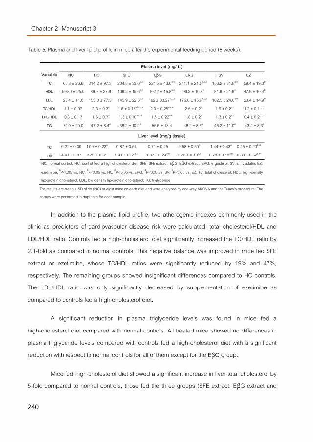
Chapter 2- Manuscript 3
240
Table 5. Plasma and liver lipid profile in mice after the experimental feeding period (8 weeks).
In addition to the plasma lipid profile, two atherogenic indexes commonly used in the clinic as predictors of cardiovascular disease risk were calculated, total cholesterol/HDL and LDL/HDL ratio. Controls fed a high-cholesterol diet significantly increased the TC/HDL ratio by 2.1-fold as compared to normal controls. This negative balance was improved in mice fed SFE extract or ezetimibe, whose TC/HDL ratios were significantly reduced by 19% and 47%, respectively. The remaining groups showed insignificant differences compared to HC controls. The LDL/HDL ratio was only significantly decreased by supplementation of ezetimibe as compared to controls fed a high-cholesterol diet.
A significant reduction in plasma triglyceride levels was found in mice fed a high-cholesterol diet compared with normal controls. All treated mice showed no differences in plasma triglyceride levels compared with controls fed a high-cholesterol diet with a significant reduction with respect to normal controls for all of them except for the EβG group.
Mice fed high-cholesterol diet showed a significant increase in liver total cholesterol by 5-fold compared to normal controls, those fed the three groups (SFE extract, EβG extract and
Variable
Plasma level (mg/dL) NC HC SFE EβG ERG SV EZ
TC 65.3 ± 26.6 214.2 ± 97.3a 204.8 ± 33.6a,e 221.5 ± 43.0a,e 241.1 ± 21.5a,d,e 156.2 ± 31.8a,e 59.4 ± 19.0b HDL 59.80 ± 25.0 89.7 ± 27.9 109.2 ± 15.6a,e 102.2 ± 15.8a,e 96.2 ± 10.3e 81.9 ± 21.9e 47.9 ± 10.4b LDL 23.4 ± 11.0 155.0 ± 77.3a 145.9 ± 22.3a,e 162 ± 33.27a,d,e 176.8 ± 15.6a,d,e 102.5 ± 24.0a,e 23.4 ± 14.9b
TC/HDL 1.1 ± 0.07 2.3 ± 0.3a 1.8 ± 0.15a,b,c,e 2.0 ± 0.25a,c,e 2.5 ± 0.2a 1.9 ± 0.2a,c 1.2 ± 0.1b,c,d LDL/HDL 0.3 ± 0.13 1.6 ± 0.3a 1.3 ± 0.10a,c,e 1.5 ± 0.22a,e 1.8 ± 0.2a 1.3 ± 0.2a,c 0.4 ± 0.2b,c.d
TG 72.0 ± 20.0 47.2 ± 8.4a 38.2 ± 10.2a 55.5 ± 13.4 48.2 ± 8.5a 46.2 ± 11.0a 43.4 ± 8.3a Liver level (mg/g tissue)
TC 0.22 ± 0.09 1.09 ± 0.23a 0.87 ± 0.51 0.71 ± 0.45 0.58 ± 0.50d 1.44 ± 0.43a 0.45 ± 0.20b,d
TG 4.49 ± 0.87 3.72 ± 0.61 1.41 ± 0.51a,b 1.87 ± 0.24a,b 0.73 ± 0.18a,b 0.78 ± 0.18a,b 0.88 ± 0.52a,b NC: normal control; HC: control fed a high-cholesterol diet; SFE: SFE extract; EβG: EβG extract; ERG: ergosterol; SV: simvastatin; EZ:
ezetimibe. aP<0.05 vs. NC; bP<0.05 vs. HC; cP<0.05 vs. ERG; dP<0.05 vs. SV; eP<0.05 vs. EZ; TC, total cholesterol; HDL, high-density
lipoprotein cholesterol; LDL, low density lipoprotein cholesterol; TG, triglyceride
The results are mean ± SD of six (NC) or eight mice on each diet and were analyzed by one-way ANOVA and the Tukey’s procedure. The assays were performed in duplicate for each sample.

Chapter 2- Influence of fungal sterols…
241
ergosterol) showed insignificant differences respect to both NC and HC control groups. Simvastatin increased liver total cholesterol compare to normal control while ezetimibe significantly reduced it by 59% respect to HC control. On the other hand, liver triglyceride levels were slightly reduced by the high-cholesterol diet, while a significant reduction with respect to NC and HC controls was noticed in the liver of all the experimental groups.
Effect of extracts on body and liver weight and sterol excretion through feces No differences were noticed in body weight before the experimental feeding period
(Table 5). The normal control group gained 5.80±1.18 g over the feeding period while the remaining groups showed a slight weight loss (0.30±1.17-1.26±1.12 g) except for the simvastatin group that showed a higher weight loss (3.82±1.31 g). No significant differences in final body weight of the treated groups were noticed compared with the control group fed a high-cholesterol diet, except for SV group where a significantly lower final body weight was recorded compared to all other groups, including the control group fed a high-cholesterol diet. Despite the weight loss observed, mice seemed healthy all over the study and tolerated well the experimental diets with a food intake similar in all groups (3.8-4.0 g/mouse/day from day 4 until the end of the experiments; data not shown). The observed weight loss was lower than the one that Ikemoto et al. [22] recorded in C57BL/6J mice fed a high-fat diet supplemented with 0.5% sodium cholate and 1% cholesterol (final body weight 22.4±0.7 g) as compared with high-fat diet alone (final body weight 38.3 ± 1.4 g). The data of liver weight (Table 5) indicated that the absolute liver weight significantly increased in the control group fed a high-cholesterol diet and the treated groups, except the ergosterol (P=0.052) and simvastatin groups, compared with the normal control group. The same tendency was observed when the hepatomegaly was expressed as liver-to-body-weight ratio, with a significant increase in all groups with respect to the normal control group. The effect of the high-cholesterol diet on liver weight and liver-to-body-weight ratio could not be counteracted by the extracts or control drugs with the SFE extract and the simvastatin group showing the highest liver-to-body-weight ratio values although in the last case, it was because of its body weight loss instead of its increase in liver weight.
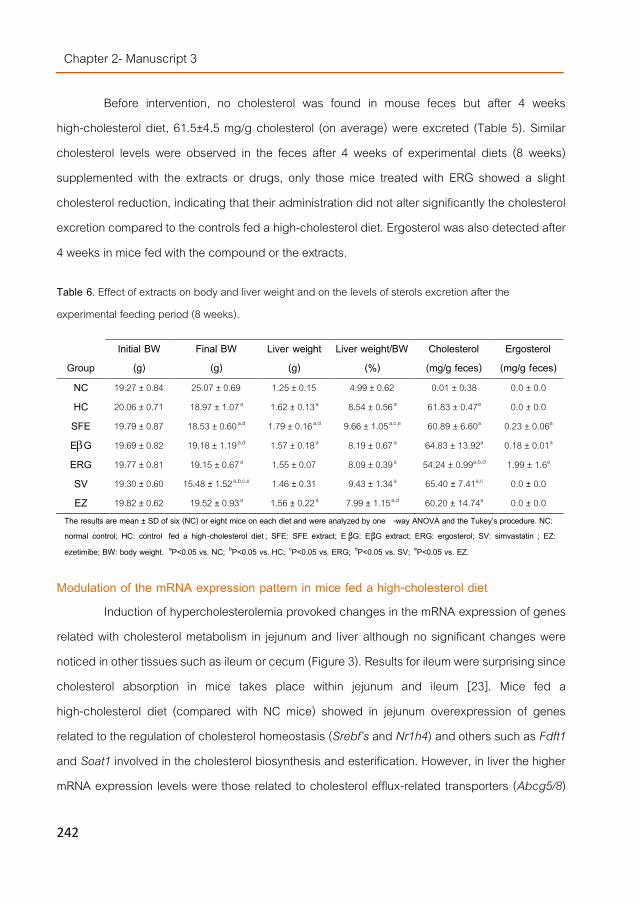
Chapter 2- Manuscript 3
242
Before intervention, no cholesterol was found in mouse feces but after 4 weeks high-cholesterol diet, 61.5±4.5 mg/g cholesterol (on average) were excreted (Table 5). Similar cholesterol levels were observed in the feces after 4 weeks of experimental diets (8 weeks) supplemented with the extracts or drugs, only those mice treated with ERG showed a slight cholesterol reduction, indicating that their administration did not alter significantly the cholesterol excretion compared to the controls fed a high-cholesterol diet. Ergosterol was also detected after 4 weeks in mice fed with the compound or the extracts.
Table 6. Effect of extracts on body and liver weight and on the levels of sterols excretion after the experimental feeding period (8 weeks).
Modulation of the mRNA expression pattern in mice fed a high-cholesterol diet Induction of hypercholesterolemia provoked changes in the mRNA expression of genes
related with cholesterol metabolism in jejunum and liver although no significant changes were noticed in other tissues such as ileum or cecum (Figure 3). Results for ileum were surprising since cholesterol absorption in mice takes place within jejunum and ileum [23]. Mice fed a high-cholesterol diet (compared with NC mice) showed in jejunum overexpression of genes related to the regulation of cholesterol homeostasis (Srebf’s and Nr1h4) and others such as Fdft1 and Soat1 involved in the cholesterol biosynthesis and esterification. However, in liver the higher mRNA expression levels were those related to cholesterol efflux-related transporters (Abcg5/8)
Group Initial BW
(g) Final BW
(g) Liver weight
(g) Liver weight/BW
(%) Cholesterol (mg/g feces)
Ergosterol (mg/g feces)
NC 19.27 ± 0.84 25.07 ± 0.69 1.25 ± 0.15 4.99 ± 0.62 0.01 ± 0.38 0.0 ± 0.0 HC 20.06 ± 0.71 18.97 ± 1.07 a 1.62 ± 0.13a 8.54 ± 0.56 a 61.83 ± 0.47a 0.0 ± 0.0 SFE 19.79 ± 0.87 18.53 ± 0.60 a,d 1.79 ± 0.16a,d 9.66 ± 1.05 a,c,e 60.89 ± 6.60a 0.23 ± 0.06a EG 19.69 ± 0.82 19.18 ± 1.19 a,d 1.57 ± 0.18a 8.19 ± 0.67 a 64.83 ± 13.92a 0.18 ± 0.01a ERG 19.77 ± 0.81 19.15 ± 0.67 a 1.55 ± 0.07 8.09 ± 0.39 a 54.24 ± 0.99a,b,d 1.99 ± 1.6a SV 19.30 ± 0.60 15.48 ± 1.52 a,b,c,e 1.46 ± 0.31 9.43 ± 1.34 a 65.40 ± 7.41a,c 0.0 ± 0.0 EZ 19.82 ± 0.62 19.52 ± 0.93 a 1.56 ± 0.22a 7.99 ± 1.15 a,d 60.20 ± 14.74a 0.0 ± 0.0
The results are mean ± SD of six (NC) or eight mice on each diet and were analyzed by one -way ANOVA and the Tukey’s procedure. NC: normal control; HC: control fed a high-cholesterol diet ; SFE: SFE extract; E βG: EβG extract; ERG: ergosterol; SV: simvastatin ; EZ: ezetimibe; BW: body weight. aP<0.05 vs. NC; bP<0.05 vs. HC; cP<0.05 vs. ERG; dP<0.05 vs. SV; eP<0.05 vs. EZ.
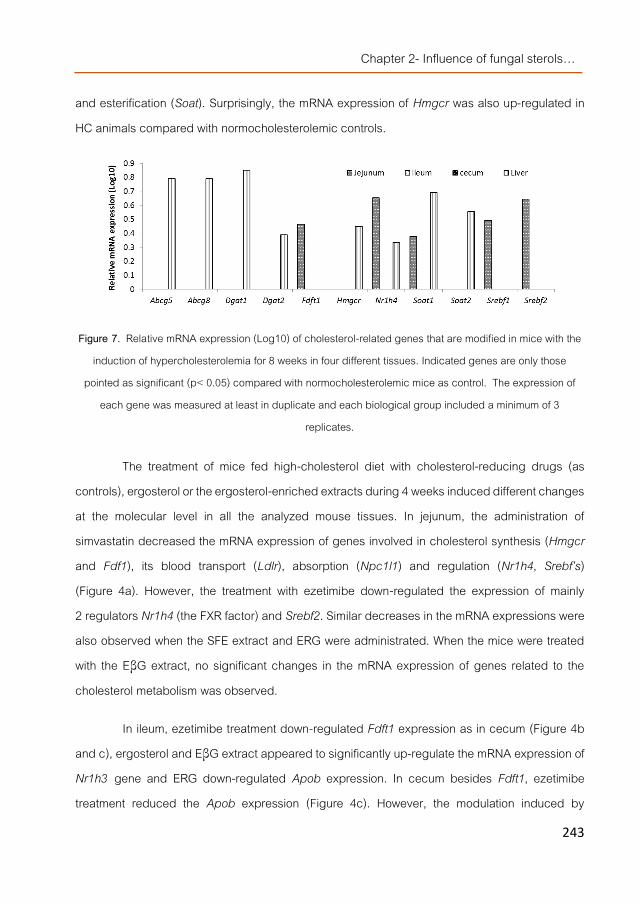
Chapter 2- Influence of fungal sterols…
243
and esterification (Soat). Surprisingly, the mRNA expression of Hmgcr was also up-regulated in HC animals compared with normocholesterolemic controls.
Figure 7. Relative mRNA expression (Log10) of cholesterol-related genes that are modified in mice with the
induction of hypercholesterolemia for 8 weeks in four different tissues. Indicated genes are only those pointed as significant (p< 0.05) compared with normocholesterolemic mice as control. The expression of
each gene was measured at least in duplicate and each biological group included a minimum of 3 replicates.
The treatment of mice fed high-cholesterol diet with cholesterol-reducing drugs (as controls), ergosterol or the ergosterol-enriched extracts during 4 weeks induced different changes at the molecular level in all the analyzed mouse tissues. In jejunum, the administration of simvastatin decreased the mRNA expression of genes involved in cholesterol synthesis (Hmgcr and Fdf1), its blood transport (Ldlr), absorption (Npc1l1) and regulation (Nr1h4, Srebf’s) (Figure 4a). However, the treatment with ezetimibe down-regulated the expression of mainly 2 regulators Nr1h4 (the FXR factor) and Srebf2. Similar decreases in the mRNA expressions were also observed when the SFE extract and ERG were administrated. When the mice were treated with the EβG extract, no significant changes in the mRNA expression of genes related to the cholesterol metabolism was observed.
In ileum, ezetimibe treatment down-regulated Fdft1 expression as in cecum (Figure 4b and c), ergosterol and EβG extract appeared to significantly up-regulate the mRNA expression of Nr1h3 gene and ERG down-regulated Apob expression. In cecum besides Fdft1, ezetimibe treatment reduced the Apob expression (Figure 4c). However, the modulation induced by

Chapter 2- Manuscript 3
244
ergosterol was, apart from Abcg5 overexpression, different than the drug control (ezetimibe) since apparently it was able to stimulate the expression of Mttp and the cholesterol transporter Npc1l1. None of the ergosterol-containing extracts were able to modulate mRNA expression in this tissue.
a)
b)
c)
Figure 4. a), b) and c). Legend in the next page

Chapter 2- Influence of fungal sterols…
245
d)
Figure 8. Relative mRNA expression (Log10) of cholesterol-related genes in a) jejunum, b) ileum, c) cecum and d) liver of mice fed high-cholesterol diet that were treated with simvastatin, ezetimibe, ergosterol and
SFE and EβG extracts. Indicated genes are only those pointed as significant (p< 0.05) compared with hypercholesterolemic mice with no treatment (only high-cholesterol diet) as control. The expression of each
gene was measured at least in duplicate and each biological group included a minimum of 3 replicates.
Expression of other few genes (Dgat1/2, Nr1h4, etc) was also down-regulated by simvastatin in liver being Nr1h4 less influenced than in jejunum (Figure 4d). ERG did not induce any changes but, administration of the SFE extract down-regulated the Abcg8 and Dgat1 expression as ezetimibe or simvastatin did. In animals treated with the SFE extract, Hmgcr gene expression was significantly reduced while Fdft1 gene expression was up-regulated.
Discussion The molecular action of non-cholesterol sterols in the cholesterol metabolism remains still
unclear [3]. Some studies indicated that supplementation of Caco2 cells with phytosterols such as β-sitosterol incorporated into synthetic micelles (containing cholesterol) reduced the level of HMGCR gene expression [24] but similarly micellated sitostanol induced the expression of the ABCA1 gene [25]. However, other publications indicated that addition of β-sitosterol did not alter ABCA1, nor ABCG5/8 gene expressions [26], probably because β-sitosterol in that case was directly applied and not micellated since apparently the micellar lipid composition profoundly

Chapter 2- Manuscript 3
246
affects cholesterol transport across Caco2 cells [27]. Supplementation of Caco2 cells with micelles generated by a less composition-controlled but more physiological-like in vitro digestion, also differently modulated cholesterol-related gene expression. Addition of generated DMM fractions containing CH+ERG or ERG did not alter HMGCR or transporters gene expression as observed for phytosterols, but, particularly the CH+SFE, induced overexpression of LDLR mRNA. However, ERG containing DMMs induced for these two genes a similar effect to CH containing DMMs (after 24 h) suggesting that the observed LDLR induction might be due to other sterols or compounds present in the SFE extract that modified the micelles composition or stimulated a synergistic effect because the DMMs including CH+ERG were also unable of inducing its gene expression. Moreover, up-regulation of the HMGCR gene after 24 h was also observed in all the samples but, it was lower in the one including CH+SFE and it was noticed concomitantly with a higher induction of the LDLR mRNA expression and repression of the FDFT1 gene that might be considered as a sort of response or compensation mechanism for the stimulated induction of the HMGCR mRNA expression. In vivo, increase of LDLR is associate with lowering of cholesterol and LDL in serum.
Further addition to hepatic cells (HepG2) of the potentially bioavailable fractions generated after Caco2 absorption of the previously mentioned DMM fractions induced a fast (1 h) molecular response stimulating the mRNA expression of most of the selected genes suggesting rearrangement in their lipid metabolic pathways in order to assimilate and metabolize the absorbed or Caco2-transformed compounds. The fraction generated from CH+SFE (and CH+ERG but to a lower extent) induced an expression profile similar to CH (including i.e. significant overexpression of the MTTP, DGAT1 (triglyceride synthesis) and NPC1L1 (cholesterol absorption) genes)). Previous observations indicated that ergosterol and other sterols integrated into the SFE extract showed low bioavailability in in vitro studies and they could also be metabolized by Caco2 into other derivatives [5]. Thus, the fraction generated by CH+SFE might have a similar composition to the CH fraction or at least a composition able to stimulate initially a similar response because as the time passed (24 h), cell transcriptional responses differed depending on the sample and although for a few genes the modulation pattern was similar, the

Chapter 2- Influence of fungal sterols…
247
bioavailable fraction generated by the CH+SFE extract induced also in hepatic cells overexpression of the LDLR mRNA suggesting a potentially beneficial effect but that it might not be due to its ergosterol content but to other generated compounds.
Although the SFE extract induced LDLR mRNA overexpression in Caco2 and HepG2 cells, no effect was noticed in in vivo studies with mice fed a high-cholesterol diet. In this regard, it should be noted that the extracts doses used in mice were calculated considering EFSA recommendation on hypocholesterolemic daily minimum intake of plant sterols (0.8 g) [28]. The human equivalent dose of sterols, that is, the sterols human dose translated from the sterols mouse dose using a body surface area normalization method [29] and considering a normal-weight adult (69 kg, 1.81m2) [30] was 0.94 g/day for SFE extract and 0.88 g/day for EβG extract. Although EFSA recommendation to achieve significant LDL lowering effect is a daily intake of 2-2.4g of phytosterols added to an appropriate food [31], a lower dose was administrated, as a first approach, to avoid undesirable potential overdosing because previous in vitro results [5] indicated that the SFE extract was 1.5-fold more effective than β-sitosterol as cholesterol displacer from DMMs and in the presence of fungal β-glucans this activity was partially enhanced. LDLR mRNA expression was also found to be increased in mononuclear blood cells of non-hypercholesterolemic humans given phytostanol esters [32] but in mice fed a high-cholesterol diets, addition of phytosterol [33] did not change Ldlr expression. Moreover, A. bisporus and Flammulina velutipes (Enokitake mushroom) fiber extracts (5%) lowered the serum total cholesterol level by enhancement of the hepatic Ldlr mRNA in rats fed with a cholesterol-free diet [21, 34] but fibers from Grifola frondosa (Maitake mushroom) and Lentinula edodes (Shiitake mushroom) [34] did not influence rat Ldlr expression. Similarly, when the mice were fed with the EβG-extract (containing similar insoluble fibers) no effect on Ldlr expression was noticed. Despite differences between animals and mushroom species, the lack of effect could also be influenced by the experimental conditions [1], perhaps overexpression is more noticeable under physiological status closer to normocholesterolemia. Further studies using sterols doses closer to their human equivalent dose according to EFSA recommendations regarding daily intake of sterols

Chapter 2- Manuscript 3
248
able of lowering LDL levels might indicate whether the lack of LDL reduction observed in our data has been due to the sterols dose used.
On the other hand, addition of the EβG extract (although a very similar fungal sterols concentration than the SFE extract was administrated), did not induce any significant change in the expression pattern of genes related to the cholesterol metabolism in the selected mice tissues. This result might indicate that, under in vivo conditions, the sterols present in the EβG extract were probably not easily absorbed in jejunum and they were transported further in the intestine to ileum (where an overexpression of the Nr1h3 gene was induced) suggesting that fungal β-glucans might have been able of partially binding the sterols pulling them along the intestine. In a previous study, β-glucans from Pleurotus ostreatus (Oyster mushrooms) were able of partially scavenge fungal sterols as well as cholesterol in an in vitro digestion model and the amount of β-glucans compared to fungal sterols was higher in the EβG extract than in that study [5]. The EβG extract did not significantly lowered hepatic total cholesterol as observed in rats fed with β-glucan-rich extracts from P. ostreatus [35]. However, EβG extract reduced liver triglyceride levels (p<0.05) compared to control fed a high-cholesterol diet, indicating that certain changes at physiological level occurred in spite of the lack of transcriptional changes.
The SFE extract seemed to be more effective than the EβG extract and ERG (administrated as standard compound) and absorbed mostly in jejunum since it appeared to modulate gene expression in this tissue (and not further in the intestine i.e. ileum, cecum) and its effect reached even the liver. Moreover, it was also able to significantly decrease the TC/HDL ratio compared to controls fed a high-cholesterol diet mostly because of the HDL increase. The TC/HDL ratio is a strong predictor for cardiovascular disease risk and its reduction is associated with reduction of cardiovascular mortality. A recent study reported that the TC/HDL ratio decreases with an increasing Mediterranean dietary pattern and it is associated with a lower atherothrombotic risk [36]. ERG was also partially absorbed in jejunum but apparently not in sufficient amounts to affect gene expression in liver but further on in the intestine including ileum and cecum being later marginally eliminated (its concentration in the feces was slightly higher than when the mice were treated with the ERG-containing extracts).

Chapter 2- Influence of fungal sterols…
249
Compared to the two drugs utilized as controls, ERG and the SFE extract induced in jejunum an effect more similar to the inhibitor of cholesterol absorption (ezetimibe) than to simvastatin, they all down-regulated the expression of Nr1h4 and Srebf2. The latter gene controls the expression of numerous genes involved in cholesterol homeostasis including HMGCR, 3-hydroxy-3-methylglutaril-CoA synthase (HMGCS), LDLR and NPC1L1 [37] while NR1H4 encodes FXR, a member of the nuclear receptors family such as LXR (NR1H3 gene). Phytosterols with an unsaturation within the side chain such as stigmasterol (and ergosterol) were shown to disrupt cholesterol biosynthesis because they were endogenous regulators of these two major cholesterol regulatory pathways (LXR- and SREBP-sensitive pathways) [38, 39] but their regulatory effect was achieved because they were acting as structural analogs of LXR and SREBP-signaling ligands and not down-regulating the SREBF2 mRNA expression. Nevertheless, the SREBF2 repression induced by ergosterol, the SFE extract and the two control drugs might negatively influence cholesterol biosynthetic pathway in jejunum (although the biosynthesis ratio in this tissue is lower than in liver).
FXR is the chief sensor of intracellular levels of bile acids, controlling their synthesis and transport and in the intestine controls the absorption of bile acids, lipids, vitamins etc., through the regulation of expression of important transporters [7]. Stigmasterol was also described as a potent antagonist of FXR [37] and these type of compounds can lower serum LDL cholesterol and triglycerides levels and increase HDL levels through its regulation of a subset of FXR targets [7]. Thus, down-regulation of FXR gene (NR1H4) might also similarly reduce the expression of genes that were activated by FXR and enhance those that were inhibited (i.e. the apolipoprotein A1 (ABCA1) involved in the nascent HDL [40]). Perhaps, this might be the reason for the tendency to increase HDL levels found in plasma of mice treated with ergosterol-containing preparations.
On the other hand, although the target of ezetimibe is Niemann-Pick C1 like 1 [41] apparently it did not interact at its transcriptional level but directly on the transporter. Phytosterols were also unable to inhibit cholesterol absorption via transcriptional changes in intestinal Npc1l1 (in mice) [26, 33] and similarly neither ERG nor the SFE-extract affected its expression in mice jejunum.

Chapter 2- Manuscript 3
250
Adding cholesterol and cholic acid to a high-fat diet induces a significant triglyceride reduction in plasma of mice as compared with high-fat diet alone [18]. Likewise, cholic acid lowers hepatic and plasmatic triglyceride levels in mouse models of hypertriglyceridemia. At the molecular level, cholic acid decreases hepatic expression of SREBP-1c and its lipogenic target genes via a pathway involving FXR (NR1H4) up-regulation and LXR (NR1H3) down-regulation [42]. However, the high-cholesterol diet (1% cholic acid) did not change hepatic triglyceride levels compared to the normal diet. Despite hepatic overexpression of Nr1h4 mRNA, the concomitant overexpression of Dgat1 and Dgat2 (suggesting increase of triglyceride biosynthesis) seemed contradictory results difficult to clarify. Differences between the composition of the experimental diet used in this mouse model and the one used in that work could likely explain their different effects. On the other hand, the diet supplementation with the extracts and control drugs led to a significant reduction of hepatic triglyceride in all treated groups as compared with HC controls. That reduction was paralleled by a down-regulated Dgat1 expression in the livers of mice fed the SFE extract, treated with ezetimibe or treated with simvastatin, suggesting that the lower hepatic triglyceride accumulation could be mediated by a reduction of the triglyceride synthesis. Concerning the EβG extract and ergosterol they did not change hepatic Dgat1, FXR (Nr1h4) or LXR (Nr1h3) mRNA expression suggesting that the hepatic triglyceride-lowering effect observed for both was not mediated by inhibition of triglyceride synthesis or via the pathway induced by cholic acid. Hepatic steatosis is an early stage of fatty liver disease that can result in more serious liver disease such as non-alcoholic steatohepatitis, fibrosis and cirrhosis, therefore new therapeutic approaches are of great interest. Previous studies have demonstrated protective effects of several mushrooms against hepatic steatosis. Specifically, the dietary intake of A. bisporus prevents the development of hepatic steatosis in a mouse model of postmenopausal women [43], and Panellus serotinus (Mukitake mushroom) consumption alleviates non-alcoholic fatty liver disease in obese, diabetic ob/ob mice [44], and in obese, diabetic db/db mice [45]. Therefore, the ergosterol-containing fungal extracts might help preventing lifestyle-related diseases such as non-alcoholic fatty liver disease, the most common liver disease in developed countries.

Chapter 2- Influence of fungal sterols…
251
Down-regulation of Hmgcr gene concomitant with up-regulation of the Fdft1 gene was also observed in mice treated with the SFE extract. The reduction of the Hmgcr gene expression observed for β-sitosterol in Caco2 was not noticed in livers of hypercholesterolemic mice and healthy humans, only in individuals with sitosterolaemia (more than 100 fold β-sitosterol) indicating that only large amounts of phytosterols in target tissues may contribute to these characteristics in vivo [3, 46]. However, in that study phytosterols were administrated to mice as standard compounds and perhaps as part of an extract (such as SFE extract) or because differences in absorption have been detected due to specific structural conformations [47], their bioavailability in vivo could have been increased having a better chance to reach their target in the liver at lower concentrations. Another possibility could be that the compounds responsible from the observed modulation of the genes expression were other molecules different than sterols. However, they might not be chemically far from sterols since dietary phytosterol supplementations also up-regulated the expression of the hepatic cholesterogenic farnesyl pyrophosphate synthase (FPPS) gene in apoE-deficient mice [34] and the SFE extract was able to up-regulate the enzyme immediately after in the cholesterol biosynthetic pathway (Fdft1). Nevertheless, the observed modulation of the gene expression profile in liver could not explain the reduction in total cholesterol observed in the liver of mice fed with i.e. SFE extract (20%) or ezetimibe (59%).
Conclusions In vitro studies indicated that the DMM fraction generated after digestion of the SFE
extract mixed with lard up-regulated LDLR mRNA expression in Caco2 and HepG2 cells although in the latter it also modulated the expression of other genes related to the lipid metabolism. Results differed from in vivo studies using mice fed a high-cholesterol diet where no effect on Ldlr expression was observed but down-regulation of cholesterol regulatory genes such as Srebf2 and Nr1h4 (FXR) (in jejunum). Similar effect was also observed in mice treated with ERG and two control drugs (simvastatin and ezetimibe). In addition to other effects, the SFE extract decreased the atherogenic index TC/HDL in plasma, which is associated with a reduction of the cardiovascular disease risk. Furthermore, the SFE extract induced in liver a similar down-regulation of Dgat1 mRNA than simvastatin or ezetimibe, suggesting that their reduced

Chapter 2- Manuscript 3
252
hepatic triglyceride levels could be produced by inhibition of triglyceride synthesis. In this tissue, the effect of the SFE extract differed from the drugs because it induced down-regulation of Hmgcr and up-regulation of Fdft1.
The observed transcriptional modulation was not followed by changes in the biochemical parameters analyzed i.e., the treatment with the EβG extract showed no effect at the transcriptional level but it reduced hepatic triglyceride in liver suggesting that post-transcriptional regulation processes might be also involved. Thus, these mushroom extracts may be useful limiting hepatic steatosis and could be a source of bioactive compounds to functionalize foods intended to prevent lifestyle-related diseases such as non-alcoholic fatty liver disease. Further studies with higher extract doses are, at the present, being carried out to clarify the effect of ergosterol and ergosterol-containing extracts on the lipid regulatory pathways and to assess their potential applicability in humans.
Acknowledgments The research was supported by AGL2010-21537 national R+D program from the Spanish
Ministry of Science and Innovation and ALIBIRD-C M S2009/AGR-1469 regional program from the Community of Madrid (Spain). CTICH (Centro Tecnológico de Investigación del Champiñón de La Rioja, Autol, Spain) is also acknowledged for the cultivation and supplying of the mushrooms fruiting bodies and Maria Navarro-Rubio for her technical assistance.
On behalf of all authors, the corresponding author states that there is no conflict of interest.

Chapter 2- Influence of fungal sterols…
253
References [1]. Gil-Ramírez, A. and C. Soler-Rivas, The use of edible mushroom extracts as bioactive ingredients to design novel
functional foods with hypocholesterolemic activities, in Mushrooms: Cultivation, Antioxidant Properties and Health Benefits, G. Pesti, Editor. 2014, Nova Science Publishers: New York. p. 43-73.
[2]. Jeong, S.C., et al., White button mushroom (Agaricus bisporus) lowers blood glucose and cholesterol levels in diabetic and hypercholesterolemic rats. Nutrition Research, 2010. 30(1): p. 49-56.
[3]. Calpe-Berdiel, L., J.C. Escolà-Gil, and F. Blanco-Vaca, New insights into the molecular actions of plant sterols and stanols in cholesterol metabolism. Atherosclerosis, 2009. 203(1): p. 18-31.
[4]. Lammert, F. and D.Q.H. Wang, New insights into the genetic regulation of intestinal cholesterol absorption. Gastroenterology, 2005. 129(2): p. 718-734.
[5]. Gil-Ramírez, A., et al., Effect of ergosterol-enriched extracts obtained from Agaricus bisporus on cholesterol absorption using an in vitro digestion model. Journal of Functional Foods, 2014. 11(0): p. 589-597.
[6]. Kaneko, E., et al., Induction of intestinal ATP-binding cassette transporters by a phytosterol-derived liver X receptor agonist. J Biol Chem, 2003. 278(38): p. 36091-8.
[7]. Matsubara, T., F. Li, and F.J. Gonzalez, FXR signaling in the enterohepatic system. Molecular and Cellular Endocrinology, 2013. 368(1–2): p. 17-29.
[8]. Miyata, M., et al., SREBP-2 negatively regulates FXR-dependent transcription of FGF19 in human intestinal cells. Biochemical and Biophysical Research Communications, 2014. 443(2): p. 477-482.
[9]. Jesch, E.D., et al., Sitosterol reduces messenger RNA and protein expression levels of Niemann-Pick C1-like 1 in FHs 74 Int cells. Nutrition Research, 2009. 29(12): p. 859-866.
[10]. Gil-Ramírez, A., et al., Sterol enriched fractions obtained from Agaricus bisporus fruiting bodies and by-products by compressed fluid technologies (PLE and SFE). Innovative Food Science & Emerging Technologies, 2013. 18(0): p. 101-107.
[11]. van de Steeg, E., et al., Combined analysis of pharmacokinetic and efficacy data of preclinical studies with statins markedly improves translation of drug efficacy to human trials. Journal of Pharmacology and Experimental Therapeutics, 2013. 347(3): p. 635-644.
[12]. Lei, Y.-F., et al., Hypolipidemic and anti-inflammatory properties of Abacopterin A from Abacopteris penangiana in high-fat diet-induced hyperlipidemia mice. Food and Chemical Toxicology, 2011. 49(12): p. 3206-3210.
[13]. Qin, Y.-w., et al., Simvastatin inhibited cardiac hypertrophy and fibrosis in apolipoprotein E-deficient mice fed a "Western-style diet" by increasing PPAR a and g expression and reducing TC, MMP-9, and Cat S levels. Acta Pharmacol Sin, 2010. 31(10): p. 1350-1358.
[14]. Zúñiga, S., et al., Ezetimibe prevents cholesterol gallstone formation in mice. Liver International, 2008. 28(7): p. 935-947.
[15]. Wang, H.H., et al., Effect of ezetimibe on the prevention and dissolution of cholesterol gallstones. Gastroenterology, 2008. 134(7): p. 2101-2110.

Chapter 2- Manuscript 3
254
[16]. Muraoka, T., et al., Ezetimibe decreases SREBP-1c expression in liver and reverses hepatic insulin resistance in mice fed a high-fat diet. Metabolism - Clinical and Experimental, 2011. 60(5): p. 617-628.
[17]. Palanisamy, M., et al., Pressurized water extraction of β-glucan enriched fractions with bile acids-binding capacities obtained from edible mushrooms. Biotechnology Progress, 2014. 30(2): p. 391-400.
[18]. Nitschke, J., et al., A new method for the quantification of chitin and chitosan in edible mushrooms. Carbohydrate Research, 2011. 346(11): p. 1307-1310.
[19]. Vandesompele, J., et al., Accurate normalization of real-time quantitative RT-PCR data by geometric averaging of multiple internal control genes. Genome Biology, 2002. 3(7): p. 1-12.
[20]. Pfaffl, M.W., A new mathematical model for relative quantification in real-time RT–PCR. Nucleic Acids Research, 2001. 29(9): p. e45.
[21]. Fukushima, M., et al., Hepatic LDL receptor mRNA in rats is increased by dietary mushroom (Agaricus bisporus) fiber and sugar beet fiber. The Journal of Nutrition, 2000. 130(9): p. 2151-2156.
[22]. Ikemoto, S., et al., Cholate inhibits high-fat diet-induced hyperglycemia and obesity with acyl-CoA synthetase mRNA decrease. Vol. 273. 1997. E37-E45.
[23]. Duan, L.-P., H.H. Wang, and D.Q.-H. Wang, Cholesterol absorption is mainly regulated by the jejunal and ileal ATP-binding cassette sterol efflux transporters Abcg5 and Abcg8 in mice. Journal of Lipid Research, 2004. 45(7): p. 1312-1323.
[24]. Field, F.J., E. Born, and S.N. Mathur, Effect of micellar beta-sitosterol on cholesterol metabolism in CaCo-2 cells. Journal of Lipid Research, 1997. 38(2): p. 348-60.
[25]. PLAT, J. and R.P. MENSINK, Increased intestinal ABCA1 expression contributes to the decrease in cholesterol absorption after plant stanol consumption. The FASEB Journal, 2002. 16(10): p. 1248-1253.
[26]. Field, F.J., E. Born, and S.N. Mathur, LXR/RXR ligand activation enhances basolateral efflux of β-sitosterol in CaCo-2 cells. Journal of Lipid Research, 2004. 45(5): p. 905-913.
[27]. Petruzzelli, M., et al., Micellar lipid composition profoundly affects LXR-dependent cholesterol transport across CaCo2 cells. FEBS Letters, 2009. 583(8): p. 1274-1280.
[28]. EFSA Panel on Dietetic Products, N.a.A.N., Scientific opinion on the substantiation of health claims related to plant sterols and plant stanols and maintenance of normal blood cholesterol concentrations (ID 549, 550, 567, 713, 1234, 1235, 1466, 1634, 1984, 2909, 3140), and maintenance of normal prostate size and normal urination (ID 714, 1467, 1635) pursuant to Article 13(1) of regulation (EC) No 1924/2006. EFSA Journal, 2010. 8: p. 1813-1834.
[29]. Reagan-Shaw, S., M. Nihal, and N. Ahmad, Dose translation from animal to human studies revisited. The FASEB Journal, 2008. 22(3): p. 659-661.
[30]. Verbraecken, J., et al., Body surface area in normal-weight, overweight, and obese adults. A comparison study. Metabolism, 2006. 55(4): p. 515-524.
[31]. EFSA Panel on Dietetic Products, N.a.A.N., Scientific opinion of the Panel on Dietetic Products Nutritiun and Allergies on a request from Unilever PLC/NV on plant sterols and lower/reduced blood cholesterol, reduced the risk of (coronary) heart disease. EFSA Journal, 2008. 781: p. 1-12.

Chapter 2- Influence of fungal sterols…
255
[32]. Plat, J. and R.P. Mensink, Effects of plant stanol esters on LDL receptor protein expression and on LDL receptor and HMG-CoA reductase mRNA expression in mononuclear blood cells of healthy men and women. The FASEB Journal, 2001.
[33]. Calpe-Berdiel, L., et al., Changes in intestinal and liver global gene expression in response to a phytosterol-enriched diet. Atherosclerosis, 2005. 181(1): p. 75-85.
[34]. Fukushima, M., et al., Cholesterol-lowering effects of maitake (Grifola frondosa) fiber, shiitake (Lentinus edodes) fiber, and enokitake (Flammulina velutipes) fiber in rats. Experimental Biology and Medicine, 2001. 226(8): p. 758-765.
[35]. Bobek, P., Ĺ. Ozdín, and Ĺ. Kuniak, Effect of oyster mushroom and isolated β-glucan on lipid peroxidation and on the activities of antioxidative enzymes in rats fed the cholesterol diet. The Journal of Nutritional Biochemistry, 1997. 8(8): p. 469-471.
[36]. Carter, S.J., et al., Relationship between Mediterranean Diet Score and atherothrombotic risk: Findings from the Third National Health and Nutrition Examination Survey (NHANES III), 1988–1994. Atherosclerosis, 2010. 210(2): p. 630-636.
[37]. Carter, B., et al., Stigmasterol, a soy lipid–derived phytosterol, is an antagonist of the bile acid nuclear receptor FXR. Pediatric Research, 2007. 62(3): p. 301-306.
[38]. Yang, C., et al., Disruption of cholesterol homeostasis by plant sterols. The Journal of Clinical Investigation, 2004. 114(6): p. 813-822.
[39]. Yang, C., et al., Sterol Intermediates from cholesterol biosynthetic pathway as Liver X Receptor ligands. Journal of Biological Chemistry, 2006. 281(38): p. 27816-27826.
[40]. Cui, J., et al., Guggulsterone is a farnesoid X receptor antagonist in coactivator association assays but acts to enhance transcription of bile salt export pump. Journal of Biological Chemistry, 2003. 278(12): p. 10214-10220.
[41]. Garcia-Calvo, M., et al., The target of ezetimibe is Niemann-Pick C1-Like 1 (NPC1L1). Proc Natl Acad Sci U S A, 2005. 102(23): p. 8132-7.
[42]. Watanabe, M., et al., Bile acids lower triglyceride levels via a pathway involving FXR, SHP, and SREBP-1c. Journal of Clinical Investigation, 2004. 113(10): p. 1408-1418.
[43]. Kanaya, N., et al., Protective effects of white button mushroom (Agaricus bisporus) against hepatic steatosis in ovariectomized mice as a model of postmenopausal women. PLoS ONE, 2011. 6(10): p. e26654.
[44]. Inoue, N., et al., Effect of Mukitake mushroom (Panellus serotinus) on the pathogenesis of lipid abnormalities in obese, diabetic ob/ob mice. Lipids in Health and Disease, 2013. 12: p. 18-18.
[45]. Inafuku, M., et al., Protective effects of fractional extracts from Panellus serotinus on non-alcoholic fatty liver disease in obese, diabetic db/db mice. British Journal of Nutrition, 2012. 107(05): p. 639-646.
[46]. Volger, O.L., et al., Dietary plant stanol esters reduce VLDL cholesterol secretion and bile saturation in Apolipoprotein E*3-Leiden Transgenic Mice. Arteriosclerosis, Thrombosis, and Vascular Biology, 2001. 21(6): p. 1046-1052.

Chapter 2- Manuscript 3
256
[47]. Igel, M., et al., Comparison of the intestinal uptake of cholesterol, plant sterols, and stanols in mice. Journal of Lipid Research, 2003. 44(3): p. 533-538.

Chapter 3 Inhibition of pancreatic lipase activity by fungal extracts

Chapter 3- Inhibition of pancreatic lipase…
259
Preface One of the hypolipidemic strategies accepted by physicians against cardiovascular
disease related to obesity is hindering dietary fatty acids release from several structures such as triglycerides during digestion process. The inhibition of one of the enzymes involved in this process, the pancreatic lipase (PL), results in a non-liberation of fatty acids from triglyceride structure in the digestion medium. Thus, inappropriate incorporation of fatty acids into DMMs occurs and consequently, their transport thought brush border membrane of the enterocyte is impaired.
Tetrahidrolipstatin (commonly known as orlistat) is a chemically stabilized structure considered as an effective pancreatic lipase inhibitor and isolated from Streptomyces toxytricini. Orlistat is a worldwide commercialized compound prescribed against obesity because it impairs fatty acid absorption (figure 1).
Figure 1. Orlistat molecule.
Other compounds from several natural sources have been described as molecules able of inhibiting this enzyme i.e. saponins from Medicago sativa, polyphenols enriched extracts from berry species, a member of the 7-hydroxyisoflavones class isolated from Derris scandens, Glycyrrhiza uralensis or Cudrania tricuspidata etc..
As previously mentioned, although PL inhibition have been observed in a large range of mushroom species with 1 till 92% efficiency in in vitro assays, only a couple of singular compounds with β-lactones configuration have been isolated from Stereum complicatum and Boreostereum vibrans. Despite these interesting in vitro results, the antiobesity effect was only demonstrated in

Chapter 3- Preface
260
vivo for a few mushrooms species using animal models. The observed reduction in the lipid absorption during digestion was due to their PL inhibitory capacity.
Occasionally, the bioactivity of many compounds determined with in vitro tests can be
altered during digestion processes due to the degradations and transformations of the initial
molecules in the presence of gastric, bile and pancreatic juices. Thus, in order to confirm the
potential PL inhibitory effect of several edible mushrooms, two analytical methods were compared
in the work entitled Testing edible mushrooms to inhibit the pancreatic lipase activity by an in
vitro digestion model. One of the in vitro tests utilized was a commercialized enzymatic kit to
determine PL activity that is frequently used in the literature and the other was an optimized
method to detect PL activity during in vitro digestion conditions that simulate the physiological
conditions of human digestion. A screening within several wild and cultivated strains using
different solvents revealed that results differed depending on the assay utilized.
Although non-promisingly results were obtained, later experiments were carried out to confirm several authors published results. As mentioned, it have been described the inhibition effect of mushrooms polysaccharides on PL activity. Thus, with the objective confirm those results-avoiding possible concentration problem- water soluble polysaccharides were extracted from Pleurotus eryngii by environmentally friendly technology as described in Additional non-published results section.

Chapter 3- Inhibition of pancreatic lipase…
261
WORKPLAN
300
400
500
600
700
800
900
1000
0 10 20 30 40 50 60 70 80 90 100 110 120
U/ml
Digestion time (min)
LP activity
Sunflower oil
Mushroom
300
400
500
600
700
800
900
1000
0 10 20 30 40 50 60 70 80 90 100 110 120
U/ml
Digestion time (min)
LP activity
Sunflower oil
Mushroom
Selection of PL inhibitors extraction solvent
material
Screening of PL inhibition by mushrooms extracts
Pleurotus eryngii
Marasmius oreades
Cantharellus cibarius
Morchella esculenta
Enzymatic activity measure (Comercial kit)
/chemic
In vitro digestion of mushrooms extracts and PL inhibition test
PL activity evaluation
/chemic
Enzymatic activity profile during
digestion process
/chemic
Food matrix: sunflower oil
/chemic
Enzymatic activity after 2h of
digestion process
/chemic
Analysis using a commercial kit
Manual analysis
/chemic
Agrocybe aegerita
Agaricus blazei
Craterellus cornucopioides
Amanita ponderosa
Lyophyllum shimeji

Chapter 3- Inhibition of pancreatic lipase…
263
Manuscript 1
Testing edible mushrooms to inhibit the pancreatic lipase activity by an in vitro digestion model
Marimuthu Palanisamy, Alicia Gil-Ramírez, Alejandro Ruíz-Rodríguez, Francisco R. Marín, Guillermo Reglero, Cristina Soler-Rivas
Department of Production and Characterization of Novel Foods, CIAL – Research Institute in Food Science (UAM+CSIC), C/ Nicolas Cabrera 9, Campus de Cantoblanco, Universidad Autónoma de Madrid, 28049 Madrid, Spain.
International Journal of Food Science & Technology Volume 47, Issue 5, pages 1004–1010, May 2012
DOI:10.1111/j.1365-2621.2011.02934.x

Chapter 3- Manuscript 1
264
Abstract One of the strategies in prevention or treatment of obesity is altering metabolism of lipids
by inhibition of dietary fat absorption. The extracts obtained with methanol, water and methanol:water (1:1) from 21 mushroom species were screened as potential sources of pancreatic lipase (PL) inhibitors using a standardized in vitro test. Lepiota procera methanol:water (1:1) extracts showed the highest inhibition activity closely followed by Grifola frondosa, Pleurotus eryngii and Lyophyllum shimeji. Other mushroom strains such as Morchella conica, Marasmius oreades, Lentinula edodes, Amanita ponderosa and Boletus edulis also showed a certain inhibitory activity. However, when the PL inhibitory activity was evaluated using an in vitro digestion model mimicking gut conditions, none of the selected mushroom extracts were able to inhibit PL activity. On the contrary, stimulation of the lipase activity levels was observed and it was not due to endogenous mushroom lipases activities.
Chapter 3- Inhibition of pancreatic lipase…
265
Introduction Obesity is the sixth most important risk factor contributing to the overall burden of disease
worldwide reaching the category of epidemic. Nowadays, pharmacotherapy arises as useful tools to approach obesity. So far, there are three major drug options for the long-term treatment of obesity: orlistat (a gastric and lipase inhibitor), sibutramine (a monoamine reuptake inhibitor) and rimonabant (an endocannabinoid receptor blocker) [1]. However, obese and specially overweighed population is reluctant to assume obesity as a medical problem and, before turning to a health professional, starts his/her own therapy by using special foods, such as reduced fat content (light) products and nutritional supplements (including herbal extracts) and more often, diets without scientific evidence.
Therefore, foods containing active principles with clear metabolic targets and scientific evidence of their activity may help in the self-fight against obesity, reaching to a higher number of individuals and in an earlier stage of their own obesity. A wide range of natural products (including crude extracts) mainly obtained from plants have been reported as effective pancreatic lipase (PL) inhibitors. For instance, berry polyphenols [2], triterpenes from Sapindus sp [3], monoterpenes from Monarda punctata [4], abietanes from Salvia sp [5] and more than 70 plant extracts [6] showed PL inhibitory activity. The list of compounds and sources could be further extended with the findings of Birari & Bhutani (2007) and Slanc et al., (2004) [7, 8]. The latter publication pointed fungi as a potential new source of PL inhibitors since within 60 edible and non-edible fungi species, PL inhibitory activities were found ranging from 1% till 97% depending on the specie considered.
Only a few interesting PL inhibitors were isolated from edible fungi, two of them were
β-lactones with unusual configurations named percyquinin (obtained from Stereum complicatum) and vibralactone (Boreostereum vibrans) with similar IC50 (0.4 μg/mL) [7, 9]. For a few mushroom species, the observed activities were also effective in vivo according to the results obtained with animal models. Ahn et al., (2007) reported the antiobesity effects of Isaria sinclairii fruiting bodies [10] and Mizutani et al., (2010) demonstrated the PL inhibitory activity of water extracts (polysaccharide-rich fraction) obtained from Pleurotus eryngii fruiting bodies [11].

Chapter 3- Manuscript 1
266
However, most of the former results were obtained from biochemical tests and no further studies to evaluate them under gut conditions were carried out. Positive scientific results in dead end may be mislead and produce misuse. For instance, a published scientific evidence of lipase inhibitory activity in some raw foodstuff or an herb does not mean that will have effect on fat absorption but can be interpreted like that, and wrongly used for that purpose.
Thus, in this work the PL inhibitory activities of several extracts obtained from edible mushrooms were determined using a fast and standardized method and compared to the PL inhibitory activities obtained using an in vitro digestion model simulating the conditions in which the human PL would be involved in order to confirm or reject, in a step ahead, their lipase inhibitory capacity before animal testing.
Materials and Methods
Biological material Mushroom species used in this investigation were Lentinula edodes S. (Berkeley),
Cantharellus cibarius (Fr.), Lactarius deliciosus (Fr.), Boletus edulis (Bull. Ex Fr.), Pleurotus ostreatus (Jacq.Ex Fr.) Kummer, Agaricus bisporus L. (Imbach), Amanita caesarea (Scop. Ex Fri.) Pers. Ex Schw., Morchella esculenta (Pers Ex Amans), Agaricus blazei Murill ss. (Heinem), Grifola frondosa (Dicks.) Gray, Ganoderma lucidum (Curtis) P.Karst., Flammulina velutipes (Curt. Ex Fr.) Singer, Pleurotus eryngii (D.C. Ex Fr.) Quel, Lyophyllum Shimeji (Kawam.), Morchella conica (Pers.), Agrocybe aegerita (Briganti) Singer, Auricularia judae (Bull. Ex St.Amans) Berck, Amanita ponderosa Malençon & R. Heim, Craterellus cornucopioides (L. Ex Fr.) Pers, Marasmius oreades (Bolt. Ex Fr.) Fr. and Lepiota procera (Scop. Ex Fr.) Singer. Fruiting bodies were purchased from the local market in Madrid, Spain. Refined Sunflower oil, milk powder, walnut, dehydrated cereals, dried banana were also obtained from local markets. All the experiments were performed using the same bottle, box or lotus.
Reagents Pancreatine (P-1750), lecithin, bile salts, maleic acid, 2,3-Dimercapto-1-propanol
tributyrate (BALB), sodium dodecyl sulfate, 5,5´-Dithiobis (2-nitrobenzoic acid) (DTNB),

Chapter 3- Inhibition of pancreatic lipase…
267
D-mannitol, phenylmethylsulfonyl fluoride (PMSF) were procured from Sigma-Aldrich Co. Steinheim, Germany. Orlistat was purchased from Glaxo Group Limited (Middlesex, UK). All other reagents and solvents were used of analytical grade.
Sample preparation Dried fruiting bodies dehydrated as described in Ramírez-Anguiano et al., (2007) [12].
The fruiting bodies were ground into fine powder using liquid nitrogen in a mortar. The mushroom powders (10 mg) were mixed with 1 mL of absolute methanol, a mixture methanol:water (1:1) or water, shacked on a Vortex (Heidolph Reax top, Germany) during 3 min and centrifuged (10000 rpm 2 min) in a mini-centrifuge (Eppendorf, Minispin, Germany). The supernatants were screened as source of pancreatic lipase inhibitors.
Pancreatic lipase activity using an standardized lipase assay kit Pancreatic lipase activity in the presence of mushroom extracts was measured using a
QuantiChromTM Lipase assay kit (DLPS 100) from BioAssay Systems (Hayward, USA). A pancreatic lipase solution was prepared by mixing 19 μg porcine pancreatin with 1 ml 50 mM trizma-maleate pH 7.5 and it was used as a lipase source. Mushroom supernatants (10 μl) were mixed with 40 μl of the pancreatic solution and 100 μl of a mixture including 5 mg DTNB, 8 μl BALB in ethanol and 100 μl tris(hydroxymethyl)aminoethane buffer pH 8.5 in a clear flat bottom 96-well plate according to the user´s manual. Absorbance increase during 20 min at 412 nm and 37 ºC was recorded using a microplate reader (Tecan Group Lt, Switzerland). Lipase activity was expressed as indicated by the user´s manual. An Orlistat solution (0.75mg/ml) was also applied (10 μl) and used as a positive inhibitor control.
Pancreatic lipase activity using an in vitro digestion model The samples (different concentrations of mushroom extracts with/without 1 g sunflower
oil) were submitted to an in vitro intestinal digestion model carried out following the method described by Martín et al., (2010) [13]. The samples were mixed with 54 ml of 50 mM trizma-maleate buffer pH 7.5 in a titrator device (Titrino plus Metrohm 877, Switzerland) maintaining temperature (37 ºC), stirring and pH (7.5) constant for 120 min. Simulation of intestinal digestion

Chapter 3- Manuscript 1
268
was started by addition of 5 mM CaCl2, 150 mM NaCl, and 6 mL of a pancreatin solution (1000 IUB m/L, 11.8 mM bile salts and 1.3 mM lecithin in the trizma-maleate buffer). Every 30 minutes (including time 0 immediately after the mixture of the mushroom sample with the pancreatic mixture), 100 μl of the digestion suspension were withdrawn, diluted with 1.8 ml 50 mM trizma-maleate buffer pH 7.5 and assayed for lipase activity (50 μl) according to the procedure of Furukawa et al., (1982) [14]. Briefly, diluted solutions were mixed with 1 ml of 0.3 mM DTNB and 20 μl PMSF. The mixture was incubated at 37 ºC for 5 min. Afterwards, 100 μl of a BALB solution (20 mM BALB and 20 mM SDS in ethanol) were added and incubated at 37 ºC for 30 min. The reaction was stopped by adding 2 ml acetone. Concomitantly, a zero sample of each assay was prepared as above described but with no substrate addition. Absorbance increase at 412 nm was recorded using a spectrophotometer (Evolution 600 Thermoscintific, England). In vitro digestions of mushroom extracts were also carried out as before described but without lecithin and bile salts and the effect of these compounds was also evaluated on the test performance. Orlistat (1.48 mg/g oil or 10 mg/ml depending on the type of experiment) was used as a positive inhibitor control.
Mushrooms lipase activities Mushroom powders (10 mg/mL) were mixed with methanol:water (1:1) and diluted in
buffer as described from samples obtained from the in vitro digestion model. Obtained mixtures (50 μl) were directly mixed with all the reagents to measure their lipase activity using the protocol described above by Furukawa et al., (1982) [15] except for the pancreatin addition in order to evaluate the potential residual lipase activity that might occur due to endogenous mushroom lipases.
Results and Discussion
Screening of pancreatic lipase inhibitory activity Several extracts were obtained from mushroom fruiting bodies using methanol,
methanol:water (1:1) and water as solvents (eight different concentrations ranging from 666 to

Chapter 3- Inhibition of pancreatic lipase…
269
5.2 μg/ml) screened for pancreatic lipase (PL) inhibitory activity using an standardized lipase assay kit and a pancreatin solution.
Extract concentrations lower than 166 μg/ml (corresponding to an enzyme:inhibitor ratio of 1:9 in the final reaction volume) showed no PL inhibitory activity and the percentage of inhibition observed for these or higher concentrations was strain and solvent dependent. None of the water extracts showed significant PL inhibitory activities compared to controls. The highest inhibitory values were found on the methanol:water extracts except for M. oreades (Table 1) and M. conica showing higher inhibition on their methanol than methanol:water (1:1) extracts (respect. 19.4% and 15.5%). Thus, results were in concordance with those of Slanc et al., (2004) [8] pointing methanol:water as the best solvent mixture to extract PL inhibitors.
Table 1. Pancreatic lipase activity (U/L) using the standardized lipase activity kit of Marasmius oreades (166 mg/mL) methanol, methanol:water (1:1, v/v) and water extracts.
The methanol:water (1:1) extracts obtained from the 21 selected mushroom strains were screened using the in vitro lipase assay kit for PL inhibitory capacities (Table 2) and many of them lacked inhibitory capacity or it was very low. However, B. edulis, A. ponderosa, L. edodes, M. oredades, M. conica, Lyophyllum shimeji, P. eryngii and G. frondosa showed between 10% to 20% and L. procera a PL inhibitory activity equivalent to the 25% of the selected orlistat concentration.
Although, the PL inhibitory activities found in the latter species were four- or five folds lower than the selected orlistat concentration (0.75 mg/ml) results might be of high interest. In fact, a foodstuff with a quarter of the orlistat activity could be recommended by nutritionists as part of
Control Extract
Water 252.14 ± 3.34 244.25 ± 6.08
Methanol: water (1:1) 255.68 ± 0.15 229.22 ± 2.84
Methanol 294.38 ± 56.76 212.83 ± 7.54
ORL
44.13 ± 0.73

Chapter 3- Manuscript 1
270
a diet to lose weight. Therefore, the same mushroom extracts were tested under simulated gut conditions.
Table 2. Pancreatic lipase (PL) activity in the presence of methanol:water (1:1) extracts obtained from several fruiting bodies.
Mushroom species PL activity assessed
using an enzymatic kit (%)
PL activity assessed using an in vitro digestion model
(%)
Cantharellus cibarius 106.89 ± 12.03 124.62 ± 1.62 Lactarius deliciosus 106.55 ± 0.96 123.21 ± 17.32 Amanita caesarea 105.08 ± 0.80 122.98 ± 7.03 Flammulina velutipes 103.01 ± 6.55 165.47 ± 11.24 Morchella esculenta 100.22 ± 0.53 131.12 ± 8.49 Ganoderma lucidum 100.61 ± 0.80 221.61 ± 9.49 Agrocybe aegerita 96.16 ± 4.00 121.56 ± 6.67 Agaricus blazei 96.13 ± 4.58 99.36 ± 12.88 Craterellus cornucopioides 95.75 ± 0.96 181.04 ± 9.83 Auricularia judae 93.42 ± 1.28 104.97 ± 6.31 Agaricus bisporus 93.27 ± 1.01 127.42 ± 5.95 Pleurotus ostreatus 92.53 ± 2.92 123.98 ± 0.47 Boletus edulis 87.35 ± 2.74 106.01 ± 16.45 Amanita ponderosa 86,18 ± 2.74 167.30 ± 24.35 Lentinula edodes 86.10 ± 3.77 165.71 ± 10.19 Marasmius oreades 85.81 ± 1.06 159.19 ± 9.81 Morcella conica 84.46 ± 0.00 96.21 ± 20.70 Lyophyllum shimeji 83.14 ± 1.01 220.87 ± 14.41 Pleurotus eryngii 82.69 ± 1.12 95.44 ± 0.94 Grifola frondosa 82.46 ± 4.00 118.22 ± 19.25 Lepiota procera 79.07 ± 0.29 152.42 ± 0.23 Orlistat 16.45 ± 1.33 33.09 ± 15.25

Chapter 3- Inhibition of pancreatic lipase…
271
Effect of in vitro digestion on the PL inhibitory activity of mushroom extracts In order to evaluate whether the mushroom extracts that showed positive PL inhibitory
activity would still be able to carry out their potential effect on pancreatic lipase during human digestion, mushroom extracts were mixed with a fatty food matrix such as oil and submitted to an in vitro digestion model mimicking the intestinal digestion.
The pancreatic lipase activity was monitorised in an in vitro digestion model during 2h using a sunflower oil digestion as control (Figure 1). As expected, the lipase activity levels showed a constant value of approx. 400 IUB/mL during the selected digestion period and conditions. The increasing volume of NaOH added into the digestion vessel by the tritiation device during the digestion indicated that the pancreatic lipases were degrading the oil triacylglycerides and releasing free fatty acids (provoking a reduction of pH that was compensated by the NaOH) [13]. Under the selected in vitro conditions, the lipase reactions followed a hyperbolic curve needing approximately one hour to liberate most of the fatty acids although, a slight degradation was still observed until 2h digestion. This effect could correspond to the Ca2+ extinction in the media needed for the proper lipase activity [15]. When the same digestion was performed in the presence of orlistat (1.48 mg/g oil), activity was reduced by approximately six folds during the complete digestion time. However, when the oil was mixed with mushroom extracts of G. frondosa (51 mg/g oil dw), M. oreades (91 mg/g), and L. procera (189 mg/g) and submitted to in vitro digestion during 2 h no inhibitory effect similar to orlistat was observed. A slightly but significantly lower PL activity was noticed at the beginning of the digestion when the samples containing the M. oreades and L. procera extracts were analyzed.
However, as the digestion proceeded, in the samples containing M. oreades extracts, after 1h PL activity reached values similar to control and in those samples containing L. procera (applied in higher concentration) the PL activity values increased up to levels even higher than control. Thus, the PL inhibitory capacity detected for some mushroom species using the lipase activity kit might not have significant relevance when the lipase assay is performed simulating the digestion conditions in which pancreatic lipase might be involved in vivo. Therefore, the PL

Chapter 3- Manuscript 1
272
inhibitory capacity of all the selected mushroom strains was measured again but adding the mushroom extracts into a pancreatic lipase solution simulating the in vitro digestion conditions.
Figure 1. Pancreatic lipase activity during an in vitro digestion model of mushroom extracts mixed with sunflower oil. Digestion of sunflower oil was used as not inhibited control and orlistat digestion as inhibited
control.
Screening for pancreatic lipase inhibitors under simulated digestion conditions The methanol:water extracts of the selected edible mushrooms were screened again
against pancreatic lipase activity but maintaining the in vitro digestion conditions and adjusting the inhibitor concentrations to similar conditions than those of the kit (enzyme:inhibitor ratio 1:8 in the final reaction volume).
Results differed from those obtained when the enzymatic assay was performed using the lipase standardized kit (Table 2). Only P. eryngii extract showed an almost insignificant PL inhibitory activity under the simulated gut conditions, being lower than 7% of the orlistat inhibitory capacity. Those methanol:water extracts that showed PL inhibitory activity using the in vitro biochemical kit did not show activity when assayed simulating the gut conditions. Wise to mention was the fact that many of the mushroom species showed higher lipase activity than the negative control (methanol: water (1:1) solution instead of the extract).

Chapter 3- Inhibition of pancreatic lipase…
273
Since the obtained results appeared so different than those obtained using the standardized lipase assay and because practically none of the prepared mushroom extracts seemed to contain PL inhibitors, extractions with other solvents were assessed again. Therefore, some mushroom extracts were obtained using 100% methanol or water as a second attempt to screen for PL inhibitors under simulated digestion conditions (Table 3). Also in this case, none of the samples showed any interesting PL inhibitory activity except for orlistat and P. eryngii methanol extracts (with an almost insignificant inhibition) and on the contrary, some of the mushroom extracts showed lipase activity values higher than their control including only water, methanol or a methanol:water (1:1) mixture.
Table 3. Pancreatic lipase activity (IUB/mL) in the presence of several mushroom extracts (using water, methanol and methanol: water (1:1)) simulating the in vitro digestion conditions.
Moreover, if the extracts were applied doubling the concentration, as it was tested for M. conica methanol extracts, P. eryngii water extracts and G. lucidum methanol:water extracts, the lipase activity was increasing and not decreasing as it would have been expected if the extracts would have contained any inhibitory compound. After the above described results it could be concluded that none of the tested mushroom extracts contained compounds in sufficient quantities able to reduce the pancreatic lipase activity under the simulated gut conditions.
However, still remains unexplained the reason for increasing PL values in the presence of some of the mushroom extracts. Thus, further studies were carried out in order to clarify this unexpected behavior. The pancreatic lipase source used to carry out this experiments was not an isolated enzyme but a porcine pancreas extract (including co-lipases and other enzymes) so it
Mush.extracts Water Methanol:water (1:1) Methanol Control 961.38 ± 99.79 1076.04 ± 23.11 1253.91 ± 45.74 G. lucidum 979.02 ± 112.26 1362.69 ± 130.97 1772.82 ± 128.90 M. conica 1290.66 ± 66.52 1858.08 ± 274.41 1571.43 ± 68.60 P. eryngii 1121.83 ± 174.13 997.89 ± 64.11 892.61 ± 158.11 L. edodes 1646.40 ± 332.62 1166.89 ± 26.19 1073.69 ± 205.81 ORL 247.62 ± 48.39 161.41 ± 71.59 155.11 ± 5.27

Chapter 3- Manuscript 1
274
could be hypothesized that the presence of lipids, lipid-related or lipidic fractions from the mushroom extracts might activate, induce or enhance the lipase activity as it was described for compounds such as triacylglicerides, etc. [15] since the presence or absence of lecithin nor bile salt in the reaction medium influenced the PL activity values. Another possible explanation could be that the mushroom extracts might also contain endogenous lipases that might interfere with the reaction since fungal lipases have been previously described in some of the mushroom strains [16].
Thus, in order to further clarify the obtained results, the lipase activity of the mushroom extracts was measured and on the other hand, the pancreatic lipase assay mimicking the gut conditions was carried out in the presence of various food extracts and pure compounds with different lipidic composition.
Methanol:water mushroom extracts were submitted to the lipase activity test under similar conditions than the in vitro digestion assay but without pancreatin addition. Thus, the observed activity could be only due to fungal lipases. As expected, results indicated that none of the selected strains showed significant lipase activity compared to the control with pancreatin (Table 4) since the extraction solvent contained methanol and mushroom enzymes (mainly exoenzymes), although they can tolerate certain level of organic solvents, they exert their activity using water or specific buffer.
Table 4. Endogenous lipase activity determined in several mushroom extracts using the in vitro digestion conditions but in absence of pancreatin.
Mushroom species PL (IUB/mL) Control (MeOH:water,1:1) 126.4 ± 8.3 Lyophillum shimeji 132.3 ± 8.3 Craterellus cornucopioides 117.6 ± 12.5 Pleurotus eryngii 111.7 ± 8.3 Agaricus blazeii 123.5 ± 8.3 Amanita caesarea 147.0 ± 8.3 Morchella conica 113.2 ± 10.4 Control (with pancreatin) 1123.1 ± 198.4

Chapter 3- Inhibition of pancreatic lipase…
275
Moreover, their lipase activity values did not correlate with the mushroom species which showed higher PL activity (in Table 2). Thus, the endogenous mushroom lipases were not the responsible for the increasing of lipase activity observed during in vitro digestion of the extracts.
Sucrose, starch or dehydrated wheat (as breakfast cereals) was selected as example of carbohydrates-rich foodstuff. Serum bovine albumin (BSA) and defatted milk powder were selected as protein rich sources. When the methanol:water (1:1) extracts were obtained from these poor lipid-containing samples and assayed for lipase activity no significant differences in PL activity levels were observed compared to control (Table 5).
However, when dehydrated banana and walnut extracts (selected as example of lipid-containing foodstuff) were tested, pancreatic lipase activities increased up to similar levels than those observed for some of the mushroom extracts (for instance M. conica). Moreover, when the powdered walnut or M. conica were mixed with a drop of sunflower oil (285mg/g) an even higher PL activity increase was observed suggesting that the enhancing of lipase activity is related to lipidic compounds.
Table 5. Pancreatic lipase activity of some lipidic and non-lipidic food matrices and pure compounds using the simulated in vitro digestion conditions.
Food extracts PL activity (IUB/mL) Control 1123.08 ± 198.42 Sugar 1261.26 ± 41.58 Starch 1149.54 ± 158.00 BSA 1145.13 ± 172.55 Dehydrated wheat (Cereal) 1056.93 ± 81.08 Defatted milk powder 1331.08 ± 76.90 Dehydrated banana 1718.59 ± 2.08 Walnut 1822.80 ± 49.90 Walnut with oil 2335.83 ± 76.90 Morchella conica 1858.08 ± 274.41 Morchella conica with oil 2475.48 ± 241.15 ORL 155.11 ± 5.27

Chapter 3- Manuscript 1
276
Edible mushrooms are highly recommended in diets for overweighed people particularly because they are considered as low-fat food [17]. Thus, a minor compound or group of compounds (of lipidic nature) could be the responsible for the stimulation of PL activity, perhaps sterols since cholesterol was described as compound able to mediate pancreatic lipase activity[18]. Mushroom fruiting bodies contain ergosterol as the major sterol constituent of the fungal membrane and it was present in the selected mushroom strains in a concentration range between 5.69 to 0.69 mg/g [19]. However, when the ergosterol levels were plotted toward PL activity measured by the in vitro digestion model no correlation was found leaving the responsible compound / group of compounds still unidentified. Other compounds such as saponins have been described as stimulating compounds of pancreatic lipases; however, edible mushrooms do not contain this type of compounds [20].
Conclusions Thus, it could be concluded that, although some mushroom extracts showed PL inhibitory
activity in an in vitro test, under the in vitro digestion model utilized mimicking the intestinal human conditions, the selected mushroom species were not able to effectively inhibit the pancreatic lipase when mixed with a food matrix as sunflower oil or directly applied as mushroom extract. These results strengthen the need of more detailed studies before concluding the ability of natural extracts to perform certain biological activities by using only in vitro biochemical tests. Therefore, the use of mushroom extracts i.e. as ingredient to formulate functional foods able to act as orlistat is, in any case, of limited significance. On the other hand, further studies are, at the present, being conducted to clarify the stimulation of PL activity observed.
Acknowledgments The research was supported by the European Union within the 7th framework programme
(7FM-PEOPLE-2009-IIF project 251285), ALIBIRD-CM S2009/AGR-1469 regional program from the Community of Madrid (Spain) and AGL2010-21537 national R+D program from the Spanish Ministry of Science and Innovation

Chapter 3- Inhibition of pancreatic lipase…
277
References [1]. Ahn, M.Y., S.D. Jee, and B.M. Lee, Antiobesity effects of Isaria sinclairii by repeated oral treatment in obese
Zucker rats over a 4-month period. Journal of Toxicology and Environmental Health, Part A, 2007. 70(15-16): p. 1395-1401.
[2]. Birari, R.B. and K.K. Bhutani, Pancreatic lipase inhibitors from natural sources: unexplored potential. Drug Discovery Today, 2007. 12(19–20): p. 879-889.
[3]. Furukawa, I., et al., Assays of serum lipase by the "BALB-DTNB method" mechanized for use with discrete and continuous-flow analyzers. Clinical Chemistry, 1982. 28(1): p. 110-3.
[4]. Gil-Ramirez, A., et al. Edible mushrooms as potential sources of new hypocholesterolemic compounds in 7th International Conference on Mushroom Biology and Mushroom Products. 2011. Arcachon, France.
[5]. Kalač, P., Chemical composition and nutritional value of European species of wild growing mushrooms: A review. Food Chemistry, 2009. 113(1): p. 9-16.
[6]. Krügener, S., et al., Heterologous expression of an extra-cellular lipase from the basidiomycete Pleurotus sapidus. Journal of Molecular Catalysis B: Enzymatic, 2009. 57(1–4): p. 16-21.
[7]. Liu, D.-Z., et al., Vibralactone: A lipase inhibitor with an unusual fused β-Lactone produced by cultures of the Basidiomycete Boreostereum vibrans. Organic Letters, 2006. 8(25): p. 5749-5752.
[8]. Martin, D., et al., Intestinal digestion of fish oils and ω-3 concentrates under in vitro conditions. European Journal of Lipid Science and Technology, 2010. 112(12): p. 1315-1322.
[9]. McDougall, G.J., N.N. Kulkarni, and D. Stewart, Berry polyphenols inhibit pancreatic lipase activity in vitro. Food Chemistry, 2009. 115(1): p. 193-199.
[10]. Mizutani, T., et al., Hypolipidemic effect of Pleurotus eryngii extract in fat-loaded mice. Journal of Nutritional Science and Vitaminology, 2010. 56(1): p. 48-53.
[11]. Morikawa, T., et al., Oleanane-type triterpene oligoglycosides with pancreatic lipase inhibitory activity from the pericarps of Sapindus rarak. Phytochemistry, 2009. 70(9): p. 1166-1172.
[12]. Ninomiya, K., et al., Carnosic acid, a new class of lipid absorption inhibitor from sage. Bioorganic & Medicinal Chemistry Letters, 2004. 14(8): p. 1943-1946.
[13]. Padwal, R.S. and S.R. Majumdar, Drug treatments for obesity: orlistat, sibutramine, and rimonabant. The Lancet. 369(9555): p. 71-77.
[14]. Porter, C.J.H., et al., Enhancing intestinal drug solubilisation using lipid-based delivery systems. Advanced Drug Delivery Reviews, 2008. 60(6): p. 673-691.
[15]. Ramírez-Anguiano, A.C., et al., Radical scavenging activities, endogenous oxidative enzymes and total phenols in edible mushrooms commonly consumed in Europe. Journal of the Science of Food and Agriculture, 2007. 87(12): p. 2272-2278.
[16]. Ros, E., Intestinal absorption of triglyceride and cholesterol. Dietary and pharmacological inhibition to reduce cardiovascular risk. Atherosclerosis, 2000. 151(2): p. 357-379.

Chapter 3- Manuscript 1
278
[17]. Sharma, N., V.K. Sharma, and S.-Y. Seo, Screening of some medicinal plants for anti-lipase activity. Journal of Ethnopharmacology, 2005. 97(3): p. 453-456.
[18]. Slanc, P., et al., Screening of wood damaging fungi and macrofungi for inhibitors of pancreatic lipase. Phytotherapy Research, 2004. 18(9): p. 758-762.
[19]. Sroka, Z., et al., Stimulation of pancreatic lipase activity by saponins isolated from Medicago sativa L. Z Naturforsch C, 1997. 52(3-4): p. 235-9.
[20]. Yamada, K., et al., A lipase inhibitor monoterpene and monoterpene glycosides from Monarda punctata. Phytochemistry, 2010. 71(16): p. 1884-1891.
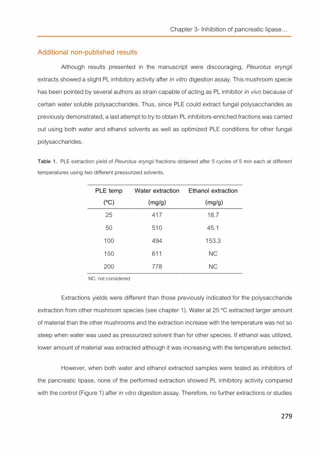
Chapter 3- Inhibition of pancreatic lipase…
279
Additional non-published results
Although results presented in the manuscript were discouraging, Pleurotus eryngii extracts showed a slight PL inhibitory activity after in vitro digestion assay. This mushroom specie has been pointed by several authors as strain capable of acting as PL inhibitor in vivo because of certain water soluble polysaccharides. Thus, since PLE could extract fungal polysaccharides as previously demonstrated, a last attempt to try to obtain PL inhibitors-enriched fractions was carried out using both water and ethanol solvents as well as optimized PLE conditions for other fungal polysaccharides.
Table 1. PLE extraction yield of Pleurotus eryngii fractions obtained after 5 cycles of 5 min each at different temperatures using two different pressurized solvents.
PLE temp (ºC)
Water extraction (mg/g)
Ethanol extraction (mg/g)
25 417 18.7 50 510 45.1
100 494 153.3 150 611 NC 200 778 NC
NC: not considered
Extractions yields were different than those previously indicated for the polysaccharide extraction from other mushroom species (see chapter 1). Water at 25 ºC extracted larger amount of material than the other mushrooms and the extraction increase with the temperature was not so steep when water was used as pressurized solvent than for other species. If ethanol was utilized, lower amount of material was extracted although it was increasing with the temperature selected.
However, when both water and ethanol extracted samples were tested as inhibitors of the pancreatic lipase, none of the performed extraction showed PL inhibitory activity compared with the control (Figure 1) after in vitro digestion assay. Therefore, no further extractions or studies

Chapter 3- Additional non-published results
280
were carried out since results were not so promising as the other compounds investigated and described in other chapters.
Figure 1. Pancreatic lipase activity (IUB/ml) in digested P. eryngii mushroom extracts obtained
from by PLE using ethanol or water as pressurized solvents.

Chapter 4 Effects of fungal compounds with HMGCR inhibitory activity on
the cholesterol metabolism.
Chapter 4- Efect of fungal compounds with HMGCR inhibitory…
283
Preface Statins are the most frequently prescribed drugs to decrease blood LDL-cholesterol levels. These molecules are acting as competitive inhibitors of 3-hydroxy-3-methylglutaryl CoA reductase (HMGCR) substrate, leading to a lower activity of this key enzyme involved in the cholesterol biosynthesis. However, in order to maintain cholesterol homeostasis, HMGCR inhibition stimulate diet cholesterol absorption and vice versa, if its absorption is limited (i.e. by any of the mechanisms previously mentioned), liver cholesterol biosynthesis is enhanced. Thus, an effective lowering-cholesterol therapy for people with hypercholesterolemia should combine statins with low cholesterol-containing diets or intake of food including compounds with the ability of reducing cholesterol absorption (as previously demonstrated in other chapters for instance for fungal sterols and β-glucans). However, although statins are widely used, they induce negative interactions with other therapies (i.e. those for Alzheimer disease) as well as undesirable secondary effects after prolonged administrations.
Besides statins and other pharmacological compounds, a wide range of natural extracts and molecules from different sources have been described as HMGCR inhibitors and gene modulators affecting not only to HMGCR mRNA but also to collateral cholesterol-related genes expression according to in vitro and in vivo studies (as mentioned in Introduction section). Moreover, a few extracts obtained from a mushroom strain (Pleurotus ostreatus) also showed interesting HMGCR inhibitory activities although no statins were detected as demonstrated by the HPLC-MS analysis described in the Preliminary studies section. Thus, in this chapter a wider screening through many mushroom species and the extraction of HMGCR inhibitors-enriched fractions from specific edible mushrooms using pressurized solvent technologies is described. In addition, the use of isolation procedures to identify the inhibitors of the enzyme present in particular fractions from specific mushrooms is detailed and then, in vitro and in vivo experiments were carried out with the extract from one mushroom to establish its ability to lower cholesterol levels and to modulate cholesterol-related genes.
After the preliminary studies carried out with P. ostreatus, the presence of HMGCR inhibitors was investigated in several edible mushroom species. These results are described in
Chapter 4- Preface
284
the works entitled Screening of edible mushrooms and extraction by pressurized water (PWE) of 3-hydroxy-3-methyl-glutaryl CoA reductase inhibitors and Study on the 3-hydroxy-3-methyl-glutaryl CoA reductase inhibitory properties of Agaricus bisporus and extraction of bioactive fractions using pressurized solvent technologies. Firstly, two types of solid:liquid extractions were performed from powdered fruiting bodies obtained from 26 edible mushrooms species in order to determine the presence/absence of HMGCR inhibitors. After the screening, three species were selected (P. ostreatus, L. edodes and A. bisporus) and more detailed studies of their HMGCR inhibitory properties were carried out such as the influence of cultivation parameters (i.e. different spawns, developmental stages, flushes or selenite addition to cultivation substrates), the tissue distribution of the HMGCR inhibitors (as previously elucidated for others compounds i.e. ergosterol) or the advantages of PWE or SFE as extraction methods to obtain fractions with high HMGCR inhibitory activities depending on their extraction parameters i.e. static extraction times, number of cycles or temperature etc., compared with the use of supercritical CO2 with or without co-solvents (depending on the mushroom strain).
In the work previously mentioned concerning A.bisporus, an attempt to further determine the nature of the HMGCR inhibitors was carried out and results indicated that no statins but other compounds with higher molecular weight might be the responsible for the observed inhibitory activities. Thus, there were low probabilities that such a large molecules could exert their observed inhibitory effect in vivo and therefore, efforts were focused in the study of the HMGCR inhibitors detected in the other two mushroom species.
The HMGCR-inhibitors were different depending of the mushroom specie, thus the most interesting inhibitors, those obtained from P. ostretaus and L. edodes, were individually studied. The work entitled Water-soluble polysaccharides from Pleurotus ostreatus with HMGRC (3-hydroxy-3-methyl-glutaryl-CoA reductase) inhibitory activity describes the experiments carried out to point out the compounds present in P. ostreatus water-extracts capable of inhibiting the HMGCR. FITR spectroscopy, GC-MS and MNR analysis of specific fractions at different purification steps were correlated with the determination of their HMGCR inhibitory activities to direct the isolation procedure toward the identification of the compounds responsible for the

Chapter 4- Efect of fungal compounds with HMGCR inhibitory…
285
inhibitory activity. Moreover, the HMGCR inhibitory extracts from P. ostreatus were submitted to an in vitro digestion model to find out whether they could be at least partially bioaccesible. Afterwards, digestates were submitted to a bioavailability tests using Caco2 cells transport assays.
The last work of this chapter, Water-soluble compounds from Lentinula edodes influencing the HMGCoA-reductase activity and the expressions of genes involved in the cholesterol metabolism, includes the results obtained after a similar isolation procedure than the one described for P. ostreatus but directed toward the identification of the HMGCR inhibitors present in L. edodes. The effect on the gene expression showing HMGCR inhibitor of HMGCR and other cholesterol-related genes of a water-soluble extract (containing no statins but eritadenine) was also reported for Caco2 and HepG2 cell lines. Moreover, the hypocholesterolemic properties of the L.edodes water extract are compared with simvastatin or ezetimibe in animal studies using normo- and hypercholesterolemic mice models. Their effect on the transcription profiles after the different dietary interventions are also described.

Chapter 4- Efect of fungal compounds with HMGCR inhibitory…
287
WORKPLAN
Extraction method develop to obtain HMGCR inhibitors-enriched fractions
vs
Analysis of fungal extracts with HMGCR inhibitory
capability HMGCR activity
commercial kit
In vitro digestion
n
Caco2 cell culture
RT-PCR
e
Identification and nature determination of HMGCR inhibitors
Agaricus bisporus Pleurotus ostreatus Lentinula edodes
Caco2 cells
β-glucans content determination
n
Caco2 cell culture In vitro and in vivo genomic effect of L. edodes water-soluble extract
Bioavailability and transport assay of P. ostreatus water-soluble extract
Mus musculus (Male C57BL/6JRj mice)

Chapter 4- Efect of fungal compounds with HMGCR inhibitory…
289
Manuscript 1
Screening of edible mushrooms and extraction by pressurized water (PWE) of 3-hydroxy-3-methyl-glutaryl CoA reductase inhibitors
Alicia Gil-Ramírez1, Cristina Clavijo2, Marimuthu Palanisamy1, Alejandro Ruíz-Rodríguez1, María Navarro-Rubio1, Margarita Pérez2,
Francisco R. Marín1, Guillermo Reglero1, Cristina Soler-Rivas1
1Department of Production and Characterization of Novel Foods, CIAL – Research Institute in Food Science (UAM+CSIC), C/ Nicolas Cabrera 9, Campus de Cantoblanco, Universidad Autónoma de Madrid, 28049 Madrid, Spain. 2Centro Tecnológico de Investigación del Champiñón de La Rioja (CTICH). Autol. Spain
Journal of Functional Foods
Volume 5, Issue 1, pages 244–250, January 2013 DOI: 10.1016/j.jff.2012.10.013

Chapter 4- Manuscript 1
290
Abstract The methanol/water and particularly the water extracts obtained from 26 mushroom
species were able to inhibit the 3-hydroxy-3-methyl-glutaryl CoA reductase (HMGCR) activity to different extent (10 to 76%). Cultivated mushrooms such as Pleurotus sp. and Lentinula edodes were among the strains which showed higher HMGCR inhibitory capacities. Their inhibitory properties were not largely influenced by cultivation parameters, mushroom developmental stage or flush number. The HMGCR inhibitory activity of L. edodes was concentrated in the cap excluding the gills while in P. ostreatus it was distributed through all the different tissues. A method to obtain aqueous fractions with high HMGCR inhibitory activity was optimized using an accelerated solvent extractor (ASE) by selecting 10.7 MPa and 25 ºC as common extraction conditions and 5 cycles of 5 min each for P. ostreatus fruiting bodies and 15 cycles of 5 min for L. edodes suggesting that the potential HMGCR inhibitors are different in the two selected mushrooms.

Chapter 4- Efect of fungal compounds with HMGCR inhibitory…
291
Introduction Coronary heart disease (CHD) is the leading cause of death in the Western world after
cancer according to the World Health Organization. Many studies have established that high total-cholesterol and low-density lipoprotein (LDL) cholesterol levels are risk factors for CHD and mortality. Several in vivo studies have demonstrated the ability of certain edible mushrooms to lower cholesterol levels in serum. Species such as Pleurotus spp. [1-4], Agaricus bisporus [5], Ganoderma lucidum [6], Lentinula edodes, Grifola frondosa, Flammulina velutipes [7], Auricularia auricular and Tremella fuciformis [8], among others [9], have been investigated in animals and human studies.
Apparently, the hypocholesterolemic effect of the mushroom fruiting bodies and several types of their extracts is reached by different mechanisms of action such as impairing dietary cholesterol absorption or inhibiting the endogenous cholesterol metabolism [10]. Mushrooms are rich in chitin (dietary fibre) and specific β-glucans which might inhibit cholesterol absorption by increasing the faecal excretion of bile acids and reducing the amount of serum LDL-cholesterol [10, 11].
Eritadenine (an adenosine analogue alkaloid) is another compound isolated from Lentinula edodes (shiitake mushroom) which is able to lower cholesterol levels. This molecule inhibits S-adenosylhomocysteine hydrolase and modifies the hepatic phospholipid metabolism [12-14].
According to previous reports, oyster mushrooms (Pleurotus spp.) contained lovastatin, a compound able to lower cholesterol levels inhibiting 3-hydroxy-3-methyl-glutaryl CoA reductase (HMGCR) [15, 16], the key-enzyme in the cholesterol metabolism. Statins are the most potent drugs available for reducing plasma low density lipoproteins (LDL) in cholesterol concentrations [17]. However, other reports found no detectable statins levels in Pleurotus sp. fruiting bodies although high HMGCR inhibition activities were recorded [4, 18]. Other compounds (obtained from Ganoderma lucidum) were also described as being able to impair the proper function of the enzyme [6].

Chapter 4- Manuscript 1
292
Thus, in this work a preliminary screening of HMGCR inhibitors was carried out using several edible mushroom species and varieties. The screening was also performed in cultivated mushrooms harvested from cultivation rooms with different cultivation parameters in an attempt to define the conditions required for the synthesis of the effective inhibitors. Once the mushrooms varieties containing higher HMGCR inhibitory activity were defined, they were submitted to pressurized solvent extractions (accelerated solvent extractions, ASE) in order to optimize environmentally friendly and GRAS methods able to obtain fungal fractions with high HMGCR inhibitory activity to further functionalize foods with potentially hypocholesterolemic properties [19].
Materials and Methods
Biological material and samples preparation Mushroom strains used in this investigation were Lentinus edodes S. (Berkeley),
Cantharellus cibarius (Fr.), Lactarius deliciosus (Fr.), Boletus edulis (Bull. Ex Fr.), Pleurotus ostreatus (Jacq.Ex Fr.) Kummer, Agaricus bisporus L. (Imbach), Amanita caesarea (Scop. Ex Fri.) Pers. Ex Schw., Morchella esculenta (Pers Ex Amans), Agaricus blazei Murill ss. (Heinem), Grifola frondosa (Dicks.) Gray, Ganoderma lucidum (Curtis) P.Karst., Flammulina velutipes (Curt. Ex Fr.) Singer, Pleurotus eryngii (D.C. Ex Fr.) Quel, Lyophyllum shimeji (Kawam.), Morchella conica (Pers.), Agrocybe aegerita (Briganti) Singer, Auricularia judea (Bull. Ex St.Amans) Berck, Amanita ponderosa Malençon & R. Heim, Craterellus cornucopioides (L. Ex Fr.) Pers, Marasmius oreades (Bolt. Ex Fr.) Fr., Lepiota procera (Scop. Ex Fr.) Singer., Pholiota nameko (T. Itô) S. Imai, Calocybe gambosa (Fr.) Donk, Hydnum repandum (Linné Ex Fr.), Cantharellus lutescens (Pers.), Pleurotus pulmonarius (Fr.) Quel.
Fruiting bodies from wild mushrooms were purchased from a local market in Madrid (Spain). The cultivable strains were grown in cultivation rooms with automatic control of cultivation parameters (temperature, R.H., CO2) at CTICH (Centro Tecnológico de Investigación del Champiñón de La Rioja, Autol, Spain) or at the cultivation facilities of some mushroom growers belonging to the La Rioja´s mushroom association using commercially available substrates

Chapter 4- Efect of fungal compounds with HMGCR inhibitory…
293
depending on the mushroom specie to cultivate. Fruiting bodies were harvested at the usual developmental stage prior to commercialization except in those experiments when the effect of the developmental stage was studied. The recorded parameters are described in Table 1.
Table 11. Cultivation parameters recorded and correlated with the HMGCR inhibitory activity values obtained within the analyzed samples (P. ostreatus and L. edodes).
Complete fruiting bodies or their separated tissues were immediately frozen,
freeze-dried, ground and sieved until the particle size smaller than 0.3 mm as described [20] and
stored at -20 ºC until further use.
To extract the potential inhibitors from the spores allowing the breaking down of the spore walls, they were first treated with methanol as described by [15].
Determination of HMGCoA-red inhibitors in mushrooms Mushroom powders (50 mg/ml) were mixed with water, methanol/water (1:1 v/v) or
methanol. Suspensions were shaken in a Vortex for 1 min and centrifuged at 12000 rpm
Parameter Different type of samples
Commercial spawn
5 different commercial varieties per specie: P. ostreatus K15 Fungisem, HK35 Sylvean, K40 Fungisem, S-300 Mispaj and H9 Gurelan and L. edodes4312 Sylvan, S05 Mispaj, 3710 Mycelia, Le Lion L8 and 4012 Amycel.
Substrate Pasteurized straw (for P. ostreatus) and
sterilized/pasteurized sawdust (for L. edodes)
Disease influence Healthy, brown blotch, thrichoderma, pests
Developmental stage Primordia, small, medium, large
Flush number First, second, third and fourth flush
Mushroom growers CTICH and 12 mushroom growers
Modern controlled chambers Yes/No
1

Chapter 4- Manuscript 1
294
(8,854 x g) for 2min (Eppendorf mini-spin, Madrid, Spain) according to the user´s manual. Supernatants were used as source of HMGCR inhibitors.
HMGCR activity was measured using the commercial HMG-CoA Reductase Assay (Sigma, Madrid) according to the user´s manual. The assay is based on the spectrophotometric measurement of the decrease in absorbance at 340 nm, which represents the oxidation of NADPH by the catalytic subunit of HMGCR in the presence of the substrate HMG-CoA according to the reaction:
HMG-CoA + 2NADPH + 2H+ →mevalonate + 2NADP+ + CoA-SH
Mushroom supernatants (20 µl) were applied into a 96 wells-plate and their absorbance change was monitorized at 37 ºC using a microplate reader (Tecan Group Lt, Männedorf, Switzerland).
Pravastatin was utilized as a control for positive inhibition. Other control samples were prepared in each assay by substituting the mushroom extract by the same solvent solution utilized in the extract. These controls were considered as 100% activity and tested samples were referred to them as percentage of inhibition or activity. Assays were performed in duplicate.
Pressurized water extractions to obtain fractions with HMGCR inhibitory activity Mushroom powders (1g) were mixed with sea sand (Sigma, Madrid, Spain) (4g) and
submitted to pressurized solvent extraction at 1500 psi (10.68 MPa) using an accelerated solvent extractor (ASE) (Dionex, ASE 350, Sunnyvale, CA, USA). Several parameters such as extraction time or cycle’s number and temperature were changed in order to optimize the extraction method to obtain fractions with high HMGCR inhibitory activity.
Obtained fractions were immediately frozen, lyophilized and stored at -18 ºC until further analysis. Extracted dry matter content was measured to calculate the extraction yields.

Chapter 4- Efect of fungal compounds with HMGCR inhibitory…
295
Statistical analysis One way analysis of variance (ANOVA) was performed using a Statgraphics® Plus 3.1
for Windows software (Statistical Graphics Corporation, Rockville, MD, USA). The mean comparison test used was Fisher’s least significant differences procedure (LSD).
Results and Discussion
Screening of mushrooms species as a source of HMGCR inhibitors Several wild and cultivated mushroom species were submitted to water and
methanol/water (1:1, v/v) extraction since both type of solvents were previously described as able to extract compounds with HMGCR inhibitory activity [18]. Methanol was also tested with a few strains but no interesting HMGCR inhibitory activity was found in any of the selected samples. Results indicated a wide variability within the different mushroom species (Figure 1). The water extracts of 8 species showed no significant HMGCR inhibitory activity and many showed inhibitory activities from 10 to 50%. However, strains such as Pleurotus sp., Cratharellus cornucopiodes, Amanita ponderosa and particularly Lentinula edodes showed inhibitory capacities ranging from 52 up to 76%. In most of the cases except for Agaricus bisporus and Cantharellus lutescens, the water extracts showed higher inhibitory activity than the methanol/water extracts. Surprisingly, A. bisporus methanol/water extracts showed almost 2 fold more capacity than the water extracts.
No similar distribution was found between genera belonging to the same family, for instance, within the Pleurotaceae family (P. ostreatus, P. pulmonarius and P. eryngii) two species showed similar and high HMGCR inhibitory capacities but P. eryngii showed only 20.7% inhibitory capacity. On the contrary, the Agaricaceae family included two species (A. bisporus and A. blazeii) with low inhibition in their water extracts while L. procera was able to inhibit almost 50% of the enzyme. The Cantharellaceae family (C. lutescens, C. Cibarius and C. cornucopioides) as well as other families such as Marasmiaceae (M. oreades and L. edodes), Morchellaceae (M. conica and M. esculenta) and Amanitaceae (A. caesarea and A. ponderosa) included species with no, middle and high inhibitory capacities.

Chapter 4- Manuscript 1
296
Figure 9. HMGCR inhibitory activities of several mushrooms extracts obtained in water or methanol/water (1:1, v/v).

Chapter 4- Efect of fungal compounds with HMGCR inhibitory…
297
Influence of the cultivation parameters on the HMGCR inhibitory activity of cultivated mushrooms
The water extracts from cultivated mushrooms such as Pleurotus sp. and L. edodes showed very interesting inhibitory activities and their growth can be more easily controlled than wild mushrooms. Environmental parameters usually influence the presence/absence of many compounds therefore; a more detailed study was carried out in those cultivated mushrooms in order to determine the effect of the cultivation parameters in their HMGCR inhibitory activities.
Substrates were inoculated with commercial spawns from several P. ostreatus and L. edodes varieties in a minimum of three independent trials but performed under similar conditions. After cultivation, the obtained fruiting bodies from the first flush were harvested and their water extracts analyzed. Results showed no significant differences between Fungisem K15 and Sylvan HK35 but they were significantly higher in Fungisem K15 than the other analyzed varieties (Figure 2a). Similar results were obtained when the different L. edodes spawns were studied (Figure 2b); significant differences were found between Sylvan 4312 and Mycelia 3710 or Le Lion L8 but the standard deviation observed within similar trials suggested that other parameters different than variety are involved in the HMGCR inhibitory activity observed.
Therefore, other parameters were investigated such as mushroom grower, automatic control of cultivation conditions (or traditional cultivation system), presence of bacterial blotch infection, pests (flies, mosquitoes etc.), good or poor quality fruiting bodies or mushroom picked from places close to a Trichoderma sp. spot. However, no correlation was found within the detected HMGCR inhibitory activities and any of the indicated parameters.
Developmental and tissue distribution of the HMGCR inhibitory activity of cultivated mushrooms
P. ostreatus and L. edodes fruiting bodies from the same cultivation trial were harvested at different developmental stages including from primordia until cap was open and gills exposed (mature). The harvesting was repeated in the second and third flush. No significant differences were observed between flushes or developmental stages in the HMGCR inhibitory activity of the water extracts obtained from P. ostreatus mushrooms. In L. edodes mushrooms a slight decrease

Chapter 4- Manuscript 1
298
of the inhibitory capacity was observed with the increase of the flush number but no significant differences were found between young or mature fruiting bodies.
a)
b)
This suggest that the compounds responsible for the HMGCR inhibitory activity are present through the whole sporophore life cycle and that they might be different between the two studied mushroom strains. This view was further supported by the fact that compounds potentially responsible for the inhibitory activity showed a different tissue distribution within the fruiting bodies. The water extracts obtained from different tissues of P. ostreatus showed similar HMGCR inhibitory activity indicating a homogeneous distribution (Figure 3a) thought the whole body while
Figure 2. HMGCR inhibitory activities of water extracts from the fruiting bodies obtained by cultivating different commercial spawns fron a) Pleurotus ostreatus and b) Lentinula edodes.
0
10
20
30
40
50
60
70
80
90
100
K15 Fungisem HK35 Sylvan K40 Fungisem S - 300 Mispaj H9 Gurelan
HMGC
R inh
ibitio
n (%
)
Commercial spawn
0
10
20
30
40
50
60
70
80
90
100
4312 Sylvan S05 Mispaj 3710 Mycelia Le Lion L8 4012 Amycel
HMGC
R inh
ibitio
n (%
)
Commercial spawn

Chapter 4- Efect of fungal compounds with HMGCR inhibitory…
299
in the selected L. edodes variety the inhibitory activity was concentrated in the dermis and epidermis excluding the gills and the stipe (Figure 3b).
a)
b)
Worth to take into consideration was the HMGCR inhibitory activity levels found in the discarded parts of the stipes, this part correspond to the lower stipe which is bound to the mycelium and the substrate, at the base of the fruiting body. Usually, this part of the mushroom is cut during harvesting because it might contain traces of substrate or it is deformed and not commercialized. The HMGCR inhibition values of the discarded by-product obtained from L. edodes varied depending on the strain since Amycell 4012 showed no HMGCR inhibitory activity but Sylvan 4312 showed a 37.1% inhibition, higher values than the 10.8% inhibition found in the discarded stipe of P. ostreatus.
Figure 3. HMGCR inhibitory activities of water extracts from different tissues from fruiting bodies from the first flush of a) Pleurotus ostreatus Sylvan HK35 and b) Lentinula edodes Amycell 4012.
0
10
20
30
40
50
60
70 80
90
100
Cap Dermis+Epidermis Gills Stipe Spores
HMGC
R inh
ibitio
n (%
)
0
10
20
30
40
50
60
70
80
90
100
Cap Dermis Epidermis Gills Stipe
HMGC
R inh
ibitio
n (%
)

Chapter 4- Manuscript 1
300
Pressurized water extraction of the cultivated mushrooms to obtain fractions with HMGCR inhibitory activity
Pressurized water extractions are economically sustainable processes that are increasingly being developed as a response to the demand (by the food and other industries) of environmentally clean extraction processes to produce new extracts or compounds with a potential use as functional ingredients or nutraceuticals [21]. Water is a non-toxic and naturally occurring solvent that can replace organic solvents when used at higher temperatures and pressures. Accelerated solvent extraction using water has been used to extract i.e. polysaccharides with different structures and biological activities from edible mushrooms [22, 23] and they are, at the present, being scaled up for industrial applications [24].
P. ostreatus and L. edodes fruiting bodies were submitted to extraction with pressurized water in order to obtain mushroom extracts with high HMGCR inhibitory activity to be in the future potentially used as hypocholesterolemic ingredient to functionalize foods. Thus, several parameters such as extraction temperature, time and cycles number were changed in order to optimize the extraction method.
A range of temperatures was studied from 25 up to 200 ºC and the obtained dry matter increased largely with the temperature, being 200 ºC the best condition to extract almost 60 and 80% of the material from P. ostreatus and L. edodes, respectively (Figure 4a). However, when the HMGCR inhibitory capacity of the extracted fractions was measured, the increase of temperature was detrimental suggesting that the compounds responsible for the inhibition were thermolabile (Figure 4b). No significant differences were found between extractions at 25 or 50 ºC in both mushroom strains neither between these extractions from L. edodes and extractions with plain water however, water extraction of P. ostreatus showed a significantly higher inhibitory activity (80.1%) than its ASE extractions. Thus, extraction temperature was fixed at 25 ºC and other ASE parameters were modified in order to improve extraction yields and inhibitory activities.

Chapter 4- Efect of fungal compounds with HMGCR inhibitory…
301
a)
b)
Extractions carried out by selecting 5 cycles of 1 min each yielded almost half of the dry matter than longer extraction times from P. ostreatus mushrooms however, no significant differences were observed for L. edodes within 1 to 10 min; apparently all the water soluble compounds at 25 ºC are easily extracted after 5 cycles of 5 min from both strains (Figure 5a). The ASE extracts obtained from both mushroom strains after 5 cycles of 1 to 10 min each showed high HMGCR inhibitory capacity while longer extractions showed a slightly lower inhibitory activity, suggesting than the compounds potentially responsible for the inhibition might be unstable if extracted for 75 min total extraction time (Figure 5b). The P. ostreatus extract obtained after
Figure 4. a) ASE extraction yield and b) HMGCR inhibitory activities of Pleurotus ostreatus and Lentinula edodes fractions obtained after 5 cycles of 5 min each at different temperatures.
0
100
200
300
400
500
600
700
800
900
25 50 100 150 200
mg/g
mush
room
(dw)
ASE temperatures (ºC)
L. edodes
P. ostreatus
0
10
20
30
40
50
60
70
80
90
100
25 50 100 150 200
HMGC
R inh
ibitio
n (%
)
ASE temperatures (ºC)
L. edodes
P.ostreatus

Chapter 4- Manuscript 1
302
5 cycles of 1 min showed similar inhibitory capacity than longer extraction times, however, less dry matter was obtained pointing the 5 min extraction as the best extraction time, thus, the optimal number of cycles was further studied.
a)
b)
Increase of the extraction cycles (of 5 min each at 25 ºC) from 1 to 15 cycles increased the extracts yields almost 3 fold for both mushroom strains (Figure 6a). However, for P. ostreatus, the ASE extracts obtained after 5 cycles showed the highest inhibitory activity while no significant differences were observed within the HMGCR inhibitory capacity of all the ASE extracts from L. edodes obtained independently of the number of cycles (Figure 6b). These results suggested once more that the compounds responsible of the inhibitory capacity might be different in both
Figure 5. a) ASE extraction yield and b) HMGCR inhibitory activities of Pleurotus ostreatus and Lentinula edodes fractions obtained at 25 °C but at different extraction times per cycle (5 cycles).
0
50
100
150
200
250
300
1 5 10 15
mg/g
mus
hroo
m (d
w)
Extraction time (min)
L. edodes
P. ostreatus
0 10 20 30
40 50 60 70 80
90 100
1 5 10 15
HMGC
R inh
ibitio
n (%
)
Extraction time (min)
L. edodes
P. ostreatus

Chapter 4- Efect of fungal compounds with HMGCR inhibitory…
303
mushrooms since 5 cycles should be the optimal to extract the potentially enzyme inhibitors from P. ostreatus while higher cycles number can be used to extract higher amount of L. edodes extracts. But, both inhibitors are easily extracted with water and their HMGCR inhibitory activity is eliminated at temperatures where the typical protein denaturalization takes place suggesting that they might share similar chemical nature and it could include a proteic part in its structure. Actually, preliminary studies are being carried out suggesting that the inhibitors might be proteoglucans.
a)
b)
Figure 6. a) ASE extraction yield and b) HMGCR inhibitory activities of Pleurotus ostreatus and Lentinula edodes fractions obtained at 25 °C but at different number of cycles (5 min each).
0
50
100
150
200
250
300
350
400
1 5 10 15
mg/g
mush
room
(dw)
Extraction cycles (number)
L. edodes
P. ostreatus
0
10
20
30
40
50
60
70
80
90
100
1 5 10 15
HMGC
R inh
ibitio
n (%
)
Extraction cycles (number)
L. edodes
P. ostreatus
Chapter 4- Manuscript 1
304
Conclusions Several wild and cultivated mushroom species contain water soluble compounds
potentially capable of inhibiting the key enzyme of the cholesterol metabolism using in vitro test. Two of the cultivated strains, Oyster and Shiitake mushrooms, were particularly interesting. Their higher or lower inhibitory capacity was probably dependent on specific requirements since no specific influence of the commercial variety, cultivation parameters or developmental stage of the mushroom was observed. Tissue distribution within the fruiting bodies was different suggesting the presence of different compounds with HMGCR inhibitory activity in both species. The potential inhibitory compounds could be easily extracted by using pressurized water extractions at 10.68 MPa and 25 ºC, 5cycles of 5 min for P. ostreatus and up to 15 cycles of 5 min for L. edodes. Thus, by using the optimized extraction methods, higher quantities of the potential inhibitors in will be extracted for further identification of the responsible compounds. The purification steps will have to be separately carried out for each mushroom since the responsible compounds might be different.
Acknowledgments The research was supported by the European Union within the 7th framework programme
(7FM-PEOPLE-2009-IFF project 251285), ALIBIRD-CM S2009/AGR-1469 regional program from the Community of Madrid (Spain),AGL2010-21537 national R+D program from the Spanish Ministry of Science and Innovation, and 2010-I-ID-00093 project by ADER (Agency for La Rioja´s economic development).

Chapter 4- Efect of fungal compounds with HMGCR inhibitory…
305
References [1]. Bajaj, M., et al., Role of oyster mushroom (Pleurotus florida) as hypocholesterolemic/antiatherogenic agent. Indian
Journal of Experimental Biology, 1997. 35(10): p. 1070-5. [2]. Bobek, P. and S. Galbavy, The oyster mushroom (Pleurotus ostreatus) effectively prevents the development of
atherosclerosis in rabbits. Ceska a Slovenska farmacie : casopis Ceske farmaceuticke spolecnosti a Slovenske farmaceuticke spolecnosti, 1999. 48(5): p. 226-30.
[3]. Opletal, L., et al., Evidence for the anti-hyperlipidaemic activity of the edible fungus Pleurotus ostreatus. British Journal of Biomedical Science, 1997. 54(4): p. 240-243.
[4]. Schneider, I., et al., Lipid lowering effects of oyster mushroom (Pleurotus ostreatus) in humans. Journal of Functional Foods, 2011. 3(1): p. 17-24.
[5]. Jeong, S.C., et al., White button mushroom (Agaricus bisporus) lowers blood glucose and cholesterol levels in diabetic and hypercholesterolemic rats. Nutrition Research, 2010. 30(1): p. 49-56.
[6]. Berger, A., et al., Cholesterol-lowering properties of Ganoderma lucidum in vitro, ex vivo, and in hamsters and minipigs. Lipids in Health and Disease, 2004. 3(1): p. 2.
[7]. Fukushima, M., et al., Cholesterol-lowering effects of maitake (Grifola frondosa) fiber, shiitake (Lentinus edodes) fiber, and enokitake (Flammulina velutipes) fiber in rats. Experimental Biology and Medicine, 2001. 226(8): p. 758-765.
[8]. Cheung, P.C.K., The hypocholesterolemic effect of two edible mushrooms: Auricularia auricula (tree-ear) and Tremella fuciformis (white jelly-leaf) in hypercholesterolemic rats1. Nutrition Research, 1996. 16(10): p. 1721-1725.
[9]. Roupas, P., et al., The role of edible mushrooms in health: Evaluation of the evidence. Journal of Functional Foods, 2012. 4(4): p. 687-709.
[10]. Guillamón, E., et al., Edible mushrooms: Role in the prevention of cardiovascular diseases. Fitoterapia, 2010. 81(7): p. 715-723.
[11]. Chen, J. and R. Seviour, Medicinal importance of fungal β-(1→3), (1→6)-glucans. Mycological Research, 2007. 111(6): p. 635-652.
[12]. Chibata, I., et al., Lentinacin-A new hypocholesterolemic subbstance in Lentinus edodes. Experientia, 1969. 25(12): p. 1237-&.
[13]. Sugiyama, K., Hypocholesterolemic action of eritadenine is mediated by a modification of hepatic phospholipid metabolism in rats. Journal of Nutrition, 1995. 125(8): p. 2134-2144.
[14]. Yamada, T., et al., Structure and function of eritadenine and its 3-deaza analogues: Potent inhibitors of S-adenosylhomocysteine hydrolase and hypocholesterolemic agents. Biochemical Pharmacology, 2007. 73(7): p. 981-989.
[15]. Gunde-Cimerman, N., A. Plemenitaš, and A. Cimerman, Pleurotus fungi produce mevinolin, an inhibitor of HMG CoA reductase. FEMS Microbiology Letters, 1993. 113(3): p. 333-337.

Chapter 4- Manuscript 1
306
[16]. Gunde-Cimerman, N., A. Plemenitaš, and A. Cimerman, A hydroxymethylglutaryl-CoA reductase inhibitor synthesized by yeasts. FEMS Microbiology Letters, 1995. 132(1–2): p. 39-43.
[17]. Shitara, Y. and Y. Sugiyama, Pharmacokinetic and pharmacodynamic alterations of 3-hydroxy-3-methylglutaryl coenzyme A (HMG-CoA) reductase inhibitors: Drug–drug interactions and interindividual differences in transporter and metabolic enzyme functions. Pharmacology & Therapeutics, 2006. 112(1): p. 71-105.
[18]. Gil-Ramirez, A., et al. Edible mushrooms as potential sources of new hypocholesterolemic compounds in 7th International Conference on Mushroom Biology and Mushroom Products. 2011. Arcachon, France.
[19]. Chen, Z.-Y., et al., Role and classification of cholesterol-lowering functional foods. Journal of Functional Foods, 2011. 3(2): p. 61-69.
[20]. Ramírez-Anguiano, A.C., et al., Radical scavenging activities, endogenous oxidative enzymes and total phenols in edible mushrooms commonly consumed in Europe. Journal of the Science of Food and Agriculture, 2007. 87(12): p. 2272-2278.
[21]. Sun, H., et al., Application of accelerated solvent extraction in the analysis of organic contaminants, bioactive and nutritional compounds in food and feed. Journal of Chromatography A, 2012. 1237(0): p. 1-23.
[22]. Lo, T.C.T., et al., Pressurized water extraction of polysaccharides as secondary metabolites from Lentinula edodes. Journal of Agricultural and Food Chemistry, 2007. 55(10): p. 4196-4201.
[23]. Di, X., et al., Fingerprint profiling of acid hydrolyzates of polysaccharides extracted from the fruiting bodies and spores of Lingzhi by high-performance thin-layer chromatography. Journal of Chromatography A, 2003. 1018(1): p. 85-95.
[24]. Pronyk, C. and G. Mazza, Design and scale-up of pressurized fluid extractors for food and bioproducts. Journal of Food Engineering, 2009. 95(2): p. 215-226.

Chapter 4- Efect of fungal compounds with HMGCR inhibitory…
307
Manuscript 2
Study on the 3-hydroxy-3-methyl-glutaryl CoA reductase inhibitory properties of Agaricus bisporus and extraction of bioactive fractions
using pressurized solvent technologies (ASE and SFE)
Alicia Gil-Ramírez1, Cristina Clavijo2, Marimuthu Palanisamy1, Alejandro Ruiz-Rodríguez1, María Navarro-Rubio1, Margarita Pérez2,
Francisco R. Marín1, Guillermo Reglero3, Cristina Soler-Rivas1
1Department of Production and Characterization of Novel Foods, CIAL – Research Institute in Food Science (UAM+CSIC), C/ Nicolas Cabrera 9, Campus de Cantoblanco, Universidad Autónoma de Madrid, 28049 Madrid, Spain. 2Centro Tecnológico de Investigación del Champiñón de La Rioja (CTICH). Autol. Spain 3IMDEA-Food Institute. CEI UAM+CSIC. Madrid, Spain.
Journal of the Science of Food and Agriculture Volume 93, Issue 11, pages 2789–2796, August 2013
DOI: 10.1002/jsfa.6102

Chapter 4- Manuscript 2
308
Abstract BACKGROUND: Agaricus bisporus mushrooms were able to lower cholesterol levels in
hypercholesterolemic rats and it was suggested that dietary fibers might inhibit cholesterol absorption. However, A. bisporus extracts were also able to inhibit the 3-hydroxy-3-methyl-glutaryl CoA reductase (HMGCR, the key enzyme in the cholesterol biosynthetic pathway) and this might also contribute to the observed lowering of cholesterol levels in serum.
RESULTS: The methanol:water extracts obtained from A. bisporus were able to inhibit up to 60% the HMGCR activity using an in vitro assay. The HMGCR inhibitory capacities depended on cultivation conditions, strains, etc. The potential inhibitors were not statins, they might be β-glucans able to scavenge the substrate and impair the enzymatic reaction. They were present during all mushroom developmental stages and similarly distributed through all the tissues including the parts discarded as a by-product. Accelerated solvent extractions using 1:1 ethanol:water as pressurized solvent (10.7 MPa, 25 ºC, 5 cycles of 5 min) were more effective in the extraction of the HMGCiR inhibitor/s than supercritical fluid extractions (9 MPa, 40 ºC) using CO2 with 10% ethanol.
CONCLUSION: A mushroom cultivation and two extraction procedures were optimized to obtain fractions from A. bisporus with high HMGCR inhibitory activities to design novel ingredients for hypocholesterolemic functional foodstuffs.

Chapter 4- Efect of fungal compounds with HMGCR inhibitory…
309
Introduction High cholesterol or low-density lipoprotein levels in serum increases the risk of
cardiovascular diseases. In public health terms, achieving a reduction in cholesterol by dietary advice (increase consumption of vegetable and fruits) is of limited effectiveness [1]. Many researchers have been exploring the possibility of increasing components in foodstuff which have hypocholesterolemic effects such as i.e. β-glucans or phytosterols [2, 3]. These novel functionalized foods reduce serum cholesterol (10 – 20%) because they impair the absorption of exogenous cholesterol during digestion [2, 4]. However, depending on genetic variations, many consumers cannot reduce their cholesterol levels because, by avoiding cholesterol intake, their 3-hydroxy-3-methyl-glutaryl CoA reductase (HMGCR) activity is stimulated, enhancing the biosynthesis of endogenous cholesterol [5]. Thus, another approach to the design of novel hypocholesterolemic functional foods might be their supplementation with compounds able to inhibit the HMGCR activity.
It has been shown that edible mushrooms are able to lower cholesterol levels in vivo (Pleurotus spp., Lentinula edodes, Ganoderma lucidum, Agaricus bisporus etc.) [6-8] and can inhibit HMGCR in vitro. Some reports pointed to lovastatin as the compound responsible for the enzyme inhibition [9-11]. Lovastatin (mevinolin) and other statins are drugs usually prescribed to hypercholesterolemic patients because they act as competitive inhibitors of the HMGCR, showing approx 200-fold more affinity for them than their real substrate [12]. Moreover, selenium supplementation combined with statins therapy was proved to be significantly beneficial to lipid therapy [13] and mushrooms can be fortified with selenium by addition of sodium selenite to their cultivation substrates to modify their biological properties [14]. Thus, if there are statin-producing mushroom strains, they might also be fortified with selenium to enhance their hypocholesterolemic effect.
However, other authors found no detectable lovastatin levels but still high HMGCR inhibition activities [6, 15]. Other compounds such as lanosteroids, ganoderols etc., were described in G. lucidum and apparently they were also able to inhibit the enzyme but through an indirect mechanism [6].

Chapter 4- Manuscript 2
310
A. bisporus basidiomata were able to lower blood glucose and cholesterol levels in diabetic and hypercholesterolemic rats and it was suggested that dietary fibre might be involved, but via a complex process [8] The mushroom dietary fibre fraction includes mainly chitins and, in higher amounts, β-glucans. Fungal β-glucans are polysaccharides of D-glucose monomers linked by β-glycosidic bonds showing preference for the β-(1→3),(1→6) bonds instead of the β-(1→3),(1→4), the characteristic pattern of cereals β-glucans [16]. In this study, the HMGCR inhibitory properties of A. bisporus extracts are reported as another pathway which might also contribute to the observed lowering of cholesterol levels in serum. The distribution of the potential inhibitor(s) depending on the cultivation parameters, different strains (including Se-fortified strains), developmental stages etc., was also monitored.
Supercritical fluid extraction (SFE) with CO2 as well as accelerated solvent extraction (ASE) are ecologically and economically sustainable processes that are increasingly being developed as a response to recent reports concerning the environmental status of our planet [17]. Moreover, both methods satisfy the demand of environmentally clean extraction processes to produce new extracts or compounds with a potential use as functional ingredients or nutraceuticals. The use of supercritical carbon dioxide, with properties such as low viscosity, high solvent strength and zero surface tension has been encouraged as a substitute for organic solvents. It has been previously used in the extraction of different compounds from mushroom strains [18-23]. On the other hand, when water is used at higher temperatures and pressures (as occurs in ASE) it can also replace organic solvents. Water can also be used in combination with ethanol, a solvent that is classified as generally recognized as safe’ and which is used in the food industry, to tailor the extract composition and therefore its beneficial properties. Accelerated solvent extraction using water has also been used to extract polysaccharides and compounds with different biological activities from L. edodes, P. ostreatus and G. lucidum [15, 24-26]. However, to our knowledge there are no studies on the screening of the HMGCR inhibitory properties of A. bisporus extracts produced by either SFE or ASE technologies.

Chapter 4- Efect of fungal compounds with HMGCR inhibitory…
311
Therefore the other aim of this work was to compare these two extraction technologies in order to optimize methods to obtain fractions from A. bisporus with high HMGCR inhibitory activities to design novel ingredients for hypocholesterolaemic functional foodstuffs.
Materials and Methods
Biological material Agaricus bisporus L. (Imbach) mushrooms were grown in cultivation rooms with
automatic control of cultivation parameters (temperature, relative humidity, CO2) at CTICH (Centro Tecnológico de Investigación del Champinón de La Rioja, Autol, Spain) or at the cultivation facilities of some mushroom growers belonging to La Rioja’s mushroom association using as substrate the commonly utilized indoor compost phase II. A more detailed explanation of the experimental trials, utilized substrates, casing layers and spawns and Se supplementations can be found elsewhere [27]. Basidiomata were harvested at stage 2 –3 according to Hammond and Nichols (1975) [28] except in those experiments when the effect of the developmental stage was studied. The recorded parameters are described in Table 1.
Table 7. Cultivation parameters recorded and correlated with the 3-hydroxy-3-methyl-glutaryl CoA reductase inhibitory activity values obtained within the Agaricus bisporus samples analyzed.
Basiodiomata or their separate tissues (manually separated with the aid of a knife) were dehydrated (lyophilized) and ground into fine powder as described by Ramírez-Anguiano et al. (2007) [29]. Dried mushroom powders were stored at -20 °C until further use.
A minimum of three sporophores were harvested from the same cultivation bed with identical conditions for each different type of sample.
Parameter Different type of samples
Selenium-enriched mushrooms Low (10.9 ppm)/ high (31.6 ppm)/ control (3.2 ppm) Se content Commercial spawn Fungisem H15 and H5,Gurelan 60, Mispaj 365 and Somycel A15
Developmental stage Primordia(stage 1), small(2), medium(3-4), large(5-6)
Flush number First, second and third flush
1
Chapter 4- Manuscript 2
312
Determination of selenium The digestion procedure and hydride generation atomic absorption spectrometry (HG-
AAS) method was optimized at CTICH based on previous reports [30, 31]. Briefly, mushroom powders (0.4 g) were introduced into a Teflon microwave digestion vessel and HNO3 7.7 M was added to a final volume of 8 mL. The vessel was closed and fastened into the rotor. The rotor with six loaded vessels was placed into the microwave oven (Milestone Ethos Touch control; Sorisole, Italy). The applied microwave digestion program was as follows: 0 – 100 °C (3 min), 100 – 150 °C (7 min), 150 – 180 °C (6 min) and 180 °C (15 min). The digested samples were adjusted to 20 mL with HCl 4M and heated at 95 °C for 20 min to ensure reduction of Se(VI) to Se(IV). Once the mixture had cooled, it was diluted with 50 ml HCl 4F. Selenium was determined by HG-AAS with optimized parameters. Detection was performed by an HG-AAS system (Solaar M6 MK2 Dualz; Thermo Scientific, Cambridge, UK). The peak areas of the absorbance were used for calculation of selenium content. The HG-AAS technique requires a selenium hollow cathode lamp to operate at a wavelength of 196.0 nm with a slit width set to 0.5 nm and an electrode-less discharge lamp set at 10 mA current without background corrector. Aspiration time was 45 s, measuring time 5 s, the height signal evaluation and argon flow 314 ml/min. The atomizing environment was a silicous cell heated to 900 °C, floated solution HCl (10 mol/l) and reducers were 0.3% NaBH4 and 0.4% NaOH. Selenium determination was performed in duplicate and quantified according to a Se calibration curve.
Accelerated solvent extractions Mushroom powder (1 g) were mixed with washed sea sand (4 g) (Panreac, Madrid,
Spain) and submitted to pressurized solvent extraction using an accelerated solvent extractor (ASE 350; Dionex Corporation, Sunnyvale, CA, USA). The sea sand was selected as an inert material to hold the sample inside the extraction cell and to improve efficiency avoiding formation of preferential flow paths. The extraction procedure (per cycle) was carried out at 10.68 MPa (1500 psi) as follows. First, the sample was loaded into an 11 ml extraction cell. Then, the cell was filled with ethanol and heated. Static extraction was carried out during the selected time (in minutes) with all system valves closed. When a cycle had finished, the cell was rinsed, the solvent was

Chapter 4- Efect of fungal compounds with HMGCR inhibitory…
313
purged from the cell with N2 gas and the cell remained depressurized. Then, fresh solvent was again added to the extraction cell to carry out another extraction cycle until the programmed number of cycles was finished. The fractions collected after the selected cycles were pooled in a vial as a single extract.
Several parameters, such as solvent, static extraction time or number of cycles, and temperature were changed in order to optimize the extraction method to obtain fractions with high HMGCR inhibitory activity.
After collection, fractions were immediately concentrated with a rotary evaporator (40 °C), frozen and lyophilized. Dried samples were stored at -18 °C until further analysis. Extracted dry matter content was measured to calculate the extraction yields. Extractions were carried out in duplicate.
Supercritical fluid extractions Supercritical fluid extractions (pilot-plant scale) with CO2 (Air-Liquid España, S.A.,
Madrid, Spain) were carried out in a plant (model SF2000; Thar Technology, Pittsburgh, PA, USA) comprising a 2 L cylinder extraction cell and two different separators (S1 and S2), each of 0.5 L capacity, with independent control of temperature and pressure. The extraction vessel has a height:diameter ratio of 5.5 (0.42 m height, 0.076 m internal diameter). A detailed explanation of the experimental device can be found elsewhere [32]. For each experiment, the extraction cell was filled with 80 g of mushroom powder and 900 g of washed sea sand (Panreac). In order to optimize the extraction method to obtain fractions with high HMGCR inhibitory activity, parameters such as the use of a co-solvent (ethanol, 10% w/w) and extraction pressure (30, 18 and 9 MPa) were tested. Extraction temperature as well as the temperature of separators 1 and 2 was set to 40 °C for all the experimental assays. The pressure of separator 1 and separator 2 using ethanol as co-solvent was maintained at 6 and 0.1 MPa, respectively; however in CO2 extraction both separators were kept at a pressure of 6 MPa. The CO2 flow was set to 2.4 kg/h and the total extraction time was 3 h. For each experiment, extracts collected in separators 1 and 2 were mixed together, concentrated to dryness with a rotary evaporator and stored at -18 °C until further

Chapter 4- Manuscript 2
314
analysis. Extracted dry matter content was measured to calculate the extraction yields. All the experiments were carried out in duplicate.
Determination of HMGCR inhibitory activity Mushroom powders or the mushroom extracts obtained (50 mg/ml) were mixed with
water (heated at 50 °C or room temperature) or with methanol:water (1:1 v/v). Suspensions were shacked in a vortex for 1 min and centrifuged at 9659 × g for 2 min. Supernatants (20 µl) were applied to a 96-well plate (Corning Incorporated Life Sciences, Tewksbury, MA, USA) according to the user’s manual of the 3-hydroxy-3-methyl-glutaryl CoA reductase (HMGCR) assay (Sigma, Madrid, Spain). Absorbance at 340 nm was monitored at 37 °C using a microplate reader (Tecan Group, Mannedorf,¨ Switzerland). Determinations were carried out in duplicate.
HPLC-MS/MS analysis of the extracts Mushroom extracts (3 mg/mL) showing HMGCR inhibitory capacity were injected (20 µL)
into an Accela HPLC-MS/MS (Thermo Electron Corporation, San Jose, CA, USA) equipped with an ACE 3 C18-AR column 150 × 4.6 mm, 3 µm particle size (Advanced Chromatography Technologies, Aberdeen, UK). The mobile phase was a mixture of acetonitrile:0.5% acetic acid in water (60:40 v/v) running isocratically for 30 min. The flow rate was 0.5 ml/min. Detection was accomplished by using photodiode array detector (200 – 700 nm) and a mass spectrometer (triple quadrupole) (TSQ-Quantum; Thermo Electron Corporation, San Jose, CA, USA) with an electrospray ionization interface.
For mass spectrometry analysis the spray voltage was set at 3500 V in positive mode, nitrogen was used as sheath gas at 350 °C and 35 arbitrary flow/pressure units. The mass analyzer was set simultaneously in full scan and single reaction monitoring (SRM) modes; in this case SRM experiments were done using one precursor ion and one daughter as recommended by the relevant European Union regulation (Comision Decission 657/2002 regarding performance of analytical methods and the interpretation of results). SRM filters were set for a better quantification of statins, precursor and product ions. Moreover, a full scan mode was used within a mass range

Chapter 4- Efect of fungal compounds with HMGCR inhibitory…
315
of 200 – 800 m/z. Statins including pravastatin, lovastatin, simvastatin and atorvastatin (Cinfa, Pamplona, Spain) were used as standards. Determinations were carried out in duplicate.
Determination of the chemical nature of the HMGCR inhibitors Agaricus bisporus dry powder (50 mg/mL) showing HMGCR inhibitory activity was mixed
with methanol:water (1:1) the vigorously shaken in a vortex for 1 min, centrifuged at 9659 × g in a microfuge for 2 min and the obtained supernatant separated. The supernatant was filtrated through a 0.45 µm filter and the filtrate was submitted to several filtrations using centrifugal filters (VWR, Barcelona, Spain) with cut-off of 10 and 3 kDa (Microcom filters; Millipore, Madrid, Spain) using a microfuge. Fractionated samples were then applied to the HMGCR activity test in order to estimate the molecular weight of the potential HMGCR inhibitors.
The mushroom samples in methanol:water (1:1) were also prepared by mixing them with enzymatic preparations such as: (1) a mixture of exo-1,3-β-glucanase (100 U/ml) and β- glucosidase (20 U/ml); (2) a mixture of invertase (500 U/ml) and amyloglucosidase (1630 U/ml); and (3) pepsin (536 U/ml). Treated samples where incubated for 1 min and centrifuged (9659 × g for 2 min.). Supernatants were submitted to filtration using the 10 kDa filters and the microfuge (13 147 × g for 30 min) in order to separate the enzymes avoiding interference. Then, the HMGCR inhibitory activity of the separated fractions was determined.
The same methanol:water extracts showing HMGCR inhibitory activity where applied to the HMGCR activity test but altering the application procedure. Usually, in this test, solutions are applied following the sequence: first the buffer, then NADPH, substrate, standard inhibitor (pravastatin) as control or sample (potential inhibitory preparation) and, finally, the HMGCoA reductase. Then, the test was performed by mixing the sample and substrate separately and applying the mixture when substrate should be added or mixing the sample with the enzyme and applied at the end. Moreover, similar mixtures were also prepared and applied but mixing the sample with a diluted solution (1/2) of pravastatin or with the enzyme but in the presence of the diluted pravastatin concentration. All the experiments were carried out in duplicate.
Chapter 4- Manuscript 2
316
Statistical analysis One way analysis of variance was performed using a Statgraphics Plus 3.1 for Windows
software (Statistical Graphics Corporation, Rockville, MD, USA). The mean comparison test used was Fisher’s least significant difference procedure.
Results and Discussion
Influence of the cultivation systems
According to preliminary results [27, 33], cultivation parameters influence the HMGCR inhibitory activity found in A. bisporus basidiomata because, when different growers cultivate the same mushroom variety, large differences in the percentage inhibition are observed [33]. However, some of those parameters, such as the casing layer utilized to induce fructification, did not significantly affect the presence of HMGCR inhibitory compounds and neither was any correlation observed with other parameters, such as crop quality or infections (bacterial, fungal and pests)[33]. Other reports also indicated that water was the best solvent to extract HMGCR inhibitors from several mushrooms except for A. bisporus, which showed higher HMGCR inhibitory activity in methanol:water extracts [15]. Thus, A. bisporus basidiomata from the same spawn variety (Fungisem H-15) were cultivated in the same cultivation room in three different batches and harvested to investigate the variability from trial to trial and between the first three flushes.
The mushroom powders obtained from the collected basidiomata were mixed with both solvents to compare and results indicated that there were no significant differences between trials. The methanol:water extracts obtained from mushrooms harvested in the first and second flush showed a lower HMGCR inhibitory capacity (respectively, 14.4 and 11.2%) than those harvested from the third flush (29.5%). Thus, the potential inhibitor(s) might be compounds related to the secondary metabolism (i.e. statins) since in the third flush, mushrooms obtain fewer nutrients from the substrate than during the first and second flushes. On the other hand, it might also suggest that the HMGCR inhibitory activity of the basidiomata might be modulated by the presence/absence of specific compounds.

Chapter 4- Efect of fungal compounds with HMGCR inhibitory…
317
Mushrooms can be fortified with selenium by the addition of sodium selenite to their cultivation substrates and this supplementation can modify their biological properties [14]. Moreover, selenium supplementation combined with statins was proved to enhance the effect of statins on the reduction of cholesterol levels [13]. Thus, if these A. bisporus strains were able to produce statins or related compounds, Se-fortified mushroom strains could perhaps enhance their HMGCR inhibitory activity. Therefore, A. bisporus mushrooms (Fungisem H15) were cultivated in substrates supplemented with a low and a high dose of selenite. The selenium absorbed by the Se-fortified basidioma was also measured and the HMGCR inhibitory activity evaluated. However, no correlation was found between the selenium supplementation or the levels of selenium absorbed by the basidioma and the inhibitory capacity. Selenium content in the basidiomata ranged from an average value of 3.2 ppm in control samples, 10.9 ppm in the lower selenium application up to 31.6 ppm in the highest applied dose and the HMGCR inhibitory activity ranged from 5.7 to 48.9% independently of the selenium content evaluated. These inhibition values were in the range of those usually found for mushrooms that had not been fortified with selenium. Therefore, no synergistic effect or enhancing of the HMGCR inhibitory activities was observed by selenium supplementation.
Variability within the Agaricus bisporus commercial strains Spawns from several commercial A. bisporus varieties were cultivated and the
basiodiomata obtained from the first flush were harvested. Only the methanol:water (1:1 v/v) extracts obtained from Somycel A-15 showed a significantly lower HMGCR inhibitory activity compared to the others varieties (Figure 1).
No differences were found between summer varieties such as Gurelan 60 and the other strains preferentially cultivated in winter. It is worth mentioning that water extracts obtained from Fungisem H15 also showed inhibitory capacities opposite to the rest of the varieties, suggesting the presence of different inhibitory compounds within the strains.

Chapter 4- Manuscript 2
318
Developmental and tissue distribution of the HMGCoA-reductase inhibitors A. bisporus basidiomata (Fungisem H15 from the third flush) from the same cultivation
tray were harvested at different developmental stages including from primordia until the veil was broken and the gills were visible (stage 5). However, the HMGCR inhibitory activity did not correlate with the basidioma developmental stage since primordia showed a slightly higher inhibitory activity (37.8%), stages 3 and 4 showed 30.8% inhibition and mature mushrooms (stages 5 – 6) an insignificant increase (32.7%). Therefore, the HMGCR inhibitory capacity was always present through the complete sporophore life cycle.
Similarly, the tissue distribution showed no preferential location of the potential inhibitor(s) within the basidioma. Epidermis, dermis, gills, veil, stipe and lower part of the stipe were analyzed and only gill extracts appeared to have a slightly lower inhibitory capacity compared to the other tissues (Figure 2). These results disagree [9] with those pointing to gills as the tissue with higher lovastatin concentration, indicating that perhaps this compound was not in this case the responsible for the observed HMGCR inhibitory activity. Interestingly, the lower part of the stipe, usually removed during harvesting and discarded, showed 56.9% inhibitory capacity, indicating that these mushroom by-products could be valorized and utilized to extract HMGCR inhibitors.
Figure 3. HMGCR inhibitory activity of A. bisporus extracts obtained from different commercial varieties. a,bDenotes statistically significant differences (P<0.05) among the
values from the same extraction solvent.
0
10
20
30
40
50
60
70
80
H5 Fungisem Fungisem H15 Mispaj 365 Gurelan 60 Somycel A15
HMGC
R inh
ibitio
n (%
)
Commercial spawn
MeOH:water Water
a
a
a
b a
a
a
a a
b

Chapter 4- Efect of fungal compounds with HMGCR inhibitory…
319
Accelerated solvent extraction to obtain fractions with high HMGCR inhibitory activity A. bisporus basidiomata were submitted to extraction with pressurized solvents in order
to obtain mushroom fractions with high HMGCR inhibitory activity. Two advanced extraction methodologies were tested, supercritical fluid extraction (SFE) and accelerated solvent extraction (ASE).
According to recent studies [15] the extraction yield of HMGCR inhibitors was highly influenced by the extraction temperature (for other mushroom species such as Pleurotus ostreatus and Lentinula edodes), while other parameters, such as extraction time and number of cycles, did not remarkably improve the extraction of the potential inhibitors. Thus, A. bisporus extractions were carried out selecting five cycles of 5 min (optimal conditions for the two other species) but at different temperatures and using water (as used for the other mushroom species) and a mixture of ethanol:water (1:1 v/v). The same extraction procedures were applied to two of the mushroom varieties which showed more interesting HMGCR inhibitory activities (Fungisem H15 and Gurelan 60).
Increasing the temperature from 25 to 200 °C resulted in a higher yield (higher percentage of extracted compounds) with the temperature. Similar results and extraction yields
Casing layer and substrate (mycelium)
Wasted
Commercialized
a
a
a
a
b
c
Figure 2. Tissue distribution of the HMGCR inhibitory activity observed within the A. bisporus basidioma. The dotted line delimits the tissues that are usually commercialized (above the line) or discarded during harvesting (lower part of the stipe and mycelium).a,b,c Denotes statistically
significant differences (P<0.05) among the values.

Chapter 4- Manuscript 2
320
were obtained for both Gurelan 60 (ranging from 243 to 787 mg/g mushroom dry weight) and Fungisem H15 (Table 2) varieties being 200◦ C the best condition to extract approx. 80% of the material using water as pressurized solvent.
Table 8. Extraction yields obtained after submission of Agaricus bisporus (Fungisem H15) basidiomata to accelerated solvent extraction (ASE) at different temperatures using two different pressurized solvent mixtures.
When ethanol:water (1:1) was used as the extraction solvent a slightly lower yield than with plain water was obtained when the extraction was performed at 25 °C. But, increasing temperatures resulted in increasing extraction yields up to similar percentages than using 100% water. Higher temperatures were not considered because, at 150 and 200 °C, the presence of the organic solvent induced a remarkable browning in the extracts provoking colour interferences when analyzing their HMGCR inhibitory activities. Moreover, the increase of temperature was detrimental for the HMGCR inhibitory capacity of the fractions suggesting that the compounds responsible for the inhibition were thermolabile (Figure 3).
Water extracts obtained at room temperature (with or without pressure) did not show interesting HMGCR inhibitory activity. Temperatures higher than room temperature should be used in order to increase the extraction of the potential HMGCR inhibitor(s) being the extracts obtained at 50 °C the fractions which showed higher inhibitory activities (ASE and standard extractions). However, the extractions carried out with ethanol:water as pressurized solvent showed higher HMGCR inhibitory activities than water extracts and, in this case, the use of temperatures higher than 25 °C was detrimental. Standard solvent extraction using a mixture of ethanol:water at 50 °C could not be carried out because of discolouration. In contrast to the situation in ASE extractions,
1
2
3
4
5
6
7
8
NC: not considered 9
a,b,c Denotes statistically significant differences (P < 0.05) among values from the same column 10
ASE temperatures
100% water
Ethanol:water (1:1)
(ºC) (mg/g dw) % (mg/g dw) % 25 185 ± 94.75a 18.5 62 ± 13.11a 6.2 50 261 ± 127.99a 26.1 181 ± 29.70b 18.1
100 283 ± 70.71a 28.3 333 ± 25.71c 33.3 150 484 ± 44.55b 48.4 NC - 200 749 ± 156.98c 74.9 NC -

Chapter 4- Efect of fungal compounds with HMGCR inhibitory…
321
the oxygen present in the solution and the organic solvent induces activation of the mushroom polyphenol oxidases [34], impairing the proper determination of the HMGCR inhibitory activity.
These results indicate that A. bisporus might contain compounds able to interfere with the extraction process or that the compounds responsible of the inhibitory capacity of A. bisporus extracts might be different than those extracted by ASE from P. ostreatus and L. edodes since in the latter mushrooms pressurized water was a more effective solvent to extract fractions with high HMGCR inhibitory activities [15].
Supercritical fluid extraction to obtain fractions with high HMGCR inhibitory activity Results obtained from the ASE experiments showed that the extraction of potential
HMGCR inhibitor(s) detected in A. bisporus basidioma was enhanced when using a mixture of water:organic solvent instead of plain water, indicating a certain apolarity in the compounds of interest and suggesting that they might also be susceptible to SFE extraction. Then, A. bisporus basidiomata were submitted to extraction with pressurized CO2 and CO2 combined with 10% ethanol as modifier. Results showed that the highest extraction yields were obtained when ethanol was added to the CO2 but higher extraction pressure did not significantly increased the extraction yield (Table 3). In terms of HMGCR inhibition, fractions obtained with the aid of ethanol and at 9 MPa showed a slightly higher inhibitory activity than those obtained at higher pressures (Figure 4). However, no significant differences were found in samples extracted without modifier indicating
Figure 3. HMGCR inhibitory activity of the ASE fractions obtained using two pressurized solvents at different temperatures from A. bisporus (Fungisem H15) compared with standardized solid:liquid
extractions. a,b Denotes statistically significant differences (P<0.05) among the values from the same extraction solvent.
j
0
10
20
30
40
50
60
70
80
90
100
Solvent 25 ºC
Solvent 50 ºC
25 ºC 50 ºC 100 ºC 150 ºC 200 ºC
HMGC
R inh
ibitio
n (%
) Water Ethanol:water (1:1, v/v)
ASE
a
a
a
a
a
a
a
a
a
b
b

Chapter 4- Manuscript 2
322
that the fractions obtained with ethanol and low pressure were more selective to isolate the potential HMGCR inhibitor(s).
Table 3. Extraction yields obtained after submission of Agaricus bisporus (Fungisem H15) basidioma to SFE at different pressures with or without ethanol as co-solvent.
These results can be explained in terms of CO2 density: at higher pressures the density
– and hence the extracting power – of supercritical CO2 increases at constant temperature, so higher extraction yields were expected when working at 30 MPa. Nevertheless, the higher number of compounds present in the fractions obtained at higher pressures could have caused a dilution effect on the compounds responsible of the HMGCR inhibition leading to better results in fractions recovered under lower extraction pressures (9 MPa).
Since it was previously indicated that the lower part of the A. bisporus stipes (which are usually discarded) showed interesting HMGCR inhibitory activities, SFE extractions were also performed using these by-products. Extractions carried out at 30 MPa with ethanol as co-solvent showed 82.74% inhibition, two-fold higher inhibitory activity than the same SFE extract obtained from the basidioma, pointing to these by-products as an interesting source of HMGCR inhibitor(s).
1
2
3
4
5
6
a,b Denotes statistically significant differences (P < 0.05) among values from the same column 7
SFE pressures
(MPa)
With 10% ethanol Without 10% ethanol Extracted weight (g)
% Extracted
weight (g) %
9 1.24 ± 0.1a 1.55 0.50 ± 0.13a 0.62 18 1.36 ± 0.22a 1.70 0.60 ± 0.20a 0.75 30 1.50 ± 0.31a 2.08 0.56 ± 0.16a 0.70

Chapter 4- Efect of fungal compounds with HMGCR inhibitory…
323
Figure 4. HMGCR inhibitory activity of the SFE fractions obtained using different pressures with or without
ethanol as modifier from A. bisporus (Fungisem H15). a,b,c Denotes statistically significant differences (P<0.05) among the values from the same extraction solvent.
However, direct extractions with methanol:water (1:1) from the powdered by-product applied at the same concentration than the SFE extract showed already 56.9% inhibition and 80 g of mushroom powder were necessary to obtain 1.42 g SFE extract with only 1.5-fold higher activity than the mushroom powder; thus, SFE was not a method highly selective to obtain fractions with high HMGCR inhibitory activity. In contrast, ASE extractions at 25 °C showed 1.6-fold higher inhibitory activity than did direct extraction when the same amount of starting material (1 g) was used. Thus, the latter method was more suitable than SFE for the extraction of potential HMGCR inhibitor(s).
Presence of statins in the Agaricus bisporus extracts A. bisporus extracts with HMGCR inhibitory activity were injected into an HPLC-MS/MS
system to investigate whether the observed inhibition was due to the presence of statins, as described by other authors [10, 11] since they also observed higher inhibitory capacity in samples obtained with methanol:water rather than with water [9]. However, no detectable peak at the retention time of pravastatin (3.82 min), atorvastatin (8.06 min), simvastatin (9.83 min) or lovastatin (20.26 min) was observed.
A. bisporus extracts showed three peaks – at 4.8, 8.8 and 13.2 min – with 567, 459 and 520 m/z different from those of the utilized standard statins (Table 4). The SRM transitions of the
0
10
20
30
40
50
60
70
80
90
100
SFE 9 MPa SFE 18 MPa SFE 30 MPa
HMGC
R inh
ibitio
n (%
) 10% EtOH No modifier
a b
a a c a

Chapter 4- Manuscript 2
324
statins were similar to those used previously by other authors [35], therefore, statins were not in this case responsible for the observed HMGCR inhibitory activity.
Table 4. Single reaction monitoring transitions and energies used for analysis of statins.
Determination of the chemical nature of the HMGCR inhibitors Since statins were not present in the A. bisporus extracts showing HMGCR inhibitory
activity, a preliminary attempt to identify the nature of the inhibitors was carried out. Thus, the methanol:water extracts (showing 47.2% inhibitory activity) were fractionated by molecular weight using specific filtering devices with cut-offs of 10 and 3 kDa. The fraction obtained, including compounds with a molecular weight higher than 10 kDa, showed 61.5% inhibitory activity. However, the fraction with molecular weight lower than 10 kDa also showed high inhibitory capacity (50.4%). Thus, the latter fraction was further fractionated using a 3 kDa filter and the higher inhibitory activity was found in the fraction with a molecular weight between 10 and 3 kDa (44.2%) being lower than 23% in the filtrate corresponding to the low molecular weight fraction. Results indicated that the potential HMGCR inhibitors might be molecules of high molecular weight, higher than 10 kDa. However, the fact that the fractions with molecular weight higher than 3 kDa also showed interesting inhibitory activity could indicate, on the one hand, that the possible degradation products obtained from the molecules with molecular weight higher than 10 kDa might still acts as inhibitors or, on the other hand, that the inhibitors were not a single compound but molecules with a wide range of molecular weights. In both cases, results suggested that the inhibitor(s) were macromolecules, thus they could be proteins or polysaccharides (or a mixture of both types of structure, proteoglucans etc.).
Compound Parent ion Parent ion
(m/z) Product ion
(m/z) Collision Energy
(V) Lovastatin M+Na+ 427.101 324.921 24 Simvastatin M+Na+ 441.106 324.812 24 Pravastatin M+Na+ 447.099 326.924 18 Atorvastatin M+H+ 559.114 439.998 24
1

Chapter 4- Efect of fungal compounds with HMGCR inhibitory…
325
Therefore, in order to define furthermore the chemical nature of the inhibitors, the mushroom extracts showing HMGCR inhibitory activities were treated with specific enzymes mixtures. When pepsin was applied to the mushroom extract only a slight reduction of the initial inhibitory capacity was observed (from 48 to 41% inhibition). The mushroom extracts mixed with enzymes able to hydrolyze β-linked glucans (exo-1,3-β-glucanase and β-glucosidase) showed only a 24.3% of HMGCR inhibition, indicating that almost half of the inhibitory capacity was lost in the presence of β-hydrolytic enzymes. When the extract was mixed with amyloglucosidase (hydrolyse α-linked polysaccharides) and invertase still 34.1% inhibitory activity was observed indicating that α-glucans (such as starch or glycogen) might not have as much involvement in the inhibitory effect as β-glucans.
Study of the potential in vitro mechanism of action of the HMGCR inhibitors
If, as above suggested, certain β-glucans might be the com-pounds responsible of the
observed inhibition, their mechanism of action cannot be similar to that of statins. The latter are
small molecules acting as competitive inhibitors for which the enzyme showed 1000-fold more
affinity (KI in the nanomolar range) than for its substrate (KM in the micromolar range) HMGCoA (3-
hydroxy-3-methulglutaryl-CoA).12 However, fungal β-glucans showed gel-forming properties able
to scavenge small molecules within their complex structures [26] thus, perhaps these
water:methanol-soluble mushroom β-glucans might be able to inhibit the enzyme by scavenging
its substrate. Moreover, these polysaccharides can also bind proteins and if they form linkages
close to the catalytic center they might also impair the enzymatic reaction.
Thus, the HMGCR inhibitory test was carried out by mixing first the mushroom extract
with the substrate or with the enzyme and then performing the standard procedure. As expected,
addition of the mushroom extract inhibited the activity; however, no significant differences were
found if the sample was first mixed with the substrate or with the enzyme. Thus, the same
procedure was carried out by mixing the samples first with pravastatin instead of the substrate
and with the enzyme in the presence of pravastatin. The selected statin dilution was able to inhibit
87.8% of the HMGCR activity; however, if the pravastatin was mixed with the mushroom extract

Chapter 4- Manuscript 2
326
and added to the test 71.5% inhibition was recorded suggesting that pravastatin might have been
partially scavenged because of the decrease (approx. 16%) of its inhibitory effect. But, when the
sample was first mixed with the enzyme, 11% reduction in the pravastatin inhibitory activity was
observed suggesting that the sample was also impairing the proper binding of the inhibitor into
the catalytic site (since the enzyme has more affinity for pravastatin than for the substrate).
Results indicated that both proposed mechanisms could be possible and might take
place simultaneously at least in the well of the in vitro test since the same mushroom extract was
acting as HMGCR inhibitor or as inhibitor of the pravastatin inhibitory action depending on the
testing conditions. However, in order to test whether these mechanisms have real significance in
vivo, animal models should be utilized. Moreover, it is wise to mention that not all the β-glucans
present in A. bisporus were able to inhibit the HMGCR activity since, for instance, the ASE extracts
obtained using pressurized water at 150 °C (Figure 3) contained large amounts of β-glucans [26]
but they showed very low HMGCR inhibitory activity.
Conclusions Agaricus bisporus extracts able to inhibit the 3-hydroxy-3-methyl-glutaryl CoA reductase
(HMGCR) activity could be obtained by environmentally friendly advanced technologies such as
supercritical fluid extraction (SFE) with CO2 and 10% ethanol and accelerated solvent extraction
(ASE) using methanol:water (1:1 v/v) as pressurized solvent, the latter being more effective than
SFE or standard extraction methods. Cultivation parameters, flush, strain and tissue type could
modulate the higher or lower HMGCR inhibitory activity but not the substrate supplementation with
selenium. LC-MS analysis confirmed the absence of statins in the inhibitory fractions obtained.
Instead, certain β-glucans that were easily extracted with methanol:water at moderate
temperatures (lower than 100 °C) and which had molecular weights higher than 10 kDa, were
considered as the compounds that might be responsible for the observed activity. They might
scavenge the HMGCR substrate and bind to the enzyme impairing the enzymatic reaction in the
in vitro HMGCR activity test.
Chapter 4- Efect of fungal compounds with HMGCR inhibitory…
327
Acknowledgements The research was supported by the European Union within the 7th framework programme
(7FM-PEOPLE-2009-IIF project 251285), by the ALIBIRD-CM S2009/AGR-1469 and ADER 2010-
I-ID-0093 regional programmes from, respectively, the Community of Madrid and La Rioja (Spain),
and by the AGL2010-21537 national R+D program from the Spanish Ministry of Science and
Innovation.

Chapter 4- Manuscript 2
328
References [1]. Tang, J.L., et al., Systematic review of dietary intervention trials to lower blood total cholesterol in free-living
subjectsCommentary: Dietary change, cholesterol reduction, and the public health—what does meta-analysis add? BMJ, 1998. 316(7139): p. 1213-1220.
[2]. Drozdowski, L.A., et al., β-Glucan extracts inhibit the in vitro intestinal uptake of long-chain fatty acids and cholesterol and down-regulate genes involved in lipogenesis and lipid transport in rats. The Journal of Nutritional Biochemistry, 2010. 21(8): p. 695-701.
[3]. Hicks, K.B. and R.A. Moreau, Phytosterols and phytostanols: Functional food cholesterol busters. Food Technology, 2001. 55: p. 63-67.
[4]. Jesch, E.D. and T.P. Carr, Sitosterol reduces micellar cholesterol solubility in model bile. Nutrition Research, 2006. 26(11): p. 579-584.
[5]. Chen, J. and X.-F. Huang, The effects of diets enriched in beta-glucans on blood lipoprotein concentrations. Journal of Clinical Lipidology, 2009. 3(3): p. 154-158.
[6]. Berger, A., et al., Cholesterol-lowering properties of Ganoderma lucidum in vitro, ex vivo, and in hamsters and minipigs. Lipids in Health and Disease, 2004. 3(1): p. 2.
[7]. Schneider, I., et al., Lipid lowering effects of oyster mushroom (Pleurotus ostreatus) in humans. Journal of Functional Foods, 2011. 3(1): p. 17-24.
[8]. Jeong, S.C., et al., White button mushroom (Agaricus bisporus) lowers blood glucose and cholesterol levels in diabetic and hypercholesterolemic rats. Nutrition Research, 2010. 30(1): p. 49-56.
[9]. Gunde-Cimerman, N., A. Plemenitaš, and A. Cimerman, A hydroxymethylglutaryl-CoA reductase inhibitor synthesized by yeasts. FEMS Microbiology Letters, 1995. 132(1–2): p. 39-43.
[10]. Chen, S.-Y., et al., Contents of lovastatin, γ-aminobutyric acid and ergothioneine in mushroom fruiting bodies and mycelia. LWT - Food Science and Technology, 2012. 47(2): p. 274-278.
[11]. Alarcon, J., et al., Production and purification of statins from Pleurotus ostreatus (basidiomycetes) strains. Zeitschrift fur Naturforschung, 2003. 58c: p. 62-64.
[12]. Istvan, E., Statin inhibition of HMG-CoA reductase: a 3-dimensional view. Atherosclerosis Supplements, 2003. 4(1): p. 3-8.
[13]. Dhingra, S. and M.P. Bansal, Modulation of hypercholesterolemia-induced alterations in apolipoprotein B and HMG-CoA reductase expression by selenium supplementation. Chemico-Biological Interactions, 2006. 161(1): p. 49-56.
[14]. Spolar, M.R., et al., Selenium-enriched Agaricus bisporus mushrooms suppress 7,12-dimethlybenz[a]anthracene bioactivation in mammary tissue. Cancer Letters, 1999. 138(1–2): p. 145-150.
[15]. Gil-Ramírez, A., et al., Screening of edible mushrooms and extraction by pressurized water (PWE) of 3-hydroxy-3-methyl-glutaryl CoA reductase inhibitors. Journal of Functional Foods, 2013. 5(1): p. 244-250.
[16]. Gil-Ramirez, A., et al. Edible mushrooms as potential sources of new hypocholesterolemic compounds

Chapter 4- Efect of fungal compounds with HMGCR inhibitory…
329
in 7th International Conference on Mushroom Biology and Mushroom Products. 2011. Arcachon, France. [17]. Turner, C. Eco-sustainable sub- and supercritical fluid extraction, biocatalysis and particle formation. in 11th
European Meeting on Supercritical Fluids. New perspectives in supercritical fluids: Nanoscience, materials and processing. 2008. Barcelona, Spain.
[18]. Young, J.C. and D.E. Games, Supercritical fluid extraction and supercritical fluid chromatography of the fungal metabolite ergosterol. Journal of Agricultural and Food Chemistry, 1993. 41(4): p. 577-581.
[19]. Ibrahim Abdullah, M., J.C. Young, and D.E. Games, Supercritical fluid extraction of carboxylic and fatty acids from Agaricus spp. mushrooms. Journal of Agricultural and Food Chemistry, 1994. 42(3): p. 718-722.
[20]. Hsu, R.C., B.H. Lin, and C.W. Chen, The study of supercritical carbon dioxide extraction for Ganoderma Lucidum. Industrial & Engineering Chemistry Research, 2001. 40(20): p. 4478-4481.
[21]. Kitzberger, C.S.G., et al., Antioxidant and antimicrobial activities of shiitake (Lentinula edodes) extracts obtained by organic solvents and supercritical fluids. Journal of Food Engineering, 2007. 80(2): p. 631-638.
[22]. Kitzberger, C.S.G., et al., Supercritical fluid extraction of shiitake oil: Curve modeling and extract composition. Journal of Food Engineering, 2009. 90(1): p. 35-43.
[23]. Wang, B.J., et al., Free radical scavenging and apoptotic effects of Cordyceps sinensis fractionated by supercritical carbon dioxide. Food and Chemical Toxicology, 2005. 43(4): p. 543-552.
[24]. Lo, T.C.T., et al., Pressurized water extraction of polysaccharides as secondary metabolites from Lentinula edodes. Journal of Agricultural and Food Chemistry, 2007. 55(10): p. 4196-4201.
[25]. Di, X., et al., Fingerprint profiling of acid hydrolyzates of polysaccharides extracted from the fruiting bodies and spores of Lingzhi by high-performance thin-layer chromatography. Journal of Chromatography A, 2003. 1018(1): p. 85-95.
[26]. Gil-Ramirez, A., et al. Functional foods including mushrooms b-glucans extracts with bile acid binding capacity during in vitro digestion model. in 2nd International Conference on Food Digestion. 2013. Madrid, Spain: COST Action FA1005 INFOGEST.
[27]. Perez, M., C. Urbina-Saenz, and C. Clavijo, The white mushroom as an alternative to increase the Selenium intake in diet., in Advances in the technology for the commercial production of the white button mushrooms and other cultivated mushrooms, J. Pardo-Nuñez, F. Gea-Alegria, and A. Pardo-Giménez, Editors. 2009. Cuenca (Spain). p. 21-32.
[28]. Hammond, J.B.W., Changes in composition of harvested mushrooms (Agaricus bisporus). Phytochemistry, 1979. 18(3): p. 415-418.
[29]. Ramírez-Anguiano, A.C., et al., Radical scavenging activities, endogenous oxidative enzymes and total phenols in edible mushrooms commonly consumed in Europe. Journal of the Science of Food and Agriculture, 2007. 87(12): p. 2272-2278.
[30]. Nóbrega, J.A., et al., Focused-microwave-assisted strategies for sample preparation. Spectrochimica Acta Part B: Atomic Spectroscopy, 2002. 57(12): p. 1855-1876.

Chapter 4- Manuscript 2
330
[31]. Capelo, J.L., et al., Trends in selenium determination/speciation by hyphenated techniques based on AAS or AFS. Talanta, 2006. 68(5): p. 1442-1447.
[32]. García-Risco, M.R., et al., Fractionation of thyme (Thymus vulgaris L.) by supercritical fluid extraction and chromatography. The Journal of Supercritical Fluids, 2011. 55(3): p. 949-954.
[33]. Clavijo, C., et al., Inhibitors of the 3-hydroxy-3-methyl-glutaryl CoA reductase in Agaricus bisporus L. (Imbach)fruiting bodies, in The 18h Congress of the International Society for Mushroom Science. 2012: Beijing, China.
[34]. Soler-Rivas, C., et al., Biochemical and physiological aspects of brown blotch disease of Agaricus bisporus. FEMS Microbiology Reviews, 1999. 23(5): p. 591-614.
[35]. Wang, D., et al., Determination of lovastatin in human plasma by ultra-performance liquid chromatography/electrospray ionization tandem mass spectrometry. Biomedical Chromatography, 2008. 22(5): p. 511-518.

Chapter 4- Efect of fungal compounds with HMGCR inhibitory…
331
Manuscript 3
Water-soluble polysaccharides from Pleurotus ostreatus with HMGCR (3-hydroxy-3-methyl-glutaryl-CoA-reductase) inhibitory activity
Alicia Gil-Ramírez1, Fhernanda R. Smiderle2, Coen Govers3, Andry Synytsya4, Jonatan Caballero1, Harry J. Wichers3, Marcello Iacomini2,
Guillermo Reglero1, Cristina Soler-Rivas1
1Department of Production and Characterization of Novel Foods, CIAL – Research Institute in Food Science (UAM+CSIC), C/ Nicolas Cabrera 9, Campus de Cantoblanco, Universidad Autónoma de Madrid, 28049 Madrid, Spain. 2Department of Biochemistry and Molecular Biology, Federal University of Parana, CP 19046, Curitiba, Parana, Brazil. 3Food & Biobased Research, Wageningen University and Research Centre, Bornse Weilanden 9, P.O. Box 17, 6700 AA Wageningen, The Netherlands. 4Department of Carbohydrates and Cereals. Institute of Chemical Technology Prague (Czech Republic).
Submitted. May, 2015

Chapter 4- Manuscript 3
332
Abstract Water extracts from Pleurotus ostreatus containing no statins showed HMGCR inhibitory
activity (in vitro) that could be due to specific water soluble polysaccharides (WSP) because when isolated and deproteinized, increasing concentrations of WSP induced higher inhibition. The WSP extract contained mainly β-glucans, mannogalactans and glycogen like α-glucans although derivatives or fragments with lower molecular weight (between 14–3.5 kDa) were present and were able of inducing the inhibitory activity. The extract contained more β-(1→3)-glucans than β-(1→3),(1→6)-glucans and they partially survived digestion and managed to pass through Caco2 cell monolayers to the lower compartment after in vitro digestion and transport experiments. The WSP might also exert a protective effect on the cell membranes.

Chapter 4- Efect of fungal compounds with HMGCR inhibitory…
333
Introduction The hypocholesterolemic properties of Oyster mushrooms (Pleurotus sp.) were
demonstrated long ago using animal models and now even with clinical trials [1]. Diabetic subjects administrated Pleurotus ostreatus for 24 days significantly reduced blood glucose, blood pressure, triglycerides and total cholesterol leaving unaffected the HDL levels [2]. Similar results were obtained using healthy volunteers drinking an oyster mushroom soup as part of a diet during 21 days [3]. The beneficial effects of such mushrooms were attributed to the presence of linoleic acid, ergosterol and ergosta-derivatives because of their antioxidant activities.
Animal studies using Wistar rats, Syrian hamsters and Chinchilla rabbits also demonstrated P. ostreatus ability to lower the cholesterol levels. When they were fed with 4–10 % fruiting bodies in specific diets, they lowered by 40–70% cholesterolemia compared with control animals after 4–7 weeks (depending on the type of animal and testing conditions). This effect was, in most of the cases, due to a decrease in the concentrations of very low density lipoproteins (VLDL), an increase in the HDL and to an induced delay in cholesterol absorption [4-9].
According to several studies, ethanol extracts obtained from the same mushroom were less efficient than the administration of the whole fruiting body or its water extract (aqueous extract reduced the VLDL fraction too) [5] and apparently, other compounds obtained from its mycelium (with dichloromethane and 95% ethanol) were also able of lowering cholesterol levels in liver of normocholesterolemic rats [10]. Although, the precise nature of the compounds responsible for the hypocholesterolemic properties and their mechanism of action still remains unclear because sometimes contradictory results were also obtained. For instance, a β-glucan fraction extracted from the fruiting bodies was unable to influence cholesterol levels in both serum and liver [7] however, specific hot water extracts (containing polysaccharides) managed to decrease the oxidative damage related to hypercholesterolemia [11, 12].
Similarly, a few publications identified a naturally occurring statin (lovastatin) as one of the potential compounds involved in the hypocholesterolemic effect by acting as inhibitor of the 3-hydroxy-3-methyl-glutaryl-CoA-reductase (HMGCR), the key enzyme in the biosynthetic cholesterol pathway [13-17]. Although, statins were not always detected in mushroom extracts

Chapter 4- Manuscript 3
334
showing high HMGCR inhibitory activity [3, 18, 19]. In other species such as Agaricus bisporus, the polysaccharide fraction was pointed as responsible for the inhibition observed in vitro [20]. However, fungal polysaccharides are large molecules that usually survive digestion and are eliminated through the faeces (as dietary fibres) reducing the potentially of their use as HMGCR inhibitors as they might not enter in the human body as such in large amounts enough for reaching easily the liver. Nevertheless, recent reports suggest that both insoluble and water-soluble β-glucans might bypass the enterocyte barrier entering via M-cells or Payer´s patches. Particularly water-soluble β-glucans might be partially bioavailable because of their large solubility in physiological fluids [21, 22].
Thus in order to bring more light into one of the possible several mechanisms influencing the cholesterol lowering properties of P. ostreatus, an attempt to further identify those compounds with HMGCR inhibitory activities is described. Their potential bioavailability was estimated using an in vitro digestion model and transport assays with Caco2 cell cultures.
Materials and Methods
Fungal material and preparation of a water extract Fresh fruiting bodies Pleurotus ostreatus (Jacq.Ex Fr.) Kummer strain Gurelan H-107 from
the first flush were harvested, cut in slices, lyophilized and ground until a dry powder was obtained following the procedure described by Gil-Ramirez et al. (2013) [23]. Resulting mushroom powders were utilized as starting material to extract the water-soluble compounds and the polysaccharide-containing fractions.
Pleurotus ostreatus dry powder (50 mg/ml) was mixed with MilliQ water and stirred gently during 1 min in a Vortex at room temperature. Afterwards, the solution was centrifuged at 12000 rpm during 2 min (Eppendorf mini-spin, Madrid, Spain), the supernatant collected and freeze-dried. Dried extract (named ´lyophilized water extract´ (LWE)) was stored at -20 ºC until further analysis.

Chapter 4- Efect of fungal compounds with HMGCR inhibitory…
335
Determination of HMGCR inhibitory activity Pleurotus ostreatus extracts (obtained at different purification steps or submitted to
different treatments) and other commercially available standard compounds were solubilized in water and applied (20 µl) into a 96 wells-plate. Their HMGCR inhibitory activity was measured using the commercial HMGCR activity assay (Sigma-Aldrich, Madrid) according to the user´s manual by monitoring their absorbance change (340 nm) at 37 ºC using a 96-wells microplate reader (Tecan Group Lt, Männedorf, Switzerland). Pravastatin was utilized as a control for positive inhibition.
Determination of β-glucans The total β-glucan content of the extracts (50 mg) was evaluated by a β-glucan
determination kit specific for mushrooms and yeasts (Megazyme, Barcelona, Spain) following the instructions of the user´s manual. Absorbance at 510 nm was measured using a spectrophotometer (Evolution 600, Fisher scientific, Madrid, Spain). The amounts of β-(1→3)-glucans and β-(1→3),(1→6)-glucans cans were also quantified using respectively the methods described by Ko and Lin (2004) and Nitschke et al. (2011) [24, 25] with modifications. Briefly, the polysaccharide extracts (4.7 mg/ml) dissolved in Tris-maleate buffer pH 6.5 (300 µl) (NaOH was not necessary since the PSC were completely solubilized and their chains allowed perfect integration of the fluorophore) were mixed with 30 µl of 6N NaOH and 630 µl of a dye solution (40:21:59 (v/v/v) 0.1% w/v aniline blue, 1N HCl, 1M glycine/NaOH buffer). The mixture was incubated at 50 ºC during 30 min in a water bath and cooled down at room temperature for 30 min. Afterwards, samples were placed (300 µl) in 96-wells black microplates specific to measure fluorescence. The fluorescence of the β-(1→3)-glucans-aniline blue complex was measured at an emission wavelength of 502 nm (20 nm slit width) with an excitation wavelength of 398 nm (20 nm slit width) and quantified using a curdlan standard curve.
Polysaccharide extracts (4.7 mg/ml) were also dissolved in Tris-maleate buffer pH 6.5 (1.3 ml) (buffer change did not significantly modify the determinations and allowed better comparison with digested samples (see later)) and mixed with 100 µl of a dye solution (0.08% w/v

Chapter 4- Manuscript 3
336
congo red in buffer) up to 1.4 ml total volume. Absorbance of the mixtures at 516 nm was determined and the β-(1→3),(1→6)-glucans quantified using an schizophyllan standard curve.
Determination of the chemical nature of the HMGCR inhibitors P. ostreatus lyophilized water extract (50 mg/ml) showing HMGCR inhibitory activity was
treated as described by Gil-Ramirez et al. (2013b) [20] in order to define the molecular weight and chemical nature of the inhibitors but using water instead of the methanol:water mixture described in the report to solubilize the extract.
Moreover, the aqueous extract of P. ostreatus (15 ml of 50 mg/ml) was also submitted to a double-membrane dialysis to separate two fractions including compounds with different molecular weight. Firstly, dialysis membranes were activated by placing them in boiling 1% EDTA (0.5 M) solution for 30 min. Then, a cellulose membrane tubing with 14 kDa cut off was inserted inside an outer membrane (3.5 kDa cut off) with the assistance of a 1.7 mm (22.5 cm long) spring. The double-membrane system was dialyzed against MilliQ water under gentle stirring at 4 ºC for 24 h with frequent renewing of the water. The inner fraction (IN) included compounds with a molecular weight higher than 14 kDa plus traces of compounds with molecular weight higher than 3.5 kDa and the fraction obtained between the two membranes (OUT) included most of the compounds with molecular weights lower than 14 kDa but higher than 3.5 kDa. Thus, OUT and IN fractions were collected, lyophilized and stored at -20 ºC until further use.
Isolation of water-soluble polysaccharides Pleurotus ostreatus dry powder (10 g) was mixed with 700 ml ethanol:water (1:1, v/v)
and stirred at 4 ºC for 20 h under darkness to remove phenolic compounds. Then, the mixture was centrifuged at 4600 rpm during 20 min at 4 ºC, the pellet collected and submitted to phenol extraction again. Afterwards, pellet was dissolved in 700 ml MilliQ water, stirred at 4 ºC for 20 h under darkness and centrifuged (at 4600 rpm during 20 min at 4 ºC). Pellet was submitted twice to extraction and supernatants were pooled together and lyophilized. The remained pellets were submitted to hot water extraction (100 ºC, 8 h) and obtained supernatant and pellet separately collected (centrifugation) and lyophilized for FT-IR comparison with the water soluble fraction. The

Chapter 4- Efect of fungal compounds with HMGCR inhibitory…
337
obtained lyophilized water soluble fraction (WSP) was mixed with 20% TCA (w/v), maintained on ice for 30 min and centrifuged until a clear deproteinized supernatant was obtained (4000 rpm for 5 min). Then, supernatant was collected, mixed with 1% NaCl and 2 volumes of absolute ethanol and incubated overnight at 4 ºC. The precipitated water-soluble polysaccharides (WSP) were collected by centrifugation (4000 rpm for 5 min), the supernatant was discarded and the pellet washed with acetone (2 ml) and dried under a N2 stream. Obtained extract (WSP) was stored at -20 ºC until further analysis.
IR analysis of fractionated extracts Pleurotus ostreatus fractions such as mushroom powder (MP), lyophilized water extract
(LWE), dialyzed fractions (IN and OUT) as well as the water-soluble polysaccharides (WSP) were monitorized by FT-IR spectroscopy. Dry fractions (1mg) were mixed with 300 mg of KBr FT-IR grade (Erba Lachema, Praha, Czech Republic) and compressed. Then, samples were analyzed in a Nicolete 6700 FT-IR spectrophotometer (Thermo Scientific, Praha, Czech Republic) in a spectral region 4000-400 cm-1 (resolution of 2cm-1) with a laser power set at 100 mW and 256 measure scans per sample. Obtained data were recorded and analyzed by respectively OMNIC and Origin 6.0 s software (Thermo Scientific).
Monosaccharide composition analysis of water-soluble polysaccharides The WSP fraction (1 mg) was hydrolyzed with 2 M TFA at 100 ºC for 8 h, followed by
evaporation to dryness. The dried carbohydrate samples were dissolved in distilled water (100 µl), held at room temperature for 10–15 min in reinforced 4 ml Pyrex tubes with Teflon lined screw caps. NaBH4 (1 mg) was added to reach pH 8.0-9.0, and the solution was kept overnight at room temperature, in order to reduce aldoses to alditols [26]. The excess of NaBH4 was neutralized by adding acetic acid (30 µl), and removed by the addition of methanol (x 2) under a N2 stream in a fume hood. The reduced product was dried and acetylation of the Me-alditols was performed in pyridine–Ac2O (200 µl; 1:1, v/v), for 30 min at 100 ºC. The resulting alditol acetates were analyzed by GC-MS. The analysis was performed using a Varian (model 3300) gas chromatograph linked to a Finnigan Ion-Trap model 810 R-12 mass spectrometer, with He as carrier gas. A capillary column (30 m x 0.25 mm i.d.) of DB-225, held at 50 ºC during injection and

Chapter 4- Manuscript 3
338
then programmed at 40 ºC /min to 220 ºC (constant temperature) was used for qualitative and quantitative analysis of alditol acetates. The sugars were identified by their typical retention times and electron impact profiles [27].
NMR analysis of water-soluble polysaccharides The WSP fraction (30 mg) was solubilized in 400 µl of D2O and added to a 5 mm thin wall
NMR sample tube. NMR spectra (1H-, 13C-, and HSQC) were obtained using a 400MHz Bruker Avance III spectrometer with a 5 mm inverse probe. The analyses were performed at 50 ºC in D2O, and the chemical shifts are expressed in ppm relative to external standard of acetone at 30.2 (13C) and 2.22 (1H). The signals were assigned according to the correlation among the NMR spectra and to literature values of similar polysaccharides.
HPSEC-MALLS analysis of water-soluble polysaccharides The WSP fraction (1 mg) was submitted to analysis of high performance size-exclusion
chromatography (HPSEC), using a refractive index (RI) detector and a Wyatt Technology Dawn-F Multi-Angle Laser Light Scattering detector (MALLS). The eluent was 0.1 M NaNO3, containing 0.5 g/l NaN3. The sample was solubilized on the eluent solution and filtered through a membrane of 0.22 µm pore size (Millipore). The chromatogram was analyzed using Astra software 4.70.
In vitro digestion Extracts were submitted to an in vitro digestion according to Palanisamy et al. (2014)[28]
with modifications. Briefly, the extracts were digested in 3 mL of 0.01 M HCl for 1 h at 37 ºC with gentle agitation (Orbital incubator S150, Stuart, Stone, UK). Afterwards, the sample pH was adjusted to 6.3 with 0.3 ml of 0.1 M NaOH. Then, 12 ml of bile extract (14.3 mg in Tris-maleate buffer pH 6.3) and 15 ml of pancreatin (10 mg in tris-maleate buffer pH 6.3) were added, the mixture was incubated for 1 h at 37 ºC and then, filtrated through filter paper. A control sample was prepared following the same digestion protocol but without the addition of polysaccharide extracts. After digestion, both control and samples were applied to Caco2 fully differentiated monolayers.

Chapter 4- Efect of fungal compounds with HMGCR inhibitory…
339
Caco2 cell cultures and transport assay Human colorectal adenocarcinoma cell line Caco-2 (ATCC HTB-37) (Rockville, MD, USA)
were maintained in Dulbecco´s Modified Eagle’s Medium (DMEM) with high glucose content (4.5 g/l) and supplemented with 10% fetal bovine serum at 37 ºC under a humidified atmosphere containing 5% CO2. At 70% of cell confluence, the cells were treated with 0.25% trypsine-EDTA and further seeded onto a 33.6 mm2 translucent PET-membrane support (0.4 μm pore size, Greiner, Bio-one Kremsmunster, Austria) at a density of 2.25 x 10⁵ cells. Afterwards, they were incubated under similar conditions changing the medium every second day until brush-border differentiation (21 days later) for both cytotoxicity and transport assays.
Firstly, the cytotoxicity was evaluated following CellTiter-Glo® Luminescent Cell Viability Assay instructions (Promega, Leiden, The Netherlands). Then, digested and non-digested WSP extracts were mixed with no-phenolic medium (1:1, v/v ratio) and applied at subtoxic concentrations (160 μl) to the upper compartment of Caco2 cell monolayers. Afterwards, plates were incubated at 37 ºC and 5% CO2 during 24 h and the lower compartments were collected to determine the amount of β-(1→3)-glucans. During the incubation time, the integrity of the cell membranes was evaluated by measuring the transepithelial electrical resistance (TEER) (Evon, Sarasota, FL) at 0, 1, 3, 6, 9 and 24 h.
Results and Discussion Previous studies indicated that extracts from Pleurotus ostreatus were able of inhibiting
the activity of the HMGCoA reductase because they contained lovastatin [15]. However, the statin was not always detected [3, 18] and still inhibition of the enzyme was recorded [19] therefore, other compounds could also be involved in the observed inhibition and an attempt to further identify those potential inhibitors was made.
Screening of specific compounds with potentially HMGCR inhibitory activity Water extracts obtained from P. ostreatus fruiting bodies showed higher HMGCR
inhibitory capacity than others such as methanol, methanol:water or other solvent combinations [19] but no statins were detected after HPLC-MS analysis [19]. Thus, other standard compounds

Chapter 4- Manuscript 3
340
potentially present in the water extract such as ergothioneine and laccase were tested because they or the laccase oxidation products could be involved in redox reactions (oxidative/antioxidant activities) interfering the assay. However, none of the tested compounds inhibited or reduced the enzyme activity at concentrations higher than they are usually present in the water extract (Table 1). Ergosterol (ergosta-5,7,22-trien-3-β-ol) was also tested since a compound analog to lanosterol (15α-fluorolanost-7-en-3-β-ol) appeared to decrease the HMGCR activity in cultured cells [29] and ergosterol structure resembles lanosterol too. Moreover, specific extracts obtained from A. bisporus with pressurized solvent technologies containing not only ergosterol but also other fungal sterols such as ergosta-7,22-dienol, ergosta-5,7-dienol and fungisterol were also able of inhibiting the enzyme [20]. However, these sterols are lipids integrated in the hyphae membranes with reduced water solubility. Thus, ergosterol was dissolved in methanol:water (1:1) and approx. 20% reduction was observed but mostly due to the methanol used to enable sterol solubilization since a control with only methanol showed similar inhibition percentage.
Table 1. HMGCR Inhibitory activity of specific standard compounds.
Extract fractionation to determine the molecular size of the potential inhibitors Firstly, a preliminary fractionation of LWE was carried out by molecular weight using
specific filtering devices with cut-off of 10 and 3 kDa and the fractions including compounds with a molecular weight lower than 10 kDa and higher than 3 kDa showed high HMGCR inhibitory activity (Table 2). Thus, the compounds responsible for the HMGCR inhibition could be molecules
Standard compounds Concentration applied (mg/ml) HMGR inhibition (%) Ergothioneine 1 0
Lacasse 1 8.45 ± 6.7 Ergosterol (dissolved in MeOH:H2O,1:1, v/v)
(MeOH reduced by 20% activity) 27.47 22.9 ±15.6
Agar-Agar 10 32 ± 14.3 Xanthan 10 14.8 ± 1.0 Alginate 10 19.8 ± 2.48
Pravastatin (Cinfa, Pamplona, Spain) 0.5 100 ± 0.9 0.01 40 ± 5.6 0.001 7.6 ± 2.9

Chapter 4- Efect of fungal compounds with HMGCR inhibitory…
341
such as proteins, small polysaccharides or oligosaccharides (or proteo-glucans as observed for another mushroom species such as Agaricus bisporus [20]). However, P. ostreatus inhibitory compounds could be smaller than for A. bisporus since the compounds responsible for the inhibition in the latter mushroom showed a molecular weight higher than 10 kDa.
Then, the LWE was also fractionated using a dialysis system to separate the fractions containing those compounds with a molecular weight higher than 14 kDa (IN fraction) and those with a molecular weight lower than 14 but higher than 3.5 kDa (OUT fraction) in sufficient amounts to monitor their composition using FT-IR spectroscopy.
Dialyzed fractions lacked small molecules present in the mushroom powder and LWE (i.e. simple sugars) as they were removed with the dialysis system (Figure 1a). They also showed a lower protein/phenol ratio than LWE and higher carbohydrate content. However, both IN and OUT fractions contained similar compositions including proteins, phenols and polysaccharides.
Table 2. HMGCR inhibitory activity of P. ostreatus extracts obtained using different fractionation procedures
Fractionation procedure
Isolated fraction Concentration applied (mg/ml)
HMGR inhibition (%)
Filtration
P. ostreatus water extract (directly obtained from the mushroom powder)
50 80.51 ± 0.91
Water fraction < 10 kDa 50* 82.69 ± 2.71
10 kDa > fraction > 3 kDa 50* 70.72 ± 2.98 Water fraction < 3 kDa 50* 0.0 ± 3.47
Dialysis
P. ostreatus water extract (directly obtained from the mushroom powder)
50 39.35 ± 0.14
Lyophilized water extract 50 31.92 ± 18.06 IN fraction (fraction > 14 kDa) 50 41.94 ± 10.32
OUT fraction (14 kDa > fraction > 3.5 kDa) 50 36.30 ± 18.10 *Initial mushroom concentration before fractionation process
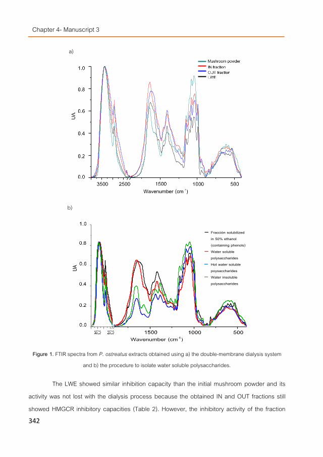
Chapter 4- Manuscript 3
342
a)
b)
Figure 1. FTIR spectra from P. ostreatus extracts obtained using a) the double-membrane dialysis system and b) the procedure to isolate water soluble polysaccharides.
The LWE showed similar inhibition capacity than the initial mushroom powder and its activity was not lost with the dialysis process because the obtained IN and OUT fractions still showed HMGCR inhibitory capacities (Table 2). However, the inhibitory activity of the fraction
40003000 1500 1000 500
0,0
0,2
0,4
0,6
0,8
1,0
Uni
dade
s de
Abs
orba
ncia
Longitud de onda (cm-1)
S1
S2
S3
P3
40003000 1500 1000 500
0,0
0,2
0,4
0,6
0,8
1,0
Uni
dade
s de
Abso
rban
cia
Longitud de onda (cm-1)
S1
S2
S3
P3
Fracción solubilized in 50% ethanol (containing phenols) Water soluble polysaccharides Hot water soluble poysaccharides Water insoluble polysaccharides

Chapter 4- Efect of fungal compounds with HMGCR inhibitory…
343
corresponding to molecular weights between 14 and 3.5 kDa was not significantly different than the one with a molecular weight higher than 14 kDa suggesting than both larger size polysaccharides and those between 14 to 3.5 kDa were able of inhibiting the enzyme. Since both fractions showed similar composition (according to FT-IR spectra) the OUT fraction could contain smaller fragments generated from the higher molecular weight polysaccharides (derivatives, partially degraded polysaccharides, broken fibers, partially synthetized pieces etc). This observation could be of interest because it suggest that the compounds responsible for the HMGCR inhibitory activity could still be acting as inhibitors when they are partially degraded as it might occur during digestion.
Moreover, the HMGCR inhibitory activity observed using the filtration devices was higher than using dialysis. The observed differences could be due to the different P. ostreatus batch utilized for fractionation (since its initial activity was also lower). However, it could also be due to the followed procedures because dialysis requested longer time and the phenolic compounds present in the fractions (particularly in the OUT fraction) could polymerize interfering in the inhibitory activity as noticed by a slightly browning of the obtained fractions. Edible mushrooms contain phenolic compounds that can be oxidized by fungal polyphenol oxidases and peroxidases into quinones [30]. The latter are very reactive molecules able to spontaneously polymerize with themselves and with almost any high molecular weight compound including polysaccharides or proteins interfering in many reactions.
Therefore, a procedure to separate the phenolic compounds from the water-soluble polysaccharides was followed.
Isolation of water-soluble polysaccharides with potentially HMGCR inhibitory activity Phenolic compounds and simple sugars were removed after washing the mushroom
powder with 50% ethanol (Figure 1b). Further extraction with cold water yielded a higher protein-rich fraction than carbohydrate fraction.
The profile was different than the typical signals from 1500 to 1200 cm-1 corresponding to more water insoluble polysaccharide mixture that is usually obtained when hot water is utilized
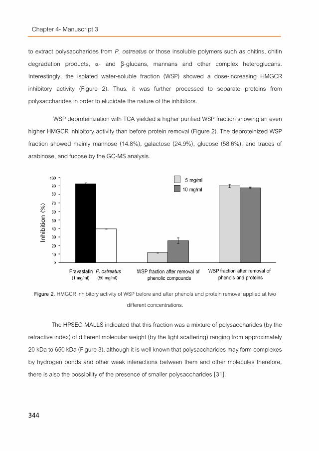
Chapter 4- Manuscript 3
344
to extract polysaccharides from P. ostreatus or those insoluble polymers such as chitins, chitin degradation products, α- and β-glucans, mannans and other complex heteroglucans. Interestingly, the isolated water-soluble fraction (WSP) showed a dose-increasing HMGCR inhibitory activity (Figure 2). Thus, it was further processed to separate proteins from polysaccharides in order to elucidate the nature of the inhibitors.
WSP deproteinization with TCA yielded a higher purified WSP fraction showing an even higher HMGCR inhibitory activity than before protein removal (Figure 2). The deproteinized WSP fraction showed mainly mannose (14.8%), galactose (24.9%), glucose (58.6%), and traces of arabinose, and fucose by the GC-MS analysis.
Figure 2. HMGCR inhibitory activity of WSP before and after phenols and protein removal applied at two different concentrations.
The HPSEC-MALLS indicated that this fraction was a mixture of polysaccharides (by the refractive index) of different molecular weight (by the light scattering) ranging from approximately 20 kDa to 650 kDa (Figure 3), although it is well known that polysaccharides may form complexes by hydrogen bonds and other weak interactions between them and other molecules therefore, there is also the possibility of the presence of smaller polysaccharides [31].
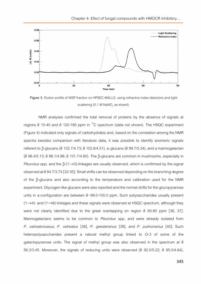
Chapter 4- Efect of fungal compounds with HMGCR inhibitory…
345
Figure 3. Elution profile of WSP fraction on HPSEC-MALLS, using refractive index detectors and light scattering (0.1 M NaNO3 as eluent).
NMR analyses confirmed the total removal of proteins by the absence of signals at regions 10-40 and 120-180 ppm in 13C spectrum (data not shown). The HSQC experiment (Figure 4) indicated only signals of carbohydrates and, based on the correlation among the NMR spectra besides comparison with literature data, it was possible to identify anomeric signals referent to β-glucans ( 102.7/4.73; 102.8/4.51), α-glucans ( 99.7/5.34), and a mannogalactan ( 98.4/5.13; 98.1/4.98; 101.7/4.80). The β-glucans are common in mushrooms, especially in Pleurotus spp. and the β-(1→3)-linkages are usually observed, which is confirmed by the signal observed at 84.7/3.74 [32-35]. Small shifts can be observed depending on the branching degree of the β-glucans and also according to the temperature and calibration used for the NMR experiment. Glycogen-like glucans were also reported and the normal shifts for the glucopyranose units in α-configuration are between ~99.0-100.0 ppm. Such polysaccharides usually present (1→4)- and (1→6)-linkages and these signals were observed at HSQC spectrum, although they were not clearly identified due to the great overlapping on region 65-80 ppm [36, 37]. Mannogalactans seems to be common to Pleurotus spp. and were already isolated from P. ostreatoroseus, P. ostreatus [38], P. geesteranus [39], and P. pulmonarius [40]. Such heteropolysaccharides present a natural methyl group linked to O-3 of some of the galactopyranose units. The signal of methyl group was also observed in the spectrum at 56.3/3.45. Moreover, the signals of reducing units were observed ( 92.0/5.22; 95.0/4.64),

Chapter 4- Manuscript 3
346
indicating that low molecular weight carbohydrates are present in the WSP fraction. By the integration of the areas of the anomeric signals it was possible to quantify the four main classes of molecules observed in this fraction totalizing 34% of mannogalactans, 38% of β-glucans, 25.7% of α-glucans, and 2.3% of low molecular weight carbohydrates.
Thus, other polysaccharides were also tested as HMGCR inhibitors in order to discard possible interferences that could be common for scavenging/binding molecules such as complex polysaccharides. Results indicated that anionic polysaccharides such as alginate (a linear β-(1→4)-linked polymer of n-mannuronic and L-guluronic acid) and xanthan (a β-(1→4)-D-glucopyranose glucan backbone with side chains of (1→3)-α-linked D-mannopyranose-(2→1)-β-D-glucuronic acid-(4→1)-β-D-mannopyranose on alternating residues) showed marginally interference (Table 1), only the presence of agar-agar (a partially neutral and anionic polysaccharide complex constituted by agarose (chains of repeating alternate units of β-(1→3)-linked- D-galactose and α-(1→4)-linked 3,6-anhydro-L-galactose) and agaropectin (a sulfated polysaccharide (3% to 10% sulfate), showed a 30.5% inhibition but significantly lower than the inhibition observed by the isolated water-soluble polysaccharide fraction isolated from P. ostreatus taking into consideration that they were applied at 10 fold higher concentrations (50 mg/ml).
In vitro bioavailability of the WSP with HMGCR inhibitory properties NMR analysis indicated that the WSP extract contained higher amount of glucans (63.7%)
than mannogalactans (34%) and the other tested polysaccharides including galactose in their backbones and mannopyranose residues in their side chains (although different linkage patterns) did not show HMGCR inhibitory activity (Table 1). Moreover, many reports pointed β-glucans as the polysaccharides with hypocholesterolemic properties thus, these compounds might potentially be the inhibitors of the HMGCR. However, in order to reach their target, they should firstly resist digestion and pass through the intestinal barrier therefore, the WSP extract was submitted to an in vitro digestion and absorption model as a preliminary attempt to investigate whether its β-glucans might be bioaccesible and bioavailable.

Chapter 4- Efect of fungal compounds with HMGCR inhibitory…
347
Figure 4. HSQC spectrum of the WSP fraction isolated from P. ostreatus, in D2O at 50 ºC (chemical shifts are expressed in ppm).
Spectrophotometrical determinations indicated that 41.4% of the WSP glucans showed an β-linkage while 58.6% of them were β-glucans coinciding with NMR analysis. Moreover, the WSP extract contained more β-(1→3)- than β-(1→3),(1→6)-glucans (Table 3) suggesting than their structural configuration might be linear more than the usual triple helix form typical for branched polysaccharides. This might explain their large solubility in water since complex folding and chains aggregations increases water insolubility. Digestion affected more to β-(1→3)-glucans than β-(1→3),(1→6)-glucans which was also expected as the complex conformation of the latter polysaccharides should protect them from the enzymatic degradation more than a linear chain. Nevertheless, part of the β-(1→3)-glucans also survived the digestive process indicating that they might be partially bioaccesible.

Chapter 4- Manuscript 3
348
Table 9. Effect of in vitro digestion on the β-(1→3)- and β-(1→3),(1→6)-glucans present in the WPS.
However, the degree of β-glucans degradation during digestion might also be highly influenced by the presence of other food matrix in the digestive track since it might protect them reducing the access of degradative enzymes to the polysaccharide thus, the bioavailability of the WSP β-(1→3)-glucans (present in higher amounts) was studied using non-digested and digested WSP extracts. Surprisingly, results indicated than the non-digested β-(1→3)-glucans were able to pass through the Caco2 cells in larger amounts than after digestion (Table 4) although it might also indicate than digested β-glucans might be easier further processed by the cells during the 24h incubation than the non-digested since they should have lower molecular weights because of the previous digestion process liberating into the lower compartment more monosaccharides than higher polymers that are no longer detectable with the method utilized since the fluorophore specifically binds β-(1→3)-linkages. Nevertheless, WSP β-(1→3)-glucans were observed in the lower compartment suggesting that they could be bioavailable thus, in vivo studies should be carried out to further investigate the significance of these results.
Table 4. β-(1→3)-Glucans applied to the Caco2 cells.
Wise to mention was the fact than when the TEER was measured to control the membrane integrity during the 24h cells incubation in the presence of digested and non-digested WSP, the obtained values were higher than the controls with no β-glucan addition (Figure 5).
β-Glucans concentration (g/100mg)
Before WSP digestion
After WSP digestion
Bioaccesible (%)
β-(1→3),(1→6)-Glucans 4.012 2.854 71.1
β-(1→3)-Glucans 394.7 85.4 21.6
Sample applied β-Glucans applied (g/mL)
β-Glucans lower compart. (g/mL)
Bioavailable β-Glucans (%)
Non digested WSP 2.3 0.38 16.7 Digested WSP 2 0.14 7

Chapter 4- Efect of fungal compounds with HMGCR inhibitory…
349
Figure 5. Relative TEER during Caco2 cells incubation in the presence of digested and non-digested WSP compared to respective control cells where a digestion mixture including no extract or only medium and
buffer were used.
The TEER of the cells treated with the digested WSP increased already after 1h application and continued to increase until 6h later while the TEER increase induced by the non-digested WSP was not noticed until 3h later and it was less pronounced than the digested one. Recent studies indicated that dietary fibres and β-glucans might have a protective effect on enterocytic membranes [41] these results might support that possibility. On the other hand, the lower β-(1→3)-glucan concentration found in the lower compartment of cells treated with the digested WSP extract compared to the non-digested WSP could perhaps be because these possibly smaller polysaccharides might be better retained or attached to the Caco2 luminal membranes (upper compartment) acting as protective layer more efficiently than higher size polysaccharides (non-digested). However, further experiments are needed to confirm this hypothesis.
Conclusions The HMGCR inhibitory activity observed in the water extracts from Pleurotus ostreatus
was not due to the presence of statins but to other compounds with higher molecular weight because when the water soluble polysaccharide fraction (WSP) was isolated, inhibition of the HMGCR was also observed. According to NMR analysis this fraction contained β-glucans, mannogalactans, α-glucans and a small amount of low molecular weight (MW) compounds. Compounds with MW lower than 3.5 kDa showed no HMGCR inhibitory activity but those with MW
98100102104106108110112114116
0 5 10 15 20 25
Relat
ive T
EER
value
s (%
)
Incubation time (h)
WSP ND
WSP

Chapter 4- Manuscript 3
350
lower than 14 but higher than 3.5 kDa did suggesting that derivatives or fragments of the indicated polysaccharides were still retaining the inhibitory activity. This could be interesting because lower MW compounds are easier absorbed by the intestine than large and complex polymers. Apparently, there were more β-glucans showing linear (β-(1→3)-glucans) than triple helix configuration enhancing their solubility in physiological fluids and their possibility of being bioavailable. In fact, the in vitro model utilized suggested that they could be not only partially bioaccesible but also partially absorbed by the enteric barrier reaching the blood stream since they were detected in the lower compartment of Caco2 cell transport experiments. However, further studies are necessary to confirm these indications and to investigate whether the HMGCR inhibitory polysaccharides are also able of performing such an inhibition under in vivo real situations therefore, animal dietary interventions are being carried out at the present to confirm the presented results.
Acknowledgments The research was supported by AGL2010-21537 national R+D program from the Spanish
Ministry of Science and Innovation and ALIBIRD-C M S2009/AGR-1469 regional program from the Community of Madrid (Spain). CTICH (Centro Tecnológico de Investigación del Champiñón de La Rioja, Autol, Spain) is also acknowledged for the cultivation and supplying of the mushrooms fruiting bodies and Maria Navarro-Rubio for her technical assistance.

Chapter 4- Efect of fungal compounds with HMGCR inhibitory…
351
References [1]. Gil-Ramírez, A. and C. Soler-Rivas, The use of edible mushroom extracts as bioactive ingredients to design novel
functional foods with hypocholesterolemic activities, in Mushrooms: Cultivation, Antioxidant Properties and Health Benefits, G. Pesti, Editor. 2014, Nova Science Publishers: New York. p. 43-73.
[2]. Khatun, K., et al., Oyster mushroom reduced blood glucose and cholesterol in diabetic subjects. Mymensingh medical journal : MMJ, 2007. 16(1): p. 94-99.
[3]. Schneider, I., et al., Lipid lowering effects of oyster mushroom (Pleurotus ostreatus) in humans. Journal of Functional Foods, 2011. 3(1): p. 17-24.
[4]. Bobek, P., et al., Cholesterol-lowering effect of the mushroom Pleurotus ostreatus in hereditary hypercholesterolemic rats. Annals of Nutrition and Metabolism, 1991. 35(4): p. 191-195.
[5]. Bobek, P., L. Kuniak, and L. Ozdín, The mushroom Pleurotus ostreatus reduces secretion and accelerates the fractional turnover rate of Very-Low-Density Lipoproteins in the rat. Annals of Nutrition and Metabolism, 1993. 37(3): p. 142-145.
[6]. Bobek, P., L. Ozdin, and L. Kuniak, Mechanism of hypocholesterolemic effect of oyster mushroom (Pleurotus ostreatus) in rats: reduction of cholesterol absorption and increase of plasma cholesterol removal. Zeitschrift für Ernährungswissenschaft, 1994. 33(1): p. 44-50.
[7]. Bobek, P., Ĺ. Ozdín, and Ĺ. Kuniak, Effect of oyster mushroom and isolated β-glucan on lipid peroxidation and on the activities of antioxidative enzymes in rats fed the cholesterol diet. The Journal of Nutritional Biochemistry, 1997. 8(8): p. 469-471.
[8]. Bobek, P. and Š. Galbavý, Hypocholesterolemic and antiatherogenic effect of oyster mushroom (Pleurotus ostreatus) in rabbits. Food / Nahrung, 1999. 43(5): p. 339-342.
[9]. Bobek PhD, P., L. Ozdín PhD, and Š. Galbavý Md, Dose- and time-dependent hypocholesterolemic effect of Oyster Mushroom (Pleurotus ostreatus) in rats. Nutrition, 1998. 14(3): p. 282-286.
[10]. Opletal, L., et al., Evidence for the anti-hyperlipidaemic activity of the edible fungus Pleurotus ostreatus. British Journal of Biomedical Science, 1997. 54(4): p. 240-243.
[11]. Xu, C., et al., The pharmacological effect of polysaccharides from Lentinus edodes on the oxidative status and expression of VCAM-1mRNA of thoracic aorta endothelial cell in high-fat-diet rats. Carbohydrate Polymers, 2008. 74(3): p. 445-450.
[12]. Zhu, M., et al., Optimizing conditions of polysaccharide extraction from Shiitake mushroom using response surface methodology and its regulating lipid metabolism. Carbohydrate Polymers, 2013. 95(2): p. 644-648.
[13]. Chen, S.-Y., et al., Contents of lovastatin, γ-aminobutyric acid and ergothioneine in mushroom fruiting bodies and mycelia. LWT - Food Science and Technology, 2012. 47(2): p. 274-278.
[14]. Alarcon, J., et al., Production and purification of statins from Pleurotus ostreatus (basidiomycetes) strains. Zeitschrift fur Naturforschung, 2003. 58c: p. 62-64.

Chapter 4- Manuscript 3
352
[15]. Gunde-Cimerman, N., et al., Screening fungi for the production of an inhibitor of HMG CoA reductase: Production of mevinolin by the fungi of the genus Pleurotus. FEMS Microbiology Letters, 1993. 111(2-3): p. 203-206.
[16]. Gunde-Cimerman, N., A. Plemenitaš, and A. Cimerman, Pleurotus fungi produce mevinolin, an inhibitor of HMG CoA reductase. FEMS Microbiology Letters, 1993. 113(3): p. 333-337.
[17]. Gunde-Cimerman, N. and A. Cimerman, Pleurotus fruiting bodies contain the inhibitor of 3-Hydroxy-3-Methylglutaryl-Coenzyme A reductase—Lovastatin. Experimental Mycology, 1995. 19(1): p. 1-6.
[18]. Berger, A., et al., Cholesterol-lowering properties of Ganoderma lucidum in vitro, ex vivo, and in hamsters and minipigs. Lipids in Health and Disease, 2004. 3(1): p. 2.
[19]. Gil-Ramirez, A., et al. Edible mushrooms as potential sources of new hypocholesterolemic compounds in 7th International Conference on Mushroom Biology and Mushroom Products. 2011. Arcachon, France.
[20]. Gil-Ramírez, A., et al., Study on the 3-hydroxy-3-methyl-glutaryl CoA reductase inhibitory properties of Agaricus bisporus and extraction of bioactive fractions using pressurised solvent technologies. Journal of the Science of Food and Agriculture, 2013. 93(11): p. 2789-2796.
[21]. Chanput, W., et al., β-Glucans are involved in immune-modulation of THP-1 macrophages. Molecular Nutrition & Food Research, 2012. 56(5): p. 822-833.
[22]. Aouadi, M., et al., Orally delivered siRNA targeting macrophage Map4k4 suppresses systemic inflammation. Nature, 2009. 458(7242): p. 1180-1184.
[23]. Gil-Ramírez, A., et al., Sterol enriched fractions obtained from Agaricus bisporus fruiting bodies and by-products by compressed fluid technologies (PLE and SFE). Innovative Food Science & Emerging Technologies, 2013. 18(0): p. 101-107.
[24]. Ko, Y.T. and Y.L. Lin, 1,3-b-glucan quantification by a fluorescence microassay and analysis of its distribution in foods. J Agric Food Chem, 2004. 52(11): p. 3313-8.
[25]. Nitschke, J., et al., A new colorimetric method to quantify β-1,3-1,6-glucans in comparison with total β-1,3-glucans in edible mushrooms. Food Chemistry, 2011. 127(2): p. 791-796.
[26]. Sassaki, G.L., et al., Application of acetate derivatives for gas chromatography–mass spectrometry: Novel approaches on carbohydrates, lipids and amino acids analysis. Journal of Chromatography A, 2008. 1208(1–2): p. 215-222.
[27]. Sassaki, G.L., et al., Rapid synthesis of partially O-methylated alditol acetate standards for GC–MS: some relative activities of hydroxyl groups of methyl glycopyranosides on Purdie methylation. Carbohydrate Research, 2005. 340(4): p. 731-739.
[28]. Palanisamy, M., et al., Pressurized water extraction of β-glucan enriched fractions with bile acids-binding capacities obtained from edible mushrooms. Biotechnology Progress, 2014. 30(2): p. 391-400.
[29]. Trzaskos, J.M., et al., Modulation of 3-hydroxy-3-methylglutaryl-CoA reductase by 15 alpha-fluorolanost-7-en-3 beta-ol. A mechanism-based inhibitor of cholesterol biosynthesis. Journal of Biological Chemistry, 1993. 268(30): p. 22591-9.

Chapter 4- Efect of fungal compounds with HMGCR inhibitory…
353
[30]. Soler-Rivas, C., et al., Biochemical and physiological aspects of brown blotch disease of Agaricus bisporus. FEMS Microbiology Reviews, 1999. 23(5): p. 591-614.
[31]. Lehtovaara, B.C. and F.X. Gu, Pharmacological, structural, and drug delivery properties and applications of 1,3-β-glucans. Journal of Agricultural and Food Chemistry, 2011. 59(13): p. 6813-6828.
[32]. Carbonero, E.R., et al., A β-glucan from the fruit bodies of edible mushrooms Pleurotus eryngii and Pleurotus ostreatoroseus. Carbohydrate Polymers, 2006. 66(2): p. 252-257.
[33]. Santos-Neves, J.C., et al., A novel branched αβ-glucan isolated from the basidiocarps of the edible mushroom Pleurotus florida. Carbohydrate Polymers, 2008. 73(2): p. 309-314.
[34]. Santos-Neves, J.C., et al., A gel-forming β-glucan isolated from the fruit bodies of the edible mushroom Pleurotus florida. Carbohydrate Research, 2008. 343(9): p. 1456-1462.
[35]. Liu, Y.-T., et al., Chemical composition of five wild edible mushrooms collected from Southwest China and their antihyperglycemic and antioxidant activity. Food and Chemical Toxicology, 2012. 50(5): p. 1238-1244.
[36]. Stanek, M., H. Falk, and A. Huber, Investigation of the branching characteristic of glycogen by means of two-dimensional1H and 13C NMR spectroscopy. Monatshefte für Chemie / Chemical Monthly, 1998. 129(4): p. 355-364.
[37]. Smiderle, F.R., et al., High molecular weight glucan of the culinary medicinal mushroom Agaricus bisporus is an α-glucan that forms complexes with low molecular weight galactan. Molecules, 2010. 15(8): p. 5818-5830.
[38]. Rosado, F.R., et al., The presence of partially 3-O-methylated mannogalactan from the fruit bodies of edible basidiomycetes Pleurotus ostreatus ‘florida’ Berk. and Pleurotus ostreatoroseus Sing. Vol. 221. 2003. 119-124.
[39]. Zhang, Y., et al., Schizophyllan: A review on its structure, properties, bioactivities and recent developments. Bioactive Carbohydrates and Dietary Fibre, 2013. 1(1): p. 53-71.
[40]. Smiderle, F.R., et al., A 3-O-methylated mannogalactan from Pleurotus pulmonarius: Structure and antinociceptive effect. Phytochemistry, 2008. 69(15): p. 2731-2736.
[41]. Rop, O., J. Mlcek, and T. Jurikova, Beta-glucans in higher fungi and their health effects. Nutr Rev, 2009. 67(11): p. 624-31.

Chapter 4- Efect of fungal compounds with HMGCR inhibitory…
355
Manuscript 4
Water-soluble compounds from Lentinula edodes influencing the HMGCoA-reductase activity and the expression of genes involved in the
cholesterol metabolism
Alicia Gil-Ramírez1, Víctor Caz2, Fhernanda R. Smiderle3, Roberto Martin-Hernandez4, Francisco R. Marín1, Carlota Largo2, María
Tabernero3, Marcello Iacomini2, Guillermo Reglero1,4
Cristina Soler-Rivas1
1Department of Production and Characterization of Novel Foods, CIAL – Research Institute in Food Science (UAM+CSIC), C/ Nicolas Cabrera 9, Campus de Cantoblanco, Universidad Autónoma de Madrid, 28049 Madrid, Spain. 2Department of Experimental Surgery, Research Institute Hospital La Paz (IdiPAZ), Paseo de la Castellana 261, 28046 Madrid, Spain 3Department of Biochemistry and Molecular Biology, Federal University of Parana, CP 19046, Curitiba, Parana, Brazil. 4Imdea-Food Institute. CEI UAM+CSIC. Pabellón Central del Antiguo Hospital Cantoblanco (Edificio nº7). Cra. Cantoblanco nº8, 28049 Madrid, Spain.
In Preparation. May, 2015

Chapter 4- Manuscript 4
356
Abstract The water extracts from Lentinula edodes (Shiitake mushroom) (LWE) able of reducing
the HMG-CoA reductase in vitro contained no lovastatin but a mixture of water soluble α- and β-glucans and fucomannogalactans. Fragments or derivatives of these polysaccharides with molecular weight down to approx. 1 kDa still retained their inhibitory activity. The extract also contained eritadenine although it was not the compound responsible for the inhibitory activity. When digested and applied to Caco2 cells no significant effect was noticed on the modulation of cholesterol-related gene expression. But, when the lower compartment of Caco2 cell monolayer (where LWE digestates were added) was applied to HepG2 modulation of some genes were noticed after 24 h incubation. The LWE extract was also administrated to normo- and hypercholesterolemic mice (4 weeks) and no significant lowering of serum cholesterol levels was observed but reduction of triglycerides in liver. However, LWE supplementation modulated the transcriptional profile of the genes involved in the cholesterol metabolism similarly to simvastatin suggesting that it could hold a potential as hypolipidemic /hypocholesterolemic extract although further dose-dependent studies should be carried out.

Chapter 4- Efect of fungal compounds with HMGCR inhibitory…
357
Introduction Cardiovascular diseases (CVD) are the major cause of death in industrialized countries
and high cholesterol levels in serum increases CVD risk. Nowadays, natural or synthetic statins are commonly prescribed to lower cholesterol levels since they are inhibitors of the HMGCR (3-hydroxy-3-methyl-glutaryl-Co A reductase), the key enzyme in the endogenous cholesterol biosynthesis [1]. However, frequent and prolonged administration of these drugs induce secondary undesirable effects therefore, other alternative are being followed such as the prescription of drugs impairing cholesterol absorption (ezetimibe) or other formulations able of inhibiting enzymes downstream the cholesterol metabolic pathway (squalene synthetase, 24-dehydrocholesterol reductase, etc. ) [2, 3].
People still with low or moderate hypercholesterolemia might choose for a less drastic alternative to drug treatment: the regular consumption of functional foods with hypocholesterolemic activity such as those including phytosterols or cereal β-glucans as compounds able to reduce mainly cholesterol absorption [4]. However, these products are not effective for everyone because if cholesterol is not absorbed, the liver synthetizes it by increasing up to 3 folds the activity of the HMGCR (observed in regular β-glucan consumers) [5]. Thus, another strategy might be designing of functional foods able to impair the endogenous cholesterol biosynthesis by disrupting the HMGCR activity or inhibiting other enzymes involved in that metabolic pathway.
Shiitake mushrooms (Lentinula edodes) and other edible mushroom species showed hypocholesterolemic effects according to in vivo tests with animals and clinical trials [6]. Apparently, their dietary fiber content [7] and eritadenine (2(R), 3(R),-dihydroxy-4-(9-adenyl)-butyric acid, lentinacin or lentysine) [8] contents were involved in their cholesterol lowering properties by different mechanisms.
Fractions enriched in water-insoluble fibers obtained from L. edodes administrated to rats for 4 weeks were able to significantly lower VLDL, IDL and LDL levels compared with cellulose fed control rats. However, the mRNA transcription levels of genes encoding key enzymes in the biosynthesis of cholesterol (HMGCR, gene encoding HMG-CoA reductase) and in the synthesis of bile acids from cholesterol (CYP7A1, cholesterol 7α-hydroxylase) and other proteins involved

Chapter 4- Manuscript 4
358
in cholesterol transport (APOB, apolipoproteins B and LDLR, LDL receptor) were not significantly modulated compared to control. Apparently, their hypocholesterolemic effect was exerted by enhancement of faecal cholesterol excretion [7] probably because of their ability to scavenge bile acids during digestion as suggested after in vitro studies [9].
Eritadenine reduced cholesterol and triacylglycerol levels in rats due to its activity as inhibitor of the S-adenosylhomocysteine hydrolase (SAHH), a key enzyme in the hepatic phospholipid metabolism [8]. The alkaloid decreased in a dose-dependent manner levels of VLDL, LDL and HDL, increased the hepatic S-adenosylhomocysteine (SAH) level and decreased the ratio of S-adenosylmethionine (SAM) to SAH indicating that its hypocholesterolemic action might be elicited through an alteration of the hepatic phospholipid metabolism that resulted from an inhibition of phosphatidylethanolamine N-methylalion due to a decreased SMA/SAH ratio in the liver [10].
However, there could be more compounds involved acting via different mechanisms because other reports pointed out that the water extracts obtained from this mushroom were able of inhibiting the HMGCR activity in vitro [11] and hot water extracts decreased oxidative damage related to hypercholesterolemia [12, 13]. They could also contain lovastatin as suggested for other mushroom species [14] or different water soluble polysaccharides as suggested for specific species such as Agaricus bisporus [15] and Pleurotus ostreatus [16]. Other publications pointed compounds such as lanosteroids, ganoderols etc. from Ganoderma lucidum as molecules able of reducing the HMGCR mRNA expression [17] although ergosterol and derivatives, the compounds from L. edodes with structures similar to those mentioned, showed no HMGCR inhibitory activity [16].
In this work, a more detailed study about those HMGCR inhibitors is carried out in order to further define the compounds responsible for the inhibition as an initial study to investigate the possibility of designing new functional foods with hypocholesterolemic properties but following different mechanisms of action than those of the actually commercialized products. Molecular approaches were also utilized (low-density arrays) in order to broaden the overview of other potential action points (other enzymes involved in the cholesterol homeostasis) which might be target of fungal compounds. The experiments were firstly conducted in vitro using Caco2 and

Chapter 4- Efect of fungal compounds with HMGCR inhibitory…
359
HepG2 cell cultures and afterwards the effect of a L. edodes extract was tested using normo- and hypercholesterolemic mice models.
Materials and Methods
Fungal material and preparation of water extracts Fresh fruiting bodies from Lentinula edodes S. (Berkeley) strain 4312 Sylvan were
harvested and processed until a dry powder was obtained as described [18]. Resulting mushroom powders were further processed to prepare a lyophilized water extract (LWE) and to isolate a water-soluble polysaccharide fraction (WSP) as explained [16].
Determination of HMGCR inhibitory activity Lentinula edodes extracts (obtained at different purification steps or submitted to
different treatments) and an eritadenine (Synchem UG & Co. KG, Altenburg, Germany) standard preparation were solubilized in water and applied (20 ml) into a 96 wells-plate. Their HMGCR inhibitory activity was measured using the commercial HMG-CoA Reductase Assay (Sigma, Madrid, Spain) according to the user´s manual by monitoring their absorbance change (340 nm) at 37 ºC using a microplate reader (Tecan Group Lt, Männedorf, Switzerland). Pravastatin was utilized as a control for positive inhibition.
Determination of statins The presence of statins in L. edodes were determined following the procedure indicated
by Gil-Ramirez et al. (2011) [19].
Determination of eritadenine Eritadenine was extracted from the mushroom powder following the procedure of [20]
with modifications. Briefly, L. edodes dry powder or LWE (5 g) were mixed with 100 ml methanol:water (80:20 v/v) and stirred at 25 ºC for 3 h. Then, the suspension was filtered through a 14-18 μm pore size paper filter (GE Healthcare Europe GmbH 1240, Barcelona, Spain) and the filtrate was dried in a water bath at 60 ºC in darkness. Afterwards, the remaining dry pellet was dissolved in 50 ml MilliQ water and washed 3 times with diethyl ether to remove undesirable

Chapter 4- Manuscript 4
360
compounds. This washed aqueous extract was stored at -20 ºC until it was injected in an HPLC system for analysis.
Identification and quantification of eritadenine were carried out using a C18 Spherisorb ODS2 4 x 250 mm analytical column with a 5 μm particle size (Waters, Missisagua, Ontario, Canada) in an HPLC system (Varian Pro-Star 330, Madrid, Spain) coupled to PDA detector (Varian Pro-Star 363 module, Madrid, Spain). Samples (10 ml) were injected and developed under a constant flow (0.5 ml/min) and an isocratic mobile phase of water:acetonitrile (98:2, v/v with 1% TFA). The alkaloid eluting after 11.6 min showed an UV-spectra similar to previously described [20] and to eritadenine thus, it was quantified at 260 nm using a standard curve of the commercialized standard.
Identification of the compounds with HMGCR inhibitory activity Firstly, the LWE (50 mg/ml) showing HMGCR inhibitory activity was filtrated as described
in [16] to fractionate the extract and estimate the molecular weight and chemical nature of the inhibitors. Then, they were also fractionated using a double-dialysis system but using membranes with smaller cut off (3.5 and 1 kDa). Finally, a water-soluble polysaccharide fraction (WSP) was also isolated from L. edodes dry powder (10 g) following the procedure of [16].
The composition of the fractions obtained during the isolation procedure was followed by
FT-IR spectroscopy. The sugar content of the isolated WSP was quantified by GC-MS and the compounds identified by NMR as described by [16].
In vitro digestion and Caco2 cell experiments LWE extracts were submitted to an in vitro digestion according to [16]. After digestion,
the samples were applied to Caco2 cell cultures at subtoxic concentrations. Human colorectal adenocarcinoma cell line Caco-2 (ATCC HTB-37) obtained from the
ATCC (Rockville, MD, USA) were maintained in Dulbecco´s Modified Eagle’s Medium (DMEM) with high glucose content (4.5 g/L) and supplemented with 10% fetal bovine serum, 1% penicillin-streptomycin, and 1% non-essential amino acids at 37 ºC in at humidified atmosphere containing

Chapter 4- Efect of fungal compounds with HMGCR inhibitory…
361
5% CO2. Firstly, the cytotoxicity was evaluated using 3-(4,5-dimethylthiazol)-2,5-diphenyl tetrazolium bromide (MTT) method [21]. Then, for the gene expression assays, cells were seeded onto a 9.5 cm2 grown area 6 well flat bottom plate (Costar, Corning, USA) at a density of 5 × 10⁵ cell per well (3.3 × 10⁵ cell/ml) and incubated at 37 ºC and 5% CO2 until confluence. The digested extracts were applied at subtoxic concentrations (4 μl/ml) and left incubating for 1 or 24 h.
For transport assays, cells were seeded at similar densities in a 4.67 cm2 permeable membrane support (0.4 μm pore size, Transwell Costar, Corning, USA). Cells were cultivated as above described and allowed to differentiate (21 days). The integrity of the cell layer was evaluated by measuring the transepithelial electrical resistance (TEER) (Evon, Sarasota, FL) and alkaline phosphatase activity (ALP) [22]. The digested extracts were applied to Caco2 cell monolayers. Each sample was mixed with medium to a total volume of 1.5 ml per insert and applied to the apical (upper) compartment. Then, plates were incubated at 37 ºC and 5% CO2 for 1 h. Afterwards, the lower compartment was collected and applied to HepG2 cell cultures.
Treatment of HepG2 with the basolateral compartment of the Caco2 monolayers Human hepatoma HepG2 cells, obtained from American Type Culture Collection (ATCC,
Manassas, VA, USA) were cultured in Eagle’s Minimum Essential Medium (EMEM) supplemented with 10% fetal bovine serum (FBS) and 1% of antibiotic solution (containing 10,000 units/ml of penicillin base and 10,000 μg/ml of streptomycin base, Gibco). The cells were maintained under standard conditions of temperature (37 ºC), humidity (95%) and CO2 (5%).
Firstly, the cytotoxicity was evaluated as above described for the Caco2 cell cultures but after 3 h incubation. Then, the HepG2 cell cultures were seeded in 6 wells plates (9.5 cm2) and cultivated until confluence. Afterwards, cells were treated with the basolateral compartment obtained 3 h after the Caco2 transport assay was carried out using as starting material the digested extracts or controls. These solutions were applied to HepG2 at subtoxic concentrations (6.25 μl/ml) and left incubating at 37 ºC, 95% humidity and 5% CO2 during 1 or 24 h. The medium of the basolateral compartment with no addition was also tested as control.

Chapter 4- Manuscript 4
362
Animals and diets Animal studies were approved by our institution’s Animal Welfare and Ethics Committee
and were carried out according to European legislation (2010/63/EU). C57BL/6JRj mice were purchased from Janvier SAS (Le Genest Saint Isle, France). They were maintained in a temperature-, humidity- and light-controlled room (24 ± 2 ºC, 40-60% humidity, 12:12 hour light: dark cycle) and food and water were provided ad libitum. Male mice were randomly divided into five groups (n=6-8 per group) and fed with normal diet (Safe Rodent Diet A04, Augy, France; 3% fat, NCD) or high-cholesterol diet (NCD diet supplemented with 2% cholesterol, 1% cholic acid, HCD). NCD and HCD were also supplemented with 0.67% LWE from L. edodes. HCD groups were also treated with 0.025% simvastatin as a positive control for inhibition of cholesterol biosynthesis, and HCD plus 0.005% ezetimibe as a positive control for inhibition of cholesterol absorption. In order to assess the effects of the mushroom extract in normo- and hyper-cholesterolemic mice, in the first case, mice were fed NCD for 4 weeks (from 5 to 9 weeks of age) supplemented with the LWE. In the second case, mice were fed HCD for 4 weeks to induce hypercholesterolemia followed by another 4 weeks of HCD plus LWE (from 10 to 13 weeks of age).
Plasma and liver lipid analyses, tissue and feces collection Mice were sacrificed at the end of experiments by intracardiac exsanguination under
anesthesia with 1.5% isoflurane, and plasma was collected and stored at -80 ºC. Jejunum, ileum, cecum, and liver samples were removed and immediately frozen in liquid nitrogen and stored at -80 ºC. Feces were collected at the beginning, after induction of the hypercholesterolemia and at the end of the experiments and they were stored at -20 ºC. Cholesterol extraction from feces (300 mg) and analysis by GC-MS were carried out as described in [23]. Plasma concentrations of total cholesterol, HDL, LDL and triglyceride were measured using a Covas C311 Autoanalyzer (Roche, Spain). Total cholesterol and triglyceride concentrations were measured in liver samples homogenized according to [23].

Chapter 4- Efect of fungal compounds with HMGCR inhibitory…
363
RNA extraction, qRT-PCR and gene expression analysis of the low-density array (LDA) Total RNA from cell cultures or mice tissues (see later) was extracted and quantified as
explained in [23]. Total RNA from each sample were reversed, loaded into the micro-fluidic cards and run in a 7900HT Fast Real-Time PCR system (Applied Biosystems).
The micro-fluid card was constructed using respectively human and mouse commercial available assays (Applied Biosystems) for several genes related to the cholesterol metabolism, some internal standard and housekeeping gene that are described in detail elsewhere [23]. The selected cholesterol-related genes were ABCA1 (ATP-Binding Cassette, Sub-Family A, Member 1), ABCG5 and ABCG8 (ATP-binding cassette, sub-family G (WHITE), members 5 and 8), ACAT1 and ACAT2 (Acetyl-CoA acetyltransferase 1 and 2), APOB (Apolipoprotein B), DGAT1 and DGAT2 (Diacylglycerol O-acyltransferase 1 and 2), FDFT1 (Farnesyl-diphosphate farnesyltransferase 1), HMGCR (3-hydroxy-3-methylglutaryl-CoA reductase), LDLR (Low density lipoprotein receptor), MTTP (Microsomal triglyceride transfer protein), NPC1L1 (NPC1 (Niemann-Pick disease, type C1 like 1), NR1H3 and NR1H4 (Nuclear receptor subfamily 1, group H, member 3 and 4), SOAT1 and SOAT2 (Sterol O-acyltransferase 1 and 2), SREBF1 and SREBF2 (Sterol regulatory element binding transcription factor 1 and 2). Comparative analysis of each of these genes was performed using specialized computer programs SDS2.3 and RQ 2.1 (Applied Biosystems) [23].
Gene expression statistical data analysis The expression of each gene was measured at least in triplicate for each sample. In order
to control for potential outliers, Grubb’s test was applied to technical replicates with a threshold of 0.05. Biological replicates were quality controlled using the Median Absolute Deviation test (MAD test). Samples of the same biological group with a MAD score higher than 3 were removed from the analysis. Genorm algorithm [24] was used to identify the most stable reference genes for normalization. Genes GUSB (Glucuronidase, beta) and HPRT1 (Hypoxanthine phosphoribosyl transferase 1) were found to be the most stably expressed in Caco2 cell cultures while GAPDH (Glyceraldehyde-3-phosphate dehydrogenase)and HPRT1 were more stably expressed in HepG2 cells. In the mouse tissue samples (see later), genes Hprt1 and Polr2a (Polimerase) were the most

Chapter 4- Manuscript 4
364
stably expressed in cecum and liver tissues, Gusb and Polr2a in jejunum and ileum tissues.
Relative gene expression levels were calculated by using the 2-ΔΔCT method [25]. Statistical significance of the results was assessed using a limma test, and Benjamini-Hochberg false discovery rate procedure was subsequently applied. Adjusted p-values lower than 0.05 were considered significant. All these calculations were carried out with Real Time StatMiner 4.5® (Integromics S.L, Spain).
Results and Discussion Water extracts obtained from Lentinula edodes were able of inhibiting the HMG-CoA
reductase using in vitro tests [11]. However, they did not contain lovastatin as suggested for Pleurotus spp. [14] nor pravastatin or simvastatin according to HLPC-MS analysis indicating that other compounds might be responsible for the observed inhibition. Thus, an isolation procedure was followed as an attempt to further identify the compounds responsible for the HMGCR inhibitory activity found in the water extracts of L. edodes fruiting bodies.
Identification of the compounds with HMGCR inhibitory activity Firstly, a preliminary fractionation of LWE was carried out by molecular weight using
specific filtering devices with cut-off of 10 and 3 kDa as previously described for other mushroom extracts [15, 16]. Opposite to the results observed for A. bisporus or P. ostreatus, the fraction obtained from L. edodes including compounds with a molecular weight lower than 3 kDa showed higher HMGCR inhibitory activity than those with higher molecular weight (Table 1) suggesting that the compounds responsible for the HMGCR inhibition could be small polypeptides or oligosaccharides or even lower molecular weight compounds such as specific phenolic compounds, acids or alkaloids. The fact that polysaccharides were pointed in the other two mushroom species as the compounds responsible for the observed inhibitory activity (and that fractions with higher molecular weight also showed interesting inhibitory activities) might also suggest that perhaps in this case, the polysaccharides of L. edodes could be smaller or that the activity showed at lower molecular weight was due to degradation products or fragments derived from larger molecules that might still retain its inhibitory activity when partially degraded. This
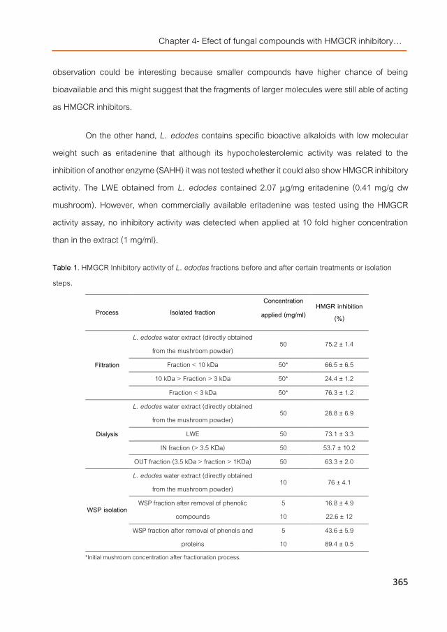
Chapter 4- Efect of fungal compounds with HMGCR inhibitory…
365
observation could be interesting because smaller compounds have higher chance of being bioavailable and this might suggest that the fragments of larger molecules were still able of acting as HMGCR inhibitors.
On the other hand, L. edodes contains specific bioactive alkaloids with low molecular weight such as eritadenine that although its hypocholesterolemic activity was related to the inhibition of another enzyme (SAHH) it was not tested whether it could also show HMGCR inhibitory activity. The LWE obtained from L. edodes contained 2.07 μg/mg eritadenine (0.41 mg/g dw mushroom). However, when commercially available eritadenine was tested using the HMGCR activity assay, no inhibitory activity was detected when applied at 10 fold higher concentration than in the extract (1 mg/ml).
Table 1. HMGCR Inhibitory activity of L. edodes fractions before and after certain treatments or isolation steps.
Process Isolated fraction Concentration
applied (mg/ml) HMGR inhibition
(%)
Filtration
L. edodes water extract (directly obtained from the mushroom powder)
50 75.2 ± 1.4
Fraction < 10 kDa 50* 66.5 ± 6.5 10 kDa > Fraction > 3 kDa 50* 24.4 ± 1.2
Fraction < 3 kDa 50* 76.3 ± 1.2
Dialysis
L. edodes water extract (directly obtained from the mushroom powder)
50 28.8 ± 6.9
LWE 50 73.1 ± 3.3 IN fraction (> 3.5 KDa) 50 53.7 ± 10.2
OUT fraction (3.5 kDa > fraction > 1KDa) 50 63.3 ± 2.0
WSP isolation
L. edodes water extract (directly obtained from the mushroom powder)
10 76 ± 4.1
WSP fraction after removal of phenolic compounds
5 16.8 ± 4.9 10 22.6 ± 12
WSP fraction after removal of phenols and proteins
5 43.6 ± 5.9 10 89.4 ± 0.5
*Initial mushroom concentration after fractionation process.

Chapter 4- Manuscript 4
366
Then, the LWE was also fractionated using a double-dialysis system to separate the fractions containing those compounds with a molecular weight higher than 3.5 kDa (IN fraction) and those with a molecular weight lower than 3.5 but higher than 1 kDa (OUT fraction) in sufficient amounts to monitor their composition using FT-IR spectroscopy and to distinguish small polypeptides or polysaccharides from lower molecular weight compounds such as phenols, monosaccharides, alkaloids etc.
Dialyzed fractions lacked the mannitol content identified in mushroom fruiting bodies (MP) samples by presence of several characteristic signals, peak in the area between 1100-1000 cm-1 , 860 cm-1 peak and a right shift of water signal (Figure 1a). Other small molecules present in MP and LWE (i.e. simple sugars) were also removed with the dialysis system. The marked lipid band (approx. 1450 cm-1) observed in LWE was also noticed in both dialyzed samples but with lower intensity suggesting a possible fractionation of lipid compounds between the IN and OUT fractions. Despite different profiles, LWE, IN and OUT fractions contained similar compositions including proteins, phenols and polysaccharides but, the ratios between those compounds were different. Phenolic compounds were not completely removed with the dialysis in none of the isolated fractions. IN fraction showed the highest protein/carbohydrate ratio, followed by OUT and LWE fractions. Compared with proteins, a lower phenolic content was found in IN sample, being higher and similar for OUT and LWE fractions. Moreover, both IN and OUT fractions showed similar HMGCR inhibitory activities (Table 1) reinforcing the possibility that the inhibitors might be polysaccharides and that their fragments still retained the inhibitory activity (probably the OUT fraction mainly contained smaller fragments from the IN fraction).
Therefore, further polysaccharide isolation was carried out following the procedure utilized for another mushroom specie [16]. The FT-IR analysis of the fractions obtained during the purification of the WSP fraction indicated that phenols and monosaccharides were removed after washing of the sample with 50% ethanol (Figure 1b) leading to fractions with higher carbohydrate and proteins content. The water soluble polysaccharide (WSP) fraction showed a specific signal of protein content from amina I and amina II compounds (peaks at 1650 and 1550 cm-1). Protein/phenol and protein/carbohydrates ratios of WSP were higher than those from the hot water

Chapter 4- Efect of fungal compounds with HMGCR inhibitory…
367
fraction since WSP fraction contained lower amounts of phenols and carbohydrates. Spectral comparison in the range 1500 to 1200 cm-1 between the water soluble fractions (cold/hot) and the water insoluble fractions indicated a peak shifting corresponding to the presence in the latter fraction of compounds such as chitins, chitins degradation products, α- and β-glucans, mannans and other complex polysaccharides. FT-IR analysis showed that although, most of phenols seemed to be removed from WSP, some peaks in the phenolic area were still present and that the profile in the area from 1200 to 1500 cm-1 could correspond to specific proteo-polysaccharide complexes.
Moreover, when the HMGCR inhibitory activity of the WSP fraction was determined before and after a treatment usually applied to remove proteins it could be observed that deproteinization increased the inhibitory activity and the inhibition degree was increasing with increasing concentrations (Table 1). Therefore, the WSP were also pointed as the compounds responsible for the inhibitory activity as observed for A. bisporus and P. ostreatus. Thus, this fraction was injected in GC-MS and NMR to further identify the type of polysaccharides.
The deproteinized WSP fraction showed a heterogeneous profile when analyzed by HPSEC-MALLS and it contains polysaccharides of different molecular weight (data not shown). The monosaccharide composition was determined by GC-MS, after total hydrolysis, and it presented fucose (8.0%), mannose (13.7%), galactose (36.7%), and glucose (41.6%) as main monosaccharides.
NMR analysis of the deproteinized WSP fraction indicated a great complexity due to the presence of high amounts of signals in the anomeric region (Figure 2a), although only carbohydrate signals could be observed on the HSQC, 13C and 1H experiments. Based on the correlation among the NMR spectra besides comparison with literature data, it was possible to identify some anomeric signals referent to β-glucans ( 102.8/4.51), α-glucans ( 99.6/5.36) [26, 27] and fucomannogalactans ( 101.6/4.79; 101.5/5.13; 101.3/5.07; 98.4/5.21; 98.3/5.12; 98.0/5.04; 98.0/4.98) [28, 29].

Chapter 4- Manuscript 4
368
a)
b)
Figure 1. IR spectra from a) the complete L. edodes, the LWE extract and IN and OUT samples and b) from the WSP fraction compared to the discarded fraction containing phenolic compounds, a hot water extraction
and a fraction containing the insoluble polysaccharides.

Chapter 4- Efect of fungal compounds with HMGCR inhibitory…
369
a)
b)
Figure 2. a) Anomeric region of the HSQC spectrum of the WSP fraction isolated from L. edodes, in D2O at 50 ºC (chemical shifts are expressed in ppm). b) HSQC and insert of 13C spectra of the WSP fraction isolated
from L. edodes, in D2O at 50 ºC (chemical shifts are expressed in ppm).
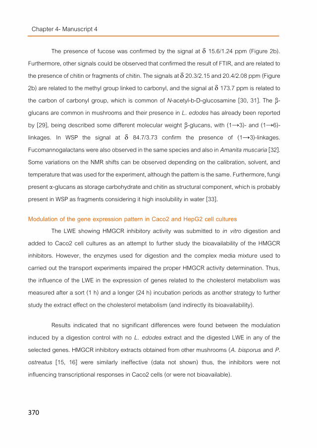
Chapter 4- Manuscript 4
370
The presence of fucose was confirmed by the signal at 15.6/1.24 ppm (Figure 2b). Furthermore, other signals could be observed that confirmed the result of FTIR, and are related to the presence of chitin or fragments of chitin. The signals at 20.3/2.15 and 20.4/2.08 ppm (Figure 2b) are related to the methyl group linked to carbonyl, and the signal at 173.7 ppm is related to the carbon of carbonyl group, which is common of N-acetyl-b-D-glucosamine [30, 31]. The β-glucans are common in mushrooms and their presence in L. edodes has already been reported by [29], being described some different molecular weight β-glucans, with (1→3)- and (1→6)-linkages. In WSP the signal at 84.7/3.73 confirm the presence of (1→3)-linkages. Fucomannogalactans were also observed in the same species and also in Amanita muscaria [32]. Some variations on the NMR shifts can be observed depending on the calibration, solvent, and temperature that was used for the experiment, although the pattern is the same. Furthermore, fungi present α-glucans as storage carbohydrate and chitin as structural component, which is probably present in WSP as fragments considering it high insolubility in water [33].
Modulation of the gene expression pattern in Caco2 and HepG2 cell cultures The LWE showing HMGCR inhibitory activity was submitted to in vitro digestion and
added to Caco2 cell cultures as an attempt to further study the bioavailability of the HMGCR inhibitors. However, the enzymes used for digestion and the complex media mixture used to carried out the transport experiments impaired the proper HMGCR activity determination. Thus, the influence of the LWE in the expression of genes related to the cholesterol metabolism was measured after a sort (1 h) and a longer (24 h) incubation periods as another strategy to further study the extract effect on the cholesterol metabolism (and indirectly its bioavailability).
Results indicated that no significant differences were found between the modulation induced by a digestion control with no L. edodes extract and the digested LWE in any of the selected genes. HMGCR inhibitory extracts obtained from other mushrooms (A. bisporus and P. ostreatus [15, 16] were similarly ineffective (data not shown) thus, the inhibitors were not influencing transcriptional responses in Caco2 cells (or were not bioavailable).

Chapter 4- Efect of fungal compounds with HMGCR inhibitory…
371
However, when the basolateral compartment of the Caco2 monolayer where the digestate of the L. edodes extract was added (containing those compounds able of passing through the cells) was applied to HepG2 cell cultures, a significantly different modulation of the expression was observed after 24 h compared to the control (Figure 3). The genes involved in the biosynthesis of cholesterol such as HMGCR and, downstream the pathway, the FDFT1 (squalene synthethase) were significantly overexpressed as well as the SOAT1 (sterol O-acyltransferase). Two cholesterol homeostasis regulators: SREBF1 and NR1H4 (nuclear factor FXR) were also inversely modulated while a cholesterol carrier was slightly down-regulated (APOB). Other enzymes involved in the lipid metabolism such as ACAT (acetyl-CoA acetyltransferases) and DGAT1 (diacylglycerol O-acyltransferase) were also up-regulated.
Results suggested that the compounds from the water extracts were not affecting the intestinal-like cell cultures but affected the hepatic cells. Thus, the responsible compounds might be bioavailable or might be transformed into something able to modulate, at the molecular level, not only cholesterol homeostasis but also the lipid metabolism.
Previous reports indicated that L. edodes was able of inducing hypocholesterolemia and reducing the triacylglycerol levels in rats due to its content in eritadenine via modifications in the hepatic phospholipid metabolism [8, 10]. Thus perhaps, eritadenine could also be involved in the observed molecular changes although it was never related to the HMGCR mRNA transcription nor activity levels. Moreover, the fact that the HMGCR and FDFT1 mRNAs were not modified shortly after application (1 h) but up-regulated (together with other cholesterol biosynthetic genes) after certain time could suggest certain response mechanism to a previous lack or insufficient HMGCR activity.
Perhaps eritadenine or the compounds derivatives from the HMGCR inhibitory polysaccharides might show certain involvement in cholesterol metabolism but at post-transcriptional level. Thus, in order to further clarify this effect in vivo studies were carried out.
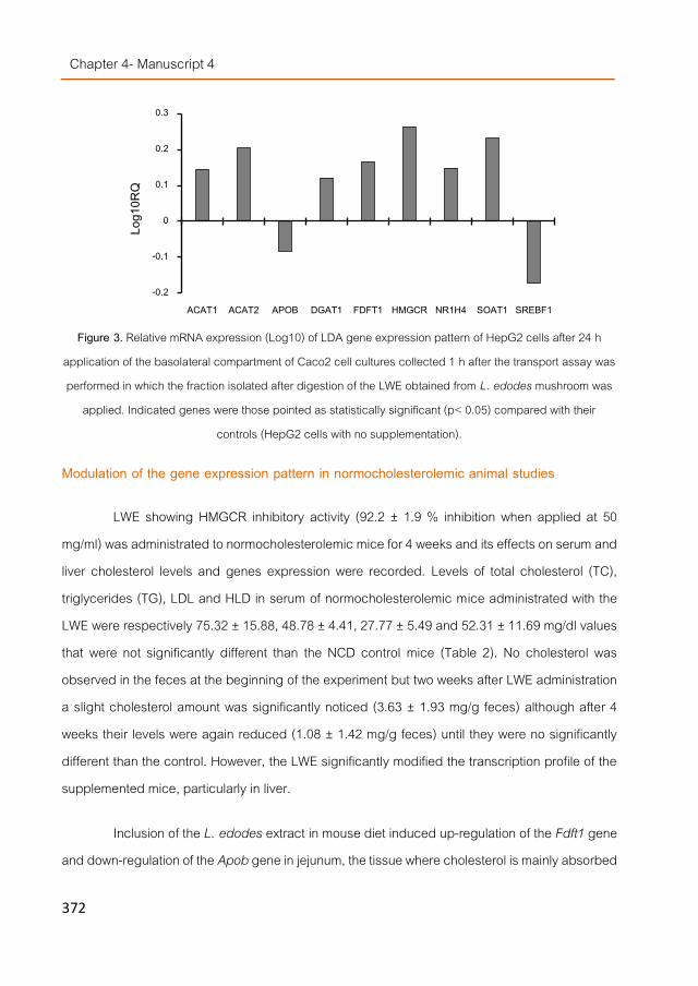
Chapter 4- Manuscript 4
372
Figure 3. Relative mRNA expression (Log10) of LDA gene expression pattern of HepG2 cells after 24 h
application of the basolateral compartment of Caco2 cell cultures collected 1 h after the transport assay was performed in which the fraction isolated after digestion of the LWE obtained from L. edodes mushroom was
applied. Indicated genes were those pointed as statistically significant (p< 0.05) compared with their controls (HepG2 cells with no supplementation).
Modulation of the gene expression pattern in normocholesterolemic animal studies
LWE showing HMGCR inhibitory activity (92.2 ± 1.9 % inhibition when applied at 50 mg/ml) was administrated to normocholesterolemic mice for 4 weeks and its effects on serum and liver cholesterol levels and genes expression were recorded. Levels of total cholesterol (TC), triglycerides (TG), LDL and HLD in serum of normocholesterolemic mice administrated with the LWE were respectively 75.32 ± 15.88, 48.78 ± 4.41, 27.77 ± 5.49 and 52.31 ± 11.69 mg/dl values that were not significantly different than the NCD control mice (Table 2). No cholesterol was observed in the feces at the beginning of the experiment but two weeks after LWE administration a slight cholesterol amount was significantly noticed (3.63 ± 1.93 mg/g feces) although after 4 weeks their levels were again reduced (1.08 ± 1.42 mg/g feces) until they were no significantly different than the control. However, the LWE significantly modified the transcription profile of the supplemented mice, particularly in liver.
Inclusion of the L. edodes extract in mouse diet induced up-regulation of the Fdft1 gene and down-regulation of the Apob gene in jejunum, the tissue where cholesterol is mainly absorbed
-0.2
-0.1
0
0.1
0.2
0.3
ACAT1 ACAT2 APOB DGAT1 FDFT1 HMGCR NR1H4 SOAT1 SREBF1
Log1
0RQ

Chapter 4- Efect of fungal compounds with HMGCR inhibitory…
373
(Figure 4). The observed increase in the mRNA levels of the squalene synthetase might involve stimulation of the cholesterol biosynthesis however, in that case probably the Hmgcr and Soat1 mRNA should have also been induced (as observed for HepG2 in the in vitro study). Since the squalene synthetase is located in the intersection of a few metabolic branches, perhaps in this case it is stimulated to generate other intermediate metabolites yielding steroids or other triterpenoids. Apob gene encode for apolipoproteins (Apolipoprotein B-48 in intestine and B-100 in liver) which are lipid and cholesterol carriers to transport them through the blood. Thus, a reduction in the Apob mRNA transcription in jejunum might negatively affect the intestinal chylomicrons formation since apoliproteins B-48 are building blocks of these particles.
Table 12. Plasma, liver and feces lipid profile in normocholesterolemic and hypercholesterolemic mice treated with a water extract from Lentinula edodes, simvastatin or ezetimibe.
As noticed in vitro, hepatic cells in mice were more affected by the L. edodes extract
than intestinal cells (no significant modulation of the gene expression was neither noticed in ileum or cecum) and showed similar modulation profiles for genes such as Hmgcr, Acat1 and Nr1h4 (FXR), they were in both cases overexpressed. Moreover, similar modulation of the transcription
Variable Plasma level (mg/dL)
NC HC LWE SV EZ TC 65.3 ± 26.6 214.2 ± 97.3a 206.0 ± 47.3a,d 156.2 ± 31.8a,d 59.4 ± 19.0b
HDL 59.8 ± 25.0 89.7 ± 27.9 91.6 ± 19.4d 81.9 ± 21.9d 47.9 ± 10.4b LDL 23.4 ± 11.0 155.0 ± 77.3a 133.5 ± 36.6a,d 102.5 ± 24.0a,d 23.40± 14.9b
TC/HDL 1.1 ± 0.0 2.3 ± 0.3a 2.2 ± 0.2a,d 1.9 ± 0.2a 1.22 ± 0.16b,c LDL/HDL 0.3 ± 0.1 1.6 ± 0.3a 1.4 ± 0.2a,d 1.3 ± 0.2a 0.46 ± 0.23b,c
TG 72.0 ± 20.0 47.2 ± 8.4a 32.5 ± 9.6a 46.2 ± 11.0a 43.4 ± 8.3a Liver level (mg/g tissue)
TC 0.22 ± 0.09 1.09 ± 0.23a 1.20 ± 0.28a,d 1.44 ± 0.43a 0.45 ± 0.20b,c
TG 4.49 ± 0.87 3.72 ± 0.61 0.63 ± 0.20a,b 0.78 ± 0.18a,b 0.88 ± 0.52a,b
Fecal level after 4 weeks (mg/g feces) TC nd 61.83 ± 0.46a 49.33 ± 7.37a,b 65.40 ± 7.41 60.20 ± 14.7
NC, normal control; HC, hypercholesterolemic control; TC, total cholesterol; HDL, high-density lipoprotein cholesterol; LDL, low density lipoprotein
cholesterol; TG, triglyceride. aP<0.05 vs. NC; bP<0.05 vs. HC; cP<0.05 vs. SV; dP<0.05 vs. EZ; nd, non-detected.
The results are mean ± SD of six-eight mice on each diet and were analyzed by one-way ANOVA and the Bonferroni’s procedure..

Chapter 4- Manuscript 4
374
of the Srebfs regulators was observed but, in vitro the Srebf1 gene was significantly down-regulated (Figure 3) while in vivo the influenced gene was Srebf2.
Figure 4. Relative mRNA expression (Log10) of cholesterol-related genes in jejunum, ileum, cecum and liver
of normocholesterolemic mice fed for 4 weeks with normal diet plus LWE. Indicated genes are only those pointed as statistically significant (p< 0.05) compared with normocholesterolemic mice with only NC diet as control. Few genes did not pass the quality control either in the target or the control group, and therefore
their expression level was omitted in the figure.
The effect on the Apob gene expression was opposite to the one observed in HepG2 since overexpression of this mRNA was recorded in liver of normocholesterolemic mice. Perhaps it was a type of compensating mechanism for the down-regulation observed in jejunum, suggesting that the potential reduction of the ApoB-48 biosynthesis was compensated by induction of ApoB-100 synthesis in liver. This might influence the lack of noticeable changes in the biochemical cholesterol parameters in plasma.
Nevertheless, the fact that in vivo the Hmgcr transcription was also stimulated (and perhaps also the Fdft1 in jejunum) could, as before suggested for the in vitro results, indicate that the mice liver was noticing certain lack of cholesterol that could be due to a possible enzyme inhibition (at the post-translational level) or due to certain cholesterol excretion (noticed after 2 weeks) which should have activated correcting mechanisms (after 4 weeks no excretion was noticed) to maintain the homeostasis thus, another in vivo experiment was carried out to investigate the effect of the extract when the cholesterol homeostasis is altered such as under hypercholesterolemic conditions.
-1
-0.5
0
0.5
1
1.5
Abca1 Abcg5 Abcg8 Acat1 Apob Dgat1 Dgat2 Fdft1 Hmgcr Ldlr Npc1l1 Nr1h4 Soat1 Soat2 Srebf1 Srebf2
Log1
0RQ
Jejunum Liver

Chapter 4- Efect of fungal compounds with HMGCR inhibitory…
375
Modulation of the gene expression pattern in hypercholesterolemic animal studies Feeding of mice with the hypercholesterolemic diet increased the cholesterol levels in
plasma 3.3 folds although levels of triglycerides were reduced (Table 2). The increase was mainly due to higher LDL concentrations in serum. Treatment with ezetimibe or simvastatin, two drugs utilized to impair respectively cholesterol absorption and biosynthesis affected differently the cholesterol levels in serum since only ezetimibe was able to significantly induce reduction in TC, HDL and LDL concentrations in serum compared to HC mice. Administration of the LWE obtained from L. edodes showed biochemical values similar to those observed after the treatment with simvastatin since they both were unable to significantly reduce cholesterol levels in plasma but reduce TG levels in liver compared to hypercholesterolemic mice. Surprisingly, cholesterol fecal excretion in mice fed LWE extract was significantly lower than HC.
Similarities between the effect of the LWE from L. edodes and simvastatin were also noticed at the molecular level (Figure 5). For instance, in both cases the Abcg8 and Acat1 genes were upregulated and the Srebf2 down-regulated in jejunum (Figure 5a and 5b). In liver, also in both cases down-regulation of the Dgat1 gene was recorded. The expression of Nr1h4 (FXR) was in both type of samples also down-regulated but in mice treated with simvastatin the effect was noticed in liver while in the mice supplemented with the mushroom extract the effect was noticed in jejunum. In this case, the modulation was more similar to the other drug since inhibition of the Nr1h4 transcription was also noticed in the jejunum of mice treated with ezetimibe (Figure 5c).
The administration of the mushroom extract showed also certain similarities at the molecular level with the effect of ezetimibe but in other tissues such as in cecum in both cases overexpression of the Abcg5 gene was observed and inhibition of the Ffdt1 gene was noticed in ileum. Moreover, ezetimibe treatment was also able of inhibiting transcription of the Srebf2 gene in jejunum as observed after simvastatin treatment or extract administration. This treatment also up-regulated the Npc1l1 gene, probably because its translation product, the Niemann-Pick C1-like 1 protein is the ezetimibe target and therefore, an inhibition of the cholesterol transporter would trigger a response mechanism such as increasing its transcription, particularly after 4 weeks of

Chapter 4- Manuscript 4
376
treatment when the biochemical parameters showed a correction for the induced hypercholesterolemia.
Wise to mention is the completely different transcription profile generated by the same mushroom extract in normo (Figure 4) or hypercholesterolemic (Figure 5a) mice. Particularly in liver, genes such as Soat2 and Dgat1 in one case were up-regulated and in the other down-regulated. Opposite behaviors were also found (but within different tissues) for the expression of other genes such as Fdft1 and Nr1h4. The inhibition of Dgat1 transcription in hypercholesterolemic mice administrated LWE or simvastatin might be related to the lower hepatic TG levels noticed in these cases as it would inhibit triglyceride biosynthetic pathway (perhaps due to the eritadenine content of the extract as it was previously described as the metabolite able of altering this mechanism).
SREBPs (Sterol Regulatory Element-Binding Proteins) directly activate the expression of more than 30 genes dedicated to the synthesis and uptake of cholesterol, fatty acids, triglycerides, and phospholipids, as well as the NADPH cofactor required to synthesize these molecules. Both SREBP-1a and -1c are derived from a single gene (SREBF1) by alternative transcription and SREBP2 is encoded by the SREBF2 gene. SREBP-1a is a potent activator of all SREBP-responsive genes, including those that mediate the synthesis of cholesterol, fatty acids, and triglycerides. The roles of SREBP-1c and SREBP-2 are more restricted than that of SREBP-1a. SREBP-1c preferentially enhances transcription of genes required for fatty acid synthesis but not cholesterol synthesis. Like SREBP-1a, SREBP-2 preferentially activates cholesterol synthesis. SREBP-1a and SREBP-2 are the predominant isoforms of SREBP in most cultured cell lines, whereas SREBP-1c and SREBP-2 predominate in the liver and most other intact tissues. Thus, the down-regulation of Srebf2 mRNA observed in the animals treated with the two drugs and the mushroom extract might indicate inhibition of cholesterol synthesis in jejunum.

Chapter 4- Efect of fungal compounds with HMGCR inhibitory…
377
a)
b)
c)
Figure 10. Relative mRNA expression (Log10) of cholesterol-related genes in jejunum, ileum, cecum and liver of hypercholesterolemic mice fed for 4 weeks with HCD supplemented with a) the LWE extract b)
simvastatin or c) ezetimibe. Indicated genes are only those pointed as significant (p< 0.05) compared with hypercholesterolemic mice fed only HCD as control. Few genes did not pass the quality control either in the
target or the control group, and therefore their expression level was omitted in the figure.
However, FXR (the nuclear receptor encoded by NR1H4) regulates not only a wide variety of genes involved in bile acid synthesis, metabolism, and transport but is also critically involved in triglyceride, cholesterol and glucose metabolism [34]. Activation of FXR by bile acids (or nonsteroidal synthetic FXR agonists) lowers plasma triglycerides by a mechanism that may involve the repression of hepatic SREBP-1c expression and/or the modulation of glucose-induced lipogenic genes. Thus, FXR activation improved hyperglycemia and dyslipidemia in diabetic
-1
-0.5
0
0.5
1
1.5
Abca1 Abcg5 Abcg8 Acat1 Apob Dgat1 Dgat2 Fdft1 Hmgcr Ldlr Npc1l1 Nr1h4 Soat1 Soat2 Srebf1 Srebf2
Log1
0RQ
Jejunum Ileum Cecum Liver
-1.5
-1
-0.5
0
0.5
1
Abca1 Abcg5 Abcg8 Acat1 Apob Dgat1 Dgat2 Fdft1 Hmgcr Ldlr Npc1l1 Nr1h4 Soat1 Soat2 Srebf1 Srebf2
Log1
0RQ
Jejunum Ileum Cecum Liver
-2
-1
0
1
Abca1 Abcg5 Abcg8 Acat1 Apob Dgat1 Dgat2 Fdft1 Hmgcr Ldlr Npc1l1 Nr1h4 Soat1 Soat2 Srebf1 Srebf2
Log1
0RQ
Jejunum Ileum Cecum Liver

Chapter 4- Manuscript 4
378
mice. Thus, the inhibition of Nr1h4 transcription observed in the animals treated with the drugs and the extract would suggest an opposite effect than above described. Moreover, FXR-deficient (FXR-/-) mice displayed elevated serum levels of triglycerides and high-density lipoprotein cholesterol.
On the other hand, overexpression of the mRNAs of both ABC carriers (ABCG5/8) was related to stimulatory effects of the cholesterol intestinal efflux although in this case the activation was noticed in cecum and not in jejunum were might be expected. The fact that non-digestible polysaccharides passes along the small intestine and get accumulated in cecum and colon might be related with this observation.
However, the real significance of all these changes at the molecular level at this moment is a bit speculative since many genes belonging to different metabolic pathways were modified including two genes (encoding the regulatory molecules SREBP2 and FXR) that should activate opposite mechanisms. However, an extract showing similar modulatory effects than two commercial drugs utilized against hypercholesterolemia and reduced TG content in liver seemed quite interesting, therefore at the present further studies are being conducted using higher doses of the LWE in order to light up the real significance of the results.
Conclusions
The water extracts of L. edodes contained specific polysaccharides able of inhibiting the HMGCoA reductase in vitro. The extracts also modified the expression of genes related to the cholesterol homeostasis in HepG2 cell cultures and similarly in normocholesterolemic mice. Extract administration to hypercholesterolemic mice induced a complete different expression profile but it was similar to the modulation observed when mice where treated with drugs utilized to lower cholesterol levels in serum, particularly to simvastatin however, neither LWE nor simvastatin were able of lowering cholesterol levels in plasma. They reduced the triglyceride content in livers which might be related to the Dgat1 down-regulation observed. Therefore, this mushroom extract might have potential to be used as functional ingredient to design hypolipidemic / hypocholesterolemic foods acting via a different mechanism that the compounds actually commercialized. The lack of statistical significance in the plasmatic biochemical

Chapter 4- Efect of fungal compounds with HMGCR inhibitory…
379
parameters encouraged further dose-related experiments since modulation of key genes involved in the cholesterol metabolism are being modulated.
Acknowledgments
The research was supported by AGL2010-21537 national R+D program from the Spanish Ministry of Science and Innovation and ALIBIRD-C M S2009/AGR-1469 regional program from the Community of Madrid (Spain). CTICH (Centro Tecnológico de Investigación del Champiñón de La Rioja, Autol, Spain) is also acknowledged for the cultivation and supplying of the mushrooms fruiting bodies and Maria Navarro-Rubio for her technical assistance.

Chapter 4- Manuscript 4
380
References [1]. Vaquero, M.P., et al., Major diet-drug interactions affecting the kinetic characteristics and hypolipidaemic
properties of statins. Nutrición Hospitalaria, 2010. 25: p. 193-206. [2]. Amin, D., et al., RPR 107393, a potent squalene synthase inhibitor and orally effective cholesterol-lowering agent:
Comparison with inhibitors of HMG-CoA reductase. Journal of Pharmacology and Experimental Therapeutics, 1997. 281(2): p. 746-752.
[3]. Zerenturk, E.J., et al., The endogenous regulator 24(S),25-epoxycholesterol inhibits cholesterol synthesis at DHCR24 (Seladin-1). Biochimica et Biophysica Acta (BBA) - Molecular and Cell Biology of Lipids, 2012. 1821(9): p. 1269-1277.
[4]. Gil-Ramírez, A. and C. Soler-Rivas, The use of edible mushroom extracts as bioactive ingredients to design novel functional foods with hypocholesterolemic activities, in Mushrooms: Cultivation, Antioxidant Properties and Health Benefits, G. Pesti, Editor. 2014, Nova Science Publishers: New York. p. 43-73.
[5]. Chen, J. and X.-F. Huang, The effects of diets enriched in beta-glucans on blood lipoprotein concentrations. Journal of Clinical Lipidology, 2009. 3(3): p. 154-158.
[6]. Cheung, P.C.K., Mushrooms as functional foods, P.C. Cheung, Editor. 2008, John Wiley & Sons, Inc., publications: New Jersey.
[7]. Fukushima, M., et al., Cholesterol-lowering effects of maitake (Grifola frondosa) fiber, shiitake (Lentinus edodes) fiber, and enokitake (Flammulina velutipes) fiber in rats. Experimental Biology and Medicine, 2001. 226(8): p. 758-765.
[8]. Yamada, T., et al., Structure and function of eritadenine and its 3-deaza analogues: Potent inhibitors of S-adenosylhomocysteine hydrolase and hypocholesterolemic agents. Biochemical Pharmacology, 2007. 73(7): p. 981-989.
[9]. Palanisamy, M., et al., Pressurized water extraction of β-glucan enriched fractions with bile acids-binding capacities obtained from edible mushrooms. Biotechnology Progress, 2014. 30(2): p. 391-400.
[10]. Sugiyama, K., Hypocholesterolemic action of eritadenine is mediated by a modification of hepatic phospholipid metabolism in rats. Journal of Nutrition, 1995. 125(8): p. 2134-2144.
[11]. Gil-Ramírez, A., et al., Screening of edible mushrooms and extraction by pressurized water (PWE) of 3-hydroxy-3-methyl-glutaryl CoA reductase inhibitors. Journal of Functional Foods, 2013. 5(1): p. 244-250.
[12]. Xu, C., et al., The pharmacological effect of polysaccharides from Lentinus edodes on the oxidative status and expression of VCAM-1mRNA of thoracic aorta endothelial cell in high-fat-diet rats. Carbohydrate Polymers, 2008. 74(3): p. 445-450.
[13]. Zhu, M., et al., Optimizing conditions of polysaccharide extraction from Shiitake mushroom using response surface methodology and its regulating lipid metabolism. Carbohydrate Polymers, 2013. 95(2): p. 644-648.
[14]. Gunde-Cimerman, N. and A. Cimerman, Pleurotus fruiting bodies contain the inhibitor of 3-Hydroxy-3-Methylglutaryl-Coenzyme A reductase—Lovastatin. Experimental Mycology, 1995. 19(1): p. 1-6.

Chapter 4- Efect of fungal compounds with HMGCR inhibitory…
381
[15]. Gil-Ramírez, A., et al., Study on the 3-hydroxy-3-methyl-glutaryl CoA reductase inhibitory properties of Agaricus bisporus and extraction of bioactive fractions using pressurised solvent technologies. Journal of the Science of Food and Agriculture, 2013. 93(11): p. 2789-2796.
[16]. Gil-Ramírez, A., et al., Water-soluble polysaccharides from Pleurotus ostreatus with HMGCR (3-hydroxy-3-methyl-glutaryl-CoA-reductase) inhibitory activity. In press, 2015.
[17]. Berger, A., et al., Cholesterol-lowering properties of Ganoderma lucidum in vitro, ex vivo, and in hamsters and minipigs. Lipids in Health and Disease, 2004. 3(1): p. 2.
[18]. Gil-Ramírez, A., et al., Sterol enriched fractions obtained from Agaricus bisporus fruiting bodies and by-products by compressed fluid technologies (PLE and SFE). Innovative Food Science & Emerging Technologies, 2013. 18(0): p. 101-107.
[19]. Gil-Ramirez, A., et al. Edible mushrooms as potential sources of new hypocholesterolemic compounds in 7th International Conference on Mushroom Biology and Mushroom Products. 2011. Arcachon, France.
[20]. Enman, J., U. Rova, and K.A. Berglund, Quantification of the bioactive compound eritadenine in selected strains of shiitake mushroom (Lentinus edodes). Journal of Agricultural and Food Chemistry, 2007. 55(4): p. 1177-1180.
[21]. Mosmann, T., Rapid colorimetric assay for cellular growth and survival: Application to proliferation and cytotoxicity assays. Journal of Immunological Methods, 1983. 65(1–2): p. 55-63.
[22]. Gil-Ramírez, A., et al., Effect of ergosterol-enriched extracts obtained from Agaricus bisporus on cholesterol absorption using an in vitro digestion model. Journal of Functional Foods, 2014. 11(0): p. 589-597.
[23]. Gil-Ramírez, A., et al., Modulation of cholesterol-related gene expression by ergosterol and ergosterol-enriched extracts obtained from A. bisporus. European Journal of Nutrition, 2014. Available online. DOI 10.1007/s00394-015-0918-x.
[24]. Vandesompele, J., et al., Accurate normalization of real-time quantitative RT-PCR data by geometric averaging of multiple internal control genes. Genome Biology, 2002. 3(7): p. 1-12.
[25]. Pfaffl, M.W., A new mathematical model for relative quantification in real-time RT–PCR. Nucleic Acids Research, 2001. 29(9): p. e45.
[26]. Stanek, M., H. Falk, and A. Huber, Investigation of the branching characteristic of glycogen by means of two-dimensional1H and 13C NMR spectroscopy. Monatshefte für Chemie / Chemical Monthly, 1998. 129(4): p. 355-364.
[27]. Smiderle, F.R., et al., High molecular weight glucan of the culinary medicinal mushroom Agaricus bisporus is an
α-glucan that forms complexes with low molecular weight galactan. Molecules, 2010. 15(8): p. 5818-5830. [28]. Carbonero, E.R., et al., Lentinus edodes heterogalactan: Antinociceptive and anti-inflammatory effects. Food
Chemistry, 2008. 111(3): p. 531-537. [29]. Wang, K.-p., et al., Structural differences and conformational characterization of five bioactive polysaccharides
from Lentinus edodes. Food Research International, 2014. 62(0): p. 223-232. [30]. Rinaudo, M., Chitin and chitosan: Properties and applications. Progress in Polymer Science, 2006. 31(7): p. 603-
632.

Chapter 4- Manuscript 4
382
[31]. Kasaai, M.R., Determination of the degree of N-acetylation for chitin and chitosan by various NMR spectroscopy techniques: A review. Carbohydrate Polymers, 2010. 79(4): p. 801-810.
[32]. Ruthes, A.C., et al., Fucomannogalactan and glucan from mushroom Amanita muscaria: Structure and inflammatory pain inhibition. Carbohydrate Polymers, 2013. 98(1): p. 761-769.
[33]. Muzzarelli, R.A.A., et al., Current views on fungal chitin/chitosan, human chitinases, food preservation, glucans, pectins and inulin: A tribute to Henri Braconnot, precursor of the carbohydrate polymers science, on the chitin bicentennial. Carbohydrate Polymers, 2012. 87(2): p. 995-1012.
[34]. Lefebvre, P., et al., Role of bile acids and bile acid receptors in metabolic regulation. Vol. 89. 2009. 147-191.

Chapter 5 Influence of food products supplemented with fungal extracts on
cholesterol metabolism

Chapter 5- Influence of food products supplemented with…
385
Preface As previously mentioned, the normal activity of cholesterol metabolism could be altered
by certain modulatory compounds able to modify one of the two major pathways regulating
cholesterol levels: the exogenous cholesterol absorption or the endogenous cholesterol
biosynthesis. Thus, if one of these pathways is impair/stimulated the human body compensate it
by enhancing/inhibiting the activity of the other to maintain cholesterol homeostasis and bring
back normal cholesterol blood levels. Thus, lower cholesterol absorption rates induce higher
activities of cholesterol biosynthetic enzymes and vice versa.
Apparently, ergosterol and ergosterol-derivatives and dietary fibers such as β-glucans
or chitins-derivatives products were modulators more oriented toward the cholesterol absorption
pathway while other mushroom polysaccharides (with HMGCR inhibitory activity), also pointed as
modulators, might probably be more involved in the cholesterol biosynthetic pathway although all
these compounds were also capable of modulating the expression of some genes involved in both
and other regulatory pathways related to the cholesterol metabolism. Therefore, the next logic step
to follow was to prepare a specific mixture of all the investigated compounds and study whether
it could lower cholesterol levels in mice by interfering at both cholesterol-generating pathways
acting then as a double targeting hypocholesterolemic mixture.
Thus, the extracts detailed in preceding chapters were individually and jointly introduced
in a specific food matrix simulating a specifically supplemented food product. Lard was selected
as ideal food matrix because of its high cholesterol and saturated lipid levels, mimicking unhealthy
eating habits related with cardiovascular diseases to evaluate whether the fungal extracts were
able to overcome its negative influence for mice health. Mice were fed a hypercholesterolemic
diet simultaneously with the supplemented food products and their influence on cholesterol
metabolism was studied by measuring cholesterol- and triglyceride-related parameters in plasma,
liver and feces. Furthermore, cholesterol-related gene expression profiles were studied in several
mice tissues (jejunum, cecum, liver).The most relevant results are summarized in the work titled

Chapter 5- Preface
386
The cholesterol-lowering effects of food products supplemented with specific fungal extracts are
independent of Niemann-Pick C1-like 1 protein and ABC sterol transporters gene expression in
mice fed an hypercholesterolemic diet.

Chapter 5- Influence of food products supplemented with…
387
WORKPLAN
Selection of starting material and preparation of food product
In vivo experimental design
Hypocholesterolemic ingredients from mushrooms
RT-PCR
e
Gene expression analysis
A B C
A+B+C
Food matrix supplemented with:
Sampling and subsequents analysis
Chapter 5- Influence of food products supplemented with…
389
Manuscript 1
The cholesterol-lowering effects of food products supplemented with specific fungal extracts are independent of Niemann-Pick C1-like 1 protein and ABC sterol transporters gene expression in mice fed a
high-cholesterol diet.
Víctor Caz1*, Alicia Gil-Ramírez2*, Mónica Santamaría1, María Tabernero1, Cristina Soler-Rivas2, Roberto Martin-Hernandez3,
Francisco R. Marín2, Guillermo Reglero2,3, Carlota Largo1
1Department of Experimental Surgery, Research Institute Hospital La Paz (IdiPAZ), Paseo de la Castellana 261, 28046 Madrid, Spain. 2Department of Production and Characterization of Novel Foods, CIAL – Research Institute in Food Science (UAM+CSIC), C/ Nicolas Cabrera 9, Campus de Cantoblanco, Universidad Autónoma de Madrid, 28049 Madrid, Spain. 3IMDEA Food Institute. Pabellón Central del Antiguo Hospital de Cantoblanco (Edificio nº 7), Crta. de Cantoblanco nº 8, 28049 Madrid, Spain.
*Same contribution
In Preparation, May 2015

Chapter 5- Manuscript 1
390
Abstract Mushrooms are a source of β-glucans and ergosterol, which have been shown to
significantly lower cholesterol. However, food matrices functionalized with mushroom extracts have been less widely studied and their underlying mechanisms remain still unclear. An animal model was used to investigate the effect of lard supplemented with mushroom extracts on the cholesterol metabolism and the transcriptional changes of cholesterol-related genes. Mice were fed a hypercholesterolemic diet, and concomitantly lard functionalized with mushroom extracts at 0% HC, 2.77% BGF, 1.23% WPF, 0.44% EGF, or 4.44% (w/w) MIF for 4 weeks. BGF was a β-glucan enriched extract, WPF was an extract containing specific water-soluble polysaccharides with HMGCR inhibitory activity and EGF was an ergosterol-enriched extract. MIF was a mixture of BGF, WPF and EGF. Functionalized foods significantly reduced plasma cholesterol levels by 22% to 42%, HDL by 18% to 40%, and LDL by 25% to 51% and they led to different tissue-specific expression profiles with a higher regulation in the intestine than in the liver. BGF and MIF significantly increased jejunal mRNA levels of Npc1l1 and Abcg5 genes and hepatic mRNA levels of Npc1l1 gene. None of the tested foods significantly modify the expression of other cholesterol-related genes. In conclusion, lard functionalized with mushroom extracts may help limiting cholesterol levels when consuming hypercholesterolemic diets and show good prospect to be developed into functional foods. Their cholesterol-lowering effect was not mediated via transcriptional changes of Npc1l1 or ABC sterol transporters. Further studies to determine mechanisms of action are needed.

Chapter 5- Influence of food products supplemented with…
391
Introduction Consumption of edible/medicinal mushrooms such as Pleurotus spp. and Lentinula
edodes has beneficial effects on cardiovascular diseases [1, 2] mainly because they were able of decreasing hypercholesterolemia, one of the major risk factor for coronary heart disease. Pleurotus ostreatus decreases plasma cholesterol levels in both humans [3] and animals [4, 5] and cholesterol-lowering effect of Lentinula edodes has also showed in rats [6]. Concerning their bioactive compounds, most evidence pointed to β-glucans [7], ergosterol and derivatives [8], lovastatin [9], eritadenine [10] and phenolic compounds [11]. However, the underlying mechanisms of these effects are only partially understood.
Hypocholesterolemic effect can take place by decreasing intestinal cholesterol absorption, where Niemann-Pick C1-Like 1 (NPC1L1) protein is essential [12]. In fact, ezetimibe, a potent cholesterol absorption inhibitor acting through NPC1L1, it is used in the clinics for management of dyslipidemias [13]. On the other hand, cholesterol absorption is mainly regulated by the sterol efflux transporters Abcg5 and Abcg8 in the small intestine of mice [14]. ABCG5 and ABCG8 form a complex that transport cholesterol from the enterocyte back into the intestinal lumen counteracting cholesterol absorption and is also expressed in liver where it promotes the excretion of cholesterol into bile. Increased expression of Abcg5 and Abcg8 in mice decreases dietary cholesterol absorption and increases biliary cholesterol secretion [15].
Competition and displacement of cholesterol by other molecules for incorporation into dietary mixed micelles decreasing intestinal cholesterol absorption have been pointed as one of the possible mechanisms underlying cholesterol-lowering effect of plant stanols, phytosterols and fungal sterols such as ergosterol [16-18].
The hypocholesterolemic mechanism of β-glucans are also still unclear, some studies pointed their ability to bind to bile acids within micelles in the intestine, preventing the entry of bile acids into blood and therefore removing them from the enterohepatic circulation as one of the potential mechanisms [19]. The decreased bile acids returned to the liver in turn activates cholesterol 7α-hydroxylase that catalyze the conversion of cholesterol into bile acids leads a compensatory increase in hepatic LDLR expression which results in reduced LDL and cholesterol

Chapter 5- Manuscript 1
392
levels. Bile-acid binding capacities for fungal β-glucans have been described but using in vitro assays [20, 21]. Moreover, the subsequent addition of mushroom β-glucans to ergosterol enriched extracts increased cholesterol displacement from the DMM [18].
Statins are a class of drugs that reduce synthesis of cholesterol in the liver by competitively inhibiting 3-hydroxy-3-methylglutaryl coenzyme A reductase (HMGCR), leading to decreased plasma cholesterol levels. Moreover, the reduction in intracellular cholesterol concentration induces low-density lipoprotein receptor (LDLR) expression on the hepatocyte cell surface, which results in a decreased concentration of circulating LDL-cholesterol [22]. However, the presence of lovastatin or other statins in mushroom is actually under discussion, some publications monitored their presence in several mushroom strains [23, 24] and other found no statins [3, 25, 26] but other compounds such as lanosteroids, ganoderols etc. able of reducing the mRNA expression of the HMG-CoA reductase or specific water-soluble polysaccharides showing HMGCR inhibitory activity with in vitro tests [26-29].
The studies carried out to demonstrate the hypocholesterolemic effects of edible mushrooms have mostly been performed using powdered fruiting bodies, broth cultured mycelia or specific water/organic extracts. However, their bioavailability and efficacy when added into a food matrix are still not studied in detail and food nature may influence the efficiency of the bioactive compounds when integrated into food matrices [18]. Therefore, in this study, in order to emulate usual inadequate eating habits related with dyslipidemia, lard was chosen as a food matrix and it was supplemented with 3 different mushrooms extracts. The extracts contained very different bioactive compounds such as β-glucans, ergosterol and derivatives and a water extract containing polysaccharides with HMGCR inhibitory capacity and eritadenine. A mixture of the 3 extracts was also integrated into the food matrix to detect possible positive or negative synergies and their hypocholesterolemic effects studied. The influence of lard supplemented with mushroom extracts was examined on plasma lipids and fecal cholesterol in mice fed a high-cholesterol diet. In addition, low-density arrays were used in order to identify genes regulated by mushroom foods in the small intestine, liver and cecum.

Chapter 5- Influence of food products supplemented with…
393
Materials and Methods
Biological material Fresh fruiting bodies of Pleurotus ostreatus (Jacq.Ex Fr.) Kummer strain ¨Gurelan H-
107¨ and Lentinula edodes S. (Berkeley) from ¨Sylvan 4312¨ from the first flush were harvested, cut in slices, lyophilized and ground until a dry powder was obtained following the procedure described by [30]. Resulting mushroom powders were utilized as starting material to prepare a dietary fiber extract rich in β-glucans, an extract containing eritadenine and water soluble polysaccharides with HMGCR inhibitory activity and an ergosterol-enriched extract.
Commercially available lard (Iberian pork lard (100 g fat/100 g food), BHT, BHA) was purchased from a local supermarket and maintained at 4 ºC until use. All the experiments were performed from the same lotus.
Preparation of a food product containing a β-glucans-enriched extract (BGF) A β-glucan-enriched fraction from Pleurotus ostreatus was prepared according to the
method of [31]. This fraction contained 39.87 mg/100mg (dw) β-glucans, 7.70 mg/100mg α-glucans and 6.10 mg/100mg chitins [32]. The extract (2.77%) was mixed with lard (12%) by slightly melting of the lipid matrix and the resulting supplemented food product (BGF) was utilized for animal feeding.
Preparation of a food product containing an extract with water-soluble polysaccharides (WPF) Lentinula edodes dry powder was mixed with MilliQ water and stirred gently during 1 min
in a Vortex at room temperature. Afterwards, the solution was centrifuged at 12000 rpm during 2 min and supernatant collected and freeze-dried [29]. The obtained extract contained no statins but 0.21 mg/100mg eritadenine, 26.49 mg/100mg proteins and 31.51 mg/100mg of polysaccharides of which only 9.74 mg/100mg were β-glucans but it also contained α-glucans and fucomannogalactans. The polysaccharides were able of inhibiting the HMGCR activity [29]. The extract (1.23%) was mixed with lard (12%) by slightly melting of the lipid matrix and the resulting supplemented food product (WPF) was utilized for animal feeding.

Chapter 5- Manuscript 1
394
Preparation of a food product containing an ergosterol-enriched extract (EGF) A fungal sterols-enriched fraction from Lentinula edodes was prepared according to the
method of [30]. Mushroom powder (80 g) were mixed with washed sea sand (900 g) and submitted to supercritical fluid extraction (SFE) with CO2. Extraction was carried out at 18 MPa and 40 ºC with a total extraction time of 3 h. Obtained fractions were immediately dried on a rotary vacuum evaporator and stored at -20 ºC. This fraction contained 37.3 mg/100mg ergosterol, 6.5 mg/100 mg ergosta-5,7-dienol, 2.6 mg/100 mg fungisterol and 1.5 mg/100 mg ergosta-7,22-dienol [33]. The extract (0.44%) was mixed with lard (12%) by slightly melting of the lipid matrix and the resulting supplemented food product (EGF) was utilized for animal feeding.
Preparation of a food product containing a mixture of BGF, WPF and EGF (MIF) A mixture of the 3 above described extracts (4.44%) was prepared and added to lard
(12%) by slightly melting of the lipid matrix and the resulting supplemented food product (MIF) containing 2.77% BGF + 1.23% WPF + 0.44% EGF was utilized for animal feeding.
Experimental design Male C57BL/6JRj mice (9 weeks old), were purchased from Janvier SAS (Le Genest Saint
Isle, France). The mice were maintained in temperature-, humidity- and light-controlled conditions (24 ± 2 ºC, 40-60% humidity, 12:12 hour light: dark cycle) and had free access to water and food. The experimental protocol was approved by our institution’s Animal Welfare and Ethics Committee and was carried out according to internationally approved guidelines (2010/63/EU). Animals were randomly divided into six groups (n=8 per group). Mice fed a high-cholesterol diet used as control (HC) were administrated a standard diet (Safe Rodent Diet A04, Augy, France) supplemented with 2% cholesterol, 1% cholic acid and 12% lard (w/w). The other groups were fed the same diet as HC but lard was substituted by BGF, WPF, EGF or MIF. Mice were fed for 4 weeks and fasted 12 hours before harvesting blood for subsequent lipid measurements, and jejunum, liver and cecum for RNA isolation. Both the mice and the food were regularly weighed to determine changes in body weight and food intake.

Chapter 5- Influence of food products supplemented with…
395
Blood and feces biochemistry Blood samples were obtained from retromandibular vein before the administration of
experimental diets (baseline samples) or intracardiac exsanguination under isoflurane anesthesia (1.5%) at the end of the study. Plasma was collected and stored at -80 ºC before use. Plasma levels of total cholesterol, HDL-cholesterol, LDL-cholesterol and triglycerides were measured using an Automated Analyzer (Covas C311, Roche, Spain). Cholesterol extraction from the stored feces (300 mg) and analysis by GC-MS were carried out as described in [33].
Statistical Analysis of biochemical data To determine statistically significant differences among dietary groups, the data were
analyzed at P<0.05 by one-way ANOVA, followed by Tukey’s test, using SPSS software (LEAD Technologies, Chicago, IL, USA). Data are expressed as means ± SEM.
RNA extraction and quantitative real-time PCR Total RNA from cell cultures or mice tissues was extracted by magnetic bead technology
using a pureLinkTM Total RNA kit TRIzol® Plus RNA Kit (Invitrogen, Carlsbad, CA, USA) in an iPrepTM Purification Instrument (Invitrogen) programmed with an iPrepTM total RNA card (Invitrogen) according to manufacturer instructions. The RNA concentration was determined by spectrophotometry at 260 nm and the purity of the extracted RNA was calculated from ratio of absorbance at 260:280 nm and 260:230 nm in a NanoDrop ND-1000 (NanoDrop Technologies Inc., Wilmington, DE, USA).An amount of 400 ng of total RNA from each sample were reversed transcribed using the High Capacity cDNA Reverse Transcription Kit (Applied Biosystems, Framinghan, MA, USA). The obtained cDNA (100 µl per port) were loaded into the fluidic cards. Before sealing them, the cards were centrifuged twice on a Sorvall centrifuge at 1200 rpm for 1 min. Finally, the micro fluidic cards were run in a 7900HT Fast Real-Time PCR system (Applied Biosystems). Amplification conditions were 50 ºC for 2 min, 94.5 ºC for 10 min, followed by 40 cycles with 97 ºC with 30 s and 59.7 ºC for 1 min.
Gene expression by Low Density Array (LDA) procedures The micro-fluid card (Applied Biosystems TaqMan Array Micro-Fluid Cards) was
constructed using respectively human and mouse commercial available assays (Applied

Chapter 5- Manuscript 1
396
Biosystems) for several genes related to the cholesterol metabolism (Table 1) and the internal standard and housekeeping genes.Comparative analysis of each of these genes was performed using specialized computer programs SDS2.3 and RQ 2.1 (Applied Biosystems).
Gene expression statistical data analysis The expression of each gene was measured at least in triplicate for each sample. In order
to control for potential outliers, Grubb’s test was applied to technical replicates with a threshold of 0.05. Biological replicates were quality controlled using the Median Absolute Deviation test (MAD test). Samples of the same biological group with a MAD score higher than 3 were removed from the analysis. Genorm algorithm [34] was used to identify the most stable reference genes for normalization. Genes Gusb and Hprt1 were found to be the most stably expressed in the mouse tissue samples. Relative gene expression levels were calculated by using the 2-ΔΔCT method [35]. Statistical significance of the results was assessed using a Limma test, and Benjamini-Hochberg false discovery rate procedure was subsequently applied. Adjusted p-values lower than 0.05 were considered significant. All these calculations were carried out with Real Time StarMiner 4.5® (Integromics S.L, Spain).
Results
Functionalized foods lower plasma levels of cholesterol, HDL and LDL Diets with or without supplemented foods were well tolerated by mice. There were no
significant differences in final body weights or dietary intake among groups (data not shown). Plasma total cholesterol, HDL, LDL and triglycerides were determined at baseline and at the end of the experiments (week 4). Total cholesterol and LDL were markedly increased in mice controls fed a high-cholesterol diet (HC) at the end of the dietary period with respect their own baseline levels (2.9, 26 fold, P<0.05, respectively), while HDL slightly increased (1.43 fold). Compared with HC, 22-42% reduction of plasma total cholesterol was observed in mice fed supplemented foods

Chapter 5- Influence of food products supplemented with…
397
Table 1. ABI (Applied Biosystems) commercial reference and NCBI reference sequence of the selected genes from Mus musculus and internal controls used in the LDA design.
ABI code for Mus musculus
Reference sequence/s Gen Description
Mm00442646_m1 NM_005502.3 Abca1 ATP-Binding Cassette, Sub-Family A (ABC1), Member
1
Mm00446241_m1 NM_022436.2 Abcg5 ATP-binding cassette, sub-family G (WHITE), member
5
Mm00445970_m1 NM_022437.2 Abcg8 ATP-binding cassette, sub-family G (WHITE), member
8
Mm00507463_m1 NM_000019.3 Acat1 Acetyl-CoA acetyltransferase 1
NM_005891.2 Acat2 Acetyl-CoA acetyltransferase 2
Mm01545156_m1 NM_000384.2 ApoB Apolipoprotein B
Mm00815354_s1 NM_004462.3 Fdft1 Farnesyl-diphosphate farnesyltransferase 1
Mm01282499_m1 NM_000859.2 NM_001130996.1 Hmgcr 3-hydroxy-3-methylglutaryl-CoA reductase
Mm00440169_m1
NM_000527.4, NM_001195798.1NM_001195799.1N
M_001195800.1 NM_001195802.1NM_001195803.1
Ldlr Low density lipoprotein receptor
Mm01191972_m1 NM_001101648.1 NM_013389.2 Npc1l1 NPC1 (Niemann-Pick disease, type C1, gene)-like 1
Mm00443451_m1 NM_001130101.2
NM_001130102.2NM_001251934. NM_001251935.1 NM_0056933
Nr1h3 (Lxrα)
Nuclear receptor subfamily 1, group H, member 3 (LXRα)
Mm00436425_m1 NM_001206977.1NM_001206978.1 NM_001206979.1 NM_001206992.1
NM_001206993.1 NM_005123.3
Nr1h4 (Fxrα)
Nuclear receptor subfamily 1, group H, member 4 (FXRα)
Mm00486279_m1 NM_001252511.1 NM_001252512.1
NM_003101.5 Soat1 Sterol O-acyltransferase 1
Mm00448823_m1 NM_003578.3 Soat2 Sterol O-acyltransferase 2
Mm00550338_m1 NM_001005291.2 NM_004176.4 Srebf1 Sterol regulatory element binding transcription factor 1
Mm01306292_m1 NM_004599.2 Srebf2 Sterol regulatory element binding transcription factor 2
Mm00839493_m1 NM_002046.4 Polr2a polimerase
Mm00446953_m1 NM_000181.3 Gusb Glucuronidase, beta (internal control)
Mm00446968_m1 NM_000194.2 Hprt1 Hypoxanthine phosphoribosyltransferase 1 (internal
control)
18S-Hs99999901_s1 Rn18s1 RNA, 18S ribosomal 1 (housekeeping gene)

Chapter 5- Manuscript 1
398
for 4 weeks, with a significant reduction for BGF, WPF and MIF (Figure 1a). HDL levels were reduced by 18-40% compared with HC, with a significant reduction for WPF (Figure 1b). LDL levels were markedly increased due to the high cholesterol diet from day 0 to the end of the experimental period. A higher reduction in LDL levels (27-51%) compared with that of HDL was observed, with a significant reduction in LDL levels for BGF and WPF mushroom foods (Figure 1c).
a) b) c)
Figure 11. Supplemented foods lower plasma levels of cholesterol, HDL, and LDL. Plasma total cholesterol
(a), HDL (b), and LDL (c) levels in mice at 0 and 4 weeks of feeding with supplemented foods. Hypercholesterolemic control (HC), -glucan containing food (BGF), water-soluble polysaccharides
containing food (WPF), ergosterol containing food (EGF), and mixture containing food (MIF). Values are expressed as mean ± SEM of eight mice per group. *P<0.05 vs. HC.
Plasma triglycerides levels showed no differences between HC and mice fed mushroom foods at the end of the dietary period (HC, BGF, WPF, EGF and MIF: 0.43 ± 0.03, 0.49 ± 0.04, 0.44 ± 0.01, 0.39 ± 0.03, and 0.38 ± 0.03 mmol/l, respectively).
Effect of mushroom foods on cholesterol excretion Before the experimental feeding period no cholesterol was detected in mice feces
whereas at the end of the experiments, WPF and MIF groups showed a significant reduction compared with HC (Table 2). BGF and EGF groups showed no significant differences with respect HC. MIF group also showed a significant reduction with respect BGF and EGF groups.

Chapter 5- Influence of food products supplemented with…
399
Table 2. Fecal cholesterol concentrations in mice fed supplemented foods for 4 weeks.
Ergosterol was not detected in the faces neither at the beginning or after 4 weeks in mice fed BGF or WPF as expected. However, it was also not found at significant levels in mice fed EGF food and only traces in some samples of mice fed MIF foods.
Effects of mushroom foods on Npc1l1, Abcg5, and Abcg8 gene expression To examine responses of major tissues involved in cholesterol homeostasis after the
different diets, the mRNA expression levels of cholesterol-related genes were studied in jejunum, liver and cecum using low density arrays. No significant differences were observed at transcriptional level of genes involved in the cholesterol biosynthesis such as Hmgcr, Fdft1 or in those related to cholesterol transport (Ldlr), to the maintenance of cholesterol homeostasis (such as Nr1h3 (Lxr), Nr1h4 (Fxr), Srebf1/2) or to the cholesterol incorporation into the blood circulation (Apob, Abca1). However, changes in cholesterol absorption, cholesterol efflux and biliary cholesterol excretion were noticed since consumption of supplemented foods produced significant changes in the expression of Npc1l1 gene and ABC transporters (Abcg5/8) in jejunum, liver, and cecum although the intensity of these changes was tissue dependent. Compared with HC, feeding BGF and MIF significantly increased expression levels of Npc1l1 and Abcg5 genes in the jejunum with a higher regulation on Abcg5, while no changes of Abcg8 gene were observed (Figure 2). WPF and EGF did not produce changes on the expression of Npc1l1, Abcg5 and Abcg8 genes in jejunum.
Diet group HC BGF WPF EGF MIF
Time on diet (mg cholesterol/g feces)
0 weeks ND ND ND ND ND 2 weeks 43.56 ± 4.28 58.98 ± 8.13* 46.92 ± 9.95 51.76 ± 18.49 44.91 ± 0.96 4 weeks 59.53 ± 2.27 54.87 ± 2.43 46.18 ± 2.55* 48.99 ± 3.07* 33.01 ± 0.16*,#,§
HC: hypercholesterolemic control; BGF: -glucan containing food; WPF: water-soluble polysaccharides containing food; EGF: ergosterol containing food; MIF: mixture containing food; ND: no detected. Values are expressed as means ± SEM for eight mice per group. *P<0.05 vs. HC, #P<0.05 vs. BGF, §P<0.05 vs. EGF.

Chapter 5- Manuscript 1
400
In liver, Npc1l1 gene expression was significantly increased by BGF and MIF as compared with HC, whereas expression of Abcg5 and Abcg8 genes were not modified by those foods.
EGF did not induce any transcriptional answer in the jejunum or liver but in cecum, it stimulated a significant overexpression of Abcg5 and Abcg8 genes while MIF induced a significant down-regulation on Npc1l1 gene in cecum. BGF and WPF did not induce significant changes on gene expression of Npc1l1, Abcg5 and Abcg8 genes.
Npc1l1 Abcg5 Abcg8 Npc1l1 Abcg5 Abcg8 Npc1l1 Abcg5 Abcg8
Figure 12. Effects of supplemented foods on Npc1l1, Abcg5, and Abcg8 gene expression in jejunum, liver, and cecum. Mice were fed each diet for 4 weeks and thereafter, mRNA expression levels were determined using low-density arrays. Data are relative to the hypercholesterolemic control (HC) and presented as mean
± SEM of three mice per group. *P<0.05 vs. HC. See legend of groups in Figure 1.
Discussion Diets supplemented with cholesterol and cholic acid facilitates cholesterol absorption
resulting in increased plasma cholesterol [36]. However, when mice were given these high cholesterol and cholic diets including specific foods supplemented with several mushroom extracts a notable decrease in plasma cholesterol as compared with controls was recorded mainly due to a higher LDL reduction than HDL reduction. The food matrix enriched with β-glucans (BGF), was probably the most beneficial of the 4 tested food formulations since it induced a significant

Chapter 5- Influence of food products supplemented with…
401
reduction of cholesterol and LDL with respect to controls and no changes of HDL compared with its baseline levels. The fungal β-glucans included in the BGF are considered as dietary fiber (DF) because with their β-(1→3) and β-(1→3),(1→6) linkages generate complex polymers that cannot be digested and previous in vitro studies indicated that they could scavenge bile acids during digestion [20] suggesting that these properties might contribute to the observed hypocholesterolemic activity. Indeed significantly higher cholesterol excretion was observed after only 2 weeks BGF supplementation compare to HC controls, although this effect was not kept until the end of the feeding period. Dietary fiber extracts obtained from other mushroom strains such as Agaricus bisporus, Grifola frondosa, and Flammulina velutipes were also able of lowering LDL [6, 37] although, DF fractions from L. edodes and A. bisporus also induced a significant HDL reduction. However, these extracts were given to rats fed a cholesterol-free diet thus, the results obtained using BGF might suggest that apparently fungal DF fractions could even exert their beneficial influence when unhealthy diets are followed (high-cholesterol diet). But, a previous report indicated that when an alkali-insoluble β-glucan extract obtained from Pleurotus ostreatus was administrated concomitant with a cholesterol-enriched (0.3%) diet, no cholesterol lowering effect was observed [38]. Perhaps, the higher isolation degree of the specific polysaccharide (higher than BGF and those previously reported DF extracts) might explain its lack of activity because when fed the complete mushroom powder from which the purified extract was obtained, cholesterol levels were reduced [38].
Likewise, the consumption of WPF also induced cholesterol and LDL levels significantly reduced but taking into account its effect on HDL, which reached levels below baseline levels, it seems less beneficial than BGF. This extract contained water soluble polysaccharides, such as glucans and fucomannogalactans with the ability of inhibiting the HMGCR (3-hydroxy-3-methyl-glutaryl-Co A reductase) in vitro and eritadenine [29]. The ability of the small alkaloid to decrease levels of LDL and HDL was associated with modifications in hepatic phospholipid metabolism [10]. Moreover, ergosterol and β-glucan-enriched extracts were also able of reducing hepatic triglyceride levels in mice [33]. Therefore, the observed results might be partially due to modifications on the lipid metabolism in liver in combination with the inhibition of the HMGCR if

Chapter 5- Manuscript 1
402
the water-soluble polysaccharides (or their degradation products) were also able of inhibiting the enzyme in vivo. In fact, the reduction in the excreted cholesterol that was noticed in this samples could reinforce this hypothesis since this observation suggest that the cholesterol-lowering effect of the supplemented food is not mediated via inhibition of intestinal cholesterol absorption or enhanced biliary cholesterol excretion.
The ergosterol enriched food (EGF) trended to reduce cholesterol, HDL and LDL but these changes did not reach statistical significance. Previous studies carried out using female hamster rats indicated that ergosterol-containing extracts obtained from Pleurotus citrinopileatus lowered cholesterol but increased HDL. Differences between results could be, despite differences in extract composition, due to the different experimental settings since in that case hypercholesterolemia was firstly induced in rats and then treated with the extract [8]. Significant cholesterol reduction was also observed in another study using apoE (-/-) mice fed a normal diet containing ergosterol [39]. Compared to phytosterols, 2% supplementations were needed to observe a slight cholesterol decrease in C57BL/6J mice fed a Western diet for 4 weeks [40] while apparently 0.44% fungal sterols were also able of stimulate a similar reduction. Perhaps this low concentration also explains their absence in the feces. The traces found in MIF foods could be due to certain scavenging degree due to the presence of β-glucans in the same food matrix [18].
The MIF containing a mix of the 3 mushroom extracts lowered cholesterol without significant reduction of HDL and LDL. The observed effects were not quantitatively higher than those induced by each individually administrated food and therefore, no synergy was observed. Its biochemical parameters (and gene expression data, see later) were more similar to BGF than to the others, which it seems reasonable taking into account that the β-glucan-enriched extract was the extract with the highest concentration in MIF.
The cholesterol-lowering effects observed after administration of supplemented foods did not correlated with changes in the expression of cholesterol-related genes. Genes were selected taking into account that cholesterol homeostasis is a highly regulated balance of dietary cholesterol absorption, biliary excretion, and de novo synthesis. Concerning cholesterol

Chapter 5- Influence of food products supplemented with…
403
absorption, which occurs in jejunum both in humans and mice, Npc1l1 [12], Abcg5 and Abcg8 genes [14] play a key role. Intestinal expression of Npc1l1 is down regulated by diets containing high levels of cholesterol [41]. Thus, the overexpression of the Npc1l1 gene noticed in the jejunum of mice fed BGF or MIF might indicate that higher levels of Npc1l1 mRNAs than the HC are found in this tissue as a response to the noticeable lower cholesterol levels in serum. The cholesterol levels in serum are lower so more Npc1l1 protein can be synthesized to absorb it.
Beside down-regulation of Npc1l1, inhibition of cholesterol absorption can also take place by up-regulation of intestinal Abcg5/8 [15]. Administration of BGF and MIF supplemented foods significantly increased Abcg5 mRNA levels in the intestine while Abcg8 mRNA levels and Lxr (liver X receptor), a transcription factor regulating ABCG5 and ABCG8 genes [42], remained unchanged (data not shown). Thus, it seems unlikely that the observed over-expression is of relevance for the cholesterol efflux mechanism or the hypocholesterolemic effect of these foods although, the possibility of post-transcriptional or activity regulation mechanisms should not be excluded. Moreover, inhibition of cholesterol absorption is associated with increased fecal cholesterol levels [43] but no changes or decrease in fecal cholesterol levels was found in mice fed supplemented foods as compared with controls. Similar transcriptional responses were also recorded in mice fed a phytosterol enriched diet where intestinal overexpression of Npc1l1, Abcg5 and Abcg8 was observed concomitant with a hypocholesterolemic effect indicating that the inhibition of cholesterol absorption by phytosterols was not mediated via transcriptional changes of these genes [40]. In other study, reduction of cholesterol absorption also did not correlate with transcriptional changes of Npc1l1, Abcg5 or Abcg8 in jejunum of mice fed a phytosterol enriched diet since no significant differences compare to control were observed after the dietary period [44]. If the cholesterol absorption pathway was not transcriptionally modulated, the supplemented foods could influence the endogenous cholesterol biosynthetic pathway. In fact that was the response that could be expected for the mice supplemented with WPF because of its HMGCR inhibitors content. However, no significant changes at the transcriptional level were found for genes such as Hmgcr, Fdft1, Srebf1/2 etc. confirming previous in vitro studies using Caco2 cell cultures [29] where no significant modulation of cholesterol-related genes was found for water-

Chapter 5- Manuscript 1
404
soluble extracts similar to the one included in WPF. These results suggested again that the modulation of cholesterol metabolism induced by supplemented foods might took place via post-transcriptional mechanisms.
The balance between hepatic transcriptional regulation of NPC1L1, and ABCG5/8 genes is involved in the cholesterol excretion into bile. Taking into account Npc1l1 is almost not expressed in mouse liver [12] it is noticeable the marked enhancement of Npc1l1 expression in liver of mice fed BGF and MIF supplemented foods. Hepatic overexpression of Npc1l1 markedly decreases biliary cholesterol concentration in transgenic mice expressing human Npc1l1 in liver [45, 46] which in turn decreases fecal cholesterol levels and increases plasma cholesterol. Lower fecal cholesterol excretion was recorded in mice fed the supplemented foods (after 4 weeks) than the HC control suggesting a lower biliary cholesterol concentration associated to their increased hepatic Npc1l1 mRNA levels. However, mice fed WPF or EGF also showed decreased feces cholesterol levels even though their no effect on hepatic Npc1l1 expression. On the other hand, decreased instead of increased plasma cholesterol levels were observed in mice fed supplemented foods. Therefore, no correlation between hepatic Npc1l1 gene expression and biliary and plasma cholesterol levels could be established.
The amount of cholesterol that circulates in the plasma as lipoproteins can be affected by the balance of cholesterol metabolism within and between the intestines and liver. Lowering LDL by ezetimibe has been proven to be associated with increase of hepatic LDLR expression [13]. Ezetimibe inhibits intestinal cholesterol absorption which in turn leads to a reduced amount of lipoprotein cholesterol circulated to the liver. The reduction of cholesterol delivery induces an up-regulation of LDLR in the liver and as a consequence, a reduction of plasma LDL levels [47]. However, hepatic Ldlr mRNA levels in mice fed supplemented foods were not different than HC control (data not shown) indicating that the plasmatic LDL decrease was not related to transcriptional changes at the LDLR. Hepatic Ldlr up-regulation was also noticed after administration of dietary fiber extracts from A. bisporus or Flammulina velutipes to normocholesterolemic rats [6, 37] but it was not significant for the fiber extracts from other mushrooms such as L. edodes and Grifola frondosa. LDLR up-regulation was also found in Caco2

Chapter 5- Influence of food products supplemented with…
405
and HepG2 cell cultures after administration of an ergosterol-enriched extract but in vivo it was unable of reproducing the observed modulation [33].
Hypocholesterolemic effect as a secondary reaction of microbial fermentation of oat β-glucans has also been proposed as β-glucans can be transformed into short-chain fatty acids (SCFAs) in the colon [48, 49] and they might lower plasma cholesterol levels by inhibiting hepatic cholesterol synthesis in humans [50, 51] and rats [52]. This inhibition was observed at enzymatic level instead of transcriptional level as some of the generated SCFAs were able of acting as competitive inhibitors of mevalonate [52]. This observation is in line with the results found for the BGF and MIF foods which β-glucans could exert their hypocholesterolemic effect via post-transcriptional modifications rather than by modifying the mRNAs levels of genes involved in the synthesis of cholesterol since no different transcriptional profiles were found compare to HC controls.
The cholesterol-lowering effect of ergosterol has been previously demonstrated in hamster rats [8], rat liver microsomes [53] and mice [54]. In the latter study, an ergosterol derivative ((22E)-ergost-22-ene-1,3-diol) but not ergosterol, was found to be a potent agonist for LXR and inhibited intestinal cholesterol absorption by up-regulation of Abcg5 and Abcg8 genes in the small intestine but not in liver of mice. EGF also induced overexpression of Abcg5 and Abcg8 in cecum but not in the small intestine or liver. Moreover, a significant down-regulation of the Npc1L1 was observed also in cecum for the MIF, perhaps as combination of the tendency observed for the 2 extracts containing polysaccharides (BGF and WPF). More studies are needed in order to assess whether these transcriptional changes might be involved in cholesterol absorption or in other processes.
Conclusions A considered unhealthy food matrix such as lard (because of its high cholesterol and fat
contents) can be functionalized with specific mushroom extracts to generate food products showing hypocholesterolemic properties. They all similarly modified the biochemical cholesterol-related parameters despite their differences in their compositions suggesting that probably

Chapter 5- Manuscript 1
406
several mechanisms are involved. Effects appeared not to be due to inhibition of intestinal cholesterol absorption or increase of cholesterol excretion through feces or into bile. The ability of β-glucans to bind to bile acids, preventing their entry into blood and forcing the cholesterol transformation into bile acids to maintain the levels needed for digestion [19] might not be the in vivo mechanism of action for fungal β-glucans extracts. The ability of sterols for competing and displacing cholesterol from dietary mixed micelles seemed not to be the main mechanism for fungal sterols. Thus, the hypocholesterolemic effect might be more related to the inhibition of the cholesterol biosynthetic pathway (although via post-translational changes) perhaps by the generation of SCFAs (from β-glucans) or other compounds (from water-soluble polysaccharides or sterols) acting as inhibitors of enzymes involved in the pathway. Nevertheless, the cholesterol-lowering effect was independent of transcriptional changes in Npc1l1 or ABC transporters as well as in other genes involved in its regulation, biosynthesis, absorption, processing or transport. Thus, the prepared functional foods may be useful in limiting hypercholesterolemia under a high-cholesterol diet. Furthermore, they could be a source for the development of novel functional foods although further studies to determine mechanisms of action are needed.
Acknowledgments The research was supported by AGL2010-21537 national R+D program from the Spanish
Ministry of Science and Innovation and ALIBIRD-CM S2009/AGR-1469 regional program from the Community of Madrid (Spain).CTICH (Centro Tecnológico de Investigación del Champiñón de La Rioja, Autol, Spain) is also acknowledged for the cultivation and supplying of the mushrooms fruiting bodies and Maria Navarro-Rubio for her technical assistance.

Chapter 5- Influence of food products supplemented with…
407
References [1]. Guillamón, E., et al., Edible mushrooms: Role in the prevention of cardiovascular diseases. Fitoterapia, 2010.
81(7): p. 715-723. [2]. Bisen, P., et al., Lentinus edodes: a macrofungus with pharmacological activities. . Current Medicinal Chemistry,
2010. 17: p. 2419-2430. [3]. Schneider, I., et al., Lipid lowering effects of oyster mushroom (Pleurotus ostreatus) in humans. Journal of
Functional Foods, 2011. 3(1): p. 17-24. [4]. Opletal, L., et al., Evidence for the anti-hyperlipidaemic activity of the edible fungus Pleurotus ostreatus. British
Journal of Biomedical Science, 1997. 54(4): p. 240-243. [5]. Bobek PhD, P., L. Ozdín PhD, and Š. Galbavý Md, Dose- and time-dependent hypocholesterolemic effect of
oyster mushroom (Pleurotus ostreatus) in rats. Nutrition, 1998. 14(3): p. 282-286. [6]. Fukushima, M., et al., Cholesterol-lowering effects of maitake (Grifola frondosa) fiber, shiitake (Lentinus edodes)
fiber, and enokitake (Flammulina velutipes) fiber in rats. Experimental Biology and Medicine, 2001. 226(8): p. 758-765.
[7]. Rop, O., J. Mlcek, and T. Jurikova, Beta-glucans in higher fungi and their health effects. Nutr Rev, 2009. 67(11): p. 624-31.
[8]. Hu, S.H., et al., Antihyperlipidemic and Antioxidant Effects of Extracts from Pleurotus citrinopileatus. Journal of Agricultural and Food Chemistry, 2006. 54(6): p. 2103-2110.
[9]. Cohen, R., L. Persky, and Y. Hadar, Biotechnological applications and potential of wood-degrading mushrooms of the genus Pleurotus. Applied Microbiology and Biotechnology, 2002. 58(5): p. 582-594.
[10]. Sugiyama, K., Hypocholesterolemic action of eritadenine is mediated by a modification of hepatic phospholipid metabolism in rats. Journal of Nutrition, 1995. 125(8): p. 2134-2144.
[11]. Kundakovic, T. and M. Kolundzic, Therapeutic properties of mushrooms in managing adverse effects in the metabolic syndrome. Current Topics in Medicinal Chemistry, 2013. 13(21): p. 2734-2744.
[12]. Altmann, S.W., et al., Niemann-Pick C1 Like 1 protein is critical for intestinal cholesterol absorption. Science, 2004. 303(5661): p. 1201-1204.
[13]. Reiner, Z., et al., ESC Comittee for Practice Guidelines (CPG) 2008-2010 and 2010-2012 Comittees. ESC/EAS Guidelines for the management of dyslipidemias: the task force fro the management of dyslipidemias of the European Society of Cardiology (ESC) and the Eurpean Atherosclerosis Society (EAS). European Heart Journal, 2010. 32: p. 1769-1818.
[14]. Duan, L.-P., H.H. Wang, and D.Q.-H. Wang, Cholesterol absorption is mainly regulated by the jejunal and ileal ATP-binding cassette sterol efflux transporters Abcg5 and Abcg8 in mice. Journal of Lipid Research, 2004. 45(7): p. 1312-1323.
[15]. Yu, L., et al., Overexpression of ABCG5 and ABCG8 promotes biliary cholesterol secretion and reduces fractional absorption of dietary cholesterol. J Clin Invest, 2002. 110(5): p. 671-80.

Chapter 5- Manuscript 1
408
[16]. Nissinen, M., et al., Micellar distribution of cholesterol and phytosterols after duodenal plant stanol ester infusion. Vol. 282. 2002. G1009-G1015.
[17]. Pouteau, E.B., et al., Non-esterified plant sterols solubilized in low fat milks inhibit cholesterol absorption - A stable isotope double-blind crossover study. European Journal of Nutrition, 2003. 42(3): p. 154-164.
[18]. Gil-Ramírez, A., et al., Effect of ergosterol-enriched extracts obtained from Agaricus bisporus on cholesterol absorption using an in vitro digestion model. Journal of Functional Foods, 2014. 11(0): p. 589-597.
[19]. Barsanti, L., et al., Chemistry, physico-chemistry and applications linked to biological activities of [small beta]-glucans. Natural Product Reports, 2011. 28(3): p. 457-466.
[20]. Palanisamy, M., et al., Pressurized water extraction of β-glucan enriched fractions with bile acids-binding capacities obtained from edible mushrooms. Biotechnology Progress, 2014. 30(2): p. 391-400.
[21]. Kim, H.J. and P.J. White, In vitro bile-acid binding and fermentation of high, medium, and low molecular weight β-Glucan. Journal of Agricultural and Food Chemistry, 2009. 58(1): p. 628-634.
[22]. Liao, J.K., Effects of statins on 3-Hydroxy-3-Methylglutaryl Coenzyme A reductase inhibition beyond low-density lipoprotein cholesterol. The American Journal of Cardiology, 2005. 96(5, Supplement): p. 24-33.
[23]. Gunde-Cimerman, N. and A. Cimerman, Pleurotus fruiting bodies contain the inhibitor of 3-Hydroxy-3-Methylglutaryl-Coenzyme A reductase—Lovastatin. Experimental Mycology, 1995. 19(1): p. 1-6.
[24]. Chen, S.-Y., et al., Contents of lovastatin, γ-aminobutyric acid and ergothioneine in mushroom fruiting bodies and mycelia. LWT - Food Science and Technology, 2012. 47(2): p. 274-278.
[25]. Berger, A., et al., Cholesterol-lowering properties of Ganoderma lucidum in vitro, ex vivo, and in hamsters and minipigs. Lipids in Health and Disease, 2004. 3(1): p. 2.
[26]. Gil-Ramírez, A., et al., Screening of edible mushrooms and extraction by pressurized water (PWE) of 3-hydroxy-3-methyl-glutaryl CoA reductase inhibitors. Journal of Functional Foods, 2013. 5(1): p. 244-250.
[27]. Gil-Ramírez, A., et al., Study on the 3-hydroxy-3-methyl-glutaryl CoA reductase inhibitory properties of Agaricus bisporus and extraction of bioactive fractions using pressurised solvent technologies. Journal of the Science of Food and Agriculture, 2013. 93(11): p. 2789-2796.
[28]. Gil-Ramírez, A., et al., Water-soluble polysaccharides from Pleurotus ostreatus with HMGCR (3-hydroxy-3-methyl-glutaryl-CoA-reductase) inhibitory activity. In press, 2015.
[29]. Gil-Ramirez, A., et al., Water-soluble compounds from Lentinula edodes influencing the HMGCoA-reductase activity and the expresssion of genes involved in the cholesterol metabolism. In Press. 2015.
[30]. Gil-Ramírez, A., et al., Sterol enriched fractions obtained from Agaricus bisporus fruiting bodies and by-products by compressed fluid technologies (PLE and SFE). Innovative Food Science & Emerging Technologies, 2013. 18(0): p. 101-107.
[31]. Jeurink, P.V., et al., Immunomodulatory capacity of fungal proteins on the cytokine production of human peripheral blood mononuclear cells. International Immunopharmacology, 2008. 8(8): p. 1124-1133.
[32]. Caz, V., et al., Modulation of cholesterol-related gene expression by dietary fiber fractions from edible mushrooms. 2015. Available online. DOI 10.1007/s00394-015-0918-x.
Chapter 5- Influence of food products supplemented with…
409
[33]. Gil-Ramírez, A., et al., Modulation of cholesterol-related gene expression by ergosterol and ergosterol-enriched extracts obtained from A. bisporus. European Journal of Nutrition, 2014. In Press.
[34]. Vandesompele, J., et al., Accurate normalization of real-time quantitative RT-PCR data by geometric averaging of multiple internal control genes. Genome Biology, 2002. 3(7): p. 1-12.
[35]. Pfaffl, M.W., A new mathematical model for relative quantification in real-time RT–PCR. Nucleic Acids Research, 2001. 29(9): p. e45.
[36]. Ando, H., et al., Regulation of cholesterol 7α-hydroxylase mRNA expression in C57BL/6 mice fed an atherogenic diet. Atherosclerosis, 2005. 178(2): p. 265-269.
[37]. Fukushima, M., et al., Hepatic LDL receptor mRNA in rats is increased by dietary mushroom (Agaricus bisporus) fiber and sugar beet fiber. The Journal of Nutrition, 2000. 130(9): p. 2151-2156.
[38]. Bobek, P., Ĺ. Ozdín, and Ĺ. Kuniak, Effect of oyster mushroom and isolated β-glucan on lipid peroxidation and on the activities of antioxidative enzymes in rats fed the cholesterol diet. The Journal of Nutritional Biochemistry, 1997. 8(8): p. 469-471.
[39]. Mori, K., et al., Antiatherosclerotic effect of the edible mushrooms Pleurotus eryngii (Eringi), Grifola frondosa (Maitake), and Hypsizygus marmoreus (Bunashimeji) in apolipoprotein E–deficient mice. Nutrition Research, 2008. 28(5): p. 335-342.
[40]. Calpe-Berdiel, L., et al., Changes in intestinal and liver global gene expression in response to a phytosterol-enriched diet. Atherosclerosis, 2005. 181(1): p. 75-85.
[41]. Davis Jr, H.R. and S.W. Altmann, Niemann–Pick C1 Like 1 (NPC1L1) an intestinal sterol transporter. Biochimica et Biophysica Acta (BBA) - Molecular and Cell Biology of Lipids, 2009. 1791(7): p. 679-683.
[42]. Repa, J.J., et al., Regulation of ATP-binding cassette sterol transporters ABCG5 and ABCG8 by the liver X receptors alpha and beta. J Biol Chem, 2002. 277(21): p. 18793-800.
[43]. Davidson, M.H., et al., Inhibition of intestinal cholesterol absorption with ezetimibe increases components of reverse cholesterol transport in humans. Atherosclerosis, 2013. 230(2): p. 322-329.
[44]. Plösch, T., et al., Reduction of cholesterol absorption by dietary plant sterols and stanols in mice is independent of the Abcg5/8 transporter. The Journal of Nutrition, 2006. 136(8): p. 2135-2140.
[45]. Temel, R.E., et al., Hepatic Niemann-Pick C1–like 1 regulates biliary cholesterol concentration and is a target of ezetimibe. The Journal of Clinical Investigation, 2007. 117(7): p. 1968-1978.
[46]. Kurano, M., et al., Modulation of lipid metabolism with the overexpression of NPC1L1 in mouse liver. Journal of Lipid Research, 2012. 53(11): p. 2275-2285.
[47]. Dujovne, C.A., et al., Efficacy and safety of a potent new selective cholesterol absorption inhibitor, ezetimibe, in patients with primary hypercholesterolemia. The American Journal of Cardiology, 2002. 90(10): p. 1092-1097.
[48]. Shen, R.-L., et al., Effects of oat β-Glucan and barley β-Glucan on fecal characteristics, intestinal microflora, and intestinal bacterial metabolites in rats. Journal of Agricultural and Food Chemistry, 2012. 60(45): p. 11301-11308.
[49]. Hughes, S.A., et al., In vitro fermentation of oat and barley derived β-glucans by human faecal microbiota. Vol. 64. 2008. 482-493.

Chapter 5- Manuscript 1
410
[50]. Queenan, K.M., et al., Concentrated oat β-glucan, a fermentable fiber, lowers serum cholesterol in hypercholesterolemic adults in a randomized controlled trial. Nutrition Journal, 2007. 6: p. 6-6.
[51]. Bridges, S.R., et al., Oat bran increases serum acetate of hypercholesterolemic men. The American Journal of Clinical Nutrition, 1992. 56(2): p. 455-9.
[52]. Wright, R.S., J.W. Anderson, and S.R. Bridges, Propionate inhibits hepatocyte lipid synthesis. Experimental Biology and Medicine, 1990. 195(1): p. 26-29.
[53]. Fernández, C., et al., Inhibition of cholesterol biosynthesis by Delta22-unsaturated phytosterols via competitive inhibition of sterol Delta24-reductase in mammalian cells. Biochem. J., 2002. 366(1): p. 109-119.
[54]. Kaneko, E., et al., Induction of intestinal ATP-binding cassette transporters by a phytosterol-derived liver X receptor agonist. J Biol Chem, 2003. 278(38): p. 36091-8.

Conclusions/Conclusiones

Conclusions/Conclusiones
413
The following conclusions can be drawn from the obtained results concerning the different hypocholesterolemic compounds obtained from edible mushrooms. They have been organized as responses to each of the initially proposed objectives.
Objective 1. Evaluation of mushrooms polysaccharides as compounds able of impairing exogenous cholesterol absorption.
- The amount of total β-glucans in mushroom fruiting bodies is specie-dependent. They also showed different structural conformations and degree of branching (ratio β-(1→3) vs β-(1→3),(1→6)).
- Environmentally friendly technologies (PWE) can be used to extract polysaccharide-enriched fractions from L. edodes, P.ostreatus and A. bisporus. Extracts contents differed depending on the selected extraction parameters and specie but in general, they included mainly β-glucans, chitins (also chitooligosaccharides) and α-glucans.
- PWE obtained extracts and fractions obtained by standardized methods showed similar bile
acids scavenging capacities during an in vitro digestion model. They showed a slightly lower activity than β-glucans from marketed cereals products claiming cholesterol lowering properties.
- The digested fractions of L. edodes, A. bisporus and P. ostreatus induced on Caco2 cultures
different short (1 h) and long (24 h) transcriptomic responses of genes involved in the cholesterol metabolism depending on the specie.
- No significant hypocholesterolemic effect of P. ostreatus fraction was noticed in mice serum
when administrated together with a high-cholesterol diet using two experimental settings. However, reduction of hepatic total triglycerides were recorded concomitant with down-regulation of Dgat1 mRNA. Modulation of other genes was similar to simvastatin or ezetimibe

Conclusions/Conclusiones
414
therefore, the lack of effect in serum could be because higher doses are needed when hypercholesterolemic models are used compared with normocholesterolemic mice.
Objective 2. Evaluation of fungal sterols as compounds able of impairing exogenous cholesterol absorption.
- Ergosterol was present in all the mushrooms (around a 80% of sterols) together with ergosta-7,22-dienol, ergosta 5,7-dienol and fungisterol but at different concentrations depending on the specie considered.
- Within the same strain (A. bisporus), sterol levels were modulated depending on their spawn variety, flush, casing soil composition, developmental stage and tissue.
- PLE and SFE could be used to obtain fungal sterols-enriched extracts from fruiting bodies and
by-products with recovery rates of respectively 5-12% and 30-50%. The use of 10% ethanol as co-solvent in SFE extractions was detrimental decreasing 20-30% the sterol yield depending of selected conditions.
- Ergosterol (49%) and SFE-extracts (67%) were able of displacing cholesterol from DMMs
isolated after an in vitro digestion process. They were more effective than β-sitosterol (47%). When sterols were digested in the presence of fungal β-glucans the cholesterol displacement from DMMs was even larger.
- DMM fraction generated after digestion of the SFE-extract induced up-regulation of LDLR
mRNA expression Caco2. The bioavailable fraction obtained after transport experiments from that sample also stimulated LDLR transcription in HepG2 and modulated a few more genes related to the cholesterol metabolism.
- The atherogenic index TC/HDL was significantly lower in hypercholesterolemic mice administrated the SFE extract than hypercholesterolemic controls due to increased HDL values

Conclusions/Conclusiones
415
but no significant HDL/LDL reduction was recorded. However, down-regulation of two important genes involved in the regulation of cholesterol homeostasis (Srebf2 and Nr1h4 (FXR)) was observed similarly for SFE extracts, ergosterol and two hypocholesterolemic drugs (simvastatin and enzetimibe). Therefore, the lack of significance in the biochemical parameters might be due to the low sterol concentration utilized.
Objective 3. Evaluation of mushroom extracts as pancreatic lipase inhibitors influencing diet lipid absoption.
- Soli-liquid extracts from several mushrooms species showed PL inhibitory activity (up to 21% inhibition) using an in vitro enzymatic test. Except for some species, the methanol:water (1:1, v/v) extracts were more effective than others obtained only with methanol.
- The attempt to extract PL inhibitory compounds from Pleurotus eryngii with PLE using different solvents and temperatures was unsuccessful since none of the generated extracts showed significant inhibitory activity.
- When the extracts of mushrooms showing higher PL inhibitory activity there tested using an in
vitro digestion model (simulating physiological conditions) no inhibition was noticed indicating that the results obtained using the enzymatic test might not have in vivo relevance.
Objective 4. Evaluation of mushroom extracts with HMGCR inhibitory activity able of impairing endogenous cholesterol biosynthesis.
- The water extracts from several mushrooms species showed higher HMGCR inhibitory capacity (up to 80% inhibition) than the methanol:water (1:1, v/v) extracts except for a few strains. Differences were also found between commercial varieties within the same strain. However, the observed inhibitory activity was not due to lovastatin or any other natural statin.

Conclusions/Conclusiones
416
- HMGCR inhibitorory fractions could be extracted with PLE and SFE technologies from L. edodes , P. ostreatus and A. bisporus taking into consideration that increasing of temperature is detrimental since the inhibitors were thermolabile.
- The compounds responsibles for the HMGCR inhibitory activity were different water soluble
polysaccharides depending on whether they were extracted from P. ostreatus, L. edodes or A. biporus. The inhibitory extracts isolated from P. ostreatus and L. edodes, contained both α- and β-glucans and respectively mannogalactans or fucomannogalactans.
- Fragments derived from the polysaccharides retained their inhibitory activity down to certain
size being L. edodes fractions the smaller (1KDa). The β-(1→3)-glucans from P. ostreatus partially passed through enterocitic barrier (7-17%) in Caco2 transport experiments. L. edodes polysaccharides did not influence Caco2 gene expression after in vitro digestion but its bioavailable fraction activated in HepG2 a retarded transcriptomic response similar to intracellular low-cholesterol conditions.
- No significant differences were found in the cholesterol-related biochemical parameters in any
of the two mice models supplemented with L. edodes water extracts compared with controls. The induced modulation affected to some genes belonging to different metabolic pathways suggesting that the transcriptomic response was due to indirect influences. Nevertheless, modulation for certain genes, i.e. Dgat1 in liver, was similar to simvastatin suggesting that higher extract doses might be needed to achieve significance in its hypocholesterolemic effect.
Objective 5. Evaluation of the hypocholesterolemic effect of a food matrix supplemented with specific mushroms extracts using animal trials.
- Administration of lard supplemented with β-glucans (2.77%), sterols (0.44%), water soluble fractions (1.23%) or a mixture of the extracts to mice simultaneously fed a high-cholesterol diet reduced cholesterol-related serum parameters compared to controls being the β-glucan

Conclusions/Conclusiones
417
extract the most effective because it reduced 34% LDL but not HDL levels. No positive synergies were noticed in the mixture.
- Modulation of the expression of cholesterol-related genes in mice administrated the supplemented food products was completely different than when direct administration of the extracts was carried out. The noticed cholesterol-lowering effects were independent of Niemann-Pick C1-like 1 protein and ABC sterol transporters gene expression reinforcing the premise that the different hypocholesterolemic mechanisms of the mushroom extrats might be mainly post-transcriptional being the observed mRNA modulations consecuences of indirect effects.

Conclusions/Conclusiones
419
A continuación se enumeran las conclusiones más relevantes extraídas del estudio de compuestos hipocolesterolemicos de origen fúngico. Dichas conclusiones han sido organizadas siguiendo los objetivos inicialmente planteados en este trabajo.
Objetivo 1. Evaluación de polisacáridos fúngicos como compuestos capaces de impedir la absorción de colesterol exógeno.
- El contenido en β-glucanos totales en los cuerpos fructíferos de hongos es dependiente de la especie, mostrando variadas conformaciones estructurales, así como diferentes patrones de ramificación (ratio β-(1→3) vs β-(1→3),(1→6)).
- Las tecnologías respetuosas con el medio ambiente, como las extracciones con agua presurizada (PWE), son de utilidad para la extracción de fracciones ricas en polisacáridos de L. edodes, P.ostreatus y A. bisporus. El contenido de dichos extractos depende tanto de la especie como de los parámetros de extracción seleccionados; sin embargo, en general, todo ellos incluyen mayoritariamente β-glucanos, quitinas (así como quito-oligosacáridos) y α-glucanos.
- Las fracciones obtenidas mediante el empleo de métodos estandarizados y avanzados de
extracción (i.e. PWE), muestran capacidades similares en cuanto a la captación de ácidos biliares durante su digestión in vitro. La actividad de dichos extractos es ligeramente menor que la mostrada por β-glucanos comerciales, obtenidos de cereales. y que alegan ser capaces de disminuir los niveles plasmáticos de colesterol.
- La respuesta en la transcripción de genes involucrados en el metabolismo del colesterol de células Caco2 tratadas a corto y largo plazo (1 y 24 h) con fracciones digeridas de L. edodes, A. bisporus y P. ostreatus, depende de la especie fúngica utilizada.
- En las condiciones descritas en el presente trabajo, los tratamientos con extractos de P. ostreatus administrados, junto a una dieta hipercolesterolémica, no mostraron efecto

Conclusions/Conclusiones
420
hipocolesterolémico alguno. No obstante, se observó una reducción en los niveles totales de triglicéridos en hígado junto con la disminución en la transcripción del gen Dgat1. La modulación de otros genes bajo estudio presentó patrones similares a los observados para los tratamientos con simvastatina y ezetemiba. Dicha falta de efecto fisiológico queda probada en las condiciones del estudio, no pudiendo descartarse que sean necesarias dosis más altas cuando se utilizan modelos hipercolesterolémicos, en comparación con ratones normocolesterolémicos
Objetivo 2. Evaluación de esteroles fúngicos como compuestos capaces de impedir la absorción de colesterol exógeno.
- En todas las especies fúngicas estudiadas se detecta ergosterol, siendo este el esterol mayoritario en todas ellas; además se encuentran otros como el ergosta-7,22-dienol, ergosta 5,7-dienol y fungisterol, variando sus concentraciones con las especie analizada.
- La cantidad de esteroles presentes en una especie determinada (v.gr. A bisporus) está sujeta a la fenología y al fenotipo, i.e. variedad de las semillas, florada, composición de la tierra de cobertura, estado del desarrollo o tipo de tejido, entre otros.
- Determinadas tecnologías de extracción, como PLE y SFE, resultan efectivas para la obtención de extractos ricos en esteroles fúngicos, cuando se utilizan como material de partida tanto el cuerpo fructífero de los hongos como sus productos de deshecho; en estos casos se alcanzan tasas de recuperación de 5-12% y 30-50% respectivamente. Asimismo, el uso de etanol (10%) como co-solvente, en las extracciones SFE, disminuye la tasa de recuperación de esteroles entre un 20-30% dependiendo de las condiciones seleccionadas.
- Tanto el ergosterol (49%) como los extractos SFE (67%) son capaces de desplazar al colesterol de manera más efectiva que el β-sitosterol (47%) en las micelas mixtas de digestión (DMMs), aisladas después de un proceso de digestión in vitro. Dicha capacidad es aún mayor cuando los esteroles son digeridos en presencia de β-glucanos de origen fúngico.

Conclusions/Conclusiones
421
- Las DMMs obtenidas tras la digestión de los extractos obtenidos mediante SFE estimulan la
expresión génica de LDLR en células Caco2. Igualmente, se observa un efecto similar en células HepG2, como respuesta a la presencia de la fracción biodisponible procedente de las células Caco2.
- Los niveles séricos de LDL y HDL en ratones hipercolesterolémicos, alimentados con una
dieta suplementada con extractos obtenidos mediante SFE, se mantienen o aumentan respectivamente en comparación con el control de hipercolesterolemia inducida; lo que se traduce en un índice aterogénico TC/HDL significativamente menor que el observado en el control de hipercolesterolemia inducida. En contraposición, no se observó una reducción significativa en el ratio HDL/LDL registrado después del tratamiento con dicho extracto.
- Los extractos obtenidos mediante SFE, el ergosterol y dos fármacos hipocolesterolemiantes (simvastatina y ezetimiba) deprimen, de manera similar, la expresión génica de Srebf2 y Nr1h4 (FXR).
Objetivo 3. Evaluación de la influencia de extractos fúngicos en la absorción de lípidos dietéticos por su potencial inhibidor de la lipasa pancreática.
- Extractos sólido-líquido de varias especies de hongos muestran inhibición de la enzima PL
(lipasa pancreática) en su análisis in vitro con test enzimáticos (hasta un 21% de inhibición).
Exceptuando algunas especies, los extractos obtenidos con la mezcla de disolventes
metanol:agua (1:1, v/v) resultan más efectivos que aquellos extraídos sólo con metanol.
- El empleo de PLE para la obtención de compuestos inhibidores de Pleurotus eryngii no es la
técnica más adecuada ya que ninguna de las fracciones obtenidas con diferentes
disolventes a diversas temperaturas muestra una actividad inhibidora relevante.

Conclusions/Conclusiones
422
- La actividad inhibidora de la Lipasa Pancreática, mostrada por los extractos fúngicos estudiados, desaparece tras someter estos extractos a una simulación in vitro del proceso de digestión fisiológica; de lo que puede inferirse su irrelevancia a nivel fisiológico.
Objetivo 4. Evaluación de extractos fúngicos con actividad inhibidora de la HMGCR capces de impedir la biosíntesis endógena de colesterol.
- Los extractos acuosos de varias especies de hongos presentan una elevada actividad inhibidora de la enzima HMGCR, llegando ésta hasta un 80% en condiciones in vitro. Asimismo, y en general, los extractos acuosos mostraron mayor actividad inhibidora que los extractos obtenidos con mezclas binarias metano-agua, en proporción 1:1 (v:v).
- Los estudios realizados mediante LC-MS descartan que la actividad inhibidora de la enzima HMGCR sea debida a estructuras químicas con naturaleza de estatinas.
- Las tecnologías de extracción PLE y SFE resultan adecuadas para la obtención de fracciones inhibidoras de la actividad HMGCR a partir de las especies L. edodes , P. ostreatus y A. bisporus. Asimismo, los extractos obtenidos a temperaturas altas mostraron menor actividad inhibidora que los obtenidos a temperaturas menores.
- Los estudios encaminados a dilucidar la naturaleza de los compoeustos responsables de la actividad inhibiora de la HMGCR obtenidos a partir de P. ostreatus, L. edodes o A. biporus, permiten concluir que éstos se corresponden con polisacáridos solubles en agua y que estos polisacáidos son característicos de cada especie. En particular, para los aislados de P. ostreatus y L. edodes se pudo concluir que contienen principalmente α- y β-glucanos, junto con manogalactanos y fucomanogalactanos respectivamente.
- De los procesos de digestión química de polisacáridos de L edodes, se concluye que los fragmentos derivados de dichos polisacaridos mantienen su actividad inhibidora hasta un determinado peso molecular, desapareciendo la actividad inhibidora para fragmentos inferiores a 1 kDa.

Conclusions/Conclusiones
423
- Parte de los β-(1→3)-glucanos de P. ostreatus son capaces de atravesar la barrera entérica
(entre un 7 y un 17%, de los inicales) en experimentos de transporte de membrana con células Caco2. Por otra parte, los polisacárdos de L. edodes digeridos mediante un modelo de digestion in vitro no modifican el patrón normal de expresión génica de las celulas Caco2. Sin embargo, la fracción biodisponible es capaz de generar a largo plazo una respuesta trasncriptomica en HepG2 similar a la registrada en condiciones de baja presencia de colesterol.
- Los estudios realizados con ratones alimentados con dietas suplementadas, con extractos acuosos de L. edodes, no mostraron diferencias significativas, respecto al control, en los marcadores bioquímicos relacionados con la hipercolesterolemia. Asimismo, el patrón de expresión génica afecta a genes involucrados en varias rutas metabólicas, lo que sugiere un efecto pleiotrópico. No obstante, el comportamiento individual de algunos genes, i.e. Dgat1 en hígado, es consistente con el observado en tratamientos con simvastatina. Asi, un patrón transcriptómico acorde con el del fármaco, pero sin manifestación clínica, podría sugerir que las dosis utilizadas han sido inferiores a las necesarias para obtener el efecto hipocolesterolémico buscado.
Objectivo 5. Evaluación del efecto hiocolesterolemico de una matriz alimentaria suplementada con extractos especificos de hognos mediante el uso de ensayos con animales.
- La administración de manteca de cerdo suplementada con β-glucanos (2,77 %), esteroles (0.44 %), fracciones solubles en agua (1,23 %) o su mezcla, a ratones alimentados con una dieta alta en colesterol, reduce los marcadores séricos relacionados con el colesterol en comparación con los controles. La alimentación con extractos que contienen β-glucanos resultó ser la más eficaz al reducir en un 34% los niveles de LDL manteniendo inalteradas las lipoproteínas de elevada densidad (HDL). Por el contrario, no se han detectado sinergias positivas en el grupo de estudio alimentado con la mezcla de extractos.

Conclusions/Conclusiones
424
- La modulación en la expresión de genes relacionados con el metabolismo de colesterol, observada en ratones alimentados con productos alimenticios suplementados, sigue un patrón totalmente diferente al obtenido tras la administración directa de los extractos. Los efectos en la reducción de colesterol son independientes de la expresión génica de los transportadores Niemann-Pick C1-like 1 y ABC. Estos resultados descartan un mecanismo de regulación transcripcional, en cuanto a transporte del colesterol en el modelo experimental estudiado, del efecto hipocolesterolemiante observado, no pudiendo descartarse mecanismos post-transcripcionales.

About the author
“Fatigas, pero no tantas, que a fuerza de muchos
golpes, hasta el hierro se quebranta”
Antonio Machado

427
BIOGRAPHY
Alicia Gil Ramirez was born in 1983 in Madrid where she carried out the obligatory studies at the elementary and high schools enabling her to go to the University. Thus, her first contact with science was in 2002 when she started her studies at the Universidad Autónoma de Madrid (UAM). Six years later, she got her degree in ´Biological Sciences´. However, she decided to study for two more years and to get her second degree in ́ Food Science and Technology´. The second year she was awarded with a mobility grant to carry out her studies at the Universidad de Santiago de Compostela (Lugo, Spain). Afterwards, she went back to her University (2009) and participated in a collaborative research project carried out partially at UAM and partially at IFI-CSIC (a research institute). As result of such collaboration, she presented her Master thesis entitled ´Removing prenylflavonoids hops by PLE. Chemical characterization and in vitro anti-inflammatory activity´ that was published later on in a scientific journal and obtained her Master degree in ´Biology and Food Sciences´ (in 2010). Immediately after, in September 2010, she started to work at CIAL (UAM+CSIC food research center) in a project called ´New ingredients able to decrease cholesterol levels obtained from edible mushrooms. Effectiveness validation according to genetic profiles (REDUCOL)´ and collaborated with other mushroom-related projects that were being simultaneously developed at the same laboratory. From those projects, several patents, book chapters and articles were published and she was co-author in 11 of them. During the latter years, Alicia got the financial support to assist to 12 congresses to present some of her results and to spent six months abroad (at the University of Prague, Czechs republic (Oct-Dic 2013) and at the Food and Bio-based Research Institute in Wageningen, The Netherlands (Mar-May 2015)) to complete her studies with other practical trainings of interest for her project. In the last year, she was also involved in teaching activities and in the supervising of experimental works developed by under-graduated and master students.

429
LIST OF PUBLICANTIONS
Articles Gil-Ramírez, A; Mendiola, JA; Arranz, E; Ruiz-Rodríguez, A Reglero, G; Ibáñez, E & Marín, FR. (2012). Highly
isoxanthohumol enriched hop extract obtained by Subcritical Water Extraction. Chemical and functional characterization. Innovative Food Science and Emerging Technologies. 16, 54-60. DOI:10.1016/j.ifset.2012.04.006
Marimuthu Palanismy, Alicia Gil-Ramírez, Alejandro Ruíz-Rodríguez, Francisco R. Marín, Guillermo Reglero, Cristina Soler-Rivas (2012).Testing edible mushrooms to inhibit the pancreatic lipase activity by an in vitro digestion model. International Journal Food Science and Technology. 47, 1004-1010. DOI: 10.1111/j.1365-2621.2011.02934.x
Alicia Gil-Ramírez, Cristina Clavijo, Marimuthu Palanismy, Alejandro Ruíz-Rodríguez, María Navarro-Rubio, Margarita Pérez, Francisco R. Marín, Guillermo Reglero, Cristina Soler-Rivas (2013). Screening of edible mushrooms and extraction by pressurized water (PWE) of 3-hydroxy-3-methyl-glutaryl CoA reductase inhibitors. Journal of Functional Foods, 5, 244-250. DOI: 10.1016/j.jff.2012.10.013
Alicia Gil-Ramírez, Laila Aldars-García, Marimuthu Palanisamy, Rodica M. Jiverdeanu, Alejandro Ruiz-Rodríguez, Francisco R. Marín, Guillermo Reglero, Cristina Soler-Rivas (2013). Sterols enriched fractions obtained from Agaricus bisporus fruiting bodies and by-products by compressed fluid technologies (PLE and SFE). Innovative Food Science and Emerging Technologies, 18, 101-107. DOI: 10.1016/j.ifset.2013.01.007
Alicia Gil-Ramírez, Cristina Clavijo, Marimuthu Palanisamy, Alejandro Ruiz-Rodríguez, María Navarro-Rubio, Margarita Pérez, Francisco R. Marín, Guillermo Reglero, Cristina Soler-Rivas (2013). Study on the 3-hydroxy-3-methyl-glutaryl CoA reductase inhibitory properties of Agaricus bisporus and extraction of bioactive fractions using pressurized solvent technologies (ASE and SFE). Journal of the Science of Food and Agriculture, 93, 2789-2796. DOI: 10.1002/jsfa.6102
Marimuthu Palanisamy, Laila Aldars-García, Alicia Gil-Ramírez, Alejandro Ruiz-Rodríguez, Francisco R. Marín, Guillermo Reglero, Cristina Soler-Rivas (2014). Pressurized water extraction of β-glucan enriched fractions with bile acids-binding capacities obtained from edible mushrooms. Biotechnology Progress, 30(2), 391-400. DOI 10.1002/btpr.1865
Alicia Gil-Ramírez, Alejandro Ruíz-Rodríguez, Francisco R. Marín., Guillermo Reglero, Cristina Soler-Rivas (2014). Effect of ergosterol-enriched extracts obtained from Agaricus bisporus on cholesterol absorption using an in vitro digestion model. Journal of Functional Foods, 11, p. 589-597. DOI: 10.1016/j.jff.2014.08.025
Alicia Gil-Ramírez, Cristina Soler-Rivas, Arantxa Rodríguez-Casado, Alejandro Ruíz-Rodríguez, Guillermo Reglero, Francisco R. Marín (2015).Effect of selenium-enriched Agaricus bisporus (Higher Basidiomycetes) extracts, obtained by pressurized water extraction, on the expression of cholesterol homeostasis related genes by Low-Density Array (2015). International Journal of Medicinal Mushrooms, 17(2), p. 105-116. DOI: 0.1615/IntJMedMushrooms.v17.i2.20

430
Alicia Gil-Ramírez, Víctor Caz, Roberto Martin-Hernandez, Francisco R. Marín, Carlota Largo, Arantxa Rodríguez-Casado, María Tabernero, Alejandro Ruiz-Rodríguez, Guillermo Reglero, Cristina Soler-Rivas (2015). Modulation of cholesterol-related gene expression by ergosterol and ergosterol-enriched extracts obtained from Agaricus bisporus. European Journal of Nutrition. Available online. DOI 10.1007/s00394-015-0918-x
Alicia Gil-Ramírez, Fhernanda Smiderle, Coen Govers, Andry Synystsya, Jonatan Caballero, HarryJ. Wichers, MArcello Iacomini, Guillermo Reglero, Cristina Soler-Rivas (2015). Water-soluble polysaccharides from Pleurotus ostreatus with HMGCR (3-hydroxy-3-methyl-glutaryl-CoA-reductase) inhibitory activity. Submited.
Book chapters Alicia Gil-Ramírez & Cristina Soler-Rivas. The use of edible mushroom extracts as bioactive ingredients to
design novel functional foods with hypocholesterolemic activities. Mushrooms: Cultivation, Antioxidant Properties and Health Benefits. Nova Publishers, Inc. 400 Oser Avenue, Suite 1600. Hauppauge, NY 11788 USA / Grégoire. 978-1-63117-521-3, 2014 (2º CUARTER)
Scientific communications (poster and oral presentations) Gil-Ramírez, A; Mendiola, JA; Marín, FR & Ibáñez, E. Hop’s (Humulus lupulus sp) prenylated flavonoids
extracted by PLE and analyzed by LC-ESI-MS/MS. 28th International Symposium of Chromatography. Valencia, Spain. 2010. Poster.
Gil-Ramírez, A; Ruiz- Rodríguez, A; Marín Martín, FR; Palanisamy, M; Reglero, G & Soler-Rivas, C. Optimization of a method for fungal sterols extraction and quantification from edible mushrooms. VII CONGRESS Internacional de Nutrición, Alimentación y Dietética. Madrid, Spain. 2011. Poster.
Gil-Ramírez, A; Clavijo, C; Palanisamy, M; Soler-Rivas, C; Ruíz-Rodríguez, A; Marín Martín, FR; Reglero, G & Pérez, M. Edible mushrooms as potencial sources of new hypocholesterolemic compounds. 7th International Conference of Mushroom Biology and Mushroom Products. Arcachon, France. 2011. Poster.
Marimuthu Palanisamy, Alicia Gil-Ramírez, Laila Aldars-García, Alejandro Ruiz-Rodríguez, Francisco R. Marín, Guillermo Reglero, Cristina Soler-Rivas. Pressurized water extraction of bioactive β-glucans from the medicinal mushroom Ganoderma lucidum. XVI Jornadas Nacionales de Nutrición Práctica. Madrid, Spain. 2012. Poster.
Laila Aldars-García, Marimuthu Palanisamy, Alicia Gil- Ramírez, Alejandro Ruiz-Rodríguez, Francisco R. Marín, Guillermo Reglero, Cristina Soler-Rivas. Extracción acelerada con disolventes (ASE) de polisacáridos de Boletus edulis. VI Reunión de Expertos en Tecnologías de Fluidos Comprimidos (FLUCOMP). Madrid, Spain. 2012. Poster
Cristina Clavijo, Alicia Gil-Ramírez, Cristina Soler-Rivas, Marimuthu Palanisamy, Alejandro Ruiz_Rodriguez, Francisco R. Marín, Margarita Pérez. Inhibitors of the 3-hydroxy-3-methyl-glutaryl CoA reductase in Agaricus bisporus L. (Imbach) fruiting bodies. The 18th Congress of the International Society for Mushroom Science. Pekin, China. 2012. Poster.

431
Gil-Ramírez, A; Palanisamy, M; Aldars-García, L; Marín, FR; Reglero, G; Ruiz-Rodríguez, A. Extracción con fluidos supercríticos de esteroles fúngicos del champiñón (Agaricus bisporus). VI Reunión de Expertos en Tecnologías de Fluidos Comprimidos (FLUCOMP). Madrid, Spain. 2012. Oral presentation.
Gil-Ramírez, A; Aldars-García, L; Ruiz-Rodríguez, A.; Marín, FR; Reglero, G; Soler-Rivas, C. Functional foods including mushrooms β-glucans extracts with bile acids binding capacity during an in vitro digestion model. 2nd International Conference on Food Digestion. Madrid, Spain. 2013. Short oral presentation.
Gil-Ramírez, A; López de las Hazas, M. C.; Ruiz-Rodríguez, A; Soler-Rivas, C; Reglero, G; Santoyo, S. Immunomodulatory properties of edible mushroom extracts obtained by pressurized solvent technologies. 6th International Symposium on High Pressure Processes Technology. Belgrade, Serbia. 2013. Poster.
Alicia Gil-Ramírez, Roman Bleha, Andriy Synytsya, Cristina Soler-Rivas, Guillermo Reglero. Isolation and evaluation of cholesterol lowering fractions from fruiting bodies of mushrooms Pleurotus ostreatus and Lentinula edodes. 9th International Conference on Polysaccharides-Glycoscience. Praha, Czech Republic. 2013. Poster.
Alicia Gil-Ramírez, Andriy Synytsya, Guillermo Reglero, Cristina Soler-Rivas. Polisacáridos fúngicos con actividad inhibidora de la 3-hidroxi-3-metilglutaril-coenima A. I Jornadas Científicas CIAL Forum. Madrid, Spain. 2014. Poster and oral presentation.
Alicia Gil-Ramírez, Víctor Caz, Roberto Martín-Hernández, Francisco R. Marín, Carlota Largo, Arantxa Rodríguez-Casado, María Tabernero, Guillermo Reglero, Cristina Soler-Rivas. Modulation of Dio1 gene expression by edible mushrooms extracts in normo- and hypercholesterolaemic mice. Euro Global Summit and Expo on Food & Beverages. Alicante, Spain. 2015. Oral presentation.

Dimidium facti, qui coepit, habet: sapere aude, incipe
(Quien ha comenzado, ya ha hecho la mitad: atrévete a saber, empieza)
Horacio