Embed Size (px)
Citation preview

F O C U S O N M O L E C U L A R I M A G I N G
New Approaches to Molecular Imaging of Multiple Myeloma
Ravi Vij1, Kathryn J. Fowler2, and Monica Shokeen2
1Division of Hematology and Oncology, Washington University School of Medicine, Saint Louis, Missouri; and 2Mallinckrodt Instituteof Radiology, Washington University School of Medicine, Saint Louis, Missouri
Molecular imaging plays an important role in detection and staging of
hematologic malignancies. Multiple myeloma (MM) is an age-related
hematologic malignancy of clonal bone marrow plasma cells character-
ized by destructive bone lesions and is fatal in most patients. Traditional
skeletal survey and bone scans have sensitivity limitations for osteolytic
lesions manifested in MM. Progressive biomedical imaging technologies
such as low-dose CT, molecularly targeted PET, MRI, and the functional–
anatomic hybrid versions (PET/CT and PET/MRI) provide incremental
advancements in imaging MM. Imaging with PET and MRI using molec-
ularly targeted probes is a promising precision medicine platform that
might successfully address the clinical ambiguities of myeloma spectrum
diseases. The intent of this focus article is to provide a concise review of
the present status and promising developments on the horizon, such as
the new molecular imaging biomarkers under investigation that can either
complement or potentially supersede existing standards.
Key Words: multiple myeloma (MM); monoclonal gammopathy of un-
determined significance (MGUS); PET/CT; PET/MRI; receptor very late
antigen-4 (VLA-4); metabolic imaging
J Nucl Med 2016; 57:1–4
DOI: 10.2967/jnumed.115.163808
Multiple myeloma (MM), the second most common age-relatedhematologic malignancy in the United States, is incurable inmost patients. MM is a malignancy of clonal bone marrow plasmacells whose DNA has undergone the characteristic class-switch recom-bination and somatic hypermutation (1). In addition to hallmark ge-netic mutations, bone microenvironmental elements play a critical rolein the pathogenesis of MM (2). MM is preceded by a premalignantstage called monoclonal gammopathy of undetermined significance(MGUS), with an incidence of progression to MM of 0.5%–1% peryear (3). Smoldering MM is an intermediate clinical stage in which therisk of progression to MM is 10% per year (3). The diagnostic criteriaof the International Myeloma Working Group for premalignant andmalignant MM have been elegantly summarized by Rajkumar et al.(3). Because the U.S. population is aging, there is expected to be anincrease in the incidence of MM, along with the associated costs. Totalhealth care costs in the first year after diagnosis of MM are $118,353(4). Advancements in targeted therapy as well as the success of stemcell transplantation have contributed to improvements in the 5-y sur-vival rate in MM (26.3% in 1975 vs. 46.6% in 2011) (5). Promisingnew agents are currently under development for relapsed and refractory
MM (6). The treatment regimen for MM is dictated by patient eligi-bility for autologous or allogeneic stem cell transplantation. About
80% of MM patients treated at our institution are transplant-eligible.
Most of these patients receive combination therapy with bortezomib (a
proteasome inhibitor), lenalidomide (an immunomodulatory drug), and
dexamethasone (a corticosteroid), although treatment is tailored aroundpatient age and comorbidities. Despite the improvement in 5-y sur-
vival, relapse and acquired drug resistance remain a challenge in MM.
Remissions are transient, and most patients eventually experience a
relapse and die from progressive disease. The mechanisms by which
premalignant myeloma (MGUS and smoldering MM) progresses to
MM are complex and not fully known. Malignant myeloma plasma
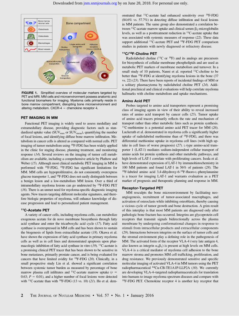
cells accumulate in the bone marrow and disrupt bone homeostasis,leading to bone destruction and marrow failure (Fig. 1). Consequently,
the risk of related skeletal events such as fractures is high in MM
patients and continues to rise even with treatment (7). Malignant
plasma cells are generally avid secretors of immunoglobulins; there-
fore, MM and its obligate precursor state, MGUS, are readily detected
in most cases using serum markers or urine markers (either intact
immunoglobulin or free light chains). However, serum markers areinsufficient to distinguish premalignant MGUS and smoldering MM
from fully transformed MM. The diagnosis of MM requires a high
monoclonal tumor burden or end organ damage such as lytic bone
lesions. Evaluation of progression and treatment response is also con-
founded in the 10% of MM patients who display an oligosecretory
phenotype (defined as serum M-protein, 1 g/dL and urine M-protein
, 200 mg/24 h) (8). The timely and accurate diagnosis of MM isimportant; a delay can be detrimental to the patient’s outcome. Imag-
ing might provide critical information such as predicting high-risk
fracture sites, visualizing nonsecretory and oligosecretory MM tumors,
and assessing treatment response at various stages of disease (9).The current clinical practice for MM includes an initial di-
agnostic full-skeleton radiographic survey for lytic bone lesions(recommended by the International Staging System) (10). This sur-
vey involves acquiring a series of radiographs (plain 2-dimensional
films) to cover the entire skeleton or common anatomic regions
appropriate for clinical indications of the whole spine. Despite the
advantage of this fast, relatively low-cost imaging option, a key
limitation of the radiographic skeletal survey is its low sensitivity
to early osteolytic lesions, as lesions typically can be detected onlyafter 30%–50% of mineralized bone destruction has occurred (11).
Low-dose whole-body (WB) CT is now frequently used in MM and
has higher sensitivity than radiographs for superimposed skeletal
regions such as the scapulae, ribs, and sternum (12). Additionally,
CT is better than conventional radiography for detecting extraosseous
lesions and for radiotherapy planning (13). PET and MRI have high
sensitivity and specificity for providing molecular, functional, andmetabolic information on MM patients. Recent advances in func-
tional PET and MRI for MM are discussed below.
Received Jul. 14, 2015; revision accepted Nov. 2, 2015.For correspondence or reprints contact: Monica Shokeen, Mallinckrodt Institute
of Radiology, Department of Radiology, 4525 Scott Ave., St. Louis, MO 63110.E-mail: [email protected] online Nov. 5, 2015.COPYRIGHT © 2016 by the Society of Nuclear Medicine and Molecular
Imaging, Inc.
MOLECULAR IMAGING OF MULTIPLE MYELOMA • Vij et al. 1
by on June 28, 2018. For personal use only. jnm.snmjournals.org Downloaded from
PET IMAGING IN MM
Functional PET imaging is widely used to assess medullary andextramedullary disease, providing diagnostic factors such as stan-dardized uptake value (SUVmax or SUVmean), quantifying the numberof focal lesions, and identifying diffuse bone marrow infiltration. Me-tabolism in cancer cells is altered as compared with normal cells. PETimaging of tumor metabolism using 18F-FDG has been widely appliedin the clinic for staging disease, planning treatment, and monitoringresponse (14). Several reviews on the imaging of tumor cell metab-olism are available, including a comprehensive article by Plathow andWeber (15). Although most clinical metabolic PET imaging in MM isperformed with 18F-FDG, 18F-FDG has significant limitations forMM. MM cells are hypoproliferative, do not consistently overexpressglucose transporter 1, and 18F-FDG does not easily distinguish betweena benign lesion and a low-metabolism MM lesion. Over a third ofintramedullary myeloma lesions can go undetected by 18F-FDG PET(16). There is an unmet need for myeloma-specific diagnostic imagingagents. New tracers targeting different molecular signatures, and there-fore biologic properties of myeloma, will enhance knowledge of dis-ease progression and lead to personalized patient management.
11C-Acetate PETA variety of cancer cells, including myeloma cells, can metabolize
exogenous acetate for de novo membrane biosynthesis through fattyacid synthase and enter the tricarboxylic acid cycle (17). Fatty acidsynthase is overexpressed in MM cells and has been shown to sustainthe biogenesis of lipids from extracellular acetate (18). Okawa et al.have shown the expression of fatty acid synthase in primary myelomacells as well as in cell lines and demonstrated apoptosis upon phar-macologic inhibition of fatty acid synthase in vitro (19). 11C-acetate isa promising clinical PET tracer that has been shown to be sensitive inbone metastases, primarily prostate cancer, and is being evaluated forcancers that have limited avidity for 18F-FDG (20). Clinically, in asmall prospective study Lin et al. showed a significant correlationbetween systemic tumor burden as measured by percentage of bonemarrow plasma cell infiltrates and 11C-acetate marrow uptake (r 50.63; P 5 0.01), and a higher number of focal lesions were detectedwith 11C-acetate than with 18F-FDG (13 vs. 10) (21). Ho et al. dem-
onstrated that 11C-acetate had enhanced sensitivity over 18F-FDG(84.6% vs. 57.7%) in detecting diffuse infiltration and focal lesionsin MM patients. The same group also demonstrated a correlation be-tween 11C-acetate marrow uptake and clinical serum b2-microglobulinlevels, as well as a posttreatment reduction in 11C-acetate uptake thatwas associated with systemic measures of response (22). These datasupport additional 11C-acetate PET and 18F-FDG PET comparisonstudies in patients with newly diagnosed or refractory disease.
11C/18F-Choline PETRadiolabeled choline (11C or 18F) and its analogs are precursors
for biosynthesis of cellular membrane phospholipids and are used asmetabolic PET markers of membrane metabolism and turnover. In asmall study of 10 patients, Nanni et al. reported 11C-choline to bebetter than 18F-FDG at identifying myeloma lesions in the bone (37vs. 22) (23). There have been reports of incidental findings of MM ora solitary plasmacytoma by radiolabeled choline PET (24). Addi-tional preclinical and clinical evaluations will help correlate myelomahallmarks with choline metabolism and uptake mechanisms.
Amino Acid PETProbes targeted to amino acid transporters represent a promising
class of imaging agents in view of their ability to reveal increasedrates of amino acid transport by cancer cells (25). Tumor uptakeof amino acid tracers primarily reflects the rate and mechanism oftransport rather than other metabolic fates such as protein synthesis.11C-methionine is a potential amino acid PET tracer for MM (26).Luckerath et al. demonstrated in myeloma cells a significantly higheruptake of radiolabeled methionine than of 18F-FDG, and there wasdifferential methionine uptake in myeloma cell lines (with high up-take in cell lines of worse prognosis) (27). L-type amino-acid trans-porter 1 (LAT-1) mediates sodium-independent cellular transport ofamino acids for protein synthesis and other metabolic pathways, andhigh levels of LAT-1 correlate with proliferating cancers. Isoda et al.have demonstrated expression of LAT-1 by immunohistochemistry in100 MM patients and found LAT-1 in 56% of patients (28). The18F-labeled amino acid 3,4-dihydroxy-6-18F-fluoro-L-phenylananineis a tracer for imaging LAT-1 and warrants evaluation as a PETmarker of prognosis and therapeutic planning and response in MM.
Receptor-Targeted PETMM resculpts the bone microenvironment by facilitating neo-
angiogenesis, recruitment of tumor-associated macrophages, andactivation of osteoclasts while inhibiting osteoblasts, thereby causinga vicious cycle of tumor growth and bone destruction. A grim resultof this interplay is that most MM patients are diagnosed only afterpathologic bone fracture has occurred. Integrins are glycoprotein cellreceptors that transmit signals bidirectionally across the plasmamembrane by undergoing conformational changes in response tostimuli from intracellular products and extracellular components(29). Interactions between integrins on the surface of tumor cells andthe stromal environment play a defining role in the pathogenesis ofMM. The activated form of the receptor VLA-4 (very late antigen 4,also known as integrin a4b1) is present at high levels on MM cells.VLA-4 is a critical mediator of myeloma cell adhesion to the bonemarrow stroma and promotes MM cell trafficking, proliferation, anddrug resistance. We previously demonstrated sensitive and specificmolecular imaging of activated VLA-4 in MM tumors using the PETradiopharmaceutical 64Cu-CB-TE1A1P-LLP2A (30). We currentlyare developing VLA-4–targeted radiopharmaceuticals for translationinto humans to image myeloma spectrum diseases and compare with18F-FDG PET. Chemokine receptor 4 is another key receptor that
FIGURE 1. Simplified overview of molecular markers targeted byPET andMRI. MM cells andmicroenvironment possess anatomic andfunctional biomarkers for imaging. Myeloma cells primarily reside inbone marrow compartment, disrupting bone microenvironment andaltering metabolism. CXCR-4 5 chemokine receptor 4.
2 THE JOURNAL OF NUCLEAR MEDICINE • Vol. 57 • No. 1 • January 2016
by on June 28, 2018. For personal use only. jnm.snmjournals.org Downloaded from
plays an important role in MM pathogenesis. Philipp-Abbrederiset al. recently demonstrated imaging of advanced MM in humans usingthe chemokine receptor 4–targeted PET probe 68Ga-pentixafor (31).
MRI IN MM
The role of MRI in imaging MM relies on 2 primary functions:improved sensitivity for detecting pathologic lesions, and the potentialfor predictive and prognostic imaging biomarkers. With regard tosensitivity of disease detection, WB MRI offers high soft-tissuecontrast and high spatial resolution, which in turn yield sensitivitysuperior to that of conventional radiography for visualization of focaland diffuse tumor infiltration of bone marrow in untreated patients(32). The updated criteria for diagnosis of MM by the InternationalMyeloma Working Group recommend MRI as part of the initial as-sessment (3), and MRI is also considered particularly beneficial inpatients with smoldering MM (33). Hillengass et al., in a study of149 patients with asymptomatic MM, demonstrated that patients withmore than one focal lesion had a significantly shorter progression-freesurvival than those without a focal lesion or with only one (P, 0.001)(34). Beyond sensitivity, there has been much interest in developingprognostic and predictive imaging biomarkers using the functionalcapabilities of MRI. One such example is dynamic contrast-enhanced(DCE) MRI using gadolinium-based contrast agents. Increased angio-genesis of the bone marrow is associated with the transition frompremalignant states to MM. In a prospective clinical trial, 30 patientswere evaluated for level of angiogenesis from MGUS to frank malig-nancy (35). The kinetic parameters Ktrans (transendothelial transport ofgadolinium from the vascular compartment to the tumor interstitium[wash in]) and Kep (reverse transport of gadolinium back into thevascular space [washout]) derived from DCE MRI of the lumbarvertebrae were compared with bone marrow microvessel density anda serum panel of 17 angiogenic markers. The study found a moderate-to-strong correlation between marrow microvessel density and Kep inall patients (r5 0.59; P5 0.001) and a weak-to-moderate correlationbetween marrow microvessel density and Ktrans in all patients (r 50.43; P 5 0.03). It should be noted that DCE is not a WB applicationand is done to evaluate a specific anatomic region such as in the case ofa plasmacytoma. To evaluate the cellularity of a lesion or to quantifythe distribution of plasma cells in bone marrow, apparent diffusioncoefficients (ADCs) derived from diffusion-weighted imaging (DWI)sequences are used. DWI can noninvasively quantify altered diffusion,
volume, and flow permeability in new vessels. The relationship be-tween tumor and background ADCs in marrow is complex and de-pends on the degree of marrow activation and the status of the tumor.In a pilot study of 11 patients with metastatic osseous lesions, medianglobal ADCs acquired by semiautomated segmentation of DWI dataallowed for differentiation of responders from nonresponders (36). Aprospective trial of 26 patients with MM and baseline/follow-up WBDWI found a significant change in posttherapy ADCs that was re-producible between multiple interpreters (37). A few additional studieshave shown similar results suggesting that ADC DWI is a potentialresponse biomarker platform (38–40). Although ADC DWI data pro-vide insight into tumor cellularity and disease activity, the interpreta-tion of these images can be complicated by physiologic factors such asage and bone marrow activation due to physical activity and infection.
PET/MRI IN MM
In recent years, simultaneous PET/MRI platforms have becomeavailable for clinical use. These hybrid systems can combine themolecular data of PETwith the anatomic and functional data of MRI.The benefits of simultaneous acquisition are that 2 previously separateexaminations can now be performed in a single imaging session, thereis improved registration between modalities, and dynamic PET andDCE MRI can be done simultaneously. The drawbacks of hybridizingPET with MRI rest mainly on issues related to attenuation correctionof the PET data. MR-based attenuation correction does not take intoaccount cortical bone; however, vendors and researchers are activelyinvestigating the potential impact of this factor on quantitative evalu-ation of osseous lesions while working toward improved technology.With regard to workflow challenges, it is essential to focus on patienttolerance and comfort when designingWB PET/MRI protocols (41). Itis advisable that MRI sequences be minimized to what is essential toanswer the clinical or research question. In the absence of WB PET/MRI, WB PET imaging and MRI of the spine and pelvis are recom-mended. Additionally, any known or suspected areas of disease in-volvement may be targeted for imaging. PET/MRI protocols that arebeing optimized at our institution for prospective use in patients withMGUS, smoldering MM, and MM are summarized in Table 1. Figure2 is an example of a fused PET/MR image showing an active site ofMM involvement in a lumbar vertebral body. Studies evaluating PET/MRI as a diagnostic tool for MM will provide more insight into thebenefits of this promising imaging platform.
TABLE 1MRI Sequences to Include in WB PET/MRI Evaluation of Marrow Lesions
Sequence Recommended use
T1-weighted turbo spin echo Evaluation of cortex (normally dark) and marrow infiltration (marrow darker than normal)
Contrast-enhanced T2-weighted fat
suppression
Evaluation for marrow edema and replacement; a T2-weighted fat-suppressed sequence (areas of edema
and replacement are often brighter than background fat-containing marrow)
T2-weighted half-Fourier acquisition
single-shot fast spin echo
Evaluation of the full body and anatomic detail on organs and soft tissues; a fast-acquisition T2-weighted
sequence
ADC DWI* Evaluation of lesion cellularity; a possible biomarker of treatment response
DCE MRI* Evaluation of limited regions such as in the case of a plasmacytoma; a surrogate for perfusion and
permeability
Attenuation-corrected T1-weighted
Dixon
Creation of a m-map for attenuation correction of PET data; a dual-echo gradient recalled echo sequence
that is acquired at in-phase and opposed-phase echo times with generation of fat-only and water-only
images
*ADC DWI and DCE MRI sequences may be applied in a more focused way to characterize specific sites of disease and potentially add value in assessing
tumor response. DCE is not a WB method.
MOLECULAR IMAGING OF MULTIPLE MYELOMA • Vij et al. 3
by on June 28, 2018. For personal use only. jnm.snmjournals.org Downloaded from
CONCLUSION
Imaging with PET and MRI using molecularly targeted probes isa promising precision medicine platform that might successfullyaddress the clinical ambiguities of myeloma spectrum diseases.
DISCLOSURE
This work is supported by 1R01CA176221 and CTSA UL1TR000448 to Monica Shokeen. No other potential conflict ofinterest relevant to this article was reported.
ACKNOWLEDGMENTS
We thank Drs. Walter J. Akers, Michael H. Tomasson, JonMcConathy, Farrokh Dehdashti, Katherine N. Weilbaecher,and Francesca Fontana for helpful discussions.
REFERENCES
1. Kuehl WM, Bergsagel PL. Molecular pathogenesis of multiple myeloma and its
premalignant precursor. J Clin Invest. 2012;122:3456–3463.
2. Roodman GD. Pathogenesis of myeloma bone disease. J Cell Biochem. 2010;109:283–291.
3. Rajkumar SV, Dimopoulos MA, Palumbo A, et al. International Myeloma Working Group
updated criteria for the diagnosis of multiple myeloma. Lancet Oncol. 2014;15:e538–e548.
4. Teitelbaum A, Ba-Mancini A, Huang H, Henk HJ. Health care costs and resource utilization,
including patient burden, associated with novel-agent-based treatment versus other therapies
for multiple myeloma: findings using real-world claims data. Oncologist. 2013;18:37–45.
5. Kristinsson SY, Anderson WF, Landgren O. Improved long-term survival in multiple
myeloma up to the age of 80 years. Leukemia. 2014;28:1346–1348.
6. Anderson KC. Therapeutic advances in relapsed or refractory multiple myeloma. J Natl
Compr Canc Netw. 2013;11:676–679.
7. Kyle RA, Gertz MA, Witzig TE, et al. Review of 1027 patients with newly diagnosed
multiple myeloma. Mayo Clin Proc. 2003;78:21–33.
8. Wang TF, Ahluwalia R, Fiala MA, et al. The characteristics and outcomes of patients with
multiple myeloma dual refractory or intolerant to bortezomib and lenalidomide in the era of
carfilzomib and pomalidomide. Leuk Lymphoma. 2014;55:337–341.
9. Pianko MJ, Terpos E, Roodman GD, et al. Whole-body low-dose computed tomography
and advanced imaging techniques for multiple myeloma bone disease. Clin Cancer Res.
2014;20:5888–5897.
10. Greipp PR, San Miguel J, Durie BG, et al. International staging system for multiple
myeloma. J Clin Oncol. 2005;23:3412–3420.
11. Healy CF, Murray JG, Eustace SJ, et al. Multiple myeloma: a review of imaging features
and radiological techniques. Bone Marrow Res. 2011;2011:583439.
12. Princewill K, Kyere S, Awan O, et al. Multiple myeloma lesion detection with whole
body CT versus radiographic skeletal survey. Cancer Invest. 2013;31:206–211.
13. Kropil P, Fenk R, Fritz LB, et al. Comparison of whole-body 64-slice multidetector
computed tomography and conventional radiography in staging of multiple myeloma.
Eur Radiol. 2008;18:51–58.
14. Zamagni E, Patriarca F, Nanni C, et al. Prognostic
relevance of 18-F FDG PET/CT in newly diagnosed
multiple myeloma patients treated with up-front
autologous transplantation. Blood. 2011;118:5989–5995.
15. Plathow C, Weber WA. Tumor cell metabolism imaging.
J Nucl Med. 2008;49(suppl 2):43S–63S.
16. van Lammeren-Venema D, Regelink JC, Riphagen II, et al.18F-fluoro-deoxyglucose positron emission tomography in
assessment of myeloma-related bone disease: a systematic
review. Cancer. 2012;118:1971–1981.
17. Lyssiotis CA, Cantley LC. Acetate fuels the cancer
engine. Cell. 2014;159:1492–1494.
18. Wang WQ, Zhao XY, Wang HY, et al. Increased fatty
acid synthase as a potential therapeutic target in multiple
myeloma. J Zhejiang Univ Sci B. 2008;9:441–447.
19. Okawa Y, Hideshima T, Ikeda H, et al. Fatty acid
synthase is a novel therapeutic target in multiple
myeloma. Br J Haematol. 2008;141:659–671.
20. Grassi I, Nanni C, Allegri V, et al. The clinical use of
PET with 11C-acetate. Am J Nucl Med Mol Imaging.
2012;2:33–47.
21. Lin C, Ho CL, Ng SH, et al. 11C-acetate as a new
biomarker for PET/CT in patients with multiple
myeloma: initial staging and postinduction response
assessment. Eur J Nucl Med Mol Imaging. 2014;41:
41–49.
22. Ho CL, Chen S, Leung YL, et al. 11C-acetate PET/CT for metabolic characterization
of multiple myeloma: a comparative study with 18F-FDG PET/CT. J Nucl Med.
2014;55:749–752.
23. Nanni C, Zamagni E, Cavo M, et al. 11C-choline vs. 18F-FDG PET/CT in assessing bone
involvement in patients with multiple myeloma. World J Surg Oncol. 2007;5:68.
24. Calabria F, Chiaravalloti A, Schillaci O. 18F-choline PET/CT pitfalls in image
interpretation: an update on 300 examined patients with prostate cancer. Clin Nucl
Med. 2014;39:122–130.
25. Huang C, McConathy J. Fluorine-18 labeled amino acids for oncologic imaging with
positron emission tomography. Curr Top Med Chem. 2013;13:871–891.
26. Nakamoto Y, Kurihara K, Nishizawa M, et al. Clinical value of 11C-methionine PET/CT
in patients with plasma cell malignancy: comparison with 18F-FDG PET/CT. Eur J Nucl
Med Mol Imaging. 2013;40:708–715.
27. Luckerath K, Lapa C, Spahmann A, et al. Targeting paraprotein biosynthesis for non-
invasive characterization of myeloma biology. PLoS One. 2013;8:e84840.
28. Isoda A, Kaira K, Iwashina M, et al. Expression of L-type amino acid transporter 1
(LAT1) as a prognostic and therapeutic indicator in multiple myeloma. Cancer Sci.
2014;105:1496–1502.
29. Shimaoka M, Springer TA. Therapeutic antagonists and conformational regulation of
integrin function. Nat Rev Drug Discov. 2003;2:703–716.
30. Soodgupta D, Hurchla MA, Jiang M, et al. Very late antigen-4 (a4b1 integrin) targeted
PET imaging of multiple myeloma. PLoS One. 2013;8:e55841.
31. Philipp-Abbrederis K, Herrmann K, Knop S, et al. In vivo molecular imaging of
chemokine receptor CXCR4 expression in patients with advanced multiple myeloma.
EMBO Mol Med. 2015;7:477–487.
32. Dutoit JC, Vanderkerken MA, Verstraete KL. Value of whole body MRI and dynamic
contrast enhanced MRI in the diagnosis, follow-up and evaluation of disease activity
and extent in multiple myeloma. Eur J Radiol. 2013;82:1444–1452.
33. Kristinsson SY, Minter AR, Korde N, Tan E, Landgren O. Bone disease in multiple
myeloma and precursor disease: novel diagnostic approaches and implications on
clinical management. Expert Rev Mol Diagn. 2011;11:593–603.
34. Hillengass J, Fechtner K, Weber MA, et al. Prognostic significance of focal lesions in
whole-body magnetic resonance imaging in patients with asymptomatic multiple
myeloma. J Clin Oncol. 2010;28:1606–1610.
35. Bhutani M, Turkbey B, Tan E, et al. Bone marrow angiogenesis in myeloma and its
precursor disease: a prospective clinical trial. Leukemia. 2014;28:413–416.
36. Blackledge MD, Collins DJ, Tunariu N, et al. Assessment of treatment response by total
tumor volume and global apparent diffusion coefficient using diffusion-weighted MRI
in patients with metastatic bone disease: a feasibility study. PLoS One. 2014;9:e91779.
37. Giles SL, Messiou C, Collins DJ, et al. Whole-body diffusion-weighted MR imaging for
assessment of treatment response in myeloma. Radiology. 2014;271:785–794.
38. Horger M, Weisel K, Horger W, et al. Whole-body diffusion-weighted MRI with
apparent diffusion coefficient mapping for early response monitoring in multiple
myeloma: preliminary results. AJR. 2011;196:W790–W795.
39. Hillengass J, Bauerle T, Bartl R, et al. Diffusion-weighted imaging for non-invasive and
quantitative monitoring of bone marrow infiltration in patients with monoclonal plasma
cell disease: a comparative study with histology. Br J Haematol. 2011;153:721–728.
40. Messiou C, Giles S, Collins DJ, et al. Assessing response of myeloma bone disease with
diffusion-weighted MRI. Br J Radiol. 2012;85:e1198–e1203.
41. Martinez-Moller A, Eiber M, Nekolla SG, et al. Workflow and scan protocol
considerations for integrated whole-body PET/MRI in oncology. J Nucl Med.
2012;53:1415–1426.
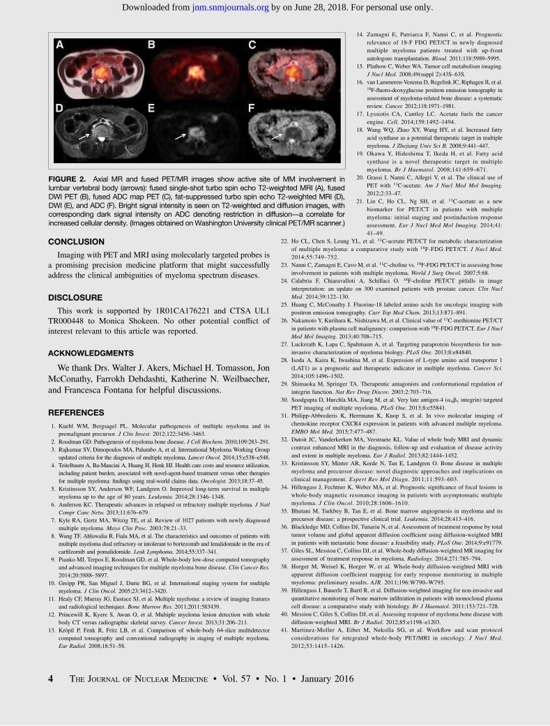
FIGURE 2. Axial MR and fused PET/MR images show active site of MM involvement inlumbar vertebral body (arrows): fused single-shot turbo spin echo T2-weighted MRI (A), fusedDWI PET (B), fused ADC map PET (C), fat-suppressed turbo spin echo T2-weighted MRI (D),DWI (E), and ADC (F). Bright signal intensity is seen on T2-weighted and diffusion images, withcorresponding dark signal intensity on ADC denoting restriction in diffusion—a correlate forincreased cellular density. (Images obtained onWashington University clinical PET/MR scanner.)
4 THE JOURNAL OF NUCLEAR MEDICINE • Vol. 57 • No. 1 • January 2016
by on June 28, 2018. For personal use only. jnm.snmjournals.org Downloaded from
Doi: 10.2967/jnumed.115.163808Published online: November 5, 2015.
2016;57:1-4.J Nucl Med. Ravi Vij, Kathryn J. Fowler and Monica Shokeen New Approaches to Molecular Imaging of Multiple Myeloma
http://jnm.snmjournals.org/content/57/1/1This article and updated information are available at:
http://jnm.snmjournals.org/site/subscriptions/online.xhtml
Information about subscriptions to JNM can be found at:
http://jnm.snmjournals.org/site/misc/permission.xhtmlInformation about reproducing figures, tables, or other portions of this article can be found online at:
(Print ISSN: 0161-5505, Online ISSN: 2159-662X)1850 Samuel Morse Drive, Reston, VA 20190.SNMMI | Society of Nuclear Medicine and Molecular Imaging
is published monthly.The Journal of Nuclear Medicine
© Copyright 2016 SNMMI; all rights reserved.
by on June 28, 2018. For personal use only. jnm.snmjournals.org Downloaded from