Embed Size (px)
Citation preview

Neutrophilic Inflammatory Response and Oxidative Stressin Premenopausal Women Chronically Exposed to IndoorAir Pollution from Biomass Burning
Anirban Banerjee,1 Nandan Kumar Mondal,1,2 Debangshu Das,1 and Manas Ranjan Ray1
Abstact—The possibility of inflammation and neutrophil activation in response to indoor air poll-ution (IAP) from biomass fuel use has been investigated. For this, 142 premenopausal, never-smoking women (median age, 34 years) who cook exclusively with biomass (wood, dung, cropwastes) and 126 age-matched control women who cook with cleaner fuel liquefied petroleum gas(LPG) were enrolled. The neutrophil count in blood and sputum was significantly higher (p<0.05)in biomass users than the control group. Flow cytometric analysis revealed marked increase in thesurface expression of CD35 (complement receptor-1), CD16 (FCγ receptor III), and β2 Mac-1integrin (CD11b/CD18) on circulating neutrophils of biomass users. Besides, enzyme-linked imm-unosorbent assay showed that they had 72%, 67%, and 54% higher plasma levels of the proinfla-mmatory cytokines tumor necrosis factor-alpha, interleukin-6, and interleukin-12, respectively, anddoubled neutrophil chemoattractant interleukin-8. Immunocytochemical study revealed significantlyhigher percentage of airway neutrophils expressing inducible nitric oxide synthase, while the serumlevel of nitric oxide was doubled in women who cooked with biomass. Spectrophotometric analysisdocumented higher myeloperoxidase activity in circulating neutrophils of biomass users, suggestingneutrophil activation. Flow cytometry showed excess generation of reactive oxygen species (ROS)by leukocytes of biomass-using women, whereas their erythrocytes contained a depleted level ofantioxidant enzyme superoxide dismutase (SOD). Indoor air of biomass-using households had twoto four times more particulate matter with diameters of <10 μm (PM10) and <2.5 μm (PM2.5) asmeasured by real-time laser photometer. After controlling potential confounders, rise in proin-flammatory mediators among biomass users were positively associated with PM10 and PM2.5 inindoor air, suggesting a close relationship between IAP and neutrophil activation. Besides, the levelsof neutrophil activation and inflammation markers were positively associated with generation ofROS and negatively with SOD, indicating a role of oxidative stress in mediating neutrophilic inf-lammatory response following chronic inhalation of biomass smoke.
KEY WORDS: biomass fuel; neutrophil; inflammation; oxidative stress; premenopausal women; India.
INTRODUCTION
A large majority of rural women of the developingworld including India are chronically exposed to highlevels of indoor air pollution (IAP) from daily householdcooking with traditional biomass fuel (BMF) such asdried cow dung cake, firewood, and agricultural wastes.Smoke emitted from burning biomass contains a wide
ABBREVIATIONS: ACD, acid citrate dextrose; BMF, biomass fuel;BSA, bovine serum albumin; DCF-DA, dichlorofluorescein diacetate;EDTA, ethylenediaminetetraacetic acid; ELISA, enzyme-linked immu-nosorbent assay; FACS, fluorescence-activated cell sorter; FITC,fluorescein isothiocyanate; HRP, horseradish peroxidase; IAP, indoorair pollution; ICC, immunocytochemistry; IL, interleukin; iNOS,
inducible nitric oxide synthase; LPG, liquefied petroleum gas; MFI,mean fluorescence intensity; MPO, myeloperoxidase; NO, nitric oxide;PAP, Papanicolaou; PBS, phosphate-buffered saline; PE, phycoerythrin;PM, particulate matter; ROS, reactive oxygen species; SOD, super-oxide dismutase; TNF, tumor necrosis factor
Anirban Banerjee and Nandan Kumar Mondal contributed equally tothis work.
1 Department of Experimental Hematology, Chittaranjan NationalCancer Institute, 37, S. P. Mukherjee Road, Kolkata, 700 026, India
2 To whom correspondence should be addressed at Department ofExperimental Hematology, Chittaranjan National Cancer Institute, 37,S. P. Mukherjee Road, Kolkata, 700 026, India. E-mail: [email protected]
0360-3997/12/0200-0671/0 # 2011 Springer Science+Business Media, LLC
Inflammation, Vol. 35, No. 2, April 2012 (# 2011)DOI: 10.1007/s10753-011-9360-2
671

spectrum of pollutants that include carbon monoxide,coarse, fine, and ultrafine particulate matters (PM),oxides of nitrogen and sulfur, formaldehyde, transitionalmetals, and volatile organic compounds includingbenzene and polycyclic aromatic hydrocarbons such asbenzo(a)pyrene [1]. Some of these pollutants like benzo(a)pyrene and benzene are potent human carcinogens[2]. Women who used to cook with BMF in poorlyventilated kitchen for 2–6 h/day are believed to beinhaling carcinogens equivalent to smoking two packs ofcigarettes per day [3]. Epidemiological studies haveshown that exposure to fine particles promotes inflam-mation in the lung [4–9] via activation of alveolarmacrophages and lung epithelial cells [10, 11]. Suchexposures may also elicit systemic inflammatoryresponse [12], associated with increased levels of severalcytokines (interleukin-6 and -1b) in the bloodstream [13]as well as increased production, release, and activationof neutrophils from the bone marrow [14].
Neutrophils are the most abundant leukocytespresent in human blood. These cells are important forphagocytosis and destruction of invading pathogens,especially bacteria [15]. They are among the first cells toleave the bloodstream and reach the tissues in case oftissue injury [16]. During the beginning of an acutephase of inflammation, particularly as a result ofbacterial infection, environmental exposure, and somecancers, neutrophils are among the first responders thatmigrate towards the site of inflammation. Such migrationof blood leukocytes to the tissues in order to fightagainst invading pathogens and to mediate tissue repairis known as inflammation. Neutrophils have beenimplicated in the development of bronchial hyperres-ponsiveness [17]. Neutrophilic inflammation in theairways and the bronchial submucosa has been reportedin patients with severe asthma [18, 19], especially duringexacerbation [20]. Neutrophil elastase has the capacity toinduce mucus gland hyperplasia and mucus secretion[21] and proliferation or apoptosis of airway smoothmuscle cells [22, 23]. Myeloperoxidase (MPO) is anenzyme found in myeloid cells, particularly in neutro-phils, and to a lesser extent in monocytes and tissuemacrophages. It plays an important role in the hostdefense against bacteria and viruses. In neutrophils,MPO is located within the primary granules and itsconcentration is used as an indicator of neutrophilactivation and corresponding inflammatory response[24]. Neutrophil migration from circulation into an areaof inflammation involves regulated expression of anumber of adhesion molecules on the cell surface.
Upregulation of these surface molecules has beenreported in patients with chronic obstructive pulmonarydisease (COPD) [25]. A recent study has shown allergicpulmonary inflammation following exposure to dieselexhaust particles [26]. Moreover, studies in laboratoryanimals have documented lung inflammation induced byair pollution [27]. In view of these reports, it isreasonable to assume that IAP from biomass burningmay lead to the activation of circulating neutrophils andconsequent inflammatory response in women who cookwith these fuels.
Neutrophils in peripheral blood may be activated orprimed with regard to superoxide production, chemotaxis,and increased expression of surface molecules such asCD35 which mediates the binding and phagocytosis ofC3b-coated particles and immune complexes [28]. Thestudy of Smith et al. [29] suggested that upregulation ofCD11b expression may be associated with neutrophilsdegranulation. Data from studies of pathological condi-tions suggest that neutrophil receptor expression is alteredin response to inflammatory stimuli and tissue injury. Forexample, the expression of CD11b and CD35 receptors isenhanced in patients suffering sepsis. In contrast, theexpression of CD11b, CD35, and CD16 is reduced inpatients who have experienced acute trauma and burns.These alterations may represent adaptive responses tocontrol the level of neutrophils and complement activationduring these pathological conditions [30–32]. Alterationsin neutrophil receptor expression could also affect neu-trophil functional activity. For example, a reduction in theneutrophil expression of CD11b and CD16 in patients withthermal injuries has been associated with defects inrespiratory burst activity, and this has resulted in anincreased susceptibility to infection [33]. Neutrophilsrespond to infection and tissue injury by recognizing andbinding immunoglobulin G (IgG) molecules and comple-ment proteins that coat the surface of foreign pathogensand host tissue fragments. This recognition-binding proc-ess is mediated by neutrophil surface receptors, includingthe complement receptor 1 CD35, the complementreceptor 3 CD11b, and the low-affinity IgG receptorCD16. The engagement of these surface receptors initiatesa cascade of intracellular events leading to the release ofenzymes (degranulation) and reactive oxygen species(ROS; respiratory burst activity) from neutrophils [34].Together, these enzymes and ROS assist in the destructionand degradation of foreign pathogens and damaged tissuefragments [35].
Our earlier studies have linked IAP from BMF withupregulation of ribosome biogenesis [36, 37], inappro-
672 Banerjee, Mondal, Das, and Ray

priate response of DNA damage repair proteins in theface of increased DNA damage [38, 39], overactivationof signal transduction pathway involving Akt, i.e.,protein kinase B [40], and hypertension with elevatedlevels of oxidized low-density lipoprotein indicatingcardiovascular disease [41]. These findings along withobservations from other studies led us to hypothesizethat chronic exposure to IAP from biomass burningcould be associated with neutrophilic inflammatoryresponses and oxidative stress in premenopausal womenof rural India who cook regularly with BMF. To ourknowledge, no such study has been carried out in Indiaor any other developing country where biomass isextensively used as a major source of household energy.Against this background, we examined in this study theimpact of IAP from BMF use on neutrophil activation ina group of never-smoking, premenopausal women fromeastern India who used to cook exclusively with BMFfor the past 5 years or more. We have compared thefindings with an age-matched group of control womenfrom same neighborhood who cooked with cleaner fuelliquefied petroleum gas (LPG).
MATERIALS AND METHODS
Subjects
A total of 268 premenopausal women aged between23 and 41 years from rural areas of West Bengal, a statein eastern India, were enrolled in this study afterobtaining written informed consent. They attendedhealth checkup camps organized in different villageswith the active cooperation of the local administrativebodies and nongovernmental organizations. Among theparticipants, 142 women (age, 23–40 years; median age,34 years) were cooking daily for 2.5–5.5 h exclusivelywith wood, cow dung, and agricultural refuse, such asbamboo, jute stick, paddy husk, hay, and dried leaves forthe past 5 years or more. Accordingly, they weregrouped as biomass users. The remaining 126 women(age, 24–41 years; median age, 33 years) were from thesame villages but they used to cook with cleaner fuelLPG and were considered as control.
Inclusion and Exclusion Criteria
The inclusion criteria were apparently healthy,premenopausal married women actively engaged inhousehold cooking for the past 5 years or more, whowere nonsmokers and nonchewers of tobacco, and had a
body mass index (BMI) >15 and <30 kg/m2. Womenusing oral contraceptive, had a history of malignancy, orwere currently under medication were excluded.
During personal interview with female members ofthe research team, each participant was requested tofurnish information about age, education, family sizeand income, habit, cooking time per day, years ofcooking, fuel and oven type, ventilation and location ofkitchen, and general health problems in the past 3 monthsand last 1 year. As most of the participants were poorlyeducated, the researchers recorded their responses instructured questionnaire forms on their behalf. TheEthics Committee of Chittaranjan National CancerInstitute approved the study protocol.
Collection of Blood and Expectorated Sputum
Venous blood (5 ml) was collected in vacutainer tubes(Becton Dickinson [BD], USA) containing K2EDTA asanticoagulant. Blood was collected at a fixed time of theday (09:30AM – 10:30 AM) tominimize diurnal variation.A drop of blood was used for smear preparation on cleanglass slide for each individual. An aliquot of whole bloodwas mixed with acid citrate dextrose (ACD) solution in theratio of 5:1 separately in sterile vacutainer tubes forisolation of circulation neutrophils. Blood in the no-additive vacutainer was allowed to clot and serum wascollected. Plasma was obtained by centrifugation at2,500×g for 10 min at 4°C.
Early morning expectorated sputa were collected insterile plastic cups for three consecutive days to harvestairway cells following the procedure of Erkilic et al.[42]. Two smears were prepared from the nontransparenthighly viscous part of each freshly collected sputumsample on clean glass slides from each day’s sample—one for Papanicolaou (PAP) staining and the other forimmunocytochemistry (ICC) of inducible nitric oxidesynthase (iNOS). After air drying, one slide marked forPAP staining was fixed with 95% ethanol and the otherfor ICC were fixed in ice-cold methanol for 20 min atthe site of collection. The remaining part of theexpectorated sputum was collected in sterile plasticscrew-cap tubes containing 20 ml of phosphate-bufferedsaline (PBS) with 0.1% dithiotheritol.
Routine Hematology
Differential counts of leukocytes were done fromLeishman-stained blood films. Routine hematologicalparameters, such as total and differential counts of whiteblood cells and total count of platelets, were carried out
673Neutrophilic Inflammatory Response and Oxidative Stress

using a hemocytometer under light microscope (Leitz,Germany).
PAP Staining for Routine Sputum Cytopathology
Ethanol-fixed slides were brought to the laboratoryand were stained with PAP following the procedure ofHughes and Dodds [43]. The slides were coded andexamined under light microscope (Leitz, Germany) at×400 and ×1,000 magnifications. At least 10 high-powerfields (hpf; ×40 objective and ×10 eyepiece) per slidewere examined. Sputum cytology and differential dis-tribution of inflammatory cells were performed follow-ing the established criteria [44].
Isolation of Circulating Neutrophils
Neutrophils were isolated following the procedureof Drabikova et al. [45] with slight modifications. Inbrief, blood in ACD was added with 6% dextransolution in 0.9% NaCl (blood–dextran ratio, 1:2 v/v).Then, it was allowed to sediment for 45 min at roomtemperature. The supernatant was centrifuged at 700×gfor 12 min at 4°C, the cell pellet was reconstituted in1.2 ml ice-cold distilled water, and within 20 s, 400 μl of0.6 M KCl was added and mixed. It was diluted to 5 mlwith PBS and kept in ice for 5 min for red blood cell(RBC) lysis. It was then centrifuged at 770×g for 6 minat 4°C. Supernatant was discarded and pellet resus-pended in 1–2 ml of ice-cold PBS. Next, the cellsuspension was layered over 3 ml of Ficoll–Histopaque(Sigma Aldrich, USA) in a 15-ml tube and centrifuged at1,200×g for 30 min. After discarding the supernatant,the pellet was dissolved in 500 μl of ice-cold Hank’sbalanced salt solution. About 99% of neutrophils wereisolated by this procedure and viability of the cells was>90% in trypan blue dye exclusion test.
Assessment of the Expression of Surface Moleculesby Flow Cytometry
Whole blood (25 μl) was added to polypropylenetubes and incubated for 20 min at room temperature inthe dark with FITC-conjugated antihuman CD11b,CD18, and CD35 monoclonal antibodies, PE-conjugatedantihuman CD16 monoclonal antibody (BD, USA), andisotype controls. Thereafter, the erythrocytes were lysedwith 2 ml of 1X FACS lysing solution (BD, USA), andsamples were centrifuged at 500×g for 5 min. The cellpellet was washed with ice-cold PBS containing 0.1%sodium azide and resuspended in 500 μl of 1%
paraformaldehyde in PBS. The samples were analyzedin a flow cytometer (FACSCalibur with Sorter, BD, SanJose, CA, USA) using Cell Quest software (BD, USA).A gate was drawn around the granulocytes showingneutrophils as CD16-positive cells with eosinophils asCD16-negative cells with high side scatter. A furthergate was then drawn around the neutrophil (CD16+) oreosinophil (CD16−) populations and reflected into ahistogram so that cells could be labeled with a furtherfluorochrome (e.g., CD11b or CD18). Fluorescence wasmeasured with a FACSCalibur flow cytometer and CellQuest software (BD, USA). Measurements were madeon the FL1 (green) and FL2 (red) channels, and the gateswere adjusted to the negative control quadrant. A total of15,000 events were collected. Results were recorded asmean fluorescence intensity (MFI), which represents thecell surface receptor density.
Measurement of Proinflammatory Cytokineand Chemokine by ELISA
The levels of proinflammatory cytokine tumornecrosis factor-alpha (TNF-α), interleukin-6 (IL-6),interleukin-12 (IL-12), and chemokine interleukin-8(IL-8) were measured in blood plasma and nitric oxide(NO) level in serum by ELISA using commerciallyavailable kits of BD Biosciences Pharmingen, SanDiego, USA (for TNF-α, IL-6, and IL-12), RocheDiagnostics GmbH, Germany (for IL-8), and OxfordBiomedical Research, Oxford, MI, USA (for NO). Theassays were performed following the procedures recom-mended by the manufacturers. The instructions of themanufacturers were followed during the study andreadings were taken in microplate reader (Bio-Rad,USA) at 450 nm.
Immunocytochemical Localization of iNOS
ICC of iNOS in isolated blood neutrophils andsputum cells were done following a standard procedure[38, 40]. In brief, slides were air dried and fixed in ice-cold methanol for 30 min, air dried, washed in PBSthrice, and blocked in 3% BSA (Sigma-Aldrich Chem-icals, Saint Louis, MO, USA) for 1 h at room temper-ature. The slides were overnight incubated in darknesswith rabbit polyclonal iNOS (Santa Cruz, CA, USA)primary antibody (diluted 1:100 in 1% BSA, respec-tively) in a humid box at 4°C. After washing with PBS,slides were incubated with antirabbit IgG, F(ab′)2-HRP(Santacruz, USA) secondary antibody diluted 1:500 in1% BSA for 90 min. The slides were developed by
674 Banerjee, Mondal, Das, and Ray

incubating with substrate for HRP for 45 min indarkness followed by washing with distilled water andcounterstaining with hematoxylin, dehydration in gradedethanol, and mounted in distrene plasticizer xylene. Theslides were coded and examined blindly. For eachparticipant, one blood neutrophil sample and three sputasamples obtained from three consecutive days wereexamined. The 3-day average values of iNOS expres-sions in sputum cells were taken as the representativedata of each participant.
Quantitative Measurement of MPO Activityby Spectrophotometry
MPO activity in isolated blood neutrophil homoge-nate was determined by a modification of the assaymethod involving O-dianisidine [46]. The principleinvolves oxidation of O-dianisidine by MPO present inthe isolated neutrophil homogenate to produce a colorcomplex which is read spectrophotometrically at460 nm. The higher the MPO activity, the more oxygenreleased from hydrogen peroxide; hence, more oxidationof O-dianisidine and consequently higher observedspectrophotometer reading at 460 nm. The assay mixturecontained 0.3 ml of 0.1 M phosphate buffer (pH 6.0),0.3 ml of 0.01 M H2O2, 0.5 ml 0.02 M of O-dianisidine(Sigma Chem, USA; freshly prepared in deionizedwater), and 10 μl neutrophil homogenate in a finalvolume of 3 ml. The neutrophil homogenate was addedlast in a cuvette with a path length of 1 cm and thechange in absorbance in 460 nm was followed for10 min. One unit of MPO is defined as that giving anincrease in absorbance of 0.001 per minute and specificactivity is given as international units per milligram ofprotein.
Flow Cytometric Measurement of ROS Generation
Generation of ROS was measured in leukocytespresent in anticoagulated venous blood and sputum cellsby flow cytometry using DCF-DA (Sigma Chem, USA)following the procedure of Rothe and Valet [47] withslight modifications [40]. For this, 10,000 events wereacquired in a flow cytometer (FACSCalibur with sorter,Becton Dickinson [BD], San Jose, CA, USA) using CellQuest software (BD). Respiratory burst and generationof ROS by cells were associated with emission of greenfluorescence that was recorded in fluorescence channel 1and was expressed as MFI in arbitrary units. In case ofblood, granulocytes, monocytes, and lymphocytes weregated on the basis of their characteristic forward and side
scatters on dot plot and the MFI of each population wasrecorded, while the MFI of the total cells present insputum was recorded.
Spectrophotometric Measurement of SOD
The activity of the antioxidant enzyme superoxidedismutase (SOD) was assayed in blood erythrocytes andsputum cell lysate spectrophotometrically following theprocedure of Paoletti et al. [48]. Whole sputum collectedin PBS with 0.1% dithiotheritol were used for cell lysatepreparation for SOD assay followed by our previouslypublished procedure [40]. Lysates were centrifuged at15,000×g for 10 min at 4°C in an Eppendorf micro-centrifuge. The supernatant was collected and used forSOD assay. Ethylenediaminetetraacetic acid (EDTA)-anticoagulated blood was centrifuged at 200×g at 4°Cfor 5 min, the RBC pellet was collected and washed in0.9% saline and then lysed with cold distilled water (1:9,v/v). Following centrifugation at 500×g for 10 min, thesupernatant was collected. In spectrophotometric cuv-ette, 800 ml of triethanolamine (TEA)–diethanolamine(DEA) buffer containing 100 mM each of TEA andDEA (Qualigens, Mumbai, India), 40 ml of 7.5 mMnicotinamide adenine dinucleotide reduced disodium salt(NADH, SRL, Mumbai, India), pH 7.4, 25 ml of amixture (1:1, v/v) of 0.2 M EDTA disodium salt (Sigma-Aldrich Chem, St. Louis, MO, USA) and 0.1 Mmanganous chloride (SD-Fine Chem, Mumbai, India),and 100 ml of sample (RBC or sputum cell lysate) wereadded and mixed well. The absorbance (OD) wasmeasured at 340 nm in a spectrophotometer (Shimadzu,Kyoto, Japan) immediately (0 min) and 1, 2, 3, 4, and5 min after the addition of mercaptoethanol. SODactivity was calculated from the standard curve andwas expressed as units per milliliter.
Measurement of PM10 and PM2.5 in Indoor Air
PM with aerodynamic diameter less than 10 mm(PM10) and 2.5 mm (PM2.5) were measured in thecooking areas with real-time laser photometer (Dust-Trak™ Aerosol Monitor, model 8520, TSI Inc., Shore-view MN, USA) that contained 10-mm nylon Dor-Oliver cyclone and operated at a flow rate of 1.7 L/min,measuring particle load in the concentration range of 1to 100 mg/m3. The monitor was calibrated to thestandard ISO 12103-1 A1 test dust. We used twomonitors for simultaneous measurement of PM10 andPM2.5. Air sampling was done in each household forthree consecutive days, 8 h/day (07:00 AM – 03:00 PM)
675Neutrophilic Inflammatory Response and Oxidative Stress

covering both cooking and noncooking time. The meanof 3 days was used as the indoor air quality of a singlehousehold. Biomass-using women cook in a sittingposition 2–3 ft away from the open chullah (oven).Accordingly, the monitor was placed in the breathingzone of the cook 2.5 ft above the floor level on a woodenstool 3 ft away from the chullah. LPG users, on the otherhand, cook in a standing position, and the monitor was,therefore, placed at a height of 4.5 ft. Since laserphotometers make an overestimation of PM levels bytwofold to threefold [49], the raw data require reductionusing correction factors. We reduced the 8-h raw data bydividing with a correction factor of 2.5 for PM10 [50]and 2.77 for PM2.5 [49, 51].
Statistical Analysis of Data
Results are presented as mean±standard deviation(SD) or median with range. Analysis between groupswas performed using Student’s t test, chi-square test, orMann–Whitney U test, as applicable. The possibility ofan association between measured parameters with age,BMI, family income, education, cooking hours per day,lifetime duration of cooking (cooking years), kitchenlocation, family size, number of smokers in the family,use of mosquito repellant, and PM10 and PM2.5 levels incooking areas was tested using univariate regressionanalysis. Variables that showed significant associationwere later included in a backward stepwise multipleregression model to adjust for their effects. Statisticalanalyses were performed using EPI info 6 and SPSSstatistical software (Statistical Package for Social Scien-ces for Windows, release 10.0, SPSS Inc., Chicago,USA) and p<0.05 was considered significant.
RESULTS
Demography
Demographic and socioeconomic characteristics ofthe study population are summarized in Table 1. TheLPG and BMF users were comparable with respect toage, BMI, cooking years, hours of cooking per day,family income, environmental tobacco smoke for thepresence of a smoker in the family, food habit, and useof mosquito repellant. However, BMF users were lesseducated (p<0.05 in Mann–Whitney U test) and theirfamily income was significantly lower than that of theirneighbors who used to cook with LPG (p<0.05 inStudent’s t test). Moreover, 40.7% of BMF-using house-
holds did not possess a separate kitchen and they used tocook in an area adjacent to the living room. In contrast,only 15.3% of LPG-using households lacked a separatekitchen (p<0.05 in chi-square test).
Particulate Pollutants in Indoor Air
The mean concentration of PM10 in biomass-usingkitchen during cooking hours was 625±150 μg/m3 (range,479–1,556 μg/m3) in contrast to 129±42 μg/m3 (range,97–254 μg/m3) in LPG-using kitchen. The correspondingPM2.5 level was 312±85 μg/m3 (range, 262–715 μg/m3)and 77±29 μg/m3 (range, 46–117 μg/m3). Duringnoncooking hours, PM10 and PM2.5 concentrations were204±98 μg/m3 (range, 142–311 μg/m3) and 82±21 μg/m3
(range, 66–126 μg/m3), respectively, in biomass-usingkitchen compared with 93±41 μg/m3 (range, 45–145 μg/m3) and 45±22 μg/m3 (range, 24–72 μg/m3) in LPG-usingkitchen. Thus, the particulate pollution levels of cookingareas in biomass-using households were significantlyhigher than those in LPG-using households duringcooking and noncooking times (p<0.001).
Hematology and Sputum Cytology
The total leukocyte count in peripheral blood wassignificantly higher (p<0.05 in Student’s t test) inBMF users compared with LPG-using controls (8.7±0.72 vs. 5.2±0.83×10³/μl, respectively). Moreover,BMF users had elevated number of total cells insputum (88.9±23.7 vs. 65.2±13.1 cells/hpf, p<0.05)than controls. Blood and sputa of BMF userscontained more inflammatory cells like neutrophils(blood, 5,867±445 vs. 3,708±281 cells/μl, p<0.01;sputum, 58.9±17.4 vs. 42.3±10.6 cells/hpf, p<0.01),eosinophils (blood, 552±63 vs. 300±48 cells/μl, p<0.01; sputum, 1.7±0.7 vs. 0.4±0.2 cells/hpf, p<0.01),and lymphocytes (blood, 2,100±244 vs. 1,929±207cells/μl, p>0.05; sputum, 4.6±1.3 vs. 3.3±1.0 vs.cells/hpf, p<0.05).
Higher Surface Expression of CD16, CD35,and CD11b/CD18 in Circulating Neutrophilsof Biomass Users
Flow cytometric analysis revealed significantlyhigher MFI of CD16, CD35, CD11b, and CD18 incirculating neutrophils of BMF users when comparedwith that of LPG-using controls (p<0.0001; Fig. 1). Inbiomass users, the MFI of neutrophilic surface receptorCD16 and CD35 was increased by 2.5-fold (3,196.1±
676 Banerjee, Mondal, Das, and Ray

337.5 vs. 1,282.1±613.3 in controls) and 1.4-fold(81.9±9.5 vs. 56.7±4.2), respectively. Moreover, theMFI of CD11b and CD18 on neutrophils was increasedby 37% and 66%, from 736.3±278.4 and 217.9±93.1 incontrols to 1,008.2±234.3 and 362.1±79.7 in biomassusers, respectively (Fig. 1).
Rise in Proinflammatory Cytokines and Chemokinein Biomass Users
Table 2 shows significantly elevated levels ofproinflammatory cytokines TNF-α, IL-6, and IL-12
and chemokine IL-8 in plasma of BMF users incomparison with that of LPG users. Compared withLPG-using control women, biomass users had 72%,67%, and 54% higher plasma levels of TNF-α, IL-6, andIL-12, respectively. Moreover, the plasma level of IL-8was doubled in biomass users (Table 2).
Overexpression of iNOS and NO in Biomass Users
iNOS enzyme was found located in the cyto-plasm of the neutrophils. Compared with LPG users,the percentage of iNOS-expressing isolated blood
Table 1. Demographic and Socioeconomic Characteristics of BMF- and LPG-using Women
Variable LPG users (n=126) BMF users (n=142) p value
Age in years, median (range) 33 (24–41) 34 (23–40) NSMedian BMI (kg/m2) 24.3 23.9 NSCooking years, median (range) 13 (5–25) 14 (5–22) NSCooking hours per day, median (range) 3 (2.5–5.5) 3 (2.5–5.5) NSYears of schooling, median (range) 8 (3–14) 3 (0–10) <0.05Homes with separate kitchen (%) 84.7 59.3 <0.05Smoker in family (%) 43.9 44.1 NSUse of mosquito repellant at home (%) 75.8 74.1 NSFood habit, mixed (%) 97.2 96.9 NSMembers in family, median (range) 4 (2–6) 4 (2–7) NSFamily income per month in US dollars (mean±SD) 58.41±8.32 31.78±7.17 <0.05
The LPG- and biomass-using groups were compared by chi-square test (for results presented as percentages), Mann–Whitney U test (for medianvalues with range), and Student’s t test (for mean±SD) and p<0.05 was considered significantn number of subjects, NS statistically not significant
Fig. 1. Histograms showing the MFI of surface expressions of CD16 (a), CD35 (b), and β2 Mac-1 integrin (CD11b/CD18; c, d) on circulatingneutrophils of rural women who cooked exclusively either with biomass or LPG for the past 5 years or more. Bars represent the SD of the mean;*p<0.0001, compared with the LPG users.
677Neutrophilic Inflammatory Response and Oxidative Stress
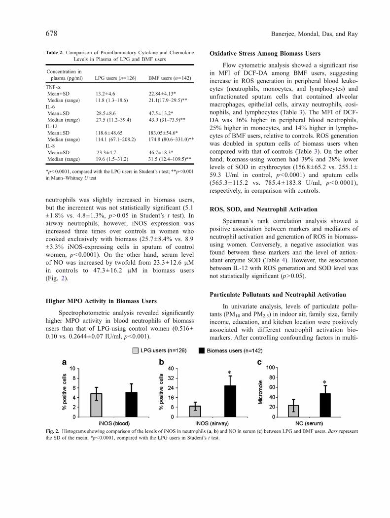
neutrophils was slightly increased in biomass users,but the increment was not statistically significant (5.1±1.8% vs. 4.8±1.3%, p>0.05 in Student’s t test). Inairway neutrophils, however, iNOS expression wasincreased three times over controls in women whocooked exclusively with biomass (25.7±8.4% vs. 8.9±3.3% iNOS-expressing cells in sputum of controlwomen, p<0.0001). On the other hand, serum levelof NO was increased by twofold from 23.3±12.6 μMin controls to 47.3±16.2 μM in biomass users(Fig. 2).
Higher MPO Activity in Biomass Users
Spectrophotometric analysis revealed significantlyhigher MPO activity in blood neutrophils of biomassusers than that of LPG-using control women (0.516±0.10 vs. 0.2644±0.07 IU/ml, p<0.001).
Oxidative Stress Among Biomass Users
Flow cytometric analysis showed a significant risein MFI of DCF-DA among BMF users, suggestingincrease in ROS generation in peripheral blood leuko-cytes (neutrophils, monocytes, and lymphocytes) andunfractionated sputum cells that contained alveolarmacrophages, epithelial cells, airway neutrophils, eosi-nophils, and lymphocytes (Table 3). The MFI of DCF-DA was 36% higher in peripheral blood neutrophils,25% higher in monocytes, and 14% higher in lympho-cytes of BMF users, relative to controls. ROS generationwas doubled in sputum cells of biomass users whencompared with that of controls (Table 3). On the otherhand, biomass-using women had 39% and 28% lowerlevels of SOD in erythrocytes (156.8±65.2 vs. 255.1±59.3 U/ml in control, p<0.0001) and sputum cells(565.3±115.2 vs. 785.4±183.8 U/ml, p<0.0001),respectively, in comparison with controls.
ROS, SOD, and Neutrophil Activation
Spearman’s rank correlation analysis showed apositive association between markers and mediators ofneutrophil activation and generation of ROS in biomass-using women. Conversely, a negative association wasfound between these markers and the level of antiox-idant enzyme SOD (Table 4). However, the associationbetween IL-12 with ROS generation and SOD level wasnot statistically significant (p>0.05).
Particulate Pollutants and Neutrophil Activation
In univariate analysis, levels of particulate pollu-tants (PM10 and PM2.5) in indoor air, family size, familyincome, education, and kitchen location were positivelyassociated with different neutrophil activation bio-markers. After controlling confounding factors in multi-
Table 2. Comparison of Proinflammatory Cytokine and ChemokineLevels in Plasma of LPG and BMF users
Concentration inplasma (pg/ml) LPG users (n=126) BMF users (n=142)
TNF-αMean±SD 13.2±4.6 22.84±4.13*Median (range) 11.8 (1.3–18.6) 21.1(17.9–29.5)**IL-6Mean±SD 28.5±8.6 47.5±13.2*Median (range) 27.5 (11.2–39.4) 43.9 (31–73.9)**IL-12Mean±SD 118.6±48.65 183.05±54.6*Median (range) 114.1 (67.1–208.2) 174.8 (80.6–331.0)**IL-8Mean±SD 23.3±4.7 46.7±18.3*Median (range) 19.6 (1.5–31.2) 31.5 (12.4–109.5)**
*p<0.0001, compared with the LPG users in Student’s t test; **p<0.001in Mann–Whitney U test
Fig. 2. Histograms showing comparison of the levels of iNOS in neutrophils (a, b) and NO in serum (c) between LPG and BMF users. Bars representthe SD of the mean; *p<0.0001, compared with the LPG users in Student’s t test.
678 Banerjee, Mondal, Das, and Ray

variate logistic regression, a strong positive associationwas observed between neutrophil activation biomarkerswith PM10 and PM2.5 as shown in Table 5.
Correlation Between Neutrophil ActivationBiomarkers and Years of Cooking with Biomass
A strong positive correlation was found in Spear-man’s rank correlation test between years of cooking withbiomass and proinflammatory cytokine TNF-α (rho [ρ]=0.9103, p<0.0001), IL-6 (ρ=0.8251, p<0.001), and IL-12(ρ=0.6316, p<0.001), proinflammatory chemokine IL-8(ρ=0.8797, p<0.0001), neutrophil surface receptor CD35(ρ=0.9912, p<0.0001) and CD16 (ρ=0.9037, p<0.0001),CD18 (ρ=0.7998, p<0.002), CD11b (ρ=0.8875, p<0.0001), neutrophil iNOS (ρ=0.6247, p<0.012), NO (ρ=0.5797, p<0.031), and MPO (ρ=0.9623, p<0.0001).Moreover, there was a strong positive associationbetween oxidative stress and exposure years of cook-ing with biomass because cooking years correlatedpositively with generation of ROS in sputum cells, in
terms of MFI of DCF-DA (ρ=0.9738, p<0.0001), andnegatively with concentration of SOD in erythrocytes(ρ=−0.8993, p<0.0001).
DISCUSSION
The objective of this study was to examine whethercumulative inhalation of biomass smoke activatesneutrophil towards the pathway of inflammation viaoxidative stress in a group of rural premenopausalwomen in India who are actively engaged in cookingfor the past 5 years of more. Significantly increasednumber of inflammatory cells was found in blood andsputa of biomass-using women than LPG users, suggest-ing inflammation. This finding tempted us to examinewhether the inflammatory response was mediated viaoverexpression of neutrophilic surface molecules, excessproinflammatory cytokine and chemokine release, andother neutrophil activation biomarkers like iNOS andMPO. As anticipated, biomass-using women of thisstudy had elevated circulating levels of proinflammatorycytokine TNF-α, IL-6, and IL-12 and proinflammatorychemokine IL-8. Besides, their neutrophils expressedmore CD16, CD35, and CD11b/CD18 on their surfacecompared with their age- and sex-matched neighborswho used to cook with cleaner fuel LPG. Collectively,these findings suggest that chronic exposures to biomasssmoke elicit inflammatory response through the upregu-lation of proinflammatory mediators and neutrophiladhesion molecules.
Table 3. Comparison of ROS Generation by Blood Leukocytes andSputum Cells between Biomass users and Control Women
MFI of DCF-DA (in arbitrary units)LPG users(n=126)
BMF users(n=142)
Blood neutrophils, mean±SD 420.6±143.3 572.5±136.5*Blood monocytes, mean±SD 245.6±102.5 306.4±124.8*Blood lymphocytes, mean±SD 157.5±72.7 179.4±91.1**Whole sputum, mean±SD 346.4±188.1 708.8±274.9*
*p<0.0001 and **p<0.05, compared with the LPG users in Student’st test
Table 4. Spearman’s Rank Correlation Analysis to Test an AssociationBetween Oxidative Stress and Markers and Mediators of Neutrophil
Activation
Neutrophil activationbiomarkers
With ROS With SOD
ρ value p value ρ value p value
CD16 0.516 0.048* −0.740 0.003*CD35 0.601 0.021* −0.529 0.022*CD11b 0.552 0.023* −0.378 0.041*CD18 0.802 0.001* −0.508 0.021*TNF-α 0.543 0.037* −0.448 0.020*IL-6 0.561 0.031* −0.396 0.038*IL-12 0.112 0.124 −0.291 0.102IL-8 0.604 0.021* −0.683 0.008*iNOS 0.681 0.012* −0.714 0.009*NO 0.536 0.042* −0.385 0.040*MPO 0.622 0.013* −0.462 0.016*
*p<0.05, considered as significant
Table 5. Multivariate Logistic Regression Analysis of the Associationbetween Neutrophil Activation Markers and PM10 and PM2.5 Levels in
Indoor Air Controlling Potential Confounders
Neutrophil activationbiomarkers
With PM10 With PM2.5
OR 95% CI OR 95% CI
CD16 1.25 1.03–1.52 1.35 1.12–1.65CD35 1.24 1.02–1.47 1.44 1.18–1.76CD11b 1.33 1.12–1.56 1.68 1.35–1.96CD18 1.37 1.15–1.63 1.64 1.25–2.13TNF-α 1.29 1.06–1.57 2.50 1.25–3.66IL-6 1.24 1.04–1.64 1.89 1.66–2.23IL-12 1.11 1.11–1.20 1.23 1.25–1.56IL-8 1.37 1.15–1.63 1.64 1.25–2.13iNOS 1.37 1.15–1.63 1.64 1.25–2.13NO 1.33 1.12–1.56 1.68 1.35–1.96MPO 1.37 1.15–1.63 1.64 1.25–2.13
All the associations were positive and statistically significantOR odds ratio, CI confidence of intervals
679Neutrophilic Inflammatory Response and Oxidative Stress

CD16 is widely expressed on the neutrophil surface,and upregulation of this receptor contributes to diseaseseverity in sepsis [52]. On the other hand, CD35 isresponsible for controlling complement activation and thebinding of soluble immune complexes [33]. The engage-ment of these receptors on the surface of neutrophilsstimulates neutrophil phagocytosis, degranulation, andgeneration of ROS. Rise in blood leukocyte number,increase in inflammatory cells in spontaneously expecto-rated sputa, overproduction of ROS, and depletion of SOD,as observed in biomass-using women of this study, areconsistent with inflammatory response [53–55]. In addition,increased expressions of neutrophil surface receptors CD16,CD35, and CD11b/CD18 among biomass users maysuggest stimulation of the immune defense. CD11b on theneutrophil surface plays an important role in the adhesionand migration of neutrophils from circulation into theairways and in the activation of neutrophils [17, 56]. Apartfrom its involvement in phagocytosis of opsonized bacteria,CD11b/CD18 contributes to neutrophil aggregation andadhesion to the endothelial surface [26]. Activated neutro-phils express β2-integrin (CD11b/CD18) on their surfacebecause it is required for their transmigration into the area ofinflammation. Neutrophil influx from blood to the airwayspace is driven largely by IL-8, a potent neutrophil chemo-attractant produced by airway epithelial cells. Biologicalcomponents of air pollution activate alveolar macrophagesto produce TNF-α which, in turn, stimulates airwayepithelial cells to produce IL-8. Therefore, upregulation ofCD11b/CD18 expression suggests accelerated migration ofneutrophils from the circulation to the tissues followingchronic exposures of the airways to biomass smoke. It isimportant to mention in this context that smoking of tobaccocauses upregulation of CD11b/CD18 expression on neu-trophil surface [57], increase in neutrophil retention in thelung, and stimulation of granulocyte–macrophage colony-stimulating factor that can modulate neutrophil function[58]. If the upregulation of CD11b/CD18 on neutrophilsurface is perpetuated, however, it can lead to adverse heathoutcome. For instance, augmented expression of neutrophilCD11b is common among patients with COPD [59].Neutrophils of biomass users also generated an excess ofMPO, suggesting neutrophil activation following cumula-tive biomass smoke exposures. MPO, a heme protein, is acritical mediator in coronary atherosclerosis [60]. The highlevel of MPO in plasma due to the activation of neutrophilsis an early event in acute myocardial infarction andpotentially precedes myocardial injury [60].
Increase in the expression of iNOS in neutrophilsand remarkable elevation in serum NO are other
significant observations among biomass users. NO isan intracellular signal transmitter. Its excessive produc-tion via iNOS and a subsequent oxidative stress reactionare thought to be critically involved in the pathophysi-ology of pulmonary sepsis [61]. Increase in iNOSmRNA expression, iNOS activity, and NO secretionhave been recorded in alveolar macrophages in hyper-oxia-induced lung injury in rat [62]. Conversely, sup-pression of NO generation via downregulation of iNOSby selective iNOS inhibitors protects from lung injury[63, 64].
The participants of this study were all never-smokers and tobacco nonchewers, and exposure toenvironmental tobacco smoke for the presence ofsmoker in the family was similar among biomass andLPG users. Therefore, it seems that neutrophilic inflam-matory changes among biomass-using women were notdue to tobacco smoking or chewing habit. The villageswhere the participants resided were far from the high-ways and busy road traffic. Bicycle and cycle rickshawwere the principal mode of transport in these villagesand there were no air-polluting industries within 5 kmradius. Thus, ambient air pollution levels in the studyareas seemed negligible. Besides, biomass and LPGusers were neighbors, so the impact of outdoor airpollution was similar in these two groups. Therefore, thedifference in indoor air quality between biomass- andLPG-using households was perhaps responsible for theinflammatory changes among biomass users. But theprecise mechanism by which IAP elicits inflammation iscurrently unknown. Combustion of biomass generatesorganic chemicals which become adsorbed on the outersurface of the respirable PM and get entry inside therespiratory tract through inhalation. Inorganic compo-nent of the PM, especially the transitional metals, candirectly stimulate epithelial cells to produce IL-8 [65]and consequent neutrophilic inflammation in the lung[66]. However, biomass smoke contains several otherpollutants such as volatile organic compounds toluene,xylene, and benzene [67] which can elicit inflammationfollowing inhalation [68].
Oxidative stress among biomass users, as evidentfrom the rise in ROS generation and depletion of SOD,is another potential mechanism of inflammation in thesewomen. Like biomass smoke, environmental tobaccosmoke and ambient (outdoor) air pollution also causeoxidative stress and airway inflammation [69, 70].Inhaled PM can induce pulmonary and systemic inflam-mation [71] resulting in ROS generation from inflam-matory cells. In essence, the present study shows that
680 Banerjee, Mondal, Das, and Ray

sustained exposures to biomass smoke during dailyhousehold cooking cause the upregulation of circulatingneutrophil surface receptor expressions facilitating theirtransmigration to the airways causing inflammation inwomen in their child-bearing age. Sustained inflamma-tion may cause tissue damage leading to various diseasesincluding COPD and cardiovascular illness. In view ofthese possibilities of great public health concern, effortsshould be made to reduce IAP from biomass burning byimproving kitchen ventilation, by introducing improvedcook stoves, and finally by switching to cleaner fueloptions.
ACKNOWLEDGEMENTS
The authors gratefully acknowledge the financialsupport received from Central Pollution Control Board,Delhi in carrying out this study.
Conflicts of interest statement. The authors declarethat there are no conflicts of interest.
REFERENCES
1. Zhang, J., and K.R. Smith. 1996. Hydrocarbon emissions andhealth risks from cook stoves in developing countries. Journal ofExposure Analysis and Environmental Epidemiology 6: 147–161.
2. Smith, K.R. 2000. National burden of disease in India fromindoor air pollution. Proceedings of the National Academy ofSciences of the United States of America 97: 3286–3293.
3. Pandey, M.R., J.S.M. Boleij, K.R. Smith, and E.M. Wafula. 1989.Indoor air pollution in developing countries and acute respiratoryinfections in children. Lancet 1: 424–429.
4. Ghio, A.J., and R.B. Devlin. 2001. Inflammatory lung injury afterbronchial instillation of air pollution particles. American Journalof Respiratory and Critical Care Medicine 164: 704–708.
5. Mukae, H., R. Vincent, K. Quinlan, D. English, J. Hards, J.C.Hogg, and S.F. van Eeden. 2001. The effect of repeated exposureto particulate air pollution (PM10) on the bone marrow. AmericanJournal of Respiratory and Critical Care Medicine 163: 201–209.
6. Nordenhall, C., J. Pourazar, A. Blomberg, J.O. Levin, T.Sandstrom, and E. Adelroth. 2000. Airway inflammation follow-ing exposure to diesel exhaust: A study of time kinetics usinginduced sputum. The European Respiratory Journal 15: 1046–1051.
7. Rudell, B., A. Blomberg, R. Helleday, M.C. Ledin, B. Lundbäck,N. Stjernberg, P. Hörstedt, and T. Sandström. 1999. Bronchoal-veolar inflammation after exposure to diesel exhaust: Comparisonbetween unfiltered and particle trap filtered exhaust. Occupationaland Environmental Medicine 56: 527–534.
8. Salvi, S., A. Blomberg, B. Rudell, F. Kelly, T. Sandström, S.T.Holgate, and A. Frew. 1999. Acute inflammatory responses in the
airways and peripheral blood after short term exposure to dieselexhaust in healthy human volunteers. American Journal ofRespiratory and Critical Care Medicine 159: 702–709.
9. Seaton, A., W. Macnee, K. Donaldson, and D. Godden. 1995.Particulate air pollution and acute health effects. Lancet 345: 176–178.
10. Ishii, T., K. Itoh, E. Ruiz, D.S. Leake, H. Unoki, M. Yamamoto,and G.E. Mann. 2004. Role of Nrf2 in the regulation of CD36 andstress protein expression in murine macrophages: Activation byoxidatively modified LDL and 4-hydroxynonenal. CirculationResearch 94: 609–616.
11. Suwa, T., J.C. Hogg, K.B. Quinlan, A. Ohgami, R. Vincent, andS.F. van Eeden. 2002. Particulate air pollution induces progressionof atherosclerosis. Journal of the American College of Cardiology39: 935–942.
12. van Eeden, S.F., W.C. Tan, T. Suwa, H. Mukae, T. Terashima, T.Fujii, D. Qui, R. Vincent, and J.C. Hogg. 2001. Cytokinesinvolved in the systemic inflammatory response induced byexposure to particulate matter air pollutants (PM10). AmericanJournal of Respiratory and Critical Care Medicine 164: 826–830.
13. Naeher, L.P., M. Brauer, M. Lipsett, J.T. Zelikoff, C.D. Simpson,J.Q. Koenig, and K.R. Smith. 2007. Wood smoke health effects:A review. Inhalation Toxicology 19: 67–106.
14. Fujii, T., S. Hayashi, J.C. Hogg, R. Vincent, and S.F. van Eeden.2001. Particulate matter induces cytokine expression in humanbronchial epithelial cells. American Journal of Respiratory Celland Molecular Biology 25: 265–271.
15. Balamayooran, G., S. Batra, M.B. Fessler, K.I. Happel, and S.Jeyaseelan. 2010. Mechanisms of neutrophil accumulation in thelungs against bacteria. American Journal of Respiratory Cell andMolecular Biology 43: 5–16.
16. Liu, Y., S.K. Shaw, and S. Ma. 2004. Regulation of leukocytetransmigration: Cell surface interactions and signaling events.Journal of Immunology 172: 7–13.
17. Chung, K.F. 1986. Role played by inflammation in the hyper-reactivity of the airways in asthma. Thorax 41: 657–662.
18. Gibson, P.G., J.L. Simpson, and N. Saltos. 2001. Heterogeneity ofairway inflammation in persistent asthma: Evidence of neutro-philic inflammation and increased sputum interleukin-8. Chest119: 1329–1336.
19. Jatakanon, A., C. Uasuf, W. Maziak, S. Lim, K.F. Chung, and P.J.Barnes. 1999. Neutrophilic inflammation in severe persistentasthma. American Journal of Respiratory and Critical CareMedicine 160: 1532–1539.
20. Tonnel, A.B., P. Gosset, and I. Tillie-Leblond. 2001. Character-istics of the inflammatory response in bronchial lavage fluidsfrom patients with status asthmaticus. International Archives ofAllergy and Immunology 124: 267–271.
21. Voynow, J.A., B.M. Fischer, D.E. Malarkey, L.H. Burch, T. Wong,M. Longphre, S.B. Ho, and W.M. Foster. 2004. Neutrophilelastase induces mucus cell metaplasia in mouse lung. AmericanJournal of Physiology. Lung Cellular and Molecular Physiology287: L1293–L1302.
22. Huang, C.D., H.H. Chen, C.H. Wang, C.L. Chou, S.M. Lin, H.C.Lin, and H.P. Kuo. 2004. Human neutrophil-derived elastaseinduces airway smooth muscle cell proliferation. Life Sciences 74:2479–2492.
23. Oltmanns, U., M.B. Sukkar, S. Xie, M. John, and K.F. Chung.2005. Induction of human airway smooth muscle apoptosis byneutrophils and neutrophil elastase. American Journal of Respi-ratory Cell and Molecular Biology 32: 334–341.
24. Ikitimur, B., and B. Karadag. 2010. Role of myeloperoxidase incardiology. Future Cardiology 6: 693–702.
25. Yamagata, T., H. Sugiura, T. Yokoyama, S. Yanagisawa, T.Ichikawa, K. Ueshima, K. Akamatsu, T. Hirano, M. Nakanishi,Y. Yamagata, K. Matsunaga, Y. Minakata, and M. Ichinose. 2007.
681Neutrophilic Inflammatory Response and Oxidative Stress
Overexpression of CD-11b and CXCR1 on circulating neutro-phils: Its possible role in COPD. Chest 132: 890–899.
26. Inoue, K., H. Takano, and Y. Zasshi. 2011. Biology of dieselexhaust effects on allergic pulmonary inflammation. YakugakuZasshi 131: 367–371.
27. Budinger, G.R., J.L. McKell, D. Urich, N. Foiles, I. Weiss, S.E.Chiarella, A. Gonzalez, S. Soberanes, A.J. Ghio, R. Nigdelioglu,E.A. Mutlu, K.A. Radigan, D. Green, H.C. Kwaan, and G.M.Mutlu. 2011. Particulate matter-induced lung inflammationincreases systemic levels of PAI-1 and activates coagulationthrough distinct mechanisms. PloS One 6: e18525.
28. Carpentier, J.L., D.P. Lew, J.P. Paccaud, R. Gil, B. Iacopetta, M.Kazatchkine, O. Stendahl, and T. Pozzan. 1991. Internalizationpathway of C3b receptors in human neutrophils and its trans-modulation by chemoattractant receptors stimulation. Cell Regu-lation 2: 41–55.
29. Smith, J., A. Gray, D. Pyne, M. Baker, R. Telford, and M.Weidemann. 1996. Moderate exercise triggers both priming andactivation of neutrophil subpopulations. American Journal ofPhysiology. Regulatory, Integrative and Comparative Physiology270: R838–R845.
30. Davey, P.C., M. Zuzel, A.S. Kamiguti, J.A. Hunt, and K.A. Aziz.2000. Activation-dependent proteolytic degradation of polymor-phonuclear CD11b. British Journal of Haematology 111: 934–942.
31. Fleit, H.B., C.D. Kobasiuk, C. Daly, R. Furie, P.C. Levy, and R.O.Webster. 1992. A soluble form of Fc gamma RIII is present inhuman serum and other body fluids and is elevated at sites ofinflammation. Blood 79: 2721–2728.
32. Sadallah, S., E. Lach, H.U. Lutz, S. Schwarz, P.A. Guerne, and J.A.Schifferli. 1997. CR1, CD35 in synovial fluid from patients withinflammatory joint diseases. Arthritis and Rheumatism 40: 520–526.
33. Babcock, G.F., J.W. Alexander, and G.D. Warden. 1990. Flowcytometric analysis of neutrophil subsets in thermally injuredpatients developing infection. Clinical Immunology and Immuno-pathology 54: 117–125.
34. Crockett-Torabi, E., and J.C. Fantone. 1990. Soluble andinsoluble immune complexes activate human neutrophil NADPHoxidase by distinct Fc gamma receptor-specific mechanisms.Journal of Immunology 145: 3026–3032.
35. Weiss, S. 1989. Tissue destruction by neutrophils. The NewEngland Journal of Medicine 320: 365–379.
36. Mondal, N.K., A. Dutta, A. Banerjee, S. Chakraborty, T. Lahiri,and M.R. Ray. 2009. Effect of indoor air pollution from biomassfuel use on argyrophilic nuclear organizer regions in buccalepithelial cells. Journal of Environmental Pathology, Toxicologyand Oncology 28: 253–259.
37. Mondal, N.K., D. Das, B. Mukherjee, and M.R. Ray. 2011.Upregulation of AgNOR expression in epithelial cells andneutrophils in the airways and leukocytes in peripheral blood ofwomen chronically exposed to biomass smoke. Analytical andQuantitative Cytology and Histology 33: 50–59.
38. Mondal, N.K., B. Mukherjee, D. Das, and M.R. Ray. 2010.Micronucleus formation, DNA damage and repair in premeno-pausal women chronically exposed to high level of indoor airpollution from biomass fuel use in rural India. Mutation Research697: 47–54.
39. Mondal, N.K., P. Bhattacharya, and M.R. Ray. 2011b. Assessmentof DNA damage by comet assay and fast halo assay in buccalepithelial cells of Indian women chronically exposed to biomasssmoke. International Journal of Hygiene and EnvironmentalHealth. doi:10.1016/j.ijheh.2011.04.003.
40. Mondal, N.K., A. Roy, B. Mukherjee, D. Das, and M.R. Ray. 2010.Indoor air pollution from biomass burning activates Akt in airwaycells and peripheral blood lymphocytes: A study among premeno-pausal women in rural India. Toxicologic Pathology 38: 1085–1098.
41. Dutta, A., B. Mukherjee, D. Das, A. Banerjee, and M.R. Ray.2011. Hypertension with elevated levels of oxidized low-densitylipoprotein and anticardiolipin antibody in the circulation ofpremenopausal Indian women chronically exposed to biomasssmoke during cooking. Indoor Air 21: 165–176.
42. Erkilic, S., C. Ozsarac, and S. Kullu. 2003. Sputum cytology forthe diagnosis of lung cancer: Comparison of smear and modifiedcell block methods. Acta Cytologica 47: 1023–1027.
43. Hughes, H.E., and T.C. Dodds. 1968. Handbook of diagnosticcytology. Edinburgh: E&S Livingstone.
44. Grubb, C. 1988. Diagnostic cytopathology—A textbook andcolour atlas. Edinburgh: Churchill Livingstone.
45. Drábiková, K., R. Nosál, V. Jancinová, M. Cíz, and A. Lojek.2002. Reactive oxygen metabolite production is inhibited byhistamine and H1-antagonist dithiaden in human PMN leuko-cytes. Free Radical Research 36: 975–980.
46. Kurutas, E.B., O. Arican, and S. Sasmaz. 2005. Superoxidedismutase and myeloperoxidase activities in polymorphonuclearleukocytes in acne vulgaris. Acta Dermatovenerologica Alpina,Panonica, et Adriatica 14: 39–42.
47. Rothe, G., and G. Valet. 1990. Flow cytometric analysis ofrespiratory burst activity in phagocytes with hydroethidine and 2,7-dichlorofluorescein. Journal of Leukocyte Biology 47: 440–448.
48. Paoletti, F., D. Aldinucci, A. Mocali, and A. Caparrini. 1986. Asensitive spectrophotometric method for the determination ofsuperoxide dismutase activity in tissue extracts. AnalyticalBiochemistry 154: 536–541.
49. Lehocky, A.H., and L.P. Williams. 1996. Comparison of respirablesamplers to direct-reading real-time aerosol monitors for measur-ing coal dust. American Industrial Hygiene Association Journal57: 1013–1018.
50. Siddiqui, A.R., K. Lee, D. Bennett, X. Yang, K.H. Brown, Z.A.Bhutta, and E.B. Gold. 2009. Indoor carbon monoxide and PM2.5concentrations by cooking fuels in Pakistan. Indoor Air 19: 75–82.
51. Chung, A., D.P. Chang, M.J. Kleeman, K.D. Perry, T.A. Cahill, D.Dutcher, E.M.McDougall, and K. Stroud. 2001. Comparison of real-time instruments used to monitor airborne particulate matter. Journalof the Air & Waste Management Association 51: 109–120.
52. Muller Kobold, A.C., J.G. Zijlstra, H.R. Koene, M. de Haas, C.G.Kallenberg, and J.W. Tervaert. 1998. Levels of soluble FcgammaRIII correlate with disease severity in sepsis. Clinicaland Experimental Immunology 114: 220–227.
53. Barregard, L., G. Sällsten, L. Andersson, A.C. Almstrand, P.Gustafson, M. Andersson, and A.C. Olin. 2008. Experimentalexposure to wood smoke: Effects on airway inflammation andoxidative stress. Occupational and Environmental Medicine 65:319–324.
54. Frampton, M.W., J.C. Stewart, G. Oberdorster, P.E. Morrow, D.Chalupa, A.P. Pietropaoli, L.M. Frasier, D.M. Speers, C. Cox, L.S. Huang, and M.J. Utell. 2006. Inhalation of ultrafine particlesalters blood leukocyte expressions of adhesion molecules inhumans. Environmental Health Perspectives 114: 51–58.
55. Il'yasova, D., A. Ivanova, J.D. Morrow, M. Cesari, and M. Pahor.2008. Correlation between two markers of inflammation, serumC-reactive protein and interleukin 6, and indices of oxidativestress in patients with high risk of cardiovascular disease.Biomarkers 13: 41–51.
56. Parkos, C.A., C. Delp, M.A. Arnaout, and J.L. Madara. 1991.Neutrophil migration across a cultured intestinal epithelium:Dependence on a CD11b/CD18-mediated event and enhancedefficiency in physiological direction. The Journal of ClinicalInvestigation 88: 1605–1612.
57. Koethe, S.M., J.R. Kuhnmuench, and C.G. Becker. 2000.Neutrophil priming by cigarette smoke condensate and a tobaccoantiidiotypic antibody. The American Journal of Pathology 157:1735–1743.
682 Banerjee, Mondal, Das, and Ray

58. Edwards, S.W., and F. Watson. 1995. The cell biology ofphagocytes. Immunology Today 16: 508–510.
59. Noguera, A., X. Busquets, J. Sauleda, J.M. Villaverde, W.MacNee, and A.G. Agustí. 1998. Expression of adhesionmolecules and G proteins in circulating neutrophils in chronicobstructive pulmonary disease. American Journal of Respiratoryand Critical Care Medicine 158: 1664–1668.
60. Goldmann, B.U., V. Rudolph, T.K. Rudolph, A.K. Holle, M.Hillebrandt, T. Meinertz, and S. Baldus. 2009. Neutrophilactivation precedes myocardial injury in patients with acutemyocardial infarction. Free Radical Biology & Medicine 47:79–83.
61. Lange, M., A. Hamahata, D.L. Traber, Y. Nakano, L.D. Traber,and P. Enkhbaatar. 2011. Specific inhibition of nitric oxidesynthases at different time points in a murine model of pulmonarysepsis. Biochemical and Biophysical Research Communications404: 877–881.
62. Shang, L.H., Z.Q. Luo, X.D. Deng, M.J. Wang, F.R. Huang, D.D.Feng, and S.J. Yue. 2010. Expression of N-methyl-D-aspartatereceptor and its effect on nitric oxide production of rat alveolarmacrophages. Nitric Oxide 23: 327–331.
63. Ding, R., J. Han, Y. Tian, R. Guo, and X. Ma. 2011. Sphingosine-1-phosphate attenuates lung injury induced by intestinal ischemia/reperfusion in mice: Role of inducible nitric-oxide synthase.Inflammation. doi:10.1007/s10753-011-9301-0.
64. Rus, A., L. Castro, M.L. Del Moral, and A. Peinado. 2010.Inducible NOS inhibitor 1400 W reduces hypoxia/re-oxygenationinjury in rat lung. Redox Report 15: 169–178.
65. Jiménez, L.A., E.M. Drost, P.S. Gilmour, I. Rahman, F. Antoni-celli, H. Ritchie, W. MacNee, and K. Donaldson. 2002. PM10-
exposed macrophages stimulate a pro inflammatory response inlung epithelial cells via TNF-α. American Journal of Physiology.Lung Cellular and Molecular Physiology 282: L237–L248.
66. Keatings, V.M., P.D. Collins, and D.M. Scott. 1996. Differences ininterleukin-8 and tumor necrosis factor-alpha in induced sputumfrom patients with chronic obstructive pulmonary disease orasthma. American Journal of Respiratory and Critical CareMedicine 153: 530–534.
67. Khalequzzaman, M., M. Kamijima, K. Sakai, B.A. Hoque, and T.Nakajima. 2010. Indoor air pollution and the health of children inbiomass- and fossil-fuel users of Bangladesh: Situation in twodifferent seasons. Environmental Health and Preventive Medicine15: 236–243.
68. Kimata, H. 2004. Effect of exposure to volatile organiccompounds on plasma levels of neuropeptides, nerve growthfactor and histamine in patients with self-reported multiplechemical sensitivity. International Journal of Hygiene andEnvironmental Health 207: 159–163.
69. D'Amato, G., L. Cecchi, M. D'Amato, and G. Liccardi. 2010.Urban air pollution and climate change as environmental riskfactors of respiratory allergy: An update. Journal of Investiga-tional Allergology & Clinical Immunology 20: 95–102.
70. Gilmour, M.I., M.S. Jaakkola, S.J. London, A.E. Nel, and C.A.Rogers. 2006. How exposure to environmental tobacco smoke,outdoor air pollutants, and increased pollen burdens influencesthe incidence of asthma. Environmental Health Perspectives 114:627–633.
71. Scapellato, M.L., and M. Lotti. 2007. Short-term effects ofparticulate matter: An inflammatory mechanism? Critical Reviewsin Toxicology 37: 461–487.
683Neutrophilic Inflammatory Response and Oxidative Stress