Embed Size (px)
Citation preview
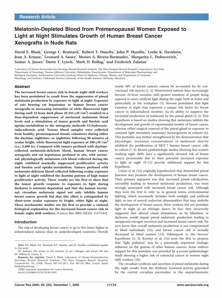
Melatonin-Depleted Blood from Premenopausal Women Exposed to
Light at Night Stimulates Growth of Human Breast Cancer
Xenografts in Nude Rats
David E. Blask,1George C. Brainard,
2Robert T. Dauchy,
1John P. Hanifin,
2Leslie K. Davidson,
1
Jean A. Krause,1Leonard A. Sauer,
1Moises A. Rivera-Bermudez,
3Margarita L. Dubocovich,
3
Samar A. Jasser,2Darin T. Lynch,
1Mark D. Rollag,
4and Frederick Zalatan
1
1Laboratory of Chrono-Neuroendocrine Oncology, Bassett Research Institute, The Mary Imogene Bassett Hospital, Cooperstown, New York;2Department of Neurology, Thomas Jefferson University, Philadelphia, Pennsylvania; 3Department of Molecular Pharmacology andBiological Chemistry, Northwestern University Feinberg School of Medicine, Chicago, Illinois; and 4Department of Anatomy,Physiology, and Genetics, Uniformed Services University of the Health Sciences, Bethesda, Maryland
Abstract
The increased breast cancer risk in female night shift workershas been postulated to result from the suppression of pinealmelatonin production by exposure to light at night. Exposureof rats bearing rat hepatomas or human breast cancerxenografts to increasing intensities of white fluorescent lightduring each 12-hour dark phase (0-345 MW/cm2) resulted in adose-dependent suppression of nocturnal melatonin bloodlevels and a stimulation of tumor growth and linoleic aciduptake/metabolism to the mitogenic molecule 13-hydroxyoc-tadecadienoic acid. Venous blood samples were collectedfrom healthy, premenopausal female volunteers during eitherthe daytime, nighttime, or nighttime following 90 minutes ofocular bright, white fluorescent light exposure at 580 MW/cm2
(i.e., 2,800 lx). Compared with tumors perfused with daytime-collected melatonin-deficient blood, human breast cancerxenografts and rat hepatomas perfused in situ , with noctur-nal, physiologically melatonin-rich blood collected during thenight, exhibited markedly suppressed proliferative activityand linoleic acid uptake/metabolism. Tumors perfused withmelatonin-deficient blood collected following ocular exposureto light at night exhibited the daytime pattern of high tumorproliferative activity. These results are the first to show thatthe tumor growth response to exposure to light duringdarkness is intensity dependent and that the human noctur-nal, circadian melatonin signal not only inhibits humanbreast cancer growth but that this effect is extinguished byshort-term ocular exposure to bright, white light at night.These mechanistic studies are the first to provide a rationalbiological explanation for the increased breast cancer risk infemale night shift workers. (Cancer Res 2005; 65(23): 11174-84)
Introduction
The risk of developing breast cancer is up to five times higher inindustrialized nations than in underdeveloped countries. Overall,
nearly 50% of breast cancers cannot be accounted for by con-ventional risk factors (1, 2). Westernized nations have increasinglybecome 24-hour societies with greater numbers of people beingexposed to more artificial light during the night both at home andparticularly in the workplace (3). Stevens postulated that lightexposure at night may represent a unique risk factor for breastcancer in industrialized societies via its ability to suppress thenocturnal production of melatonin by the pineal gland (4, 5). Thishypothesis is based on studies showing that melatonin inhibits thedevelopment and growth of experimental models of breast cancer,whereas either surgical removal of the pineal gland or exposure toconstant light stimulates mammary tumorigenesis in rodents (6).This postulate was further strengthened by the demonstration thatphysiologic, nocturnal concentrations of melatonin directlyinhibited the proliferation of MCF-7 human breast cancer cellsin culture (7, 8). Recent epidemiologic studies showing that womenworking night shifts have a significantly elevated risk of breastcancer presumably due to their potential increased exposureto light at night (9–11) provide additional support for thishypothesis.Cohen et al. (12) originally hypothesized that diminished pineal
function may promote the development of human breast cancer.Their primary argument was that increased pineal calcification,ostensibly leading to lowered melatonin production, was moststrongly associated with increased breast cancer risk. Althoughthey were the first to refer to, in general terms, environmentallighting, which necessarily includes both sunlight and artificiallight, as one of several endocrine abnormalities that may underliethe development of breast cancer, these workers did not postulatelight at night as an etiologic factor. In fact, they incorrectlysuggested that altered visual stimulation, as by blindness ordarkness, would impair pineal melatonin production leading tounopposed estrogen secretion and increased breast cancer risk. Wenow know that overall melatonin production is not compromisedin blind individuals (13), and breast cancer risk is actuallydecreased in blind women (14). Subsequent to the Stevens’hypotheses (4, 5), Kerenyi et al. (15) were the first to proposethat ‘‘light pollution’’ may be a potentially important etiologicinfluence on the genesis of other human cancers. Some indirectsupport for this postulate was provided by a recent epidemiologicstudy showing a higher risk of colorectal cancer in women nightshift workers (16).The circadian synthesis and secretion of pineal melatonin during
the night results from the rhythmic neuronal activity generatedby the central circadian pacemaker in the suprachiasmatic
Note: D.E. Blask, G.C. Brainard, R.T. Dauchy, and J.P. Hanifin contributed equallyto this work.
We dedicate this article to the memory of our colleague and friend, the lateDr. William B. Guiney, Jr.
Requests for reprints: David E. Blask, Laboratory of Chrono-NeuroendocrineOncology, Bassett Research Institute, The Mary Imogene Bassett Hospital,Cooperstown, NY 13326. Phone: 607-547-3677; Fax: 607-547-3061; E-mail: [email protected].
I2005 American Association for Cancer Research.doi:10.1158/0008-5472.CAN-05-1945
Cancer Res 2005; 65: (23). December 1, 2005 11174 www.aacrjournals.org
Research Article
nuclei of the hypothalamus, which is entrained (i.e., synchronized)by the light/dark cycle. The duration of melatonin productionduring the night is directly proportional to the length of the darkperiod (17–19). The alternating light/dark cycle entrains circadianmelatonin production to a 24-hour cycle, whereas ocular exposureto light during darkness rapidly suppresses melatonin productiondepending on the intensity, wavelength, and duration of the lightexposure (20, 21). In experimental systems, melatonin plays afundamental role in the regulation of seasonal reproduction,circadian rhythm activity, retinal physiology, cardiovascular effects,immune function, and cancer (17–19).It has been generally assumed that the breast cancer–promoting
effects of either pinealectomy (i.e., pineal removal) or constantlight exposure in rodents are due to the absence of the nocturnalmelatonin signal because pharmacologic melatonin replacementeither attenuates or prevents these tumorigenic effects (6).Physiologic nocturnal circulating levels of melatonin inhibit thegrowth of tissue-isolated rat hepatoma 7288CTC via melatoninreceptor–mediated suppression of tumor linoleic acid uptake/metabolism to the mitogenic signaling molecule 13-hydroxyocta-decadienoic acid (13-HODE; refs. 22, 23). Pinealectomy or lightexposure during darkness prevents these effects (22, 24, 25).Inasmuch as the Stevens’ hypothesis (4, 5) has not been
experimentally tested, we evaluated the response of rats, bearingeither rat hepatomas or human breast cancer xenografts, toincreasing intensities of ocular light exposure during darkness ontumor growth, linoleic acid uptake/metabolism, and signaltransduction activity. More importantly, we also determined theproliferative, metabolic, and signal transduction responses ofhepatomas and breast cancer xenografts to perfusion in situ withblood collected from premenopausal human female volunteerswhose physiologic circadian melatonin signal was suppressed byocular exposure to bright, white fluorescent light at night. Our solerationale for including tissue-isolated rat hepatomas was based onthe fact that they exhibit remarkable sensitivity to melatonin, andmost of the information we have on the biochemical/molecularmechanisms of melatonin’s anticancer action in vivo has beenobtained in this model system (22–25). We wanted the ability tocompare the biological/molecular responses to light at night in thenew tissue-isolated human breast cancer xenograft model withthose in our highly melatonin-sensitive rat liver cancer modelserving as a positive control. It is important to note, however, thatunlike breast cancer, human liver cancer is much more common inthe developing world (i.e., less light pollution) than in industrial-ized nations (i.e., more light pollution), and its incidence decreasesas societies become more westernized (26).
Materials and Methods
Animals, tumors, and histopathology. Adult male Buffalo [BUF(BUF/
Ncr)] rats were implanted with rat hepatocarcinoma syngeneic grafts(Morris 7288CTC), and adult inbred female nude rats (HSD:RH-rnu) were
implanted with either steroid receptor–negative (SR�; i.e., no estrogen or
progesterone receptor expression) or steroid receptor–positive (SR+) MCF-7human breast cancer xenografts in a tissue-isolated manner as previously
described (27, 28). SR� tumors evolved over several passages from a subset
of SR+ xenografts that had become estrogen unresponsive. Both SR+ and
SR� human breast cancer xenografts were histopathologically confirmed tobe poorly differentiated, grade 3, infiltrating ductal breast adenocarcino-
mas. Adult male rats (Hsd:Sprague-DawleySD) were used as whole-blood
donors for some tumor perfusions. Immunoshistochemical staining of cell
nuclei for estrogen and progesterone receptor expression in tissue sections
of human breast cancer xenografts was done by IMPATH (New York, NY).SR+ tumors were highly estrogen receptor positive (80% staining) and
minimally progesterone receptor positive (10% staining); in SR� xenografts
no staining for either receptor was evident (data not shown). Animals were
maintained in an Association for Assessment and Accreditation of
Laboratory Animal Care International–accredited facility on a 12-hour
light/12-hour dark cycle (lights on 06:00 to 18:00 hours) and provided with
standard laboratory chow and water ad libitum in accordance with an
Institutional Animal Care and Use Committee–approved protocol. A series
of ceiling fixtures containing 32-W fluorescent tubes (GE Watt Miser, cool
white, F40CW.RS.WM) provided 7.5 AW/cm2 of white light at rodent eye
level during the light phase.
Tissue-isolated tumor perfusions in situ . SR+ MCF-7 human breast
cancer xenografts were perfused in situ with rat donor whole blood to
which either synthetic melatonin (Sigma, St. Louis, MO), and/or 13-HODE(Cayman Chemicals, Ann Arbor, MI) was added as previously described
(22, 23). Sets (three tumors per perfusion) of tissue-isolated rat hepatomas
and human breast cancer xenografts were perfused in situ for 1 hour aspreviously described (22, 23) with whole blood collected from either donor
rats or human subjects exposed to each of the lighting conditions outlined
below.
Tumor reverse transcription-PCR. Rat hepatoma 7288CTC and SR+
human breast cancer xenograft tissues were harvested from rats under
nembutal anesthesia between 08:30 and 10:30 hours (zeitgeber time),
quickly frozen, and stored at �80jC until subsequent isolation of total
RNA. Total cellular RNA was isolated from human breast cancer xenografttissue and rat hepatoma using the SV Total RNA Isolation System (Promega
Corp., Madison, WI) The RNA was quantitated spectrophotometrically, and
its integrity was checked by agarose gel. The mRNA was reversedtranscribed with Moloney murine leukemia virus reverse transcriptase
(Invitrogen Corp., Carlsbad, CA). The resulting cDNA was amplified using
the PCR for MT1 or MT2 melatonin receptor product with Taq Polymerase
(Promega) at 1.5 mmol/L MgCl2 and a 58jC annealing temperature for36 cycles using a RoboCycler (Stratagene, La Jolla, CA). Forward and reverse
primers used for MT1 and MT2 melatonin receptors in tumor tissues are
the same as previously published for rat (29) and for human (30, 31).
Tumor in situ hybridization. At 15:30 hours (zeitgeber time), rathepatoma and SR� human breast cancer xenograft tissue wedges were
immediately harvested from rats under nembutal anesthesia, quickly
frozen, and stored at �80jC until subsequent isolation of total RNA. Rathepatoma and SR� human breast cancer tissue wedges were embedded in
ornithine carbamyl transferase compound, rapidly frozen in 2-methylbu-
tane kept in dry ice, and stored frozen at �80jC until tissue sections were
cut and thaw mounted onto glass slides. Tumor sections were processed forin situ hybridization using digoxigenin-labeled oligonucleotide antisense
and sense probes for MT1 and MT2 melatonin receptors as previously
described (32). The MT1 oligonucleotide (GGGGTCGTACTGGA-
GAGTTCCGGTTTGCAGG, mer 31) corresponds to bases 108 to 138 ofthe partial rat MT1 receptor sequence (Genbank accession no. U14409) and
to bases 518 to 548 of the human MT1 receptor sequence (Genbank
accession no. U14108). The MT2 oligonucleotide (CGGGTCATATTCTAGA-
GACCCCACAAAGAAA, mer 31) corresponds to bases 111 to 141 of thepartial rat MT2 receptor sequence (Genbank accession no. U28218) and to
bases 537 to 556 of human MT2 receptor sequence (Genbank accession no.
U25341).Nocturnal light exposure studies in rats and effects on tumor
growth. Groups of tumor-bearing rats were housed in light exposure
chambers that contained two separate solid-state, electromagnetic
fluorescent ballasts with rapid-start, cool-white lamps connected toseparate 24-hour timers and separated by a metal baffle (20). One
ballast/lamp system (GE Watt-Miser, F34CW-RS-WM, 34-W bulb) in each
chamber was set up to provide a direct, steady, bright light stimulus (345
AW/cm2) at the animals’ eye level during the light portion of a 12-hourlight/12-hour dark cycle. The second ballast/lamp system (GE Starcoat,
F32T8-SP-11, 32-W bulb) was adjustable with a combination of neutral filter
density filter material (CINEGEL 3403, N-9) and electronic dimmer modulesto emit steady, indirect light reflected off of the rear chamber wall
Light-Induced Melatonin Suppression and Cancer Growth
www.aacrjournals.org 11175 Cancer Res 2005; 65: (23). December 1, 2005
measured at the animals’ eye level during the dark phase. The entirechamber was painted with Pro-Mar flat white paint (Sherwin Williams,
St. Simon Island, GA), which has a high content of titanium dioxide that
reflects light relatively uniformly across the visible spectrum. The light
intensities during each 12-hour dark phase were different in each chamberas follows: 0 (total darkness), 0.02, 0.05, 0.06, 0.08, and 345 AW/cm2
(constant light). These intensities were measured and regularly monitored
at animals’ eye level in the center of each cage using a Model IL 1400a
Radiometer/Photometer (International Light, Inc., Newburyport, MA).Animals were exposed to the various light intensities beginning 2 weeks
before tumor implantation and continuing thereafter until the end of each
tumor growth period. Throughout each experiment, animal cage positions
were rotated on a daily basis to minimize the potential effects of slightvariations in the target, reflected light intensity within each chamber.
Nocturnal light exposure studies in human subjects and effects ontumor proliferative activity. At Thomas Jefferson University (Philadel-phia, PA), healthy, premenopausal female volunteers (n = 12; mean age,
22.6 F 0.6 years) were recruited without regard to either race or ethnic
background and signed approved institutional review board consent forms.
All subjects were free of any medication, including birth control pills,melatonin supplements, and/or h-blockers, and had regular sleep patterns.
Each subject had antecubital venous blood samples drawn by a
phlebotomist during the daytime (10:00 to 14:00 hours), nighttime (02:00
hours) following 2 hours of complete darkness, and nighttime (03:30 hours)following 90 minutes of ocular, bright, white light exposure at 580 AW/cm2
(i.e., 2,800 lx) at eye level reflected from the wall. This exposure protocol
is described in detail elsewhere (33). On the day following the final bloodcollection, whole-blood samples were shipped on ice via overnight express
mail to the Bassett Research Institute (Cooperstown, NY), where they were
stored refrigerated with constant gentle vortexing until tumor perfusions
were done 24 hours later.Assay of blood hormone levels in rats and human subjects. Rat
serum and human plasma melatonin levels were measured via RIA as
previously described (21, 23). Rat serum corticosterone and estradiol levels
were measured using a commercial RIA kits (Diagnostic Products Corp.,Los Angeles, CA).
Determination of tumor growth, linoleic acid uptake, and13-hydroxyoctadecadienoic acid formation. Tumor growth rates, uptakeof linoleic acid, and formation of 13-HODE were determined as previously
described (22, 23). Due to variations in the absolute circulating
concentrations of linoleic acid, tumor linoleic acid uptake is expressed as
a percent of the actual arterial supply of linoleic acid to the tumor.Determination of tumor cyclic AMP levels, DNA content, and
[3H]thymidine incorporation into DNA. Tumor levels of cyclic AMP
(cAMP) were determined via ELISA (Amersham Biosciences, Piscataway,
NJ) and [3H]thymidine incorporation into DNA, and DNA content weredetermined as previously described (22, 23, 28).
Determination of tumor mitogen-activated protein/extracellularsignal-regulated kinase kinase and extracellular signal-regulatedkinase 1/2 activation. Western blot analysis of tumor mitogen-activatedprotein/extracellular signal-regulated kinase kinase (MEK) and extracellular
signal-regulated kinase 1/2 (ERK1/2) activation was done as previously
described (34).Statistical analyses. Statistical differences among group means were
determined by a one-way ANOVA followed by Student-Neuman-Keul’s post
hoc test. Differences in the slopes of regression lines (i.e., tumor growth
rates) among groups were determined by regression analyses and tests forparallelism (Student’s t test). Four-variable parabolic fluence (dose)-response
curve fitting of the above variables was done as previously described (35).
Differences were considered statistically significant at P < 0.05.
Results
Melatonin responsiveness of human breast cancer xeno-grafts perfused in situ . We previously reported that tissue-isolatedrat hepatoma 7288CTC was highly responsive to the inhibitoryeffects of physiologic nocturnal melatonin concentrations with
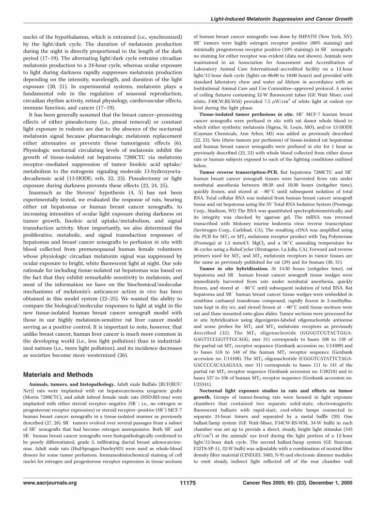
respect to tumor linoleic acid uptake/metabolism and proliferativeactivity following perfusion in situ (22, 23). Furthermore, thetumor inhibitory effects of melatonin were blocked by both MT1and MT2 receptor antagonists (36). We now show that SR+ MCF-7human breast cancer xenografts were also very sensitive to theinhibitory actions of nocturnal melatonin concentrations byperfusing tissue-isolated tumors in situ with exogenous syntheticmelatonin (1 nmol/L; see perfusion diagram in Fig. 5B). Following2 hours of perfusion with melatonin, tumor cAMP levels werereduced by 40%, whereas both tumor linoleic acid uptake and13-HODE formation were completely blocked (Table 1). Thesechanges were accompanied by a decrease in ERK1/2 activation(Fig. 1) and a 71% decrease in tumor [3H]thymidine incorporationinto DNA, which were completely prevented by the coperfusion oftumors with both melatonin and 13-HODE (Fig. 1; Table 1). Thesefindings indicated that, similar to rat hepatoma 7288CTC (22, 23),SR+ MCF-7 human breast cancer xenografts were highly sensitiveto a nocturnal, physiologic concentration of melatonin, which isconsistent with the expression of MT1 receptors in SR+ MCF-7cells (37). This high degree of melatonin sensitivity was alsoexhibited in tissue-isolated SR� MCF-7 breast cancer xenograftsthat had evolved from and retained the histopathologic phenotypeof the SR+ MCF-7 parental tumor (see Materials and Methods) andalso expressed MT1 receptors (see below). Therefore, SR� MCF-7xenografts were used in testing the effects of melatoninsuppression, via ocular light exposure during darkness in eitherrats or premenopausal human subjects, on breast cancer growthprogression.Melatonin receptor mRNA expression in rat hepatoma and
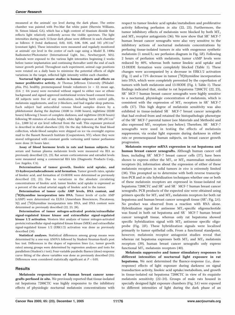
human breast cancer xenografts. Although human cancer celllines, including SR+ MCF-7 breast cancer cells (37), have beenshown to express either the MT1 or MT2 mammalian melatoninreceptors (6), information about the expression of either of thesemelatonin receptors in solid tumors in vivo is extremely limited(38). This prompted us to determine with both reverse transcrip-tion-PCR and in situ hybridization techniques whether one or bothof these melatonin receptors are expressed in tissue-isolated rathepatoma 7288CTC and SR+ and SR� MCF-7 human breast cancerxenografts. PCR products of the expected size were obtained usingprimers specific for MT1 and MT2 melatonin receptors for both rathepatoma and human breast cancer xenograft tissue (SR+; Fig. 2A).No product was observed from a reaction with RNA alone.Hybridization signal for antisense MT1-specific oligonucleotidewas found in both rat hepatoma and SR� MCF-7 human breastcancer xenograft tissue, whereas only rat hepatoma showedspecific hybridization signal with MT2 antisense specific oligo-probe (Fig. 2B). These hybridization signals were localizedprimarily to tumor epithelial cells. From a functional standpoint,however, melatonin receptor antagonist studies reveal thatwhereas rat hepatoma expresses both MT1 and MT2 melatoninreceptors (39), human breast cancer xenografts only expressfunctional MT1 melatonin receptors (40).Melatonin suppressive and tumor stimulatory responses to
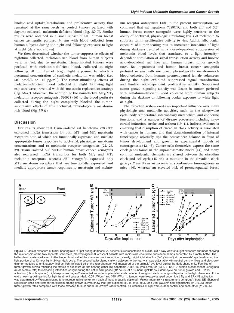
different intensities of nocturnal light exposure in rathepatoma. We next determined the fluence-response (i.e., dose-response) effects of light exposure during darkness on signaltransduction activity, linoleic acid uptake/metabolism, and growthin tissue-isolated rat hepatoma 7288CTC in view of its exquisitemelatonin sensitivity (22–24). Groups of male rats housed inspecially designed light exposure chambers (Fig. 3A) were exposedto different intensities of light during the dark phase of an
Cancer Research
Cancer Res 2005; 65: (23). December 1, 2005 11176 www.aacrjournals.org
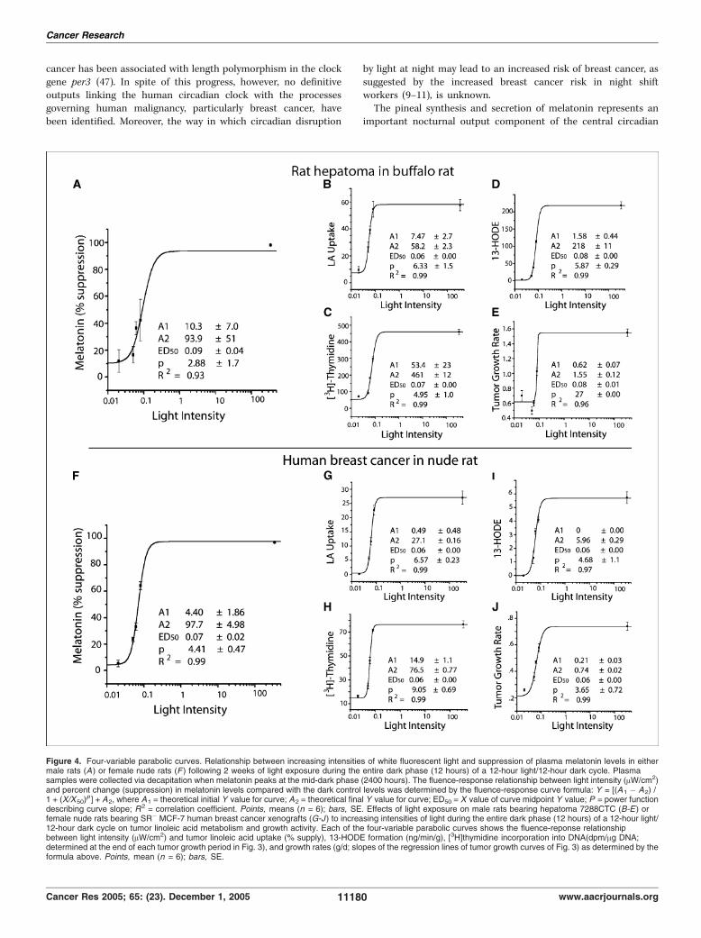
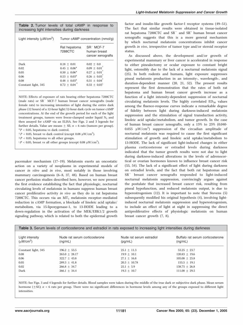
alternating 12-hour light/12-hour dark cycle beginning 2 weeksbefore tumor implantation and continuing thereafter until the endof the tumor growth experiment. Following 2 weeks of exposure ofrats to increasing intensities of reflected light [0 (total darkness),0.02, 0.05, 0.06, 0.08, and 345 (constant bright light) AW/cm2]measured at rodent eye level during darkness, there was a dose-dependent increase in the percent suppression of serum melatoninlevels (Fig. 4A). Continued exposure of rats to these lightintensities following tumor implantation resulted in a corres-ponding dose-related increase in tumor growth rates (0.62-1.55 g/d;Fig. 3B and Fig. 4E), [3H]thymidine incorporation into DNA(Fig. 4C), and DNA content (data not shown). In addition, the timeto tumor onset decreased as the light intensity increased (Fig. 3B).Similarly, tumor linoleic acid uptake and 13-HODE formation(Fig. 4B and D), ERK1/2 activation (Fig. 3B), and cAMP levels(Table 2) increased as a function of increased light intensities duringthe dark phase. Four-variable parabolic fluence-response curves(Fig. 4A-E) showed a tight correlation (r2 = 0.93-0.99) betweenpercent melatonin suppression and tumor linoleic acid uptake,13-HODE production, [3H]thymidine incorporation into DNA, andgrowth rates. The ED50 values (i.e., half maximal suppression orstimulation) for melatonin suppression and tumor stimulatoryresponses to light during darkness ranged from 0.06 to 0.09 AW/cm2.No significant effects of light exposure at any intensity was observedon serum corticosterone levels during the mid-dark period, albeitlevels were depressed in the constant light-exposed group (Table 3).These results confirmed, in a highly melatonin-sensitive murinetumor model (22–24), that tumor signal transduction, linoleic aciduptake, and 13-HODE formation and growth could be modulated ina dose-dependent manner by changes in light intensity exposureduring the dark phase of a diurnal lighting cycle.Melatonin suppressive and tumor stimulatory responses to
different intensities of nocturnal light exposure in humanbreast cancer xenografts. Previously, we reported that the femalenude rat exhibits a robust nocturnal circadian melatonin rhythm,similar to that in humans in terms of its amplitude, phasing, andduration, that is completely suppressed by constant brightlight (i.e., 300 lx or f120 AW/cm2 at rodent eye level; ref. 27).Furthermore, in nude rats bearing tissue-isolated SR+ MCF-7human breast cancer xenografts, constant bright light exposureresulted in a significant increase in tumor linoleic acid uptake/
metabolism to 13-HODE and a marked stimulation of tumorgrowth compared with control rats on 12-hour light/12-hourdark cycle (27). We found that like tissue-isolated SR+ MCF-7xenografts (Fig. 1; Table 1), SR� xenografts (Fig. 5E and G)perfused in situ were highly responsive to the tumor-inhibitingeffects of a physiologic, nocturnal circulating concentration ofmelatonin (see below). We assessed the dose-response effects oflight exposure during darkness on growth, linoleic acid uptake/metabolism to 13-HODE, and signal transduction activity inSR� breast cancer xenografts under the same lighting conditionsas outlined above for rats bearing rat hepatoma 7288CTC. As thelight intensity increased, there was a corresponding increase inthe percent suppression of serum melatonin levels in nuderats (Fig. 4F). There was also an accompanying marked, dose-related increase in tumor growth rates (0.22-0.74 g/d; Fig. 3C andFig. 4J), [3H]thymidine incorporation into DNA (Fig. 4H), andDNA content (data not shown). Latency to tumor onset decreasedas the intensity of light during darkness increased (Fig. 3C).Tumor linoleic acid uptake and 13-HODE formation (Fig. 4Gand I), ERK1/2 activation (Fig. 3C), and cAMP levels (Table 2)increased as a function of the light intensity exposure duringthe dark phase. As in the case of rat hepatoma, four-variableparabolic fluence-response curves (Fig. 4F-J) showed a tightcorrelation (r2 = 0.97-0.99) between percent melatonin suppres-sion and tumor linoleic acid uptake, 13-HODE formation,
Table 1. Response of SR+ MCF-7 human breast cancer xenografts to perfusion in situ with a physiologic nocturnalconcentration of melatonin
Treatment [3H]Thymidine incorporation(dpms/Ag DNA)
Linoleic acid uptake(% supply)
13-HODE production(ng/min/g)
cAMP(nmol/g)
Controls 47.4 F 3.9 16.7 F 1.7 0.97 F 0.17 0.55 F 0.11Melatonin 13.6 F 1.6* 0 0 0.33 F 0.12*
Melatonin + 13-HODE 74.8 F 6.3 0 333.17 F 19.02 0.68 F 0.06
13-HODE 78.0 F 6.5 17.2 F 3.2 363.18 F 10.62 0.78 F 0.17
NOTE: Effects of perfusion of tissue-isolated SR+ MCF-7 human breast cancer xenografts in situ with synthetic melatonin (1 nmol/L) in the presence or
absence of 13-HODE (12 Ag/mL) on [3H]thymidine incorporation into DNA, linoleic acid uptake, 13-HODE formation, and cAMP levels. Individual
tumors (n = 3-4) were perfused over a 2-hour period (06:30 to 08:30 h) with whole blood collected from tumor-free donor rats during the early light
phase when endogenous melatonin levels were low. Synthetic melatonin and 13-HODE were added to the whole-blood perfusate to achieve finalconcentrations of 1 nmol/L (232 pg/mL) and 12 Ag/mL, respectively. Values are means F SE.
*P < 0.05 vs controls and melatonin + 13-HODE (comparisons of relevance).
Figure 1. Tumor expression of ERK1/2. Western blot analysis of the effectsof perfusion of tissue-isolated SR+ MCF-7 human breast cancer xenograftsin situ for 1 hour with melatonin (1 nmol/L) in the presence or absenceof 13-HODE (12 Ag/mL) on ERK1/2 (mitogen-activated protein kinase p44/p42)activation. Top, phosphorylated ERK1/2; bottom, total ERK1/2. Totalprotein (27 Ag) was loaded per well. Controls (lanes 1-3), melatonin-treated(lanes 4-6), melatonin + 13-HODE (lanes 7-9 ), 13-HODE (lanes 10-12 ), andMW markers (lane 13). See Table 2 for further details.
Light-Induced Melatonin Suppression and Cancer Growth
www.aacrjournals.org 11177 Cancer Res 2005; 65: (23). December 1, 2005
[3H]thymidine incorporation into DNA, and growth rates. ED50
values for these processes ranged from 0.06 to 0.07 AW/cm2. Nosignificant effects of light exposure at any intensity was observedon serum either corticosterone or estradiol levels during the mid-dark period, albeit levels of these hormones tended to bedecreased or increased, respectively, in the constant light-exposedgroup (Table 3). These results confirmed that suppression ofphysiologic, nocturnal melatonin levels was dependent upon themagnitude of the light intensity to which nude rats were exposedduring the dark phase. This suppression of melatonin levelstranslated to an intensity-dependent stimulation of human breastcancer signal transduction activity, linoleic acid uptake/metabo-lism, and growth.Proliferative activity in rat hepatoma and human breast
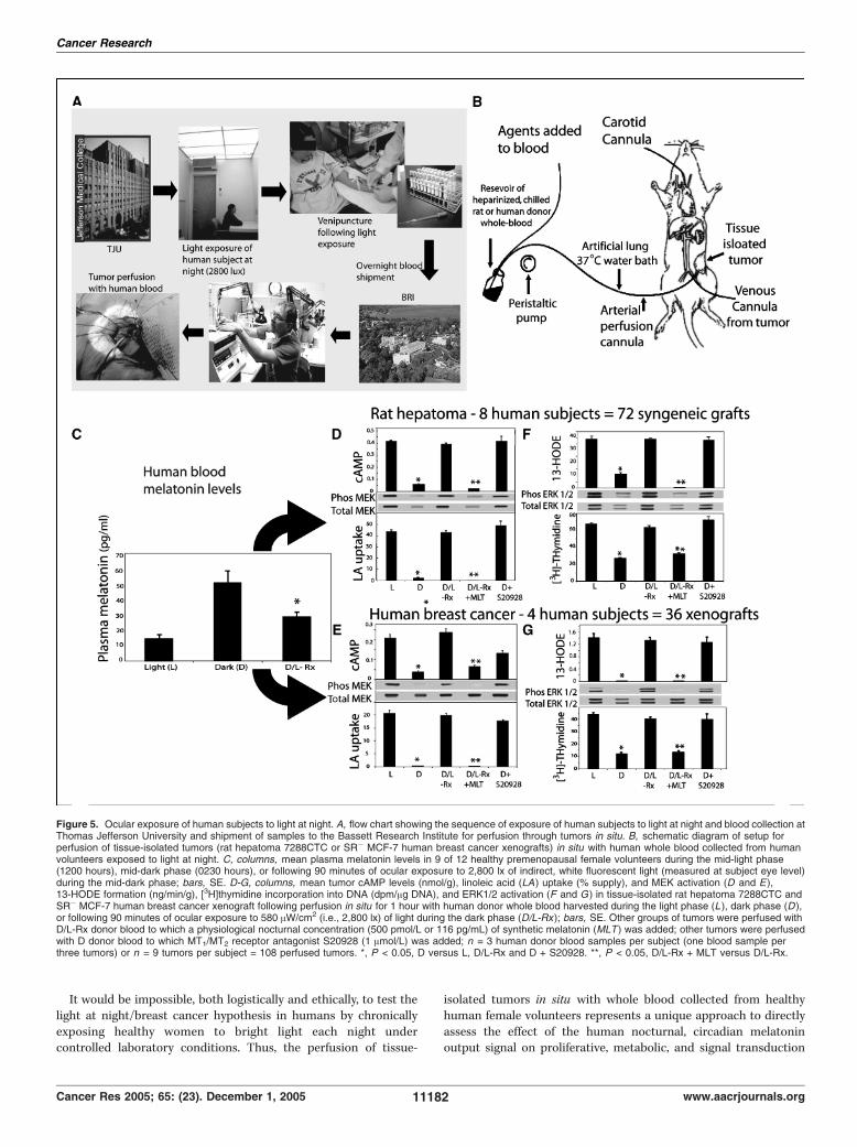
cancer xenografts perfused in situ with blood collected fromhuman subjects following ocular exposure to bright while lightat night. We next evaluated the acute effect of the humancircadian melatonin signal and its suppression by light at nighton signal transduction activity, linoleic acid uptake/metabolism,and proliferative activity in rat hepatomas and human breastcancer xenografts. This involved the unique approach of perfusingthese tumors, in situ , with whole blood collected from healthy,premenopausal human volunteers either during the daytime,nighttime, or following exposure to 580 AW/cm2 (i.e., 2,800 lx) ofwhite fluorescent light at night (ref. 33; Fig. 5A and B). As
expected, plasma melatonin titers in human volunteers were5-fold higher during the night compared with daytime levels ofmelatonin (Fig. 5C). The perfusion of tissue-isolated rat hepato-mas and human breast cancer xenografts in situ with melatonin-rich blood collected during the night resulted in tumor cAMPlevels that were reduced by 86% compared with control tumorsperfused with melatonin-deficient blood collected during thedaytime (Fig. 5D and E). Similarly, tumor linoleic acid uptake and13-HODE formation were reduced by as much as 96% followingperfusion with melatonin-rich blood collected from humansubjects during darkness compared with control tumors perfusedwith daytime-collected, melatonin-deficient blood (Fig. 5D-G).Furthermore, tumor incorporation of [3H]thymidine into DNAfollowing perfusion with melatonin-rich blood collected duringthe night was decreased by 63% to 73% compared with controltumors perfused with daytime-collected blood (Fig. 5F and G).Tumor DNA content was also significantly (P < 0.05) reducedfollowing perfusion with nighttime-collected versus daytime-collected human blood (data not shown). Following perfusionwith nighttime-collected, melatonin-rich blood, tumor MEK andERK1/2 activation was markedly reduced compared with tumorsperfused with daytime-collected, melatonin-deficient blood(Fig. 5D-G). In contrast, tumors perfused with melatonin-deficientblood obtained from subjects during the night following 90minutes of light exposure evinced signal transduction activity,
Figure 2. A, expression of MT1 and MT2 melatonin receptor mRNA transcripts in tissue-isolated rat hepatoma 7288CTC and SR+ MCF-7 human breast cancerxenograft tissue. Lane 1, reverse transcription-PCR reaction with rat hepatoma cDNA using primers specific for the rat MT1 melatonin receptor, yielding the expected397-bp product. Lane 2, negative control reaction with the MT1 primers using an equivalent amount of rat hepatoma total RNA. Lane 3, reaction with rat hepatomacDNA using primers specific for the rat MT2 melatonin receptor, yielding the expected 368-bp product. Lane 4, negative control reaction with the MT2 primersusing an equivalent amount of rat hepatoma total RNA. Lane 5, reaction with human breast cancer xenograft cDNA using primers for the human MT1 melatoninreceptor, yielding the expected 285-bp product. Lane 6, negative control reaction with MT1 primers using an equivalent amount of human breast cancer xenograft totalRNA. Lane 7, reaction with human breast cancer xenograft cDNA using primers specific for the human MT2 melatonin receptor, yielding the expected 321-bpproduct. Lane 8, negative control reaction with MT2 primers using an equivalent amount of human breast cancer xenograft total RNA. B, representativephotomicrographs showing in situ hybridization of tumor tissue from tissue-isolated rat hepatoma 7288CTC and an SR� MCF-7 human breast cancer xenografttissue using digoxigenin-labeled sense and antisense oligonucleotides specific for either MT1 or MT2 melatonin receptors. Hybridization signal for the antisenseMT1-specific oligonucleotide was found in both rat hepatoma and human breast cancer, whereas only hepatoma showed specific hybridization signal with the MT2
antisense-specific oligoprobe.
Cancer Research
Cancer Res 2005; 65: (23). December 1, 2005 11178 www.aacrjournals.org
linoleic acid uptake/metabolism, and proliferative activity thatremained at the same levels as control tumors perfused withdaytime-collected, melatonin-deficient blood (Fig. 5D-G). Similarresults were obtained in a small subset of SR+ human breastcancer xenografts perfused in situ with blood collected fromhuman subjects during the night and following exposure to lightat night (data not shown).We then determined whether the tumor-suppressive effects of
nighttime-collected, melatonin-rich blood from human subjectswere, in fact, due to melatonin. Tissue-isolated tumors wereperfused with melatonin-deficient blood, collected at nightfollowing 90 minutes of bright light exposure, to which anocturnal concentration of synthetic melatonin was added (i.e.,500 pmol/L or 116 pg/mL). The tumor-stimulating effects ofmelatonin-deficient blood collected at night following lightexposure were prevented with this melatonin replacement strategy(Fig. 5D-G). Moreover, the addition of the nonselective MT1/MT2
melatonin receptor antagonist S20928 (36) to the blood perfusatecollected during the night completely blocked the tumor-suppressive effects of this nocturnal, physiologically melatonin-rich blood (Fig. 5D-G).
Discussion
Our results show that tissue-isolated rat hepatoma 7288CTCexpressed mRNA transcripts for both MT1 and MT2 melatoninreceptors both of which are functionally expressed and mediateappropriate tumor responses to nocturnal, physiologic melatoninconcentrations and to melatonin receptor antagonists (22, 23,39). Tissue-isolated SR+ MCF-7 human breast cancer xenograftsalso expressed mRNA transcripts for both MT1 and MT2
melatonin receptors, whereas SR� xenografts expressed onlyMT1 melatonin receptors that are functionally expressed andmediate appropriate tumor responses to melatonin and melato-
nin receptor antagonists (40). In the present investigation, weconfirmed that rat hepatoma 7288CTC, and both SR+ and SR�
human breast cancer xenografts were highly sensitive to theability of nocturnal, physiologic circulating levels of melatonin tosuppress tumor proliferative activity in vivo . Additionally, ocularexposure of tumor-bearing rats to increasing intensities of lightduring darkness resulted in a dose-dependent suppression ofmelatonin blood levels that translated to a light intensity–dependent stimulation of signal transduction activity and linoleicacid–dependent rat liver and human breast tumor growthrates. Rat hepatomas and human breast cancer xenograftsperfused in situ with nocturnal, physiologically melatonin-richblood collected from human, premenopausal female volunteersduring the night exhibited suppressed signal transductionand linoleic acid–dependent proliferative activity. Suppressedtumor growth signaling activity was absent in tumors perfusedwith melatonin-deficient blood collected from human subjectsduring the daytime or following ocular exposure to white lightat night.The circadian system exerts an important influence over many
physiologic and metabolic activities, such as the sleep/wakecycle, body temperature, intermediary metabolism, and endocrinefunctions, and a number of disease processes, including myo-cardial infarction, stroke, and asthma (19, 41). Indirect evidence isemerging that disruption of circadian clock activity is associatedwith cancer in humans, and that desynchronization of internaltimekeeping adversely tips the host/cancer balance in favor oftumor development and growth in experimental models oftumorigenesis (42, 43). Cancer cells themselves express the sameclock genes found in the suprachiasmatic nuclei (44), and manycommon molecular elements are shared between the circadianclock and cell cycle (45, 46). A mutation in the circadian clockgene per2 results in an increase in spontaneous tumorigenesis inmice (46), whereas an elevated risk of premenopausal breast
Figure 3. Ocular exposure of tumor-bearing rats to light during darkness. A, schematic representation of a side, cut-a-way view of a light exposure chamber showingthe relationship of the two separate solid-state, electromagnetic fluorescent ballasts with rapid-start, cool-white fluorescent lamps separated by a metal baffle. Theballast/lamp system adjacent to the hinged front wall of the chamber provides a direct, steady, bright light stimulus (345 AW/cm2) at the animals’ eye level during thelight portion of a 12-hour light/12-hour dark cycle. The second ballast/lamp system adjacent to the rear wall was adjustable with neutral density filters and electronicdimmer modules to emit steady, indirect light reflected off of the rear chamber wall measured at the animals’ eye level during the dark phase only. Families oftumor growth curves reflecting the effects of exposure of rats bearing either (B ) hepatoma 7288CTC (male rats) or (C ) SR� MCF-7 human breast cancer xenografts(nude female rats) to increasing intensities of light during the entire dark phase (12 hours) of a 12-hour light/12-hour dark cycle on tumor growth and ERK1/2activation (phosphorylation). Light exposures began 2 weeks before tumor implantation and continued throughout each tumor growth period in the light chambers. At theend of each growth period for light treatment groups (dark, 0.05 AW/cm2 and 345 AW/cm2), tumors were freeze-clamped under liquid N2 and ERK1/2 activationwas determined by Western blotting (one representative tumor from each of these groups is depicted). Points, mean (n = 6 rats, tumors per group); bars, SE. Slopes ofregression lines and tests for parallelism among growth curves show that rats exposed to 345, 0.08, 0.06, and 0.05 AW/cm2 had significantly (P < 0.05) fastertumor growth rates compared with those exposed to 0.02 and 0.00 AW/cm2 (dark control). All intensities of light versus dark control and each other (P < 0.05).
Light-Induced Melatonin Suppression and Cancer Growth
www.aacrjournals.org 11179 Cancer Res 2005; 65: (23). December 1, 2005
cancer has been associated with length polymorphism in the clockgene per3 (47). In spite of this progress, however, no definitiveoutputs linking the human circadian clock with the processesgoverning human malignancy, particularly breast cancer, havebeen identified. Moreover, the way in which circadian disruption
by light at night may lead to an increased risk of breast cancer, assuggested by the increased breast cancer risk in night shiftworkers (9–11), is unknown.The pineal synthesis and secretion of melatonin represents an
important nocturnal output component of the central circadian
Figure 4. Four-variable parabolic curves. Relationship between increasing intensities of white fluorescent light and suppression of plasma melatonin levels in eithermale rats (A) or female nude rats (F ) following 2 weeks of light exposure during the entire dark phase (12 hours) of a 12-hour light/12-hour dark cycle. Plasmasamples were collected via decapitation when melatonin peaks at the mid-dark phase (2400 hours). The fluence-response relationship between light intensity (AW/cm2)and percent change (suppression) in melatonin levels compared with the dark control levels was determined by the fluence-response curve formula: Y = [(A1 � A2) /1 + (X/X50)
p ] + A2, where A1 = theoretical initial Y value for curve; A2 = theoretical final Y value for curve; ED50 = X value of curve midpoint Y value; P = power functiondescribing curve slope; R2 = correlation coefficient. Points, means (n = 6); bars, SE. Effects of light exposure on male rats bearing hepatoma 7288CTC (B-E ) orfemale nude rats bearing SR� MCF-7 human breast cancer xenografts (G-J ) to increasing intensities of light during the entire dark phase (12 hours) of a 12-hour light/12-hour dark cycle on tumor linoleic acid metabolism and growth activity. Each of the four-variable parabolic curves shows the fluence-reponse relationshipbetween light intensity (AW/cm2) and tumor linoleic acid uptake (% supply), 13-HODE formation (ng/min/g), [3H]thymidine incorporation into DNA(dpm/Ag DNA;determined at the end of each tumor growth period in Fig. 3), and growth rates (g/d; slopes of the regression lines of tumor growth curves of Fig. 3) as determined by theformula above. Points, mean (n = 6); bars, SE.
Cancer Research
Cancer Res 2005; 65: (23). December 1, 2005 11180 www.aacrjournals.org
pacemaker mechanism (17–19). Melatonin exerts an oncostaticaction on a variety of neoplasms in experimental models ofcancer in vitro and in vivo , most notably in those involvingmammary carcinogenesis (6–8, 37, 48). Based on human breastcancer perfusion studies described here, however, we now presentthe first evidence establishing the fact that physiologic, nocturnalcirculating levels of melatonin in humans suppress human breastcancer proliferative activity in vivo as they do in rat hepatoma7288CTC. This occurs via an MT1 melatonin receptor–mediatedreduction in cAMP formation, a blockade of linoleic acid uptake/metabolism, via 15-lipoxygenase-1, to 13-HODE leading to adown-regulation in the activation of the MEK/ERK1/2 growthsignaling pathway, which is related to both the epidermal growth
factor and insulin-like growth factor-I receptor systems (49–51).The fact that similar results were obtained in tissue-isolatedrat hepatoma 7288CTC and SR� and SR+ human breast cancerxenografts suggests that this is a more general mechanismby which nocturnal melatonin concentrations inhibit cancergrowth in vivo , irrespective of tumor type and/or steroid receptorstatus.As discussed above, the development and/or growth of
experimental mammary or liver cancer is accelerated in responseto either pinealectomy or ocular exposure to constant brightlight, ostensibly due to the lack of a nocturnal melatonin signal(25). In both rodents and humans, light exposure suppressespineal melatonin production in an intensity-, wavelength-, andduration-dependent manner (20, 21, 52). The present resultsrepresent the first demonstration that the rates of both rathepatoma and human breast cancer growth increase as afunction of a light intensity-dependent suppression of nocturnalcirculating melatonin levels. The highly correlated ED50 valuesamong the fluence-response curves indicate a remarkable degreeof fidelity between light during darkness-induced melatoninsuppression and the stimulation of signal transduction activity,linoleic acid uptake/metabolism, and tumor growth. In the caseof human breast cancer xenografts, only a 15% to 25% (0.040-0.055 AW/cm2) suppression of the circadian amplitude ofnocturnal melatonin was required to cause the first significantstimulation of growth and linoleic acid uptake/metabolism to13-HODE. The lack of significant light-induced changes in eitherplasma corticosterone or estradiol levels during darknessindicated that the tumor growth results were not due to lightduring darkness-induced alterations in the levels of adrenocor-tical or ovarian hormones known to influence breast cancer risk(53, 54). The lack of a significant effect of light during darknesson estradiol levels, and the fact that both rat hepatomas andSR� breast cancer xenografts responded to light-inducednocturnal melatonin suppression, convincingly argues againstthe postulate that increased breast cancer risk, resulting frompineal hypofunction, and reduced melatonin output, is due tohyperestrogenism (12). It is important to note that Stevens (5)subsequently modified his original hypothesis (4), involving light-induced nocturnal melatonin suppression and hyperestrogenism,to include an effect of light at night in suppressing the directantiproliferative effects of physiologic melatonin on humanbreast cancer growth (7, 8).
Table 2. Tumor levels of total cAMP in response toincreasing light intensities during darkness
Light intensity (AW/cm2) Tumor cAMP concentration (nmol/g)
Rat hepatoma
7288CTC
SR� MCF-7
human breastcancer xenografts
Dark 0.18 F 0.01 0.02 F 0.00.02 0.45 F 0.06* 0.09 F 0.01
c
0.05 0.50 F 0.06* 0.27 F 0.01c
0.06 0.53 F 0.03* 0.26 F 0.03c
0.08 0.48 F 0.03* 0.31 F 0.04c
Constant light, 345 0.72 F 0.04b
0.33 F 0.03x
NOTE: Effects of exposure of rats bearing either hepatoma 7288CTC
(male rats) or SR� MCF-7 human breast cancer xenografts (nudefemale rats) to increasing intensities of light during the entire dark
phase (12 hours) of a 12-hour light/12-hour dark cycle on tumor cAMP
concentrations. At the end of the growth period for each of the lighttreatment groups, tumors were freeze-clamped under liquid N2 and
then assayed for cAMP via an ELISA. See Figs. 2 and 3 legends for
further details. Value are means F SE; n = 6 rats (tumors per group).
*P < 0.05, hepatoma vs dark control.cP < 0.05, breast vs dark control (except 0.08 AW/cm2).bP < 0.05, hepatoma vs all other groups.xP < 0.05, breast vs all other groups (except 0.08 AW/cm2).
Table 3. Serum levels of corticosterone and estradiol in rats exposed to increasing light intensities during darkness
Light intensity(AW/cm2)
Nude rat serum corticosterone(ng/mL)
Nude rat serum estradiol(pg/mL)
Buffalo rat serum corticosterone(ng/mL)
Constant light, 345 196.2 F 53.5 25.1 F 11.3 53.25 F 23.70.08 265.0 F 28.17 19.9 F 10.1 120.83 F 19.6
0.06 322.7 F 43.8 27.1 F 16.8 105.08 F 25.8
0.05 289.3 F 41.8 20.3 F 10.78 155.5 F 19.1
0.02 266.8 F 34.7 25.1 F 5.9 138.75 F 26.8Dark 266.1 F 34.4 19.3 F 10.7 111.08 F 29.5
NOTE: See Figs. 3 and 4 legends for further details. Blood samples were taken during the middle of the true dark or subjective dark phase. Mean serum
hormone (FSE); n = 6 rats per group. There were no significant differences in hormone levels among any of the groups exposed to different lightintensities.
Light-Induced Melatonin Suppression and Cancer Growth
www.aacrjournals.org 11181 Cancer Res 2005; 65: (23). December 1, 2005
It would be impossible, both logistically and ethically, to test thelight at night/breast cancer hypothesis in humans by chronicallyexposing healthy women to bright light each night undercontrolled laboratory conditions. Thus, the perfusion of tissue-
isolated tumors in situ with whole blood collected from healthyhuman female volunteers represents a unique approach to directlyassess the effect of the human nocturnal, circadian melatoninoutput signal on proliferative, metabolic, and signal transduction
Figure 5. Ocular exposure of human subjects to light at night. A, flow chart showing the sequence of exposure of human subjects to light at night and blood collection atThomas Jefferson University and shipment of samples to the Bassett Research Institute for perfusion through tumors in situ. B, schematic diagram of setup forperfusion of tissue-isolated tumors (rat hepatoma 7288CTC or SR� MCF-7 human breast cancer xenografts) in situ with human whole blood collected from humanvolunteers exposed to light at night. C, columns, mean plasma melatonin levels in 9 of 12 healthy premenopausal female volunteers during the mid-light phase(1200 hours), mid-dark phase (0230 hours), or following 90 minutes of ocular exposure to 2,800 lx of indirect, white fluorescent light (measured at subject eye level)during the mid-dark phase; bars, SE. D-G, columns, mean tumor cAMP levels (nmol/g), linoleic acid (LA) uptake (% supply), and MEK activation (D and E),13-HODE formation (ng/min/g), [3H]thymidine incorporation into DNA (dpm/Ag DNA), and ERK1/2 activation (F and G ) in tissue-isolated rat hepatoma 7288CTC andSR� MCF-7 human breast cancer xenograft following perfusion in situ for 1 hour with human donor whole blood harvested during the light phase (L ), dark phase (D ),or following 90 minutes of ocular exposure to 580 AW/cm2 (i.e., 2,800 lx) of light during the dark phase (D/L-Rx); bars, SE. Other groups of tumors were perfused withD/L-Rx donor blood to which a physiological nocturnal concentration (500 pmol/L or 116 pg/mL) of synthetic melatonin (MLT ) was added; other tumors were perfusedwith D donor blood to which MT1/MT2 receptor antagonist S20928 (1 Amol/L) was added; n = 3 human donor blood samples per subject (one blood sample perthree tumors) or n = 9 tumors per subject = 108 perfused tumors. *, P < 0.05, D versus L, D/L-Rx and D + S20928. **, P < 0.05, D/L-Rx + MLT versus D/L-Rx.
Cancer Research
Cancer Res 2005; 65: (23). December 1, 2005 11182 www.aacrjournals.org

activity in human breast cancer xenografts. In fact, in our opinion,this is the only strategy available to evaluate the potentialconsequences of circadian disruption of the human melatoninsignal, by ocular exposure of human subjects to bright, white lightat night, on human tumor growth activity. Consistent with theresults of perfusion in situ of rat hepatomas (22, 23) and humanbreast cancer xenografts (see above) with rat donor whole blood towhich exogenous melatonin was added at a nocturnal physiologicconcentration, the present findings show that physiologicallymelatonin-rich blood, collected during the night from humansubjects, markedly inhibited signal transduction activity (i.e.,cAMP, MEK, ERK1/2), linoleic acid uptake/metabolism to 13-HODE and proliferative activity (i.e., [3H]thymidine incorporationinto DNA, DNA content) in both rat hepatoma and human breastcancer xenografts (i.e., both SR� and SR+ tumors). Each of theseoncostatic effects were completely eliminated upon suppression ofthe nocturnal, circadian melatonin signal following the exposure ofhuman subjects to 90 minutes of 2,800 lx (580 AW/cm2) of indirect,white fluorescent light. These tumor-suppressive effects ofnighttime-collected, melatonin-rich blood were also totally blockedby the nonselective melatonin receptor (MT1/MT2) antagonistS20928 (36), indicating that they were, in fact, due to the presenceof physiologically elevated levels of circulating melatonin actingvia a melatonin receptor–mediated process. This is furthersupported by the ability of melatonin, added at a physiologicnocturnal concentration, to melatonin-deficient blood collectedfollowing light exposure at night, to restore the tumor-inhibitoryresponses.To our knowledge, the present findings are the first to establish a
role for the nocturnal, physiologic melatonin signal from the pinealin the prevention and progression of an overt human disease. Morespecifically, melatonin is now the first soluble, nocturnal anticancersignal to be identified in humans that directly links the centralcircadian clock with some of the important mechanisms regulatinghuman breast carcinogenesis and possibly the progression of othermalignancies as well. These findings also provide the first definitive
nexus between the exposure of healthy premenopausal femalehuman subjects to bright white light at night and the enhancementof human breast oncogenesis via disruption (i.e., suppression) ofthe circadian, oncostatic melatonin signal. The suppression ofcircadian melatonin production by ocular exposure to bright whitelight at night, leading to augmented nocturnal tumor uptake ofdietary linoleic acid and its conversion to mitogenically active 13-HODE, can now be afforded serious consideration as a new riskfactor for human breast cancer (4, 5) and a significant public healthissue (55). The high nocturnal dietary intake of fat, particularlylinoleic acid, reported for night shift workers (56, 57), coupled withmelatonin suppression by exposure to light at night provide a firmmechanistic basis upon which to explain, in part, the increased riskof breast cancer in some women who work night shifts for manyyears (9–11). Thus, strategies to preserve the integrity of thecircadian melatonin signal (i.e., avoidance of bright light at night,intelligent lighting design, circadian-timed physiologic melatoninsupplementation) coupled with modifications in nocturnal dietaryfat intake may offer a unique approach to the prevention of breastcancer, and perhaps other melatonin-sensitive cancers, in ourincreasingly 24-hour society.
Acknowledgments
Received 6/6/2005; revised 9/12/2005; accepted 9/13/2005.Grant support: NIH USPHS grants RO1CA85408 (D.E. Blask), RO1CA76197
(D.E. Blask), 1R21ES-11659 (D.E. Blask and G.C. Brainard), MH52685 (M.L.Dubocovich), and #MH52685 minority supplement (M.A. Rivera-Bermudez); LauraEvans Memorial Breast Cancer Research Award of the Edwin W. Pauley Foundation(D.E. Blask); Louis Busch Hager Cancer Center Research Support Grant (D.E. Blask);and Stephen C. Clark Foundation (D.E. Blask).
The costs of publication of this article were defrayed in part by the payment of pagecharges. This article must therefore be hereby marked advertisement in accordancewith 18 U.S.C. Section 1734 solely to indicate this fact.
We thank Dr. Monica I. Masana for advice with the in situ hybridization studies,Dr. Marina for capturing the images, the Institute de Recherches InternationalesServier (Courbevoie Cedex, France) for the generous gift of the melatonin receptorantagonist S20928, Nancy Edger-Hall (Head of Jefferson’s Blood Donor Center) and herstaff for blood sample collections, and Stephanie Tavener and Kirsten Tollefson fortheir help with the human studies.
References1. Stevens RG, London SJ. Breast cancer. In: StevensRG, Wilson BW, Anderson LE, editors. The melato-nin hypothesis: breast cancer and the use ofelectric power. Columbus (OH): Battelle Press;1997. p. 9–24.
2. Stevens RG, Rea MS. Light in the built environment:potential role of circadian disruption in endocrinedisruption and breast cancer. Cancer Causes Control2001;12:279–87.
3. Rajaratnam SA, Arendt J. Health in a 24-h society.Lancet 2001;358:999–1005.
4. Stevens RG. Electric power use and breast cancer:a hypothesis. Am J Epidemiol 1987;125:556–61.
5. Stevens RG. Electric power, melatonin, and breastcancer. In: Gupta A, Attanasio A, Reiter RJ, editors. Thepineal gland and cancer. Tubingen: Brain ResearchPromotion; 1988. p. 233–44.
6. Blask DE, Sauer LA, Dauchy RT. Melatonin as achronobiotic/anticancer agent: cellular, biochemical,and molecular mechanisms of action and their impli-cations for circadian-based cancer therapy. Curr TopMed Chem 2002;2:113–32.
7. Blask DE, Hill SM. Effects of melatonin on cancer:studies on MCF-7 human breast cancer cells in culture.J Neural Transm 1986;Suppl 21:433–49.
8. Hill SM, Blask DE. Effects of the pineal hormone,melatonin, on the proliferation and morphological
characteristics of human breast cancer cells (MCF-7)in culture. Cancer Res 1988;48:6121–8.
9. Hansen J. Increased breast cancer risk among womenwho work predominantly at night. Epidemiology 2001;12:74–7.
10. Davis S, Mirick DK, Stevens RG. Night shift work,light at night, and risk of breast cancer. J Natl CancerInst 2001;93:1557–62.
11. Schernhammer ES, Laden F, Speizer FE, et al.Rotating night shifts and risk of breast cancer inwomen participating in the Nurse’s Health Study. J NatlCancer Inst 2001;93:1563–8.
12. Cohen M, Lippman M, Chabner B. Role of the pinealgland in the aetiology and treatment of breast cancer.Lancet 1978;2:814–6.
13. Lockley SW, Skene DJ, Arendt J, Tabandeh H, Bird AC,Defrance R. Relationship between melatonin rhythmsand visual loss in the blind. J Clin Endocrinol Metab1997;82:3763–70.
14. Verkasalo P, Pukkala E, Stevens RG, Ojama M,Rudanko S-L. Inverse association between breast cancerincidence and degree of visual impairment in Finland.Br J Cancer 1999;80:1459–60.
15. Kerenyi NA, Pandula E, Feuer G. Why the incidenceof cancer is increasing: the role of ‘light pollution’. MedHypotheses 1990;33:75–8.
16. Schernhammer ES, Laden F, Speizer FE, et al.Night-shift work and risk of colorectal cancer in the
Nurses’ Health Study. J Natl Cancer Inst 2003;95:825–8.
17. Reiter RJ. Pineal gland: interface between thephotoperiodic environment and the endocrine system.Trends Endocrinol Metab 1991;2:13–9.
18. Reiter RJ. Pineal melatonin: cell biology of itssynthesis and of its physiological interactions. EndocrRev 1991;12:151–80.
19. Dubocovich ML, Rivera-Bermudez MA, Gerdin MJ,Masana MI. Molecular pharmacology, regulation andfunction of mammalian melatonin receptors. FrontBiosci 2003;8:1093–108.
20. Brainard GC, Richardson BA, King TS, Matthews SA,Reiter RJ. The suppression of pineal melatonin contentand N -acetyltransferase activity by different lightirradiances in the Syrian hamster: a dose-responserelationship. Endocrinology 1983;113:293–6.
21. Brainard GC, Rollag MD, Hanifin JP. Photic regula-tion of melatonin in humans: ocular and neural signaltransduction. J Biol Rhythms 1997;12:537–46.
22. Blask DE, Sauer LA, Dauchy RT, Holowachuk EW,Ruhoff MS, Kopff HS. Melatonin inhibition of cancergrowth in vivo involves suppression of tumor fatty acidmetabolism via melatonin-receptor-mediated signaltransduction events. Cancer Res 1999;59:463–70.
23. Blask DE, Dauchy RT, Sauer LA, Krause JA.Melatonin uptake and growth prevention in rathepatoma 7288CTC in response to dietary melatonin:
Light-Induced Melatonin Suppression and Cancer Growth
www.aacrjournals.org 11183 Cancer Res 2005; 65: (23). December 1, 2005
melatonin receptor-mediated inhibition of tumorlinoleic acid metabolism to the growth signalingmolecule 13-hydroxyoctadecadienoic acid and thepotential role of phytomelatonin. Carcinogenesis 2004;25:951–60.
24. Dauchy RT, Blask DE, Dauer LA, Brainard GC,Krause JA. Dim light during darkness stimulates tumorprogression by enhancing tumor fatty acid uptake andmetabolism. Cancer Lett 1999;144:131–6.
25. Blask DE, Brainard GC, McGowan T, Kesselring J.In: relationship between light and development andgrowth of internal solid cancers: a review of currentresearch and the potential implications for lightingpractice. EPRI 1011162. Palo Alto (CA) and Conyers(GA): McClung Foundation; 2004. p. 1–46.
26. Parkin DM, Pisani P, Ferlay J. Global cancer statistics.Ca Cancer J Clin 1999;49:33–64.
27. Blask DE, Dauchy RT, Sauer LA, Krause JA, BrainardGC. Growth and fatty acid metabolism of human breastcancer (MCF-7) xenografts in nude rats: impact ofconstant light- induced nocturnal melatonin suppres-sion. Breast Cancer Res Treat 2003;79:313–20.
28. Sauer LA, Dauchy RT. Identification of linoleic andarachidonic acids as factors in hyperlipemic bloodthat increase [3H]thymidine incorporation in hepato-ma 7288CTC perfused in situ . Cancer Res 1988;48:3106–11.
29. Zalatan F, Krause JA, Blask DE. Inhibition ofisoproterenol-induced lipolysis in rat inguinal adipo-cytes in vitro by physiological melatonin via areceptor-mediated mechanism. Endocrinology 2001;142:3783–90.
30. Reppert M, Weaver DR, Ebisawa T. Cloning andcharacterization of a mammalian melatonin receptorthat mediates reproductive and circadian responses.Neuron 1994;13:1177–85.
31. Reppert M, Codson C, Mahle C, Weaver DR,Slaugenhaupt SA, Gusella JF. Molecular characterizationof a second melatonin receptor expressed in humanretina and brain: the Mel1b melatonin receptor. ProcNatl Acad Sci U S A 1995;92:8734–8.
32. Hunt AE, Al-Ghoul WM, Gillette MU, DubocovichML. Activation of MT2 melatonin receptors in the ratsuprachiasmatic nucleus phase advances the circadianclock. Am J Physiol Cell Physiol 2001;280:C110–8.
33. Bernecker CA, Brainard GC, Fernsler FJ, et al.Biological effects of architectural lighting and theirassociated energy utilization. J Illuminat Engineer Soc1994;23:31–9.
34. Sauer LA, Dauchy RT, Blask DE, et al. Conjugatedlinoleic acid isomers and trans fatty acids inhibitfatty acid transport in hepatoma 7288CTC andinguinal fat pads in Buffalo rats. J Nutr 2004;134:1989–97.
35. Brainard GC, Hanifin JG, Greeson JM, et al. Actionspectrum for melatonin regulation in humans: evidencefor a novel circadian photoreceptor. J Neurosci 2001;21:6405–12.
36. Audinot V, Mailliet F, Lahaye-Brasseur C, et al.New selective ligands of human cloned melatoninMT1 and MT2 receptors. Arch Pharmc (Weinheim)2003;367:553–61.
37. Ram PT, Dai J, Yuan L, et al. Involvement of the mt1melatonin receptor in human breast cancer. Cancer Lett2002;179:141–50.
38. Dillon DC, Easley SE, Asch BB, et al. Differentialexpression of high-affinity melatonin receptors (MT1)in normal and malignant breast tissue. Am J ClinPathol 2002;118:451–8.
39. Dauchy RT, Blask DE, Sauer LA, Krause JA,Davidson LK. Evidence for MT1 and MT2 receptor-mediated pathway in melatonin inhibition of fattyacid transport by hepatoma 7288CTC perfused in vivo[abstract]. Proc Amer Assoc Cancer Res 2003;44:126.
40. Blask DE, Dauchy RT, Sauer LA, et al. Oral melatoninsupplementation in rats and a human subject sup-presses the growth activity of steroid receptor negativehuman breast cancer xenografts in female nude rats viaan MT1 receptor-mediated suppression of signal trans-duction and linoleic acid uptake and metabolism[abstract]. Proc Amer Assoc Cancer Res 2005;46:1358.
41. Hrushesky WJM. Timing is everything. The Sciences1994;2:32–7.
42. Hrushesky WJM. The temporal organization of life:the impact of multifrequency non-linear biologic timestructure upon the host-cancer balance. Jpn J Clin Oncol2000;30:529–33.
43. Hrushesky W. Tumor chronobiology. J ControlRelease 2001;74:27–30.
44. Rutter J, Reick M, McKnight SL. Metabolism and
the control of circadian rhythms. Annu Rev Biochem2002;71:307–71.
45. Fu L, Lee CC. The circadian clock: pacemaker andtumour suppressor. Nat Rev 2003;3:350–61.
46. Canaple L, Kakizawa T, Laudet V. The days andnights of cancer cells. Cancer Res 2003;63:7545–52.
47. Zhu Y, Brown HW, Zhang Y, Stevens RG, Zheng T.Period 3 structural variation: a circadian biomarkerassociated with breast cancer in young women. CancerEpidemiol Biomarkers Prev 2005;14:268–70.
48. Hill SM, Ram PT, Molis TM, Spriggs LL. Melatoninat the neoplastic cellular level. In: Shafii M, Shafi SL,editors. Melatonin in psychiatric and neoplastic dis-orders. Washington (DC): Amer Psychiat Press; 1998.p. 191–241.
49. Glasgow WC, Hui R, Everhart AL, et al. The linoleicacid metabolite, (13s)-hydroperoxyoctadecadienoic ac-id, augments the epidermal growth factor receptorsignaling pathway by attenuation of receptor dephos-phorylation. J Biol Chem 1997;272:19269–76.
50. Hsi LC, Wilson LC, Eling TE. Opposing effects of 15-lipoxygenase-1 and -2 metabolites on MAPK signaling inprostate. J Biol Chem 2002;277:40549–56.
51. Kelavkar UP, Cohen C. 15-lipoxygenase-1 expres-sion upregulates and activates insulin- like growthfactor-1 receptor in prostate cancer cells. Neoplasia 2004;6:41–52.
52. Brainard GC, Richardson BA, Petterborg LJ, ReiterRJ. The effect of different light intensities on pinealmelatonin content. Brain Res 1982;233:75–81.
53. Sephton SE, Sapolsky RM, Kraemer HC, Spiegel D.Diurnal cortisol rhythm as a predictor of breast cancersurvival. J Natl Cancer Inst 2000;92:994–1000.
54. Clemons M, Goss P. Estrogen and the risk of breastcancer. N Engl J Med 2001;344:276–85.
55. Pauley SM. Lighting for the human circadianclock: recent research indicates that lighting hasbecome a public health issue. Med Hypotheses 2004;63:588–96.
56. Holmback U, Forslund A, Forslund J, et al. Metabolicresponses to nocturnal eating in men are affected bysources of dietary energy. J Nutr 2002;132:1892–9.
57. Morgan L, Hampton S, Gibbs M, Arendt J. Circadianaspects of postprandial metabolism. Chronobiol Int2003;20:795–808.
Cancer Research
Cancer Res 2005; 65: (23). December 1, 2005 11184 www.aacrjournals.org


![Feasibility of melatonin for treatment (MEL-T) of …...Perioperative melatonin & delirium • >20 years; elective Sx with planned post-op ICU stay >48h [plasma] melatonin 08:00 before](https://img.pdfslide.us/doc/110x75/5f1f61cce84d081c1e42da29/feasibility-of-melatonin-for-treatment-mel-t-of-perioperative-melatonin-.jpg)