Embed Size (px)
Citation preview
J Neurosurg Spine Volume 29 • November 2018608
LETTERS TO THE EDITORNeurosurgical Forum
J Neurosurg Spine 29:608–613, 2018
Transoral odontoidectomy: a time-honored rescue procedure
TO THE EDITOR: We have read with great interest the article by Inoue et al.4 (Inoue T, Hattori N, Ganaha T, et al: Delayed neurological deterioration following atlan-toaxial facet joint distraction and fixation in a patient with Chiari malformation type I. J Neurosurg Spine 28:262–267, March 2018).
Since the discovery of Arnold Chiari Malformation (ACM) as a surgical entity, a cafeteria choice of interven-tions has been proposed, each citing a different hypothesis
underlying this still unexplained phenomenon. Foramen magnum decompression has been considered to be the treatment of choice in the majority of the patients. Many times, ACM is found coincidental to other anomalies of the craniovertebral junction (CVJ), such as atlantoaxial dislocation (AAD) and/or basilar invagination (BI). Pro-fessor Atul Goel is credited with his unique concept of basilar invagination and Chiari formation as a pathologi-cal manifestation of underlying atlantoaxial dislocation. He conceptualized abnormal movement at the atlantoaxial joint and its resolution with his innovative and effective technique of C1–2 joint fixation with spacers in situ.1–6 Goel’s technique contradicts the need for additional an-
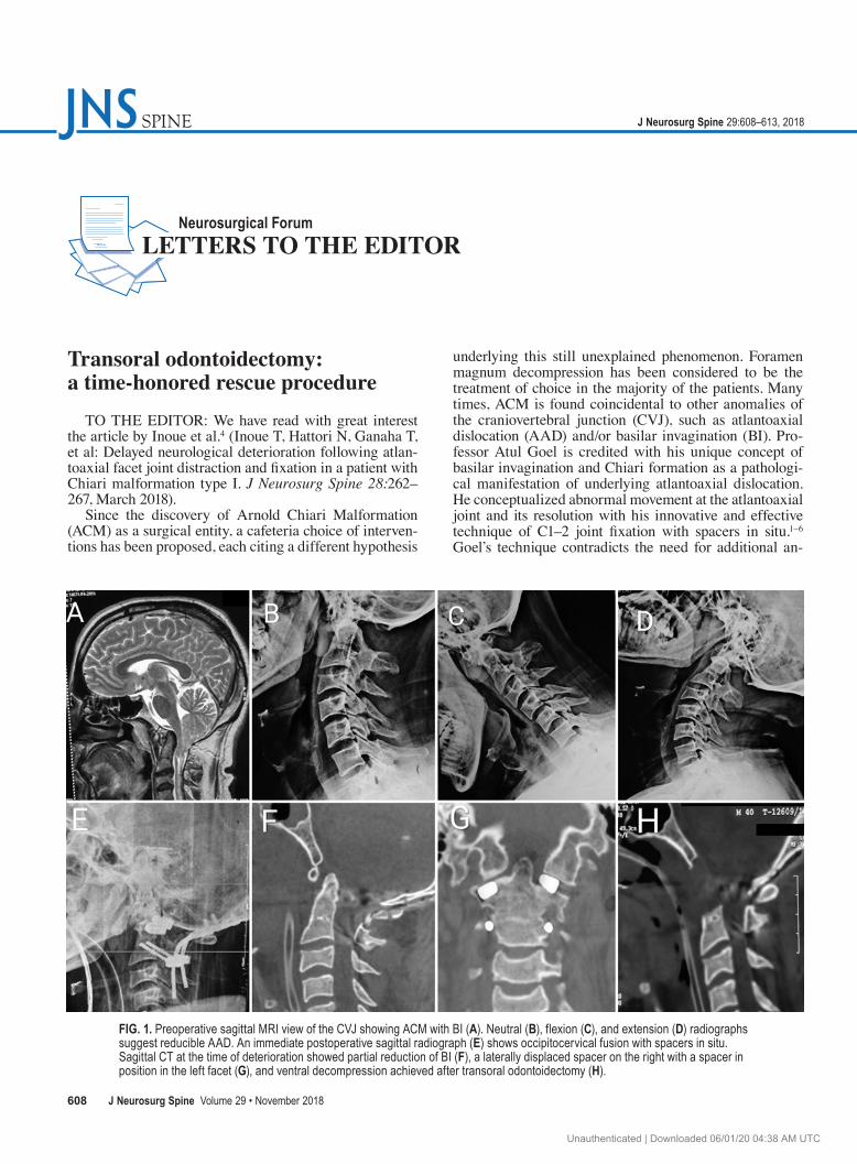
FIG. 1. Preoperative sagittal MRI view of the CVJ showing ACM with BI (A). Neutral (B), flexion (C), and extension (D) radiographs suggest reducible AAD. An immediate postoperative sagittal radiograph (E) shows occipitocervical fusion with spacers in situ. Sagittal CT at the time of deterioration showed partial reduction of BI (F), a laterally displaced spacer on the right with a spacer in position in the left facet (G), and ventral decompression achieved after transoral odontoidectomy (H).
Unauthenticated | Downloaded 06/01/20 04:38 AM UTC
J Neurosurg Spine Volume 29 • November 2018 609
Neurosurgical forum
terior odontoidectomy in the majority of patients and we have also found this technique quite useful in the majority of our patients.1–3
However, we recently had a similar experience in one patient with ACM type 1, BI, and reducible AAD (Fig. 1A–D) as that reported by Inoue et al.4 A 40-year-old man presented with features of progressive suboccipital neck pain, spastic quadriparesis, and lower cranial nerve weak-ness for 3 months’ duration. Similar to Goel’s technique, we tried distraction and C1 lateral mass and C2 pars screw fixation. However, due to technical difficulties and the presence of an anomalous vertebral artery (identified in-traoperatively), C1 lateral mass screws could not be placed and occipitoaxial fusion with contoured rod and screw placement with spacers in C1–2 facet joints was achieved (Fig. 1E and F).
The immediate postoperative period was uneventful and the patient improved symptomatically in the form of reduced spasticity. On postoperative day 6 the patient developed respiratory distress and sudden-onset flaccid quadriparesis (1–2/5). Postoperative CT of the CVJ re-vealed partial correction of BI with a spacer in situ in the left facet joint, with a slightly displaced right-sided spacer (which was not compressing the spinal cord). The dens was still angulated at the anterior surface of the cord (Fig. 1G). The patient was completely ventilator-dependent with persistent spasticity and worsened quadriparesis (2/5). After a month of ventilator support, the patient underwent transoral odontoidectomy and ventral decompression of the cord (Fig. 1H). The patient made a remarkable recov-ery in the immediate postoperative period, with motor recovery to his preoperative status, and was successfully weaned off ventilatory support within 10 days and dis-charged home.
We would like to congratulate and thank the authors for their insightful article.4 Fortunately, this article was pub-lished at the time of the similar clinical dilemma in our patient and we opted for anterior odontoidectomy with a good clinical outcome.
Ravi Bharatbhai Chauhan, MChAyusman Satapathy, MCh Sandeep Mohindra, MCh
Manjul Tripathi, MCh Aman Batish, MSSumit Dave, MCh
Postgraduate Institute of Medical Education and Research, Chandigarh, India
References 1. Goel A: Basilar invagination, syringomyelia and Chiari
formation and their relationship with atlantoaxial instability. Neurol India 66:940–942, 2018
2. Goel A: Is atlantoaxial instability the cause of Chiari malfor-mation? Outcome analysis of 65 patients treated by atlanto-axial fixation. J Neurosurg Spine 22:116–127, 2015
3. Goel A: Treatment of basilar invagination by atlantoaxial joint distraction and direct lateral mass fixation. J Neuro-surg Spine 1:281–286, 2004
4. Inoue T, Hattori N, Ganaha T, Kumai T, Tateyama S, Hirose Y: Delayed neurological deterioration following atlantoaxial
facet joint distraction and fixation in a patient with Chiari malformation type I. J Neurosurg Spine 28:262–267, 2018
5. Mummaneni PV, Haid RW: Transoral odontoidectomy. Neu-rosurgery 56:1045–1050, 2005
6. Salunke P: Delineate, yet not dread: anomalous vertebral artery in pediatric congenital atlantoaxial dislocation and basilar invagination. J Pediatr Neurosci 12:227–231, 2017
DisclosuresThe authors report no conflict of interest.
CorrespondenceSandeep Mohindra: [email protected].
INCLUDE WHEN CITING Published online August 3, 2018; DOI: 10.3171/2018.4.SPINE18424.
Response We are very happy to hear that our report was support-
ive of the patient rescue in your case. After the publication of our paper, we experienced two other similar cases (CM type I with BI). From these recent cases, we realized that some data were not included in our previous report.
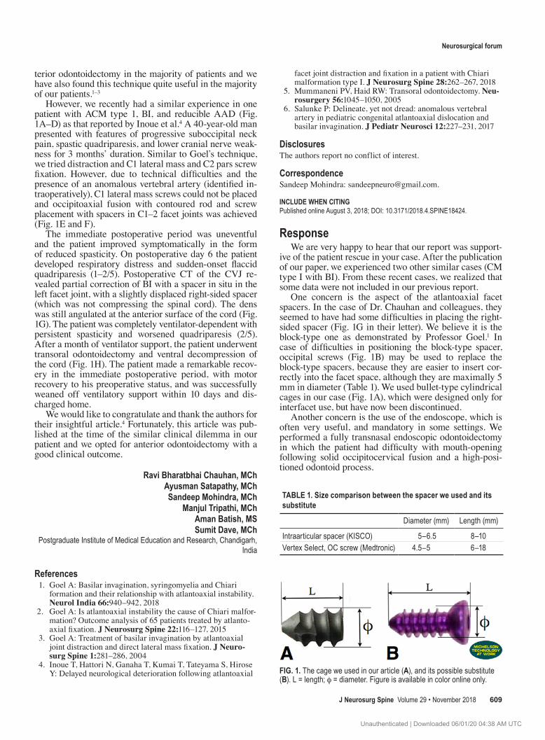
One concern is the aspect of the atlantoaxial facet spacers. In the case of Dr. Chauhan and colleagues, they seemed to have had some difficulties in placing the right-sided spacer (Fig. 1G in their letter). We believe it is the block-type one as demonstrated by Professor Goel.1 In case of difficulties in positioning the block-type spacer, occipital screws (Fig. 1B) may be used to replace the block-type spacers, because they are easier to insert cor-rectly into the facet space, although they are maximally 5 mm in diameter (Table 1). We used bullet-type cylindrical cages in our case (Fig. 1A), which were designed only for interfacet use, but have now been discontinued.
Another concern is the use of the endoscope, which is often very useful, and mandatory in some settings. We performed a fully transnasal endoscopic odontoidectomy in which the patient had difficulty with mouth-opening following solid occipitocervical fusion and a high-posi-tioned odontoid process.
TABLE 1. Size comparison between the spacer we used and its substitute
Diameter (mm) Length (mm)
Intraarticular spacer (KISCO) 5–6.5 8–10Vertex Select, OC screw (Medtronic) 4.5–5 6–18
FIG. 1. The cage we used in our article (A), and its possible substitute (B). L = length; f = diameter. Figure is available in color online only.
Unauthenticated | Downloaded 06/01/20 04:38 AM UTC

Neurosurgical forum
J Neurosurg Spine Volume 29 • November 2018610
We believe meticulous preparation, including spacers and endoscopes, would enhance the rescue options and lead to the best clinical results.
Tatsushi Inoue, MD, PhDKeisuke Ito, MD
Yuya Nishiyama, MD, PhDYohei Takahashi, MD, PhD
Takashi Tsuji, MD, PhDYuichi Hirose, MD, PhD
Fujita Health University, Aichi, Japan
References 1. Goel A: Treatment of basilar invagination by atlantoaxial
joint distraction and direct lateral mass fixation. J Neuro-surg Spine 1:281–286, 2004
INCLUDE WHEN CITING Published online August 3, 2018; DOI: 10.3171/2018.6.SPINE18533.
©AANS 2018, except where prohibited by US copyright law
Proximal junctional kyphosis and proximal junctional failure
TO THE EDITOR: We read with great interest the out-standing article by Safaee and colleagues4 (Safaee MM, Deviren V, Dalle Ore C, et al: Ligament augmentation for prevention of proximal junctional kyphosis and proximal junctional failure in adult spinal deformity. J Neurosurg Spine 28:512–519, May 2018). The authors compared out-comes of proximal junctional kyphosis/failure (PJK/PJF) between a historical cohort and a treatment group of adult spinal deformity (ASD) patients receiving ligamentous augmentation at the upper instrumented vertebra (UIV), 1 level above the UIV (UIV+1), and 1 level below the UIV (UIV-1). Ligamentous augmentation was performed by passing 2 sublaminar cables through each of these levels, tightening them to achieve the desired tension, and lock-ing them into place onto bilateral rods. The results suggest that ligamentous augmentation at the junctional levels (the junction of fused and adjacent unfused segments) may in-deed be an effective strategy for PJK/PJF prevention in ASD cases.
These results are consistent with our own institution’s retrospective analysis of consecutive ASD operations per-formed with a similar ligamentous augmentation tech-nique. We previously reported that posteriorly anchored junctional tethers (tethering the proximal terminus of fusion constructs to UIV+1 with polyethylene tape) sig-nificantly reduced the occurrence of PJK.2 Despite these positive clinical results, the exact mechanism for how ligamentous augmentation prevents PJK is incompletely understood. Current preliminary evidence from a finite el-ement analysis by Bess et al. suggests that posterior tethers mitigate adjacent-segment stress by attenuating the abrupt biomechanical transition between the rigid instrumented spine and adjacent unfused segments.1
Safaee et al. astutely pointed out that their ligament augmentation cohort contained a greater proportion of pa-tients with UIV hook fixation and vertebroplasty.4 These techniques may also prevent PJK.3,5 The authors con-trolled for the differences in rates of UIV hook fixation and vertebroplasty with multivariate analysis.
For future studies investigating PJK/PJF prophylaxis, Safaee et al. proposed a more standardized metric by re-porting the magnitude of change in the proximal junction-al angle (PJA) rather than the binary outcome of PJK. We agree that reporting the change in PJA would be helpful for more reliable analysis across studies, since definitions of PJK are variable. In our prior study, the mean change in PJA was 8° (ligamentous augmentation with tether-cross-link) compared with 13° (historical control).2 Again, our results are consistent with the current study: Safaee et al. reported that the mean change in PJA was 6° (ligamentous augmentation with sublaminar cables) compared with 14° (historical control).
The current investigation and our prior study both re-port possible benefit from ligamentous augmentation tech-niques, but they do not offer guidelines for pretensioning. Tension of the ligamentous augmentation technique (tether or sublaminar cable) is left to the discretion of the surgeon. We think that there is likely an optimal range of preten-sioning for maximal PJK prevention. In fact, overtighten-ing may actually predispose to PJK at the level above the proximal attachment of the tether or cable. Therefore, if tension meters are utilized and pretensioning is reported in future investigations, this could help create more stan-dardized guidelines for ligamentous augmentation tech-niques in ASD.
Again, we commend the authors on this excellent study.
Thomas J. Buell, MDDavis G. Taylor, MD
Ching-Jen Chen, MDChristopher I. Shaffrey, MD
Justin S. Smith, MD, PhDUniversity of Virginia Health System, Charlottesville, VA
Shay Bess, MDPresbyterian St. Luke’s/Rocky Mountain Hospital for Children, Denver, CO
References 1. Bess S, Harris JE, Turner AW, LaFage V, Smith JS, Shaffrey
CI, et al: The effect of posterior polyester tethers on the bio-mechanics of proximal junctional kyphosis: a finite element analysis. J Neurosurg Spine 26:125–133, 2017
2. Buell TJ, Buchholz AL, Quinn JC, Bess S, Line BG, Ames CP, et al: A pilot study on posterior polyethylene tethers to prevent proximal junctional kyphosis after multilevel spinal instrumentation for adult spinal deformity. Oper Neurosurg (Hagerstown) [epub ahead of print], 2018
3. Ghobrial GM, Eichberg DG, Kolcun JPG, Madhavan K, Lebwohl NH, Green BA, et al: Prophylactic vertebral cement augmentation at the uppermost instrumented vertebra and rostral adjacent vertebra for the prevention of proximal junctional kyphosis and failure following long-segment fusion for adult spinal deformity. Spine J 17:1499–1505, 2017
4. Safaee MM, Deviren V, Dalle Ore C, Scheer JK, Lau D, Osorio JA, et al: Ligament augmentation for prevention of proximal junctional kyphosis and proximal junctional failure
Unauthenticated | Downloaded 06/01/20 04:38 AM UTC

J Neurosurg Spine Volume 29 • November 2018 611
Neurosurgical forum
in adult spinal deformity. J Neurosurg Spine 28:512 –519, 2018
5. Thawrani DP, Glos DL, Coombs MT, Bylski-Austrow DI, Sturm PF: Transverse process hooks at upper instrumented vertebra provide more gradual motion transition than pedicle screws. Spine (Phila Pa 1976) 39:E826–832, 2014
DisclosuresBess: research support from K2M, NuVasive, Medtronic, DePuy Synthes, ZimmerBiomet, Allosource, Orthofix, and EOS; con-sultant for K2M, Allosource, DePuy Synthes, Misonix, and EOS; and patent holder with K2M and Innovasis. Shaffrey: consultant for Medtronic, NuVasive, Zimmer Biomet, and K2M; and pat-ent holder with and royalties from Medtronic, NuVasive, and Zimmer Biomet; stock holder with NuVasive. Smith: royalties from Zimmer Biomet; consultant for Zimmer Biomet, Cerapedics, NuVasive, K2M, and AlloSource; honoraria from Zimmer Biomet, NuVasive, and K2M; research support from DePuy Synthes and ISSGF; and fellowship support from NREF and AOSpine.
CorrespondenceThomas J. Buell: [email protected].
INCLUDE WHEN CITING Published online August 3, 2018; DOI: 10.3171/2018.5.SPINE18636.
ResponseWe appreciate the careful attention to our paper by
Buell et al. and are pleased with their results. Of particular interest are the similarities between PJA changes in liga-ment augmentation groups compared with historical con-trols across both studies. These data provide preliminary evidence that our reductions in PJK and PJF with liga-ment augmentation are indeed reproducible. With respect to tensioning of the tether, the force should be adjusted to account for bone quality and robustness of the spinous pro-cess fixation points. We prefer hand tensioning to allow the surgeon to determine the ultimate force applied in light of the above considerations. In anticipation of future work in this area, we would emphasize that technique is critical to success, and not all tethering techniques are similar. We agree that pretensioning is critical to impart an extension moment to the spine. Dr. Buell and his colleagues should be commended for their work, and we are optimistic that ligament augmentation will play an increasingly important role in PJK/PJF prevention in surgery for adult spinal de-formity.
Michael M. Safaee, MDChristopher P. Ames, MD
University of California, San Francisco, CA
INCLUDE WHEN CITING Published online August 3, 2018; DOI: 10.3171/2018.7.SPINE18654.
©AANS 2018, except where prohibited by US copyright law
Class imbalance in machine learning for neurosurgical outcome prediction: are our models valid?
TO THE EDITOR: The article by Scheer and col-leagues3 on predicting major complications in adult spinal deformity surgery was very much appreciated (Scheer JK, Smith JS, Schwab F, et al: Development of a preoperative predictive model for major complications following adult spinal deformity surgery. J Neurosurg Spine 26:736–743, June 2017).
Dr. Scheer and colleagues trained an accurate ma-chine learning (ML) model to predict complications us-ing a range of preoperatively available patient and surgi-cal features. We applaud the sound methodology that was implemented. Using multiple bootstrapped decision trees, the authors trained a highly effective predictive model that achieved an area under the curve (AUC) of 0.89 and ac-curacy of 87% at internal validation. However, no sensi-tivity or specificity was reported. We believe that, due to the rigorous methodology that Dr. Scheer and colleagues applied, the reported AUC and accuracy probably give a valid impression of their powerful ML model. However, we would like to stress the general importance of consid-ering class imbalance in this context.
Class imbalance is present when one class, the minority class, is much rarer than the majority class. ML models extract features better and are most robust if all classes are approximately equally distributed. If considerable class imbalance is present, ML models will often become “lazy” in learning how to discriminate between classes and instead choose to simply vote for the majority class. This bias provides synthetically high AUC, accuracy, and specificity but unemployable sensitivity. The “accuracy paradox” denotes the situation when synthetically high accuracy only reflects the underlying class distribution in unbalanced data.
As an example, one might want to predict complica-tions from a registry containing 90% of patients without complications. By largely voting for the majority class (no complication), the model would achieve an accuracy and specificity of around 90% and very low sensitivity without actually learning from the data. This can be countered by adjusting class weights within the model, by undersam-pling and thus removing observations from the majority class, or by oversampling the minority class.1 Specifically, the synthetic minority oversampling technique (SMOTE) has been validated, shows robust performance, and is easy to employ.2 SMOTE simulates new observations for the minority class by using k-means clustering.
Neurosurgical data are often prone to class imbalance. With the emergence of many studies that aim to predict neurosurgical outcomes using ML, it is crucial to ensure methodological quality. The study by Scheer et al. showed only moderate class imbalance and probably represents a valid predictive model. In general, if class imbalance is present, care should be taken to weight classes or to under- or oversample using data science techniques like SMOTE.
Unauthenticated | Downloaded 06/01/20 04:38 AM UTC

Neurosurgical forum
J Neurosurg Spine Volume 29 • November 2018612
Accuracy and AUC alone do not always give a full repre-sentation of an ML model’s performance. In our view, ad-ditionally reporting the sensitivity and specificity is central.
Victor E. Staartjes, BMedMarc L. Schröder, MD, PhD
Bergman Clinics, Amsterdam, the Netherlands
References 1. Batista GEAPA, Prati RC, Monard MC: A study of the
behavior of several methods for balancing machine learning training data. SIGKDD Explor Newsl 6:20–29, 2004
2. Chawla NV, Bowyer KW, Hall LO, Kegelmeyer WP: SMOTE: Synthetic Minority Over-sampling Technique. J Artif Intell Res 16:321–357, 2002
3. Scheer JK, Smith JS, Schwab F, Lafage V, Shaffrey CI, Bess S, et al: Development of a preoperative predictive model for major complications following adult spinal deformity surgery. J Neurosurg Spine 26:736–743, 2017
DisclosuresThe authors report no conflicts of interest.
CorrespondenceVictor E. Staartjes: [email protected].
INCLUDE WHEN CITING Published online August 17, 2018; DOI: 10.3171/2018.5.SPINE18543.
ResponseNo response was received from the authors of the origi-
nal article.©AANS 2018, except where prohibited by US copyright law
Second cancer risk in patients with spinal ependymomas
TO THE EDITOR: We read with great interest the re-cent article by Wostrack et al.5 on surgical outcome and progression-free survival (PFS) of patients treated for spinal ependymomas (Wostrack M, Ringel F, Eicker SO, et al: Spinal ependymoma in adults: a multicenter investi-gation of surgical outcome and progression-free survival. J Neurosurg Spine 28:654–662, June 2018). The authors evaluated the clinical and histological parameters of 158 patients with spinal ependymomas in order to identify pre-dictive factors for postoperative morbidity, tumor resect-ability, and recurrence. The results showed that gross-total resection (GTR) can be performed in 80% of patients. In the follow-up examinations, there was improvement of the functional outcome mainly in patients without disabling deficits before surgery. The 5-year PFS rate was 80%. The GTR, WHO grade, and Ki-67 index were independent prognostic factors for PFS.5
We have previously reported on a series of 14 adult patients, with histologically confirmed spinal ependymo-mas, who underwent resection in our institute over a 10-year period.4 Based on Frankel scale score, 8 patients had
postoperative improvement. Preoperative longer symptom duration was associated with poorer functional outcome. Of interest, after a mean follow-up period of 5.1 years, a second primary neoplasia occurred in 4 patients. There were 2 cases of gastric cancer, 1 case of lung cancer, and 1 case of anaplastic astrocytoma.4 Second primary cancer in patients with spinal ependymomas has also been report-ed previously.2,3 Halvorsen et al. studied 86 patients with spinal ependymomas, and during the follow-up period, 5 patients developed a second primary malignancy.3 In gen-eral, various reasons have been suggested for patients di-agnosed with more than one primary cancer, and several pairs of primary malignancies have been identified, but not for ependymomas.1 Since the prognosis of spinal epen-dymomas after GTR is favorable, we believe that patients diagnosed with this disease might have an increased risk for developing a second malignancy. Such information is clinically important in order to emphasize the importance of regular cancer screening tests and propose modifica-tions of individuals’ lifestyle choices, such as smoking ces-sation. Further studies are obviously needed to quantify this cancer risk and to identify patient and treatment-relat-ed factors, such as radiotherapy, that might influence the second cancer risk.
George A. Alexiou, MD, PhDSpyridon Voulgaris, MD, PhD
University Hospital of Ioannina, Greece
References 1. Bajdik CD, Abanto ZU, Spinelli JJ, Brooks-Wilson A, Gal-
lagher RP: Identifying related cancer types based on their in-cidence among people with multiple cancers. Emerg Themes Epidemiol 3:17, 2006
2. Boström A, von Lehe M, Hartmann W, Pietsch T, Feuss M, Boström JP, et al: Surgery for spinal cord ependymomas: outcome and prognostic factors. Neurosurgery 68:302–308, 2011
3. Halvorsen CM, Kolstad F, Hald J, Johannesen TB, Krossnes BK, Langmoen IA, et al: Long-term outcome after resection of intraspinal ependymomas: report of 86 consecutive cases. Neurosurgery 67:1622–1631, 2010
4. Voulgaris S, Alexiou GA, Zigouris A, Fotakopoulos G, Michos E, Katsiafas I, et al: Spinal ependymomas: prognostic factors and treatment results. J Cancer Res Ther 9:60–63, 2013
5. Wostrack M, Ringel F, Eicker SO, Jägersberg M, Schaller K, Kerschbaumer J, et al: Spinal ependymoma in adults: a multi-center investigation of surgical outcome and progression-free survival. J Neurosurg Spine 28:654–662, 2018
DisclosuresThe authors report no conflict of interest.
CorrespondenceGeorge Alexiou: [email protected]; [email protected].
INCLUDE WHEN CITING Published online August 24, 2018; DOI: 10.3171/2018.6.SPINE18719.
ResponseWe thank the authors very much for their interest in
our article.
Unauthenticated | Downloaded 06/01/20 04:38 AM UTC

J Neurosurg Spine Volume 29 • November 2018 613
Neurosurgical forum
Indeed, this is a very interesting comment on the devel-opment of secondary cancer in patients with spinal epen-dymomas. Unfortunately, we did not examine this feature in our series.
The association between primary malignancies and glial tumors has been previously reported.1,3
The British Childhood Cancer Survivor Study report-ed the overall standardized incidence ratio of subsequent neoplasms in patients surviving primary pediatric cancer to be 4 times higher than expected, whereas the majority of the lesions were located in the central nervous system (CNS).5 Both therapy-induced malignancy and cancer pre-disposition syndromes, such as neurofibromatosis types 1 and 2 and Li-Fraumeni syndrome, resulting in acquired or congenital mutation of tumor suppressor and mismatch repair genes have been discussed as potential causes for the development of secondary CNS tumors.4
In fact, the main problem is the lack of information concerning the involved molecular genetics of ependymo-mas and associated specific growth factors. Only a very limited number of studies focusing on this issue are avail-able. Recent transcriptional and copy number profiling of ependymomas has identified that DNA methylation may be critical to ependymoma pathogenesis.2 Spinal ependy-momas seem to display a hypermethylated phenotype that includes the loss of tumor suppressor genes involved in the control of cell growth and death.6
In our opinion, a focused study of the molecular biol-ogy of spinal ependymomas would not only clarify the is-sue of coincidence with secondary cancer but also bring clarity to the currently totally disparate WHO classifica-tion of this disease. By the way, in their series, Voulgaris et al. also observed a trend toward a higher risk of recur-rence in presumably benign WHO grade I myxopapillary ependymomas.7
Maria Wostrack, MDKlinikum rechts der Isar, Technical University of Munich, Germany
References 1. Izycka-Swieszewska E, Bien E, Stefanowicz J, Szurowska
E, Szutowicz-Zielinska E, Koczkowska M, et al: Malignant gliomas as second neoplasms in pediatric cancer survivors: neuropathological study. Biomed Res Int 2018:4596812, 2018
2. Mack SC, Witt H, Wang X, Milde T, Yao Y, Bertrand KC, et al: Emerging insights into the ependymoma epigenome. Brain Pathol 23:206–209, 2013
3. Maluf FC, DeAngelis LM, Raizer JJ, Abrey LE: High-grade gliomas in patients with prior systemic malignancies. Cancer 94:3219–3224, 2002
4. Marks AM, Packer RJ: A review of secondary central ner-vous system tumors after treatment of a primary pediatric malignancy. Semin Pediatr Neurol 19:43–48, 2012
5. Reulen RC, Frobisher C, Winter DL, Kelly J, Lancashire ER, Stiller CA, et al: Long-term risks of subsequent primary neoplasms among survivors of childhood cancer. JAMA 305:2311–2319, 2011
6. Rogers HA, Kilday JP, Mayne C, Ward J, Adamowicz-Brice M, Schwalbe EC, et al: Supratentorial and spinal pediatric ependymomas display a hypermethylated phenotype which includes the loss of tumor suppressor genes involved in the control of cell growth and death. Acta Neuropathol 123:711–725, 2012
7. Voulgaris S, Alexiou GA, Zigouris A, Fotakopoulos G, Michos E, Katsiafas I, et al: Spinal ependymomas: prognostic factors and treatment results. J Cancer Res Ther 9:60–63, 2013
INCLUDE WHEN CITING Published online August 24, 2018; DOI: 10.3171/2018.7.SPINE18723.
©AANS 2018, except where prohibited by US copyright law
Unauthenticated | Downloaded 06/01/20 04:38 AM UTC