Embed Size (px)
Citation preview

Final Scientific Program
Time schedule of keynote lectures and symposiaList of Poster SessionsList of Participants
Abstracts SpeakersAbstracts Posters
Program Commi*ee:
Tobias BonhoefferNils BroseAlois SariaStephan Schwarzacher
Organizer:brainplatform.net e.U.
Conference Chair:Alois Saria, Austria
Contributors:
-‐ Austrian Neuroscience AssociaBon-‐ InternaBonal Society for Neurochemistry -‐ Das Central-‐ AB Sciex Austria GmbH-‐ Zeiss-‐ Acal-‐BFI-‐ HORIBA ScienBfic-‐ Peqlab-‐ TSE Systems GmbH
Exhibitors:
-‐ Sigma Aldrich-‐ Novus Biologicals
NEUROSCIENCE WINTER CONFERENCE
Sölden AustriaApril 9 -April 13 2013
Das Central
15th
Inte
rnat
iona
l

PRO
GRA
M 2
013
TUES
DAY
TO T
HURS
DAY Tuesday
April 9
15:00 – 16:30 RegistraBon
16:30 -‐ 17:00 Welcome Cocktail
17:00 -‐ 19:00 Symposium 1MulBple facets of serotonin receptors in regulaBon of brain funcBonsChair: Evgeni Ponimaskin (Germany)
Vera Niederkofler (USA) Decoding the brain serotonergic system: Intersec4onal gene4cs and func4onal probingValerie Compan (France) Toggling the serotonin 5-‐HT4 receptors between acDve and silencing state switches moDvaDon from restricDve diet towards overeaDngAlexandre Dayer (Switzerland) RegulaDon of neuronal migraDon by the 5-‐HT6 receptorEvgeni Ponimaskin (Germany) Development-‐dependent regulaDon of synaptogenesis and synapDc plasDcity via serotonin receptors
WednesdayApril 10 Morning
08:15 – 09:00 Keynote Lecture 1Peter Scheiffele (Switzerland) Molecular diversity, recogniDon, and synapDc differenDaDon
09:00 – 11:00 Symposium 2Cell signaling in chronic diseasesChair: Gregor Wenning (Austria)
Poul Henning Jensen (Denmark) Oligodendroglial degeneraDon in mulDple systems atrophy – FAS and NF-‐kB related signalingTimothy Bredy (Australia) NeocorDcal Tet3-‐mediated DNA hydroxymethylaDon promotes rapid behavioural adaptaDonNicolas Singewald (Austria) Vulnerability to emoDonal trauma: Novel treatment strategies Jörg Striessnig (Austria) Voltage gated L-‐type calcium channels as drug targets in brain disorders
11:00 -‐ 11:30 Coffee Break
11:30 -‐ 13:30 Symposium 3RegulaBon of growth factor signals during neuronal development and regeneraBonChair: Lars Klimaschewski (Austria)
Georg Dechant (Austria) Role of the chromaDn organizer Satb2 in growth factor driven neuronal plasDcityPeter Claus (Germany) FGF signaling in neuronal developmentLars Klimaschewski (Austria) Novel approaches to accelerate axon elongaDon by enhancing FGF signalingKarel Dorey (UK) Molecular and cellular mechanisms regulaDng axonal branching
WednesdayApril 10 Afternoon16:00 – 16:45 Keynote Lecture 2Pierre-‐Marie Lledo (France) Impact of adult neurogenesis on olfacDon
16:45 – 17:15 Coffee Break
17:15 – 19:15 Symposium 4Stem cells and adult neurogenesisChairs: SebasDan Jessberger (Switzerland) Stephan Schwarzacher (Germany) Dieter Chichung Lie (Germany) SoxC transcripDon factors: BifuncDonal regulators of adult neurogenesisVerdon Taylor (Switzerland) Heterogeneous neural stem cells in health and regeneraDonSebasBan Jessberger (Germany) Metabolic control of adult neural stem cell acDvityBenedikt Berninger (Germany) Oligodendrogliogenic and neurogenic adult subependymal zone neural stem cells consDtute disDnct lineages and exhibit differenDal responsiveness to Wnt signaling
19:30 Gala Dinner (free for Das Central residents, others book at registraDon desk for 50,-‐ €)
ThursdayApril 11 Morning08:15 – 09:00 Keynote Lecture 3Michael Brecht (Germany) Social neural constructs in the rat barrel cortex
09:00 – 11:00 Symposium 5ISN Symposium: OptogeneBc control of moBon and moBvaBonChair: Ilka Diester (Germany)
IIka Diester (Germany) OptogeneDcs in the motor systemOfer Yizhar (Israel) OptogeneDc tool design and applicaDon in corDcal microcircuitsClaire Wyart (France) OptogeneDc dissecDon of spinal circuits underlying locomoDon in vertebratesChrisBan Lüscher (Switzerland) OptogeneDcs and addicDon
11:00 -‐ 11:30 Coffee Break
ThursdayApril 11 Afternoon16:00 – 18:00 Symposium 6Mouse models for auBsm spectrum disordersChairs: Yuri Bozzi (Italy) Michela Fagiolini (USA)
Michael Saxe (Switzerland) Rodent models of auDsm in drug discoveryYuri Bozzi (Italy) GABAergic dysfuncDon in engrailed-‐2 mutant miceJennifer Brielmaier (USA) Pharmacological reversal of depression-‐related and social deficits in engrailed-‐2 knockout miceMichela Fagiolini (USA) Circuit dissecDon in neurodevelopmental disorders
18:00 – 18:30 Coffee Break
18:30 – 20:30 Symposium 7Altered synapBc funcBon in mouse models for auBsm spectrum disordersChairs: Tobias Böckers (Germany)Carlo Sala (Italy)
Stephan Schwarzacher (Germany) Neuroligins as candidate genes for auDsm spectrum disorders: RegulaDon of synapDc acDvity and plasDcity in vivoMarkus Missler (Germany) Towards a pathomechanism of auDsm: RegulaDon of synapDc funcDon through Neurexophilin-‐1/a-‐Neurexin complex formaDonTobias Böckers (Germany) ProSAP/Shanks at the synapse: FuncDon, dynamics and their role in auDsm spectrum disordersCarlo Sala (Italy) The funcDon of the X-‐linked intellectual disability IL1RAPL1 protein complex at synapses
PRO
GRA
M 2
013
FRID
AY T
O SA
TURD
AY
FridayApril 12 Morning
08:15 – 09:00 Keynote Lecture 4Botond Roska (Switzerland) Cell type specific computaDons in reDna and cortex
09:00 – 11:00 Symposium 8Reinforcement and decision making in the developing brainChair: Gunter Schumann (UK)
Georgy Bakalkin (Sweden) Shid in epigeneDc regulaDon of opioid genes in brain of human alcoholicsSylvane Desrivieres (UK) IdenDficaDon and funcDonal characterisaDon of neurodevelopmental genes involved in human cogniDonGunter Schumann (UK) A genome-‐wide associaDon study of co-‐expression networks of brain acDvaDon during reward anDcipaDon
11:00 -‐ 11:30 Coffee Break
11:30 -‐ 13:30 Symposium 9FuncBon of the auditory system -‐ From the cochlea to auditory brainstemChair: Josef Syka (Czech Republic)
Pavel Mistrík and Jonathan Ashmore (UK) Molecular motor presDn in a cellular network and its role in sound amplificaDonWei Liu (Sweden) The structure of human cochleaMarlies Knipper (Germany) Learning about hearing from cell specific deleDon of genesJosef Syka (Czech Republic) Influence of postnatal acousDc sDmulaDon on neuronal responsiveness in the adult auditory midbrain in rat
FridayApril 12 Afternoon
16:00 – 18:00 Symposium 10Physiology of oxytocin and vasopressin in the central and peripheral systemChair: Eva Sykova (Czech Republic)Govindan Dayanithi (France)
Govindan Dayanithi (France) Calcium homeostasis in the hypothalamic vasopressin and oxytocin neurons and terminalsEva Sykova (Czech Republic) ExtrasynapDc volume transmission and diffusion parameters of the extracellular spaceHana Zemkova (Czech Republic) PotenDaDon of neurotransmifer release in neurons of supraopDc nuclus by presynapDc P2X receptorsIzumi Shibuya (Japan) AcDons of vasopressin in dorsal root ganglion
18:00 – 19:30 Coffee and Poster Session
Saturday April 13
08:15 – 09:00 Keynote Lecture 5Karl Deisseroth (USA) OpDcal deconstrucDon of fully-‐assembled biological systems
09:00 – 09:30 Coffee Break
09:30 – 11:30 Symposium 11Signal integraBon by astroglia: new insightsChair: Dmitri Rusakov (UK)
ChrisBan Steinhaeuser (Germany) DisDnct astrocyte network communicaDon in the thalamus Alfonso Araque (Spain) SynapDc funcDon regulated by astroglia via endocannabinoid and cholinergic signalingBrian MacVicar (Canada) Neuron-‐Glia signaling to maintain a healthy brainDmitri Rusakov (UK) Deciphering key elements of Ca2+ signal processing in astrocytes
11:30 End of meeBng and departure
POST
ER P
RESE
NTAT
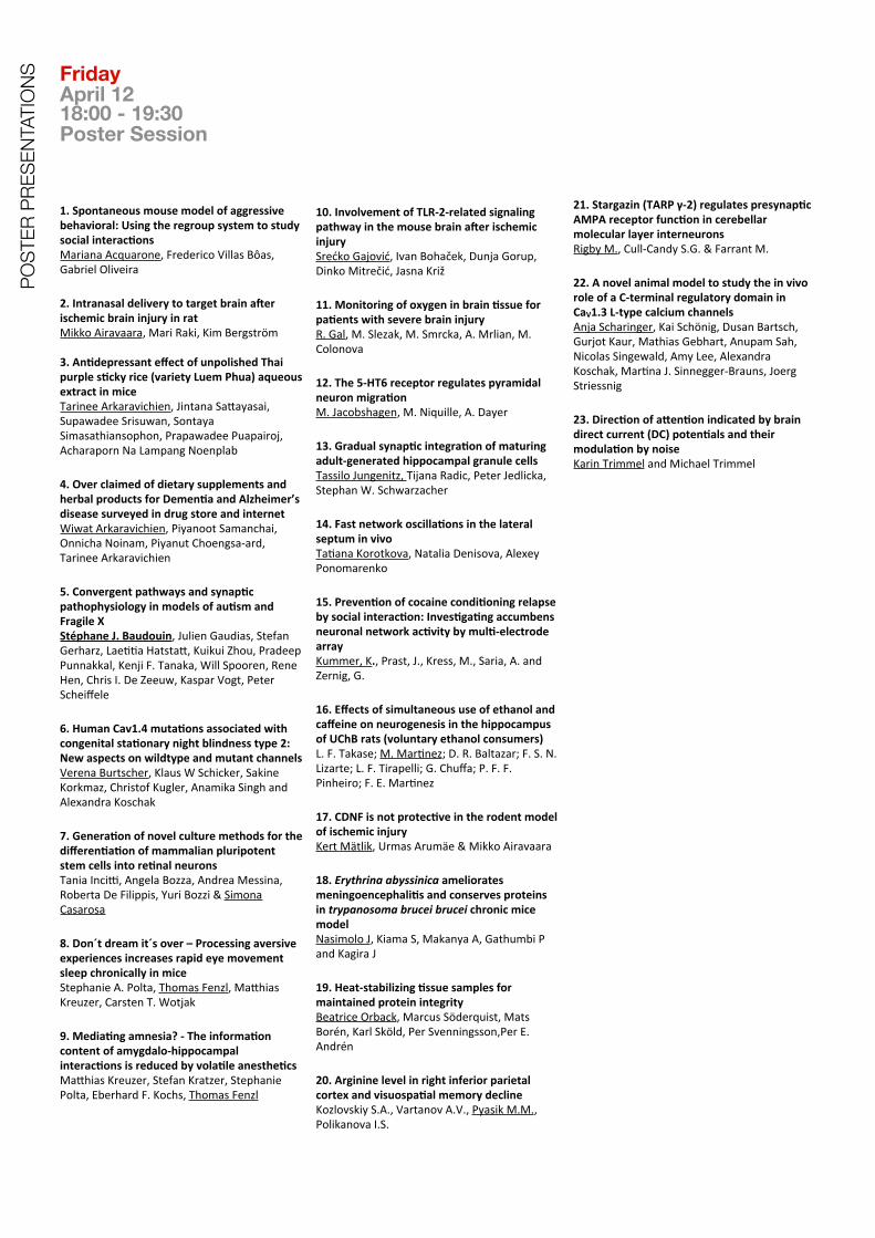
IONS Friday
April 1218:00 - 19:30 Poster Session
1. Spontaneous mouse model of aggressive behavioral: Using the regroup system to study social interacBonsMariana Acquarone, Frederico Villas Bôas, Gabriel Oliveira
2. Intranasal delivery to target brain aler ischemic brain injury in ratMikko Airavaara, Mari Raki, Kim Bergström
3. AnBdepressant effect of unpolished Thai purple sBcky rice (variety Luem Phua) aqueous extract in miceTarinee Arkaravichien, Jintana Safayasai, Supawadee Srisuwan, Sontaya Simasathiansophon, Prapawadee Puapairoj, Acharaporn Na Lampang Noenplab
4. Over claimed of dietary supplements and herbal products for DemenBa and Alzheimer’s disease surveyed in drug store and internetWiwat Arkaravichien, Piyanoot Samanchai, Onnicha Noinam, Piyanut Choengsa-‐ard, Tarinee Arkaravichien
5. Convergent pathways and synapBc pathophysiology in models of auBsm and Fragile XStéphane J. Baudouin, Julien Gaudias, Stefan Gerharz, LaeDDa Hatstaf, Kuikui Zhou, Pradeep Punnakkal, Kenji F. Tanaka, Will Spooren, Rene Hen, Chris I. De Zeeuw, Kaspar Vogt, Peter Scheiffele
6. Human Cav1.4 mutaBons associated with congenital staBonary night blindness type 2: New aspects on wildtype and mutant channelsVerena Burtscher, Klaus W Schicker, Sakine Korkmaz, Christof Kugler, Anamika Singh and Alexandra Koschak
7. GeneraBon of novel culture methods for the differenBaBon of mammalian pluripotent stem cells into reBnal neuronsTania Incil, Angela Bozza, Andrea Messina, Roberta De Filippis, Yuri Bozzi & Simona Casarosa
8. Don´t dream it´s over – Processing aversive experiences increases rapid eye movement sleep chronically in miceStephanie A. Polta, Thomas Fenzl, Mafhias Kreuzer, Carsten T. Wotjak
9. MediaBng amnesia? -‐ The informaBon content of amygdalo-‐hippocampal interacBons is reduced by volaBle anestheBcsMafhias Kreuzer, Stefan Kratzer, Stephanie Polta, Eberhard F. Kochs, Thomas Fenzl
10. Involvement of TLR-‐2-‐related signaling pathway in the mouse brain aler ischemic injurySrećko Gajović, Ivan Bohaček, Dunja Gorup, Dinko Mitrečić, Jasna Križ
11. Monitoring of oxygen in brain Bssue for paBents with severe brain injuryR. Gal, M. Slezak, M. Smrcka, A. Mrlian, M. Colonova
12. The 5-‐HT6 receptor regulates pyramidal neuron migraBonM. Jacobshagen, M. Niquille, A. Dayer
13. Gradual synapBc integraBon of maturing adult-‐generated hippocampal granule cellsTassilo Jungenitz, Tijana Radic, Peter Jedlicka, Stephan W. Schwarzacher
14. Fast network oscillaBons in the lateral septum in vivoTaDana Korotkova, Natalia Denisova, Alexey Ponomarenko
15. PrevenBon of cocaine condiBoning relapse by social interacBon: InvesBgaBng accumbens neuronal network acBvity by mulB-‐electrode arrayKummer, K., Prast, J., Kress, M., Saria, A. and Zernig, G.
16. Effects of simultaneous use of ethanol and caffeine on neurogenesis in the hippocampus of UChB rats (voluntary ethanol consumers)L. F. Takase; M. MarDnez; D. R. Baltazar; F. S. N. Lizarte; L. F. Tirapelli; G. Chuffa; P. F. F. Pinheiro; F. E. MarDnez
17. CDNF is not protecBve in the rodent model of ischemic injuryKert Mätlik, Urmas Arumäe & Mikko Airavaara
18. Erythrina abyssinica ameliorates meningoencephaliBs and conserves proteins in trypanosoma brucei brucei chronic mice modelNasimolo J, Kiama S, Makanya A, Gathumbi P and Kagira J
19. Heat-‐stabilizing Bssue samples for maintained protein integrityBeatrice Orback, Marcus Söderquist, Mats Borén, Karl Sköld, Per Svenningsson,Per E. Andrén
20. Arginine level in right inferior parietal cortex and visuospaBal memory declineKozlovskiy S.A., Vartanov A.V., Pyasik M.M., Polikanova I.S.
21. Stargazin (TARP γ-‐2) regulates presynapBc AMPA receptor funcBon in cerebellar molecular layer interneuronsRigby M., Cull-‐Candy S.G. & Farrant M.
22. A novel animal model to study the in vivo role of a C-‐terminal regulatory domain in CaV1.3 L-‐type calcium channelsAnja Scharinger, Kai Schönig, Dusan Bartsch, Gurjot Kaur, Mathias Gebhart, Anupam Sah, Nicolas Singewald, Amy Lee, Alexandra Koschak, MarDna J. Sinnegger-‐Brauns, Joerg Striessnig
23. DirecBon of a*enBon indicated by brain direct current (DC) potenBals and their modulaBon by noiseKarin Trimmel and Michael Trimmel
LIST
OF
PART
ICIP
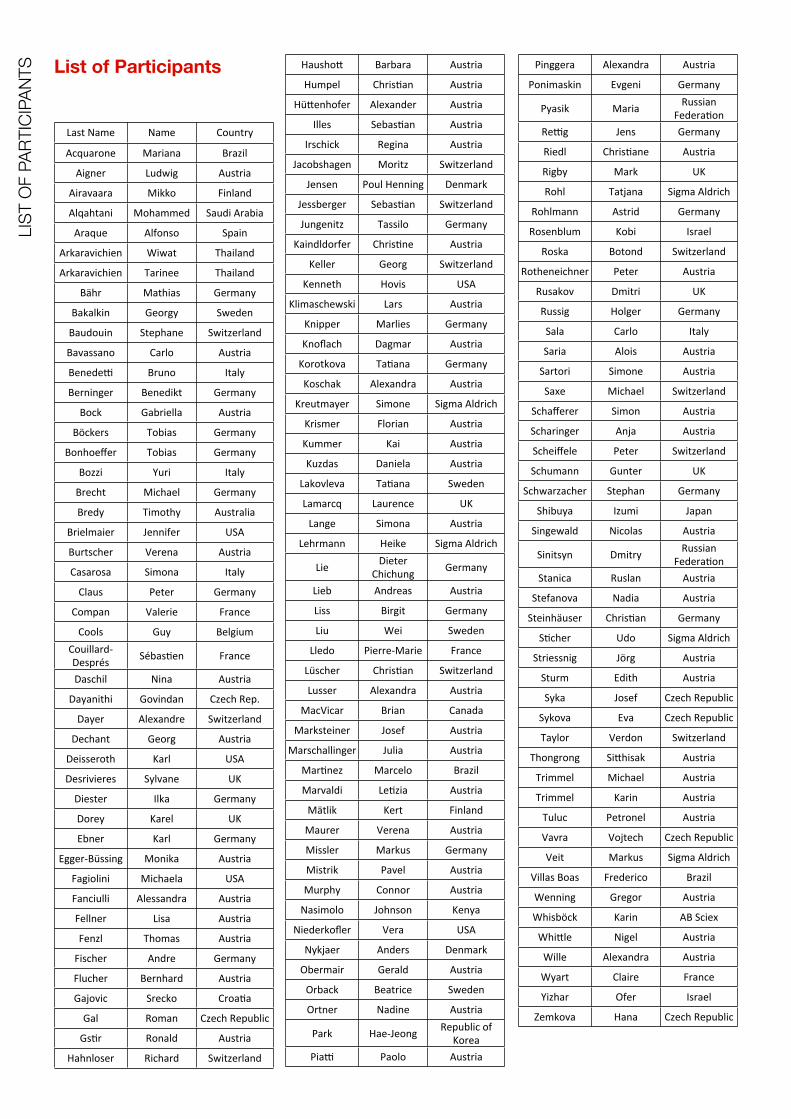
ANTS List of Participants
Last Name Name Country
Acquarone Mariana Brazil
Aigner Ludwig Austria
Airavaara Mikko Finland
Alqahtani Mohammed Saudi Arabia
Araque Alfonso Spain
Arkaravichien Wiwat Thailand
Arkaravichien Tarinee Thailand
Bähr Mathias Germany
Bakalkin Georgy Sweden
Baudouin Stephane Switzerland
Bavassano Carlo Austria
Benedel Bruno Italy
Berninger Benedikt Germany
Bock Gabriella Austria
Böckers Tobias Germany
Bonhoeffer Tobias Germany
Bozzi Yuri Italy
Brecht Michael Germany
Bredy Timothy Australia
Brielmaier Jennifer USA
Burtscher Verena Austria
Casarosa Simona Italy
Claus Peter Germany
Compan Valerie France
Cools Guy BelgiumCouillard-‐Després SébasDen France
Daschil Nina Austria
Dayanithi Govindan Czech Rep.
Dayer Alexandre Switzerland
Dechant Georg Austria
Deisseroth Karl USA
Desrivieres Sylvane UK
Diester Ilka Germany
Dorey Karel UK
Ebner Karl Germany
Egger-‐Büssing Monika Austria
Fagiolini Michaela USA
Fanciulli Alessandra Austria
Fellner Lisa Austria
Fenzl Thomas Austria
Fischer Andre Germany
Flucher Bernhard Austria
Gajovic Srecko CroaDa
Gal Roman Czech Republic
GsDr Ronald Austria
Hahnloser Richard Switzerland
Haushof Barbara Austria
Humpel ChrisDan Austria
Hüfenhofer Alexander Austria
Illes SebasDan Austria
Irschick Regina Austria
Jacobshagen Moritz Switzerland
Jensen Poul Henning Denmark
Jessberger SebasDan Switzerland
Jungenitz Tassilo Germany
Kaindldorfer ChrisDne Austria
Keller Georg Switzerland
Kenneth Hovis USA
Klimaschewski Lars Austria
Knipper Marlies Germany
Knoflach Dagmar Austria
Korotkova TaDana Germany
Koschak Alexandra Austria
Kreutmayer Simone Sigma Aldrich
Krismer Florian Austria
Kummer Kai Austria
Kuzdas Daniela Austria
Lakovleva TaDana Sweden
Lamarcq Laurence UK
Lange Simona Austria
Lehrmann Heike Sigma Aldrich
Lie Dieter Chichung Germany
Lieb Andreas Austria
Liss Birgit Germany
Liu Wei Sweden
Lledo Pierre-‐Marie France
Lüscher ChrisDan Switzerland
Lusser Alexandra Austria
MacVicar Brian Canada
Marksteiner Josef Austria
Marschallinger Julia Austria
MarDnez Marcelo Brazil
Marvaldi LeDzia Austria
Mätlik Kert Finland
Maurer Verena Austria
Missler Markus Germany
Mistrik Pavel Austria
Murphy Connor Austria
Nasimolo Johnson Kenya
Niederkofler Vera USA
Nykjaer Anders Denmark
Obermair Gerald Austria
Orback Beatrice Sweden
Ortner Nadine Austria
Park Hae-‐Jeong Republic of Korea
Pial Paolo Austria
Pinggera Alexandra Austria
Ponimaskin Evgeni Germany
Pyasik Maria Russian FederaDon
Relg Jens Germany
Riedl ChrisDane Austria
Rigby Mark UK
Rohl Tatjana Sigma Aldrich
Rohlmann Astrid Germany
Rosenblum Kobi Israel
Roska Botond Switzerland
Rotheneichner Peter Austria
Rusakov Dmitri UK
Russig Holger Germany
Sala Carlo Italy
Saria Alois Austria
Sartori Simone Austria
Saxe Michael Switzerland
Schafferer Simon Austria
Scharinger Anja Austria
Scheiffele Peter Switzerland
Schumann Gunter UK
Schwarzacher Stephan Germany
Shibuya Izumi Japan
Singewald Nicolas Austria
Sinitsyn Dmitry Russian FederaDon
Stanica Ruslan Austria
Stefanova Nadia Austria
Steinhäuser ChrisDan Germany
SDcher Udo Sigma Aldrich
Striessnig Jörg Austria
Sturm Edith Austria
Syka Josef Czech Republic
Sykova Eva Czech Republic
Taylor Verdon Switzerland
Thongrong Sifhisak Austria
Trimmel Michael Austria
Trimmel Karin Austria
Tuluc Petronel Austria
Vavra Vojtech Czech Republic
Veit Markus Sigma Aldrich
Villas Boas Frederico Brazil
Wenning Gregor Austria
Whisböck Karin AB Sciex
Whifle Nigel Austria
Wille Alexandra Austria
Wyart Claire France
Yizhar Ofer Israel
Zemkova Hana Czech Republic

Abstracts Speakers Abstracts are listed alphabetically according to presenting author
_________________________________________Sponsored by:
Synaptic function regulated by astroglia via endocannabinoid and cholinergic signaling
A. Araque
Instituto Cajal (CSIC), Madrid, Spain
We investigated the astrocyte calcium responsiveness to neurotransmitters and its modulatory consequences on synaptic transmission and plasticity in mouse hippocampal slices as well as in vivo. We have found that astrocytes respond to endocannabinoids (ECBs) released by pyramidal neurons increasing their intracellular calcium, which stimulates the release of glutamate that transiently increase the probability of transmitter release at single CA3-CA1 synapses through activation of presynaptic metabotropic glutamate receptors (mGluRs). Furthermore, the coincidence of ECB-mediated signaling in astrocytes and neuronal activity induced the long-term potentiation (LTP) of the synaptic efficacy. This LTP was absent in IP3R2-/- mice and in the presence of group I mGluR antagonists, suggesting the involvement of calcium-dependent glutamate release from astrocytes. Therefore, ECBs can induce the long-term potentiation of hippocampal synaptic transmission through stimulation of the astrocyte calcium signal and gliotransmission.We also investigated whether astrocyte-mediated synaptic plasticity occurs in vivo. We found that cholinergic activity evoked in vivo by either sensory stimulation or electrical stimulation of the medial septum elevated astrocytic calcium and induced hippocampal LTP, which required cholinergic as well as mGluR activation. This LTP required G protein-mediated astrocyte Ca2+ signal because it was reduced by selective loading of astrocytes with BAPTA or GDPβS. LTP was restored by the coincidence of astrocyte Ca2+ uncaging and postsynaptic depolarization of pyramidal neurons. These results support the idea that astrocyte calcium signal and gliotransmission are relevant in the physiology of synaptic transmission and plasticity, indicating that astrocytes play active roles in the transfer and storage of synaptic information.
Supported by: MICINN ((BFU2010-15832; CSD2010-00045), Cajal Blue Brain, and European Union (HEALTH-F2-2007-202167).
Shift in epigenetic regulation of opioid genes in brain of human alcoholics
G. Bakalkin, T. Yakovleva, H. Watanabe, M.M.H. Taqi, O. Kononenko and I. Bazov,
Dept. Pharmaceutical Biosciences, Uppsala University, Uppsala, Sweden.
The genetic, epigenetic and environmental factors may influence the risk for neuropsychiatric disease through their effects on gene transcription. These effects may be integrated through changes in methylation of CpG dinucleotides overlapping with single-nucleotide polymorphisms (SNPs) associated with a disorder. We addressed this hypothesis by analyzing methylation of prodynorphin (PDYN) CpG-SNPs associated with alcohol dependence, in the brain of human alcoholics. Postmortem human brain analysis demonstrated that PDYN expression is activated in the dl-PFC in alcoholics. This activation may contribute to cognitive dysfunctions relevant for “preoccupation / anticipation” stages of addiction and disrupted inhibitory control. Three of five PDYN SNPs associated with alcoholism were overlap with CpG dinucleotides. In alcoholics, methylation levels of one of these three CpG-SNPs, the C, non-risk variant of 3´-untranslated region (3´-UTR) SNP (rs2235749; C>T) were increased. This methylation positively correlated with PDYN mRNA and dynorphins. A DNA-binding factor that differentially targeted the T, risk allele and methylated and unmethylated C allele of this SNP was identified. This factor may be involved in PDYN transcription through binding to the methylated 3′-UTR SNP C or T allele. The findings suggest a causal link between alcoholism-associated PDYN 3´-UTR CpG-SNP methylation, activation of PDYN transcription, and vulnerability to develop alcohol dependence in subjects with the non-risk SNP variant. Methylation of CpG-SNPs associated with a disease under environmental influences may be a general phenomenon affecting gene expression and contributing to disease susceptibility.
Oligodendrogliogenic and neurogenic adult subependymal zone neural stem cells constitute distinct lineages and exhibit differential
responsiveness to Wnt signaling
Ortega F 1,2, Gascón S 1,3, Masserdotti G 1,3, Deshpande A 1, Simon C 1, Fischer J 3, Dimou L 1,3, Lie DC 4, Schroeder T 5, Berninger B 1,2,3
1 Department of Physiological Genomics, Institute of Physiology, Ludwig-Maximilians University Munich, Germany
2 Institute of Physiological Chemistry, University Medical Center Johannes Gutenberg University, Mainz, Germany
3 Institute of Stem Cell Research, Helmholtz Zentrum München, Neuherberg, Germany4 Institute of Biochemistry, Emil Fischer Center, University Erlangen-Nürnberg, Germany
5 Stem Cell Dynamics research unit, Helmholtz Zentrum München, Neuherberg, Germany6 Focus Program Translational Neuroscience UMC Johannes Gutenberg University Mainz,
The adult mouse subependymal zone (SEZ) harbors adult neural stem cells (aNSCs) that give rise to neuronal and oligodendroglial progeny. However it is not known whether the same aNSC can give rise to neuronal and oligodendroglial progeny or whether these distinct progenies constitute entirely separate lineages. Continuous live imaging and single cell tracking of aNSCs and their progeny isolated from the mouse SEZ revealed that aNSCs exclusively generate oligodendroglia or neurons, but never both within a single lineage. Moreover, activation of canonical Wnt signaling selectively stimulated proliferation within the oligodendrogliogenic lineage, resulting in a massive increase in oligodendrogliogenesis without changing lineage choice or proliferation within neurogenic clones. In vivo activation or inhibition of canonical Wnt signaling respectively increased or decreased the number of Olig2 and PDGFR-α positive cells, suggesting that this pathway contributes to the fine tuning of oligodendrogenesis in the adult SEZ.
Tobias BöckersAbstract not received
GABAergic dysfunction in Engrailed-2 mutant mice
Paola Sgadò1, Sacha Genovesi1, Giulia Zunino1, Luca Pangrazzzi1, Giovanni Provenzano1, and Yuri Bozzi1,2*
(1) Laboratory of Molecular Neuropathology, Centre for Integrative Biology (CIBIO), Univ. Trento, Italy.
(2) CNR Neuroscience Institute, Pisa, Italy.* presenting author
Genome-wide association studies indicated the human En2 gene (coding for the homeobox−containing transcription factor Engrailed-2) as a candidate gene for autism spectrum disorders (ASD). Recent studies indicated that En2 knockout (En2-/-) mice represent a suitable animal model to study the neurodevelopmental basis of ASD. En2-/- mice display cerebellar hypoplasia and a reduced number of Purkinje cells, as well as a number of “ASD-like” behaviours, such as decreased attitude to play, spatial learning deficits and increased seizure susceptibility. En2 controls the patterning and neuronal differentiation in the midbrain/hindbrain region, where it is mainly expressed. However, our recent data indicate that En2 is also expressed in the adult hippocampus and cerebral cortex, suggesting that this gene might also control the function of telencephalic regions.Using quantitative RT-PCR and immunohistochemistry for GABAergic markers, we found that En2-/- mice have a partial loss (about 30%) of GABAergic interneurons in the hippocampus and cerebral cortex, as compared to WT mice. Parvalbumin, somatostatin and neuropeptide Y expressing interneurons were mostly affected in mutant mice (Sgadò et al., 2013). These anatomical changes are accompanied by a profound alteration of the gene expression profile in the En2-/- forebrain. Microarray experiments showed that a significant number of genes changed in the En2-/- hippocampus, as compared to WT littermates. Among the genes differentially-expressed in the En2-/- hippocampus, we found many “ASD-related” genes, according to the SFARI database. Quantitative real-time RT-PCR confirmed the microarray expression data for most of these genes both in the hippocampus and cerebral cortex. We propose that En2 may play as a “master regulator” of the expression of a number of ASD-related genes.
Reference: Loss of GABAergic neurons in the hippocampus and cerebral cortex of Engrailed-2 null mutant mice: Implications for autism spectrum disorders. Sgadò P, Genovesi S, Kalinovsky A, Zunino G, Macchi F, Allegra M, Murenu E, Provenzano G, Tripathi PP, Casarosa S, Joyner AL, Bozzi Y. Exp Neurol. 2013 Jan 26.doi:pii: S0014-4886(13)00034-4. 10.1016/j.expneurol.2013.01.021. [Epub ahead of print]
Social neural constructs in the rat barrel cortex
Michael Brecht
[email protected], Bernstein Center for Computational Neuroscience, Humboldt University
Philippstr. 13 Haus 6, 10115 Berlin, Germany
Responses in sensory cortices to simple stimuli have been well characterized, but we are ignorant about how cortical neurons represent the complex sensory patterns evoked by social interactions. We addressed this question in barrel cortex by recording from neurons in rats engaging in social facial touch. A large fraction of barrel cortex neurons responds to facial touch. Whisker trimming abolishes responses. Intact and trimmed stimulus animals, which differ in shape, evoked similar responses, whereas stuffed animals (similar in shape but behaviorally aversive stimuli compared to intact rats) evoked strongly inhibitory responses. Neural activity was sexually dimorphic and mirrored interaction preferences. Males interacted to the same extent with both sexes and male neurons responded similarly and strongly to both sexes. Females interacted preferentially with males. Female neurons responded less than male cells and with an excitation bias to males and an inhibition bias to females. Response patterns could not be predicted by whisker motion parameters. A synopsis of our data suggests that barrel cortex responses represent the behavioral meaning rather than the mechanics of social stimuli. If time permits I will also discuss social representations in other parts of the rodent forebrain.
Neocortical Tet3-mediated DNA hydroxymethylation promotes rapid behavioural adaptation
Timothy Bredy
Queensland Brain Institute, Psychiatric Epigenomics, University of Queensland, Australia
Previous work from our lab and others has advanced the understanding of experience-dependent effects on brain function by demonstrating that epigenetic mechanisms, including histone modifications and DNA methylation, are necessary for neural plasticity associated with cognition and long-term memory. We have now discovered that active DNA demethylation is associated with an inhibitory learning process known as extinction. This process is related toactivity of the Ten-eleven translocation (Tet) family of enzymes, which mediate the conversion of 5-methylcytosine to 5-hydroxymethylcytosine (5-hmc); a critical component of the active DNA demethylation pathway. Genome-wide sequencing analysis has revealed a dramatic learning-dependent redistribution of 5-hmc across the genome, particularly within inter- and intra-genic regions proximal to coding genes related to neural plasticity and fear extinction. Our data suggest that active DNA demethylation within the adult prefrontal cortex is more extensively involved in experience-dependent plasticity than currently realized, and that this epigenetic mechanism may be particularly important for the extinction of conditioned fear.
Pharmacological Reversal of Depression-Related and Social Deficits in Engrailed-2 Knockout Mice
*J.Brielmaier1, J.M. Senerth1, P.G. Matteson2, J.L. Silverman1, J.H. Millonig2,3, E. DiCicco-Bloom3,4, and J.N. Crawley1
1Laboratory of Behavioral Neuroscience, National Institute of Mental Health, Bethesda, MD, 20892, 2Center for Advanced Biotechnology and Medicine, UMDNJ-Robert Wood
Johnson Medical School, Piscataway, NJ, 08854, 3Neuroscience and Cell Biology, Robert Wood Johnson Medical School, Piscataway, NJ, 08854, 4Pediatrics, Robert Wood
Johnson Medical School, New Brunswick, NJ, 08854
Engrailed-2 (En2) is a homeobox transcription factor that regulates neurodevelopmental processes including neuronal connectivity and elaboration of monoaminergic neurons in the ventral hindbrain (Joyner, 1996; Sillitoe et al.; 2010; Simon et al., 2005). We previously reported abnormalities in brain noradrenergic concentrations in En2 null mutant mice that were accompanied by increased immobility in the depression-relevant forced swim test (Lin et al., 2010). Single nucleotide polymorphisms (SNPs) in EN2 are significantly associated with autism spectrum disorders (ASD), and one of these SNPs is functional (Benayed et al., 2009; Choi et al., 2012). To understand additional consequences of En2 mutations on behaviors relevant to autism, we conducted comprehensive behavioral phenotyping of En2 wildtype (+/+), heterozygote (+/-) and null mutant (-/-) mutant mice, employing social, communication, repetitive, and cognitive behavioral assays, and a series of control measures for physical abilities. Expanding on previous studies, we reported that En2 -/- mice exhibited robust social deficits, impaired fear conditioning and water maze learning, and high immobility in the forced swim test (Brielmaier et al., 2012). More recently we evaluated the ability of chronic treatment with desipramine (DMI), a selective norepinephrine reuptake inhibitor and classical antidepressant, to reverse behavioral abnormalities in En2 -/- mice. DMI treatment significantly reduced immobility in the tail suspension and forced swim tests, restored sociability in the three-chambered social approach task, and reversed impairments in contextual fear conditioning in En2 -/- mice. Our findings indicate that modulation of brain noradrenergic systems rescues the depression-related phenotype in En2 -/- mice and suggest new roles for norepinephrine in the pathophysiology of the social and cognitive deficits seen in autism and other psychiatric disorders.
FGF signaling in neuronal development
Peter Claus
Hannover Medical School, Institute of Neuroanatomy, OE 4140, Carl-Neuberg-Str. 1, D-30625 Hannover, Germany. E-mail: [email protected]
FGF-receptor 1 (FGFR1) and its ligand FGF-2 are both part of extracellular signaling cascades as well as nuclear molecules. We have studied their nuclear functions during development of mesencephalic dopaminergic neurons. Nuclear FGFR1 interacts with the transcription factor Nurr1 and the cooperation of both is important for dopaminergic differentiation and maintenance. In a different biological context, nuclear FGF-2 directly interacts with the survival of motoneuron protein crucially involved in the neurodegenerative disease Spinal Muscular Atrophy. Taken together, we present models for the functions of nuclear FGF-signaling.
Toggling the serotonin 5-HT4 receptors between active and silencing state switches motivation from anorexia towards overeating
Laetitia Laurent, Maud Pratlong, Sabira Delaunay and Valérie Compan
CNRS, UMR-5203, Institut de Génomique Fonctionnelle, F-34000 Montpellier, France; INSERM, U661, University of Montpellier 1 & 2 and of Nîmes, France.
Anorexia and bulimia nervosa are deadly mental diseases related to either insufficient or/and excessive feeding despite energy requirements. Here, we focused on an unexplored aspect of anorexia, where patients in addition to anorexia, overeat. Neural underpinnings of this alternation are unknown. By modeling these eating disorders, we tested, here, whether the transition from anorexia to overeating depends on an abnormal constitutive activity of the serotonin 4 receptors (5-HTR4), which is their spontaneous capacity to activate their G-protein coupled signaling pathways without 5-HT stimulation. The physiological consequences of this property remain unknown, as for all G-protein coupled receptors except one (melanocortin receptor). Activation of the main signaling pathway (cAMP/PKA) of 5-HTR4 in the nucleus accumbens (NAc), a brain structure involved in reward, favors anorexia. Injecting in the NAc, a mutated 5-HTR4, “locked to 5-HT”, and more constitutive active than the native 5-HTR4 favors anorexia. Inhibiting the constitutive activity of the 5-HTR4, described here in vivo, causes overeating. From activation to total inactivation of the 5-HTR4, cAMP levels increase then decrease in the NAc. Analyses downstream molecular changes show that activation of NAc-5-HTR4 promotes anorexia because the levels of an anorectic peptide CART increase. Total inactivation of the NAc-5-HTR4 decreases CART and increases the mRNA levels of the neuropeptide Y (NPY), an orexigenic peptide in the NAc. siRNA-mediated NPY knock-down in the NAc suppresses overeating induced by the total inactivation (silence) of 5-HTR4. Collectively, findings provide a first example of a molecular mechanism in the brain underlying the transition from anorexia to overeating.
Calcium homeostasis in the hypothalamic vasopressin and oxytocin neurons and terminals
Govindan Dayanithi
Department of Molecular Neurophysiology, Institute of Experimental Medicine, Academy of Sciences of the Czech Republic, Czech Republic; and INSERM U710, Université
Montpellier 2-EPHE, France
The Ca2+ ion is an important intracellular messenger and is essential for various cellular functions. Many signal-transduction processes cause cytoplasmic Ca2+ to increase. However, Ca2+ becomes toxic at high levels, and several cellular mechanisms are known that restrict or control or simply negate the increases in cytoplasmic Ca2+ concentrations ([Ca2+]i), which bring Ca2+ to a resting level. These Ca2+ clearance mechanisms include mainly Ca2+ pumps operating in the plasmalemma and the endoplasmic reticulum membrane, uniporter-assisted mitochondrial Ca2+ uptake, and the plasmalemmal Na+/Ca2+ exchanger. Every cell/neurone type utilizes its own well organized mechanisms to maintain Ca2+ homeostasis depending on its physiological needs.
To understand this specificity, the magnocellular neurones and terminals of the hypothalamo-neurohypophysial system were chosen for this study. They are located in the supraoptic and paraventricular nuclei of the hypothalamus, show a specific bioelectrical activity, and represent the ultimate example of Ca2+-dependent neurosecretion of both oxytocin and vasopressin, both of which they synthesize and secrete at the axonal terminal and at the somatodendritic level.
In these neurones, (i) all four Ca2+ homeostatic pathways: the Na+/ Ca2+ exchanger, the endoplasmic reticulum Ca2+ pump, the plasmalemmal Ca2+ pump and mitochondria, act in a complementary fashion in clearing Ca2+ loads; (ii) somatodendritic vasopressin release closely correlates with intracellular Ca2+ dynamics; (iii) the Ca2+ homeostatic systems in the somatas of supraoptic neurones differ from those expressed in their terminals; and iv) in the terminals, mainly Ca2+ extrusion through the Ca2+ pump in the plasma membrane and uptake by mitochondria contributes to the Ca2+ clearance mechanisms.
The physiological significance of the complexity of Ca2+ signalling/homeostatic mechanisms in the somatodendritic region of supraoptic neurones and their terminals can be multifaceted, attributable, in part, to their specialized electrical activity and Ca2+-dependent neurohormone release.
Acknowledgement: This work was supported by the grant GACR P303/11/0192 from the Grant Agency of the Czech Republic.

Alexandre DayerAbstract not received
Georg DechantAbstract not received
Identification and functional characterization of neurodevelopmental genes involved in human cognition
Sylvane Desrivières
Genetic factors have a significant contribution in defining brain structure and cognition. Particularly, cortical thickness is heritable, with the strongest genetic influences showing region- and age- specific variations that seem to follow patterns of brain maturation from childhood to early adulthood. Cortical thickness has also been found to closely correlate with intellectual ability in normally developing children and adolescents. Yet, little is known about the genetic factors accounting for inter-individual differences in these traits. I will describe a recent study that we have undertaken, using a sample of 1583 healthy, typically developing adolescents, participants of the IMAGEN study, to investigate the genetic and neural basis of this brain plasticity. Using a combination of transcriptional profiling of human neural progenitor cells for targeted SNP selection and association analyses with structural neuroimaging and cognitive phenotypes, we have identified a genetic variation that may contribute to individual differences in brain development and verbal intelligence.
Optogenetics in the motor system
Ilka Diester
Optogenetic manipulation is a new technique which allows modifying neurons and neural circuits in a more defined way than any other technique has been able to. While electrical stimulation provides a high degree of temporal precision but no cell type specificity, pharmacological agents enable cell type specific manipulations but on a slower time scale. Optogenetics combines the advantages of both techniques thus providing a new and more precise way to manipulate neurons based on their molecular and cellular properties. In this sense, optical manipulations represent a complimentary alternative for electrical stimulation allowing targeting neurons with unique neurochemical profiles with higher temporal precision. In the presentation, a direct comparison of electrical and optogenetic manipulations will be discussed. We will focus on the motor cortex and the impact of both methods on motor behavior and neural activity in rodents and rhesus monkeys.
Molecular and cellular mechanisms regulating axonal branching
Tomasz Gwozdz, Meredith Lees, Jamie Casswell and Karel Dorey
The Healing Foundation Centre, Faculty of Life Sciences, University of Manchester, UK
The formation, refinement, and maintenance of neural circuits require exquisite control of axonal growth, guidance and branching. While axonal growth and guidance have been extensively studied, much less is known about the mechanisms controlling axonal branching despite its importance in development and plasticity of neural systems.
We have recently identified Sprouty3 as a new negative regulator of signalling downstream the Brain Derived Neurotrophin Factor (BDNF). Sprouty3 is expressed specifically in the trigeminal and in spinal motor and sensory neurons in a BDNF-dependent manner. Biochemically, Sprouty3 does not regulate MAPK or Akt activity but regulates the PLCg - Ca2+ pathway downstream of BDNF. Interestingly, loss-of-function experiments in Xenopus embryos revealed that Sprouty3 specifically represses axonal branching in motor neurons (MNs) in vivo. Time-lapse DIC imaging of spinal cord neurons in culture showed that knockdown of Sprouty3 expression leads to an increase in the number and the stability of filopodia. It suggests that Sprouty3 could regulate the dynamic of the cytoskeleton rearrangement, required for axonal branching, downstream of BDNF in a calcium-dependant manner.
We are currently investigating this hypothesis using live imaging of spinal cord neurons expressing fluorescently tagged Moesin (labelling F-Actin), Tau (labelling microtubules) and Sprouty3. Finally, we have started to use TALENs technology to generate sprouty3 knockout in Xenopus tropicalis in order to further investigate the function of Sprouty3 in axonal branching in vivo.
Michela FagioliniAbstract not received
Oligodendroglial degeneration in multiple systems atrophy - FAS and NF-kB related signaling
Poul Henning Jensen
Dept. Biomedicine, Aarhus University, Denmark
Alpha-synuclein (AS) is a neuronal protein that aggregates and form intracellular inclusions in so-called synucleinopathies. The AS inclusions are neuronal in Parkinson disease and Lewy body dementia where they are designated Lewy bodies. In multiple systems atrophy (MSA) the inclusions appear aberrantly in oligodendrocytes as part of the rapidly progressing neurodegeneration that leads to motor and autonomic dysfunctions. Based on investigations in cells, animals and human tissue will AS aggregate- and phosphorylation-dependent dysfunctions be described including novel protective (NF-kB) and prodegenerative (FAS) pathways along with general concept of how AS aggregates truly obtain “gain of functions” that impact on cellular homeostatic mechanisms e.g. calcium regulatory mechanisms.
Metabolic control of adult neural stem cell activity
Sebastian Jessberger
Brain Research Institute, Faculty of Medicine, University of Zurich, 8057 Zurich, Switzerland
Controlling the proliferative activity of neural stem/progenitor cells (NSPCs) is critical for life-long neurogenesis in the mammalian brain. We here analysed how metabolic programs are coupled with NSPC activity. We show that fatty acid synthase (FASN), the key enzyme of de novo lipogenesis, is highly active in adult NSPCs and that conditional deletion of FASN in NSPCs impairs adult neurogenesis. Levels of de novo lipid synthesis in NSPCs and subsequent proliferation are regulated by Spot14, a gene selectively expressed in quiescent adult NSPCs. Using metabolomics, lipidomics, and radioactive tracing experiments we provide mechanistic evidence for the requirement of a specialized lipid metabolism in adult NSCPs. Thus, we here identified a functional coupling between the metabolic state and adult NSPC proliferation.
Novel approaches to accelerate axon elongation by enhancing FGF signaling
Lars Klimaschewski, Letizia Marvaldi and Barbara Hausott
Department of Anatomy and Histology, Division of Neuroanatomy, Muellerstrasse 59, A-6020 Innsbruck
Peripheral nerve lesions cause motor and sensory deficits with often serious clinical consequences such as prolonged paralysis, anaesthesia and neuropathic pain. Therefore, improvement of long-distance axon growth is required for fast regeneration of axons to the skin and into target muscles which atrophy in the absence of reinnervation. Primary sensory neurons derived from adult dorsal root ganglia are particularly suitable to study regeneration-associated neuronal plasticity. Their axons rapidly regenerate after lesion because of the permissive environment provided by Schwann cells, extracellular matrix and neurotrophic factors. Fibroblast growth factors (FGFs) and their receptors play an important role in axon growth during brain development and regeneration in the adult nervous system. FGF-2 is up-regulated in response to nerve injury and has been shown to promote neuronal survival and neurite outgrowth mainly via activation of FGF receptor type 1 (FGFR1). Negative feedback regulators of FGFR signaling have been described, but their significance for axon growth has not been investigated so far. Our laboratory focusses on the signaling pathways activated by FGFR1 to exert neurotrophic effects and to influence different modes of axon regeneration, such as elongation, branching and maintenance. FGFR1 overexpression and inhibition of receptor degradation strongly stimulate the neuronal ERK pathway and promote elongative axon growth of adult sensory neurons. Degradation of FGFR1 is reduced by the lysosomal inhibitor leupeptin which also leads to enhanced receptor recycling. FGFR1 overexpression promotes FGF-induced axon growth as well. Therefore, inhibition of receptor degradation concomitant with ligand stimulation represents a new mechanism of tyrosine kinase receptor mediated stimulation of axon elongation which is primarily mediated by the MAP kinase/ERK pathway. Sprouty proteins act as negative feedback inhibitors of the ERK pathway. Down-regulation of Sprouty2 via transfection of shRNA promotes elongative axon growth of peripheral and central primary neurons. In response to Sprouty2 knockdown, enhanced FGF-induced activation of ERK and Ras is observed, but phosphorylation of Akt and p38 remains unaffected. Moreover, Sprouty2-knockout mice reveal improved axonal outgrowth and regeneration in vitro and in vivo. Our data indicate that Sprouty2 is highly expressed in adult peripheral neurons and its down-regulation strongly promotes elongative axon growth by activation of the Ras/Raf/ERK pathway suggesting novel therapeutic strategies to promote nerve regeneration.
Learning about hearing from cell specific deletion of genes
Marlies Knipper, Lewis Lee, Annalisa Zuccotti, Thomas Schimmang, Wibke Singer, Lukas Rüttiger
The precision of sound information transmitted to the brain depends on the transfer characteristics of the inner hair cell (IHC) ribbon synapse and its multiple contacting auditory fibers (Buran et al., 2010) A permanent IHC ribbon loss and deafferentation occurs after acoustic trauma that is discussed in the context of age-dependent hearing loss, hyperacusis or tinnitus (Kujawa and Liberman, 2009; Lin et al., 2011; Rüttiger et al., 2012). Brain-derived nerve growth factor (BDNF) has been discussed since long as a factor that is essential for survival of spiral ganglia neurons and sprouting of its afferent dendrites (Pettingill et al., 2011). Voltage-activated L-type Ca2+ channels like Cav1.2 are assumed to play a crucial role for controlling release properties of neurotrophic peptides including brain-derived nerve growth factor (BDNF). We conditionally inactivated BDNF and CaV1.2 in the auditory system using Cre recombinase under the promoter of Pax2 that would lead to a deletion of genes in the cochlea, dorsal cochlear nucleus (DCN), inferior colliculus (IC) and cerebellum (Ohyama and Groves, 2004; Zuccotti et al., 2012). The results are discussed in the context of a presumptive crucial role of BDNF and Cav1.2 for sound coding through setting feedback crosstalk between the peripheral and central auditory system.
Supported by a grant from the Marie Curie Research Training Network CavNET MRTN-CT-2006-035367, Deutsche Forschungsgemeinschaft, grant DFG-Kni316-8-1.
SoxC Transcription Factors: Bifunctional regulators of adult hippocampal neurogenesis
K. DOBERAUER1, L. MU1, G. DAVIES-SALA2, G. MASSERDOTTI3, A. URBANCZYK4, M. WEGNER4, E. SOCK4, V. LEFEBVRE5, B. BERNINGER3, A. F. SCHINDER2, D. C. LIE1,4
1Helmholtz Ctr. Munich, Germany; 2Fundacion Inst. Leloir, Buenos Aires, Argentina; 3Ludwig Maximilian Univ., Munich, Germany; 4Inst. of Biochem., Univ. of Erlangen,
Germany; 5Cleveland Clin. Lerner Res. Inst., USA
The continuous generation of new functional hippocampal dentate granule neurons from stem cells has emerged as an essential contributor to hippocampus-depending learning and memory. The ability of new dentate granule neurons to powerfully modulate the hippocampal network indicates that the rate of neurogenesis and the integration of new neurons have to be precisely regulated in space and time. Indeed, decreased neurogenesis and genetically induced modulation of the timing of newborn neuron development severely impede hippocampus-dependent behaviour. The SoxC transcription factors Sox4 and Sox11 are transiently expressed in the adult neurogenic lineage. Their expression is initiated upon neuronal fate commitment of stem cells and is terminated upon maturation and synaptic integration. Ablation of Sox4/Sox11 prevents neuronal fate commitment of adult NSCs, demonstrating that Sox4/Sox11 are key regulators of neuronal differentiation. Intriguingly, prolonged expression of Sox11 slows dendritic growth and delays synaptic integration of new dentate granule neurons, indicating that Sox11 expressing neurons are maintained in an immature state. Our data identify SoxC proteins as bifunctional transcriptional regulators in adult hippocampal neurogenesis, which control neuronal fate determination and the timing of maturation of new dentate granule neurons.
The structure of human cochlea
Wei Liu1, Helge Rask-Andersen1, Rudolph Glueckert2 and Anneliese Schrott-Fischer2
1Department of Otolaryngology, University of Uppsala, Sweden and 2Department of Otolaryngology, Medical University Innsbruck, Austria
The human cochlea is elaborate to examine since it is located deep in the skull base, surrounded by hard bone, and rapidly undergoes autolytic change. Regardless of these obstacles outstanding morphological results (LM, TEM and SEM) have been obtained during years using both post-mortal perilymphatic perfusion and peri-operative biopsy techniques. Here, we display TEM, SEM and immunohistochemical results based on inner ear specimens obtained at surgery as well as after post-mortal perilymphatic fixation. Cellular molecular preservation in collected samples at surgery allows protein identification, localization and quantification using confocal immunohistochemistry and chemical analyses. Cell culture can also be used. Here, we present some recent results obtained at our laboratories in Innsbruck and Uppsala with particular focus on clinical relevancy.
Optogenetics and addiction
Christian [email protected]
Drug-evoked synaptic plasticity can be observed at several synapses of the mesolimbic circuitry and is believed to represent a correlate of drug-adaptive behaviour. To test this hypothesis we will present experiments where we establish cocaine self-administration in mice that is followed by cue associated seeking behaviour, a model of human relapse. We then characterise ex vivo drug-evoked synaptic plasticity in neurons of the nucleus accumbens. In a second step we explore various activity-dependent protocols to reverse the cocaine effects on excitatory transmission and translate them into optogenetic treatment protocols that can be applied in vivo. In the last part I will then examine whether these protocols affect the cue-associated cocaine seeking.
Neuron-Glia signaling to maintain a healthy brain
Brian A. MacVicar, PhD, FRSC, FCAHS
Brain Research Centre, University of British Columbia, Vancouver BC Canada
Astrocytes have complex interactions with neurons that are important for allowing glia to sense and react to neuronal activity. Astrocytes in turn modify cerebral blood flow via arachidonic acid metabolites 20-HETE and PGE2 to cause arteriole constriction or dilation depending on the metabolic state of the surrounding brain tissue. We have recently found that glutathione levels modulate the efficacy of the PGE2 pathway in astrocytes thereby regulating the degree of cerebral blood flow changes that occur as a result of astrocyte calcium signals. Impairment of cerebral blood flow regulation by oxidative stress and reductions of astrocyte contributions to vessel dilations could contribute to neuronal impairment after stroke or from aging. In addition astrocytes respond to depolarization from increased extracellular [K+] released by neuronal activity with influx of HCO3- via the electrogenic Na-bicarbonate cotransporter. Increased levels of HCO3- in astrocytes activates soluble adenylyl cyclase which we have shown to be expressed in astrocytes. Increased cAMP from this pathway leads to astrocyte glycogen degradation and lactate release. This newly liberated lactate from astrocytes can protect synaptic activity during brief periods of low glucose. Therefore astrocytes can both influence the supply of energy (glucose and O2) to neurons by modifying cerebral blood flow as needed and can directly supply a replacement energy source, lactate, to at least transiently support synaptic activity in times of metabolic need.
Towards a pathomechanism of autism: Regulation of Synaptic Function through Neurexophilin-1/α−Neurexin Complex Formation
Markus Missler
Neurotransmission at different synapses is remarkably variable and synaptic cell-adhesion molecules such as neurexins are candidates to regulate this process. Impairments caused by rare mutations and copy-number variations in neurexins lead to an imbalance of excitatory to inhibitory activity in neuronal circuits which has been implicated in the pathomechanisms of autism spectrum disorders and schizophrenia. While presynaptic neurexins affect transmission and contact formation by acting in concert with several postsynaptic binding partners, the role of the α−neurexin-specific ligand neurexophilin, small glycoproteins expressed in subpopulations of neurons, is not understood. Here, we demonstrate that neurexophilin-1 affects GABAB receptor-dependent short-term plasticity of inhibitory synapses in the reticular thalamic nucleus where this molecule is highly expressed. Neurexophilin-1 depends on complex formation with α−neurexins which reduces their surface mobility at synapses to augment release activity at inhibitory terminals. Ectopic overexpression of the neurexophilin-1/α−neurexin complex at excitatory synapses consequently impairs short-term plasticity by recruiting GABAB and GABAA receptors. Our findings suggest that neurexophilin-1 serves as a local modulator of inhibitory neurotransmission through complex formation with α−neurexins, and propose that diffusion dynamics of α−neurexins is one key principle to regulate synaptic function. Dysregulation of this process may represent an important aspect of “synaptopathies” such as autism.
Molecular motor prestin in a cellular network and its role in sound amplification
Pavel Mistrík1 & Jonathan Ashmore
UCL Ear Institute, London UK,1Current address: Med-El, Innsbruck, Austria
Prestin is a molecular electro-motor expressed in cochlear sensory cells, called outer hair cells (OHCs). It generate forces sufficient to amplify sound, but large membrane capacitance of OHCs makes this difficult at frequencies higher than 1 kHz. Simulations with a large-scale computational model show that the experimentally-known tonotopical gradient in the OHC conductance is sufficient to counter-balance the single-cell membrane capacitance with increasing frequency of sound. Therefore, prestin can operate as a single force generator in a whole auditory frequency range. Furthermore, this in silico model identified that the OHC receptor potential, and prestin function, can be reduced by mutations in the connexin genes, the most common source of inherited deafness.
Next we investigated experimentally the prestin function. It belongs to a family of solute carrier 26 transporters (SLC26), which exchange halides for SO42- or HCO3-. To determine if also mammalian prestin transports bicarbonate, found in high levels in cochlear fluids, we used a pH sensitive variant of GFP to monitor intracellular pH (pHin). Measurements of the initial rate of the pHin recovery from the CO2-induced acidification in the presence or absence of extracellular HCO3- and different Cl- concentrations, allowed us to conclude that prestin acts as a weak HCO3-/ Cl- antiporter, although the effects are anticipated to be much greater in OHCs than in HEK expression systems due 30x higher copy number of prestin.
Decoding the brain serotonergic system: intersectional genetics and functional probing
Vera Niederkofler1, Russell S. Ray1, Andrea E. Corcoran2, Rachael D. Brust1, Tedi E. Asher1, Benjamin W. Okaty1, Eugene E. Nattie2, Susan M. Dymecki1
1 Department of Genetics, Harvard Medical School, 77 Avenue Louis Pasteur, Boston, MA 02115, USA
2Department of Physiology and Neurobiology, Geisel School of Medicine at Dartmouth, Lebanon, NH 03756, USA
Central serotonin-producing neurons are heterogeneous – differing in embryonic origin, final location, morphology, firing properties, and associated clinical disorders – but the underpinnings and functional implications of this heterogeneity are largely unknown. To examine this heterogeneity, we have generated intersectional genetic tools for use in mice that allow multiple features of a neuron type to be delineated and linked in vivo, for example, its origin in the embryo, fate in the adult, and function in particular circuits as relates to physiology and behavior. Neuronal silencing tools to plot cellular and behavioral functions to these different serotonergic lineages will be presented. Emphasis will be given to our recent results as relates to respiratory control, CO2 chemosensitivity, and behavioral aggression and their respective relationships to specific subtypes of serotonergic neurons. Through these approaches, we are redefining serotonin neuron subtypes and their contributions to the regulation of specific behaviors and physiological processes.
Development-dependent regulation of synaptogenesis and synaptic plasticity via serotonin receptors
Evgeni Ponimaskin
Cellular Neurophysiology, Medical School Hannover, Germanye-mail: [email protected]
The common neurotransmitter serotonin controls different aspects of early neuronal differentiation, although the underlying mechanisms are poorly understood. Here we report that activation of the serotonin 5-HT7 receptor promotes synaptogenesis and enhances synaptic activity in hippocampal neurons at early postnatal stages. An analysis of Gα12-deficient mice reveals a critical role of G12 protein for 5-HT7 receptor-mediated effects in neurons. In organotypic preparations from the hippocampus of juvenile mice stimulation of 5-HT7R/G12 signaling potentiates formation of dendritic spines, increases neuronal excitability and modulates synaptic plasticity. In contrast, morphogenetic, synaptogenic and behavioural effects of 5-HT7/G12 signaling were abolished in adult animals, and expression analysis revealed that production of 5-HT7 receptors in hippocampus continuously decreases during postnatal development. In addition we have shown that 5-HT1A and 5-HT7 receptors form heterodimers both in vitro and in vivo and demonstrated that relative concentration of 5-HT1A-5-HT7 heterodimers and, consequently, their functional importance undergoes pronounced developmental changes. Thus, regulated expression of 5-HT7 may represent a molecular mechanism by which serotonin specifically modulates formation of initial neuronal networks during early postnatal development.
Cell type specific computations in retina and cortex
Botond Roska
My talk will compare computations in the periphery and in the central domains of the brain. First I describe how the sensory environment instructs cell types in the retina to take different functions in the neuronal circuit they participate. Second, I discuss how defined neuronal computations in the retina are related to computations in cortex.
Deciphering key elements of Ca2+ signal processing in astrocytes
Dmitri A. Rusakov
UCL Institute of Neurology, University College London, Queen Square, London WC1N 3BG
Recent findings suggest that intracellular communication medium of astroglia involves a wide variety of Ca2+ signals ranging from rapid local hotspots to slow rises throughout the cell. To obtain basic insights into the organisation principles and cellular mechanisms controlling this signalling diversity, we have developed a realistic multi-compartmental, NEURON-based model of a generic protoplasmic astrocyte. The model allows exploration of key morphological features (from nanoscale to macroscale), physiological parameters and molecular machinery that contribute to formation and propagation of intracellular Ca2+ signals. High-resolution Ca2+ imaging in astrocytes in situ combined with 3D electron microscopy and model exploration suggests that both intercellular and reflexive (autaptic) gap-junctions contribute substantially to astroglial Ca2+ signalling associated with neural function.
The function of the X-linked intellectual disability IL1RAPL1 protein complex at synapses
Caterina Montani1, Chiara Verpelli1 and Carlo Sala1,2
1CNR Institute of Neuroscience, Milan, Italy, 2Neuromuscular Diseases and Neuroimmunology, Neurological Institute Foundation ‘Carlo Besta’, Milan, Italy
Mutations of the Interleukin-1-receptor accessory protein like 1 (IL1RAPL1) gene are associated with cognitive impairment ranging from non-syndromic X-linked mental retardation to autism. IL1RAPL1 belongs to the family of IL1/Toll receptors and is localized at excitatory synapses, where it interacts with PSD-95, a major component of excitatory postsynaptic compartment. The Ig-like extracellular domains of IL1RAPL1 induce excitatory pre-synapse formation by interacting with protein tyrosine phosphatase delta (PTPδ). The IL1RAPL1 TIR domains interact with RhoGAP2 and more interestingly, the IL1RAPL1/PTPδ complex recruits RhoGAP2 at excitatory synapses to induce dendritic spine formation. Deletions in Ig-like extracellular domains of IL1RAPL1 have been found in patients with intellectual disability and autism. Moreover, given that dendritic abnormalities are the most consistent anatomical correlates of mental retardation, we counted the total number of secondary dendrites and the number secondary dendrites that branch of neurons over-expressing full length IL1RAPL1, and several IL1RAPL1 mutants. Interestingly we show that the over-expression of full length proteins and IL1RAPL1ΔC mutant (laking the C-terminal domain) in rat neuron primary culture, leads to a simplification of neuronal arborisation. This effect is abolished when we overexpress mutant lacking the N-terminal domains. These results confirm the importance of the extracellular domains of IL1RAPL1 not only in in synaptogenesis but also in dendrite development.Understanding how these mutants act on synapse formation and dendritic morphology can help us to clarify how any changes in IL1RAPL1 pathways can lead to development of cognitive disorders in humans.
Rodent Models of Autism in Drug Discovery
Michael Saxe, Ph.D.
Senior Scientist, F. Hoffmann-La Roche
Autism spectrum disorders (ASD) are developmental disorders characterized by social and communication deficits and repetitive or restricted interests that are present early in childhood, and persist throughout life. In addition to these core symptoms, multiple co-morbidities occur including mood disorders, sleep disorders, seizure, and GI dysfunction. Although the incidence of ASD is now estimated to be as high as 1 in 88 people, no effective pharmacotherapies are approved for the core symptoms, and treatment options are limited to a few co-morbid symptoms using drugs that have severe side-effects. Thus, there is an urgent need for effective therapies for ASD patients.Over the last decade, discovery of multiple genetic and environmental risk factors for ASD, coupled with advances in animal model generation and behavioral testing procedures, has enabled researchers in both academia and industry to progress toward finding novel medicines for ASD. This presentation will introduce the most relevant rodent models of autism currently used in ASD drug discovery, and examples of how drug efficacy is assessed in preclinical studies. In addition, I will highlight some key limitations in using rodents to discover medicines for ASD, and improvements in animal model generation and behavioral testing that may help to overcome them.
Gunter SchumannAbstract not received
Neuroligins as candidate genes for autism spectrum disorders: Regulation of synaptic activity and plasticity in vivo
Stephan W. Schwarzacher, Tassilo Jungenitz, Matej Vnencak, Peter Jedlicka
Institute of Clinical Neuroanatomy, Goethe-University of Frankfurt, Theodor-Stern-Kai 7, D-60590 Frankfurt am Main, Germany
Processing of information in neuronal networks requires a stable balance of excitation and inhibition. A dysbalance of excitatory (glutamatergic) and inhibitory (GABAergic) neurotransmission leads to defects in responsiveness to excitatory input, a widely discussed hypothesis for Autism spectrum disorders (ASD). In cases of genetic forms of human autism spectrum disorders, candidate genes were found, that show deficits in synaptogenesis and glutamatergic excitation. Comparable gene-defects lead to an autistic phenotype in a number of recently discovered animal models. Autistic transgenic animals show various degrees of impaired social interactions and repetitive behaviour, the core symptoms of human ASD, as well as mental retardation and hyperactivity, two symptoms that frequently co-occur with ASD and are associated with the hippocampus. Our project aims to elucidate the effects of the underlying abnormalities in synaptic glutamate receptor expression to the processing of contextual input to the local hippocampal network. Disorganization of excitation could cause a reduced adjustment of the neuronal network to enhanced or novel input, e.g. due to impairments in Hebbian learning.
Neuroligins (NLs) are a family of postsynaptic adhesion proteins that interact with presynaptic neurexins. While NL-1, NL-3 and possibly NL-4 are associated with glutamatergic excitatory synapses, NL-2 is selectively localized at inhibitory synapses. Deletion of NLs has been shown to induce autistic phenotypes and a complex impairment of glutamatergic, GABAergic and glycinergic synaptic transmission and network activity in transgenic mice. Therefore, it has been suggested that expression levels and localization of different NLs may control the balance between excitatory and inhibitory (E/I) synapses.
We study network activity under in vivo conditions in the dentate gyrus of urethane-anesthetized mice following perforant-path stimulation. Dentate gyrus network conditions such as granule cell excitability and GABAergic inhibition are investigated with various stimulation tests. Long term potentiation (LTP) can be induced with both high frequency tetanic stimulation and theta burst stimulation protocols. We have performed in vivo recordings in various transgenic mice including NL-knockout mice, with defects in glutamatergic and/or GABAergic synaptic transmission. So far, we detected various forms of excitation/inhibition dysbalances that were further analyzed on a systemic level with the aid of a computer based network model of the hippocampal dentate gyrus. We found altered GABAergic feed-forward and feedback inhibition that partly counteracted excitatory dysbalances under control conditions. However, changes in contextual input such as high frequency stimulation or LTP-induction led to hyperactivity or impaired LTP, indicating that these mechanisms may ultimately be insufficient to compensate the dysbalance.
Taken together, our results indicate that Neuroligins are important regulators of the excitation/inhibition balance of neuronal networks under in vivo conditions. Supported by the LOEWE-Program “Neuronal Coordination Research Focus Frankfurt” (NeFF)
Actions of vasopressin in dorsal root ganglia
Izumi Shibuya, Taiki Moriya, Tomohiko Kayano and Naoki Kitamura
Department of Veterinary Physiology, Faculty of Agriculture, Tottori University, Japan
Arginine Vasopressin (AVP), a neuropeptide synthesized in neurons of the paraventricular and supraoptic nuclei of the hypothalamus, is known to not only regulate water balance in the body, but also has been shown to exert important cognitive and physiological functions in neurons and terminals of both the central and peripheral nervous systems. It has been reported that AVP-like immunoreactivity can be detected and that AVP induces phosphatidylinositol turnover in dorsal root ganglia (DRG). Here, we examined and identified the types of cells in DRG cultures that were responsive to AVP using the techniques of intracellular Ca2+ concentration ([Ca2+]i) imaging and immunocytochemistry. AVP induced marked [Ca2+]i increases in a large population of cells in DRG cell cultures. These cells were non-responsive to 60 mM K+ depolarization, and the immunocytochemical results using anti-S-100 antibody revealed that these AVP-responsive cells showed S-100-like immunoreactivity, suggesting the existence of non-neuronal cells (glial cells) in the DRG cultures. The AVP-induced [Ca2+]i increase in glial cells was concentration-dependent, observed in the absence of external Ca2+ suggesting a release of Ca2+ from intracellular stores and abolished in the presence of specific V1-type AVP receptor antagonists. The application of cyclopiazonic acid, an inhibitor of Ca2+-ATPase of intracellular Ca2+ stores, abolished the AVP-induced [Ca2+]i increase. These results confirm that the main source of Ca2+ in the AVP-evoked [Ca2+]i response is the intracellular Ca2+ stores. In addition, the responses were inhibited by the presence of inhibitors of phospholipase C, indicating a metabotropic response involving inositol trisphosphate, and were mediated by V1 AVP receptors. We conclude that AVP may play a role in mediating the interaction between neurons and glial cells in DRG.
Acknowledgements: This study was supported by KAKENHI (Grants #: 16780200 and 18380175)-Japan
Vulnerability to emotional trauma: novel treatment strategies
N. Singewald 1, SB Sartori1, A. Holmes2, N. Whittle1
1 Department of Pharmacology and Toxicology, Institute of Pharmacy and Centre for Molecular Biosciences Innsbruck (CMBI), University of Innsbruck, Innsbruck, Austria.
2 Section on Behavioral Science and Genetics, Laboratory for Integrative Neuroscience, NIAAA, USA.
There are individual differences in coping with emotional trauma, including the ability to extinguish learned fear responses, which is impaired in anxiety disorders including PTSD, phobia and panic contributing to treatment resistance and return of fear after treatment. Animal models of deficient extinction could be particularly useful to study underlying mechanisms and identify novel targets to inhibit pathological fear persistently, which is a major aim of extinction based cognitive behavioral therapy. We investigated different pharmacological and non-pharmacological treatments for their fear extinction-promoting effects using classical conditioning/extinction paradigms in a mouse model of impaired fear extinction, the 129/SvImJ (129S1) mouse (Holmes&Singewald, TINS 2013). Novel treatments targeting the zinc system, histone acetylation, mGluR7-mediated transmission or deep brain stimulation were identified to rescue the highly impaired fear extinction in this model. We observed that in particular multitarget approaches involving histone acetylation and zinc systems very efficiently promoted extinction and protected against spontaneous recovery and fear renewal in a novel context. Rescue of impaired extinction was associated with normalisation of aberrant functional brain activity specifically in key regions of fear/extinction circuitries including the prefrontal cortex and amygdala. Quantifying gene expression changes following successful fear extinction revealed a restricted number of regulated genes, which indicate novel pathways important in rescue of impaired fear inhibition. Taken together, these studies in a psychopathologically relevant animal model identified extinction-enhancing treatments that promoted sustained inhibition of fear and furthermore, revealed the neural target correlates and first insight into important affected signaling pathways of such interventions. These findings should provide a basis for the development of novel therapeutic adjuncts in extinction-driven therapy. Supported by the Austrian Science Fund (FWF) SFB-F4410
Distinct astrocyte network communication in the thalamus
Stephanie Griemsmann, Simon Höft, Christian Steinhäuser
University of Bonn, Institute of Cellular Neurosciences, Bonn, Germany
Astrocytes are connected with each other via gap junctions. These astroglial networks fulfil a variety of functions in the brain, including K+ buffering and metabolite transport. We have compared connexin expression, gap junction coupling and antigen profiles of the glial networks in different brain regions, by combining electrophysiology and immunohistochemstry with semi-quantitative RT-PCR and Western blot analysis. Experiments were performed in wild type and transgenic mice with glia-specific fluorescence labelling as well as in Cx30ko mice. Gap junction networks in the CA1 region of the hippocampus and the ventrobasal thalamus show abundant coupling. Intriguingly, we found significant coupling between oligodendrocytes and astrocytes in the thalamus, while in the hippocampus panglial coupling was less abundant. We also found that a fluorescent glucose analogue, 2-NBDG, propagates through the thalamic panglial network. The function of these panglial networks remains unclear. In heterozygous Cx43-ECFPki mice, deletion of one allele of Cx43 significantly reduced the number of coupled astrocytes only in the hippocampus, while the thalamic networks remained unchanged. SR101 labelling of astrocytes and subsequent 2P microscopy identified a significant subset of thalamic SR101+ cells lacking Cx43-ECFP expression. SR101 did not label oligodendrocytes as analysed in PLP-GFP mice. Semi-quantitative RT-PCR and Western blot revealed stronger expression of Cx30 in thalamic nuclei while Cx43 levels were higher in the hippocampus. This indicates a minor role for Cx43 in gap junction coupling of astrocytes in the thalamus. Consistent with these findings, the thalamus of Cx30ko mice displayed a strong decrease in astrocytic coupling compared to wild type littermates.Together, these results indicate that thalamic astrocytes differ in various aspects from their counterpart in other brain regions and support the emerging concept of astrocyte heterogeneity.
Supported by DFG (SFB-TR3) and EU (FP7-202167 Neuroglia).
Jörg StriessnigAbstract not received
Influence of postnatal acoustic stimulation on neuronal responsiveness in the adult auditory midbrain in rat
Josef Syka
Institute of Experimental Medicine, Academy of Sciences, Prague, Czech Republic
Numerous experiments in the visual system as well as in the auditory system have shown that the central nervous system is very plastic during the early developmental period. Most of the experiments in the auditory system have demonstrated plasticity in the auditory cortex; however, less information is available about the plasticity of neurons in the subcortical nuclei of the auditory pathway. We exposed young rats in the early postnatal period (14 to 28 postnatal days) to different types of acoustical stimuli and investigated the parameters of responses to sound in the midbrain nucleus – the inferior colliculus (IC) - when the animals became adult. Even a brief exposure to noise (8 min, broad-band noise, 125 dB) on the 14th postnatal day resulted in profound changes in the responsiveness of IC neurons: the frequency selectivity was decreased, particularly in high-frequency neurons, many neurons lacked inhibitory sidebands, first-spike latency was longer, and neurons had a narrower dynamic range, lower maximum response magnitudes, and a steeper slope of the rate-intensity functions. In contrast, when animals were exposed to an acoustically enriched environment (a complex spectrally and temporally modulated sound containing several target acoustic stimuli, one of which triggered a release of glucose water) for two weeks starting from postnatal day 14, the neurons in the exposed animals had lower excitatory thresholds, sharper frequency tuning, and a wider dynamic range compared with age-matched controls raised under standard conditions. The influence of the acoustically enriched environment on the neuronal responsiveness to sound occurred only when the sound environment contained active listening with a stimulus-reward paradigm. Our results demonstrate that major changes in the responsiveness of neurons after early postnatal acoustical stimulation are present in adult rats not only in the auditory cortex, but also in the inferior colliculus.
Extrasynaptic Volume Transmission and Diffusion Parameters of the Extracellular Space
Eva Syková
Institute of Experimental Medicine, Academy of Science of the Czech Republic, Prague and Department of Neuroscience, Charles University, Second Medical Faculty, Vídeňská
1083, 142 20 Prague 4, Czech Republic ([email protected])
Extrasynaptic communication between neurons or neurons and glia is mediated by the diffusion of neuroactive substances in the volume of the extracellular space (ECS). The size and irregular geometry of the diffusion channels in the ECS substantially differ not only around individual cells, but also in different CNS regions, and thus affect and direct the movement of various neuroactive substances in the ECS.
Interactions between separate synaptic inputs converging on the same target appear to contribute to the fine-tuning of information processing in the central nervous system. Intersynaptic crosstalk is made possible by transmitter spillover from the synaptic cleft and its diffusion over a distance to neighboring synapses. This is the case for glutamate, which inhibits γ-aminobutyric acid (GABA)ergic transmission in several brain regions through the activation of presynaptic receptors. Such heterosynaptic modulation depends on factors that influence diffusion in the extracellular space (ECS). Because glial cells represent a physical barrier to diffusion and, in addition, are essential for glutamate uptake, the physiological contribution of the astrocytic environment of neurons to glutamate-mediated intersynaptic communication in the brain was investigated (1). We found that the reduced astrocytic coverage of magnocellular neurons occurring in the supraoptic nucleus of lactating rats facilitates diffusion in the ECS, as revealed by tortuosity and volume fraction measurements. Under these conditions, glutamate spillover, monitored through metabotropic glutamate receptor-mediated depression of GABAergic transmission, is greatly enhanced. Conversely, impeding diffusion with dextran largely prevents crosstalk between glutamargic and GABAergic afferent inputs. Astrocytes, by hindering diffusion in the ECS, therefore regulate intersynaptic communication between neighboring synapses and, probably, overall volume transmission in the brain.
(1) Piet R., Vargová L., Syková E. et al., PNAS 101(7):2151-2155, 2004
Acknowledgement: This work was supported by the grant GACR 13-11867S and the grant GACR P304/12/G069, from the Grant Agency of the Czech Republic.
Verdon TaylorAbstract not received
Claire WyartAbstract not received
Optogenetic tool design and application in cortical microcircuits
Yizhar O.
Weizmann Institute of Science, Rehovot, Israel
Excitation and inhibition in neocortical circuits are finely tuned and the balance between them is tightly regulated. Multiple lines of evidence, from psychiatric disease patients and animal models of disease, have led to the hypothesis that changes in the cellular balance between excitation and inhibition (E/I balance) could lead to the severe behavioral deficits associated with diseases such as epilepsy, autism and schizophrenia. To dissect the contribution of these distinct cell types to cortical circuit physiology and function, we have developed several new optogenetic tools that allow prolonged modulation of neurons, dual-channel optical control, and ultra-fast control for use in fast-spiking cells . I will describe the strategies we employed for engineering several new channelrhodopsin-based optogenetic tools, and the experiments in which we applied them to study the neocortical excitation/inhibition balance.
Potentiation of neurotransmitter release in neurons of supraoptic nucleus by presynaptic P2X receptors
Hana Zemkova
Department of Cellular and Molecular Neuroendocrinology, Institute of Physiology of the Academy of Sciences
of the Czech Republic, Czech Republic.
Nucleotides are emerging as ubiquitous family of extracellular signaling molecules. Their effects are mediated through a specific class of plasma membrane receptors called purinergic P2 receptors that, according to the molecular structure, are further subdivided into P2Y and P2X. The P2X receptors (P2XRs) are ATP-gated non-selective channels which are permeable to Na+, K+, Ca2+. Seven distinct genes encode the P2XR subunits (P2X1-7). In excitable cells, P2XR activation causes an increase in the cytosolic Ca2+ concentration via two distinct mechanisms: by membrane depolarization resulting in voltage-dependent Ca2+ entry and by Ca2+ entry through the pore of P2XR itself. In neurons of supraoptic nuclei (SON), ATP increases the frequency of GABAergic or glutamatergic spontaneous synaptic currents without changing their amplitude, indicating an involvement of presynaptic receptors, and evokes inward current in about 80% of SON neurons. Suramin and PPADS, the P2X2R blockers, inhibit ATP-induced effects, whereas ivermectin, a specific allosteric modulator of the P2X4R subtype, potentiates the ATP-induced currents. ADP and the P2X7 receptor agonist, BzATP, do not induce an inward current, but they increase intracellular calcium in non-neuronal SON cells in slices These results indicate that ATP induces depolarization and acts to stimulate transmitter release via activation of somatic and presynaptic P2X receptors, respectively. ATP selectively increases glutamatergic and not GABAergic transmission in cells that received both inputs, indicating that potentiation of glutamate release is the main stimulatory effect of ATP. As ambient ATP can be released from glial cells, it is thus possible that ATP-stimulated somatic current may play a role during intense neurohypophyseal hormone secretion.
Acknowledgement: This work was supported by the Grant Agency of the Czech Republic, grant P304/12/G069.

Abstracts Posters Abstracts are listed alphabetically according to presenting author
_________________________________________Sponsored by:
Spontaneous mouse model of aggressive behavioral: using the regroup system to study social interactions
Mariana Acquarone, Frederico Villas Bôas, Gabriel Oliveira
Aim: Violence is a serious health risk and is recognized by the World Health Organization as an essential public health priority (1). Although research on aggressive behavioral is growing, there is a deficiency in animal models to better understand the impact of violence on health. We established a spontaneous mouse model that is able to determine the emergence of aggressive episodes. We could also monitor the behavior of a single individual from childhood to adulthood.
Methods and Results: Swiss Webster male mice (n= 100) were divided into 10 groups of 10 animals per group (n= 3) and the following behavioral tests were applied: tail suspension test (TST), open field (OFT), social preference test (SPT) and ethogram for mouse social investigation. After 10 weeks of interaction all animals were randomly regrouped into 10 groups of 10 animals and the behavioral tests were redone. Our results showed that 90% of the groups have at least one mice with aggressive behavioral after the regroup process, with 4% of all individuals with high aggressive level. In addition, in 10% of all individuals we observed serious injuries resulting from attacks or fights. In TST we were able to divided all animals in tree groups before and after the regroup process: (1) 40-60% of animals with low activity classified as “depressive-like animals”; (2) 20-40% of animals with intermediated active mice classified as “normal animals; and (3) 10-20% of animals with high active mice classified as “anxiety-like animals”. The OF test showed that anxiety-like animals were less active, however they spend more time interacting with each other in the SPT.
Conclusion: We conclude that our system of randomly regroup animals provide an efficient method to select spontaneous aggressive animals and that TST is a rapid and effective way to identify those individuals. We propose the use of this mouse model to study different parameters of the aggressive behavioral.
(1) World Health Organization: World Report on Violence and Health. Geneva, Switzerland: World Health Organization; 2002.
Intranasal delivery to target brain after ischemic brain injury in rat
Mikko Airavaara*, Mari Raki#, Kim Bergström#
*Institute of Biotechnology, P.O. Box 56, FIN-00014 University of Helsinki, Finland# Centre for drug research, Faculty of Pharmacy, P.O. Box 56, FIN-00014 University of
Helsinki, Finland
Stroke is the third leading cause of death and number one cause for reduction in quality of life in elderly people. There is a great need for developing treatments that would promote recovery and increase the number of years with good life quality after ischemic brain injury. In order to translate the potentiality of neurotrophic factors as a possible treatment (NTF) there needs to be a way to increase the expression of NTFs in ischemic brain in a non-invasive manner. We are using a rat model of stroke where right middle cerebral artery (MCA) is ligated with 10-0 suture and bilateral common carotids (CCA) are ligated. After sixty minutes the suture around the MCA and arterial clips on CCAs are removed. By using NanoSPECT-CT (Bioscan Inc., USA) and technetium radiolabelled compound Tc-99m-albumin (kit for the preparation of Tc-99m labeled human serum albumin: Vasculocis, CIS bio international, France) we measured the activity in the brain following intranasal administration in stroked in Sprague Dawley rats. We found that one hour after intranasal administration there was 2.1-4.5 ‰ Tc-99m-albumin activity found in the brain from the total activity, when it was administered two days after stroke surgery. Furthermore, in naive rat Tc-99m-albumin rat right/left side activity ratio was 1.0 and lesion (right side) / non lesion (left side) activity ratios were 1.5 in sham rat, and 1.6, 2.1 and 3.8 in stroke rats. These results suggest that via intranasal delivery even large protein such as albumin (67 kDa) can enter the brain.
Antidepressant effect of unpolished Thai purple sticky rice (variety Luem Phua) aqueous extract in mice
Tarinee Arkaravichien1, Jintana Sattayasai1, Supawadee Srisuwan1, Sontaya Simasathiansophon1, Prapawadee Puapairoj1, Acharaporn Na Lampang Noenplab2
1 Department of Pharmacology, Faculty of Medicine, Khon Kaen University, 40002, Thailand
2 Phitsanulok Rice Research Center, Wangthong, Phitsanulok, 65130 Thailand
Purple or black sticky rice variety Luem Phua, is one of the aromatic and indigenous rice varieties in Thailand. It has various bioactive phytochemical compounds, such as anthocyanins, tocopherals and polyphenols. In this study purple sticky rice variety Luem Phua aqueous extract (RE) was tested for the antidepressant effect using the forced swim test and the effect on muscle strength using sieve test. Mice were fed with water or RE, 2.5 or 5g/kg, based on dried grain. Forty five minutes after feeding, the tests were performed. For forced swim test, the animals were gently placed into the water–filled basin without a way to climb up for 5 minutes. The total immobility time during the last 3 minutes of the 5 minutes period were recorded. In sieve test, the animals were placed on the sieve then turn the sieve for 180 degrees position. Time the animals hang on sieve, for maximum 1 minute, were recorded. Mice received RE showed a significant decrease in immobility time when compared to the control. There was no difference in immobility time between the 2.5 and 5g/kg RE treatment groups in the forced swim test. In sieve test, muscle strength in the RE treatment groups was higher than that in the control. Muscle strength in the group of 5 g/kg RE dose was higher than 2.5 g/kg dose. The results suggested that the aqueous extract of unpolished Thai Luem Phua rice possessed a promising antidepressant effect in mice measured by forced swim test.
Over claimed of dietary supplements and herbal products forDementia and Alzheimer’s disease surveyed in drug store and internet
Wiwat Arkaravichien*1, Piyanoot Samanchai1, Onnicha Noinam1, Piyanut Choengsa-ard1, Tarinee Arkaravichien2
1 Faculty of Pharmaceutical Sciences, Khon Kaen University, Khon Kaen, Thailand. (* Responding author [email protected])
2 Faculty of Medicine, Khon Kaen University, Khon Kaen, Thailand.
Existing modern medicines for the Alzheimer’s disease have limited efficacy and most of them cause undesirable side effects. Herbal remedies and dietary supplements have been used as alternative treatment for a long time. Even if the efficacy of these products is questionable, they are widely sold in drug store and advertised thru internet. Claims about the effectiveness of these products often lacked of scientific proof. The objectives of this study were to (1) survey for products claimed for prevention or treatment of dementia / Alzheimer’s disease and (2) compare the claims with evidence based data. The survey was conducted in drug stores in Khon Kaen city, Thailand and in products advertised or sold thru internet during June-July 2012. Internet survey was conducted through google.com search engine using these terms: food/dietary supplement for dementia/Alzheimer’s disease, product to improve memory, herbal product for dementia/Alzheimer’s disease, herbal product to improve memory. Quite a numbers of product claimed for dementia or Alzheimer’s disease including: Ginkgo bioba, lecithin, fish oil, omega 3, EPA, DHA, soy protein, coenzyme Q 10, grape seed, Bacopa monniera extract and malt extract. The claimed indications were: brain tonic, prevent dementia/Alzheimer’s disease, treatment of dementia/Alzheimer’s disease, delay progression of dementia, improve brain function, improve memory, reduce risk of Alzheimer’s disease, repair and recover of brain cells, etc. When compared the claims with information from meta-analysis and clinical trial data, it was found that the claimed indications were almost always exaggerating what summarized by evidence based data. The over claimed of products surveyed in the internet trended to be more exaggerating than products sold in drug store. The finding of this study implied that it might need to have a more strictly regulation of advertisement and indication labeling of dietary products and herbal medicines for this neuro degenerative disease.
Convergent pathways and synaptic pathophysiology in models of autism and Fragile X
Stéphane J. Baudouin1, Julien Gaudias1, Stefan Gerharz1, Laetitia Hatstatt1, Kuikui Zhou2, Pradeep Punnakkal1, Kenji F. Tanaka3,4, Will Spooren5, Rene Hen3, Chris I. De
Zeeuw2,6, Kaspar Vogt1, Peter Scheiffele1
1Biozentrum of the University of Basel, Switzerland, 2Department of Neuroscience, Erasmus MC, Rotterdam, Netherlands, 3Department of Neuroscience, Columbia
University, New York, NY, USA, 4Department of Neuropsychiatry, School of Medicine, Keio University, Tokyo, Japan, 5Hoffmann-La Roche, Basel, Switzerland, 6Netherlands Institute for Neuroscience, Royal Netherlands Academy of Arts & Science, Amsterdam, Netherland
The genetic heterogeneity of autism poses a major challenge for identifying mechanism-based treatments. A number of rare mutations, as found in the Nlgn3 and 4 genes for example, are associated with autism, and it is unclear whether these result in common neuronal alterations. Monogenic syndromes, such as Fragile X, include autism as one of their multi-facetted symptoms and have revealed specific defects in synaptic plasticity. At the molecular level we identified Neuroligin 3, the Fragile X mental retardation protein and the metabotropic glutamate receptor 1 as part of the same pathway. Through a phenotypic analysis we discovered an unexpected convergence of synaptic pathophysiology between Nlgn3 related autism and the Fragile X syndrome. Neuroligin-3 knock-out mice (a model for non-syndromic autism) exhibited disrupted hetero-synaptic competition and perturbed metabotropic glutamate receptor-dependent synaptic plasticity, a hallmark of Fragile X. These phenotypes could be rescued by re-expression of neuroligin-3 in juvenile mice, highlighting the possibility for reverting neuronal circuit alterations in autism after completion of development.
Human Cav1.4 mutations associated with Congenital Stationary Night Blindness type 2: new aspects on wildtype and mutant channels
Verena Burtscher1†, Klaus W Schicker1†, Sakine Korkmaz1, Christof Kugler1, Anamika Singh2 and Alexandra Koschak1,*
† Equal contributors; *correspondence: [email protected]; 1Department of Neurophysiology and Neuropharmacology, Center for Physiology and
Pharmacology, Medical University of Vienna, Vienna, Austria; 2Department of Pharmacology and Toxicology, Institute of Pharmacy, University of Innsbruck, Innsbruck,
Austria
Cav1.4 L-type channels play an important role for retinal signal transduction at synapses between photoreceptor cells and second order neurons. Mutations in the CACNA1F gene encoding the Cav1.4 α1-subunit lead to retinal disorders in humans, and are most frequently observed in patients showing Congenital Stationary Night Blindness Type 2 (CSNB2). Here we report new aspects on the biophysical properties of Cav1.4 wildtype (wt), and mutant (Arg1816stop, RX and Leu849Pro, LP) channels (whole-cell-patch-clamp analysis; charge carrier: 2-15 mM Ca2+). We show for the first time that wt Cav1.4 channels expressed in tsA-201 cells display very slow recovery from inactivation with a maximal recovery of about 90% after 2 min. As expected mutant RX which lacks the distal domain of its intrinsic C-terminal modulator exhibited calcium-dependent inactivation (f-value: wt: 0.06 (n=21); RX: 0.63 (n=27)) and showed a -13mV-shift in the voltage-dependence of activation (15Ca2+; p<0.001). Wt properties were however restored in presence of the distal Cav1.4-C-terminus, (last 122 aminoacid residues). At physiological voltages, RX channels showed fast calcium inactivation that was frequency-dependent. Mutant LP was mainly characterized by a reduced current density ([pA/pF]: wt: 13.9±1.0 (n=92), LP: 4.3±1.9 (n=13), p<0.001; 15Ca2+), and minor, not significant changes in the activation properties were observed. In presence of the dihydropyridine channel activator BayK8644 (5 µM) LP current density was increased 5.8-fold (p<0.001 vs. control), which was significantly less than the 10-fold increase found in wt (p<0.05 vs LP). No gating currents were observed in mutant LP compared to wt further indicating that a reduction in channel expression. Non-stationary noise analysis confirmed that that less functional LP channels (channels/pF: wt: 266±28 (n=28), LP: 190±29 (n=14); p=0.08) were expressed at the plasma membrane whereas no difference was found in single-channel conductance and open-probability. We hypothesize that the loss-of-function observed for mutant LP is explained by misfolding of the channel protein either at ER or at plasma membrane level. Investigation of the C-terminal modulatory mechanism in Cav1.4 channels, by which the functional phenotype of RX was fully restored, might be of further pharmacotherapeutic interest. Financial support: FWF, P22526, SFB-4402 (A.K.) and the Medical University Vienna.
Generation of novel culture methods for the differentiation of mammalian pluripotent stem cells into retinal neurons
Tania Incitti1, Angela Bozza1, Andrea Messina1, Roberta De Filippis1, Yuri Bozzi1,2 & Simona Casarosa1,2
1. Centre for Integrative Biology (CIBIO), University of Trento, Italy2. Neuroscience Institute, National Research Council (CNR), Pisa, Italy
Degenerative diseases of the retina cause irreversible blindness and no cure is presently available to treat patients affected by retinal dystrophies. These diseases are all characterized by degeneration of photoreceptors and/or cells of the retinal pigmented epithelium. Among the possible therapeutic approaches, strong interest is raised by cell replacement protocols based on manipulation of stem cells or photoreceptor progenitors. We have published a successful approach in the differentiation of non-mammalian stem cells into retinal neurons (Lan et al., 2009). We are currently investigating whether this approach can also be successful in the generation of retinal neurons from mouse embryonic stem cells (ESCs). Our culture approach includes a cocktail of growth factros, but unlike recently published protocols (Eiraku et al., 2011, West et al., 2012), no extracellular matrix derivatives are added to the culture plates. Preliminary results indicate that retinal precursors are obtained in suspension conditions with the addition of a specific set of growth factors. Studies are now ongoing to improve the final steps of differentiation and to explore other possible approaches to 3D culture systems.
References:Eiraku M et al., Nature 472, 2011.Lan L et al., Stem Cells 27, 2009.West EL et al., Stem Cells 30, 2012.
Don´t dream it´s over – Processing Aversive Experiences increases Rapid Eye Movement Sleep chronically in Mice
Stephanie A. Polta, Thomas Fenzl, Matthias Kreuzer, Carsten T. Wotjak
Two major symptom clusters of Posttraumatic Stress Disorder (PTSD) are re-experiencing and hyperarousal phenomena. Both symptoms involve sleep disturbances manifested in nightmares, insomnia and fragmentation of sleep (disturbed sleep architecture). So far animal models of PTSD could show that immediately after the shock paradigm and up to fourteen days later changes in rapid eye movement sleep (REM sleep) could be observed. Starting from healthy sleep behavior/sleep architecture we investigated the causal and temporal relationships between altered sleep quality, fear memory consolidation and the development of PTSD-like symptoms in our mouse model.After performing baseline sleep recordings for three consecutive days in freely behaving male C57/BL6N mice, the animals were exposed to an electrical foot shock (1.5mA, 2s; PTSD paradigm). Sleep patterns were longitudinally monitored for four consecutive days immediately after the traumatic event (foot shock) and again two months later after behavioral screening for PTSD-like symptomatology. The results clearly show normal sleep behavior for all animals tested at baseline level. As expected, dramatic changes in sleep behavior could be detected immediately after the foot shock, when data were normalized and compared to its baseline. REM sleep was increased in both the active and the inactive period of the mice. NREM sleep (nonREM) was decreased and WAKE was increased only during the inactive period following the foot shock. One day after the traumatic event the abnormalities in REM sleep were even more pronounced. After a positive evaluation on freezing behavior and acoustic startle response 28 days after the foot shock (incubation time), sleep behavior 55 days after the shock revealed for the first time that a single traumatic event can lead to strong alterations in sleep architecture. Clearly, REM sleep was fragmented and the total amount of REM sleep was increased, especially during the second half of the inactive period. We propose that the magnitude of shock-related changes in sleep architecture and the amount of REM sleep per se may hold the potential to predict the severity of PTSD- symptomatology, at least in our mouse model.
Mediating Amnesia? - The Information Content of Amygdalo-Hippocampal Interactions is reduced by Volatile Anesthetics.
Matthias Kreuzer, Stefan Kratzer, Stephanie Polta, Eberhard F. Kochs, Thomas Fenzl
One of the three major targets of general anesthesia is amnesia… the volitional opposite of memory consolidation. The amygdala seems to be involved in the formation of long-term memories, especially mediating the strength of memories. Strongly based on Θ-oscillations, the basolateral amygdala (BLA) interacts with the hippocampus through neuronal input from this brain region. We recorded extracellular local field potentials (LFP) from BLA and the CA1-area of the hippocampus during application of isoflurane in freely behaving mice to evaluate if this anesthetic quantitatively affects these particular interactions.Extracellular LFP in BLA and CA1 were recorded continuously (fs=1kHz) from five freely moving mice at different isoflurane conditions until deep anesthesia was reached. During recordings, mice were placed in a sealed acryl glass box that was continuously flowed with the respective gas mixture of oxygen and isoflurane. Concentrations of O2 and isoflurane were monitored at the bottom of the chamber. The parameter of mutual information (MI) was applied to quantify the dependence of two channels (CA1 and BLA), i.e. the shared information content between both areas in amygdalo-hippocampal interactions. A MI of zero describes two independent channels. We clearly found that MI(Θ-oscillations) between CA1 and BLA dose-dependently decreased with increasing isoflurane concentrations. During recovery from anesthesia (decreasing isoflorane concentrations) the opposite effect could be measured dose-dependently. The results indicate reduced shared information content and hence reduced dependence of amygdalo-hippocampal interactions during isoflurane anesthesia. We conclude that information processing between these regions is impaired during anesthetic treatment, at least when isoflorane is applied. For the first time, the presented results might represent the electrophysiological correlate of neuronal interactions between BLA and CA1 contributing to the process of mediating amnesia.
Involvement of TLR-2-related signaling pathway in the mouse brain after ischemic injury
Srećko Gajović1, Ivan Bohaček1, Dunja Gorup1, Dinko Mitrečić1, Jasna Križ2
1Laboratory for Neurogenetics and Developmental Genetics, Croatian Institute for Brain, Research, School of Medicine, University of Zagreb, Salata 12, Zagreb HR-10000, Croatia
2Department of Psychiatry and Neuroscience, Faculty of Medicine, Université Laval, Québec, Canada, 3Centre de Recherche du Centre Hospitalier de l’Université Laval, 2705
boulevard, Laurier, Québec, QC, G1V 4G2 Canada
Toll-like receptor 2 (TLR2) is involved in innate immunity in the brain and in the events following the ischemic stroke. In order to analyze TLR-2-related signaling pathway after transient medial cerebral artery occluison (MCAO) in the mouse brain, TLR2−/− mice were compared to the wild type mice.TLR2 deficiency affected post-stroke immune response resulting in delayed increase of the brain injury, which was in the acute phase smaller, but 7 days onwards bigger than in the wild type mice. Real-time PCR expression of genes involved in the signaling pathway showed that in the wild type mice proinflammatory response reached its maximum 1 week after introduced brain ischemia. TLR2 loss resulted in the decrease of proinflammatory response after brain ischemia. The expression of Casp8, as a hallmark of apoptosis, was increased in TLR2−/− mice, in particular in the late phase of recovery. This was followed by increase of apoptotic cells in these mice, which were more numerous 7 and 14 days after ischemic injury, but not in the acute phase (3 days after ischemia).These results suggested that TLR-2-related signaling is important in the regulation and timing of inflammation and apoptosis after ischemic injury in the mouse brain.
The study was supported by EU FP7 grant GlowBrain (REGPOT–2012–CT2012–316120).
Monitoring of Oxygen in Brain Tissue for Patients with Severe Brain Injury
R. Gal, M. Slezak, M. Smrcka, A. Mrlian, M. Colonova
Department of Anaesthesiology and Intensive Care, Department of Neurosurgery, Medical Faculty of Masaryk University, University Hospital Brno, Jihlavská 20, Brno, Czech
Republic
Introduction: Multimodal monitoring is of vital importance in neurointensive care. In recent years, multimodal monitoring has gradually incorporated also the measurement of tissue oxygen in the brain tissue. Materials and Methods: We present a study of 19 patients. All patients had severe brain injury (GCS≤7), 10 patients were treated with subdural haematoma, 4 with brain contusions, 2 with epidural bleeding and 3 patients were treated with diffuse axonal injury. All patients were initially implanted sensor for measurement of intracranial pressure (ICP), 10 patients were also monitored by a sensor for measuring PbtO2. The data were recorded every hour. Patients were treated conservatively and surgically according to standard protocol of EBIC. Within the group, we analyzed the effect of different monitoring modalities and their combination in treatment strategy in these patients. Results: Studied variables were in the following ranges (mean ± standard deviation): ICP 15.7 ± 4.1 mmHg, PbtO2 22.8 ± 5.1 mmHg, MAP 91.5 ± 12.6 mmHg. Statistical evaluation of the data (Pearson correlation coefficient) was found a high correlation between ICP and PbtO2 with a correlation coefficient of 0.74. Total data was taken from 1870 hours measurement, the shortest record includes data from 68 hours, the longest of 173 hours. The group achieved these results by medical GOS: good recovery - 3 patients, moderate disability - 3 patients, severe disability - 7, vegetative state-3patients,death-3patients.Conclusions: Measurement of PtiO2 provides continuous quantitative data which contributes to the correct management of treatment and important prognostic and pathophysiological data for the detection of secondary brain injuries. It was confirmed by a strong correlation between levels of PbtO2 and ICP. More detailed assessment of the benefit measurement of tissue oxygen in the evaluation of outcome requires inclusion of a larger number of patients
Supported by grant from the Ministry of Health of the Czech Republic IGA MZČR n. NT12116-4.
The 5-HT6 receptor regulates pyramidal neuron migration
M. Jacobshagen (1), M. Niquille (1), A. Dayer (1)
(1) Department of Mental Health and General Psychiatry and Department of Basic Neuroscience
1 Rue Michel-Servet, 1211 Geneva, Switzerland
Serotonin is a neurodevelopmental signal that regulates a variety of cellular processes involved in the formation of cortical circuits. Rodent and human studies indicate that early-life serotonin dysregulation leads to vulnerability to psychiatric-related disorders. In this study we aimed to determine the role of the 5-HT6 receptor in pyramidal neuron migration. Pyramidal neuron migration was studied using an in vivo cell-specific genetic approach. Using in utero electroporation targeted to the E14.5 ventricular zone of the dorsal pallium, we studied the consequences of 5-HT6 receptor down-regulation on pyramidal neuron migration and positioning at different embryonic and postnatal time points. shRNA mediated down-regulation of 5-HT6R in upper layer pyramidal neurons lead to striking migratory defects. Analysis at E19 revealed that a large fraction of 5-HT6R shRNA+ pyramidal neurons were misplaced in the lower cortical layers as well as in the subventricular zone in contrast to control neurons. Conversely, 5-HT6R shRNA-mediated down-regulation did not appear to affect the proliferation or early differentiation of neuronal progenitors as assessed by BrdU proliferation index at E15.5 and TBR2 immunohistochemistry. Analysis of brains at P7 indicated that a fraction of 5-HT6R shRNA+ neurons were abnormally misplaced in lower cortical layers and formed an ectopy in the white matter. Cux1 and SATB2 staining revealed that misplaced neurons conserved their upper cortical layer molecular identity. These data indicate that 5-HT6R shRNA-mediated down-regulation produces persistent alterations in upper layer pyramidal positioning without altering their layer-specific molecular identity. Neurolucida analysis revealed that misplaced 5-HT6R shRNA+ neurons located in the E17.5 intermediate zone did not retain a normal bipolar migratory morphology and very significantly more branched compared to controls. These morphological changes were correlated to dynamic alterations in the migration process. Confocal time-lapse imaging indicated that the migration speed of 5-HT6R shRNA+ neurons was significantly decreased compared to controls. Furthermore the percentage of neurons that made the transition switch between a multipolar to a migratory bipolar morphology in the subventricular zone was significantly decreased in the 5-HT6R shRNA condition. Interestingly over-expression of the CdK5/p35 complex significantly rescued the migration of shRNA 5-HT6R electroporated neurons as well as their positioning. Overall these results indicate that the 5-HT6R regulates pyramidal neuron migration through CDK5-dependent pathways.
Gradual synaptic integration of maturing adult-generated hippocampal granule cells
Tassilo Jungenitz, Tijana Radic, Peter Jedlicka, Stephan W. Schwarzacher
Institute of Clinical Neuroanatomy, Goethe-University, Theodor-Stern-Kai 7, D-60590 Frankfurt am Main, Germany
Adult neurogenesis of dentate gyrus granule cells has been implicated in hippocampal forms of learning and memory. In the present study, we analyzed at which stage of structural maturation and age newly formed granule cells are functionally integrated into the existing adult dentate gyrus network.
High frequency stimulation (HFS) of the perforant pathway in urethane anesthetized rats elicited expression of the immediate early genes (IEGs) c-fos, Arc, zif268 and pCREB133 in almost 100% of mature, calbindin-positive granule cells. Young maturing granule cells were identified with doublecortin, but in contrast to mature granule cells, HFS failed to induce expression of c-fos and Arc. In addition to this massive stimulation a less extensive theta-burst stimulation and novel environment exposure were carried out, but c-fos and Arc remained absent in doublecortin-positive cells. Zif268 and pCREB133 were endogenously present and correlated closely to structural maturation with pronounced expression in young cells with more elaborated dendrites, however expression was not further enhanced by HFS.
Labeling of adult newborn granule cells with bromodeoxyuridine revealed cell age dependence of stimulation-induced c-fos, Arc and zif268 expression, starting with few cells reacting at 21 days, but increasing up to 75% of cells activated at 35-77 days of age.
Our results indicate that the increasing expression of synaptic markers corresponds closely to cell age and structural maturation, starting at 21 days of cell age, but suggest a lack of ability to respond to synaptic activation on the transcriptional level as long as maturing cells express doublecortin.
Supported by the LOEWE-Program “Neuronal Coordination Research Focus Frankfurt” (NeFF)
Fast network oscillations in the lateral septum in vivo
Tatiana Korotkova, Natalia Denisova, Alexey Ponomarenko
Animal's behavior relies on the processing of environmental cues and signals about bodily state yet physiology of their coordination remains poorly understood. Hippocampal and cortical information is relayed to hypothalamus and midbrain in rodents and other mammals mainly via the lateral septal nucleus (LS). We used high-density single and dual-site electrophysiological registration of local field potentials and of neuronal discharge in behaving mice to study network synchronization in LS and its coordination with inputs from the medial prefrontal cortex (mPFC) and hippocampus. Local field potentials (LFP) and neuronal discharge in LS displayed intermittent episodes (40-120 ms duration) of synchronization at frequencies between 40 and 90 Hz. These fast oscillations were behavioral state dependent, coordinated within LS and, strikingly, were coherent with concurrently recorded gamma oscillations in the mPFC and, less prominently, in the hippocampus during slow- and theta-rhythmic epochs. respectively. Neuronal activity in LS was modulated by locally recorded LFP gamma oscillations: the discharge of the vast majority of recorded cells was significantly modulated by the locally recorded rhythm. As a population, LS units fired near troughs of gamma cycles. LFP and the neuronal discharge recorded simultaneously from LS and mPFC displayed coordinated patterns of activity between the two regions in the gamma frequency band. Furthermore, our recordings of LFP and neuronal activity in LS in novel and familiar environments suggest experience-dependence of network synchrony in LS and its potential relevance for shaping behavioral responses to novelty processed by hippocampus and mPFC.
Prevention of cocaine conditioning relapse by social interaction: Investigating accumbens neuronal network activity by multi-electrode
array
Kummer, K.1*, Prast, J.1, Kress, M.2, Saria, A.1 and Zernig, G.1
1 Experimental Psychiatry Unit, Innsbruck Medical University, Innsbruck, Austria2 Division of Physiology, Innsbruck Medical University, Innsbruck, Austria
* e-mail: [email protected]
A main challenge in the therapy of drug dependent individuals is to help them reactivate interest in non-drug-associated activities. Among these activities, social interaction is doubly important because treatment adherence itself depends on it. We recently developed a rat experimental model based on the conditioned place preference (CPP) paradigm in which only four 15-min episodes of social interaction with a sex- and weight-matched male conspecific (i) reverse CPP from cocaine to social interaction and (ii) inhibit the reinstatement of cocaine CPP. In concurrently trained animals for CPP pairing cocaine with one compartment and social interaction with the other (i.e., mutually exclusive stimulus presentation during CPP training), excitotoxic lesioning of nucleus accumbens core (AcbC) or basolateral amygdala shifted CPP toward social interaction, whereas nucleus accumbens shell (AcbSh) inactivation shifted CPP toward cocaine. We are currently investigating possible corresponding changes in network activity in AcbC and AcbSh with the use of multi-electrode arrays (MEAs). Network activity will be recorded from electrodes covering both these regions in brain slices obtained from (1) naive, (2) cocaine conditioned, and (3) social interaction conditioned Sprague Dawley rats. We expect to see a change in firing frequency and overall network activity in both the AcbC and AcbSh paralleling the behavioral change.
Supported by FWF grants W1206-B18 (SPIN graduate program, KK and JP).
Effects of simultaneous use of ethanol and caffeine on neurogenesis in the hippocampus of UChB rats (voluntary ethanol consumers)
L. F. Takase; M. Martinez; D. R. Baltazar; F. S. N. Lizarte; L. F. Tirapelli; G. Chuffa; P. F. F. Pinheiro; F. E. Martinez
Federal University of São Carlos/SP, Brazil
Alcoholism is a serious public health problem with important socio-economic impacts. The combination of ethanol with caffeine by consuming "energy drinks”, is becoming increasingly popular among young people. The study analyzed the effects of simultaneous use of ethanol and caffeine on cell proliferation, apoptosis and neurogenesis in the dentate gyrus of the hippocampus of UChB rats. The adult rats were divided into three groups (n=14/group): UChB group: rats fed with 1:10 (v/v) ethanol ad libitum (free choice for water or ethanol) drinking from >1.9mL of ethanol/Kg body weight/day, Control group and UChB/caffeine group (free choice for water or ethanol+caffeine 300mg/L). The treatments occurred from day 100 till day 150, totalizing 50 days of ethanol/caffeine ingestion. Ki-67 immunohistochemistry was used to analyze cell proliferation. Cresil violet staining were used to analyze volumetric and picnotic cells. The volume of the dentate gyrus showed significant decrease in UChB and UChB/caffeine rats. Significant difference in cell proliferation was noted with UChB exhibiting the lowest number of immunoreactive cells, followed by UChB/caffeine and control rats. The intake of ethanol and the association with caffeine can be stimulating neurodegenerative processes in the UChB rat hippocampus.
Acknowledgements: FAPESP processes nº 2011/50366-6, 2012/10870-0 and 2007/52445-5.
CDNF is not protective in the rodent model of ischemic injury
Kert Mätlik, Urmas Arumäe & Mikko Airavaara
Institute of Biotechnology, University of Helsinki
The paralogous neurotrophic factors MANF (mesencephalic astrocyte-derived neurotrophic factor) and CDNF (cerebral dopamine neurotrophic factor) hold promise as the treatment for Parkinson’s disease, having shown great therapeutic potential in animal models of the disease. In addition to its positive effect on lesioned dopaminergic neurons, MANF has been shown to protect cortical neurons in rat model of ischemic brain injury, thus having potential for the treatment of stroke as well. We investigated whether CDNF has similar potential, using the stroke model of middle cerebral artery occlusion in rats, followed by behavioural analysis and quantification of lesion size. Rather surprisingly, CDNF, either injected as a recombinant protein or delivered by an adeno-associated virus (AAV) vector, has no neuroprotective effect in stroke. The experiments are currently on-going to investigate whether AAV-mediated overexpression of CDNF or MANF in striatum has any effect on behavioural recovery after ischemic injury. In addition, since nothing is known about the possible uptake or subcellular distribution of CDNF after its injection into brain parenchyma, we have investigated the issue by using immunohistochemical techniques and confocal microscopy. We report that the great majority of recombinant CDNF protein injected into the striatum and cerebral cortex of rat brain is cleared already by 24 hours. Analysis of the injected brains at earlier time-points (2h and 6h) revealed widespread internalization of CDNF by the great majority of neurons, with several distinct patterns of subcellular distribution. These results are significant for design of preclinical and clinical trials involving CDNF and for defining the limits of its therapeutic usefulness.
Erythrina abyssinica ameliorates meningoencephalitis and conserves proteins in trypanosoma brucei brucei chronic mice model
Nasimolo J1, Kiama S1, Makanya A1, Gathumbi P2 and Kagira J3
1 Department of Veterinary anatomy and Physiology, University of Nairobi2 Department of Veterinary Pathology, Microbiology and Parasitolgy, University of Nairobi
3 Jomo Kenyatta University of Agriculture and Technology
Human African trypanosomiasis is prevalent in Sub-sahara African countries that lie between 14° North and 29° South of the equator.Trypanosoma brucei gambesience occurs in West and Central Africa while Trypanosoma brucei rhodesience occurs in East and Southern Africa. In this region, close to 60 million people are at risk of infection. (WHO).The CNS stage of the disease is characterized by neuroinflammation and 10% of patients treated with the recommended drug develop PTRE (Post treatment reactive encephalopathy) which is fatal. Our study aimed at screening medicinal plants used by local communities for potential activity in reducing these effects.Erythrina abbysinica was selected based on its wide use by different communities in Kenya and other parts of Africa. We used immunohistochestry, histology, scanning and transmission electron microscopy to study the pathogenesis and grading neuroinflammation.We also used SDS-PAGE electrophoresis to compare the protein profiles of the different test groups.Data was analysed by one way ANOVA to compare difference between treatment groups.Results indicated water extract ameliorates neuroinflammation and conserves some high molecular weight proteins.
Acknowledgement: This work was supported by a fellowship grant from Carnegie Newyork through RISE-AFFNET.
Heat-stabilizing tissue samples for maintained protein integrity
Beatrice Orback1, Marcus Söderquist1, Mats Borén1, Karl Sköld1, Per Svenningsson2,Per E. Andrén3
1 Denator AB, 2 Karolinska Institutet, Department of Clinical Neuroscience; 3 Uppsala University, Department of Pharmaceutical Biosciences
Tissue sampling leads to major disturbance of the tissue homeostasis. The action of protein-modifying enzymes rapidly changes the composition of the proteome and post-translational modifications (PTMs). Subsequent analytical results reflect a mix of in vivo proteome and degradation products and increased inter-sample variation. This may be misleading when drawing conclusions since vital information about the ‘pre-sampling’ state may be destroyed or distorted.
A heat stabilization system was used to stabilize and stop degradation in different kinds of tissue. The samples were compared to snap frozen samples and compared between different post mortem intervals. The protein and peptide content, including their PTMs, of the samples were examined using mass spectrometry and western blot.
We show that proteins, endogenous peptides, and PTMs, are subjected to substantial degradation in brain tissue already 3 min post mortem. The number of detected protein fragments increase with post mortem times. However, heat stabilization efficiently eliminates enzymatic activity and maintains the proteome integrity judging from the number, intensity, and identity of the detected protein degradation fragments. Accordingly, assayed enzymatic functions show clear inactivation after stabilization. The levels of phosphorylated forms of CREB, GSK and MAPK remained unchanged after 2 hours in room temperature after stabilization as the levels of the same proteins in non-stabilized tissue decreased in only 10 minutes.A fragment from stathmin (stathmin 2-20) was detected in increasing levels post mortem in human, bovine, and mouse tissue. This fragments correlation with the general level of tissue degradation suggests it to be suitable as a quality indicator of the sample.The ability to study close to in vivo distribution and state of proteins, peptides and their modifications can have a major impact on the understanding, early detection, or the ability to identify and monitor targets of therapy for several diseases.
ReferencesK. Sköld, M. Svensson, M. Norrman et al., Proteomics, 7 (24), 4445-4456 (2007)M. Svensson, M. Borén, K. Sköld et al., J Proteome Res, 8 (2), 974-981(2009)
Arginine level in right inferior parietal cortex and visuospatial memory decline
Kozlovskiy S.A., Vartanov A.V., Pyasik M.M., Polikanova I.S.
Lomonosov Moscow State University, Moscow, Russia
Arginine is one of the most metabolically versatile amino acids, which can be metabolized to form a number of bioactive molecules. Research data on animals demonstrate possible arginine involvement in visuospatial memory processes functioning (Utkan et al., 2012; Hosseini et al., 2010).
We compared arginine levels (1.72, 1.90, 3.77 ppm) in hippocampi of both hemispheres and right inferior parietal cortex with neuropsychological visuospatial memory characteristics of 18 females, mean age – 58.2 years old. For each subject we performed 3T proton magnetic resonance spectroscopy. Visuospatial memory capacity, permanency and the amount of different memory errors were assessed with Luria’s neuropsychological tests. We calculated non-parametric correlations (p<0.05) between individual neuropsychological and biochemical measurements.
Visuospatial memory capacity correlates negatively (r=-0.47) with arginine (3.77 ppm) level. Furthermore, higher arginine (1.72 ppm) level is related to the increase of confabulations (r=0.58), contaminations (r=0.47) and sequence errors (r=0.64 and r=0.61 for 1.90 ppm). No significant correlations for both hippocampi were revealed.According to the results, high arginine level in right inferior parietal cortex is related to the increased amount of different visuospatial memory errors and the decrease of visuospatial memory capacity. Our results correspond well with the Hosseini et al. (2010) study, which demonstrates that administration of arginine leads to visuospatial memory impairment in rats. Although there is no certain explanation of the revealed fact, we can suggest that either arginine itself has a negative effect on the memory processes or it is not utilized properly in the brain, causing the decrease in nitric oxide synthesis.
Stargazin (TARP γ-2) regulates presynaptic AMPA receptor function in cerebellar molecular layer interneurons
Rigby M., Cull-Candy S.G. & Farrant M.
The discovery of transmembrane AMPA receptor regulatory proteins (TARPs; γ-2, -3, -4, -5, -7, and -8) that act as auxiliary subunits, influencing receptor trafficking and function, has added greatly to the molecular diversity of AMPA-type glutamate receptors. The prototypical TARP stargazin (γ-2) is intensely expressed in the cerebellum, notably in molecular layer interneurons (MLIs). The association with γ-2 modifies the function of postsynaptic AMPARs in these cells. AMPARs are also targeted to sites outside the postsynaptic density, but it remains unknown whether all such AMPARs are similarly associated with auxiliary proteins. We have investigated the role of γ-2 in the functioning of presynaptic AMPARs in MLIs by examining AMPAR-mediated responses in cerebellar slices from the stargazer mouse, which lacks functional γ-2. We found that responses to activation of presynaptic AMPARs were greatly attenuated, suggesting a common role for γ-2 in AMPARs irrespective of their location.
Supported by the Wellcome Trust and Medical Research Council.
A novel animal model to study the in vivo role of a C-terminal regulatory domain in CaV1.3 L-type calcium channels
Anja Scharinger1, Kai Schönig2, Dusan Bartsch2, Gurjot Kaur1, Mathias Gebhart1, Anupam Sah1, Nicolas Singewald1, Amy Lee3, Alexandra Koschak4, Martina J. Sinnegger-
Brauns1, Joerg Striessnig1.
1Institute of Pharmacy, Pharmacology and Toxicology, and Center for Molecular Biosciences, University of Innsbruck, Innsbruck, Austria, 2Department of Molecular
Biology, Central Institute of Mental Health, Heidelberg University, Mannheim, Germany, 3Department of Molecular Physiology & Biophysics, University of Iowa, Iowa City, IA, USA
CaV1.3 channels-mediated calcium-signals are crucial for hearing, cardiac pacemaking, and for shaping activity patterns in neurons and endocrine cells. We previously found that CaV1.3 activity is strongly modulated by alternative splicing of their pore-forming α1-subunit. α1-variants with a long C-terminus can form a C-terminal modulatory domain (CTM) that reduces open probability, slows inactivation and decreases sensitivity to activation voltage. These modulatory properties are absent in short splice variants, which results in different dynamics of calcium inward current. Long and short splice variants are expressed together in brain and other tissues. However, the (patho-)physiological role of this CTM is unknown.We therefore generated a mutant mouse strain in which CTM function is disrupted by an HA-tag in one of the putative α-helices (DCRD) forming the CTM. Homozygous mutants (CaV1.3-DCRDHA/HA mice) are viable and reproduce normally. Heterozygous mice show no overt differences in locomotive activity. As predicted, HA-immunoreactivity in Western blots of mutant mouse brains was only associated with the long CaV1.3 splice variant (230 kDa), and the mutation did not interfere with its protein expression level. Anti-CaV1.3 alpha1-antibodies recognizing all C-terminal splice variants revealed also the presence of short variants (180 kDa). These may arise from alternative splicing and/or from C-terminal post-translational proteolytic processing as described for CaV1.1 and CaV1.2 channels. Proteolytic processing would generate an HA-tagged low molecular mass fragment in CaV1.3-DCRDHA/HA tissues, a possibility which we currently evaluate. Using these animals we will also study the physiological role of CTM function in vivo. Furthermore, the HA-tagged α1-subunit will present an excellent target for specific detection with anti-HA antibodies in mouse tissues.
Support: Austrian Science Fund (SFB F4402), EC-project MRTN-CT-2006-35367 ("CavNet"), University of Innsbruck
Direction of Attention Indicated by Brain Direct Current (DC) Potentials and their Modulation by Noise
Karin Trimmel1 and Michael Trimmel2
1Department of Neurology, Medical University of Vienna, Austria2Institute for Environmental Hygiene, Center for Public Health, Medical University of
Vienna, Austria
Facilitation and inhibition of information processing are basic mechanisms of selective attention. Such mechanisms can be investigated by analysing information processing under conditions of external directed attention (intake of environmental information) versus internal directed attention (rejection of environmental stimuli). This study investigated effects of noise on brain direct current (DC) potential shifts – which are discussed to represent different states of cortical activation – of intake and of rejection tasks. It was hypothesized that without noise rejection tasks are associated with more positive DC potential changes compared to intake tasks and that under noise both kinds of tasks are associated with positive DC shifts as an expression of cortical inhibition caused by noise. DC potential shifts were analyzed at 14 standard locations in 46 persons. Noise effects were investigated by irrelevant speech and by white noise, and attention tasks consisted of a figural and a verbal task to take into account modality effects. Without noise, rejection tasks were associated with more positive DC potential changes compared to intake tasks, whereas background noise led to positive DC shifts for intake and rejection tasks, respectively. Results suggest that noise modulates selective attention mechanisms by acting as an environmental rejection mode.
Keywords: Direction of Attention, Brain DC Potentials, Selective Attention, Filtering of Information, Noise, Facilitation and Inhibition
16th InternaBonal Neuroscience Winter Conference
April 8 -‐ 12, 2014
Das Central, Sölden, Austria
www.winterneuroscience.org/2014