Embed Size (px)
Citation preview
© 2015 Can et al. This work is published by Dove Medical Press Limited, and licensed under Creative Commons Attribution – Non Commercial (unported, v3.0) License. The full terms of the License are available at http://creativecommons.org/licenses/by-nc/3.0/. Non-commercial uses of the work are permitted without any further
permission from Dove Medical Press Limited, provided the work is properly attributed. Permissions beyond the scope of the License are administered by Dove Medical Press Limited. Information on how to request permission may be found at: http://www.dovepress.com/permissions.php
Drug Design, Development and Therapy 2015:9 2819–2829
Drug Design, Development and Therapy Dovepress
submit your manuscript | www.dovepress.com
Dovepress 2819
O r i g i n a l r e s e a r c h
open access to scientific and medical research
Open access Full Text article
http://dx.doi.org/10.2147/DDDT.S83067
neuroprotective and antioxidant effects of ghrelin in an experimental glaucoma model
nagehan can1
Onur catak2
Burak Turgut2
Tamer Demir2
nevin ilhan3
Tuncay Kuloglu4
ibrahim hanifi Ozercan5
1Department of Ophthalmology, Elazıg Training and Research Hospital, 2Department of Ophthalmology, 3Department of Biochemistry, 4Department of histology and embryology, 5Department of Pathology, School of Medicine, Fırat University, Elazıg, Turkey
Abstract: Damage to retinal ganglion cells due to elevation of intraocular pressure (IOP) is
responsible for vision loss in glaucoma. Given that loss of these cells is irreversible, neuropro-
tection is crucial in the treatment of glaucoma. In this study, we investigated the possible anti-
oxidant and neuroprotective effects of ghrelin on the retina in an experimental glaucoma model.
Twenty-one Sprague–Dawley rats were randomly assigned to three groups comprising seven rats
each. The rats in the control group were not operated on and did not receive any treatment. In all
rats in the other groups, IOP was increased by cauterization of the limbal veins. After creation
of the IOP increase, saline 1 mL/kg or ghrelin 40 μg/kg was administered intraperitoneally
every day for 14 days in the vehicle control group and ghrelin groups, respectively. On day 14
of the study, the eyes were enucleated. Levels of malondialdehyde (MDA), nitric oxide (NO),
and nitric oxide synthase-2 (NOS2) in anterior chamber fluid were measured. The retinas were
subjected to immunohistochemistry staining for glial fibrillary acidic protein (GFAP), S-100,
and vimentin expression. Mean levels of MDA, NO, and NOS2 in the aqueous humor were
higher in the vehicle control group than in the control group (P,0.05). Mean levels of MDA,
NO, and NOS2 in the ghrelin group did not show a significant increase compared with levels in
the control group (P.0.05). Retinal TUNEL and immunohistochemistry staining in the vehicle
control group showed an increase in apoptosis and expression of GFAP, S-100, and vimentin
compared with the control group (P,0.05). In the ghrelin group, apoptosis and expression of
GFAP, S-100, and vimentin was significantly lower than in the vehicle control group (P,0.05).
This study suggests that ghrelin has antioxidant and neuroprotective effects on the retina in an
experimental glaucoma model.
Keywords: experimental glaucoma, ganglion cells, ghrelin, neuroprotective, antioxidant
IntroductionGlaucoma is a multifactorial, progressive optic neuropathy characterized by death
of retinal ganglion cells (RGCs), visual field loss, and excavation of the optic nerve
head. Elevated intraocular pressure (IOP) is considered to be one of the important
factors in initiation or progression of glaucoma and in the loss of RGCs. Character-
istic glaucomatous changes in the retinal layers are thinning in the retinal nerve fiber
layer and a decrease in the number of RGCs.1 RGCs are the retinal cells that are most
sensitive to IOP elevation, and damage to these cells is responsible for vision loss in
glaucoma. Given that loss of RGCs is irreversible, neuroprotection is crucial in the
treatment of glaucoma.2
Ghrelin, first described by Kojima et al is a hormone with a polypeptide structure
and is synthesized by a number of types of tissue and by many inflammatory cells, in
particular by enteroendocrine cells.3–5 Ghrelin also has antioxidant properties,6,7 and
induces appetite, lipogenesis, and secretion of growth hormone. Studies in rat eyes
correspondence: Onur catakDepartment of Ophthalmology, school of Medicine, Fırat University, 23119 Elazıg, TurkeyTel +90 42 4233 3555Fax +90 42 4238 8096email [email protected]
Journal name: Drug Design, Development and TherapyArticle Designation: Original ResearchYear: 2015Volume: 9Running head verso: Can et alRunning head recto: Neuroprotective and antioxidant effects of ghrelin in glaucomaDOI: http://dx.doi.org/10.2147/DDDT.S83067
Drug Design, Development and Therapy 2015:9submit your manuscript | www.dovepress.com
Dovepress
Dovepress
2820
can et al
have demonstrated the presence of ghrelin mRNA in the
anterior chamber. Rocha-Sousa et al also found ghrelin in
the anterior chamber of the human eye.8 In another study,
Katsanos et al established that ghrelin levels in the anterior
chamber of patients with glaucoma were significantly lower
than levels in controls.9 Erşahin et al showed that ghrelin
had neuroprotective effects in rats with induced oxidative
brain damage.10 Thus, we believe that ghrelin could have
neuroprotective effects in retinas with glaucomatous damage.
A search of the PubMed database did not reveal any previous
research concerning the neuroprotective effects of ghrelin in
an experimental glaucoma model. In this study, we investi-
gated the potential neuroprotective and antioxidant effects
of ghrelin on the retina in a rat model of glaucoma.
Materials and methodsanimals and study ethicsThe study included 21 Sprague–Dawley rats of mean weight
250 g and aged 2–3 months. Throughout the study, the rats
were maintained in the experimental research center at Fırat
University. The animals were housed in wire-bottomed cages
at room temperature and on a 12-hour light–dark cycle. All
were fed with standard rat chow, but were given only water
12 hours before surgery.
With approval from the Fırat University Ethics Commit-
tee, Elazığ, Turkey, the study was carried out using one eye
from each animal. All procedures were performed with strict
adherence to the guidelines for animal care and experimenta-
tion as prepared by the Association for Research in Vision
and Ophthalmol ogy and Guidelines for the Housing of Rats
in Scientific Institutions.
groupsThe rats were randomly assigned to three groups, with seven
rats in each group. Group 1 (controls) included rats that were
not operated on and did not receive any treatment. Group 2
(vehicle controls) included rats in which induction of an IOP
increase was performed and which received saline 1 mL/kg via
the intraperitoneal route each day for 14 days. Group 3 (ghrelin
group) included rats in which induction of an IOP increase was
performed and which received ghrelin 40 μg/kg via the intrap-
eritoneal route each day for 14 days. On day 14, the eyes were
enucleated after induction of analgesia and anesthesia.
anesthetic techniqueThe rats were injected with a combination of intramuscular
ketamine hydrochloride 50 mg/kg (Ketalar, Eczacıbaşı, Turkey) and xylazine hydrochloride 5 mg/kg (Rompun, Bayer,
Turkey) to induce anesthesia and analgesia. Proparacaine
hydrochloride 1% was administered as a topical anesthetic to
both eyes of each animal prior to surgical intervention.
induction of iOP elevationAfter induction of anesthesia and analgesia, the episcleral
veins, including the three branches rooted from the limbal
veins, with the exception of one placed at the nasal quadrant,
were cauterized using unipolar ophthalmic cautery, and ocu-
lar hypertension was induced. The eyes were then washed
with saline, and antibiotic drops were administered.
intraocular pressure measurementA Tono-Pen tonometer was used to perform accurate, repeat-
able, and noninvasive IOP measurements in the rats. On
average, ten measurements with a percent age error of less
than 5% were obtained. IOP measurements were performed
before surgery and on postoperative days 5 and 10, following
instillation of 1% proparacaine hydrochloride.
histopathologic preparationAnalgesia and anesthesia were administered to the animals
before enucleating the eyes. Samples of aqueous humor
from the eyes of the sacrificed rats were obtained by aspira-
tion using a 27-gauge needle, and sent to the biochemistry
laboratory to measure malondialdehyde (MDA) and nitric
oxide (NO) levels. Iris and ciliary body samples were also
obtained and sent to the biochemistry laboratory for mea-
surement of nitric oxide synthase-2 (NOS2) levels. Retinal
and optic nerve dissection was performed on the remaining
posterior segments of the globe and the specimens were
sent to the pathology laboratory to determine expression of
glial fibrillary acidic protein (GFAP), vimentin, and S-100
proteins by immunohistochemistry and expression of apop-
tosis by TUNEL (terminal deoxyribonucleotidyl transferase-
mediated dUTP-biotin end labeling) assay.
Determination of MDa, nO, and nOs2 levelsEqual amounts of aqueous humor were obtained from the ante-
rior chamber of each eye. Levels of MDA, as an indicator of lipid
peroxidation, were analyzed using an MDA kit (Immuchrom
GmbH, Hessen, Germany) with high performance liquid
chromatography. After transforming the MDA into fluorescent
products with a derivatization reagent, the reaction solution
was added to achieve an optimum pH level. MDA-generated
fluorescence was measured in the isocratic high performance
liquid chromatography system with a spectrofluorometer
Drug Design, Development and Therapy 2015:9 submit your manuscript | www.dovepress.com
Dovepress
Dovepress
2821
neuroprotective and antioxidant effects of ghrelin in glaucoma
detector at 553 nm (emission) and 515 nm (excitation). MDA
levels in the samples were given as μmol/L.
NO levels were determined using an enzyme-linked
immunosorbent assay device with a nitrite/nitrate calorimet-
ric assay kit (Cayman Chemicals, Ann Arbor, MI, USA).
For Western blot analysis, frozen ciliary samples were
weighed and dissolved in RIPA lysis buffer containing
protease and phosphatase inhibitors. First, 3 mL of RIPA
lysis buffer was used per gram of tissue. Then, 10 μL from
the phenylmethylsulfonyl fluoride, 10 μL from sodium
orthovanadate, and 10 μL from protease inhibi tor cocktail
were added per gram of tissue. All procedures were carried
out at 4°C; in accordance with the manufacturer’s instruc-
tions, a Bullet Blender tissue homogenizer (Next Advanced
Inc, Averill Park, NY, USA) was then used to complete
the homogenization process. Homogenized samples were
centrifuged at 4°C and 10,000× g for 10 minutes to obtain
the supernatant and then recentrifuged to form a clear lysate.
Samples were stored at -80°C. The amount of protein in
the samples was determined with a Qubit fluorometer using
a Quant-iT™ protein kit. A chromogenic detection method
was used to load 50 μg of protein into the protein gel to pre-
pare the samples. In addition to the sample added to the micro-
centrifuge tube, 2.5 μL of NuPAGE® lithium dodecyl sulfate
sample buffer (4×) and 1 μL of NuPAGE reducing agent (10×)
were added and denatured at 70°C for 10 minutes. The dena-
tured sample was chilled on ice. SeeBlue® Plus2 was added
to the first well in the Surelock X Cell vertical gel system
(Invitrogen, Thermo Fisher Scientific, Waltham, MA, USA),
and the prepared samples were loaded into the other wells.
The electrophoresis process was complete after conducting
the gel at 100 V for 35 minutes. After electrophoresis, gel
casette was opened and gel was transferred into distilled water
carefully. The proteins were transferred to PVDF membranes
using an iBlot® dry blotting system to complete the blotting
process, and then incubated for 1 hour in a 1:200 dilution of
the anti-NOS2 polyclonal antibody (rabbit IgG, obtained from
Cruz Biotechnology, Santa Cruz, CA) at 4°C Membranes
were washed with antibody wash solution and then incubated
for 30 minutes with prepared secondary antibody solution.
The membranes were washed, and the protein bands were
visualized by chromogenic substrates.
TUnel and retinal immunohistochemistry stainingapoptotic cell screeningSections 5 μm thick were taken from the paraffin blocks
and placed on slides with polylysine. In accordance with the
manufacturer’s instructions, an ApopTag® Plus Peroxidase in
situ apoptosis detection kit (Chemicon, Temecula, CA, USA)
was used to identify apoptotic cells. Tissue deparaffinized
with xylene was put through a series of graded alcohols for
dehydration and then washed with phosphate-buffered saline.
The tissue that was incubated with 0.05% proteinase K for
10 minutes was incubated with 3% hydrogen peroxide to
prevent the activity of endogenous peroxidase. After wash-
ing, the tissue was incubated for 6 minutes with equilibration
buffer and for 60 minutes with a 3°C moist-setting operation
solution (70%/30% TdT mu l enzyme reaction buffer). The
tissue was held in stop/wash buffer for 10 minutes and then
incubated with antidigoxigenin peroxidase. Apoptotic cells
were visualized using a diaminobenzidine substrate. The
sections were cross-stained with Harris hematoxylin. Breast
tissue was used as a positive control. Reaction buffer was
used instead of TdT enzyme in the negative control tissue.
The preparations were observed using a research microscope
(BX50, Olympus, Tokyo, Japan) and photographed. In the
TUNEL assay, nuclei stained blue with Harris hematoxylin
were considered normal, and cells with brown nuclear staining
were deemed apoptotic. The assessment of TUNEL staining
was performed based on the extent of the staining of apoptotic
cells. The extent of TUNEL staining was scored semiquanti-
tatively as 0 (no), 1 (light), 2 (medium), and 3 (intense).
gFaP immunohistochemistry stainingSections (4 μm thick) that passed through the retina and
optic disc were prepared from paraffin blocks for immuno-
histochemical staining. The sections were stained using a
GFAP kit (Lifespan BioSciences, Seattle, WA, USA) with
an automatic immunohistochemistry staining instrument
(Benchmark XT, Ventana Medical Systems, Oro Valley,
AZ, USA). The preparations were covered with special
sealing material and randomly examined with an Olympus
light microscope. Using an Olympus micrograph attachment,
photographs were taken of the tissue at a magnification of
40×. Nuclear positivity was evaluated as weak (+), moderate
(++), or strong (+++).
s-100 immunohistochemical stainingSections (4 μm thick) that passed through the retina and
optic disc were prepared from paraffin blocks for immuno-
histochemical staining to show S-100 immune staining pat-
terns. The sections were stained with an S-100 kit (Lifespan
BioSciences) using an automatic immunohistochemistry
staining device (Ventana Medical Systems). The preparations
were covered with special sealing material and randomly
Drug Design, Development and Therapy 2015:9submit your manuscript | www.dovepress.com
Dovepress
Dovepress
2822
can et al
examined with an Olympus light microscope. Using an
Olympus microscope photographic attachment, photographs
were taken of the tissue at a magnification of 40×. Nuclear
positivity was evaluated as weak (+), moderate (++), or
strong (+++).
Vimentin immunohistochemistry stainingSections (4 μm thick) that passed through the retina and
optic disc were prepared from paraffin blocks for immu-
nohistochemical staining. The sections were stained using
a vimentin kit (Lifespan BioSciences) with an automatic
immunohistochemistry staining device (Ventana Medical
Systems). The preparations were covered with special seal-
ing material and randomly examined with an Olympus light
microscope. Using an Olympus micrographic attachment,
photographs were taken of the tissue at a magnification of
40×. Nuclear positivity was evaluated as weak (+), moderate
(++), or strong (+++).
statistical analysisThe statistical analysis was performed using Statistical
Package for the Social Sciences version 16.0 software
(SPSS Inc, Chicago, IL, USA). All data are presented as
the mean ± standard deviation. P,0.05 was considered to
be statistically significant. For data conforming to a normal
distribution, parametric single-direction variance analysis
and Tukey’s post hoc test were applied. For data that did
not conform to a normal distribution, the nonparametric
Kruskal–Wallis analysis of variance was used, followed by
the Mann–Whitney U-test. In addition, the Wilcoxon test for
related variables was applied.
Resultsintraocular pressuresThe mean IOPs in the study groups on days 0, 5, and 10 are
shown in Table 1. In the vehicle control and ghrelin groups,
the increase in IOP following cauterization was statistically
significant (P,0.05, see Table 1). The change in IOP in the
control group was not statistically significant (P.0.05). The
increase in IOP in the vehicle control group was statistically
significant when compared with the control group (P,0.05).
In the vehicle control group, the differences between preop-
erative IOP and the IOP on postoperative days 5 and 10 were
statistically significant (P,0.05).
MDa levelsMean MDA levels in aqueous humor for the study groups are
shown in Table 2. Mean MDA levels in the vehicle control
group were significantly higher (P,0.01) than in the control
group. Mean MDA levels in the ghrelin group were sig-
nificantly lower than in the vehicle control group (P,0.01).
When mean MDA levels in the ghrelin and control groups
were compared, there was no statistically significant differ-
ence between the two groups (P.0.05).
nO levelsMean NO levels in aqueous humor for the study groups are
shown in Table 2. Mean NO levels in the vehicle control group
were significantly increased (P,0.01) compared with the
control group. There was no significant difference in NO lev-
els between the ghrelin group and the control group (P.0.05).
The mean NO level in the ghrelin group was significantly
lower than that in the vehicle control group (P,0.05).
nOs2 levelsMean NOS2 levels in the study groups are shown in Table 2.
The mean NOS2 level in the vehicle control group was sig-
nificantly higher than that in the control group (P,0.05).
However, the mean NOS2 level in the ghrelin group was
significantly lower than that in the control group (P,0.01).
The mean NOS2 level in the ghrelin group was significantly
lower than that in the vehicle control group (P,0.01).
retinal immunohistochemistry and TUnel stainingapoptosisMicrophotographs showing TUNEL staining for retinal
apoptosis from an animal in each study group are shown in
Table 1 Mean iOP changes
IOP day 0 IOP day 5 IOP day 10
control 14.57±1.13 14.00±2.08 14.42±1.51Vehicle control 14.42±1.13 28.85±1.95a 31.85±1.06b
ghrelin 14.14±1.34 26.85±2.11a 27.57±1.90b
Notes: aCompared to the control group there is a significant difference in IOP on day 5 (P,0.05). bCompared to the control group there is a significant difference in IOP on day 10 (P,0.05).Abbreviation: iOP, intraocular pressure.
Table 2 MDa, nO, and nOs2 levels in the study groups
Control Vehicle control Ghrelin
MDa (μmol/l) 1.68±0.22 3.61±0.57a 2.19±0.19b,c
nO (μM/l) 5.19±1.0 9.8±3.26a 6.12±1.87b,d
nOs2 (μM/l) 17.0±2.16 28.1±10.2e 9.28±1.49a,c
Notes: The data are shown as the mean ± standard deviation. acompared to the control group there is a significant difference (P,0.01). bcompared to the control group there is no a significant difference (P.0.05). ccompared to the vehicle control group there is a significant difference (P,0.01). dcompared to the vehicle control group there is a significant difference (P,0.05). ecompared to the control group there is a significant difference (P,0.05). Abbreviations: MDa, malondialdehyde; nO, nitric oxide; nOs2, nitric oxide synthase-2.
Drug Design, Development and Therapy 2015:9 submit your manuscript | www.dovepress.com
Dovepress
Dovepress
2823
neuroprotective and antioxidant effects of ghrelin in glaucoma
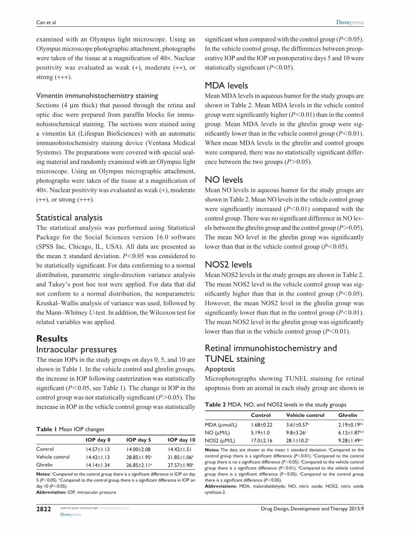
Figure 1. TUNEL positivity was observed to be weak in the
control group (Figure 1A). When compared with the control
group, TUNEL positivity in the vehicle control group was
strong (Figure 1B). More apoptotic cells were seen in the
outer and inner plexiform layers (OPL and IPL, respectively).
The ghrelin group showed significantly decreased apopto sis
compared with the vehicle control group, however, retinal
apoptosis in the ghrelin group was close to that in the control
group (Figure 1C). When retinal sections were examined,
TUNEL-positive RGC layers were observed in the IPL and
inner nuclear layer (INL). Compared with the control group,
TUNEL staining was significantly increased in the vehicle
control group (P,0.05). In the group treated with ghrelin,
TUNEL positivity was significantly decreased (P,0.05).
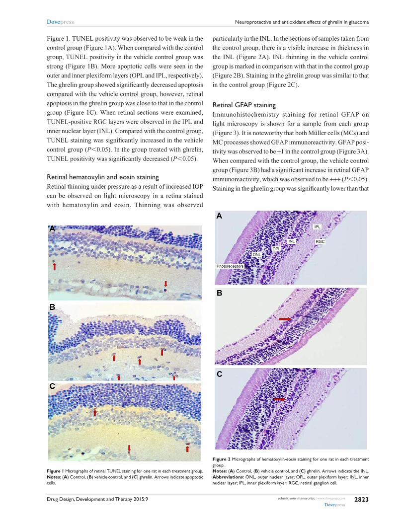
retinal hematoxylin and eosin stainingRetinal thinning under pressure as a result of increased IOP
can be observed on light microscopy in a retina stained
with hematoxylin and eosin. Thinning was observed
particularly in the INL. In the sections of samples taken from
the control group, there is a visible increase in thickness in
the INL (Figure 2A). INL thinning in the vehicle control
group is marked in comparison with that in the control group
(Figure 2B). Staining in the ghrelin group was similar to that
in the control group (Figure 2C).
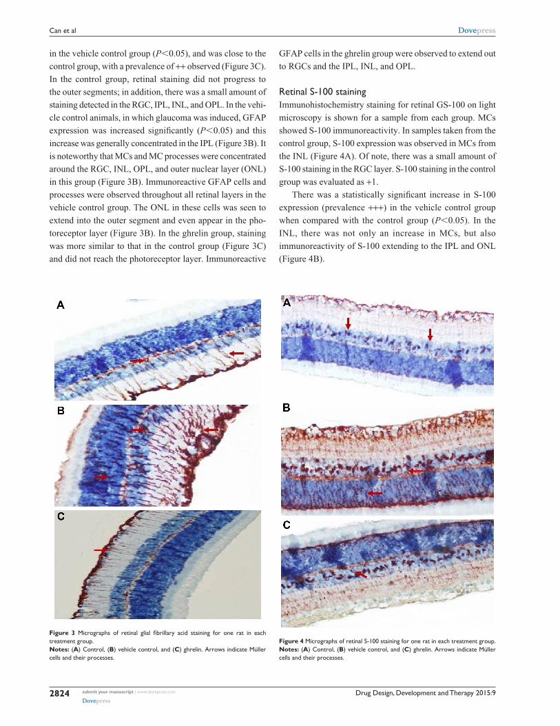
retinal gFaP stainingImmunohistochemistry staining for retinal GFAP on
light microscopy is shown for a sample from each group
(Figure 3). It is noteworthy that both Müller cells (MCs) and
MC processes showed GFAP immunoreactivity. GFAP posi-
tivity was observed to be +1 in the control group (Figure 3A).
When compared with the control group, the vehicle control
group (Figure 3B) had a significant increase in retinal GFAP
immunoreactivity, which was observed to be +++ (P,0.05).
Staining in the ghrelin group was significantly lower than that
Figure 1 Micrographs of retinal TUnel staining for one rat in each treatment group.Notes: (A) control, (B) vehicle control, and (C) ghrelin. arrows indicate apoptotic cells.
Figure 2 Micrographs of hematoxylin-eosin staining for one rat in each treatment group.Notes: (A) control, (B) vehicle control, and (C) ghrelin. arrows indicate the inl.Abbreviations: Onl, outer nuclear layer; OPl, outer plexiform layer; inl, inner nuclear layer; iPl, inner plexiform layer; rgc, retinal ganglion cell.
Drug Design, Development and Therapy 2015:9submit your manuscript | www.dovepress.com
Dovepress
Dovepress
2824
can et al
Figure 3 Micrographs of retinal glial fibrillary acid staining for one rat in each treatment group. Notes: (A) control, (B) vehicle control, and (C) ghrelin. arrows indicate Müller cells and their processes.
in the vehicle control group (P,0.05), and was close to the
control group, with a prevalence of ++ observed (Figure 3C).
In the control group, retinal staining did not progress to
the outer segments; in addition, there was a small amount of
staining detected in the RGC, IPL, INL, and OPL. In the vehi-
cle control animals, in which glaucoma was induced, GFAP
expression was increased significantly (P,0.05) and this
increase was generally concentrated in the IPL (Figure 3B). It
is noteworthy that MCs and MC processes were concentrated
around the RGC, INL, OPL, and outer nuclear layer (ONL)
in this group (Figure 3B). Immunoreactive GFAP cells and
processes were observed throughout all retinal layers in the
vehicle control group. The ONL in these cells was seen to
extend into the outer segment and even appear in the pho-
toreceptor layer (Figure 3B). In the ghrelin group, staining
was more similar to that in the control group (Figure 3C)
and did not reach the photoreceptor layer. Immunoreactive
GFAP cells in the ghrelin group were observed to extend out
to RGCs and the IPL, INL, and OPL.
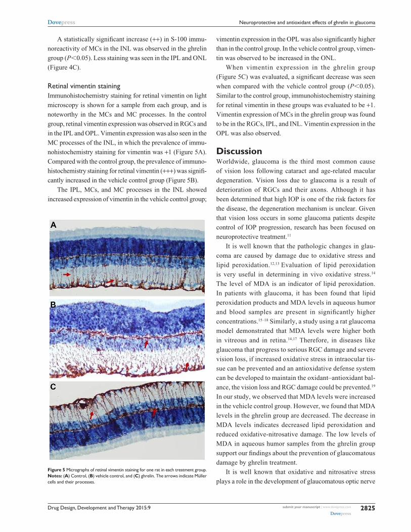
retinal s-100 stainingImmunohistochemistry staining for retinal GS-100 on light
microscopy is shown for a sample from each group. MCs
showed S-100 immunoreactivity. In samples taken from the
control group, S-100 expression was observed in MCs from
the INL (Figure 4A). Of note, there was a small amount of
S-100 staining in the RGC layer. S-100 staining in the control
group was evaluated as +1.
There was a statistically significant increase in S-100
expression (prevalence +++) in the vehicle control group
when compared with the control group (P,0.05). In the
INL, there was not only an increase in MCs, but also
immunoreactivity of S-100 extending to the IPL and ONL
(Figure 4B).
Figure 4 Micrographs of retinal s-100 staining for one rat in each treatment group.Notes: (A) control, (B) vehicle control, and (C) ghrelin. arrows indicate Müller cells and their processes.
Drug Design, Development and Therapy 2015:9 submit your manuscript | www.dovepress.com
Dovepress
Dovepress
2825
neuroprotective and antioxidant effects of ghrelin in glaucoma
A statistically significant increase (++) in S-100 immu-
noreactivity of MCs in the INL was observed in the ghrelin
group (P,0.05). Less staining was seen in the IPL and ONL
(Figure 4C).
retinal vimentin stainingImmunohistochemistry staining for retinal vimentin on light
microscopy is shown for a sample from each group, and is
noteworthy in the MCs and MC processes. In the control
group, retinal vimentin expression was observed in RGCs and
in the IPL and OPL. Vimentin expression was also seen in the
MC processes of the INL, in which the prevalence of immu-
nohistochemistry staining for vimentin was +1 (Figure 5A).
Compared with the control group, the prevalence of immuno-
histochemistry staining for retinal vimentin (+++) was signifi-
cantly increased in the vehicle control group (Figure 5B).
The IPL, MCs, and MC processes in the INL showed
increased expression of vimentin in the vehicle control group;
vimentin expression in the OPL was also significantly higher
than in the control group. In the vehicle control group, vimen-
tin was observed to be increased in the ONL.
When vimentin expression in the ghrelin group
(Figure 5C) was evaluated, a significant decrease was seen
when compared with the vehicle control group (P,0.05).
Similar to the control group, immunohistochemistry staining
for retinal vimentin in these groups was evaluated to be +1.
Vimentin expression of MCs in the ghrelin group was found
to be in the RGCs, IPL, and INL. Vimentin expression in the
OPL was also observed.
DiscussionWorldwide, glaucoma is the third most common cause
of vision loss following cataract and age-related macular
degeneration. Vision loss due to glaucoma is a result of
deterioration of RGCs and their axons. Although it has
been determined that high IOP is one of the risk factors for
the disease, the degeneration mechanism is unclear. Given
that vision loss occurs in some glaucoma patients despite
control of IOP progression, research has been focused on
neuroprotective treatment.11
It is well known that the pathologic changes in glau-
coma are caused by damage due to oxidative stress and
lipid peroxidation.12,13 Evaluation of lipid peroxidation
is very useful in determining in vivo oxidative stress.14
The level of MDA is an indicator of lipid peroxidation.
In patients with glaucoma, it has been found that lipid
peroxidation products and MDA levels in aqueous humor
and blood samples are present in significantly higher
concentrations.15–18 Similarly, a study using a rat glaucoma
model demonstrated that MDA levels were higher both
in vitreous and in retina.14,17 Therefore, in diseases like
glaucoma that progress to serious RGC damage and severe
vision loss, if increased oxidative stress in intraocular tis-
sue can be prevented and an antioxidative defense system
can be developed to maintain the oxidant–antioxidant bal-
ance, the vision loss and RGC damage could be prevented.19
In our study, we observed that MDA levels were increased
in the vehicle control group. However, we found that MDA
levels in the ghrelin group are decreased. The decrease in
MDA levels indicates decreased lipid peroxidation and
reduced oxidative-nitrosative damage. The low levels of
MDA in aqueous humor samples from the ghrelin group
support our findings about the prevention of glaucomatous
damage by ghrelin treatment.
It is well known that oxidative and nitrosative stress
plays a role in the development of glaucomatous optic nerve
Figure 5 Micrographs of retinal vimentin staining for one rat in each treatment group. Notes: (A) control, (B) vehicle control, and (C) ghrelin. The arrows indicate Müller cells and their processes.
Drug Design, Development and Therapy 2015:9submit your manuscript | www.dovepress.com
Dovepress
Dovepress
2826
can et al
damage. The finding of NOS2 in the iris ciliary body, retina,
and optic disc in recent research suggests that NO might play
a role in the pathogenesis of glaucoma.20 NO is an important
mediator that serves to regulate IOP and modulates ocular
blood flow. It is synthesized via the NOS enzyme.21 The
NOS2 form is expressed in response to immunologic and
inflammatory stimulation, and the NO made by this enzyme
is pathological.22,23 Intraocular synthesis of NO has been
studied in the past 5 years, and the presence of all forms of
the NOS enzyme in the eye has been shown.24 The effects
of NO, such as vasodilatation and neurotransmission, are
established via cyclic guanosine monophosphate by means
of the guanylate cyclase enzyme.25 In experimental glaucoma
models in animals, elevated IOP increases apoptosis in cells,
stimulates expression of NOS2, and leads to nitration of
protein.26 Studies in glaucomatous optic discs in rats and
humans have shown the presence of NOS2 on nitrotyrosine
staining and demonstrated the role of NO in glaucomatous
optic neuropathy.13,20 In clinical situations accompanied
by ocular degeneration, nitrative stress worsens the disease
course and increases NOS2 expression, pointing to the role
of NO in ocular pathologies such as glaucoma.25 These data
indicate that reactive nitrogen types may contribute to the
death of RGCs associated with IOP.21,24
The main mechanism of vision loss in glaucoma is weak-
ening of the INL and apoptosis of RGCs, which causes axonal
loss in the optic nerve.27 It has been reported that these cytotoxic
and apoptotic effects are stimulated by NO in macrophages,
astrocytes, and neuronal cells. A study by Aslan et al dem-
onstrated that elevated IOP increases expression of NOS2.23
It has also been shown that inhibition of NOS2 protects
against the degeneration caused by glaucoma in RGCs.28 It is
known that ghrelin indirectly inhibits expression of NOS2 in
gastric mucosal cells.29 An ischemia-reperfusion model in rats
demonstrated that ghrelin is protective against damage due
to an increase in NO.30 In our study, the ghrelin group had
low levels of NOS2 when compared with the vehicle control
group, which is consistent with the literature.
NO assignment can be used as a diagnostic marker of
oxidants that generate from NO, both in humans and animals.
In cases of elevated IOP, expression of NOS2 increases retinal
protein nitration and apoptosis.23 Pharmacologic studies in a rat
model of chronic glaucoma showed that aminoguanidine with
inhibition of NOS2 supports neuronal protection in RGCs.28
Elevated levels of NOS2 observed in glaucoma cases suggests
that NOS2 may contribute to the RGC death associated with
IOP.21,24 Cytotoxicity and apoptosis mediated by NO has been
reported in macrophages, astrocytes, and neuronal cells.31–33
Although the mechanism for NO-mediated apoptosis is still
unclear, it may be the result of activation of p53, leading to
DNA damage.34 Erdurmuş et al found that serum NO levels
are significantly higher in patients with pseudoexfoliation
glaucoma and those with primary open angle glaucoma.15
In our study, we found that NO levels in the vehicle control
group were higher than those in the control group. However,
NO levels in the ghrelin group were significantly lower than
those in the vehicle control group, and there was no significant
difference in NO levels between the control and ghrelin groups.
Many previous studies have shown that ghrelin reduces NO
levels in different types of tissue.35,36 In our study, we observed
that NO levels were lower in aqueous humor, which is con-
sistent with the literature. The finding of low levels of NO
(which had an important role in RGC death) in the ghrelin
group demonstrates that glaucomatous damage was less in
this group.
These hypotheses are the blockage of axoplasmic flow
and consequently the withdrawal of the neurotro phins, the
increase of intravitreal glutamate concentration, and ocular
vasospasm.37 Recent TUNEL findings in rats with elevated
IOP support apoptotic cell death. In a study by Gross et al,
it was reported that TUNEL-positive cells were seen in
the RGC layer, and that they were rarely in the con trol group
compared to the experiment group.38 In previous studies of
the sequence of normal retinal tissue, histogenetic cell death
has been shown to start at the RGC layer, and progress toward
the INL and ONL.39 Similarly, in the sequence of death due
to glaucomatous damage, IOP increased as a result of axonal
degeneration, microglial activation in the tissue, secretion of
tumor necrosis factor-a, secretion of cytokines, and activa-
tion of the complement pathway, culminating in death of
RGCs.40,41 In our study, microscopic DNA fragmentation,
the most defining feature of apoptosis, was performed to
determine apoptosis in the retinal layers. In our study, in
accordance with the literature, apoptosis determined by the
TUNEL method was shown to be less in the control and
ghrelin groups, and more in the vehicle control group, so
ghrelin might have protective effects on RGCs.
Astrocytes, MCs, and microglial cells have both protective
and destructive roles in the retina.42,43 It has also been demon-
strated that MCs can recognize various neuronal signals and
actively modulate the levels of some ions such as K+ and H+,
and some neurotransmitters including glutamate and gamma
aminobutyric acid, in the extracellular space in the retina.44
It is also claimed that microglia are sensitive to potassium
conductance and play an important role in processes such
as production of aggressive oxygen radicals and secretion of
glutamate, which may have an effect on the pathophysiologic
mechanism of the cell death in glaucoma.44 An experimental
Drug Design, Development and Therapy 2015:9 submit your manuscript | www.dovepress.com
Dovepress
Dovepress
2827
neuroprotective and antioxidant effects of ghrelin in glaucoma
study of the role of retinal glial cells in a rat glaucoma model
reported that macroglia such as astrocytes and MCs might
be involved in the pathophysiology of RGC death, and that
activated microglial/phagocytic cells have an important role
in modulating the changes in glaucomatous opti nerve heads.45
A similar report suggests that glial cells were activated by
increased IOP and that reactivity of these cells may be associ-
ated with neuronal degeneration in the glaucomatous retina.46
The same study suggests that glial cell activation increased
in order to clean the products from neurodegeneration. It has
been determined that the GFAP levels taken in MC, which is
the retina’s main glial cell, are significantly higher in many
retinal pathologies, such as glaucoma.47 It is therefore believed
that GFAP-positive immunostaining of MCs is a reliable
marker for both acute and chronic pathology.46 In a study
by Woldemussi et al47 formation of GFAP in MCs in response
to IOP elevation was found as early as day 4. Increased expres-
sion of GFAP in glaucoma is well known, and our finding that
GFAP was elevated in the vehicle control group and decreased
in the ghrelin group is consistent with the literature.
Vimentin, a primary member of the intermediate
filamentous family of proteins, comprises a cytoskeleton
with microfilaments and microtubules in eukaryotic cells.48
Previous experimental glaucoma models have shown that
vimentin in MCs and astrocytes increases damage to the
retinal nerve fiber layer and that the increase of glutamine,
a marker of glutamate metabolism, is connected to vimentin
level.49–51 Carter-Dawson et al reported that the increase in
glutamine in MCs was not a consequence of their loss and
that MC function in the glutamate-glutamine cycle continued
in glaucomatous eyes.50 Accordingly, it has been concluded
that the immune positivity for vimentin seen to increase in
the early period may drop due to the decrease in vitreous
concentration of glutamate in the chronic period.51 There is
some suggestion that tumor necrosis factor-a released from
astrocytes and vimentin from RGC is generated as a response
to glaucomatous damage triggered by NO and other cytok-
ines.52 In our study, only responses in the early period were
examined, and in this period the vehicle control group showed
increased expression of vimentin when compared with the
control group. It was observed that vimentin immunoreactiv-
ity in MCs decreased in the ghrelin group.
S-100 protein is a specific marker of retinal MCs in adult
mammals and it acts intracellularly as a calcium ion-signaling
or a calcium ion-buffering protein. Phosphorylation of S-100
protein plays an important role in many intracellular activities,
including enzyme activity, the dynamics of the building blocks
of the cytoskeleton, and protection of cells against oxidative
damage.53 It also has a role in chemoattraction of leukocytes
in the extracellular field and activation of macrophages.54
S-100 protein has both protective and destructive effects,
depending on extracellular and intracellular activity. Previous
studies in an experimental rat glaucoma model have shown
that S-100, which also plays a role in metabolic activity, is
a marker of MC damage in the eye.45,55 Our data are similar
to those in the literature. Compared with our control group,
the vehicle control group showed increased S-100 expression,
particularly in MCs in the INL. However, in the group treated
with ghrelin, S-100 immunoreactivity decreased.
First discovered in 1999 by Kojima et al ghrelin is basi-
cally a hormone with a 28-amino acid lipopeptide structure
and is excreted by the fundus of the stomach. These hor-
mones are also synthesized in the hypothalamus, pituitary
gland, salivary gland, thyroid gland, small intestine, kidneys,
heart, pancreas, central nervous system, lung, placenta,
gonads, immunologic system, breasts, and teeth.3,4 It is
known that ghrelin affects many systems, including growth
hormone, adrenocorticotropic hormone, prolactin secre-
tion, nutrition, gastric acid secretion, gastric motility, and
cell proliferation. The anti-inflammatory and antioxidant
effects of ghrelin have been shown previously.56,57 In addi-
tion to inhibiting lipid peroxidation, it increases catalase,
glutathione peroxidase, and superoxide dismutase enzyme
activity.58 By inhibiting apoptotic stimuli, ghrelin shows a
proactive effect in many cells, including adipocytes, osteo-
blasts, cardiomyocytes, and endothelial cells.59–61 It has been
reported that, in low doses, ghrelin prevents cell death by
inhibition of apoptosis in hypothalamic neuronal cells.62
The proactive effects of ghrelin on cells have been shown
in models of ischemia-reperfusion.30,63 The neuroprotective
activity of ghrelin is occur via the growth hormone secret-
agogue (GHS) receptor upon activation of GHS-R1a.59
In rat models of Parkinson’s disease, it has been reported
that intraperitoneal ghrelin injections reduced dopamine cell
loss, and that this effect might be associated with reorgani-
zation of Bcl-2 and Bax molecules.64,65 Increased activation
of microglia, which contributes to Parkinson’s disease, was
inhibited after injection of ghrelin.65 The effect of ghrelin on
neuroprotection is achieved by mitochondrial biogenesis, reor-
ganization of proteins in the mitochondrial respiratory chain,
and increased suppression of reactive oxygen species.66
The effect of ghrelin on the eye has not been clearly
established as yet. In studies done in rat eyes, mRNA for
ghrelin has been found in the anterior chamber.8 In a study
conducted by Rocha-Sousa et al, it was reported that ghrelin
was identified in humor aqueous which filled the human ante-
rior chamber.8 Ghrelin crosses the blood–brain barrier easily,
so can reach the ocular tissue. Determination of mRNA for
Drug Design, Development and Therapy 2015:9submit your manuscript | www.dovepress.com
Dovepress
Dovepress
2828
can et al
ghrelin in studies performed in rat also demonstrates that
ghrelin can be present locally in the human eye. Studies
by Katsanos et al showed that ghrelin levels in the ante-
rior chamber in patients with glaucoma were significantly
lower than those in a control group.9 Following on from these
studies, ghrelin was used in experimental glaucoma models.
Ghrelin has a protective effect on RGCs, and can attenuate
the harmful effects of glaucomatous damage. In vivo and
in vitro studies in recent years have reported that the neu-
roprotective activity of ghrelin is increased in models of
ischemic stroke. In rat models of ischemia-reperfusion,
intraperitoneal or intravenous administration of ghrelin had
a significant neuroprotective effect, reducing the infarct
volume in the brain and decreasing cell death.62,67
ConclusionTo the best of our knowledge, there have been no previous
reports in the literature on the impact of ghrelin on oxidative
damage in glaucoma. Although the small number of experi-
mental animals used and the lack of retrograde labeling of
RGCs and determination of RGC survival can be regarded as
the main limita tions of this study, our findings suggest that
ghrelin had antioxidant and neuroprotective effects on the
retina in an experimental model of glaucoma.
AcknowledgmentThis work was funded by an unrestricted grant from the Fırat
University Scientific Research Unit.
Author contributionsNC and TD performed the study; NC, NI, IHO, and TK
collected the data; BT and OC translated, typed, prepared,
edited and reviewed the manuscript; NC and TD performed
the statistical analysis; and all authors approved the final
draft of the manuscript. All authors contributed toward data
analysis, drafting and revising the paper, and agree to be
accountable for all aspects of the work.
DisclosureThe authors report no conflicts of interest in this work.
References1. Quigley HA. Glaucoma: macrocosm to microcosm the Friedenwald
lecture. Invest Ophthalmol Vis Sci. 2005;46:2662–2670.2. Shields MB. Textbook of Glaucoma. 4th ed. Baltimore, MD, USA:
Williams and Wilkins; 1998.3. Kojima M, Hosoda H, Date Y. Ghrelin is a growth-hormone-releasing
acylated peptide from stomach. Nature. 1999;402:656–660.4. Kojima M, Kangawa K. Ghrelin: structure and function. Physiol Rev.
2005;85:495–522.5. Aydin S, Ozkan Y, Caylak E, et al. Ghrelin and its biochemical functions.
Turkiye Klinikleri J Med Sci. 2006;26:272–283.
6. Wren AM, Seal LJ, Cohen MA, et al. Ghrelin enhances appetite and increases food intake in humans. J Clin Endocrinol Metab. 2001;86:5992–5994.
7. Tschop M, Smiley DL, Heiman ML. Ghrelin induces adiposity in rodents. Nature. 2000;407:908–913.
8. Rocha-Sousa A, Saraiva J, Henriques-Coelho T, et al. Ghrelin as a novel locally produced relaxing peptide of the iris sphincter and dilator muscles. Exp Eye Res. 2006;83:1179–1187.
9. Katsanos A, Dastiridou A, Georgoulias P, et al. Plasma and aqueous humour levels of ghrelin in open-angle glaucoma patients. Clin Experi-ment Ophthalmol. 2011;39:324–329.
10. Erşahin M, Toklu HZ, Erzik C, et al. The antinflammatory and neu-roprotective effects of ghrelin in subarachnoid hemorrhage-induced oxidative brain damage in rats. J Neurotrauma. 2010;27:1143–1155.
11. Naskar R, Wissing M, Thanos S. Detection of early neuron degeneration and accompanying microglial reponses in the retina of a rat model of glaucoma. Invest Ophthalmol Vis Sci. 2002;43:2962–2968.
12. Moreno MC, Campanelli J, Sande P, Sanez DA, Keller MI. Retinal oxidative stress induced by high intraocular pressure. Free Radic Biol Med. 2004;37:803–812.
13. Tezel G, Yang X, Cai J. Proteomic identification of oxidatively modified retinal proteins in a chronic pressure-induced rat model of glaucoma. Invest Ophthalmol Vis Sci. 2005;46:3177–3187.
14. Ko ML, Peng PH, Ma MC, Ritch R, Chen CF. Dynamic changes in reactive oxygen species and antioxidant levels in retinas in experimental glaucoma. Free Radic Biol Med. 2005;39:365–373.
15. Erdurmuş M, Yağcı R, Atış Ö, Karadağ R, Akbaş A, Hepşen IF. Anti-oxidant status and oxidative stress in primary open angle glaucoma and pseudoexfoliative glaucoma. Curr Eye Res. 2011;36:713–718.
16. Babizhayev MA, Bunin A. Lipid peroxidation in open-angle glaucoma. Acta Ophthalmol. 1989;67:371–377.
17. Yucel I, Akar Y, Yucel G, Çiftçioğlu MA, Keles N, Aslan M. Effect of hypercholesterolemia on inducible nitric oxide synthase expression in a rat model of elevated intraocular pressure. Vision Res. 2005;45:1107–1114.
18. Ghanem AA, Arafa LF, El-Baz A. Oxidative stress markers in patients with primary open-angle glaucoma. Curr Eye Res. 2010;35:295–301.
19. Ferreira SM, Lerner SF, Brunzini R, Reides CG, Evelson PA, Llesuy SF. Time course changes of oxidative stress markers in a rat experimental glaucoma model. Invest Ophthalmol Vis Sci. 2010;51:4635–4640.
20. Pang IH, Johnson EC, Jia L, et al. Evaluation of inducible nitric oxide synthase in glaucomatous optic neuropathy and pressure-induced optic nerve damage. Invest Ophthalmol Vis Sci. 2005;46:1313–1321.
21. Liu BA, Neufeld H. Expression of nitric oxide synthase-2 (NOS-2) in reactive astrocytes of the human glaucomatous optic nerve head. Glia. 2000;30:178–186.
22. Petros A, Lamb G, Leone A, Moncada S, Bennett D, Vallance P. Effects of a nitric oxide synthase inhibitor in humans with septic shock. Cardiovasc Res. 1994;28:34–39.
23. Aslan M, Yucel I, Akar Y, Yucel G, Çiftçioğlu MA, Şanlıoğlu S. Nitrotyrosine formation and apoptosis in rat models of ocular injury. Free Radic Res. 2006;40:147–153.
24. Shareef S, Sawada A, Neufeld AH. Isoforms of nitric oxide synthase in the optic nerves of rat eyes with chronic moderately elevated intraocular pressure. Invest Ophthalmol Vis Sci. 1999;40:2884–2891.
25. Goureau O, Bellot J, Thillaye B, Courtois Y, Kozak Y. Increased nitric oxide production in endotoxin-induced uveitis. Reduction of uveitis by an inhibitor of nitric oxide synthase. J Immunol. 1995;154:6518–6523.
26. Sacca SC, Izzotti A, Rossi P, Traverso C. Glaucomatous outflow path-way and oxidative stress. Exp Eye Res. 2007;84:389–399.
27. Fechtner RD, Weinreb RN. Mechanisms of optic nerve damage in primary open angle glaucoma. Surv Ophthalmol. 1994;39:23–42.
28. Neufeld AH, Sawada A, Becker B. Inhibition of nitric-oxide synthase 2 by aminoguanidine provides neuroprotection of retinal ganglion cells in a rat model of chronic glaucoma. Proc Natl Acad Sci U S A. 1999;96: 9944–9948.
29. Slomiany BL, Slomiany A. Helicobacter pylori induces disturbances in gastric mucosal Akt activation through inducible nitric oxide synthase-dependent s-nitrosylation: effect of ghrelin. Gastroenterology. 2011;308:727–728.
Drug Design, Development and Therapy
Publish your work in this journal
Submit your manuscript here: http://www.dovepress.com/drug-design-development-and-therapy-journal
Drug Design, Development and Therapy is an international, peer-reviewed open-access journal that spans the spectrum of drug design and development through to clinical applications. Clinical outcomes, patient safety, and programs for the development and effective, safe, and sustained use of medicines are a feature of the journal, which
has also been accepted for indexing on PubMed Central. The manu-script management system is completely online and includes a very quick and fair peer-review system, which is all easy to use. Visit http://www.dovepress.com/testimonials.php to read real quotes from published authors.
Drug Design, Development and Therapy 2015:9 submit your manuscript | www.dovepress.com
Dovepress
Dovepress
Dovepress
2829
neuroprotective and antioxidant effects of ghrelin in glaucoma
30. Konturek PC, Brzozowski T, Walter B, Burnat G, Hess T, Hahn EG. Ghrelin induced gastroprotection against ischemia-reperfusion injury involves an activation of sensory afferent nerves and hyperemia medi-ated by nitric oxide. Eur J Pharmacol. 2006;536:171–181.
31. Sarih M, Souvannavong V, Adam A. Nitric oxide synthase induces macrophage death by apoptosis. Biochem Biophys Res Commun. 1993; 191:503–508.
32. Hu J, Van Eldik LJ. S100 beta induces apoptotic cell death in cultured astrocytes via a nitric oxide-dependent pathway. Biochim Biophys Acta. 1996;1313:239–245.
33. Heneka MT, Loschmann PA, Gleichmann M, et al. Induction of nitric oxide synthase and nitric oxide-mediated apoptosis in neuronal PC12 cells after stimulation with tumor necrosis factor-alpha/lipopolysac-charide. J Neurochem. 1998;71:88–94.
34. Kim YM, Bombeck CA, Billiar TR. Nitric oxide as a bifunctional regulator of apoptosis. Circ Res. 1999;84:253–256.
35. Aslan A, Yildirim M, Ayyildiz M, Güven A, Ağar E. The role of nitric oxide in the inhibitory effect of ghrelin against penicillin-induced epileptiform activity in rat. Neuropeptides. 2009;43:295–302.
36. Alderton WK, Cooper CE, Knowles RG. Nitric oxide synthases: struc-ture, function and inhibition. Biochem J. 2001;357:593–615.
37. Levin LA, Louhab A. Apoptosis of retinal ganglion cells in anterior ischemic optic neuropathy. Arch Ophthalmol. 1996;114:488–491.
38. Gross RL, Ji J, Chang P, Pennesi ME. A Mouse model of elevated intraocular pressure: retina and optic nerve findings. Trans Am Oph-thalmol Soc. 2003;101:163–169.
39. Beazley LD, Perry VH, Baker B, Darby JE. An investigation into the role of ganglion cells in the regulation of division and death of other retinal cells. Dev Brain Res. 1987;33:169–184.
40. Ju KR, Kim HS, Kim JH, Lee NY, Park CK. Retinal glial cell responses and Fas/FasL activation in rats with chronic ocular hypertension. Brain Res. 2006;1122:209–221.
41. Kuehn MH, Kim CY, Ostojic J, et al. Retinal synthesis and deposition of complement components induced by ocular hypertension. Exp Eye Res. 2006;83:620–628.
42. Battisti WP, Wang J, Bozek K. Macrophages, microglia, and astrocytes are rapidly activated after crush injury of the goldfish optic nerve: a light and electron microscopic analysis. J Comp Neurol. 1995;354: 306–320.
43. Hughes EH, Schlichtenbrede FC, Murphy CC, et al. Minocycline delays photoreceptor death in the rds mouse through a microglia-independent mechanism. Exp Eye Res. 2004;78:1077–1084.
44. Newman E, Reichenbach A. The Muller cell: a functional element of the retina. Trends Neurosci. 1996;19:307–312.
45. Lam TT, Kwong JMK, Tso OM. Early glial responses after acute elevated intraocular pressure in rats. Invest Ophthalmol Vis Sci. 2003;44: 638–645.
46. Wang X, Tay SS, Ng YK. An immunohistochemical study of neuronal and glial cell reactions in retina of rats with experimental glaucoma. Exp Brain Res. 2000;132:476–484.
47. Woldemussie E, Wijono M, Ruiz G. Müller cell response to laser-induced increase in intraocular pressure in rats. Glia. 2004;47:109–119.
48. Dabbs DJ. Diagnostic İmmunohistochemistry. Philadelphia, PA, USA: Saunders Elsevier; 2006.
49. Hernandez M, Rodriguez FD, Sharma SC, Vecino E. Immunohis-tochemical changes in rat retinas at various time periods of elevated intraocular pressure. Mol Vis. 2009;15:2696–2709.
50. Carter-Dawson L, Shen F, Harwerth R, Smith EL III, Crawford ML, Chuang A. Glutamine immunoreactivity in Muller cells of monkey eyes with experimental glaucoma. Exp Eye Res. 1998;66:537–545.
51. Dreyer EB, Zurakowski D, Schumer RA, Podos SM, Lipton SA. Elevated glutamate levels in the vitreous body of human and monkeys with glaucoma. Arch Ophthalmol. 1996;114:299–305.
52. Tezel G, Wax MB. Increased production of tumor necrosis factor-alpha by glial cells exposed to simulated ischemia or elevated hydrostatic pres-sure induces apoptosis in cocultured retinal ganglion cells. J Neurosci. 2000;20:8693–8700.
53. Donato R. S100: a multigenic family of calcium-modulated proteins of the EF-hand type with intracellular and extracellular functional roles. Int J Biochem Cell Biol. 2001;33:637–668.
54. Barger SW, Van Eldik LJ, Mattson MP. S100 beta protects hippocam-pal neurons from damage induced by glucose deprivation. Brain Res. 1995;677:167–170.
55. Sommer I, Lagenaur C, Schachner M. Recognition of Bergmann glial and ependymal cells in the mouse nervous system by monoclonal antibody. J Cell Biol. 1981;90:448–458.
56. Dixit VD, Schaffer EM, Pyle RS, et al. Ghrelin inhibits leptin- and activation-induced proinflammatory cytokine expression by human monocytes and T cells. J Clin Invest. 2004;114:57–66.
57. Brzozowski T, Konturek PC, Konturek SJ, et al. Exogenous and endogenous ghrelin in gastroprotection against stress-induced gastric damage. Regul Pept. 2004;120:39–51.
58. Obay BD, Taşdemir E, Tümer C, Bilgin HM, Atmaca M. Dose depen-dent effects of ghrelin on pentylenetetrazole-induced oxidative stress in a rat seizure model. Peptides. 2008;29:448–455.
59. Kim MS, Yoon CY, Jang PG, Park SJ. The mitogenic and anti-apoptotic actions of ghrelin in 3T3-L1 adipocytes. Mol Endocrinol. 2004;18:2291–2301.
60. Kim S, Her SJ, Park SJ. Ghrelin stimulate proliferation and differen-tiation and inhibits apoptosis in osteoblastic MC3T3-E1 cells. Bone. 2005;37:359–369.
61. Baldanzi G, Filigheddu N, Cutrupi S, Catapano F, Bonissoni S. Ghrelin and des acyl ghrelin inhibit cell death in cardiomyocytes and endothelial cells through ERK1/2 and PI 3-kinase/AKT. J Cell Biol. 2006;159:1029–1037.
62. Chung H, Kim E, Lee DH, Seo S, Ju S, Lee D. Ghrelin inhibits apoptosis in hypothalamic neuronal cells during oxygen-glucose deprivation. Endocrinology. 2007;148:148–159.
63. Chang L, Ren Y, Liu X, Li WG, Yang J, Geng B. Protective effects of ghrelin on ischemia/reperfusion injury in the isolated rat heart. J Cardiovasc Pharmacol. 2004;43:165–170.
64. Andrews ZB, Eroin D, Beiler R, Liu ZW, Abizaid A. Ghrelin promotes and protects nigrostriatal dopamine function via UCP2-dependent mitochondrial mechanism. J Neurosci. 2009;29:14057–14065.
65. Jiang H, Li LJ, Wang J, Xie JX. Ghrelin antagonizes MPTP-induced neurotoxicity to the dopaminergic neurons in mouse substantia nigra. Exp Neurol. 2008;212:532–537.
66. Andrews ZB. The extra hypothalamic actions ghrelin on neuronal function. Trends Neurosci. 2011;34:31–40.
67. Hwang S, Moon M, Kim S, Hwang L, Ahn KJ, Park S. Neuroprotective effect of ghrelin is associated with decreased expression of prostate apoptosis response. Endocrinol J. 2009;56:609–617.