Embed Size (px)
Citation preview

Neurons, Synapses, and Signaling

The Neuron is the functional unit of the nervous system.
Neurons are composed of a cell body, which contains the nucleus and organelles; Dendrites which are extensions that received incoming messages from other cells; and Axons that transmit messages to other cells.

Many Axons are covered by a fatty myelin sheathe. This sheath acts as insulation and speeds up the rate of impulse transmission.


The Synapse is a junction between two neurons (or a neuron and a muscle fiber or gland.)
Neurotransmitters are chemical messengers released from vesicles in the synaptic terminals into the synapse. They will diffuse across the synapse and bind to receptors on the neuron, muscle fiber, or glands across the synapse, effecting a change in the second cell.
Examples: acetycholine, dopamine, serotonin


Central nervous system (CNS) consists of the brain and spinal cord.
Peripheral nervous system (PNS) consists of the nerves that communicate motor and sensory signals throughout the body.
Sensory receptors collect information about the world outside the body as well as inside the body.
Sensory neurons transmit information from sensors to the brain and spinal cord for processing.
Interneurons connect sensory neurons and motor neruorns.
Motor neurons transmit signals to effectors, such as muscle cells or glands.
Nerves are bundles of neurons.

Ion pumps and ion channels maintain the resting potential of a neuron
Membrane potential describes the difference in electrical change across a cell membrane.
The membrane potential of a nerve cell at rest is called its resting potential.
The resting potential is the result of the difference in the ionic composition between the extracellular fluid and the intracellular fluids across the plasma membrane.

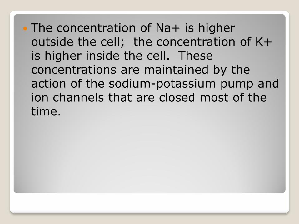
The concentration of Na+ is higher outside the cell; the concentration of K+ is higher inside the cell. These concentrations are maintained by the action of the sodium-potassium pump and ion channels that are closed most of the time.


Changes is the membrane potential of a neuron are what gives rise to nerve impulses.
A stimulus first affects the membranes permeability to ions, and this is a graded potential with a magnitude proportional to the size of the stimulus.

Action Potentials are the signals conducted by axons.
An action potential (nerve impusle) is an all-or-none response to depolarization of the membrane of a nerve cell.
A stimulus opens voltage gated sodium channels and Na+ ions enter the cell, changing the membrane potential to a positive value.

Threshold value
In order to generate an action potential, a certain level of depolarization must be achieved, known as the threshold.


The membrane potential is restored to its normal resting value by the inactivation of Na+ channels and the opening of K+ channels, which increase K+ leaving the cell.
The action potential regenerates itself, traveling along the length of the axon.
The rapid influx of Na+ ions following the action potential serves as an electrical current that depolarizes neighboring regions of the axon membrane and is large enough for that region to reach threshold causing the action potential to be regenerated.
This process is repeated until the action potential travels the length of the axon.


A refractory period (period of time when nerve cannot be stimulated) follows the action potential. This is an interval in which the Na+ channels are inactivated.
Action potentials move along neurons by saltatory conduction, which is the jumping of the nerve impulse between nodes of Ranvier(areas not covered by the myelin sheath). This speeds up the conduction of the nerve impulse.


Neurons communicate with other cells at synapses.
The signal is conducted from the axon of a presynaptic cell to the dendrite of a postsynaptic cell by electrical or chemical synapse.
Neurotransmitters are released by the presynaptic membrane into the synaptic cleft.
They bind to receptors on the postsynaptic membrane initiating depolariztion of the postsynaptic cell.
Neurotransmitters are then broken down by enzymes, or taken back into surrounding cells.


There are two categories of neurotransmitters.
◦ Excititory causes depolarization of the postsynaptic membrane.
◦ Inhibitory cause hyperpolarization of the postsynaptic membrane and prevents transmission of message.
◦ Acetycholine is a common neurotransmitter, which can be excitatory or inhibitory.
◦ Other common neurotransmitters are epinephrine, norepinephrine, dopamine, and serotonin.