Embed Size (px)
Citation preview
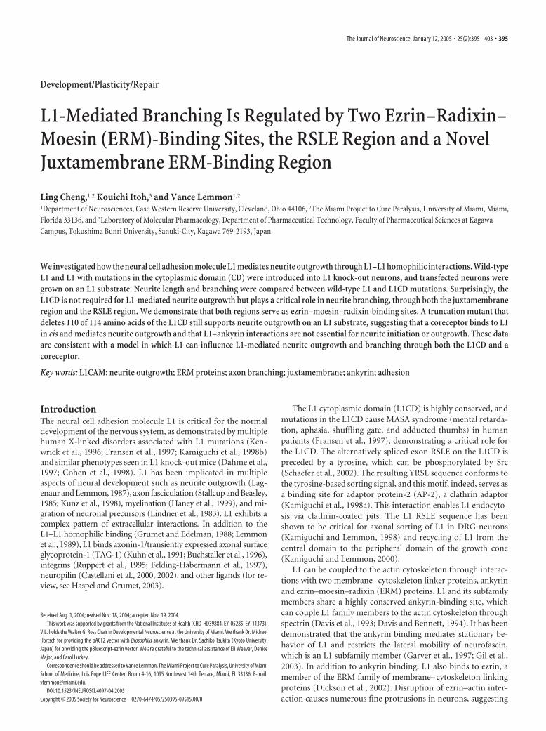
Development/Plasticity/Repair
L1-Mediated Branching Is Regulated by Two Ezrin–Radixin–Moesin (ERM)-Binding Sites, the RSLE Region and a NovelJuxtamembrane ERM-Binding Region
Ling Cheng,1,2 Kouichi Itoh,3 and Vance Lemmon1,2
1Department of Neurosciences, Case Western Reserve University, Cleveland, Ohio 44106, 2The Miami Project to Cure Paralysis, University of Miami, Miami,Florida 33136, and 3Laboratory of Molecular Pharmacology, Department of Pharmaceutical Technology, Faculty of Pharmaceutical Sciences at KagawaCampus, Tokushima Bunri University, Sanuki-City, Kagawa 769-2193, Japan
We investigated how the neural cell adhesion molecule L1 mediates neurite outgrowth through L1–L1 homophilic interactions. Wild-typeL1 and L1 with mutations in the cytoplasmic domain (CD) were introduced into L1 knock-out neurons, and transfected neurons weregrown on an L1 substrate. Neurite length and branching were compared between wild-type L1 and L1CD mutations. Surprisingly, theL1CD is not required for L1-mediated neurite outgrowth but plays a critical role in neurite branching, through both the juxtamembraneregion and the RSLE region. We demonstrate that both regions serve as ezrin–moesin–radixin-binding sites. A truncation mutant thatdeletes 110 of 114 amino acids of the L1CD still supports neurite outgrowth on an L1 substrate, suggesting that a coreceptor binds to L1in cis and mediates neurite outgrowth and that L1–ankyrin interactions are not essential for neurite initiation or outgrowth. These dataare consistent with a model in which L1 can influence L1-mediated neurite outgrowth and branching through both the L1CD and acoreceptor.
Key words: L1CAM; neurite outgrowth; ERM proteins; axon branching; juxtamembrane; ankyrin; adhesion
IntroductionThe neural cell adhesion molecule L1 is critical for the normaldevelopment of the nervous system, as demonstrated by multiplehuman X-linked disorders associated with L1 mutations (Ken-wrick et al., 1996; Fransen et al., 1997; Kamiguchi et al., 1998b)and similar phenotypes seen in L1 knock-out mice (Dahme et al.,1997; Cohen et al., 1998). L1 has been implicated in multipleaspects of neural development such as neurite outgrowth (Lag-enaur and Lemmon, 1987), axon fasciculation (Stallcup and Beasley,1985; Kunz et al., 1998), myelination (Haney et al., 1999), and mi-gration of neuronal precursors (Lindner et al., 1983). L1 exhibits acomplex pattern of extracellular interactions. In addition to theL1–L1 homophilic binding (Grumet and Edelman, 1988; Lemmonet al., 1989), L1 binds axonin-1/transiently expressed axonal surfaceglycoprotein-1 (TAG-1) (Kuhn et al., 1991; Buchstaller et al., 1996),integrins (Ruppert et al., 1995; Felding-Habermann et al., 1997),neuropilin (Castellani et al., 2000, 2002), and other ligands (for re-view, see Haspel and Grumet, 2003).
The L1 cytoplasmic domain (L1CD) is highly conserved, andmutations in the L1CD cause MASA syndrome (mental retarda-tion, aphasia, shuffling gate, and adducted thumbs) in humanpatients (Fransen et al., 1997), demonstrating a critical role forthe L1CD. The alternatively spliced exon RSLE on the L1CD ispreceded by a tyrosine, which can be phosphorylated by Src(Schaefer et al., 2002). The resulting YRSL sequence conforms tothe tyrosine-based sorting signal, and this motif, indeed, serves asa binding site for adaptor protein-2 (AP-2), a clathrin adaptor(Kamiguchi et al., 1998a). This interaction enables L1 endocyto-sis via clathrin-coated pits. The L1 RSLE sequence has beenshown to be critical for axonal sorting of L1 in DRG neurons(Kamiguchi and Lemmon, 1998) and recycling of L1 from thecentral domain to the peripheral domain of the growth cone(Kamiguchi and Lemmon, 2000).
L1 can be coupled to the actin cytoskeleton through interac-tions with two membrane– cytoskeleton linker proteins, ankyrinand ezrin–moesin–radixin (ERM) proteins. L1 and its subfamilymembers share a highly conserved ankyrin-binding site, whichcan couple L1 family members to the actin cytoskeleton throughspectrin (Davis et al., 1993; Davis and Bennett, 1994). It has beendemonstrated that the ankyrin binding mediates stationary be-havior of L1 and restricts the lateral mobility of neurofascin,which is an L1 subfamily member (Garver et al., 1997; Gil et al.,2003). In addition to ankyrin binding, L1 also binds to ezrin, amember of the ERM family of membrane– cytoskeleton linkingproteins (Dickson et al., 2002). Disruption of ezrin–actin inter-action causes numerous fine protrusions in neurons, suggesting
Received Aug. 1, 2004; revised Nov. 18, 2004; accepted Nov. 19, 2004.This work was supported by grants from the National Institutes of Health (CHD-HD39884, EY-05285, EY-11373).
V.L. holds the Walter G. Ross Chair in Developmental Neuroscience at the University of Miami. We thank Dr. MichaelHortsch for providing the pACT2 vector with Drosophila ankyrin. We thank Dr. Sachiko Tsukita (Kyoto University,Japan) for providing the pBluescript-ezrin vector. We are grateful to the technical assistance of Eli Weaver, DeniceMajor, and Carol Luckey.
Correspondence should be addressed to Vance Lemmon, The Miami Project to Cure Paralysis, University of MiamiSchool of Medicine, Lois Pope LIFE Center, Room 4-16, 1095 Northwest 14th Terrace, Miami, FL 33136. E-mail:[email protected].
DOI:10.1523/JNEUROSCI.4097-04.2005Copyright © 2005 Society for Neuroscience 0270-6474/05/250395-09$15.00/0
The Journal of Neuroscience, January 12, 2005 • 25(2):395– 403 • 395

that the ERM–actin interaction is involved in the regulation ofbranching (Dickson et al., 2002).
In this study, we examined the role of the L1CD in L1–L1homophilic interaction-mediated neurite outgrowth. Usingtransfected L1 knock-out neurons growing on L1 substrates, wedemonstrate that the L1CD is not necessary for L1-mediated neu-rite outgrowth, but it can influence branching through the jux-tamembrane region and the RSLE region. We provide evidencethat both the juxtamembrane region and the RSLE region areessential for the L1–ERM interaction.
Materials and MethodsMaterials and animals. The monoclonal anti-human L1 antibodies (7B5)were described previously (Cheng and Lemmon, 2004). The polyclonalanti-chicken L1 (8D9) antibodies were described previously (Lemmonand McLoon, 1986). Fluorescent secondary antibodies were purchasedfrom Molecular Probes (Eugene, OR). Tissue culture reagents were pur-chased from Invitrogen (Carlsbad, CA). Mouse neuron nucleofector kitwas from Amaxa (Cologne, Germany). Chick L1 was purified as de-scribed previously (Lagenaur and Lemmon, 1987). Coverslips were pur-chased from Corning (Acton, MA). Chemicals were purchased fromSigma (St. Louis, MO) and Pierce (Rockford, IL). All experiments usingmice were approved by the Case Western Reserve University and Uni-versity of Miami Animal Care and Use Committees. The L1 knock-outmice used have been described previously (Fransen et al., 1998).
DNA constructs. The wild-type (WT) human L1 (hL1) vector inpcDNA3 was described previously (Wong et al., 1995). The L1�RSLE,L1-1176, and L1-1180 were described previously (Kamiguchi and Lem-mon, 1998). The L1-1147 was described previously (Wong et al., 1995).The L1-4A and L1-1151Y�A constructs were generated by QuickChangeXL-mutagenesis kit (Stratagene, La Jolla, CA) using the wild-type humanL1 in pcDNA3 as the template. The oligonucleotides used are as follows:5�-TCAAGCGCAGCGCGGGCGGCGCAGCCTCAGCGAAGGATAA-GGAGG-3� (L1-4A sense), 5�-CCTCCTTATCCTTCGCTGAGGCTG-CGCCGCCCGCGCTGCGCTTGA-3� (L1-4A antisense), 5�-GCAAG-GGCGGCAAAGCCTCAGTGAAGGATAAGG-3� (L1-1151Y�A sense),and 5�-CCTTATCCTTCACTGAGGCTTTGCCGCCCTTGC-3� (L1-1151Y�A antisense). The mutations were confirmed by sequencing. Allthe constructs included 206 bp of the 3� untranslated region from the L1cDNA, which appear to increase L1 expression in transfected cells.
Preparation of substrate. Preparation of L1 substrates was describedpreviously (Cheng and Lemmon, 2004). Briefly, purified chick L1 (�100�g/ml) was coated to silanized coverslips by a covalent cross-linkingmethod. Silicon gaskets (Grace Biolabs, Bend, OR) were used to create L1spots on coverslips. Coverslips were washed extensively and blocked with5% hemoglobin for 1 hr at room temperature before plating cells.
Neuron culture and electroporation. Postnatal day 8 mouse cerebellawere dissected and dissociated as described previously (Beattie and Sie-gel, 1993). Dissociated cerebellar granule cells were transfected with theNucleofector machine (Amaxa) as described previously (Cheng andLemmon, 2004).
Immunocytochemistry. Fixation and staining were performed as de-scribed previously (Cheng and Lemmon, 2004). Briefly, cells were fixedand permeabilized 48 hr after electroporation. Then cells were incubatedwith primary antibodies (the monoclonal anti-human L1 antibody 7B5undiluted supernatant plus 1:500 rabbit anti-8D9) for 1 hr at room tem-perature, followed by incubation with secondary antibodies (1:200 Ore-gon green 514 anti-mouse IgG plus 1:200 Texas red-X anti-rabbit IgG)for 1 hr at room temperature. The coverslips were mounted onto slideswith the SlowFade light kit (Molecular Probes). For some experiments,neurons growing on L1 were stained with rabbit anti-MAP2 (1:500;Chemicon, Temecula, CA) and mouse anti-Tau1 (1:200; Chemicon) si-multaneously, followed by Alexa488 goat anti-mouse IgG and Alexa594goat anti-rabbit IgG as secondary antibodies.
Live/dead staining. To stain live neurons, transfected L1 knock-outneurons were stained with 2 �M calcein AM (Molecular Probes) for 45min at 37°C before fixation. Only live cells can cleave the nonfluorescentcell-permeant calcein AM to produce an intense green fluorescence in
both cell bodies and processes. After fixation and permeabilization, L1-positive neurons were stained with 7B5 and Texas red-X anti-mouse IgG.Images for both the green channel and the red channel were acquiredwith a Spot RT slider CCD camera coupled to a Leica DLMB microscopewith a 20� objective. On the overlay images, all L1-positive neurons,with or without neurites, were scored as live or dead.
Image acquisition and analysis. Images were acquired with a Spot CCDcamera RT slider (Diagnostic Instruments, Sterling Heights, MI) coupledto a Leica (Nussloch, Germany) DLMB microscope with a 20� objective(numerical aperture � 0.7). Image analysis was performed with NIHImageJ and Neurolucida (MicroBrightField, Williston, VT). Figureswere prepared with Adobe Systems (San Jose, CA) Photoshop 7.
Quantification of neurite outgrowth. The analysis of neurite outgrowthwas described previously (Cheng and Lemmon, 2004). Briefly, neurontracing was performed manually with the Neurolucida. Total neuritelength is the sum of all neuritic branches elaborated by a single neuron. Abranching point is the point at which a neurite extends from the cell bodyor from another neurite. A process has to be at least 10 �m to be consid-ered a branch. Branching number is the sum of every branching pointfrom a single neuron. It includes both the origin from the cell body andthe point at which neurites bifurcate into two processes. Primary neuritesare those branches that extend from the soma. Nodes are branch pointsalong a neurite that do not arise directly from the soma. Approximately80 –100 neurons were analyzed for each construct in one experiment, andeach construct was tested at least three times. In each experiment, WThL1 was transfected into L1KO neurons and used as a control. Data froma particular experiment were normalized with the WT hL1 as the stan-dard. We have shown previously that WT hL1 is expressed at levels in-distinguishable from mouse L1 in WT neurons and that there is highcorrelation (�0.8) between cell surface expression of WT hL1 in neuritesand total L1 in permeabilized neurites. Because expression of L1 withtruncated L1 was low for some constructs, permeabilized neurites gavebetter images for analysis (Cheng and Lemmon, 2004). ANOVA (Fisher’sPLSD) was analyzed using Statview 4.5 (SAS Institute, Cary, NC).
Measurement of relative expression level. Relative expression level wasmeasured as described previously (Cheng and Lemmon, 2004). Briefly,all images were acquired with the same exposure time corresponding tounsaturated acquisitions. Pixel values on the neurite were quantified atfive different locations on the shaft of the longest neurite with NIHImageJ. The average pixel value was determined for each neuron, and thebackground was subtracted. Approximately 20 neurons for each muta-tion in the same experiment were quantified. Because absolute fluores-cence intensity varied among different experiments, the values for themutant-transfected neurons were always normalized by the values ofneurons transfected with WT hL1 stained with the same conditions in thesame experiment.
Yeast two-hybrid assay. The bait vector pAS2 with the wild-type L1CDand the prey vector pACT2 containing the �2 chain of AP-2 were de-scribed previously (Kamiguchi et al., 1998a). The prey vector pACT2containing the Drosophila ankyrin was described previously (Dubreuil etal., 1996). The bait vectors containing the L1CD mutants were made byusing L1-4A, L1-1151Y�A, L1�RSLE, L1�RSLE-4A, and L1�RSLE-1151Y�A in pcDNA3 as the PCR template. The primers used were 5�-CGCCATGCCATGGTCAAGCGCAGCAAGGGC-3� (forward primerfor L1-1151Y�A, L1�RSLE, and L1�RSLE-1151Y�A), 5�-CGCCAT-GCCATGGTCAAGCGCAGCGCGGGC-3� (forward primer for L1-4Aand L1�RSLE-4A), and 5�-GCGGATCCACTATTCTAGGGCCAC-3�(reverse primer for all mutants). The PCR products were cloned intopGEM-T-Easy vectors (Promega, Madison, WI) by a TA-cloning kit. Thefragments containing the mutant L1CD were released by NcoI–BamHIdigestion and then subcloned into the bait pAS2 vector. The mutationswere verified by sequencing. The N-terminal ezrin fragment (1– 404 aa)was obtained by digesting the complete encoding sequence of mouseezrin (a gift from Dr. S. Tukita, Kyoto University, Kyoto, Japan) withEcoRI–XhoI and subcloned into the pACT2 vector. The AH109 strain(Clontech, Palo Alto, CA) was cotransformed with the pAS2 bait vectorcontaining wild-type or mutant forms of the L1CD and the pACT2 preyvector (ezrin, ankyrin, or AP-2). Cotransformed colonies were selectedby growing on yeast dropout medium lacking leucine and tryptophan
396 • J. Neurosci., January 12, 2005 • 25(2):395– 403 Cheng et al. • The L1 Cytoplasmic Domain Regulates Axon Branching

(C-Leu-Trp). Then, cotransformed colonies were streaked to C-Leu-Trp-His plates in the presence of 5 or 10 mM 3-amino-triazole (3-AT).Growth was scored after 2–3 d. As a positive control for the growth, thediploid of AH109 [pGBKT7-53] � Y187 [pTD1-1] (Clontech) wasstreaked on the same plate. As a negative control, the diploid of AH109[pAS2-L1CD] � Y187 [pTD1-1] was streaked on the same plate.
ResultsAs described previously (Cheng and Lemmon, 2004), we haveestablished an assay to investigate how L1 mutations affect L1-mediated neurite outgrowth. Cerebellar granule neurons fromL1KO mice were transfected with L1 cDNA expression vectorsand grown on an L1 substrate. After 2 d in vitro (DIV), the neu-rites on L1 substrates are relatively long, with average total neu-rite lengths of �250 �m, but double staining with antibodies toMAP2 and tau1 indicates that these markers overlap and cells arenot highly polarized (Fig. 1). However, L1 is concentrated onaxons rather than dendrites in polarized cells, so we are likely tobe studying immature axon-like neurites. Because L1KO neuronslack endogenous L1, L1 molecules expressed by transfected neu-rons are exclusively from the exogenous construct. In this system,the L1KO cells express WT hL1 at levels indistinguishable fromL1 expressed by WT neurons (Cheng and Lemmon, 2004). Wehave shown previously that L1KO neurons expressing WT hL1are able to attach and send neurites on L1 substrates, and theneurite length from WT hL1-transfected L1KO neurons is indis-tinguishable from the neurite length of WT neurons on an L1substrate, suggesting that the WT hL1 expressed in L1KO neu-rons is able to support neurite outgrowth to a similar degree asendogenous L1 (Cheng and Lemmon, 2004). We have performedadditional experiments and found that the branching numberof WT neurons is indistinguishable from the branching exhib-ited by hL1-transfected L1KO neurons (total branching num-ber: WT:L1KO � 1.11:1.0; Student’s t test shows no significantdifference). By comparing neurite outgrowth from mutantL1-transfected neurons with the neurite growth from WThL1-transfected L1KO neurons, we are able to evaluate the effectof L1 mutations on L1-mediated neurite outgrowth. We quanti-fied three parameters for neurite outgrowth, longest neuritelength, branching number, and total neurite length. We also cal-culated the number of primary branches from the soma and thenumber of branches from neurites (nodes). Three independentexperiments were analyzed for each mutation, and the results aresummarized in supplemental data table 1 (available at www.jneu-rosci.org as supplemental material). To compare results fromindependent experiments, we normalize the numbers from themutants by the control value (WT hL1 in the same experiment).
Absence of the RSLE sequence does notaffect neurite length but decreasesbranching numberPrevious studies have shown that theYRSLE region is the binding site for ERMproteins and AP-2. This sequence is criti-cal for clathrin-mediated L1 endocytosisand L1 sorting to axons in DRG neurons(Kamiguchi and Lemmon, 1998; Kamigu-chi et al., 1998a; Dickson et al., 2002). Tocharacterize the role of RSLE in neuriteoutgrowth, we have generated the non-neuronal form L1�RSLE (the RSLE is de-leted) and two truncation forms, L1-1176(truncation before RSLE) and L1-1180(truncation after RSLE) (Fig. 2). Both L1-1176 and L1-1180 lack the C-terminal 77
residues after the RSLE, including the highly conserved ankyrin-binding region.
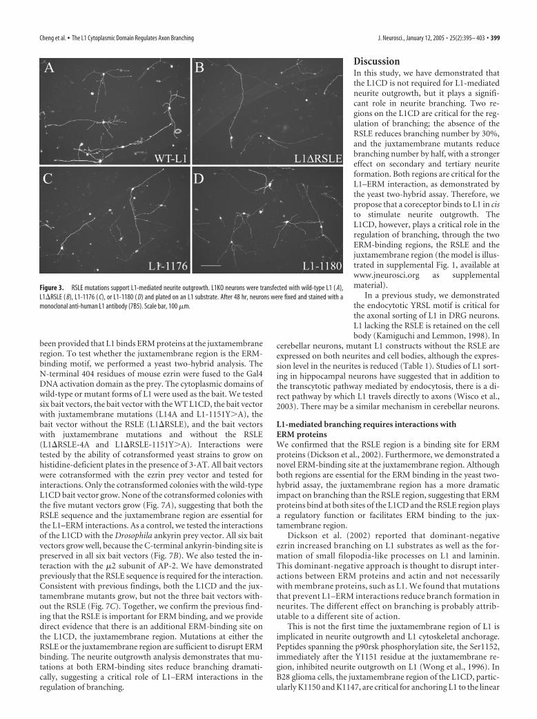
With all three mutants, transfected neurons are able to sendout long neurites on L1 substrates. It has been reported previ-ously that mutations that alter the tyrosine-based sorting motifreduce or block transport of L1 into neurites of DRG neurons inculture (Kamiguchi and Lemmon, 1998). However, in hip-pocampal neurons, L1 was still able to transport to axons via adirect pathway even when the YRSLE sequence was mutated(Wisco et al., 2003). In cerebellar neurons transfected with RSLEmutations, we did observe reduced L1 expression on neurites ifthe RSLE is deleted (Table 1), but nonetheless, neurites are able togrow on L1 substrates robustly, in marked contrast to L1KOneurons lacking L1 (Fransen et al., 1998; Itoh et al., 2004). This isconsistent with our previous report of the poor correlation be-tween neurite outgrowth and L1 cell surface expression level(Cheng and Lemmon, 2004). Representative pictures are shownin Figure 3B–D. The longest neurite length, the number ofbranches, and the total neurite length are quantified and the re-sults are shown in Figure 4. Figure 5 shows a secondary analysis ofchanges in primary neurites emerging from the soma versus sec-ondary and tertiary branches that emerge from neurites (nodes).
In both L1�RSLE and L1-1176, the branching number is re-duced to �70% of the control level, whereas the branching num-ber of L1-1180 is very close to the control level. This suggests thatthe RSLE sequence is involved in the regulation of branching.Figure 5 reveals that the primary effect was not on the number ofneurites emerging from the cell body but rather on branching ofneurites (an �50% reduction). Interestingly, the longest neuritelength of L1�RSLE and L1-1176 is very close to the WT L1 level,whereas there is a 20% decrease in the L1-1180 mutant. In allthree mutants, total neurite length is �80% of the control value.For L1-1180, it is probably attributable to the decrease of longestneurite length. For L1�RSLE and L1-1176, it is probably attrib-utable to the decrease of branch number.
The L1CD is not required for L1-mediated neurite outgrowthbut does influence branchingTo further elucidate the role of L1CD in neurite outgrowth, weexamined another mutant, L1-1147, which deletes 110 of 114amino acids of the L1CD (Fig. 2). Surprisingly, neurons express-ing L1-1147 can send out neurites on an L1 substrate. Represen-tative pictures are shown in Figure 6B. However, neurons ex-pressing L1-1147 seem to have a much simpler neuritic tree thanneurons expressing WT L1. Most neurons have only one or twomain neurites extending from the cell body. The quantitative
Figure 1. Cerebellar neurons growing on L1 are not highly polarized. Double labeling of wild-type neurons growing on L1 (2DIV) with the axonal marker anti-tau1 ( A) and the dendritic marker anti-MAP2 ( B) reveals colocalization of tau1 and MAP2 ( C).Scale bar, 100 �m.
Cheng et al. • The L1 Cytoplasmic Domain Regulates Axon Branching J. Neurosci., January 12, 2005 • 25(2):395– 403 • 397

results are shown in Figures 4 and 5. The average longest neuritelength of L1-1147 is 82% of the control value. Remarkably, thetotal branch number is reduced to 50% of the control value ( p �0.001 in all three trials), with this primarily being caused by a lossof secondary and tertiary branches (Fig. 5B). Thus, the L1CD isnot required for L1-mediated neurite initiation or outgrowth.However, the L1CD does play a critical role in branching, becausethe loss of the L1CD dramatically reduces the branching number.
In our initial studies of neurite growth, we noticed that therewere some brightly stained spots about the size of granule neuroncell bodies but without any neurites. To rule out the possibilitythat those L1-positive spots may be neurons that express L1 butfail to send out neurites, we performed live/dead staining on WTL1-transfected neurons and L1-1147-transfected neurons. Forboth constructs, the majority (�90%) of those L1-positive spotswithout processes are dead cells. Similarly, �90% of live neuronssend out neurites, so the L1-1147 construct does not appear toxicnor does it prevent neurite initiation.
Juxtamembrane mutations of the L1CD reduce branchingnumber significantlyNext, we wanted to determine the specific region on the L1CDthat is critical for the regulation of branching. The RSLE sequence
plays a role, but the effect of RSLE deletion is not as dramatic asthe L1-1147 truncation. We predicted there are other regions ofthe L1CD, nearer the transmembrane region, also involved in theregulation of branching. Ezrin, which directly binds to L1, hasbeen demonstrated to play a role in neurite branching (Dicksonet al., 2002). The ERM-binding site on L1 was mapped to theRSLE region (Dickson et al., 2002). However, the ERM-bindingsite on other transmembrane proteins such as CD44 and ICAM-2is located at a positively charged region adjacent to the membrane(Yonemura et al., 1998), which matches the consensus ERM-binding motif, the RxxTYxVxxA motif, determined by the crystalstructural studies (Hamada et al., 2003). By homology alignment,the ERM-binding site on L1 is also mapped to the juxtamem-brane region. In particular, four residues on the L1 juxtamem-brane region, K1147, K1150, Y1151 and V1153, match the “R,”“T,” “Y,” and “V” residues in the consensus ERM-binding motif(Fig. 2). Based on these findings, we hypothesized that L1 binds toERM through the juxtamembrane region, and the L1–ERM in-teractions regulate branching. The L1-1147 construct, which istruncated at K1147, loses the juxtamembrane consensus ERMbinding site and thus reduces the branching numbersignificantly.
To test our hypothesis, we made point mutations at the jux-tamembrane region (Fig. 2). The first mutant, L1-4A, mutates allfour residues, K1147, K1150, Y1151, and V1153 to alanine. Thesecond mutant, L1-1151Y�A, mutates only one residue, Y1151,to alanine, because the corresponding tyrosine residue in the con-sensus ERM-binding motif is the most critical residue for inter-actions with the ERM proteins (Hamada et al., 2003). After beingtransfected into L1KO neurons, both mutants support neuriteoutgrowth on an L1 substrate, but the branching number is sig-nificantly reduced (Fig. 6C,D). The quantitative analysis revealsthat L1-4A and L1-1151Y�A reduce branching number to 44and 48%, respectively (Fig. 4). It is comparable with the branch-ing number of L1-1147 (50%). To assess whether the reduction inbranching was caused by alterations in neurite initiation from thesoma or changes in branching along the neurites (either attribut-able to de novo initiation of a branch from a neurite shaft ordivision of a growth cone into two daughter branches), we calcu-lated the number of primary neurites and the number of nodes(branches on neurites) using Neurolucida. Whereas the numberof primary neurites was reduced by �30%, there was a dramaticeffect on secondary and tertiary branches (nodes), being reducedby 70 – 80% (Fig. 5). The longest neurite length of L1-4A andL1-1151Y�A are �80 –90% of the control level, similar to thelevel of L1-1147. Together, our results strongly suggest that L1CDregulates branching through the juxtamembrane region, partic-ularly the Y1151 residue.
Both the RSLE region and the juxtamembrane region areessential for the L1–ERM interactionsThe dramatic effect of juxtamembrane mutants on branchingand the homology alignment with the consensus ERM-bindingmotif strongly suggest that the juxtamembrane region is an ERM-binding site and the L1–ERM interaction is critical for branching.However, previous studies mapped the ERM-binding region tothe RSLE region (Dickson et al., 2002). No direct evidence has
Table 1. Expression level of L1 intracellular mutations
Mutation L1-1176 L1�RSLE L1-1180 L1-1147 L1-4A L1-1151Y�A
Percentage expression 70 12% 68 9% 95 12% 57 7% 80 12% 95 9%
Mutant expression level is expressed as a percentage compared with L1KO neurons transfected with WT hL1. Neurite total expression is from neurites fixed and permeabilized and then stained with a monoclonal antibody that recognizeshuman L1 (7B5). All values are given as mean SEM.
Figure 2. Schematic demonstration of the L1 intracellular mutations. Number of the aminoacids in the L1CD of wild-type L1 (aa 1144 –1257) is numbered by their position in the openreading frame of the human L1 gene. The juxtamembrane region, the YRSLE sequence, and theankyrin-binding sites are highlighted. The numbers of the key residues are indicated on top or atthe bottom of the corresponding residue. The underlined residues at the juxtamembrane regionare predicted to be the ERM binding site by homology alignment to ICAM-2. The L1-1176construct is truncated after the Y1176 residue. The L1-1147 construct is truncated after theK1147 residue. The L1-1180 residue is truncated after the E1180 residue. The L1�RSLE con-struct has an internal deletion from the R1177 to E1180 residue. The juxtamembrane mutantsL1-4A and L1-1151Y�A change critical residues in the juxtamembrane region to alanine. Themutated residues are underlined.
398 • J. Neurosci., January 12, 2005 • 25(2):395– 403 Cheng et al. • The L1 Cytoplasmic Domain Regulates Axon Branching
been provided that L1 binds ERM proteins at the juxtamembraneregion. To test whether the juxtamembrane region is the ERM-binding motif, we performed a yeast two-hybrid analysis. TheN-terminal 404 residues of mouse ezrin were fused to the Gal4DNA activation domain as the prey. The cytoplasmic domains ofwild-type or mutant forms of L1 were used as the bait. We testedsix bait vectors, the bait vector with the WT L1CD, the bait vectorwith juxtamembrane mutations (L14A and L1-1151Y�A), thebait vector without the RSLE (L1�RSLE), and the bait vectorswith juxtamembrane mutations and without the RSLE(L1�RSLE-4A and L1�RSLE-1151Y�A). Interactions weretested by the ability of cotransformed yeast strains to grow onhistidine-deficient plates in the presence of 3-AT. All bait vectorswere cotransformed with the ezrin prey vector and tested forinteractions. Only the cotransformed colonies with the wild-typeL1CD bait vector grow. None of the cotransformed colonies withthe five mutant vectors grow (Fig. 7A), suggesting that both theRSLE sequence and the juxtamembrane region are essential forthe L1–ERM interactions. As a control, we tested the interactionsof the L1CD with the Drosophila ankyrin prey vector. All six baitvectors grow well, because the C-terminal ankyrin-binding site ispreserved in all six bait vectors (Fig. 7B). We also tested the in-teraction with the �2 subunit of AP-2. We have demonstratedpreviously that the RSLE sequence is required for the interaction.Consistent with previous findings, both the L1CD and the jux-tamembrane mutants grow, but not the three bait vectors with-out the RSLE (Fig. 7C). Together, we confirm the previous find-ing that the RSLE is important for ERM binding, and we providedirect evidence that there is an additional ERM-binding site onthe L1CD, the juxtamembrane region. Mutations at either theRSLE or the juxtamembrane region are sufficient to disrupt ERMbinding. The neurite outgrowth analysis demonstrates that mu-tations at both ERM-binding sites reduce branching dramati-cally, suggesting a critical role of L1–ERM interactions in theregulation of branching.
DiscussionIn this study, we have demonstrated thatthe L1CD is not required for L1-mediatedneurite outgrowth, but it plays a signifi-cant role in neurite branching. Two re-gions on the L1CD are critical for the reg-ulation of branching; the absence of theRSLE reduces branching number by 30%,and the juxtamembrane mutants reducebranching number by half, with a strongereffect on secondary and tertiary neuriteformation. Both regions are critical for theL1–ERM interaction, as demonstrated bythe yeast two-hybrid assay. Therefore, wepropose that a coreceptor binds to L1 in cisto stimulate neurite outgrowth. TheL1CD, however, plays a critical role in theregulation of branching, through the twoERM-binding regions, the RSLE and thejuxtamembrane region (the model is illus-trated in supplemental Fig. 1, available atwww.jneurosci.org as supplementalmaterial).
In a previous study, we demonstratedthe endocytotic YRSL motif is critical forthe axonal sorting of L1 in DRG neurons.L1 lacking the RSLE is retained on the cellbody (Kamiguchi and Lemmon, 1998). In
cerebellar neurons, mutant L1 constructs without the RSLE areexpressed on both neurites and cell bodies, although the expres-sion level in the neurites is reduced (Table 1). Studies of L1 sort-ing in hippocampal neurons have suggested that in addition tothe transcytotic pathway mediated by endocytosis, there is a di-rect pathway by which L1 travels directly to axons (Wisco et al.,2003). There may be a similar mechanism in cerebellar neurons.
L1-mediated branching requires interactions withERM proteinsWe confirmed that the RSLE region is a binding site for ERMproteins (Dickson et al., 2002). Furthermore, we demonstrated anovel ERM-binding site at the juxtamembrane region. Althoughboth regions are essential for the ERM binding in the yeast two-hybrid assay, the juxtamembrane region has a more dramaticimpact on branching than the RSLE region, suggesting that ERMproteins bind at both sites of the L1CD and the RSLE region playsa regulatory function or facilitates ERM binding to the jux-tamembrane region.
Dickson et al. (2002) reported that dominant-negativeezrin increased branching on L1 substrates as well as the for-mation of small filopodia-like processes on L1 and laminin.This dominant-negative approach is thought to disrupt inter-actions between ERM proteins and actin and not necessarilywith membrane proteins, such as L1. We found that mutationsthat prevent L1–ERM interactions reduce branch formation inneurites. The different effect on branching is probably attrib-utable to a different site of action.
This is not the first time the juxtamembrane region of L1 isimplicated in neurite outgrowth and L1 cytoskeletal anchorage.Peptides spanning the p90rsk phosphorylation site, the Ser1152,immediately after the Y1151 residue at the juxtamembrane re-gion, inhibited neurite outgrowth on L1 (Wong et al., 1996). InB28 glioma cells, the juxtamembrane region of the L1CD, partic-ularly K1150 and K1147, are critical for anchoring L1 to the linear
Figure 3. RSLE mutations support L1-mediated neurite outgrowth. L1KO neurons were transfected with wild-type L1 ( A),L1�RSLE ( B), L1-1176 ( C), or L1-1180 ( D) and plated on an L1 substrate. After 48 hr, neurons were fixed and stained with amonoclonal anti-human L1 antibody (7B5). Scale bar, 100 �m.
Cheng et al. • The L1 Cytoplasmic Domain Regulates Axon Branching J. Neurosci., January 12, 2005 • 25(2):395– 403 • 399

arrays, which are colocalized with actin stress fibers (Dahlin-Huppe et al., 1997).
Analysis of the effects of L1 intracellular mutations on L1-mediated neurite outgrowth reveals that neurite branching andneurite length are differentially regulated. Mutations at the ERM-binding sites affect branching, but the neurite length is primarilyunaffected. Similarly, we have shown previously that some L1
extracellular mutations specifically disrupt branching but notneurite length (Cheng and Lemmon, 2004). Although axongrowth and axon branching are related processes, axon growthand axon branching can be differentially regulated. It has beendemonstrated that guidance cues such as Sema3A and netrin-1regulate axon branching but not axon length (Dent et al., 2004).Genetic studies in Drosophila have shown that axon branching,axon guidance, and axon outgrowth are distinct processes, withaxon branching being the most sensitive to loss of Rac GTPaseactivity (Ng et al., 2002).
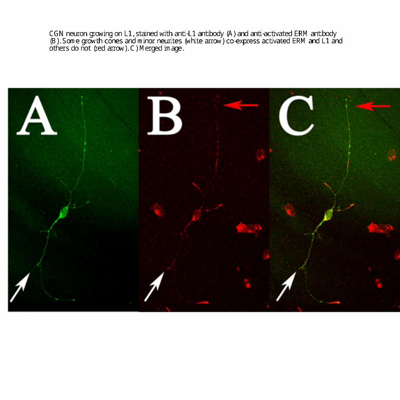
The mechanism underlying branching is not well understood.However, actin dynamics seem essential for the process; treat-ment of neurons with cytoskeleton-disrupting drugs inhibitsbranching but not axon length (Dent and Kalil, 2001). RhoGTPases, critical regulators of the actin cytoskeleton, have beenimplicated in the regulation of branching (Ng et al., 2002). ERMproteins are ideal candidates to regulate actin cytoskeleton down-stream of L1 because they can directly bind to L1 and actin filaments.Previous studies have shown that ERM proteins are essential forneuronal morphogenesis and growth cone motility (Paglini et al.,1998; Castelo and Jay, 1999). In hippocampal neurons, ERM pro-teins were localized to growth cones and colocalized with L1(Dickson et al., 2002; Haas et al., 2004). We have observedsimilar colocalization of activated ERMs and L1 in growthcones of cerebellar neurons (supplemental Fig. 2, available at
Figure 4. The effects of L1 cytoplasmic domain mutations on L1-mediated neurite out-growth. L1KO neurons were transfected with missense mutations and grown on an L1 sub-strate. Total neurite length, longest neurite length, and branching number were quantified. Themean SEM values of the mutant-transfected neurons were always normalized by the meanvalues of neurons transfected with WT hL1 in the same experiment. Values shown are theaverage of the mean SEM percentage values from three experiments. ANOVA (Fisher’s PLSD)was done using Statview 4.5. Statistical significance is shown. ***p � 0.001 in all three exper-iments; **p � 0.05 in all three experiments; *p � 0.05 in two of the three experiments.
Figure 5. The effects of L1 cytoplasmic domain mutations on L1-mediated branching, pri-mary neurites (branches extending from the soma), and nodes (branches arising from a neurite,not the soma). Primary neurites and nodes were quantified. The mean SEM values of themutant-transfected neurons were always normalized by the mean values of neurons trans-fected with WT hL1 in the same experiment. Values shown are the average of the mean SEMpercentage values from three experiments. ANOVA (Fisher’s PLSD) was done using Statview 4.5.Statistical significance is shown. ***p � 0.001 in all three experiments; **p � 0.05 in all threeexperiments.
400 • J. Neurosci., January 12, 2005 • 25(2):395– 403 Cheng et al. • The L1 Cytoplasmic Domain Regulates Axon Branching

www.jneurosci.org as supplemental material). It has also beenshown that ERM proteins colocalize with L1 in vivo when axonsare growing, but ERM protein expression then decreases (Mintzet al., 2003).
A coreceptor is implicated in L1-mediated neurite outgrowthThe L1-1147 construct, which deletes 110 of 114 amino acids ofthe L1CD, still supports neurite outgrowth. This is a very surpris-ing finding, given the high degree of conservation of the L1CDand the existence of MASA patients with mutations in the L1CD.It is widely assumed that the L1CD itself is part of the signaltransducer for L1–L1-mediated neurite outgrowth, because theL1CD can be coupled to cytoskeleton and it interacts with Erkand src, signaling molecules implicated in L1-mediated neuritegrowth. This result suggests that the L1 extracellular domain can
interact in cis with a coreceptor to stimu-late neurite outgrowth (supplemental Fig.1, available at www.jneurosci.org as sup-plemental material). However, corecep-tors have been implicated in L1-mediatedoutgrowth previously. The FGF receptor(Williams et al., 1994), TAG-1/axonin-1(Buchstaller et al., 1996), neuropilin-1(Castellani et al., 2000), and activated leu-kocyte cell adhesion molecule (DeBer-nardo and Chang, 1996) have been sug-gested to be L1 coreceptors. The fact thatL1-1147 has good single neurite out-growth but significantly reduced branch-ing number suggests that the coreceptorcan support single neurite outgrowth butdoes not participate in the initiation ofbranching. Another study from our labo-ratory, using L1 extracellular mutations,also suggests that a coreceptor is involvedin L1-mediated neurite outgrowth (Chengand Lemmon, 2004). For example, of twomutations that inhibit L1 homophilicbinding to a similar degree, only one altersbranching, suggesting that one mutationprevents interactions with a cis bindingpartner, whereas the other does not. It ispossible that L1 may have more than onecoreceptor, and the coreceptor(s) mayplay different roles under different situ-ations. Interestingly, it has been recentlyreported that NrCAM lacking its cyto-plasmic domain is still coupled to theactin cytoskeleton and that if the fi-bronectin (FN) domains are removed,then it becomes uncoupled from retro-grade flow, arguing that the FN domainsare involved in a cis interaction with a mol-ecule that is coupled to the actin retro-grade flow (Falk et al., 2004).
Although many observations point toL1 acting with a coreceptor to regulateneurite length and branching, there areother possible explanations for our dataon mutation of the L1CD. These muta-tions might alter L1–L1 cis interactions.They could also alter the conformationof the L1 extracellular domain, altering
outside-in signaling. Finally, as mentioned above, disruptionsof the L1–ERM interactions could prevent stabilization of newbranches.
L1–ankyrin binding may regulate neurite length but is notrequired for neurite initiation or extensionAnkyrin binds to L1 at a highly conserved C-terminal site. Arecent study proposed that L1–ankyrin binding mediates L1 ret-rograde flow at the perisomatic lamellae and thus regulates L1-stimulated neurite initiation (Nishimura et al., 2003). In contrast,Gil et al. (2003) have demonstrated that the ankyrin bindingregulates the stationary behavior of L1 but not the retrogradeflow. In this study, we tested several deletion mutants lacking theankyrin-binding site in our neurite outgrowth assay. The resultsof L1-1180 demonstrate that the loss of ankyrin-binding site may
Figure 6. The L1CD is not necessary for L1-mediated neurite outgrowth, but the L1CD regulates branching through thejuxtamembrane region. L1KO neurons were transfected with WT L1 ( A), L1-1147 ( B), L1-4A ( C), or L1-1151Y�A ( D) and plated on an L1substrate. After 48 hr, neurons were fixed and stained with a monoclonal anti-human L1 antibody (7B5). Scale bar, 100 �m.
Figure 7. Both the juxtamembrane region and the RSLE region are critical for the L1–ERM interaction. The pAS2 bait vectorscontaining wild-type and mutant forms of the L1CD fused to the Gal4 DNA-binding domain are cotransformed with the prey vectorpACT2 containing ezrin ( A), Drosophila ankyrin ( B), or �2 subunit of AP-2 ( C) fused to the Gal4 activation domain and tested forinteractions on histidine-deficient plates containing 10 mM 3-AT (A, B) or 5 mM 3-AT ( C). Each plate includes one positive controlp53�SV40, which is a diploid of AH109 [pGBKT7-53] (p53 was fused to GAL4 DNA-binding domain), and Y187 [pTD1-1] (SV40large T antigen was fused to GAL4 activation domain), because the p53 and the SV40 large T antigen was known to interact in theyeast two-hybrid assay. A negative control was included on each plate, which is a diploid of AH109 [pAS2-L1CD] and Y187[pTD1-1].
Cheng et al. • The L1 Cytoplasmic Domain Regulates Axon Branching J. Neurosci., January 12, 2005 • 25(2):395– 403 • 401
alter neurite length to a very small degree but does not signifi-cantly affect neurite initiation or branching. The L1-1147 con-struct, which deletes most of the L1CD, also reduces the longestneurite length to a small degree. However, another truncationconstruct, L1-1176, which also abolishes the ankyrin binding,does not affect the neurite length. The difference in neurite lengthbetween L1 constructs lacking the ankyrin-binding region versusthose with disrupted RSLE region is intriguing. It has been shownthat for L1-family members, adhesion is decreased if the ankyrin-binding region is mutated (Hortsch et al., 1998), that mutatingthe RSLE region leads to increased adhesion (Long et al., 2001),and that removing the entire L1 cytoplasmic domain producesnormal L1-mediated adhesion (Wong et al., 1995; Hortsch et al.,1998). These results suggest the L1CD has distinct regions thatcan positively and negatively regulate adhesion and, perhaps,neurite growth.
In summary, this study has demonstrated that the L1CD is notrequired for L1-mediated neurite outgrowth. The L1CD, how-ever, does play a significant role in branching. Both the jux-tamembrane region and the RSLE region, which bind ERM pro-teins, play roles in branching. After binding to an L1 substrate, L1on the cell surface may recruit a coreceptor to stimulate neuriteoutgrowth and use the L1CD to regulate branching.
ReferencesBeattie CE, Siegel RE (1993) Developmental cues modulate GABAA recep-
tor subunit mRNA expression in cultured cerebellar granule neurons.J Neurosci 13:1784 –1792.
Buchstaller A, Kunz S, Berger P, Kunz B, Ziegler U, Rader C, Sonderegger P(1996) Cell adhesion molecules NgCAM and axonin-1 form het-erodimers in the neuronal membrane and cooperate in neurite outgrowthpromotion. J Cell Biol 135:1593–1607.
Castellani V, Chedotal A, Schachner M, Faivre-Sarrailh C, Rougon G (2000)Analysis of the L1-deficient mouse phenotype reveals cross-talk betweenSema3A and L1 signaling pathways in axonal guidance. Neuron27:237–249.
Castellani V, De Angelis E, Kenwrick S, Rougon G (2002) Cis and transinteractions of L1 with neuropilin-1 control axonal responses to sema-phorin 3A. EMBO J 21:6348 – 6357.
Castelo L, Jay DG (1999) Radixin is involved in lamellipodial stability dur-ing nerve growth cone motility. Mol Biol Cell 10:1511–1520.
Cheng L, Lemmon V (2004) Pathological missense mutations of neural celladhesion molecule L1 affect neurite outgrowth and branching on an L1substrate. Mol Cell Neurosci 27:522–530.
Cohen NR, Taylor JS, Scott LB, Guillery RW, Soriano P, Furley AJ (1998)Errors in corticospinal axon guidance in mice lacking the neural celladhesion molecule L1. Curr Biol 8:26 –33.
Dahlin-Huppe K, Berglund EO, Ranscht B, Stallcup WB (1997) Mutationalanalysis of the L1 neuronal cell adhesion molecule identifies membrane-proximal amino acids of the cytoplasmic domain that are required forcytoskeletal anchorage. Mol Cell Neurosci 9:144 –156.
Dahme M, Bartsch U, Martini R, Anliker B, Schachner M, Mantei N (1997)Disruption of the mouse L1 gene leads to malformations of the nervoussystem. Nat Genet 17:346 –349.
Davis JQ, Bennett V (1994) Ankyrin binding activity shared by the neuro-fascin/L1/NrCAM family of nervous system cell adhesion molecules.J Biol Chem 269:27163–27166.
Davis JQ, McLaughlin T, Bennett V (1993) Ankyrin-binding proteins re-lated to nervous system cell adhesion molecules: candidates to providetransmembrane and intercellular connections in adult brain. J Cell Biol121:121–133.
DeBernardo AP, Chang S (1996) Heterophilic interactions of DM-GRASP:GRASP-NgCAM interactions involved in neurite extension. J Cell Biol133:657– 666.
Dent EW, Kalil K (2001) Axon branching requires interactions between dy-namic microtubules and actin filaments. J Neurosci 21:9757–9769.
Dent EW, Barnes AM, Tang F, Kalil K (2004) Netrin-1 and semaphorin 3Apromote or inhibit cortical axon branching, respectively, by reorganiza-tion of the cytoskeleton. J Neurosci 24:3002–3012.
Dickson TC, Mintz CD, Benson DL, Salton SR (2002) Functional bindinginteraction identified between the axonal CAM L1 and members of theERM family. J Cell Biol 157:1105–1112.
Dubreuil RR, MacVicar G, Dissanayake S, Liu C, Homer D, Hortsch M(1996) Neuroglian-mediated cell adhesion induces assembly of themembrane skeleton at cell contact sites. J Cell Biol 133:647– 655.
Falk J, Thoumine O, Dequidt C, Choquet D, Faivre-Sarrailh C (2004) Nr-CAM coupling to the cytoskeleton depends on multiple protein domainsand partitioning into lipid rafts. Mol Biol Cell 15:4695– 4709.
Felding-Habermann B, Silletti S, Mei F, Siu CH, Yip PM, Brooks PC, ChereshDA, O’Toole TE, Ginsberg MH, Montgomery AM (1997) A singleimmunoglobulin-like domain of the human neural cell adhesion mole-cule L1 supports adhesion by multiple vascular and platelet integrins.J Cell Biol 139:1567–1581.
Fransen E, Van Camp G, Vits L, Willems PJ (1997) L1-associated diseases:clinical geneticists divide, molecular geneticists unite. Hum Mol Genet6:1625–1632.
Fransen E, D’Hooge R, Van Camp G, Verhoye M, Sijbers J, Reyniers E, Sori-ano P, Kamiguchi H, Willemsen R, Koekkoek SK, De Zeeuw CI, De DeynPP, Van der Linden A, Lemmon V, Kooy RF, Willems PJ (1998) L1knockout mice show dilated ventricles, vermis hypoplasia and impairedexploration patterns. Hum Mol Genet 7:999 –1009.
Garver TD, Ren Q, Tuvia S, Bennett V (1997) Tyrosine phosphorylation at asite highly conserved in the L1 family of cell adhesion molecules abolishesankyrin binding and increases lateral mobility of neurofascin. J Cell Biol137:703–714.
Gil OD, Sakurai T, Bradley AE, Fink MY, Cassella MR, Kuo JA, Felsenfeld DP(2003) Ankyrin binding mediates L1CAM interactions with static com-ponents of the cytoskeleton and inhibits retrograde movement of L1CAMon the cell surface. J Cell Biol 162:719 –730.
Grumet M, Edelman GM (1988) Neuron-glia cell adhesion molecule inter-acts with neurons and astroglia via different binding mechanisms. J CellBiol 106:487–503.
Haas MA, Vickers JC, Dickson TC (2004) Binding partners L1 cell adhesionmolecule and the ezrin-radixin-moesin (ERM) proteins are involved indevelopment and the regenerative response to injury of hippocampal andcortical neurons. Eur J Neurosci 20:1436 –1444.
Hamada K, Shimizu T, Yonemura S, Tsukita S, Hakoshima T (2003) Struc-tural basis of adhesion-molecule recognition by ERM proteins revealed bythe crystal structure of the radixin-ICAM-2 complex. EMBO J22:502–514.
Haney CA, Sahenk Z, Li C, Lemmon VP, Roder J, Trapp BD (1999) Hetero-philic binding of L1 on unmyelinated sensory axons mediates Schwann celladhesion and is required for axonal survival. J Cell Biol 146:1173–1184.
Haspel J, Grumet M (2003) The L1CAM extracellular region: a multi-domain protein with modular and cooperative binding modes. FrontBiosci 8:s1210 –s1225.
Hortsch M, Homer D, Malhotra JD, Chang S, Frankel J, Jefford G, DubreuilRR (1998) Structural requirements for outside-in and inside-out signal-ing by Drosophila neuroglian, a member of the L1 family of cell adhesionmolecules. J Cell Biol 142:251–261.
Itoh K, Cheng L, Kamei Y, Fushiki S, Kamiguchi H, Gutwein P, Stoeck A,Arnold B, Altevogt P, Lemmon V (2004) Brain development in micelacking L1–L1 homophilic adhesion. J Cell Biol 165:145–154.
Kamiguchi H, Lemmon V (1998) A neuronal form of the cell adhesion mol-ecule L1 contains a tyrosine-based signal required for sorting to the axonalgrowth cone. J Neurosci 18:3749 –3756.
Kamiguchi H, Lemmon V (2000) Recycling of the cell adhesion molecule L1in axonal growth cones. J Neurosci 20:3676 –3686.
Kamiguchi H, Long KE, Pendergast M, Schaefer AW, Rapoport I, Kirch-hausen T, Lemmon V (1998a) The neural cell adhesion molecule L1interacts with the AP-2 adaptor and is endocytosed via the clathrin-mediated pathway. J Neurosci 18:5311–5321.
Kamiguchi H, Hlavin ML, Lemmon V (1998b) Role of L1 in neural devel-opment: what the knockouts tell us. Mol Cell Neurosci 12:48 –55.
Kenwrick S, Jouet M, Donnai D (1996) X linked hydrocephalus and MASAsyndrome. J Med Genet 33:59 – 65.
Kuhn TB, Stoeckli ET, Condrau MA, Rathjen FG, Sonderegger P (1991)Neurite outgrowth on immobilized axonin-1 is mediated by a hetero-philic interaction with L1(G4). J Cell Biol 115:1113–1126.
Kunz S, Spirig M, Ginsburg C, Buchstaller A, Berger P, Lanz R, Rader C, VogtL, Kunz B, Sonderegger P (1998) Neurite fasciculation mediated by
402 • J. Neurosci., January 12, 2005 • 25(2):395– 403 Cheng et al. • The L1 Cytoplasmic Domain Regulates Axon Branching
complexes of axonin-1 and Ng cell adhesion molecule. J Cell Biol143:1673–1690.
Lagenaur C, Lemmon V (1987) An L1-like molecule, the 8D9 antigen, is apotent substrate for neurite extension. Proc Natl Acad Sci USA84:7753–7757.
Lemmon V, McLoon SC (1986) The appearance of an L1-like molecule inthe chick primary visual pathway. J Neurosci 6:2987–2994.
Lemmon V, Farr KL, Lagenaur C (1989) L1-mediated axon outgrowth oc-curs via a homophilic binding mechanism. Neuron 2:1597–1603.
Lindner J, Rathjen FG, Schachner M (1983) L1 mono- and polyclonal anti-bodies modify cell migration in early postnatal mouse cerebellum. Nature305:427– 430.
Long KE, Asou H, Snider MD, Lemmon V (2001) The role of endocytosis inregulating L1-mediated adhesion. J Biol Chem 276:1285–1290.
Mintz CD, Dickson TC, Gripp ML, Salton SR, Benson DL (2003) ERMscolocalize transiently with L1 during neocortical axon outgrowth. J CompNeurol 464:438 – 448.
Ng J, Nardine T, Harms M, Tzu J, Goldstein A, Sun Y, Dietzl G, Dickson BJ,Luo L (2002) Rac GTPases control axon growth, guidance and branch-ing. Nature 416:442– 447.
Nishimura K, Yoshihara F, Tojima T, Ooashi N, Yoon W, Mikoshiba K,Bennett V, Kamiguchi H (2003) L1-dependent neuritogenesis involvesankyrinB that mediates L1-CAM coupling with retrograde actin flow.J Cell Biol 163:1077–1088.
Paglini G, Kunda P, Quiroga S, Kosik K, Caceres A (1998) Suppression ofradixin and moesin alters growth cone morphology, motility, and processformation in primary cultured neurons. J Cell Biol 143:443– 455.
Ruppert M, Aigner S, Hubbe M, Yagita H, Altevogt P (1995) The L1adhesion molecule is a cellular ligand for VLA-5. J Cell Biol 131:1881–1891.
Schaefer AW, Kamei Y, Kamiguchi H, Wong EV, Rapoport I, Kirchhausen T,Beach CM, Landreth G, Lemmon SK, Lemmon V (2002) L1 endocytosisis controlled by a phosphorylation-dephosphorylation cycle stimulatedby outside-in signaling by L1. J Cell Biol 157:1223–1232.
Stallcup WB, Beasley L (1985) Involvement of the nerve growth factor-inducible large external glycoprotein (NILE) in neurite fasciculation inprimary cultures of rat brain. Proc Natl Acad Sci USA 82:1276 –1280.
Williams EJ, Furness J, Walsh FS, Doherty P (1994) Activation of the FGFreceptor underlies neurite outgrowth stimulated by L1, N-CAM, andN-cadherin. Neuron 13:583–594.
Wisco D, Anderson ED, Chang MC, Norden C, Boiko T, Folsch H, WincklerB (2003) Uncovering multiple axonal targeting pathways in hippocam-pal neurons. J Cell Biol 162:1317–1328.
Wong EV, Cheng G, Payne HR, Lemmon V (1995) The cytoplasmic domainof the cell adhesion molecule L1 is not required for homophilic adhesion.Neurosci Lett 200:155–158.
Wong EV, Schaefer AW, Landreth G, Lemmon V (1996) Involvement ofp90rsk in neurite outgrowth mediated by the cell adhesion molecule L1.J Biol Chem 271:18217–18223.
Yonemura S, Hirao M, Doi Y, Takahashi N, Kondo T, Tsukita S (1998)Ezrin/radixin/moesin (ERM) proteins bind to a positively charged aminoacid cluster in the juxta-membrane cytoplasmic domain of CD44, CD43,and ICAM-2. J Cell Biol 140:885– 895.
Cheng et al. • The L1 Cytoplasmic Domain Regulates Axon Branching J. Neurosci., January 12, 2005 • 25(2):395– 403 • 403


Supplemental Table 1. Raw data of the effects of intracellular mutations on L1-
medaited neurite outgrowth
Mutation NM/NW Total neurite length -microns
Longest neurite length -microns
Branches Primary neurites
Nodes
L1-1176 50/88 479±25/ 591±19
288±14/ 288±10
5±0.67/ 6.22±0.35
3.54±0.41/ 3.06±0.17
1.46±0.32/ 3.1±0.26
79/98 451±18/ 536±16
284±12/ 253±8
5.54±0.45/ 8.2±0.4
2.28±0.11/ 2.92±0.13
3.2±0.39/ 5.07±0.35
79/85 458±20/ 630±21
261±12/ 290±11
5.10±0.39/ 8.59±0.54
2.84±0.19/ 3.15±0.16
2.24±0.25/ 5.18±0.42
L1-1180 73/88 543±21/ 591±19
254±11/ 288±10
6.92±0.44/ 6.22±0.35
4.01±0.34/ 3.06±0.17
2.84±0.24/ 3.1±0.26
80/98 464±19/ 536±16
223±11/ 253±8
8.4±0.57/ 8.2±0.4
3.1±0.21/ 2.92±0.13
5.15±0.44/ 5.07±0.35
92/93 584±22/ 698±28
239±10/ 288±9
10.84±0.69 10.31±0.62
5.11±0.39/ 4.06±0.26
5.54±0.44/ 6.02±0.50
L1∆RSLE 79/98 451±18/ 536±16
263±11/ 253±8
5.65±0.35/ 8.2±0.4
2.7±0.14/ 2.92±0.13
2.84±0.29/ 5.07±0.35
88/88 455±15/ 591±19
307±12/ 288±10
4.42±0.35/ 6.22±0.35
2.92±0.22/ 3.06±0.17
1.47±0.19/ 3.1±0.26
36/85 444±35/ 642±22
244±14/ 273±10
5.69±0.71/ 8.28±0.54
3.08±0.35/ 2.99±0.13
2.5±0.42/ 5.13±0.46
L1-1147 106/104 276±10/ 516±14
200±8/ 243±6
3.29±0.23/ 5.29±0.25
2.52±0.18/ 2.72±0.12
0.77±0.12/ 2.58±0.19
96/79 325±12/ 652±20
290±10/ 285±11
2.45±0.19/ 6.91±0.44
1.79±0.14/ 3.25±0.18
0.64±0.11/ 3.54±0.32
76/97 219±11/ 506±18
177±10/ 264±10
2.61±0.25/ 4.88±0.29
2.13±0.19/ 2.62±0.13
0.47±0.14/ 2.21±0.22
L1-4A 93/67 204±7/ 357±15
153±5/ 190±6
2.14±0.09/ 4.93±0.30
1.83±0.05/ 2.51±0.14
0.31±0.08/ 2.39±0.23
103/103 280±10/ 540±20
210±8/ 260±9
2.53±0.15/ 6.85±0.40
1.84±0.06/ 3.12±0.15
0.67±0.11/ 3.64±0.29
87/80 266±10/ 480±14
195±8/ 251±8
2.59±0.14/ 5.10±0.34
2±0.08/ 2.35±0.1
0.57±0.1/ 2.7±0.31
L1-1151Y>A
97/67 232±9/ 357±15
174±7/ 190±6
2.24±0.11/ 4.93±0.30
1.91±0.06/ 2.51±0.14
0.32±0.08/ 2.39±0.23
95/103 325±14/ 540±20
233±11/ 260±9
3.32±0.19/ 6.85±0.40
2.08±0.02/ 3.12±0.15
1.22±0.16/ 3.64±0.29
92/87 318±15/ 493±18
225±11/ 247±10
3.45±0.22 6.99±0.50
2.11±0.08/ 2.91±0.15
1.32±0.18/ 4±0.4
In all experiments mutant L1 or WT HL1 was transfected into L1KO neurons and plated
on L1 substrates. About 80-100 of the L1KO neurons expressing HL1 were analyzed and
used as reference values for determining the degree of neurite growth and branching for
the different mutants. For each mutant, values from three independent trials were listed.

The values before the slash were the mean± SEM of mutant L1-expression neurons. The
values after the slash were the mean± SEM of WT HL1-expression neurons in the same
set of experiment. NM is the number of mutant L1-expressing neurons measured and NW
is the number of WT HL1-expressing neurons measured in the same set of experiment.
Values for the total neurite length, longest neurite length, branches (primary, secondary
and tertiary neurites), primary neurites from the soma, and nodes (secondary and tertiary
neurites) were listed.