Embed Size (px)
Citation preview

Neuronal Apoptosis Associated with Morphine Tolerance: Evidencefor an Opioid-Induced Neurotoxic Mechanism
Jianren Mao,1 Backil Sung,1 Ru-Rong Ji,2 and Grewo Lim1
1Massachusetts General Hospital Pain Center and 2Neural Plasticity Research Group, Department of Anesthesia andCritical Care, Massachusetts General Hospital, Harvard Medical School, Boston, Massachusetts 02114
Tolerance to the analgesic effect of an opioid is a pharmaco-logical phenomenon that occurs after its prolonged administra-tion. Activation of the NMDA receptor (NMDAR) has been im-plicated in the cellular mechanisms of opioid tolerance.However, activation of NMDARs can lead to neurotoxicity undermany circumstances. Here we demonstrate that spinal neuro-nal apoptosis was induced in rats made tolerant to morphineadministered through intrathecal boluses or continuous infu-sion. The apoptotic cells were predominantly located in thesuperficial spinal cord dorsal horn, and most apoptotic cellsalso expressed glutamic acid decarboxylase, a key enzyme forthe synthesis of the inhibitory neurotransmitter GABA. Consis-tently, increased nociceptive sensitivity to heat stimulation wasobserved in these same rats. Mechanistically, the spinal gluta-matergic activity modulated morphine-induced neuronal apo-ptosis, because pharmacological perturbation of the spinalglutamate transporter activity or coadministration of morphinewith the NMDAR antagonist (�)-5-methyl-10,11-dihydro-5H-
dibenzo [a,d] cyclohepten-5,10-imine maleate affected bothmorphine tolerance and neuronal apoptosis. At the intracellularlevel, prolonged morphine administration resulted in an upregu-lation of the proapoptotic caspase-3 and Bax proteins but adownregulation of the antiapoptotic Bcl-2 protein in the spinalcord dorsal horn. Furthermore, coadministration with morphineof N-benzyloxycarbonyl-Val-Ala-Asp-fluoromethyl ketone (apan-caspase inhibitor) or acetyl-aspartyl-glutamyl-valyl-aspart-1-aldehyde (a relatively selective caspase-3 inhibitor) blockedmorphine-induced neuronal apoptosis. Blockade of the spinalcaspase-like activity also partially prevented morphine toler-ance and the associated increase in nociceptive sensitivity.These results indicate an opioid-induced neurotoxic conse-quence regulated by the NMDAR–caspase pathway, a mecha-nism that may have clinical implications in opioid therapy andsubstance abuse.
Key words: apoptosis; opioid tolerance; analgesia; NMDA;glutamate transporter; caspase-3; Bax; Bcl-2
Opioids are a class of powerful analgesics that are commonly usedin acute and chronic pain management. Prolonged exposure to anopioid results in the development of analgesic tolerance, whichsignificantly hampers the clinical utility of opioids necessitatingrepeated dose escalation regardless of disease progression. Assuch, the cellular and molecular mechanisms of opioid tolerancehave been a focus of extensive research interest. Over a decade,the NMDA receptor (NMDAR), a subgroup of glutamate recep-tors, has been implicated in the development of opioid tolerance,particularly �-opioid tolerance (Marek et al. 1991a,b; Trujillo andAkil, 1991; Elliott et al., 1994; Mao et al., 1994). Although themechanisms of NMDAR involvement in opioid analgesic toler-ance remain unclear, Ca2�-regulated intracellular protein kinaseC (PKC) is likely to be a link in this process (Mao et al., 1994,1995c; Mayer et al., 1995; Narita et al., 1996, 2001; Zeitz et al.,2002). PKC may directly or indirectly regulate the activity ofNMDARs by removing the Mg2� blockade from the NMDAR–Ca2� channel site (Chen and Huang, 1992; Woolf and Salter,2000), regulating NMDAR trafficking and gating (Lan et al.,2001), or both. Thus, NMDARs could become involved in thecellular mechanisms of opioid tolerance after prolonged admin-
istration of an opioid such as morphine (Trujillo and Akil, 1991;Mao, 1999).
Activation of NMDARs can lead to neurotoxicity under manycircumstances (Rothman and Olney, 1986; Swan and Meldrum,1990; Moncada et al., 1992; Catania et al., 1993). For instance,peripheral nerve injury has been shown to activate spinal cordNMDARs, which results in not only intractable neuropathic painbut also neuronal cell death by means of apoptosis (Mao et al.1992b, 1997; Kawamura et al., 1997; Whiteside and Munglani,2001). Furthermore, cross talk between the cellular mechanismsof opioid tolerance and neuropathic pain has been proposed,suggesting that a common cellular mechanism may be involved inboth neuropathic pain and opioid tolerance (Mao et al. 1995b;Mayer et al., 1999). Thus, it is possible that the cellular processleading to the development of opioid tolerance may also causeneurotoxic changes in response to prolonged opioid administra-tion. Here we examined the hypothesis that neurotoxicity in theform of apoptotic cell death would be induced in association withthe development of morphine tolerance.
At the synaptic level, glutamate, an endogenous ligand for theNMDAR, is actively and tightly regulated by the glutamatetransporter (GT) system (Robinson and Dowd, 1997; Semba andWakuta, 1998; Jabaudon et al., 2000). Indeed, the expression ofGLT-1 (a glial GT) has been shown to be reduced after exposureto opioids in both cortical cell cultures (Thorlin et al., 1998) andbrain regions (Ozawa et al., 2001), which may influence thedevelopment of morphine tolerance and dependence (Nakagawaet al., 2001). At the intracellular level, glutamate-induced neuro-
Received April 12, 2002; revised June 21, 2002; accepted June 21, 2002.This work was supported by United States Public Health Service Grant RO1
DA08835 to J.M. We thank Leslie Keniston, Joachim Scholz, and the NeuralPlasticity Research Group at Massachusetts General Hospital for technical help.
Correspondence should be addressed to Dr. Jianren Mao, Massachusetts GeneralHospital Pain Center, Suite WACC 324, Massachusetts General Hospital, HarvardMedical School, 15 Parkman Street, Boston, MA 02114. E-mail: [email protected] © 2002 Society for Neuroscience 0270-6474/02/227650-12$15.00/0
The Journal of Neuroscience, September 1, 2002, 22(17):7650–7661

nal apoptosis has been shown to be modulated by commonintracellular regulators of apoptosis, including Bax, Bcl-2, andcaspases (Du et al., 1997; Tenneti et al., 1998; Allen et al., 1999;Springer et al., 1999; Kwong and Lam, 2000; Nath et al., 2000;Puka-Sundvall et al., 2000; Qin et al., 2000; Tenneti and Lipton,2000; Bachis et al., 2001; Chan et al., 2001). Thus, if prolongedexposure to an opioid induces apoptosis that is regulated throughNMDAR-mediated glutamate neurotoxicity, one would expect tosee the modulation of opioid-induced apoptosis by spinal GTsand NMDARs as well as by common intracellular regulators ofapoptosis such as caspases. These possibilities were examined inthe present study to investigate the cellular mechanisms of neu-ronal apoptosis associated with the development of morphinetolerance.
MATERIALS AND METHODSExperimental animalsAdult male Sprague Dawley rats (Charles River Laboratories, Wilming-ton, MA) weighing 300–350 gm were used. Animals were housed incages with water and food pellets available ad libitum. The animal roomwas artificially illuminated from 7 A.M. to 7 P.M. The experimentalprotocol was approved by our Institutional Animal Care and UseCommittee.
Intrathecal catheter and osmotic pump implantationAn intrathecal PE 10 catheter was implanted in each rat according to apreviously published method (Yaksh and Rudy, 1976). Those animalsthat exhibited neurological deficits after intrathecal catheter implanta-tion were excluded from the experiments. Drugs were delivered via anintrathecal catheter in a total volume of 10 �l followed by a saline flush.The following drugs were purchased from Sigma (St. Louis, MO):(�)-5-methyl-10,11-dihydro-5H-dibenzo [a,d] cyclohepten-5,10-iminemaleate (MK-801), morphine, riluzole, L-trans-pyrrolidine-2–4-dicarboxylate (PDC), N-benzyloxycarbonyl-Val-Ala-Asp-fluoromethylketone (Z-VAD-FMK), and acetyl-aspartyl-glutamyl-valyl-aspart-1-aldehyde (AC-DEVD-CHO).
For continuous intrathecal infusion, osmotic minipumps (Alza, Moun-tain View, CA) were used as described previously (Granados-Soto et al.,2000; Vanderah et al., 2000). An osmotic pump, placed in a subcutaneousspace after a surgical procedure, was connected to an intrathecal cathetervia a piece of PE 60 catheter. The filled minipumps were soaked innormal saline for 4 hr before the insertion to ensure immediate drugdelivery. The integrity of the pump delivery system was reexamined atthe end of each experiment when the spinal cords were harvested.
Induction of morphine tolerance and behavioral testTolerance to the antinociceptive effect of morphine was induced usingtwo intrathecal treatment regimens: repeated boluses and continuousinfusion. Morphine was given twice daily for 7 d in the repeated bolusregimen, whereas continuous morphine infusion was made for 7 d via animplanted osmotic pump system delivering at 1 �l /hr for 7 d. Differencesin morphine antinociception among treatment groups were assessed onday 8 by the tail flick test at 30 min after a probe dose of either10 �g ofmorphine (intrathecal) for repeated bolus groups or 5 mg/kg morphine(intraperitoneal) for continuous infusion groups. Additionally, foot with-drawal latencies were compared among groups between day 0 (baseline)and the last day (day 8) of the experimental period to determine whethermorphine-tolerant rats would develop increased sensitivity to noxiousheat stimulation as shown in previous studies (Mao et al. 1995a; Ossipovet al., 1995; Vanderah et al., 2000). Because the osmotic pump infusionbegan on day 1, day 8 was the last day of a full 7 d delivery using anosmotic pump.
The routine tail flick test was made with baseline latencies of 4–5 secand a cutoff time of 10 sec (D’Amour and Smith, 1941; Akil and Mayer,1972). Three trials were made with an intertrial interval of 1 min andwith changes of the tail position receiving radiant heat stimulation ateach trial. The percent maximal possible antinociceptive effect(%MPAE) was determined by comparing the tail flick latency before[baseline (BL)] and after a drug injection (TL) using the equation%MPAE � [(TL � BL)/(10 � BL)] � 100 (the constant 10 refers to thecutoff time). To examine changes in baseline nociceptive responses
before and after a prolonged morphine administration, the foot with-drawal test with baseline latencies of 9–11 sec and a cutoff time of 20 secwas used as described previously (Hargreaves et al., 1988). The footwithdrawal test was used because this test has been shown to be sensitivein detecting moderate changes in baseline nociceptive responses (Mao etal., 1994).
In situ terminal deoxynucleotidyl transferase-mediatedbiotinylated UTP nick end-labeling stainingThe DNA fragmentation indicative of apoptosis can be demonstratedusing several methods, including terminal deoxynucleotidyl transferase-mediated biotinylated UTP nick end labeling (TUNEL), gel electro-phoresis, and in situ nick translation (Gavrieli et al., 1992; Baba et al.,1999). In this study, we used the TUNEL method because this methodallows us to examine the topographic distribution of apoptotic cellswithin the spinal cord dorsal horn, which cannot be shown using gelelectrophoresis. Besides, it has been shown that apoptotic changes re-vealed by the TUNEL method are consistent with the gel electrophoresisdata under several experimental conditions (Gavrieli et al., 1992; Lo etal., 1995). As described below, both positive and negative controls wereincluded in the staining process and the costaining with Hoechst (for thein vivo detection of DNA), and TUNEL was used to ensure the consis-tency of the data collection. In addition, the morphology of TUNEL- andHoechst-stained nuclei also was examined under a high-magnificationmicroscopic view to identify features of apoptotic cells (e.g., condensedDNA segments and nuclear fragmentation).
A modified TUNEL staining protocol described in previous studieswas followed (Gavrieli et al., 1992; Hara et al., 1995, 1998). Spinal cordsfrom each group were collected after the final behavioral test on day 8after transaortic perfusion with saline and a fixative containing 4%paraformaldehyde and cut into 10-�m-thick sections with a cryostat. Oneof every five such sections was mounted on a precoated slide. TheTUNEL staining was performed using the apoptosis detection kit pur-chased from Roche Molecular Biochemicals (Indianapolis, IN). Briefly,the sections were first incubated in a solution containing 0.1% TritonX-100 and 0.1% sodium citrate for 2 min on ice (4°C) to increase thepermeability. After being washed twice in PBS, pH 7.4, the sections wereimmersed in the TUNEL reaction mixture, containing biotinylateddUTP and terminal deoxynucleotidyl transferase (TdT) conjugated withfluorochromes (tetramethylrhodamine red) for 60 min at 37°C in a dark,humidified atmosphere. The process was terminated by washing thesections twice in a blocking buffer (PBS, Triton X-100, and BSA). In eachassay, negative controls were included using the same incubation proce-dure but omitting TdT in the process, whereas positive controls wereperformed by incubating the permeated sections with DNase (1 �g/ml)to induce DNA strand breakage.
Immunocytochemical and Hoechst stainingRoutine immunocytochemical staining (Ji et al., 1995) was used to detectneuronal-specific nuclear protein (NeuN) (1:500, a marker for neuronalnuclear protein; Mullen et al., 1992), glutamic acid decarboxylase 67(GAD67; 1:1000), Bax (1:250), caspase-3 (1:500), and cleaved caspase-3(1:50). All antibodies were purchased from Chemicon (Temecula, CA)except for the cleaved caspase-3 antibody (Cell Signaling Technology,Beverly, MA). The process of harvesting, fixing, and slicing spinal cordsamples was the same as that used for the TUNEL procedure. After theTUNEL staining, sections were blocked with 1% goat serum in 0.3%Triton X-100 for 1 hr at room temperature and incubated overnight at4°C with a primary antibody. The sections were then incubated for 1 hrat room temperature with a corresponding FITC-conjugated secondaryantibody (1:300; Chemicon). The Hoechst staining (Hoechst 33342) wasused for the in vivo detection of DNA in spinal sections. Colocalizationof TUNEL with NeuN, GAD67, caspase-3, Bax, or Hoechst staining wasexamined by an imaging program (Adobe Photoshop).
Cell countingFive or six sections randomly selected from each animal were analyzed byusing a fluorescence microscope linked to a digital camera. Numbers ofapoptotic cells were counted in a blinded manner for both sides of eachspinal section, in which three regions (laminas I–II, III–IV, and V–VI)were divided based on the laminar delineation described previously(Molander et al., 1984; Mao et al. 1992a, 1993). These regions werechosen because they represent functional subdivisions of the spinal corddorsal horn (Price, 1988). Two approaches were used to display these
Mao et al. • Neuronal Apoptosis after Morphine Tolerance J. Neurosci., September 1, 2002, 22(17):7650–7661 7651
laminar divisions. Spinal sections were either counterstained with Nisslstaining or costained with NeuN staining. Both methods have beencommonly used to outline spinal cord dorsal horn divisions based ondistinct laminar patterns described by Molander et al. (1984). Becausethe TUNEL staining is only visible in the cell body, and sections selectedfor the analysis were chosen from at least 50 �m apart (see above), thisanalysis avoided double counting the number of apoptotic cells. Toanalyze sections with costainings (e.g., TUNEL and caspase-3), imagesfrom each staining were digitized and then merged using an imagingprogram (Adobe) to examine the presence of colocalization.
Western analysis of Bax, Bcl-2, and caspase-3For Western blotting, rats were rapidly (�1 min) killed in a CO2chamber, and the dorsal horns of the lumbar spinal cord segments wereremoved and homogenized in SDS sample buffer containing a mixture ofproteinase inhibitors (Sigma). The lumbar segments were harvestedbecause an intrathecal catheter was aimed to deliver drugs at this site.Protein samples were separated on an SDS-PAGE gel (4–15% gradientgel; Bio-Rad, Hercules, CA) and transferred to polyvinylidene difluoridefilters (Millipore, Bedford, MA). The filters were blocked with 3% milkand incubated overnight at 4°C with a primary antibody (Bax, 1:100;caspase-3, 1:1000 for 19 and 32 kDa; and Bcl-2, 1:5000) and 1 hr at roomtemperature with an HRP-conjugated secondary antibody (1:5000; Am-ersham Biosciences, Arlington Heights, IL). The blots were then visual-ized in ECL solution (PerkinElmer Life Sciences, Emeryville, CA) for 1min and exposed onto hyperfilms (Amersham) for 1–10 min. The graydensity of each blot was obtained for each experimental group. The sameamounts of protein for each loading lane were estimated by the Bio-Radprotein assay, and the extracellular signal-regulated kinase (ERK) pro-tein was used as a loading control.
Experimental designExperiment 1: induction of apoptosis af ter morphine tolerance. Sevengroups of rats were used in this experiment. To investigate whetherrepeated exposure to morphine boluses would result in the induction ofapoptosis, three groups of rats (n � 5) were each given 10 or 20 �g ofintrathecal morphine or saline twice daily for 7 d. In addition, three moregroups of rats (n � 5) were infused, via an intrathecal osmotic pump for7 d, with 10 or 20 nmol � �l �1 � hr �1 morphine or saline to determinewhether apoptosis would be induced using continuous infusion. Thistreatment regimen was included because repeated morphine boluseshave been suggested to cause intermittent opioid withdrawals, a processthat could increase NMDAR activity via glutamate release independentof the intracellular mechanisms of opioid tolerance (Ibuki et al., 1997;Dunbar and Pulai, 1998). In either treatment regimen, morphine doseswere chosen on the basis of the previous studies that showed the devel-opment of morphine tolerance using these doses (Mao et al., 1994; Ibukiet al., 1997). An additional group of rats (n � 4) was included to examinewhether apoptosis occurred in response to an acute morphine effect aftera single intrathecal injection of 20 �g of morphine. In all groups, spinalcords were harvested after the final behavioral test as described above.
To investigate the neurochemical nature of apoptotic cells, the spinalsections were examined for the colocalization of TUNEL and GAD, akey enzyme for the synthesis of the inhibitory neurotransmitter GABA.Furthermore, neuronal apoptosis was identified by examining the colo-calization of TUNEL and NeuN as described above.
Experiment 2: role of the spinal GT and NMDAR in morphine-inducedapoptosis. To investigate whether perturbation of spinal GT activitywould affect opioid-induced apoptosis, PDC, a GT inhibitor (Lievens etal., 2000; Matthews et al., 2000), and riluzole, a positive GT regulator(Azbill et al., 2000), were used in this set of experiments. Althoughriluzole was initially considered as an inhibitor of the presynaptic gluta-mate release (Cheramy et al., 1992; Doble, 1996), this agent has beenshown recently to be a positive GT regulator that increases glutamateuptake in synaptosomes under both in vivo and in intro conditions (Azbillet al., 2000). Four groups of rats (n � 5) were used, including (1) 10 �gof morphine plus 20 �g of riluzole, (2) 20 �g of riluzole alone, (3) 10 �gof morphine plus 20 �g of PDC, and (4) 20 �g of PDC alone. The drugsor their combinations were given intrathecal twice daily for 7 d and thesegroups were compared with those receiving 10 �g of morphine or salinealone in experiment 1. The dose for riluzole or PDC was selected on thebasis of a pilot study showing a reliable effect of each agent at this doseon modulating the development of morphine (10 �g, intrathecal) toler-ance. In addition, the equivalent doses of PDC and riluzole have beenshown to be effective in regulating the extracellular glutamate concen-
tration and NMDAR-mediated activity under both in vivo and in vitroexperimental conditions (Semba and Wakuta, 1998; Azbill et al., 2000;Jabaudon et al., 2000; Lievens et al., 2000; Matthews et al., 2000).
To determine the role of NMDARs in the induction of neuronalapoptosis, two more groups of rats each received a 7 d intrathecalinfusion with either 20 nmol � �l �1 � hr �1 morphine plus 1nmol � �l �1 � hr �1 MK-801 (n � 9) or 1 nmol � �l �1 � hr �1 MK-801 alone(n � 6). The dose for MK-801 was selected on the basis of a previousstudy showing the blockade of morphine tolerance by this dose ofMK-801 (Ibuki et al., 1997). The data from these groups were comparedwith those from the 20 nmol � �l �1 � hr �1 morphine- or saline-alonegroups in experiment 1.
Experiment 3: role of intracellular apoptosis regulators in morphine-induced apoptosis. To examine the intracellular mechanisms ofmorphine-induced apoptosis, two approaches were used. First, changesin Bax, caspase-3, and Bcl-2 were examined using the methods of West-ern blotting and immunocytochemistry in rats receiving either 20 �g ofmorphine or saline (n � 8–10 per group for separate sample collections)twice daily for 7 d. Second, additional groups of rats (n � 4–6 per group)were used to examine whether blockade of caspases including caspase-3would prevent morphine-induced apoptosis. Thus, each group of ratsreceived intrathecal (1) saline, (2) 20 �g of morphine plus vehicle, (3) 20�g of morphine plus 5 �g of Z-VAD-FMK (a pan-caspase inhibitor), or(4) 20 �g of morphine plus 5 �g of AC-DEVD-CHO (a relativelyselective caspase-3 inhibitor) twice daily for 7 d. The selected dose foreach agent was based on previous studies showing a reliable inhibition ofthe caspase-like activity in vivo within this intrathecal dose range (Qin etal., 2000; Chan et al., 2001). For additional controls, rats (n � 3–4 pergroup) received a 7 d intrathecal treatment (twice daily) with the samedose of Z-VAD-FMK or AC-DEVD-CHO alone. A single dose of 10 �gof morphine was given on day 8 to examine whether Z-VAD-FMK orAC-DEVD-CHO alone would affect morphine-induced antinociception.
Statistical analysisData obtained from the tail flick test were first calculated to yield themean %MPAE as shown previously (Mao et al., 1994). The data werethen analyzed by using two-way ANOVA to detect overall differencesamong treatment groups. When significant main effects were observed,Waller-Duncan (WD) K ratio t tests (WD) were performed to determinesources of differences. The histological data (cell counting) from eachspinal section were first averaged for each dorsal horn region and thenanalyzed using ANOVA followed by the WD test to determine thestatistical differences. Paired Student’s t test was used to examine statis-tical differences in the gray density of Western blots.
RESULTSInduction of neuronal apoptosis associated withmorphine toleranceTwice daily administration of 10 or 20 �g of morphine for 7 dproduced, dose-dependently, tolerance to the antinociceptive ef-fect of morphine when tested on day 8 (Fig. 1A) ( p � 0.01). Thismorphine treatment regimen induced apoptotic cells within thespinal cord dorsal horn of the same rats (Fig. 2B). The in situdetection of DNA fragmentation by the TUNEL method wasfurther indicated by the colocalization of both TUNEL andHoechst (detecting in situ DNA) staining in the same cells (Fig.2D–F). Moreover, features of apoptotic cells were observed inthe TUNEL and Hoechst costained nuclei including nuclearfragmentation and condensed DNA segments (Fig. 2D�–F�). Incontrast, apoptotic cells were hardly detectable in saline-treatedrats (Figs. 2A, 3A) ( p � 0.01), indicating that the induction ofapoptosis is specifically associated with morphine treatment.Likewise, few apoptotic cells were present in rats receiving asingle intrathecal treatment of 20 �g of morphine (Fig. 3A) ( p �0.05), indicating that the induction of apoptosis is not attributableto an acute morphine effect.
Similar to the results obtained using the bolus treatment regi-men, tolerance to the morphine antinociception developed dose-dependently when tested on day 8 in rats receiving 10 or 20
7652 J. Neurosci., September 1, 2002, 22(17):7650–7661 Mao et al. • Neuronal Apoptosis after Morphine Tolerance

nmol � �l�1 � hr�1 morphine, but not saline, infusion for 7 d (Fig.1A) ( p � 0.01). Consistently, apoptotic cells were observed inmorphine- but not saline-infused rats (Fig. 3A) ( p � 0.01). Boththe distribution and quantity of apoptotic cells were comparablewith those seen in rats treated with morphine boluses (Fig. 3A). Inboth experiments, more apoptotic cells were observed in ratsreceiving a high dose (20 �g or 20 nmol � �l�1 � hr�1) than a lowdose (10 �g or 10 nmol � �l�1 � hr�1) of morphine (Fig. 3A) ( p �0.05), indicating that the induction of apoptosis wasdose-dependent.
Topographically, these apoptotic cells were primarily located inlaminas I–II of the spinal cord dorsal horn of rats receiving eitherrepeated or continuous morphine administration (Table 1). Fur-thermore, most apoptotic cells were identified as neuronal cells
(Fig. 3B), because both apoptosis (TUNEL) and neuronal(NeuN) markers were colocalized in the same cells (Fig. 2G–I).Because the total number of apoptotic cells exceeded that ofneuronal apoptotic cells (Fig. 3B), it is likely that some of theseapoptotic cells were glial cells (Fig. 2G–I). Thus, apoptosis wasinduced in the spinal cord dorsal horn of rats made tolerant tomorphine after either repeated bolus or continuous intrathecaladministration.
Expression of the GABA-synthesizing enzyme GAD inapoptotic neuronal cellsSpinal sections from rats receiving either repeated 20 �g mor-phine boluses or 20 nmol � �l�1 � hr�1 morphine infusion for 7 dwere costained with TUNEL and GAD67. Colocalization of bothTUNEL and GAD67 immunostaining was clearly observedwithin the superficial spinal cord dorsal horn laminas (Fig. 2J–L).As shown in Figure 3B, �50% of apoptotic cells from each groupdisplayed positive GAD67 immunostaining. Given the observa-tion that a portion of apoptotic cells are likely to be glial cells(Fig. 2G–I) and that GAD is expected to be visualized only inneuronal cells, the data indicate that many apoptotic neuronalcells are likely to be GABAergic inhibitory neurons.
Increased nociceptive heat sensitivity in rats showingneuronal apoptosisThe baseline foot withdrawal latency was compared between day0 (baseline) and day 8 in each group. There was no significantdifference in the foot-withdrawal latency between days 0 and 8 inthe saline-treated rats (Fig. 4A) ( p � 0.05). In contrast, thebaseline foot withdrawal latency was reduced on day 8, comparedwith that on day 0 (Fig. 4A) ( p � 0.01) in rats receiving eitherrepeated boluses or continuous infusion with morphine for 7 d.Similar to that observed with morphine tolerance (Fig. 1) andassociated neuronal apoptosis (Fig. 3A), the magnitude of in-crease in nociceptive sensitivity was also dose-dependent. That is,a significantly lower baseline foot withdrawal latency was observedin rats treated with a high dose (20 �g or 20 nmol � �l�1 � hr�1)than a low dose (10 �g or 10 nmol � �l�1 � hr�1) of morphine (Fig.4A) ( p � 0.05). These results indicate that the induction ofneuronal apoptosis in morphine-tolerant rats was accompanied byan increase in the sensitivity to noxious heat stimulation, a findingconsistent with that demonstrated by previous studies (Mao et al.1995a; Ossipov et al., 1995; Vanderah et al., 2000).
Contribution of the spinal GT and NMDAR to theinduction of neuronal apoptosisIntrathecal coadministration (twice daily) of morphine (10 �g)and the GT inhibitor PDC (20 �g) for 7 d further increased thenumber of apoptotic cells within the superficial spinal cord dorsalhorn compared with that of the morphine-alone (10 �g) group(Figs. 2A–C, 5A) ( p � 0.01). Conversely, apoptotic cells werereduced in rats receiving combined morphine (10 �g) and ri-luzole (20 �g, a positive regulator of GT activity) for 7 d com-pared with that of the morphine-alone group (Fig. 5A) ( p � 0.05).Neither PDC nor riluzole alone at the current dose inducedapoptotic changes (Fig. 5A). Furthermore, riluzole and PDC at itscurrent dose also reduced and enhanced, respectively, the devel-opment of morphine tolerance and changes in nociceptive sensi-tivity in the behavioral tests (Figs. 1B, 4B). Thus, regulation ofthe spinal GT activity contributes to the induction of apoptosisassociated with the development of morphine tolerance and theincrease in nociceptive heat sensitivity.
Consistent with the role of spinal GT activity, apoptosis was
Figure 1. Modulation of morphine tolerance by regulating spinal GT andNMDAR activity. A, When tested on day 8, %MPAE in response to 20�g of intrathecal morphine was reduced in rats treated with 10 or 20 �gof intrathecal morphine (B10, B20) twice daily for 7 d. Similarly, %MPAEin response to 5 mg/kg morphine (intraperitoneally) on day 8 was reducedin rats receiving a 7 d infusion, via an intrathecal osmotic pump, with 10or 20 nmol � �l �1 � hr �1 morphine (C10, C20). In both treatment regi-mens, the development of morphine tolerance was dose-dependent. B,MK-801 (1 nmol � �l �1 � hr �1) blocked the development of tolerancewhen given with morphine (20 nmol � �l �1 � hr �1) for 7 d (C20�MK ).Twice daily coadministration of 10 �g of morphine and 20 �g of PDC (aGT inhibitor; B10�P) for 7 d potentiated, whereas combined 10 �g ofmorphine and 20 �g of riluzole (a positive GT regulator; B10�R) re-duced, the development of morphine tolerance when tested on day 8 witha probe dose of 20 �g of morphine (intrathecal). *p � 0.05; **p � 0.01compared with the corresponding saline group; �p � 0.05 compared withthe corresponding low-dose morphine group (A) or the morphine-alonegroup (B).
Mao et al. • Neuronal Apoptosis after Morphine Tolerance J. Neurosci., September 1, 2002, 22(17):7650–7661 7653

clearly blocked in rats receiving coadministration of morphine(20 nmol � �l�1 � hr�1) and MK-801 (1 nmol � �l�1 � hr�1, a non-competitive NMDAR antagonist) via an intrathecal osmoticpump for 7 d compared with the corresponding morphine-alonegroup (Fig. 5B) ( p � 0.01). The combined administration withmorphine and MK-801 also effectively prevented the develop-ment of morphine tolerance (Fig. 1B) as well as the increase innociceptive heat sensitivity (Fig. 4B) when tested on day 8.Neither apoptosis nor changes in morphine antinociception wereobserved on day 8 in rats infused with MK-801 (1nmol � �l�1 � hr�1, intrathecal) alone for 7 d, indicating that MK-801 specifically blocked the process of morphine tolerance andneuronal apoptosis.
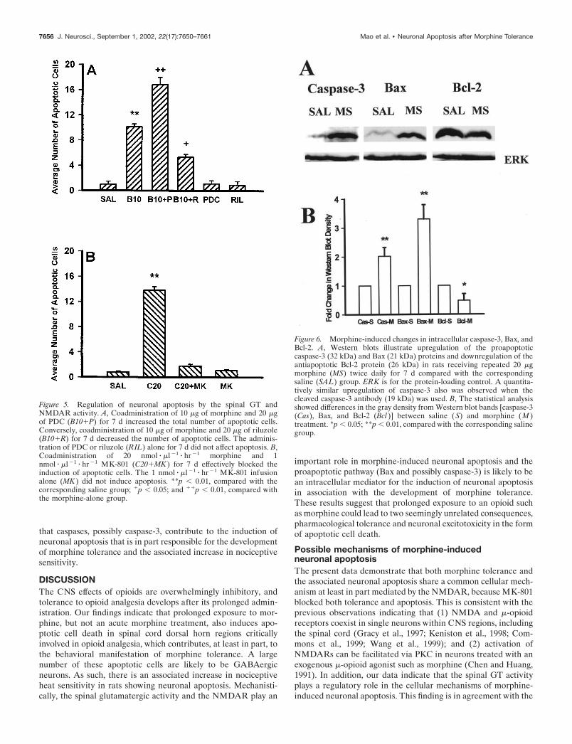
Changes in the spinal caspase-3, Bax, and Bcl-2protein content in morphine-tolerant ratsIntrathecal administration (twice daily) of 20 �g of morphine for7 d induced an upregulation of Bax and caspase-3 but a down-regulation of Bcl-2 in the spinal cord dorsal horn, as shown incorresponding Western blots (Fig. 6). Consistently, there was an
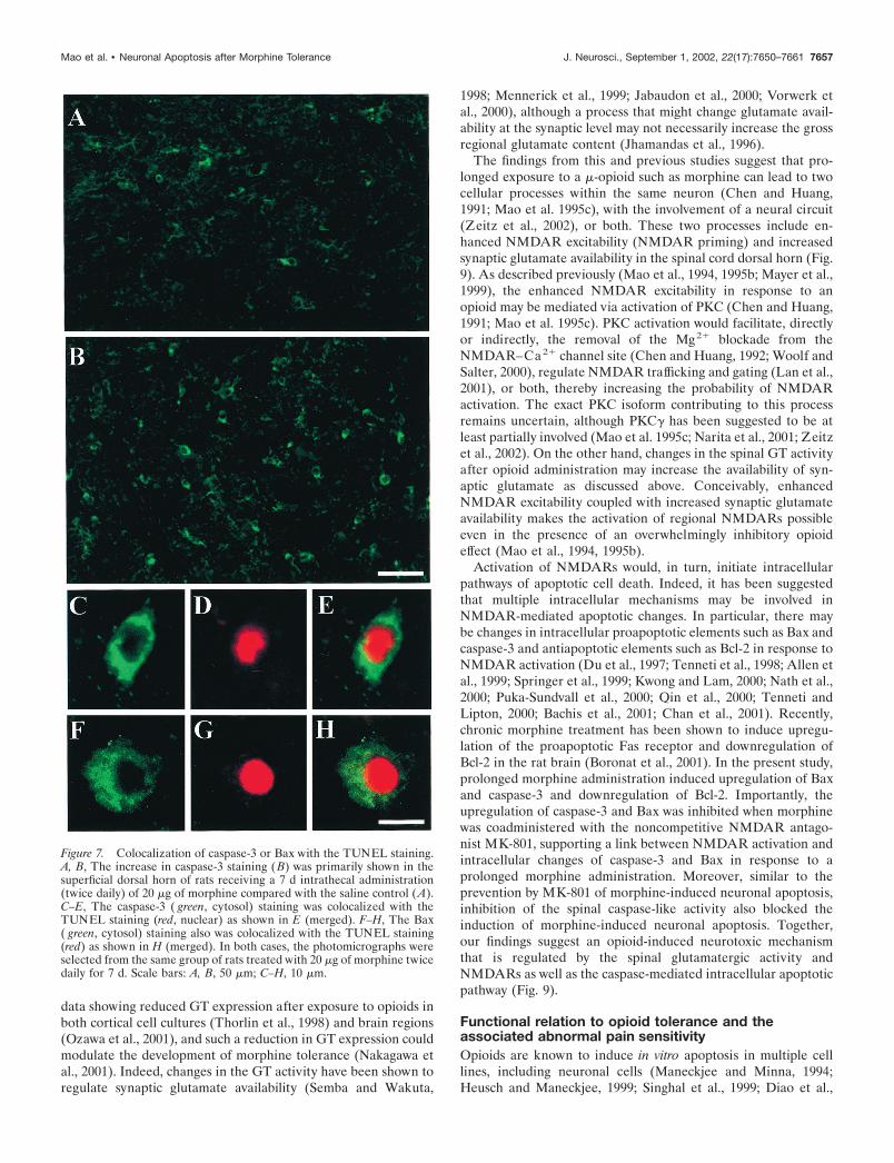
increase in cell bodies positively stained with cleaved caspase-3 inthe superficial dorsal horn of rats treated with 20nmol � �l�1 � hr�1 morphine but not saline for 7 d (Figs. 7A,B,8A). Such an increase in caspase-3-positive cells was blocked bythe coadministration of morphine (20 nmol � �l�1 � hr�1) withMK-801 (1 nmol � �l�1 � hr�1) for 7 d (Fig. 8A), suggesting thatNMDARs play an important role in the caspase-3 increase inmorphine-tolerant rats. Importantly, caspase-3 or Bax was colo-calized with the TUNEL staining in the spinal cord dorsal horn(Fig. 7C–H), indicating a morphological correlation at the cellu-lar level between caspase-3 or Bax changes and neuronalapoptosis.
Contribution of the spinal caspase-like activity to theinduction of neuronal apoptosisConsistent with the immunocytochemical and Western blot find-ings, coadministration of 20 �g of morphine with the pan-caspaseinhibitor Z-VAD-FMK (5 �g) or the relatively selectivecaspase-3 inhibitor AC-DEVD-CHO (5 �g) for 7 d prevented theinduction of neuronal apoptosis compared with that of the
Figure 2. Induction of neuronal apoptosis af-ter prolonged morphine treatment. A–C, Mi-crographs from the superficial spinal cord dor-sal horn illustrate apoptotic cells in ratsreceiving repeated intrathecal saline (A), 10�g morphine boluses (B), and the combinationof 10 �g of morphine and 20 �g of PDC ( C)for 7 d. D–F, The TUNEL (red) staining co-localizes with condensed and fragmented nu-clei, as shown by the Hoechst staining (blue)and the merged image in F, indicating that theTUNEL staining specifically detected in vivoDNA fragmentation (arrows). Note that allTUNEL-positive cells were colocalized withthe Hoechst staining. D�–F�, Note the high-magnification views of these insets (arrows)from D–F showing nuclear fragmentation andcondensed DNA segments. G–I, The TUNEL(red) staining was colocalized with the NeuNstaining ( green) as shown in I (merged), indi-cating that the costained apoptotic cells wereneurons in the dorsal horn. Note that someTUNEL-positive cells were not colocalizedwith NeuN, indicating the presence of apopto-tic glial cells. J–L, The TUNEL (red) stainingwas colocalized with the GAD67 staining( green) as shown in L (merged), indicatingthat the costained apoptotic cells contain theGABA-synthesizing enzyme and are likely tobe GABAergic neurons. Images from D–Lwere taken from rats receiving intrathecal 20�g morphine boluses or 20 nmol � �l �1 � hr �1
morphine infusion for 7 d. Scale bars: A–C, 30�m; D–L, 15 �m; D�–F�, 5 �m.
7654 J. Neurosci., September 1, 2002, 22(17):7650–7661 Mao et al. • Neuronal Apoptosis after Morphine Tolerance
morphine-alone (20 �g) group (Fig. 8B) ( p � 0.01). NeitherZ-VAD-FMK nor AC-DEVD-CHO alone induced apoptoticchanges. Furthermore, both tolerance to the antinociceptive ef-fects of morphine and the increase in nociceptive sensitivity werepartially prevented in rats receiving the coadministration of mor-
phine and Z-VAD-FMK or AC-DEVD-CHO (Fig. 8C,D),whereas Z-VAD-FMK or AC-DEVD-CHO (5 �g, intrathecal)alone for 7 d did not change baseline latencies and the responseto the antinociceptive effects of morphine. These results indicate
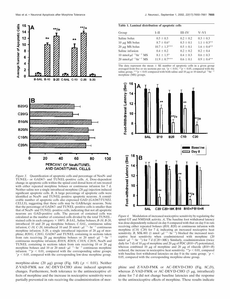
Figure 3. Quantification of apoptotic cells and percentage of NeuN- andTUNEL- or GAD67- and TUNEL-positive cells. A, Dose-dependentchange in apoptotic cells within the spinal cord dorsal horn of rats treatedwith either repeated morphine boluses or continuous infusion for 7 d.Neither saline nor a single intrathecal morphine (20 �g) injection inducedsignificant apoptotic cells. B, A large percentage of apoptotic cells wereidentified as NeuN- and TUNEL-positive apoptotic neurons. A consid-erable number of apoptotic cells also expressed GAD (GAD67/TUNELCELLS), suggesting that these cells may be GABAergic neurons. Notethat the percentage of GAD67- and TUNEL-positive cells is smaller thanthat of NeuN- and TUNEL-positive cells, indicating that not all apoptoticneurons are GAD-positive cells. The percent of costained cells wascalculated as the number of costained cells divided by the total TUNEL-stained cells in each category � 100%. B-SAL, Saline boluses; B-10, B-20,intrathecal 10 and 20 �g morphine boluses; C-SAL, continuous salineinfusion; C-10, C-20, intrathecal 10 and 20 nmol � �l �1 � hr �1 continuousmorphine infusion; S-20, a single intrathecal injection of 20 �g of mor-phine; B20/G, C20/G, GAD67 and TUNEL costaining in sections takenfrom rats receiving 20 �g morphine boluses or 20 nmol � �l �1 � hr �1
continuous morphine infusion; B10/N, B20/N, C10/N, C20/N, NeuN andTUNEL costaining in sections taken from rats receiving 10 or 20 �gmorphine boluses and 10 or 20 nmol � �l �1 � hr �1 continuous morphineinfusion. **p � 0.01, compared with the corresponding saline groups;�p � 0.05, compared with the corresponding low-dose morphine group.
Table 1. Laminal distribution of apoptotic cells
Group I–II III–IV V–VI
Saline bolus 0.5 � 0.3 0.2 � 0.2 0.3 � 0.310 �g MS bolus 8.7 � 0.6* 0.3 � 0.1 1.1 � 0.3**20 �g MS bolus 10.7 � 1.3*** 0.5 � 0.1 1.6 � 0.4**Saline infusion 0.4 � 0.2 0.2 � 0.2 0.2 � 0.410 nmol��l�1�hr�1 MS 8.1 � 1.5* 0.4 � 0.3 0.6 � 0.320 nmol��l�1�hr�1 MS 11.9 � 0.7*** 0.6 � 0.1 0.9 � 0.4**
The data represent the mean � SE number of apoptotic cells in a given groupaveraged from five or six sections per rat. *p � 0.01; **p � 0.05, compared with thesaline group, ***p � 0.01 compared with both saline and 10 �g or 10 nmol��l�1�hr�1
morphine (MS) groups.
Figure 4. Modulation of increased nociceptive sensitivity by regulating thespinal GT and NMDAR activity. A, The baseline foot withdrawal latencywas dose-dependently reduced on day 8 compared with that on day 0 in ratsreceiving either repeated boluses (B10, B20) or continuous infusion withmorphine (C10, C20) for 7 d, indicating an increased nociceptive heatsensitivity. B, MK-801 (1 nmol � �l �1 � hr �1) blocked the increased noci-ceptive heat sensitivity when coadministered with morphine (20nmol � �l �1 � hr �1) for 7 d (C20�MK ). Similarly, coadministration (twicedaily for 7 d) of 10 �g of morphine and 20 �g of PDC (B10�P) potentiated,whereas combined 10 �g of morphine and 20 �g of riluzole (B10�R)reduced, the increase in nociceptive heat sensitivity. **p � 0.01, comparedwith baseline foot withdrawal latencies on day 0 in the same group; �p �0.05, compared with the corresponding morphine-alone group.
Mao et al. • Neuronal Apoptosis after Morphine Tolerance J. Neurosci., September 1, 2002, 22(17):7650–7661 7655
that caspases, possibly caspase-3, contribute to the induction ofneuronal apoptosis that is in part responsible for the developmentof morphine tolerance and the associated increase in nociceptivesensitivity.
DISCUSSIONThe CNS effects of opioids are overwhelmingly inhibitory, andtolerance to opioid analgesia develops after its prolonged admin-istration. Our findings indicate that prolonged exposure to mor-phine, but not an acute morphine treatment, also induces apo-ptotic cell death in spinal cord dorsal horn regions criticallyinvolved in opioid analgesia, which contributes, at least in part, tothe behavioral manifestation of morphine tolerance. A largenumber of these apoptotic cells are likely to be GABAergicneurons. As such, there is an associated increase in nociceptiveheat sensitivity in rats showing neuronal apoptosis. Mechanisti-cally, the spinal glutamatergic activity and the NMDAR play an
important role in morphine-induced neuronal apoptosis and theproapoptotic pathway (Bax and possibly caspase-3) is likely to bean intracellular mediator for the induction of neuronal apoptosisin association with the development of morphine tolerance.These results suggest that prolonged exposure to an opioid suchas morphine could lead to two seemingly unrelated consequences,pharmacological tolerance and neuronal excitotoxicity in the formof apoptotic cell death.
Possible mechanisms of morphine-inducedneuronal apoptosisThe present data demonstrate that both morphine tolerance andthe associated neuronal apoptosis share a common cellular mech-anism at least in part mediated by the NMDAR, because MK-801blocked both tolerance and apoptosis. This is consistent with theprevious observations indicating that (1) NMDA and �-opioidreceptors coexist in single neurons within CNS regions, includingthe spinal cord (Gracy et al., 1997; Keniston et al., 1998; Com-mons et al., 1999; Wang et al., 1999); and (2) activation ofNMDARs can be facilitated via PKC in neurons treated with anexogenous �-opioid agonist such as morphine (Chen and Huang,1991). In addition, our data indicate that the spinal GT activityplays a regulatory role in the cellular mechanisms of morphine-induced neuronal apoptosis. This finding is in agreement with the
Figure 5. Regulation of neuronal apoptosis by the spinal GT andNMDAR activity. A, Coadministration of 10 �g of morphine and 20 �gof PDC (B10�P) for 7 d increased the total number of apoptotic cells.Conversely, coadministration of 10 �g of morphine and 20 �g of riluzole(B10�R) for 7 d decreased the number of apoptotic cells. The adminis-tration of PDC or riluzole (RIL) alone for 7 d did not affect apoptosis. B,Coadministration of 20 nmol � �l �1 � hr �1 morphine and 1nmol � �l �1 � hr �1 MK-801 (C20�MK ) for 7 d effectively blocked theinduction of apoptotic cells. The 1 nmol � �l �1 � hr �1 MK-801 infusionalone (MK ) did not induce apoptosis. **p � 0.01, compared with thecorresponding saline group; �p � 0.05; and ��p � 0.01, compared withthe morphine-alone group.
Figure 6. Morphine-induced changes in intracellular caspase-3, Bax, andBcl-2. A, Western blots illustrate upregulation of the proapoptoticcaspase-3 (32 kDa) and Bax (21 kDa) proteins and downregulation of theantiapoptotic Bcl-2 protein (26 kDa) in rats receiving repeated 20 �gmorphine (MS) twice daily for 7 d compared with the correspondingsaline (SAL) group. ERK is for the protein-loading control. A quantita-tively similar upregulation of caspase-3 also was observed when thecleaved caspase-3 antibody (19 kDa) was used. B, The statistical analysisshowed differences in the gray density from Western blot bands [caspase-3(Cas), Bax, and Bcl-2 (Bcl )] between saline ( S) and morphine (M )treatment. *p � 0.05; **p � 0.01, compared with the corresponding salinegroup.
7656 J. Neurosci., September 1, 2002, 22(17):7650–7661 Mao et al. • Neuronal Apoptosis after Morphine Tolerance
data showing reduced GT expression after exposure to opioids inboth cortical cell cultures (Thorlin et al., 1998) and brain regions(Ozawa et al., 2001), and such a reduction in GT expression couldmodulate the development of morphine tolerance (Nakagawa etal., 2001). Indeed, changes in the GT activity have been shown toregulate synaptic glutamate availability (Semba and Wakuta,
1998; Mennerick et al., 1999; Jabaudon et al., 2000; Vorwerk etal., 2000), although a process that might change glutamate avail-ability at the synaptic level may not necessarily increase the grossregional glutamate content (Jhamandas et al., 1996).
The findings from this and previous studies suggest that pro-longed exposure to a �-opioid such as morphine can lead to twocellular processes within the same neuron (Chen and Huang,1991; Mao et al. 1995c), with the involvement of a neural circuit(Zeitz et al., 2002), or both. These two processes include en-hanced NMDAR excitability (NMDAR priming) and increasedsynaptic glutamate availability in the spinal cord dorsal horn (Fig.9). As described previously (Mao et al., 1994, 1995b; Mayer et al.,1999), the enhanced NMDAR excitability in response to anopioid may be mediated via activation of PKC (Chen and Huang,1991; Mao et al. 1995c). PKC activation would facilitate, directlyor indirectly, the removal of the Mg2� blockade from theNMDAR–Ca2� channel site (Chen and Huang, 1992; Woolf andSalter, 2000), regulate NMDAR trafficking and gating (Lan et al.,2001), or both, thereby increasing the probability of NMDARactivation. The exact PKC isoform contributing to this processremains uncertain, although PKC� has been suggested to be atleast partially involved (Mao et al. 1995c; Narita et al., 2001; Zeitzet al., 2002). On the other hand, changes in the spinal GT activityafter opioid administration may increase the availability of syn-aptic glutamate as discussed above. Conceivably, enhancedNMDAR excitability coupled with increased synaptic glutamateavailability makes the activation of regional NMDARs possibleeven in the presence of an overwhelmingly inhibitory opioideffect (Mao et al., 1994, 1995b).
Activation of NMDARs would, in turn, initiate intracellularpathways of apoptotic cell death. Indeed, it has been suggestedthat multiple intracellular mechanisms may be involved inNMDAR-mediated apoptotic changes. In particular, there maybe changes in intracellular proapoptotic elements such as Bax andcaspase-3 and antiapoptotic elements such as Bcl-2 in response toNMDAR activation (Du et al., 1997; Tenneti et al., 1998; Allen etal., 1999; Springer et al., 1999; Kwong and Lam, 2000; Nath et al.,2000; Puka-Sundvall et al., 2000; Qin et al., 2000; Tenneti andLipton, 2000; Bachis et al., 2001; Chan et al., 2001). Recently,chronic morphine treatment has been shown to induce upregu-lation of the proapoptotic Fas receptor and downregulation ofBcl-2 in the rat brain (Boronat et al., 2001). In the present study,prolonged morphine administration induced upregulation of Baxand caspase-3 and downregulation of Bcl-2. Importantly, theupregulation of caspase-3 and Bax was inhibited when morphinewas coadministered with the noncompetitive NMDAR antago-nist MK-801, supporting a link between NMDAR activation andintracellular changes of caspase-3 and Bax in response to aprolonged morphine administration. Moreover, similar to theprevention by MK-801 of morphine-induced neuronal apoptosis,inhibition of the spinal caspase-like activity also blocked theinduction of morphine-induced neuronal apoptosis. Together,our findings suggest an opioid-induced neurotoxic mechanismthat is regulated by the spinal glutamatergic activity andNMDARs as well as the caspase-mediated intracellular apoptoticpathway (Fig. 9).
Functional relation to opioid tolerance and theassociated abnormal pain sensitivityOpioids are known to induce in vitro apoptosis in multiple celllines, including neuronal cells (Maneckjee and Minna, 1994;Heusch and Maneckjee, 1999; Singhal et al., 1999; Diao et al.,
Figure 7. Colocalization of caspase-3 or Bax with the TUNEL staining.A, B, The increase in caspase-3 staining (B) was primarily shown in thesuperficial dorsal horn of rats receiving a 7 d intrathecal administration(twice daily) of 20 �g of morphine compared with the saline control (A).C–E, The caspase-3 ( green, cytosol) staining was colocalized with theTUNEL staining (red, nuclear) as shown in E (merged). F–H, The Bax( green, cytosol) staining also was colocalized with the TUNEL staining(red) as shown in H (merged). In both cases, the photomicrographs wereselected from the same group of rats treated with 20 �g of morphine twicedaily for 7 d. Scale bars: A, B, 50 �m; C–H, 10 �m.
Mao et al. • Neuronal Apoptosis after Morphine Tolerance J. Neurosci., September 1, 2002, 22(17):7650–7661 7657

2000; Kugawa et al., 2000; Yoshida et al., 2000). This opioid-induced apoptosis is considered beneficial for fighting malignanttumor cells. In this experimental paradigm, however, in vivoapoptosis occurred in the spinal cord dorsal horn regions criticallyinvolved in opioid analgesia in response to clinically relevantmorphine analgesic doses given through a common administra-tion route. Because a large portion of apoptotic cells are likely tobe GABAergic neurons within the superficial spinal cord dorsalhorn, this morphine-induced neuronal excitotoxic process couldlead to changes in spinal neural circuits involved in pain and painmodulation, thereby enhancing pain sensitivity by means of spinaldisinhibition.
Indeed, signs of abnormal pain such as hyperalgesia have beenobserved both in animals exposed to opioids such as morphineand heroin (Mao et al., 1994; Ossipov et al., 1995; Wegert et al.,1997; Vanderah et al., 2000; Celerier et al., 2001) and in humansubjects undergoing acute or chronic opioid therapy (Sjogren etal., 1993; Devulder, 1997). This is further supported by thepresent data showing increased nociceptive heat sensitivity in ratswith neuronal apoptosis, which was potentiated by the GT inhib-itor PDC. However, both apoptosis and increased nociceptiveheat sensitivity were prevented by the noncompetitive NMDAR
antagonist MK-801. Of significance to note is that the presentdata indicate a functional link between morphine-induced apo-ptosis, morphine tolerance, and tolerance-related abnormal no-ciceptive sensitivity, because inhibition of morphine-induced ap-optosis by the relatively selective caspase-3 inhibitor AC-DEVD-CHO partially prevented morphine tolerance and the associatedincrease in nociceptive heat sensitivity. An interesting distinctionbetween the effects of MK-801 and AC-DEVD-CHO is that bothtolerance and increased nociceptive sensitivity were effectivelyprevented by MK-801 but incompletely prevented by AC-DEVD-CHO, suggesting that morphine-induced neuronal apo-ptosis may contribute to the cellular mechanisms of morphinetolerance and the associated increase in nociceptive sensitivity.
Clinical implicationsThe present findings suggest important implications in chronicopioid therapy. First, if neuronal apoptosis is a part of the neuralmechanisms of opioid tolerance, tolerance to opioids would beless likely to fully recover and more likely to be exacerbated in thesubsequent opioid therapy in the clinical setting. Second, opioid-induced apoptosis would be of particular concern in cancer painand chronic nonmalignant pain treatment that often requires
Figure 8. Effects of the inhibition of caspases on apoptosis and morphine tolerance. A, B, The caspase-3-positive cells were increased in rats receivinga 7 d intrathecal administration (twice daily) of 20 �g of morphine (B20) compared with the saline control (SAL). The increase in caspase-3-positivecells was blocked in rats receiving 20 �g of morphine and 10 nmol of MK-801 (B20�MK ) twice daily for 7 d. MK-801 alone (MK ) did not affect thebaseline of caspase-3-positive cells. Coadministration of 20 �g of morphine with 5 �g of either Z-VAD-FMK (B20�ZVF ) or AC-DEVD-CHO(B20�ADC) for 7 d nearly abolished morphine-induced apoptosis. C, D, Coadministration of 20 �g of morphine with 5 �g of either Z-VAD-FMK orAC-DEVD-CHO for 7 d also partially prevented the development of morphine tolerance and the increase in nociceptive heat sensitivity when testedon day 8. **p � 0.01, compared with the saline control; �p � 0.05, compared with the morphine-alone group (A–C) or the baseline value of the samegroup (D).
7658 J. Neurosci., September 1, 2002, 22(17):7650–7661 Mao et al. • Neuronal Apoptosis after Morphine Tolerance

prolonged use of opioids. A loss of opioid analgesic efficacyattributable to tolerance in combination with enhanced painsensitivity secondary to neurotoxicity would compromise the out-come of chronic opioid therapy and more importantly would leadto persistent changes in the neural circuits involved in pain andpain modulation. This may be reflected as repeated dose escala-tion during opioid therapy regardless of disease progression andan intractable chronic pain state refractory to opioid treatment,because opioid therapy itself may be the driving force for such acondition. Third, such a consequence could be further exacer-bated in neuropathic pain treatment with opioids, because neu-ropathic pain itself may be associated with the CNS neurotoxicchanges (Mao et al. 1992b, 1997; Kawamura et al., 1997; White-side and Munglani, 2001). As indicated by the present data,however, blockade of NMDARs, modulation of the spinal GTactivity, inhibition of intracellular proapoptotic elements, or acombination of the three during opioid therapy may help preventthe development of opioid-induced neurotoxic changes.
Another clinical implication of the present findings is related tothe field of substance abuse. Neurobehavioral changes indicativeof drug addiction can be seen after exposure to a substance ofabuse such as cocaine, alcohol, or heroin. A common feature ofsuch neurobehavioral changes is the difficulty of rehabilitation,with a high tendency of relapse. Critically, activation ofNMDARs has been extensively implicated in the neural mecha-nisms of such neurobehavioral changes (De Montis et al., 1992;Cebere et al., 1999; Churchill et al., 1999; Cornish et al., 1999;Huber et al., 2001), and neuronal apoptosis has indeed beenobserved in the prenatal and early postnatal stages of animalsexposed to a substance of abuse (Nassogne et al., 1997; He et al.,1999). Therefore, a corollary of our findings showing opioid-induced apoptosis in adult rats is that persistent CNS changes inthe form of apoptosis may be triggered by a substance of abuseand may contribute to clinical features of substance abuse.
In summary, we found that a subgroup of neurons, as well asglial cells, primarily located in the superficial laminas of the spinalcord dorsal horn and likely to be inhibitory GABAergic neurons,undergo the NMDAR- and caspase-mediated apoptotic processin association with the development of morphine tolerance.These findings may have significant clinical implications in rela-tion to chronic opioid therapy and substance abuse.
REFERENCESAkil H, Mayer DJ (1972) Antagonization of stimulation produced anal-
gesia by p-CPA, a serotonin synthesis inhibitor. Brain Res 44:692–697.Allen JW, Knoblach SM, Faden AI (1999) Combined mechanical
trauma and metabolic impairment in vitro induces NMDA receptor-dependent neuronal cell death and caspase-3-dependent apoptosis.FASEB J 13:1857–1882.
Azbill RD, Mu X, Srpinger JE (2000) Riluzole increases high-affinityglutamate uptake in rat spinal cord synaptosome. Brain Res871:175–180.
Baba N, Koji T, Itho M, Akio M (1999) Reciprocal changes in theexpression of Bcl-2 and Bax in hypoglossal nucleus after axotomy inadult rats: possible involvement in the induction of neuronal cell death.Brain Res 827:122–129.
Bachis A, Colangelo AM, Vicini S, Doe PP, Bernardi A, Brooker G,Mocchetti I (2001) Interleukin-10 prevents glutamate-mediated cere-bellar granule cell death by blocking caspase-3-like activity. J Neurosci21:3104–3112.
Boronat MA, Garcia-Fuster MJ, Garcia-Sevilla JA (2001) Chronic mor-phine induces up-regulation of the pro-apoptotic Fas receptor anddown-regulation of the anti-apoptotic Bcl-2 oncoprotein in rat brain.Br J Pharmacol 134:1263–1270.
Catania MV, Hollingsworth Z, Penney JB, Young AB (1993) Phospho-lipase A2 modulates different subtypes of excitatory amino acid recep-tors: autoradiographic evidence. J Neurochem 60:236–245.
Cebere A, Cebers G, Liljequist S (1999) Enhancement of NMDA-induced functional responses without NMDA receptor changes follow-ing chronic ethanol exposure in granule cells. Naunyn SchmiedebergsArch Pharmacol 360:623–632.
Celerier E, Laulin JP, Corcuff JB, Le Moal M, Simonnet G (2001)Progressive enhancement of delayed hyperalgesia induced by repeatedheroin administration: a sensitization process. J Neurosci21:4074–4080.
Chan YM, Wu W, Yip HK, So KF, Oppenheim RW (2001) Caspaseinhibitors promote the survival of avulsed spinal motoneurons in neo-natal rats. NeuroReport 12:541–545.
Chen L, Huang LYM (1991) Sustained potentiation of NMDA receptor-mediated glutamate responses through activation of protein kinase C bya �-opioid. Neuron 7:319–326.
Chen L, Huang LYM (1992) Protein kinase C reduces Mg 2� block ofNMDA-receptor channels as a mechanism of modulation. Nature356:521–523.
Cheramy A, Barbeito L, Godeheu G, Glowinski J (1992) Riluzole inhib-its the release of glutamate in the caudate nucleus of the cat in vivo.Neurosci Lett 147:209–212.
Churchill L, Swanson CJ, Urbina M, Kalivas PW (1999) Repeated co-caine alters glutamate receptor subunit levels in the nucleus accumbensand ventral tegmental area of rats that develop behavioral sensitization.J Neurochem 72:2397–2403.
Commons KG, Van Bockstaele EJ, Pfaff NW (1999) Frequent colocal-ization of Mu opioid and NMDA-type glutamate receptors at postsyn-aptic sites in periaqueductal gray neurons. J Comp Neurol 408:549–559.
Figure 9. Possible mechanisms of opioid-induced neuronal apoptosis.The data from both previous and present studies suggest that NMDARactivation may be initiated after prolonged exposure to a �-opioid agonistsuch as morphine by means of increased NMDAR excitability and re-gional glutamate availability. NMDAR activation would enhance intra-cellular positive apoptosis regulators such as Bax and caspases and de-crease negative apoptosis regulators such as Bcl-2. The resultantapoptosis contributes, at least in part, to the neural mechanisms of opioidtolerance and the associated increase in abnormal pain sensitivity. Dashedlines indicate the involvement of additional intermittent steps.
Mao et al. • Neuronal Apoptosis after Morphine Tolerance J. Neurosci., September 1, 2002, 22(17):7650–7661 7659

Cornish JL, Duffy P, Kalivas PW (1999) A role of nucleus accumbensglutamate transmission in the relapse to cocaine-seeking behaviors.Neuroscience 93:1359–1367.
D’Amour FE, Smith DL (1941) A method for determining loss of painsensation. J Pharmacol Exp Ther 72:74–79.
De Montis MG, Devoto P, Meloni D, Ganbarana C, Giorgi G, Tangli-amonte A (1992) NMDA receptor inhibition prevents tolerance tococaine. Pharmacol Biochem Behav 42:179–182.
Devulder J (1997) Hyperalgesia induced by high-dose intrathecal sufen-tanil in neuropathic pain. J Neurosurg Anesthesiol 9:146–148.
Diao CT, Li L, Lau SY, Wong TM, Wong NS (2000) Kappa-opioidreceptor potentiates apoptosis via a phospholipase C pathway in theCNE2 human epithelial tumor cell line. Biochem Biophys Acta1499:49–62.
Doble A (1996) The pharmacology and mechanism of action of riluzole.Neurology 47:S233–S241.
Du Y, Bales KR, Dodel RC, Hamilton-Byrd E, Horn JW, Czilli DL,Simmons LK, Ni B, Paul SM (1997) Activation of a caspase-e-relatedcysteine protease is required in glutamate-mediated apoptosis of cul-tured cerebellar granule neurons. Proc Natl Acad Sci USA94:11657–11662.
Dunbar SA, Pulai IJ (1998) Repetitive opioid abstinence causes progres-sive hyperalgesia sensitive to N-methyl-D-aspartate receptor blockadein the rat. J Pharmacol Exp Ther 284:678–686.
Elliott K, Minami N, Kolesnikov YA, Pasternak GW, Inturrisi CE (1994)The NMDA receptor antagonists, LY274614 and MK-801, and thenitric oxide synthase inhibitor, NG-nitro-L-arginine, attenuate analgesictolerance to the mu-opioid morphine but not to kappa opioids. Pain56:69–75.
Gavrieli Y, Sherman Y, Ben-Saaon SA (1992) Identification of pro-grammed cell death in situ via specific labeling of nuclear DNA frag-mentation. J Cell Biol 119:493–501.
Gracy KN, Svingos AL, Pickel VM (1997) Dual ultrastructural localiza-tion of mu-opioid receptors and NMDA-type glutamate receptors inthe shell of the rat nucleus accumbens. J Neurosci 17:4839–4848.
Granados-Soto V, Kalcheva I, Hua X, Newton A, Yaksh TL (2000)Spinal PKC activity and expression: role in tolerance produced bycontinuous spinal morphine infusion. Pain 85:395–404.
Hara A, Yoshimi N, Hirose Y, Ino N, Tanaka T, Mori H (1995) DNAfragmentation in granular cells of human cerebellum following globalischemia. Brain Res 697:247–250.
Hara A, Niwa M, Nakashima M, Iwai T, Uematsu T, Yoshimi N, Mori H(1998) Protective effect of apoptosis-inhibitory agent, N-tosyl-L-phenyllalanyl chloromethyl ketone against ischemia-induced hippocam-pal neuronal damage. J Cereb Blood Flow Metab 18:819–823.
Hargreaves K, Dubner R, Brown F, Flores C, Joris J (1988) A new andsensitive method for measuring thermal nociception in cutaneous hy-peralgesia. Pain 32:77–88.
He N, Song Z, Lidow MS (1999) Cocaine induces cell death within theprimate fetal cerebral wall. Neuropathol Appl Neurobiol 6:504–512.
Heusch WL, Maneckjee R (1999) Effects of bombesin on methadone-induced apoptosis of human lung cancer cells. Cancer Lett136:177–185.
Huber JD, Darling SF, Park K, Soliman KF (2001) The role of NMDAreceptors in neonatal cocaine-induced neurotoxicity. Pharmacol Bio-chem Behav 69:451–459.
Ibuki T, Dunbar SA, Yaksh TL (1997) Effect of transient naloxoneantagonism on tolerance development in rats receiving continuousspinal morphine infusion. Pain 70:125–132.
Jabaudon D, Scanziani M, Gahwiler BH, Gerber U (2000) Acute de-crease in net glutamate uptake during energy deprivation. Proc NatlAcad Sci USA 97:5610–5615.
Jhamandas KH, Marsala M, Ibuki T, Yaksh TL (1996) Spinal aminoacid release and precipitated withdrawal in rats chronically infused withspinal morphine. J Neurosci 16:2758–2766.
Ji RR, Zhang Q, Law PY, Low HH, Elde R, Hokfelt T (1995) Expres-sion of mu-, delta-, and kappa-opioid receptor-like immunoreactivitiesin rat dorsal root ganglia after carrageenan-induced inflammation.J Neurosci 15:8156–8166.
Kawamura T, Akira T, Watanabe H, Kagitani Y (1997) ProstaglandinE1 prevents apoptotic cell death in superficial dorsal horn of rat spinalcord. Neuropharmacology 36:1023–1030.
Keniston L, Mao J, Price DD, Lu J, Mayer DJ (1998) Co-localization of�-opioid and N-methyl-D-aspartate receptors to single neurons in thespinal cord dorsal horn of the adult rat. Soc Neurosci Abstr 24:390.
Kugawa F, Ueno A, Aoki M (2000) Apoptosis of NG108–5 cells inducedby buprenorphine hydrochloride occurs via the caspase-3 pathway. BiolPharmacol Bull 23:930–935.
Kwong JM, Lam TT (2000) N-Methyl-D-aspartate (NMDA) inducedapoptosis in adult rabbit retinas. Exp Eye Res 71:437–444.
Lan JY, Skeberdis VA, Jover T, Grooms SY, Lin Y, Araneda RC, ZhengX, Bennett MVL, Zukin RS (2001) Protein kinase C modulatesNMDA receptor trafficking and gating. Nat Neurosci 4:382–390.
Lievens JC, Bernal F, Forni C, Mahy N, Kerkerian-Le Goff L (2000)Characterization of striatal lesions produced by glutamate uptake al-
teration: cell death, reactive gliosis, and changes in GLT-1 andGADD45 mRNA expression. Glia 29:222–232.
Lo AC, Houenou LJ, Oppenheim RW (1995) Apoptosis in the nervoussystem: morphological features, methods, pathology, and prevention.Arch Histol Cytol 58:139–149.
Maneckjee R, Minna JD (1994) Opioids induce while nicotine sup-presses apoptosis in human lung cancer cells. Cell Growth Differ5:1033–1040.
Mao J (1999) NMDA and opioid receptors: their interactions in antino-ciception, tolerance and neuroplasticity. Brain Res Brain Res Rev30:289–304.
Mao J, Price DD, Mayer DJ, Hayes RL (1992a) Pain-related increases inspinal cord membrane-bound protein kinase C following peripheralnerve injury. Brain Res 588:144–149.
Mao J, Mayer DJ, Hayes RL, Lu J, Price DD (1992b) Differential rolesof NMDA and non-NMDA receptor activation in induction and main-tenance of thermal hyperalgesia in rats with painful peripheral monon-europathy. Brain Res 598:271–278.
Mao J, Mayer DJ, Hayes RL, Price DD (1993) Spatial patterns of in-creased spinal cord membrane-bound protein kinase C and their rela-tion to increases in 14C-2-deoxyglucose metabolic activity in rats withpainful peripheral mononeuropathy. J Neurophysiol 70:470–481.
Mao J, Price DD, Mayer DJ (1994) Thermal hyperalgesia in associationwith the development of morphine tolerance in rats: roles of excitatoryamino acid receptors and protein kinase C. J Neurosci 14:2301–2312.
Mao J, Price DD, Mayer DJ (1995a) Experimental mononeuropathyreduces the antinociceptive effects of morphine: implications for com-mon intracellular mechanisms involved in morphine tolerance andneuropathic pain. Pain 61:353–364.
Mao J, Price DD, Mayer DJ (1995b) Mechanisms of hyperalgesia andopiate tolerance: a current view of their possible interactions. Pain62:259–274.
Mao J, Price DD, Phillips LL, Lu J, Mayer DJ (1995c) Increases inprotein kinase C gamma immunoreactivity in the spinal cord of ratsassociated with tolerance to the analgesic effects of morphine. BrainRes 677:257–267.
Mao J, Price DD, Zhu J, Lu J, Mayer DJ (1997) The inhibition of nitricoxide-activated poly(ADP-ribose) synthetase attenuates transsynapticalteration of spinal cord dorsal horn neurons and neuropathic pain inthe rat. Pain 72:355–366.
Marek P, Ben Eliyahu S, Gold M, Liebeskind JC (1991a) Excitatoryamino acid antagonists (kynurenic acid and MK-801) attenuate thedevelopment of morphine tolerance in the rat. Brain Res 547:77–81.
Marek P, Ben Eliyahu S, Vaccarino AL, Liebeskind JC (1991b) Delayedapplication of MK-801 attenuates development of morphine tolerancein rats. Brain Res 558:163–165.
Matthews CC, Zielke HR, Wollack JB, Fishman PS (2000) Enzymaticdegradation protects from glutamate excitotoxicity. J Neurochem75:1045–1052.
Mayer DJ, Mao J, Price DD (1995) The development of morphinetolerance and dependence is associated with translocation of proteinkinase C. Pain 61:365–374.
Mayer DJ, Mao J, Holt J, Price DD (1999) Cellular mechanisms ofneuropathic pain, morphine tolerance, and their interactions. Proc NatlAcad Sci USA 96:7731–7736.
Mennerick S, Shen W, Xu W, Benz A, Tanaka K, Shimamoto K, IsenbKE, Krause JE, Zorumski CF (1999) Substrate turnover by transport-ers curtails synaptic glutamate transients. J Neurosci 19:9242–9251.
Molander C, Xu Q, Grant G (1984) The cytoarchitectonic organizationof the spinal cord in the rat. I. The lower thoracic and lumbosacral cord.J Comp Neurol 230:133–141.
Moncada C, Lekieffre D, Arvin B, Meldrum B (1992) Effect of NOsynthase inhibition on NMDA- and ischaemia-induced hippocampallesions. NeuroReport 3:530–532.
Mullen RJ, Buck CR, Smith AM (1992) NeuN, a neuronal specific nu-clear protein in vertebrates. Development 116:201–211.
Nakagawa T, Ozawa T, Shige K, Yamamoto R, Minami M, Satoh M(2001) Inhibition of morphine tolerance and dependence by MS-153, aglutamate transporter activator. Eur J Pharmacol 419:39–45.
Narita M, Mizoguchi H, Kampine JP, Tseng LF (1996) Role of proteinkinase C in desensitization of spinal delta-opioid-mediated antinoci-ception in the mouse. Br J Pharmacol 118:1829–1835.
Narita M, Mizoguchi H, Nagase H, Suzuki T, Tseng LF (2001) Involve-ment of spinal protein kinase C-gamma in the attenuation of opioid-mu-receptor-mediated G-protein activation after chronic intrathecaladministration of [D-Ala2, N-MePhe4, Gly-Ol5]enkephalin. J Neurosci21:3715–3720.
Nassogne MC, Louahed J, Evrard P, Courtoy PJ (1997) Cocaine inducesapoptosis in cortical neurons of fetal mice. J Neurochem 68:2442–2450.
Nath R, Scott M, Nadimpalli R, Gupta R, Wang KK (2000) Activationof apoptosis-linked caspase(s) in NMDA-injured brain in neonatal rats.Neurochem Int 36:119–126.
Ossipov MH, Lopez Y, Nichols ML, Bian D, Porreca F (1995) The lossof antinociceptive efficacy of spinal morphine in rats with nerve ligation
7660 J. Neurosci., September 1, 2002, 22(17):7650–7661 Mao et al. • Neuronal Apoptosis after Morphine Tolerance

injury is prevented by reducing spinal afferent drive. Neurosci Lett199:87–90.
Ozawa T, Nakagawa T, Shige K, Minami M, Satoh M (2001) Changes inthe expression of glial glutamate transporters in the rat brain accom-panied with morphine dependence and naloxone-precipitated with-drawal. Brain Res 905:254–258.
Price DD (1988) Psychological and neural mechanisms of pain. NewYork: Raven.
Puka-Sundvall M, Hallin U, Zhu C, Wang X, Karlsson JO, Blomgren K,Hagberg H (2000) NMDA blockade attenuates caspase-3 activationand DNA fragmentation after neonatal hypoxia-ischemia. NeuroRe-port 11:2833–2836.
Qin ZH, Wang Y, Chasea TN (2000) A caspase-3-like protease is in-volved in NF-kB activation induced by stimulation of N-methyl-D-aspartate receptors in rat striatum. Brain Res Mol Brain Res80:111–122.
Robinson MB, Dowd LA (1997) Heterogeneity and functional proper-ties of subtypes of sodium-dependent glutamate transporters in themammalian central nervous system. Adv Pharmacol 37:69–115.
Rothman SM, Olney JW (1986) Glutamate and the pathophysiology ofhypoxic-ischemic brain damage. Ann Neurol 19:105–111.
Semba J, Wakuta MS (1998) Regional differences in the effects of glu-tamate uptake inhibitor trans-pyrrolidine-2,4-bicarboxylic acid on ex-tracellular amino acids and dopamine in rat brain: an in vivo microdi-alysis study. Gen Pharmacol 31:399–404.
Singhal PC, Kapasi AA, Reddy K, Franki N, Gibbons N, Ding G (1999)Morphine promotes apoptosis in Jurkat cells. J Leukoc Biol66:650–658.
Sjogren P, Josson T, Jesen NH, Drenck NE, Jensen TS (1993) Hyper-algesia and myoclonus in terminal cancer patients treated with contin-uous intravenous morphine. Pain 55:93–97.
Springer JE, Azbill RD, Knapp PE (1999) Activation of the caspase-3apoptotic cascade in traumatic spinal cord injury. Nat Med 5:943–946.
Swan JH, Meldrum BS (1990) Protection by NMDA antagonists againstselective cell loss following transient ischaemia. J Cereb Blood FlowMetab 10:343–351.
Tenneti L, Lipton SA (2000) Involvement of activated caspase-3-likeproteases in N-methyl-D-aspartate-induced apoptosis in cerebrocorticalneurons. J Neurochem 74:134–142.
Tenneti L, D’Emilia DM, Troy CM, Lipton SA (1998) Role of caspasesin N-methyl-D-aspartate-induced apoptosis in cerebrocortical neurons.J Neurochem 71:946–959.
Thorlin T, Roginski RS, Choudhury K, Nilsson M, Ronnback L, HanssonE, Eriksson PS (1998) Regulation of the glial glutamate transporterGLT-1 by glutamate and delta-opioid receptor stimulation. FEBS Lett425:453–459.
Trujillo KA, Akil H (1991) Inhibition of morphine tolerance and depen-dence by the NMDA receptor antagonist MK-801. Science 251:85–87.
Vanderah TW, Gardell LR, Burgess SE, Ibrahim M, Dogrul A, ZhongCM, Malan TP, Ossipov MH, Lai J, Porreca F (2000) Dynorphinpromotes abnormal pain and spinal cord opioid antinociceptive toler-ance. J Neurosci 20:7074–7079.
Vorwerk CK, Naskar R, Schuettauf F, Quinto K, Zurakowski D,Gochenauer G, Robinson, Mackler SA, Dreyer EB (2000) Depressionof retinal glutamate transporter function leads to elevated intravitrealglutamate levels and ganglion cell death. Invest Ophthalmol Vis Sci41:3615–3621.
Wang H, Gracy KN, Pickel VM (1999) �-Opioid and NMDA-typeglutamate receptors are often colocalized in spiny neurons withinpatches of the caudate-putamen nucleus. J Comp Neurol 412:132–146.
Wegert S, Ossipov MH, Nichols ML, Bian D, Vanderah TW, Malan JrTP, Porreca F (1997) Differential activities of intrathecal MK-801 ormorphine to alter responses to thermal and mechanical stimuli innormal and nerve-injured rats. Pain 71:57–64.
Whiteside GT, Munglani R (2001) Cell death in the superficial dorsalhorn in a model of neuropathy. J Neurosci Res 64:168–173.
Woolf CJ, Salter MW (2000) Neuronal plasticity: increasing the gain inpain. Science 288:1765–1769.
Yaksh TL, Rudy TA (1976) Chronic catheterization of the spinal sub-arachnoid space. Physiol Behav 17:1031–1036.
Yoshida A, Tokuyama S, Iwamura T, Ueda H (2000) Opioid analgesic-induced apoptosis and caspase-independent death in human lung car-cinoma A549 cells. Int J Mol Med 6:329–335.
Zeitz KP, Malmberg AB, Gilbert H, Basbaum AI (2002) Reduced de-velopment of tolerance to the analgesic effects of morphine andclonidine in PKC� mutant mice. Pain 94:245–253.
Mao et al. • Neuronal Apoptosis after Morphine Tolerance J. Neurosci., September 1, 2002, 22(17):7650–7661 7661