Embed Size (px)
Citation preview
Detection of Morphine andMorphine-Glucuronide in Saliva andUrine with a Single, Rapid, LC IonTrap Method Without Derivatization
Author
Jason S. Wood
Agilent Technologies, Inc.
5301 Stevens Creek Boulevard
Santa Clara, CA 95051
USA
Application Note
Drugs of Abuse Testing
Abstract
Rapid (5 min), dilute-and-shoot method for the determination of morphine and one of
its major metabolites (morphine-glucuronide) in biological fluids (saliva and urine)
using the Agilent 500 Ion Trap LC/MS. It does not require lengthy derivatization
processes and can be used in a high-throughput screening environment.
Introduction
Drugs of abuse (DoA) testing is becoming more prevalent in many environments aswell as for participants in sporting events. Add to the already existing markets offorensics and social justice, a steady increase in DoA testing is evident in severalmarkets worldwide. Non-heroin opiates such as morphine, oxycodone, andhydrocodone are appearing increasingly in drug indicator data. A US Department ofJustice report from the Bureau of Justice Statistics in 2004 showed that the topfour drugs utilized by high school seniors (and the percentage of those reportingtheir use in the last 12 months) were: 1) Alcohol (70.6%), 2) Marijuana (34.3%), 3) Stimulants (10.0%) and 4) Non-heroin Opiates (9.5%) [2]. Moreover, the US DrugEnforcement Administration (USDEA) reports that since 1990, there has been abouta 3-fold increase in morphine products in the United States [3].
This application note describes a rapid (5 min), « dilute-andshoot » method for thedetermination of morphine and one of its major metabolites (morphine-glucuronide)in biological fluids (saliva and urine). It has the advantage that it does not requirelengthy derivatization processes (utilized in GC/MS) and, because of its speed, canbe used in a high-throughput screen environment. It utilizes the advanced featuresof the Agilent 500 Ion Trap to divert salts and contaminating proteins away from themass spec ion source and includes Agilent's OnTrak OraTube as a sampling mechanism for the saliva.
2
HTS CombiPAL conditionsInjection mode LC
Read bar code Never
Required syringe 100 µL liquid
Pre-inj washes solvent 1 0
Pre-inj washes solvent 2 2
Pre-inj sample flushes 0
Sample vial penetration depth pct 90%
Plunger fill speed 5.0 µL/sec
Fill strokes 0
Viscosity delay 0.3 sec
Air volume below sample 0 µL
Injector LC Vlv1
Pre-injection delay 0.5 sec
Plunger inject speed 5 µL/sec
Post-injection delay 0.5 sec
Post-inj washes solvent 1 0
Post-inj washes solvent 2 3
Post-inj valve washes solvent 1 0
Post-inj valve washes solvent 2 2
LC cycle time (prep ahead) OFF
LC conditionsColumn Agilent Pursuit C18 3 mm 100 × 2 mm
Solvent A 0.1% acetic acid in water
Solvent B 0.1% acetic acid in methanol
Flow Rate 0.2 mL/min
Injection volume 10 µL
LC program Time %A %B
0:00 95 5
0:30 95 5
1:30 95 5
2:00 57 43
3:45 5 95
4:30 5 95
4:45 95 5
5:00 95 5
500 Ion Trap segment parametersProd Prod
Time Ex Ex ion ion RF# Name (min) Precursor stor amp start end load
1 Divert 0–1 NA NA NA NA NA NA
2 Mor 1.01–2 286.1 94.5 2 95 296 85
3 MGluc 2.01–5 462.2 155.7 1 200 350 60
Instrumentation
• The Agilent 500 Ion Trap LC/MS equipped with an ESIsource
• Agilent ProStar 210 Solvent Delivery System (2)
• CTC Analytics HTS PAL AutoSampler
Materials and Reagents
All chemicals were reagent or HPLC grade from Sigma-Aldrich(St. Louis, MO) with the exception of the morphine and mor-phine-glucuronide (Cerilliant, Little Rock, TX). Drugs of abuseurine control were obtained from Utak Laboratories, Inc(Valencia, CA). OnTrak OraTube was obtained from the localAgilent sales office.
Sample Preparation
All stock solutions were prepared in 50:50 methanol:water at1 mg/mL. All dilutions were prepared in 5% aqueousmethanol + 0.1% acetic acid.
Agilent OnTrak OraTube (Cat# 6902051) was used to obtain asaliva sample and was used according to instructions.
Utak Laboratories drugs of abuse urine standard (Cat# 98815)was prepared, according to instructions, by adding 10 mL of HPLC grade water to the dried samples provided.
All saliva and urine samples were diluted 1:10 with 5% aqueous methanol containing 0.1% acetic acid.
Conditions
Mass spectrometry conditionsIonization mode ESI (positive)
Isolation window 5
API drying gas 37 psi at 350 ºC
API nebulizing gas 45 psi
Data rate 0.45 Hz
Mulitplier offset 200
Needle 5000 V
Capillary 100 V
RF storage (See 500 Ion Trap segment parameters)
Shield 600 V
Damping gas 0.8 mL/min
Excitation time 20 msec
Waveform Resonant
3
Discussion
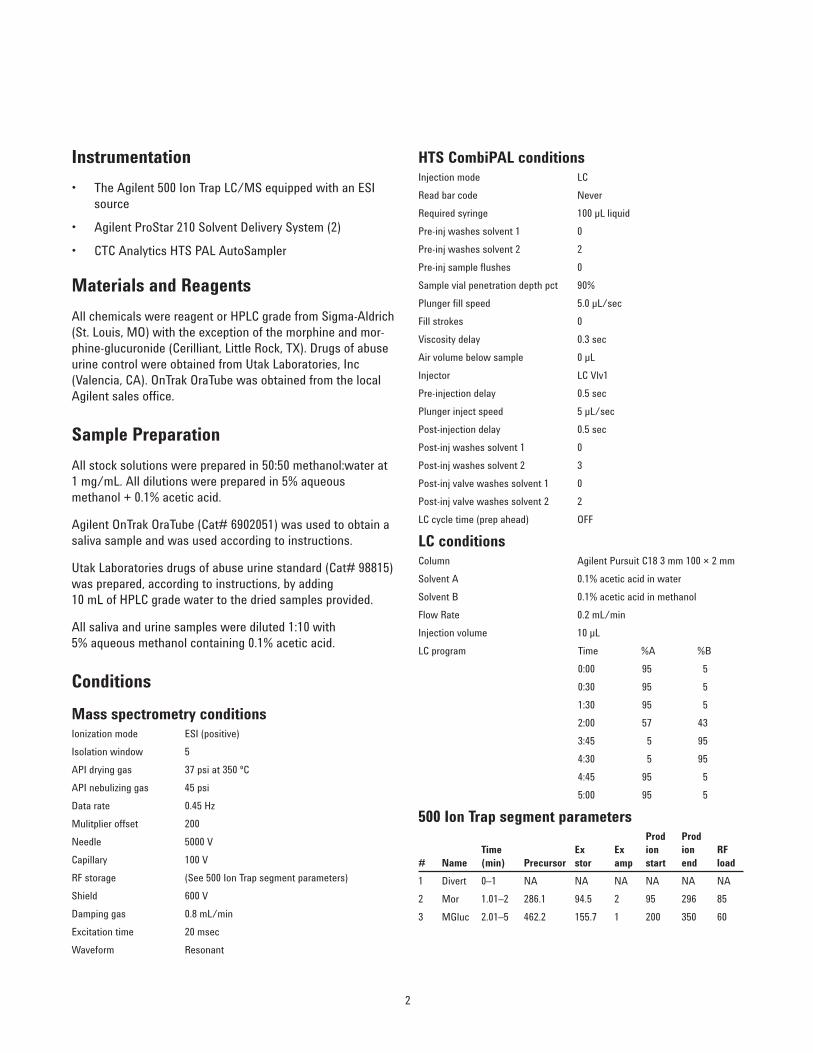
The structures of morphine and one of its metabolites areshown in Figure 1. Glucuronidation (catalyzed by uridineglu-curonic transferase or UGTs) is a Phase II metabolism reactionand involves the addition of glucuronic acid from uridine-diphosphoglucuronide to the xenobiotic being metabolized. Ingeneral, morphine metabolism is undertaken by UGT2B7.
Typically, simple onsite kits such as the Agilent OnTrakTesTcard perform preliminary screening for morphine usage,either on a random basis or as an incident-driven event.However, to eliminate false positives a second, more specificchemical method, such as GC/MS, is used as a confirmatorytest. However, GC/MS methods require the use of solid-phaseextraction and derivatization with pentafluoropropionic anhydride/pentafluoropropanol.
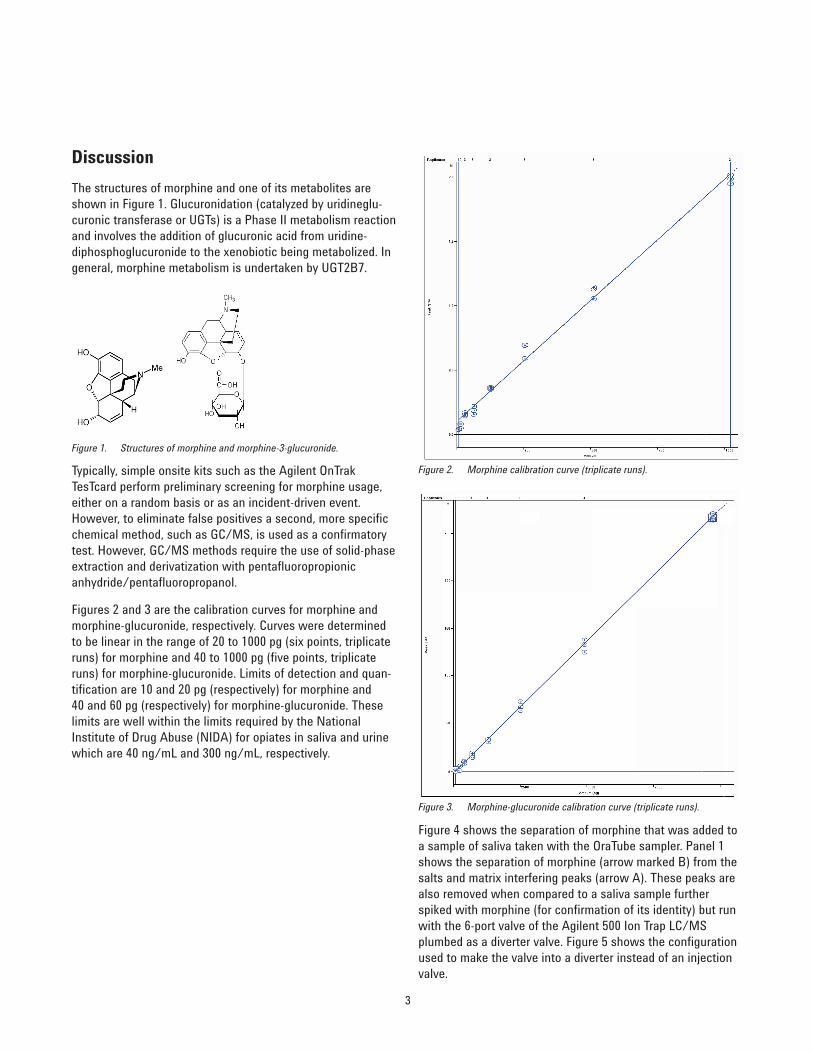
Figures 2 and 3 are the calibration curves for morphine andmorphine-glucuronide, respectively. Curves were determinedto be linear in the range of 20 to 1000 pg (six points, triplicateruns) for morphine and 40 to 1000 pg (five points, triplicateruns) for morphine-glucuronide. Limits of detection and quan-tification are 10 and 20 pg (respectively) for morphine and 40 and 60 pg (respectively) for morphine-glucuronide. Theselimits are well within the limits required by the NationalInstitute of Drug Abuse (NIDA) for opiates in saliva and urinewhich are 40 ng/mL and 300 ng/mL, respectively.
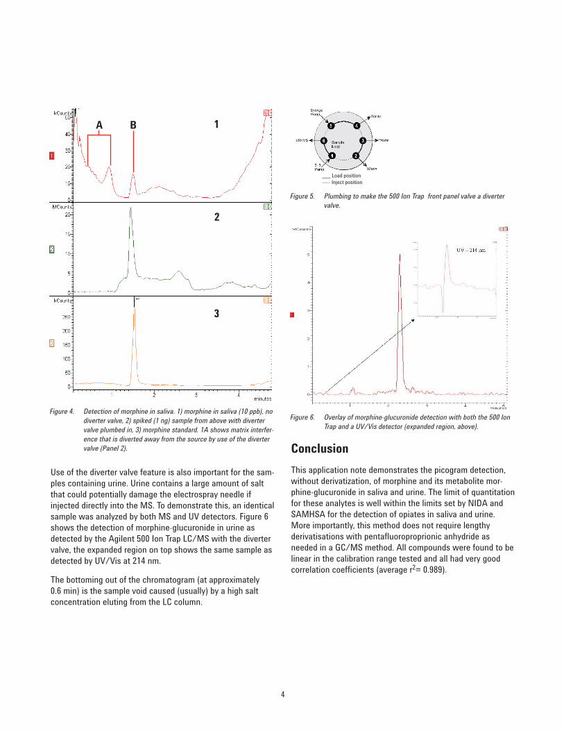
Figure 4 shows the separation of morphine that was added toa sample of saliva taken with the OraTube sampler. Panel 1shows the separation of morphine (arrow marked B) from thesalts and matrix interfering peaks (arrow A). These peaks arealso removed when compared to a saliva sample furtherspiked with morphine (for confirmation of its identity) but runwith the 6-port valve of the Agilent 500 Ion Trap LC/MSplumbed as a diverter valve. Figure 5 shows the configurationused to make the valve into a diverter instead of an injectionvalve.
Figure 1. Structures of morphine and morphine-3-glucuronide.
Figure 3. Morphine-glucuronide calibration curve (triplicate runs).
Figure 2. Morphine calibration curve (triplicate runs).
4
Use of the diverter valve feature is also important for the sam-ples containing urine. Urine contains a large amount of saltthat could potentially damage the electrospray needle ifinjected directly into the MS. To demonstrate this, an identicalsample was analyzed by both MS and UV detectors. Figure 6shows the detection of morphine-glucuronide in urine asdetected by the Agilent 500 Ion Trap LC/MS with the divertervalve, the expanded region on top shows the same sample asdetected by UV/Vis at 214 nm.
The bottoming out of the chromatogram (at approximately 0.6 min) is the sample void caused (usually) by a high saltconcentration eluting from the LC column.
Conclusion
This application note demonstrates the picogram detection,without derivatization, of morphine and its metabolite mor-phine-glucuronide in saliva and urine. The limit of quantitationfor these analytes is well within the limits set by NIDA andSAMHSA for the detection of opiates in saliva and urine.More importantly, this method does not require lengthyderivatisations with pentafluoroproprionic anhydride asneeded in a GC/MS method. All compounds were found to belinear in the calibration range tested and all had very goodcorrelation coefficients (average r2= 0.989).
Figure 4. Detection of morphine in saliva. 1) morphine in saliva (10 ppb), nodiverter valve, 2) spiked (1 ng) sample from above with divertervalve plumbed in, 3) morphine standard. 1A shows matrix interfer-ence that is diverted away from the source by use of the divertervalve (Panel 2).
A B 1
2
3
Figure 5. Plumbing to make the 500 Ion Trap front panel valve a divertervalve.
Figure 6. Overlay of morphine-glucuronide detection with both the 500 IonTrap and a UV/Vis detector (expanded region, above).
___ Load position----- Inject position
5
This application demonstrates two unique features of theAgilent 500 Ion Trap LC/MS, the use of the built-in 6-portinjection valve and the syringe pump. Together they can beused as a diverter valve to avoid potential damage to the elec-trospray interface by diverting salts away from the source andapplying a make-up volume of solvent.
Finally, it should be noted that this method does separateboth of the major metabolites of morphine, morphine- 3-glu-curonide and morphine-6-glucuronide, if required. Therefore itwas found that most, if not all, of the Utak urine standard wasfound to be morphine-3-glucuronide, consistent with theinformation provided in their catalog.
References
1. NIDA InfoFacts – Sept. 2004 (www.drugabuse.gov)
2. US Dept. Of Justice – Bureau of Justice Statistics(www.ojp.usdoj.gov/bjs/dcf/du.html)
3. DEA Briefs and Background, Drugs and Drug Abuse(www.usdoj.gov/dea/concern/morphine.html)
For More Information
These data represent typical results. For more information onour products and services, visit our Web site at www.agilent.com/chem.

www.agilent.com/chem
Agilent shall not be liable for errors contained herein orfor incidental or consequential damages in connectionwith the furnishing, performance, or use of this material.
Information, descriptions, and specifications in this publication are subject to change without notice.
© Agilent Technologies, Inc., 2011Printed in the USAFebruary 24, 2011SI-A-1022