Embed Size (px)
Citation preview
Pakistan Journal ofNeurological Sciences (PJNS)
Volume 13 | Issue 3 Article 7
9-2018
Neuromyelitis Optica (nmo); A case reportSaad WaheedAyub Teaching Hospital, Abbottabad
Jawad HussianAyub Teaching Hospital, Abbottabad
Yasir SaoodAyub Teaching Hospital, Abbottabad
Aqsa ShehzadiAyub Teaching Hospital, Abbottabad
Follow this and additional works at: https://ecommons.aku.edu/pjns
Part of the Neurology Commons
Recommended CitationWaheed, Saad; Hussian, Jawad; Saood, Yasir; and Shehzadi, Aqsa (2018) "Neuromyelitis Optica (nmo); A case report," PakistanJournal of Neurological Sciences (PJNS): Vol. 13 : Iss. 3 , Article 7.Available at: https://ecommons.aku.edu/pjns/vol13/iss3/7
NEUROMYELITIS OPTICA (NMO);A Case Report:
Saad Waheed1, Jawad Hussian1, Yasir Saood1, Aqsa Shehzadi1 1Department Of Internal Medicine, Ayub Teaching Hospital, Abbottabad
Correspondence to: Dr.Saad Waheed, Email: [email protected] Date of submission: March 21, 2018 Date of revision: May 26, 2018 Date of acceptance: May 30, 2018
ABSTRACT:
Devic’s disease or syndrome also known as Neuromyelitis optica (NMO) which is a demyelinating central nervous system (immune-mediated) ailment that predominantly targets the optic nerves and spinal cord. NMO-IgG seropositivity & longitudinally extensive spinal-cord lesions typically represents the Devic’s disease & differentiates it from Multiple Sclerosis (MS).Treatment for NMO is based upon its presentation such as for acute phase(steroids, IVIG etc), prevention of relapse (steroid sparing immunosuppression etc) and symptom based therapy (e.g spasticity etc). Here, we present a case of NMO in a young female (26 years old), married patient who presented to us with total vision loss, urinary & fecal incontinence & paraplegia. She was being treated as a case of Multiple Sclerosis (MS) several years ago but we further investigated & found NMO-IgG seropositivity and other findings in the history that conforms the diagnostic criteria of NMOSD for this patient. Furthermore, the patient was treated with IV glucocorticoids for its acute phase of disease & sent home when symptoms of transverse myelitis improved.
KEYWORDS: Neuromyelitis optica (NMO), Multiple Sclerosis (MS), Neuromyelitis optica-immunoglobulinG(NMO-IgG), aquaporin-4 antibody(AQP4-Ab), Cranial nerve(CN)
INTRODUCTION:
Neuromyelitis optica (a demyelinating disease of central nervous system)1was described by Albutt in 1870 and after two decades Devic narrated its clinical features i.e. acute transverse myelitis &optic neuritis. The incidence &prevalence of NMO is quite low as it is a rare disease to be found in general population. The worldwide incidence of this disease is 0.053 to 0.40 per 100,000, while the prevalence is ranged from 0.52 to 4.4 per 100,0002 .This disease (NMO) is more to be found among Asians, Blacks & Indian populations & it is estimated that the prevalence in Northwest of England is approximately 0.72 per 100,0003 & in Japan is 14 per 100,0004,5 While the mean age for NMO is 40 years(approx.) but it could be present among young ones6,7 & there is a female predominance with a female to male ratio of3:1. Neuromyelitis optica-immunoglobulin is a highly specific & sensitive auto antibody for diagnosis of NMO which binds to the aquaporin-4 (AQP4); channels that regulate water homeostasis in the CNS4,7,8. CASE REPORT:
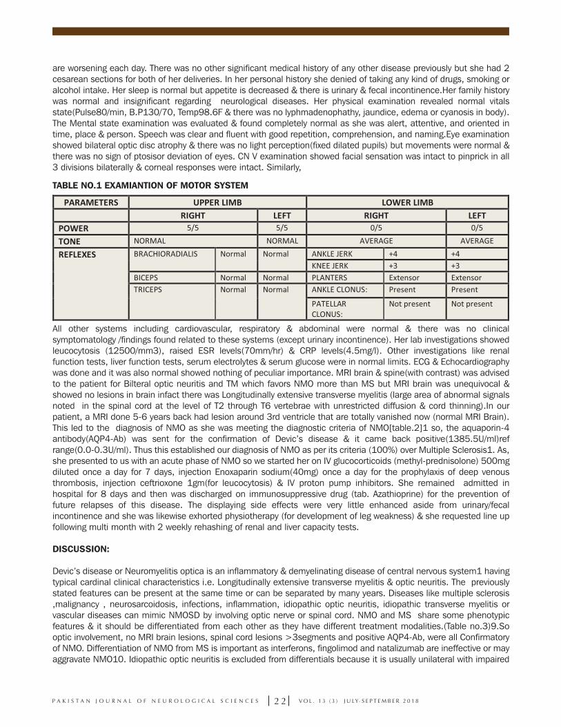
A 26 year old female, married having two children (a girl & boy) presented to us with bilateral vision loss (total blindness) for the last 6 years(2012-18), paraplegia for the last 2 months(2018) with urinary & fecal incontinence (on/off). There was sudden loss of vision in the right eye with extreme pain 9 years ago(2010), followed by other eye vision loss after 3 years of the previous one but this time it was a gradual loss of sight over 2-3 months. In the same year she had her first episode of leg weakness (left) & urinary incontinence which was gradual & slow in nature but improved after 1 month of hospital treatment (drugs included steroids). After 3 years(2013) she felt moderate weakness in both upper limbs and left leg weakness with urinary & fecal incontinence. It was gradual in onset & power was much reduced according to the patient. It took almost 6 months to improve back to normal again after a hospital treatment (diagnosed as MS patient about 7 years ago & was treated in hospital as accordingly but not recovered according to patient’s history). Now, she presented to us with the above stated symptoms for the last 2 months which
are worsening each day. There was no other significant medical history of any other disease previously but she had 2 cesarean sections for both of her deliveries. In her personal history she denied of taking any kind of drugs, smoking or alcohol intake. Her sleep is normal but appetite is decreased & there is urinary & fecal incontinence.Her family history was normal and insignificant regarding neurological diseases. Her physical examination revealed normal vitals state(Pulse80/min, B.P130/70, Temp98.6F & there was no lyphmadenophathy, jaundice, edema or cyanosis in body). The Mental state examination was evaluated & found completely normal as she was alert, attentive, and oriented in time, place & person. Speech was clear and fluent with good repetition, comprehension, and naming.Eye examination showed bilateral optic disc atrophy & there was no light perception(fixed dilated pupils) but movements were normal & there was no sign of ptosisor deviation of eyes. CN V examination showed facial sensation was intact to pinprick in all 3 divisions bilaterally & corneal responses were intact. Similarly,
TABLE NO.1 EXAMIANTION OF MOTOR SYSTEM
All other systems including cardiovascular, respiratory & abdominal were normal & there was no clinical symptomatology /findings found related to these systems (except urinary incontinence). Her lab investigations showed leucocytosis (12500/mm3), raised ESR levels(70mm/hr) & CRP levels(4.5mg/l). Other investigations like renal function tests, liver function tests, serum electrolytes & serum glucose were in normal limits. ECG & Echocardiography was done and it was also normal showed nothing of peculiar importance. MRI brain & spine(with contrast) was advised to the patient for Bilteral optic neuritis and TM which favors NMO more than MS but MRI brain was unequivocal & showed no lesions in brain infact there was Longitudinally extensive transverse myelitis (large area of abnormal signals noted in the spinal cord at the level of T2 through T6 vertebrae with unrestricted diffusion & cord thinning).In our patient, a MRI done 5-6 years back had lesion around 3rd ventricle that are totally vanished now (normal MRI Brain). This led to the diagnosis of NMO as she was meeting the diagnostic criteria of NMO[table.2]1 so, the aquaporin-4 antibody(AQP4-Ab) was sent for the confirmation of Devic’s disease & it came back positive(1385.5U/ml)ref range(0.0-0.3U/ml). Thus this established our diagnosis of NMO as per its criteria (100%) over Multiple Sclerosis1. As, she presented to us with an acute phase of NMO so we started her on IV glucocorticoids (methyl-prednisolone) 500mg diluted once a day for 7 days, injection Enoxaparin sodium(40mg) once a day for the prophylaxis of deep venous thrombosis, injection ceftrioxone 1gm(for leucocytosis) & IV proton pump inhibitors. She remained admitted in hospital for 8 days and then was discharged on immunosuppressive drug (tab. Azathioprine) for the prevention of future relapses of this disease. The displaying side effects were very little enhanced aside from urinary/fecal incontinence and she was likewise exhorted physiotherapy (for development of leg weakness) & she requested line up following multi month with 2 weekly rehashing of renal and liver capacity tests.
DISCUSSION:
Devic’s disease or Neuromyelitis optica is an inflammatory & demyelinating disease of central nervous system1 having typical cardinal clinical characteristics i.e. Longitudinally extensive transverse myelitis & optic neuritis. The previously stated features can be present at the same time or can be separated by many years. Diseases like multiple sclerosis ,malignancy , neurosarcoidosis, infections, inflammation, idiopathic optic neuritis, idiopathic transverse myelitis or vascular diseases can mimic NMOSD by involving optic nerve or spinal cord. NMO and MS share some phenotypic features & it should be differentiated from each other as they have different treatment modalities.(Table no.3)9.So optic involvement, no MRI brain lesions, spinal cord lesions >3segments and positive AQP4-Ab, were all Confirmatory of NMO. Differentiation of NMO from MS is important as interferons, fingolimod and natalizumab are ineffective or may aggravate NMO10. Idiopathic optic neuritis is excluded from differentials because it is usually unilateral with impaired
C A S E R E P O R T
2 1P A K I S T A N J O U R N A L O F N E U R O L O G I C A L S C I E N C E S V O L . 1 3 ( 3 ) J U LY- S E P T E M B E R 2 0 1 8
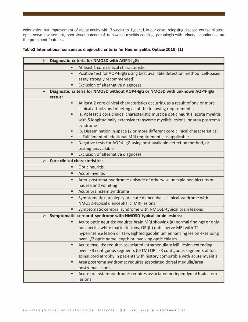
color vision but improvement of visual acuity with 3 weeks to 1year11.In our case, relapsing disease course,bilateral optic nerve involvement, poor visual outcome & transverse myelitis causing paraplegia with urinary incontinence are the prominent features.
Table2 International consensus diagnostic criteria for Neuromyelitis Optica(2015) [1]
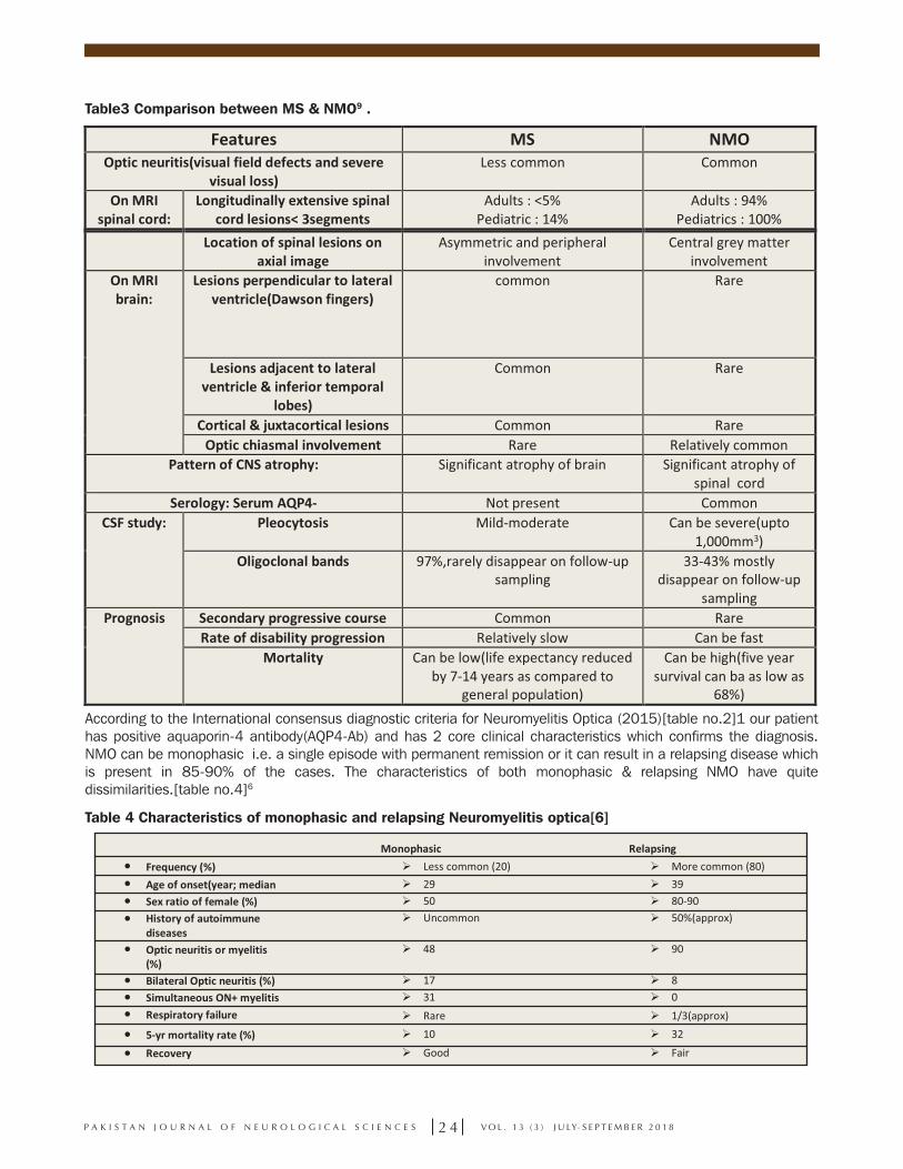
Table3 Comparison between MS & NMO9 .
According to the International consensus diagnostic criteria for Neuromyelitis Optica (2015)[table no.2]1 our patient has positive aquaporin-4 antibody(AQP4-Ab) and has 2 core clinical characteristics which confirms the diagnosis. NMO can be monophasic i.e. a single episode with permanent remission or it can result in a relapsing disease which is present in 85-90% of the cases. The characteristics of both monophasic & relapsing NMO have quite dissimilarities.[table no.4]6
Table 4 Characteristics of monophasic and relapsing Neuromyelitis optica[6]
Identification of NMO is by neurological examination, blood test(positive anti AQP4-Ab),MRI,CSF findings .Here auto-antibodies develop against aquaporin 4(water channel protein) and there is disruption of blood–brain barrier, glutamate homeostasis impairment and induction of necrotic cell death by AQP4-Ab-positive serum10.this leads to astrocyte damage, perivascular and spinal cord inflammation and cavitations of optic nerve12.AQP4-IgG is 73%sensitive and 91% specific in NMO12.NMO is a disease with severe sequelae so acute and maintenance therapy both are very important. In acute cases, pulse steroid therapy is given. If response is not good, IVIG and plasmapheresis may play a role12for long term relapse prevention, Azathioprine with oral steroids and mycophenolate mofetil with oral steroids are given13.For refractory cases, Rituximab and mitoxantrone show some efficacy13.There is no role of interferons10.In our case patient showed improvement with 7 days IV steroids and then discharged on Azathioprine as maintenance therapy. As we had advised patient for follow up & she came with same presentation at which she was discahrged after 1 month. We ran all her baseline investigations(Cbc, Rft, Lft, Serum electrolytes & urine/RE) & they came in normal parameters. To sum up, 5 year survival of NMO is as low as 68%13. CONCLUSION: After all this inferencing we emphasize early and right diagnosis with management of acute and chronic phase as damage & disability is slowly cumulative by recurrent attacks that damage new areas of myelin. we also need attention to significance of strict patient-physician collaboration in follow-up-periods.
REFERENCES:
1. Wingerchuk, D. M., B. Banwell, J. L. Bennett, P. Cabre, W.Carroll, T. Chitnis, et al. 2015. International consensuscriteria for neuromyelitis optica spectrum disorders. Neurology 85:177–189.
2. Marrie, R. A., & Gryba, C. (2013). The Incidence and Prevalence of Neuromyelitis Optica: A Systematic Review.
International Journal of MS Care, 15(3), 113–118. http://doi.org/10.7224/1537-2073.2012-048
3. Jacob A, Panicker J, Lythgoe D, et al. The epidemiology of neuromyelitis optica amongst adults in the Merseyside county of United Kingdom. J Neurol 2013; 260: 2134–2137.
4. Kira JI. Multiple sclerosis in the Japanese population. LancetNeurol 2003;2:117−27.
5. Jacob A, Boggild M. Neuromyelitis optica. Pract Neurol 2006;6:180−4.
6. Wingerchuk DM, Hogancamp WF, O’Brien PC, et al. The clinical course of neuromyelitis optica (Devic’s syndrome).Neurology 1999;53:1107−14.
7. Wingerchuk DM, Lennon VA, Pittock SJ, Lucchinetti CF,Weinshenker BG. Revised diagnostic criteria for neuromyelitisoptica. Neurology 2006;66:1485−9.
8. Lennon VA, Wingerchuk DM, Kryzer TJ, et al. A serumautoantibody marker of Neuromyelitis optica: distinctionfrom multiple sclerosis. Lancet 2004;364:2106−12.
9. Sung-Min Kim, Seong-Joon Kim, Haeng Jin Lee, Hiroshi Kuroda, Jacqueline Palace, Kazuo Fujihara.Differential diagnosis of neuromyelitis optica spectrum disorders. Therapeutic Advances in Neurological Disorders. https://doi.org/10.1177/1756285617709723
10. Jarius, S, Wildemann, B, Paul, F. Neuromyelitis optica: clinical features, immunopathogenesis and treatment. Clin Exp Immunol 2014; 176: 149–164.
11. Beck, R, Cleary, P, Backlund, J. The course of visual recovery after optic neuritis: experience of the Optic Neuritis Treatment Trial. Ophthalmology 1994; 101: 1771–1778.
12. Gürgen, Nurgül & Yalcin, Destina & Taner Gözükızıl, Salim & Polisci, Deniz. (2016). Nöromiyelitis optika: Olgu sunumu. SiSli Etfal Hastanesi Tip Bulteni / The Medical Bulletin of Sisli Hospital. 166-70. http://10.5350/SEMB.20150806062945.
13. Marcelo Matiello, Brian G WeinshenkerUS Neurology, 2009;5(1):56-58. http://doi.org/10.17925/USN.2009.05.01.56
ABSTRACT:
Devic’s disease or syndrome also known as Neuromyelitis optica (NMO) which is a demyelinating central nervous system (immune-mediated) ailment that predominantly targets the optic nerves and spinal cord. NMO-IgG seropositivity & longitudinally extensive spinal-cord lesions typically represents the Devic’s disease & differentiates it from Multiple Sclerosis (MS).Treatment for NMO is based upon its presentation such as for acute phase(steroids, IVIG etc), prevention of relapse (steroid sparing immunosuppression etc) and symptom based therapy (e.g spasticity etc). Here, we present a case of NMO in a young female (26 years old), married patient who presented to us with total vision loss, urinary & fecal incontinence & paraplegia. She was being treated as a case of Multiple Sclerosis (MS) several years ago but we further investigated & found NMO-IgG seropositivity and other findings in the history that conforms the diagnostic criteria of NMOSD for this patient. Furthermore, the patient was treated with IV glucocorticoids for its acute phase of disease & sent home when symptoms of transverse myelitis improved.
KEYWORDS: Neuromyelitis optica (NMO), Multiple Sclerosis (MS), Neuromyelitis optica-immunoglobulinG(NMO-IgG), aquaporin-4 antibody(AQP4-Ab), Cranial nerve(CN)
INTRODUCTION:
Neuromyelitis optica (a demyelinating disease of central nervous system)1was described by Albutt in 1870 and after two decades Devic narrated its clinical features i.e. acute transverse myelitis &optic neuritis. The incidence &prevalence of NMO is quite low as it is a rare disease to be found in general population. The worldwide incidence of this disease is 0.053 to 0.40 per 100,000, while the prevalence is ranged from 0.52 to 4.4 per 100,0002 .This disease (NMO) is more to be found among Asians, Blacks & Indian populations & it is estimated that the prevalence in Northwest of England is approximately 0.72 per 100,0003 & in Japan is 14 per 100,0004,5 While the mean age for NMO is 40 years(approx.) but it could be present among young ones6,7 & there is a female predominance with a female to male ratio of3:1. Neuromyelitis optica-immunoglobulin is a highly specific & sensitive auto antibody for diagnosis of NMO which binds to the aquaporin-4 (AQP4); channels that regulate water homeostasis in the CNS4,7,8. CASE REPORT:
A 26 year old female, married having two children (a girl & boy) presented to us with bilateral vision loss (total blindness) for the last 6 years(2012-18), paraplegia for the last 2 months(2018) with urinary & fecal incontinence (on/off). There was sudden loss of vision in the right eye with extreme pain 9 years ago(2010), followed by other eye vision loss after 3 years of the previous one but this time it was a gradual loss of sight over 2-3 months. In the same year she had her first episode of leg weakness (left) & urinary incontinence which was gradual & slow in nature but improved after 1 month of hospital treatment (drugs included steroids). After 3 years(2013) she felt moderate weakness in both upper limbs and left leg weakness with urinary & fecal incontinence. It was gradual in onset & power was much reduced according to the patient. It took almost 6 months to improve back to normal again after a hospital treatment (diagnosed as MS patient about 7 years ago & was treated in hospital as accordingly but not recovered according to patient’s history). Now, she presented to us with the above stated symptoms for the last 2 months which
are worsening each day. There was no other significant medical history of any other disease previously but she had 2 cesarean sections for both of her deliveries. In her personal history she denied of taking any kind of drugs, smoking or alcohol intake. Her sleep is normal but appetite is decreased & there is urinary & fecal incontinence.Her family history was normal and insignificant regarding neurological diseases. Her physical examination revealed normal vitals state(Pulse80/min, B.P130/70, Temp98.6F & there was no lyphmadenophathy, jaundice, edema or cyanosis in body). The Mental state examination was evaluated & found completely normal as she was alert, attentive, and oriented in time, place & person. Speech was clear and fluent with good repetition, comprehension, and naming.Eye examination showed bilateral optic disc atrophy & there was no light perception(fixed dilated pupils) but movements were normal & there was no sign of ptosisor deviation of eyes. CN V examination showed facial sensation was intact to pinprick in all 3 divisions bilaterally & corneal responses were intact. Similarly,
TABLE NO.1 EXAMIANTION OF MOTOR SYSTEM
All other systems including cardiovascular, respiratory & abdominal were normal & there was no clinical symptomatology /findings found related to these systems (except urinary incontinence). Her lab investigations showed leucocytosis (12500/mm3), raised ESR levels(70mm/hr) & CRP levels(4.5mg/l). Other investigations like renal function tests, liver function tests, serum electrolytes & serum glucose were in normal limits. ECG & Echocardiography was done and it was also normal showed nothing of peculiar importance. MRI brain & spine(with contrast) was advised to the patient for Bilteral optic neuritis and TM which favors NMO more than MS but MRI brain was unequivocal & showed no lesions in brain infact there was Longitudinally extensive transverse myelitis (large area of abnormal signals noted in the spinal cord at the level of T2 through T6 vertebrae with unrestricted diffusion & cord thinning).In our patient, a MRI done 5-6 years back had lesion around 3rd ventricle that are totally vanished now (normal MRI Brain). This led to the diagnosis of NMO as she was meeting the diagnostic criteria of NMO[table.2]1 so, the aquaporin-4 antibody(AQP4-Ab) was sent for the confirmation of Devic’s disease & it came back positive(1385.5U/ml)ref range(0.0-0.3U/ml). Thus this established our diagnosis of NMO as per its criteria (100%) over Multiple Sclerosis1. As, she presented to us with an acute phase of NMO so we started her on IV glucocorticoids (methyl-prednisolone) 500mg diluted once a day for 7 days, injection Enoxaparin sodium(40mg) once a day for the prophylaxis of deep venous thrombosis, injection ceftrioxone 1gm(for leucocytosis) & IV proton pump inhibitors. She remained admitted in hospital for 8 days and then was discharged on immunosuppressive drug (tab. Azathioprine) for the prevention of future relapses of this disease. The displaying side effects were very little enhanced aside from urinary/fecal incontinence and she was likewise exhorted physiotherapy (for development of leg weakness) & she requested line up following multi month with 2 weekly rehashing of renal and liver capacity tests.
DISCUSSION:
Devic’s disease or Neuromyelitis optica is an inflammatory & demyelinating disease of central nervous system1 having typical cardinal clinical characteristics i.e. Longitudinally extensive transverse myelitis & optic neuritis. The previously stated features can be present at the same time or can be separated by many years. Diseases like multiple sclerosis ,malignancy , neurosarcoidosis, infections, inflammation, idiopathic optic neuritis, idiopathic transverse myelitis or vascular diseases can mimic NMOSD by involving optic nerve or spinal cord. NMO and MS share some phenotypic features & it should be differentiated from each other as they have different treatment modalities.(Table no.3)9.So optic involvement, no MRI brain lesions, spinal cord lesions >3segments and positive AQP4-Ab, were all Confirmatory of NMO. Differentiation of NMO from MS is important as interferons, fingolimod and natalizumab are ineffective or may aggravate NMO10. Idiopathic optic neuritis is excluded from differentials because it is usually unilateral with impaired
2 2P A K I S T A N J O U R N A L O F N E U R O L O G I C A L S C I E N C E S V O L . 1 3 ( 3 ) J U LY- S E P T E M B E R 2 0 1 8
color vision but improvement of visual acuity with 3 weeks to 1year11.In our case, relapsing disease course,bilateral optic nerve involvement, poor visual outcome & transverse myelitis causing paraplegia with urinary incontinence are the prominent features.
Table2 International consensus diagnostic criteria for Neuromyelitis Optica(2015) [1]
Table3 Comparison between MS & NMO9 .
According to the International consensus diagnostic criteria for Neuromyelitis Optica (2015)[table no.2]1 our patient has positive aquaporin-4 antibody(AQP4-Ab) and has 2 core clinical characteristics which confirms the diagnosis. NMO can be monophasic i.e. a single episode with permanent remission or it can result in a relapsing disease which is present in 85-90% of the cases. The characteristics of both monophasic & relapsing NMO have quite dissimilarities.[table no.4]6
Table 4 Characteristics of monophasic and relapsing Neuromyelitis optica[6]
Identification of NMO is by neurological examination, blood test(positive anti AQP4-Ab),MRI,CSF findings .Here auto-antibodies develop against aquaporin 4(water channel protein) and there is disruption of blood–brain barrier, glutamate homeostasis impairment and induction of necrotic cell death by AQP4-Ab-positive serum10.this leads to astrocyte damage, perivascular and spinal cord inflammation and cavitations of optic nerve12.AQP4-IgG is 73%sensitive and 91% specific in NMO12.NMO is a disease with severe sequelae so acute and maintenance therapy both are very important. In acute cases, pulse steroid therapy is given. If response is not good, IVIG and plasmapheresis may play a role12for long term relapse prevention, Azathioprine with oral steroids and mycophenolate mofetil with oral steroids are given13.For refractory cases, Rituximab and mitoxantrone show some efficacy13.There is no role of interferons10.In our case patient showed improvement with 7 days IV steroids and then discharged on Azathioprine as maintenance therapy. As we had advised patient for follow up & she came with same presentation at which she was discahrged after 1 month. We ran all her baseline investigations(Cbc, Rft, Lft, Serum electrolytes & urine/RE) & they came in normal parameters. To sum up, 5 year survival of NMO is as low as 68%13. CONCLUSION: After all this inferencing we emphasize early and right diagnosis with management of acute and chronic phase as damage & disability is slowly cumulative by recurrent attacks that damage new areas of myelin. we also need attention to significance of strict patient-physician collaboration in follow-up-periods.
REFERENCES:
1. Wingerchuk, D. M., B. Banwell, J. L. Bennett, P. Cabre, W.Carroll, T. Chitnis, et al. 2015. International consensuscriteria for neuromyelitis optica spectrum disorders. Neurology 85:177–189.
2. Marrie, R. A., & Gryba, C. (2013). The Incidence and Prevalence of Neuromyelitis Optica: A Systematic Review.
International Journal of MS Care, 15(3), 113–118. http://doi.org/10.7224/1537-2073.2012-048
3. Jacob A, Panicker J, Lythgoe D, et al. The epidemiology of neuromyelitis optica amongst adults in the Merseyside county of United Kingdom. J Neurol 2013; 260: 2134–2137.
4. Kira JI. Multiple sclerosis in the Japanese population. LancetNeurol 2003;2:117−27.
5. Jacob A, Boggild M. Neuromyelitis optica. Pract Neurol 2006;6:180−4.
6. Wingerchuk DM, Hogancamp WF, O’Brien PC, et al. The clinical course of neuromyelitis optica (Devic’s syndrome).Neurology 1999;53:1107−14.
7. Wingerchuk DM, Lennon VA, Pittock SJ, Lucchinetti CF,Weinshenker BG. Revised diagnostic criteria for neuromyelitisoptica. Neurology 2006;66:1485−9.
8. Lennon VA, Wingerchuk DM, Kryzer TJ, et al. A serumautoantibody marker of Neuromyelitis optica: distinctionfrom multiple sclerosis. Lancet 2004;364:2106−12.
9. Sung-Min Kim, Seong-Joon Kim, Haeng Jin Lee, Hiroshi Kuroda, Jacqueline Palace, Kazuo Fujihara.Differential diagnosis of neuromyelitis optica spectrum disorders. Therapeutic Advances in Neurological Disorders. https://doi.org/10.1177/1756285617709723
10. Jarius, S, Wildemann, B, Paul, F. Neuromyelitis optica: clinical features, immunopathogenesis and treatment. Clin Exp Immunol 2014; 176: 149–164.
11. Beck, R, Cleary, P, Backlund, J. The course of visual recovery after optic neuritis: experience of the Optic Neuritis Treatment Trial. Ophthalmology 1994; 101: 1771–1778.
12. Gürgen, Nurgül & Yalcin, Destina & Taner Gözükızıl, Salim & Polisci, Deniz. (2016). Nöromiyelitis optika: Olgu sunumu. SiSli Etfal Hastanesi Tip Bulteni / The Medical Bulletin of Sisli Hospital. 166-70. http://10.5350/SEMB.20150806062945.
13. Marcelo Matiello, Brian G WeinshenkerUS Neurology, 2009;5(1):56-58. http://doi.org/10.17925/USN.2009.05.01.56
ABSTRACT:
Devic’s disease or syndrome also known as Neuromyelitis optica (NMO) which is a demyelinating central nervous system (immune-mediated) ailment that predominantly targets the optic nerves and spinal cord. NMO-IgG seropositivity & longitudinally extensive spinal-cord lesions typically represents the Devic’s disease & differentiates it from Multiple Sclerosis (MS).Treatment for NMO is based upon its presentation such as for acute phase(steroids, IVIG etc), prevention of relapse (steroid sparing immunosuppression etc) and symptom based therapy (e.g spasticity etc). Here, we present a case of NMO in a young female (26 years old), married patient who presented to us with total vision loss, urinary & fecal incontinence & paraplegia. She was being treated as a case of Multiple Sclerosis (MS) several years ago but we further investigated & found NMO-IgG seropositivity and other findings in the history that conforms the diagnostic criteria of NMOSD for this patient. Furthermore, the patient was treated with IV glucocorticoids for its acute phase of disease & sent home when symptoms of transverse myelitis improved.
KEYWORDS: Neuromyelitis optica (NMO), Multiple Sclerosis (MS), Neuromyelitis optica-immunoglobulinG(NMO-IgG), aquaporin-4 antibody(AQP4-Ab), Cranial nerve(CN)
INTRODUCTION:
Neuromyelitis optica (a demyelinating disease of central nervous system)1was described by Albutt in 1870 and after two decades Devic narrated its clinical features i.e. acute transverse myelitis &optic neuritis. The incidence &prevalence of NMO is quite low as it is a rare disease to be found in general population. The worldwide incidence of this disease is 0.053 to 0.40 per 100,000, while the prevalence is ranged from 0.52 to 4.4 per 100,0002 .This disease (NMO) is more to be found among Asians, Blacks & Indian populations & it is estimated that the prevalence in Northwest of England is approximately 0.72 per 100,0003 & in Japan is 14 per 100,0004,5 While the mean age for NMO is 40 years(approx.) but it could be present among young ones6,7 & there is a female predominance with a female to male ratio of3:1. Neuromyelitis optica-immunoglobulin is a highly specific & sensitive auto antibody for diagnosis of NMO which binds to the aquaporin-4 (AQP4); channels that regulate water homeostasis in the CNS4,7,8. CASE REPORT:
A 26 year old female, married having two children (a girl & boy) presented to us with bilateral vision loss (total blindness) for the last 6 years(2012-18), paraplegia for the last 2 months(2018) with urinary & fecal incontinence (on/off). There was sudden loss of vision in the right eye with extreme pain 9 years ago(2010), followed by other eye vision loss after 3 years of the previous one but this time it was a gradual loss of sight over 2-3 months. In the same year she had her first episode of leg weakness (left) & urinary incontinence which was gradual & slow in nature but improved after 1 month of hospital treatment (drugs included steroids). After 3 years(2013) she felt moderate weakness in both upper limbs and left leg weakness with urinary & fecal incontinence. It was gradual in onset & power was much reduced according to the patient. It took almost 6 months to improve back to normal again after a hospital treatment (diagnosed as MS patient about 7 years ago & was treated in hospital as accordingly but not recovered according to patient’s history). Now, she presented to us with the above stated symptoms for the last 2 months which
are worsening each day. There was no other significant medical history of any other disease previously but she had 2 cesarean sections for both of her deliveries. In her personal history she denied of taking any kind of drugs, smoking or alcohol intake. Her sleep is normal but appetite is decreased & there is urinary & fecal incontinence.Her family history was normal and insignificant regarding neurological diseases. Her physical examination revealed normal vitals state(Pulse80/min, B.P130/70, Temp98.6F & there was no lyphmadenophathy, jaundice, edema or cyanosis in body). The Mental state examination was evaluated & found completely normal as she was alert, attentive, and oriented in time, place & person. Speech was clear and fluent with good repetition, comprehension, and naming.Eye examination showed bilateral optic disc atrophy & there was no light perception(fixed dilated pupils) but movements were normal & there was no sign of ptosisor deviation of eyes. CN V examination showed facial sensation was intact to pinprick in all 3 divisions bilaterally & corneal responses were intact. Similarly,
TABLE NO.1 EXAMIANTION OF MOTOR SYSTEM
All other systems including cardiovascular, respiratory & abdominal were normal & there was no clinical symptomatology /findings found related to these systems (except urinary incontinence). Her lab investigations showed leucocytosis (12500/mm3), raised ESR levels(70mm/hr) & CRP levels(4.5mg/l). Other investigations like renal function tests, liver function tests, serum electrolytes & serum glucose were in normal limits. ECG & Echocardiography was done and it was also normal showed nothing of peculiar importance. MRI brain & spine(with contrast) was advised to the patient for Bilteral optic neuritis and TM which favors NMO more than MS but MRI brain was unequivocal & showed no lesions in brain infact there was Longitudinally extensive transverse myelitis (large area of abnormal signals noted in the spinal cord at the level of T2 through T6 vertebrae with unrestricted diffusion & cord thinning).In our patient, a MRI done 5-6 years back had lesion around 3rd ventricle that are totally vanished now (normal MRI Brain). This led to the diagnosis of NMO as she was meeting the diagnostic criteria of NMO[table.2]1 so, the aquaporin-4 antibody(AQP4-Ab) was sent for the confirmation of Devic’s disease & it came back positive(1385.5U/ml)ref range(0.0-0.3U/ml). Thus this established our diagnosis of NMO as per its criteria (100%) over Multiple Sclerosis1. As, she presented to us with an acute phase of NMO so we started her on IV glucocorticoids (methyl-prednisolone) 500mg diluted once a day for 7 days, injection Enoxaparin sodium(40mg) once a day for the prophylaxis of deep venous thrombosis, injection ceftrioxone 1gm(for leucocytosis) & IV proton pump inhibitors. She remained admitted in hospital for 8 days and then was discharged on immunosuppressive drug (tab. Azathioprine) for the prevention of future relapses of this disease. The displaying side effects were very little enhanced aside from urinary/fecal incontinence and she was likewise exhorted physiotherapy (for development of leg weakness) & she requested line up following multi month with 2 weekly rehashing of renal and liver capacity tests.
DISCUSSION:
Devic’s disease or Neuromyelitis optica is an inflammatory & demyelinating disease of central nervous system1 having typical cardinal clinical characteristics i.e. Longitudinally extensive transverse myelitis & optic neuritis. The previously stated features can be present at the same time or can be separated by many years. Diseases like multiple sclerosis ,malignancy , neurosarcoidosis, infections, inflammation, idiopathic optic neuritis, idiopathic transverse myelitis or vascular diseases can mimic NMOSD by involving optic nerve or spinal cord. NMO and MS share some phenotypic features & it should be differentiated from each other as they have different treatment modalities.(Table no.3)9.So optic involvement, no MRI brain lesions, spinal cord lesions >3segments and positive AQP4-Ab, were all Confirmatory of NMO. Differentiation of NMO from MS is important as interferons, fingolimod and natalizumab are ineffective or may aggravate NMO10. Idiopathic optic neuritis is excluded from differentials because it is usually unilateral with impaired
2 3P A K I S T A N J O U R N A L O F N E U R O L O G I C A L S C I E N C E S V O L . 1 3 ( 3 ) J U LY- S E P T E M B E R 2 0 1 8
color vision but improvement of visual acuity with 3 weeks to 1year11.In our case, relapsing disease course,bilateral optic nerve involvement, poor visual outcome & transverse myelitis causing paraplegia with urinary incontinence are the prominent features.
Table2 International consensus diagnostic criteria for Neuromyelitis Optica(2015) [1]
Table3 Comparison between MS & NMO9 .
According to the International consensus diagnostic criteria for Neuromyelitis Optica (2015)[table no.2]1 our patient has positive aquaporin-4 antibody(AQP4-Ab) and has 2 core clinical characteristics which confirms the diagnosis. NMO can be monophasic i.e. a single episode with permanent remission or it can result in a relapsing disease which is present in 85-90% of the cases. The characteristics of both monophasic & relapsing NMO have quite dissimilarities.[table no.4]6
Table 4 Characteristics of monophasic and relapsing Neuromyelitis optica[6]
Identification of NMO is by neurological examination, blood test(positive anti AQP4-Ab),MRI,CSF findings .Here auto-antibodies develop against aquaporin 4(water channel protein) and there is disruption of blood–brain barrier, glutamate homeostasis impairment and induction of necrotic cell death by AQP4-Ab-positive serum10.this leads to astrocyte damage, perivascular and spinal cord inflammation and cavitations of optic nerve12.AQP4-IgG is 73%sensitive and 91% specific in NMO12.NMO is a disease with severe sequelae so acute and maintenance therapy both are very important. In acute cases, pulse steroid therapy is given. If response is not good, IVIG and plasmapheresis may play a role12for long term relapse prevention, Azathioprine with oral steroids and mycophenolate mofetil with oral steroids are given13.For refractory cases, Rituximab and mitoxantrone show some efficacy13.There is no role of interferons10.In our case patient showed improvement with 7 days IV steroids and then discharged on Azathioprine as maintenance therapy. As we had advised patient for follow up & she came with same presentation at which she was discahrged after 1 month. We ran all her baseline investigations(Cbc, Rft, Lft, Serum electrolytes & urine/RE) & they came in normal parameters. To sum up, 5 year survival of NMO is as low as 68%13. CONCLUSION: After all this inferencing we emphasize early and right diagnosis with management of acute and chronic phase as damage & disability is slowly cumulative by recurrent attacks that damage new areas of myelin. we also need attention to significance of strict patient-physician collaboration in follow-up-periods.
REFERENCES:
1. Wingerchuk, D. M., B. Banwell, J. L. Bennett, P. Cabre, W.Carroll, T. Chitnis, et al. 2015. International consensuscriteria for neuromyelitis optica spectrum disorders. Neurology 85:177–189.
2. Marrie, R. A., & Gryba, C. (2013). The Incidence and Prevalence of Neuromyelitis Optica: A Systematic Review.
International Journal of MS Care, 15(3), 113–118. http://doi.org/10.7224/1537-2073.2012-048
3. Jacob A, Panicker J, Lythgoe D, et al. The epidemiology of neuromyelitis optica amongst adults in the Merseyside county of United Kingdom. J Neurol 2013; 260: 2134–2137.
4. Kira JI. Multiple sclerosis in the Japanese population. LancetNeurol 2003;2:117−27.
5. Jacob A, Boggild M. Neuromyelitis optica. Pract Neurol 2006;6:180−4.
6. Wingerchuk DM, Hogancamp WF, O’Brien PC, et al. The clinical course of neuromyelitis optica (Devic’s syndrome).Neurology 1999;53:1107−14.
7. Wingerchuk DM, Lennon VA, Pittock SJ, Lucchinetti CF,Weinshenker BG. Revised diagnostic criteria for neuromyelitisoptica. Neurology 2006;66:1485−9.
8. Lennon VA, Wingerchuk DM, Kryzer TJ, et al. A serumautoantibody marker of Neuromyelitis optica: distinctionfrom multiple sclerosis. Lancet 2004;364:2106−12.
9. Sung-Min Kim, Seong-Joon Kim, Haeng Jin Lee, Hiroshi Kuroda, Jacqueline Palace, Kazuo Fujihara.Differential diagnosis of neuromyelitis optica spectrum disorders. Therapeutic Advances in Neurological Disorders. https://doi.org/10.1177/1756285617709723
10. Jarius, S, Wildemann, B, Paul, F. Neuromyelitis optica: clinical features, immunopathogenesis and treatment. Clin Exp Immunol 2014; 176: 149–164.
11. Beck, R, Cleary, P, Backlund, J. The course of visual recovery after optic neuritis: experience of the Optic Neuritis Treatment Trial. Ophthalmology 1994; 101: 1771–1778.
12. Gürgen, Nurgül & Yalcin, Destina & Taner Gözükızıl, Salim & Polisci, Deniz. (2016). Nöromiyelitis optika: Olgu sunumu. SiSli Etfal Hastanesi Tip Bulteni / The Medical Bulletin of Sisli Hospital. 166-70. http://10.5350/SEMB.20150806062945.
13. Marcelo Matiello, Brian G WeinshenkerUS Neurology, 2009;5(1):56-58. http://doi.org/10.17925/USN.2009.05.01.56
ABSTRACT:
Devic’s disease or syndrome also known as Neuromyelitis optica (NMO) which is a demyelinating central nervous system (immune-mediated) ailment that predominantly targets the optic nerves and spinal cord. NMO-IgG seropositivity & longitudinally extensive spinal-cord lesions typically represents the Devic’s disease & differentiates it from Multiple Sclerosis (MS).Treatment for NMO is based upon its presentation such as for acute phase(steroids, IVIG etc), prevention of relapse (steroid sparing immunosuppression etc) and symptom based therapy (e.g spasticity etc). Here, we present a case of NMO in a young female (26 years old), married patient who presented to us with total vision loss, urinary & fecal incontinence & paraplegia. She was being treated as a case of Multiple Sclerosis (MS) several years ago but we further investigated & found NMO-IgG seropositivity and other findings in the history that conforms the diagnostic criteria of NMOSD for this patient. Furthermore, the patient was treated with IV glucocorticoids for its acute phase of disease & sent home when symptoms of transverse myelitis improved.
KEYWORDS: Neuromyelitis optica (NMO), Multiple Sclerosis (MS), Neuromyelitis optica-immunoglobulinG(NMO-IgG), aquaporin-4 antibody(AQP4-Ab), Cranial nerve(CN)
INTRODUCTION:
Neuromyelitis optica (a demyelinating disease of central nervous system)1was described by Albutt in 1870 and after two decades Devic narrated its clinical features i.e. acute transverse myelitis &optic neuritis. The incidence &prevalence of NMO is quite low as it is a rare disease to be found in general population. The worldwide incidence of this disease is 0.053 to 0.40 per 100,000, while the prevalence is ranged from 0.52 to 4.4 per 100,0002 .This disease (NMO) is more to be found among Asians, Blacks & Indian populations & it is estimated that the prevalence in Northwest of England is approximately 0.72 per 100,0003 & in Japan is 14 per 100,0004,5 While the mean age for NMO is 40 years(approx.) but it could be present among young ones6,7 & there is a female predominance with a female to male ratio of3:1. Neuromyelitis optica-immunoglobulin is a highly specific & sensitive auto antibody for diagnosis of NMO which binds to the aquaporin-4 (AQP4); channels that regulate water homeostasis in the CNS4,7,8. CASE REPORT:
A 26 year old female, married having two children (a girl & boy) presented to us with bilateral vision loss (total blindness) for the last 6 years(2012-18), paraplegia for the last 2 months(2018) with urinary & fecal incontinence (on/off). There was sudden loss of vision in the right eye with extreme pain 9 years ago(2010), followed by other eye vision loss after 3 years of the previous one but this time it was a gradual loss of sight over 2-3 months. In the same year she had her first episode of leg weakness (left) & urinary incontinence which was gradual & slow in nature but improved after 1 month of hospital treatment (drugs included steroids). After 3 years(2013) she felt moderate weakness in both upper limbs and left leg weakness with urinary & fecal incontinence. It was gradual in onset & power was much reduced according to the patient. It took almost 6 months to improve back to normal again after a hospital treatment (diagnosed as MS patient about 7 years ago & was treated in hospital as accordingly but not recovered according to patient’s history). Now, she presented to us with the above stated symptoms for the last 2 months which
are worsening each day. There was no other significant medical history of any other disease previously but she had 2 cesarean sections for both of her deliveries. In her personal history she denied of taking any kind of drugs, smoking or alcohol intake. Her sleep is normal but appetite is decreased & there is urinary & fecal incontinence.Her family history was normal and insignificant regarding neurological diseases. Her physical examination revealed normal vitals state(Pulse80/min, B.P130/70, Temp98.6F & there was no lyphmadenophathy, jaundice, edema or cyanosis in body). The Mental state examination was evaluated & found completely normal as she was alert, attentive, and oriented in time, place & person. Speech was clear and fluent with good repetition, comprehension, and naming.Eye examination showed bilateral optic disc atrophy & there was no light perception(fixed dilated pupils) but movements were normal & there was no sign of ptosisor deviation of eyes. CN V examination showed facial sensation was intact to pinprick in all 3 divisions bilaterally & corneal responses were intact. Similarly,
TABLE NO.1 EXAMIANTION OF MOTOR SYSTEM
All other systems including cardiovascular, respiratory & abdominal were normal & there was no clinical symptomatology /findings found related to these systems (except urinary incontinence). Her lab investigations showed leucocytosis (12500/mm3), raised ESR levels(70mm/hr) & CRP levels(4.5mg/l). Other investigations like renal function tests, liver function tests, serum electrolytes & serum glucose were in normal limits. ECG & Echocardiography was done and it was also normal showed nothing of peculiar importance. MRI brain & spine(with contrast) was advised to the patient for Bilteral optic neuritis and TM which favors NMO more than MS but MRI brain was unequivocal & showed no lesions in brain infact there was Longitudinally extensive transverse myelitis (large area of abnormal signals noted in the spinal cord at the level of T2 through T6 vertebrae with unrestricted diffusion & cord thinning).In our patient, a MRI done 5-6 years back had lesion around 3rd ventricle that are totally vanished now (normal MRI Brain). This led to the diagnosis of NMO as she was meeting the diagnostic criteria of NMO[table.2]1 so, the aquaporin-4 antibody(AQP4-Ab) was sent for the confirmation of Devic’s disease & it came back positive(1385.5U/ml)ref range(0.0-0.3U/ml). Thus this established our diagnosis of NMO as per its criteria (100%) over Multiple Sclerosis1. As, she presented to us with an acute phase of NMO so we started her on IV glucocorticoids (methyl-prednisolone) 500mg diluted once a day for 7 days, injection Enoxaparin sodium(40mg) once a day for the prophylaxis of deep venous thrombosis, injection ceftrioxone 1gm(for leucocytosis) & IV proton pump inhibitors. She remained admitted in hospital for 8 days and then was discharged on immunosuppressive drug (tab. Azathioprine) for the prevention of future relapses of this disease. The displaying side effects were very little enhanced aside from urinary/fecal incontinence and she was likewise exhorted physiotherapy (for development of leg weakness) & she requested line up following multi month with 2 weekly rehashing of renal and liver capacity tests.
DISCUSSION:
Devic’s disease or Neuromyelitis optica is an inflammatory & demyelinating disease of central nervous system1 having typical cardinal clinical characteristics i.e. Longitudinally extensive transverse myelitis & optic neuritis. The previously stated features can be present at the same time or can be separated by many years. Diseases like multiple sclerosis ,malignancy , neurosarcoidosis, infections, inflammation, idiopathic optic neuritis, idiopathic transverse myelitis or vascular diseases can mimic NMOSD by involving optic nerve or spinal cord. NMO and MS share some phenotypic features & it should be differentiated from each other as they have different treatment modalities.(Table no.3)9.So optic involvement, no MRI brain lesions, spinal cord lesions >3segments and positive AQP4-Ab, were all Confirmatory of NMO. Differentiation of NMO from MS is important as interferons, fingolimod and natalizumab are ineffective or may aggravate NMO10. Idiopathic optic neuritis is excluded from differentials because it is usually unilateral with impaired
2 4P A K I S T A N J O U R N A L O F N E U R O L O G I C A L S C I E N C E S V O L . 1 3 ( 3 ) J U LY- S E P T E M B E R 2 0 1 8
color vision but improvement of visual acuity with 3 weeks to 1year11.In our case, relapsing disease course,bilateral optic nerve involvement, poor visual outcome & transverse myelitis causing paraplegia with urinary incontinence are the prominent features.
Table2 International consensus diagnostic criteria for Neuromyelitis Optica(2015) [1]
Table3 Comparison between MS & NMO9 .
According to the International consensus diagnostic criteria for Neuromyelitis Optica (2015)[table no.2]1 our patient has positive aquaporin-4 antibody(AQP4-Ab) and has 2 core clinical characteristics which confirms the diagnosis. NMO can be monophasic i.e. a single episode with permanent remission or it can result in a relapsing disease which is present in 85-90% of the cases. The characteristics of both monophasic & relapsing NMO have quite dissimilarities.[table no.4]6
Table 4 Characteristics of monophasic and relapsing Neuromyelitis optica[6]
Identification of NMO is by neurological examination, blood test(positive anti AQP4-Ab),MRI,CSF findings .Here auto-antibodies develop against aquaporin 4(water channel protein) and there is disruption of blood–brain barrier, glutamate homeostasis impairment and induction of necrotic cell death by AQP4-Ab-positive serum10.this leads to astrocyte damage, perivascular and spinal cord inflammation and cavitations of optic nerve12.AQP4-IgG is 73%sensitive and 91% specific in NMO12.NMO is a disease with severe sequelae so acute and maintenance therapy both are very important. In acute cases, pulse steroid therapy is given. If response is not good, IVIG and plasmapheresis may play a role12for long term relapse prevention, Azathioprine with oral steroids and mycophenolate mofetil with oral steroids are given13.For refractory cases, Rituximab and mitoxantrone show some efficacy13.There is no role of interferons10.In our case patient showed improvement with 7 days IV steroids and then discharged on Azathioprine as maintenance therapy. As we had advised patient for follow up & she came with same presentation at which she was discahrged after 1 month. We ran all her baseline investigations(Cbc, Rft, Lft, Serum electrolytes & urine/RE) & they came in normal parameters. To sum up, 5 year survival of NMO is as low as 68%13. CONCLUSION: After all this inferencing we emphasize early and right diagnosis with management of acute and chronic phase as damage & disability is slowly cumulative by recurrent attacks that damage new areas of myelin. we also need attention to significance of strict patient-physician collaboration in follow-up-periods.
REFERENCES:
1. Wingerchuk, D. M., B. Banwell, J. L. Bennett, P. Cabre, W.Carroll, T. Chitnis, et al. 2015. International consensuscriteria for neuromyelitis optica spectrum disorders. Neurology 85:177–189.
2. Marrie, R. A., & Gryba, C. (2013). The Incidence and Prevalence of Neuromyelitis Optica: A Systematic Review.
International Journal of MS Care, 15(3), 113–118. http://doi.org/10.7224/1537-2073.2012-048
3. Jacob A, Panicker J, Lythgoe D, et al. The epidemiology of neuromyelitis optica amongst adults in the Merseyside county of United Kingdom. J Neurol 2013; 260: 2134–2137.
4. Kira JI. Multiple sclerosis in the Japanese population. LancetNeurol 2003;2:117−27.
5. Jacob A, Boggild M. Neuromyelitis optica. Pract Neurol 2006;6:180−4.
6. Wingerchuk DM, Hogancamp WF, O’Brien PC, et al. The clinical course of neuromyelitis optica (Devic’s syndrome).Neurology 1999;53:1107−14.
7. Wingerchuk DM, Lennon VA, Pittock SJ, Lucchinetti CF,Weinshenker BG. Revised diagnostic criteria for neuromyelitisoptica. Neurology 2006;66:1485−9.
8. Lennon VA, Wingerchuk DM, Kryzer TJ, et al. A serumautoantibody marker of Neuromyelitis optica: distinctionfrom multiple sclerosis. Lancet 2004;364:2106−12.
9. Sung-Min Kim, Seong-Joon Kim, Haeng Jin Lee, Hiroshi Kuroda, Jacqueline Palace, Kazuo Fujihara.Differential diagnosis of neuromyelitis optica spectrum disorders. Therapeutic Advances in Neurological Disorders. https://doi.org/10.1177/1756285617709723
10. Jarius, S, Wildemann, B, Paul, F. Neuromyelitis optica: clinical features, immunopathogenesis and treatment. Clin Exp Immunol 2014; 176: 149–164.
11. Beck, R, Cleary, P, Backlund, J. The course of visual recovery after optic neuritis: experience of the Optic Neuritis Treatment Trial. Ophthalmology 1994; 101: 1771–1778.
12. Gürgen, Nurgül & Yalcin, Destina & Taner Gözükızıl, Salim & Polisci, Deniz. (2016). Nöromiyelitis optika: Olgu sunumu. SiSli Etfal Hastanesi Tip Bulteni / The Medical Bulletin of Sisli Hospital. 166-70. http://10.5350/SEMB.20150806062945.
13. Marcelo Matiello, Brian G WeinshenkerUS Neurology, 2009;5(1):56-58. http://doi.org/10.17925/USN.2009.05.01.56
ABSTRACT:
Devic’s disease or syndrome also known as Neuromyelitis optica (NMO) which is a demyelinating central nervous system (immune-mediated) ailment that predominantly targets the optic nerves and spinal cord. NMO-IgG seropositivity & longitudinally extensive spinal-cord lesions typically represents the Devic’s disease & differentiates it from Multiple Sclerosis (MS).Treatment for NMO is based upon its presentation such as for acute phase(steroids, IVIG etc), prevention of relapse (steroid sparing immunosuppression etc) and symptom based therapy (e.g spasticity etc). Here, we present a case of NMO in a young female (26 years old), married patient who presented to us with total vision loss, urinary & fecal incontinence & paraplegia. She was being treated as a case of Multiple Sclerosis (MS) several years ago but we further investigated & found NMO-IgG seropositivity and other findings in the history that conforms the diagnostic criteria of NMOSD for this patient. Furthermore, the patient was treated with IV glucocorticoids for its acute phase of disease & sent home when symptoms of transverse myelitis improved.
KEYWORDS: Neuromyelitis optica (NMO), Multiple Sclerosis (MS), Neuromyelitis optica-immunoglobulinG(NMO-IgG), aquaporin-4 antibody(AQP4-Ab), Cranial nerve(CN)
INTRODUCTION:
Neuromyelitis optica (a demyelinating disease of central nervous system)1was described by Albutt in 1870 and after two decades Devic narrated its clinical features i.e. acute transverse myelitis &optic neuritis. The incidence &prevalence of NMO is quite low as it is a rare disease to be found in general population. The worldwide incidence of this disease is 0.053 to 0.40 per 100,000, while the prevalence is ranged from 0.52 to 4.4 per 100,0002 .This disease (NMO) is more to be found among Asians, Blacks & Indian populations & it is estimated that the prevalence in Northwest of England is approximately 0.72 per 100,0003 & in Japan is 14 per 100,0004,5 While the mean age for NMO is 40 years(approx.) but it could be present among young ones6,7 & there is a female predominance with a female to male ratio of3:1. Neuromyelitis optica-immunoglobulin is a highly specific & sensitive auto antibody for diagnosis of NMO which binds to the aquaporin-4 (AQP4); channels that regulate water homeostasis in the CNS4,7,8. CASE REPORT:
A 26 year old female, married having two children (a girl & boy) presented to us with bilateral vision loss (total blindness) for the last 6 years(2012-18), paraplegia for the last 2 months(2018) with urinary & fecal incontinence (on/off). There was sudden loss of vision in the right eye with extreme pain 9 years ago(2010), followed by other eye vision loss after 3 years of the previous one but this time it was a gradual loss of sight over 2-3 months. In the same year she had her first episode of leg weakness (left) & urinary incontinence which was gradual & slow in nature but improved after 1 month of hospital treatment (drugs included steroids). After 3 years(2013) she felt moderate weakness in both upper limbs and left leg weakness with urinary & fecal incontinence. It was gradual in onset & power was much reduced according to the patient. It took almost 6 months to improve back to normal again after a hospital treatment (diagnosed as MS patient about 7 years ago & was treated in hospital as accordingly but not recovered according to patient’s history). Now, she presented to us with the above stated symptoms for the last 2 months which
are worsening each day. There was no other significant medical history of any other disease previously but she had 2 cesarean sections for both of her deliveries. In her personal history she denied of taking any kind of drugs, smoking or alcohol intake. Her sleep is normal but appetite is decreased & there is urinary & fecal incontinence.Her family history was normal and insignificant regarding neurological diseases. Her physical examination revealed normal vitals state(Pulse80/min, B.P130/70, Temp98.6F & there was no lyphmadenophathy, jaundice, edema or cyanosis in body). The Mental state examination was evaluated & found completely normal as she was alert, attentive, and oriented in time, place & person. Speech was clear and fluent with good repetition, comprehension, and naming.Eye examination showed bilateral optic disc atrophy & there was no light perception(fixed dilated pupils) but movements were normal & there was no sign of ptosisor deviation of eyes. CN V examination showed facial sensation was intact to pinprick in all 3 divisions bilaterally & corneal responses were intact. Similarly,
TABLE NO.1 EXAMIANTION OF MOTOR SYSTEM
All other systems including cardiovascular, respiratory & abdominal were normal & there was no clinical symptomatology /findings found related to these systems (except urinary incontinence). Her lab investigations showed leucocytosis (12500/mm3), raised ESR levels(70mm/hr) & CRP levels(4.5mg/l). Other investigations like renal function tests, liver function tests, serum electrolytes & serum glucose were in normal limits. ECG & Echocardiography was done and it was also normal showed nothing of peculiar importance. MRI brain & spine(with contrast) was advised to the patient for Bilteral optic neuritis and TM which favors NMO more than MS but MRI brain was unequivocal & showed no lesions in brain infact there was Longitudinally extensive transverse myelitis (large area of abnormal signals noted in the spinal cord at the level of T2 through T6 vertebrae with unrestricted diffusion & cord thinning).In our patient, a MRI done 5-6 years back had lesion around 3rd ventricle that are totally vanished now (normal MRI Brain). This led to the diagnosis of NMO as she was meeting the diagnostic criteria of NMO[table.2]1 so, the aquaporin-4 antibody(AQP4-Ab) was sent for the confirmation of Devic’s disease & it came back positive(1385.5U/ml)ref range(0.0-0.3U/ml). Thus this established our diagnosis of NMO as per its criteria (100%) over Multiple Sclerosis1. As, she presented to us with an acute phase of NMO so we started her on IV glucocorticoids (methyl-prednisolone) 500mg diluted once a day for 7 days, injection Enoxaparin sodium(40mg) once a day for the prophylaxis of deep venous thrombosis, injection ceftrioxone 1gm(for leucocytosis) & IV proton pump inhibitors. She remained admitted in hospital for 8 days and then was discharged on immunosuppressive drug (tab. Azathioprine) for the prevention of future relapses of this disease. The displaying side effects were very little enhanced aside from urinary/fecal incontinence and she was likewise exhorted physiotherapy (for development of leg weakness) & she requested line up following multi month with 2 weekly rehashing of renal and liver capacity tests.
DISCUSSION:
Devic’s disease or Neuromyelitis optica is an inflammatory & demyelinating disease of central nervous system1 having typical cardinal clinical characteristics i.e. Longitudinally extensive transverse myelitis & optic neuritis. The previously stated features can be present at the same time or can be separated by many years. Diseases like multiple sclerosis ,malignancy , neurosarcoidosis, infections, inflammation, idiopathic optic neuritis, idiopathic transverse myelitis or vascular diseases can mimic NMOSD by involving optic nerve or spinal cord. NMO and MS share some phenotypic features & it should be differentiated from each other as they have different treatment modalities.(Table no.3)9.So optic involvement, no MRI brain lesions, spinal cord lesions >3segments and positive AQP4-Ab, were all Confirmatory of NMO. Differentiation of NMO from MS is important as interferons, fingolimod and natalizumab are ineffective or may aggravate NMO10. Idiopathic optic neuritis is excluded from differentials because it is usually unilateral with impaired
2 5P A K I S T A N J O U R N A L O F N E U R O L O G I C A L S C I E N C E S V O L . 1 3 ( 3 ) J U LY- S E P T E M B E R 2 0 1 8
color vision but improvement of visual acuity with 3 weeks to 1year11.In our case, relapsing disease course,bilateral optic nerve involvement, poor visual outcome & transverse myelitis causing paraplegia with urinary incontinence are the prominent features.
Table2 International consensus diagnostic criteria for Neuromyelitis Optica(2015) [1]
Table3 Comparison between MS & NMO9 .
According to the International consensus diagnostic criteria for Neuromyelitis Optica (2015)[table no.2]1 our patient has positive aquaporin-4 antibody(AQP4-Ab) and has 2 core clinical characteristics which confirms the diagnosis. NMO can be monophasic i.e. a single episode with permanent remission or it can result in a relapsing disease which is present in 85-90% of the cases. The characteristics of both monophasic & relapsing NMO have quite dissimilarities.[table no.4]6
Table 4 Characteristics of monophasic and relapsing Neuromyelitis optica[6]
Identification of NMO is by neurological examination, blood test(positive anti AQP4-Ab),MRI,CSF findings .Here auto-antibodies develop against aquaporin 4(water channel protein) and there is disruption of blood–brain barrier, glutamate homeostasis impairment and induction of necrotic cell death by AQP4-Ab-positive serum10.this leads to astrocyte damage, perivascular and spinal cord inflammation and cavitations of optic nerve12.AQP4-IgG is 73%sensitive and 91% specific in NMO12.NMO is a disease with severe sequelae so acute and maintenance therapy both are very important. In acute cases, pulse steroid therapy is given. If response is not good, IVIG and plasmapheresis may play a role12for long term relapse prevention, Azathioprine with oral steroids and mycophenolate mofetil with oral steroids are given13.For refractory cases, Rituximab and mitoxantrone show some efficacy13.There is no role of interferons10.In our case patient showed improvement with 7 days IV steroids and then discharged on Azathioprine as maintenance therapy. As we had advised patient for follow up & she came with same presentation at which she was discahrged after 1 month. We ran all her baseline investigations(Cbc, Rft, Lft, Serum electrolytes & urine/RE) & they came in normal parameters. To sum up, 5 year survival of NMO is as low as 68%13. CONCLUSION: After all this inferencing we emphasize early and right diagnosis with management of acute and chronic phase as damage & disability is slowly cumulative by recurrent attacks that damage new areas of myelin. we also need attention to significance of strict patient-physician collaboration in follow-up-periods.
REFERENCES:
1. Wingerchuk, D. M., B. Banwell, J. L. Bennett, P. Cabre, W.Carroll, T. Chitnis, et al. 2015. International consensuscriteria for neuromyelitis optica spectrum disorders. Neurology 85:177–189.
2. Marrie, R. A., & Gryba, C. (2013). The Incidence and Prevalence of Neuromyelitis Optica: A Systematic Review.
International Journal of MS Care, 15(3), 113–118. http://doi.org/10.7224/1537-2073.2012-048
3. Jacob A, Panicker J, Lythgoe D, et al. The epidemiology of neuromyelitis optica amongst adults in the Merseyside county of United Kingdom. J Neurol 2013; 260: 2134–2137.
4. Kira JI. Multiple sclerosis in the Japanese population. LancetNeurol 2003;2:117−27.
5. Jacob A, Boggild M. Neuromyelitis optica. Pract Neurol 2006;6:180−4.
6. Wingerchuk DM, Hogancamp WF, O’Brien PC, et al. The clinical course of neuromyelitis optica (Devic’s syndrome).Neurology 1999;53:1107−14.
7. Wingerchuk DM, Lennon VA, Pittock SJ, Lucchinetti CF,Weinshenker BG. Revised diagnostic criteria for neuromyelitisoptica. Neurology 2006;66:1485−9.
8. Lennon VA, Wingerchuk DM, Kryzer TJ, et al. A serumautoantibody marker of Neuromyelitis optica: distinctionfrom multiple sclerosis. Lancet 2004;364:2106−12.
9. Sung-Min Kim, Seong-Joon Kim, Haeng Jin Lee, Hiroshi Kuroda, Jacqueline Palace, Kazuo Fujihara.Differential diagnosis of neuromyelitis optica spectrum disorders. Therapeutic Advances in Neurological Disorders. https://doi.org/10.1177/1756285617709723
10. Jarius, S, Wildemann, B, Paul, F. Neuromyelitis optica: clinical features, immunopathogenesis and treatment. Clin Exp Immunol 2014; 176: 149–164.
11. Beck, R, Cleary, P, Backlund, J. The course of visual recovery after optic neuritis: experience of the Optic Neuritis Treatment Trial. Ophthalmology 1994; 101: 1771–1778.
12. Gürgen, Nurgül & Yalcin, Destina & Taner Gözükızıl, Salim & Polisci, Deniz. (2016). Nöromiyelitis optika: Olgu sunumu. SiSli Etfal Hastanesi Tip Bulteni / The Medical Bulletin of Sisli Hospital. 166-70. http://10.5350/SEMB.20150806062945.
13. Marcelo Matiello, Brian G WeinshenkerUS Neurology, 2009;5(1):56-58. http://doi.org/10.17925/USN.2009.05.01.56
ABSTRACT:
Devic’s disease or syndrome also known as Neuromyelitis optica (NMO) which is a demyelinating central nervous system (immune-mediated) ailment that predominantly targets the optic nerves and spinal cord. NMO-IgG seropositivity & longitudinally extensive spinal-cord lesions typically represents the Devic’s disease & differentiates it from Multiple Sclerosis (MS).Treatment for NMO is based upon its presentation such as for acute phase(steroids, IVIG etc), prevention of relapse (steroid sparing immunosuppression etc) and symptom based therapy (e.g spasticity etc). Here, we present a case of NMO in a young female (26 years old), married patient who presented to us with total vision loss, urinary & fecal incontinence & paraplegia. She was being treated as a case of Multiple Sclerosis (MS) several years ago but we further investigated & found NMO-IgG seropositivity and other findings in the history that conforms the diagnostic criteria of NMOSD for this patient. Furthermore, the patient was treated with IV glucocorticoids for its acute phase of disease & sent home when symptoms of transverse myelitis improved.
KEYWORDS: Neuromyelitis optica (NMO), Multiple Sclerosis (MS), Neuromyelitis optica-immunoglobulinG(NMO-IgG), aquaporin-4 antibody(AQP4-Ab), Cranial nerve(CN)
INTRODUCTION:
Neuromyelitis optica (a demyelinating disease of central nervous system)1was described by Albutt in 1870 and after two decades Devic narrated its clinical features i.e. acute transverse myelitis &optic neuritis. The incidence &prevalence of NMO is quite low as it is a rare disease to be found in general population. The worldwide incidence of this disease is 0.053 to 0.40 per 100,000, while the prevalence is ranged from 0.52 to 4.4 per 100,0002 .This disease (NMO) is more to be found among Asians, Blacks & Indian populations & it is estimated that the prevalence in Northwest of England is approximately 0.72 per 100,0003 & in Japan is 14 per 100,0004,5 While the mean age for NMO is 40 years(approx.) but it could be present among young ones6,7 & there is a female predominance with a female to male ratio of3:1. Neuromyelitis optica-immunoglobulin is a highly specific & sensitive auto antibody for diagnosis of NMO which binds to the aquaporin-4 (AQP4); channels that regulate water homeostasis in the CNS4,7,8. CASE REPORT:
A 26 year old female, married having two children (a girl & boy) presented to us with bilateral vision loss (total blindness) for the last 6 years(2012-18), paraplegia for the last 2 months(2018) with urinary & fecal incontinence (on/off). There was sudden loss of vision in the right eye with extreme pain 9 years ago(2010), followed by other eye vision loss after 3 years of the previous one but this time it was a gradual loss of sight over 2-3 months. In the same year she had her first episode of leg weakness (left) & urinary incontinence which was gradual & slow in nature but improved after 1 month of hospital treatment (drugs included steroids). After 3 years(2013) she felt moderate weakness in both upper limbs and left leg weakness with urinary & fecal incontinence. It was gradual in onset & power was much reduced according to the patient. It took almost 6 months to improve back to normal again after a hospital treatment (diagnosed as MS patient about 7 years ago & was treated in hospital as accordingly but not recovered according to patient’s history). Now, she presented to us with the above stated symptoms for the last 2 months which
are worsening each day. There was no other significant medical history of any other disease previously but she had 2 cesarean sections for both of her deliveries. In her personal history she denied of taking any kind of drugs, smoking or alcohol intake. Her sleep is normal but appetite is decreased & there is urinary & fecal incontinence.Her family history was normal and insignificant regarding neurological diseases. Her physical examination revealed normal vitals state(Pulse80/min, B.P130/70, Temp98.6F & there was no lyphmadenophathy, jaundice, edema or cyanosis in body). The Mental state examination was evaluated & found completely normal as she was alert, attentive, and oriented in time, place & person. Speech was clear and fluent with good repetition, comprehension, and naming.Eye examination showed bilateral optic disc atrophy & there was no light perception(fixed dilated pupils) but movements were normal & there was no sign of ptosisor deviation of eyes. CN V examination showed facial sensation was intact to pinprick in all 3 divisions bilaterally & corneal responses were intact. Similarly,
TABLE NO.1 EXAMIANTION OF MOTOR SYSTEM
All other systems including cardiovascular, respiratory & abdominal were normal & there was no clinical symptomatology /findings found related to these systems (except urinary incontinence). Her lab investigations showed leucocytosis (12500/mm3), raised ESR levels(70mm/hr) & CRP levels(4.5mg/l). Other investigations like renal function tests, liver function tests, serum electrolytes & serum glucose were in normal limits. ECG & Echocardiography was done and it was also normal showed nothing of peculiar importance. MRI brain & spine(with contrast) was advised to the patient for Bilteral optic neuritis and TM which favors NMO more than MS but MRI brain was unequivocal & showed no lesions in brain infact there was Longitudinally extensive transverse myelitis (large area of abnormal signals noted in the spinal cord at the level of T2 through T6 vertebrae with unrestricted diffusion & cord thinning).In our patient, a MRI done 5-6 years back had lesion around 3rd ventricle that are totally vanished now (normal MRI Brain). This led to the diagnosis of NMO as she was meeting the diagnostic criteria of NMO[table.2]1 so, the aquaporin-4 antibody(AQP4-Ab) was sent for the confirmation of Devic’s disease & it came back positive(1385.5U/ml)ref range(0.0-0.3U/ml). Thus this established our diagnosis of NMO as per its criteria (100%) over Multiple Sclerosis1. As, she presented to us with an acute phase of NMO so we started her on IV glucocorticoids (methyl-prednisolone) 500mg diluted once a day for 7 days, injection Enoxaparin sodium(40mg) once a day for the prophylaxis of deep venous thrombosis, injection ceftrioxone 1gm(for leucocytosis) & IV proton pump inhibitors. She remained admitted in hospital for 8 days and then was discharged on immunosuppressive drug (tab. Azathioprine) for the prevention of future relapses of this disease. The displaying side effects were very little enhanced aside from urinary/fecal incontinence and she was likewise exhorted physiotherapy (for development of leg weakness) & she requested line up following multi month with 2 weekly rehashing of renal and liver capacity tests.
DISCUSSION:
Devic’s disease or Neuromyelitis optica is an inflammatory & demyelinating disease of central nervous system1 having typical cardinal clinical characteristics i.e. Longitudinally extensive transverse myelitis & optic neuritis. The previously stated features can be present at the same time or can be separated by many years. Diseases like multiple sclerosis ,malignancy , neurosarcoidosis, infections, inflammation, idiopathic optic neuritis, idiopathic transverse myelitis or vascular diseases can mimic NMOSD by involving optic nerve or spinal cord. NMO and MS share some phenotypic features & it should be differentiated from each other as they have different treatment modalities.(Table no.3)9.So optic involvement, no MRI brain lesions, spinal cord lesions >3segments and positive AQP4-Ab, were all Confirmatory of NMO. Differentiation of NMO from MS is important as interferons, fingolimod and natalizumab are ineffective or may aggravate NMO10. Idiopathic optic neuritis is excluded from differentials because it is usually unilateral with impaired
2 6P A K I S T A N J O U R N A L O F N E U R O L O G I C A L S C I E N C E S V O L . 1 3 ( 3 ) J U LY- S E P T E M B E R 2 0 1 8
Conflict of interest: Author declares no conflict of interest. Funding disclosure: Nil
Author’s contribution:Saad Waheed; concept, data collection, data analysis, manuscript writing, manuscript reviewJawad Hussain; data collection, data analysis, manuscript writing, manuscript reviewYasir Saood; data analysis, manuscript writing, manuscript reviewAqsa Shehzadi: data analysis, manuscript writing, manuscript review
color vision but improvement of visual acuity with 3 weeks to 1year11.In our case, relapsing disease course,bilateral optic nerve involvement, poor visual outcome & transverse myelitis causing paraplegia with urinary incontinence are the prominent features.
Table2 International consensus diagnostic criteria for Neuromyelitis Optica(2015) [1]
Table3 Comparison between MS & NMO9 .
According to the International consensus diagnostic criteria for Neuromyelitis Optica (2015)[table no.2]1 our patient has positive aquaporin-4 antibody(AQP4-Ab) and has 2 core clinical characteristics which confirms the diagnosis. NMO can be monophasic i.e. a single episode with permanent remission or it can result in a relapsing disease which is present in 85-90% of the cases. The characteristics of both monophasic & relapsing NMO have quite dissimilarities.[table no.4]6
Table 4 Characteristics of monophasic and relapsing Neuromyelitis optica[6]
Identification of NMO is by neurological examination, blood test(positive anti AQP4-Ab),MRI,CSF findings .Here auto-antibodies develop against aquaporin 4(water channel protein) and there is disruption of blood–brain barrier, glutamate homeostasis impairment and induction of necrotic cell death by AQP4-Ab-positive serum10.this leads to astrocyte damage, perivascular and spinal cord inflammation and cavitations of optic nerve12.AQP4-IgG is 73%sensitive and 91% specific in NMO12.NMO is a disease with severe sequelae so acute and maintenance therapy both are very important. In acute cases, pulse steroid therapy is given. If response is not good, IVIG and plasmapheresis may play a role12for long term relapse prevention, Azathioprine with oral steroids and mycophenolate mofetil with oral steroids are given13.For refractory cases, Rituximab and mitoxantrone show some efficacy13.There is no role of interferons10.In our case patient showed improvement with 7 days IV steroids and then discharged on Azathioprine as maintenance therapy. As we had advised patient for follow up & she came with same presentation at which she was discahrged after 1 month. We ran all her baseline investigations(Cbc, Rft, Lft, Serum electrolytes & urine/RE) & they came in normal parameters. To sum up, 5 year survival of NMO is as low as 68%13. CONCLUSION: After all this inferencing we emphasize early and right diagnosis with management of acute and chronic phase as damage & disability is slowly cumulative by recurrent attacks that damage new areas of myelin. we also need attention to significance of strict patient-physician collaboration in follow-up-periods.
REFERENCES:
1. Wingerchuk, D. M., B. Banwell, J. L. Bennett, P. Cabre, W.Carroll, T. Chitnis, et al. 2015. International consensuscriteria for neuromyelitis optica spectrum disorders. Neurology 85:177–189.
2. Marrie, R. A., & Gryba, C. (2013). The Incidence and Prevalence of Neuromyelitis Optica: A Systematic Review.
International Journal of MS Care, 15(3), 113–118. http://doi.org/10.7224/1537-2073.2012-048
3. Jacob A, Panicker J, Lythgoe D, et al. The epidemiology of neuromyelitis optica amongst adults in the Merseyside county of United Kingdom. J Neurol 2013; 260: 2134–2137.
4. Kira JI. Multiple sclerosis in the Japanese population. LancetNeurol 2003;2:117−27.
5. Jacob A, Boggild M. Neuromyelitis optica. Pract Neurol 2006;6:180−4.
6. Wingerchuk DM, Hogancamp WF, O’Brien PC, et al. The clinical course of neuromyelitis optica (Devic’s syndrome).Neurology 1999;53:1107−14.
7. Wingerchuk DM, Lennon VA, Pittock SJ, Lucchinetti CF,Weinshenker BG. Revised diagnostic criteria for neuromyelitisoptica. Neurology 2006;66:1485−9.
8. Lennon VA, Wingerchuk DM, Kryzer TJ, et al. A serumautoantibody marker of Neuromyelitis optica: distinctionfrom multiple sclerosis. Lancet 2004;364:2106−12.
9. Sung-Min Kim, Seong-Joon Kim, Haeng Jin Lee, Hiroshi Kuroda, Jacqueline Palace, Kazuo Fujihara.Differential diagnosis of neuromyelitis optica spectrum disorders. Therapeutic Advances in Neurological Disorders. https://doi.org/10.1177/1756285617709723
10. Jarius, S, Wildemann, B, Paul, F. Neuromyelitis optica: clinical features, immunopathogenesis and treatment. Clin Exp Immunol 2014; 176: 149–164.
11. Beck, R, Cleary, P, Backlund, J. The course of visual recovery after optic neuritis: experience of the Optic Neuritis Treatment Trial. Ophthalmology 1994; 101: 1771–1778.
12. Gürgen, Nurgül & Yalcin, Destina & Taner Gözükızıl, Salim & Polisci, Deniz. (2016). Nöromiyelitis optika: Olgu sunumu. SiSli Etfal Hastanesi Tip Bulteni / The Medical Bulletin of Sisli Hospital. 166-70. http://10.5350/SEMB.20150806062945.
13. Marcelo Matiello, Brian G WeinshenkerUS Neurology, 2009;5(1):56-58. http://doi.org/10.17925/USN.2009.05.01.56