Embed Size (px)
Citation preview

Elizabeth Papp, RN, MSN,
CNS
June, 2018
Neurological and Neuromuscular Disorders
Neuromuscular Birth Injuries:Overview
• Nerve damage caused by trauma during delivery
• Abnormal labor time (long or short)
• LGA
• CPD
• Abnormal presentation
• Instrument-assisted delivery
• Nerves most commonly implicated
• Cervical nerves 5, 6, 7, and 8
• Thoracic nerve 1
• Cranial nerve VII
• Phrenic nerve
2
Neuromuscular Birth Injuries:Brachial Plexus Injuries
• Presentation
– Erb’s palsy:
• No spontaneous abduction or external rotation of affected arm (absent Moro)
• Hand function is often preserved (grasp reflex present)
• “waiter’s tip position”
– Global plexus palsy (Erb-Duchenne-Klumpke):
• Flaccidity of affected arm and hand
• Absent Moro and grasp reflexes
– Klumpke palsy:
• Flaccidity of hand and fingers of affected arm (present Moro, absent grasp)
• “claw-hand deformity”
3

Neuromuscular Birth Injuries:Brachial Plexus Injuries
• Cause
– Erb’s palsy:
• Most common, injury to nerve roots C5 and C6
– Global plexus palsy (Erb-Duchenne-Klumpke):
• Second most common, injury to nerve roots C5 through T1
– Klumpke palsy:
• Injury to nerve roots C8 and T1 only
4

Neuromuscular Birth Injuries:Brachial Plexus Injuries
• Management
– Physical examination to assess extent of neurological involvement
– X-ray if concern for fracture or shoulder dislocation
– Neurology, orthopedic, and PT consultation
– Passive ROM exercises when post-injury neuritis has resolved (7-10 days)
– Use of wrist and/or finger splints, if indicated
– Caregiver education regarding importance of passive exercise to maintain joint function
• Complications
– Contractures may develop without passive exercise
– Decreased sensation may lead to developmental deficits in affected arm
• Outcome
– Spontaneous resolution generally occurs within 12 months
– Best predictor of recovery is return of biceps function by 3 months of age
5
Neuromuscular Birth Injuries:Phrenic Nerve Injury
• Presentation
– Typically associated with brachial plexus injury, but can occur alone
– Respiratory distress often requiring oxygen and supportive ventilation
• Cause
– Difficult extraction, esp. breech
– Damage to phrenic nerve impairs nervous system stimulation of ipsilateral half of diaphragm; cervical nerves III, IV & V
• Management
– Diagnosed via US, NOT CXR
– Affected side down
– Supportive therapies including respiratory support
– Surgical plication of diaphragm, if indicated
• Complications
– Respiratory failure, pulmonary infection, growth failure, death
• Outcome
– Mortality rate is 10 – 15%
– Surviving infants generally recover within a year
6

Neuromuscular Birth Injuries:Facial Nerve Palsy
• Presentation
– Persistent open eye on affected side
– Suck with drooling on affected side
– Mouth drawn to normal side when crying
• Cause
– Trauma to nerve sheath (CN VII) during birth
– Associated with instrument-assisted deliveries (forceps)
• Management
– Provide artificial tears to open eye, a patch may be needed
– Family support
• Complications
– Feeding impairment
• Outcome
– Spontaneous resolution is common (> 90% recover without intervention)
7
Hypoxic Ischemic Encephalopathy (HIE):Overview
• Cerebral injury associated with hypoxia and ischemia
• Incidence: 1-2 cases per 1000 term births with a mortality rate of 10 – 20%
• Hypoxemia: decrease in amount of oxygen circulating in the blood
• Ischemia: decrease in blood flow to brain (decreased perfusion)
– Decreased glucose available
• Asphyxia:
– Impairment of oxygen and carbon dioxide exchange
– Initially causes increase in cerebral blood flow
– Increasing levels of carbon dioxide contribute to acidosis
• Associated with widespread systemic injury secondary to hypoxic-ischemic insult
8

Hypoxic Ischemic Encephalopathy (HIE):Overview
• Associated antepartum conditions (20% of cases):
– Maternal hypotension, placental vasculopathy
– Contribute to decreased fetal reserves
• Intrapartum events (35% of cases):
– Prolapsed cord, abruption, traumatic birth
• Combination of antepartum and intrapartum (35% of cases)
• Neonatal conditions (10% of cases):
– Severe pulmonary disease, recurrent apnea
– Congenital heart disease
• Preterm infant is at greater risk of HIE than term infant
9
HIE:Presentation
• Stage I (mild encephalopathy)
– Hyperalert, normal muscle tone, active suck, strong Moro reflex, (+) myoclonus, hyper-responsive to stimuli
• Stage II (moderate)
– Lethargy and hypotonic, (+) myoclonus, seizures common, weak reflexes with overall increased tendon reflexes, overactive Doll’s eye
• Stage III (severe)
– Comatose, apnea and bradycardia, mechanical ventilation, seizures typical within 12 hours of birth, severe hypotonia and flaccidity, absent reflexes, pupils often unequal, variable reactivity, poor light reflex, Doll’s eye weak or absent
10
HIE:Management
• Diagnostic testing:
– Neurologic examination (Sarnat criteria)
– Conventional EEG (cEEG)
– Amplitude-integrated EEG (aEEG)
– Neuroimaging
• Head ultrasound
• CT scan
• MRI
• Interventions:
– Resuscitation and stabilization
– Therapeutic hypothermia
– Family support and education
– Palliative care
– Treat seizures
11
HIE:Complications
• Multisystem disorders are common with stage II and III HIE
– Renal and cardiac abnormalities
– Pulmonary hypertension
– Liver function abnormalities
– Thrombocytopenia
– Disseminated intravascular coagulation (DIC)
12

HIE:Outcome
• Mild encephalopathy:
– Recovery expected
– Good outcome with very small risk of long-term disability
• Moderate encephalopathy (in absence of therapeutic hypothermia):
– 6% death, 30% disability
• Severe encephalopathy (in absence of therapeutic hypothermia):
– 60% death, 100% disability
• Early onset of seizures and ability to control szs are good predictors of outcome
• Rapid turnaround after initial insult also good
predictor
13
Intraventricular Hemorrhage (IVH):Overview
• Significant injury in the preterm brain
• Germinal matrix hemorrhage:
– Germinal matrix is immature and highly vascularized area of preterm infant brain
– Site of neuron and glia development
– Poorly supported and fragile blood vessels, sensitive to blood pressure fluctuation and reperfusion injury
• Hypotension/hypertension, perinatal asphyxia, rapid volume infusions, myocardial failure, hypothermia, hyperosmolarity, etc.
– Involution of germinal matrix occurs with advancing gestational age, germinal matrix disappears by 36 weeks, GM hemorrhage less common in infants > 32 weeks
• Germinal matrix hemorrhage may extend to fill lateral ventricles and worsening IVH
14

Intraventricular Hemorrhage (IVH):Overview
• Incidence:
– 30 – 40% of infants <1500 grams or <30 weeks PMA
– <28 weeks PMA have a 3-fold higher risk than 28 – 31 weeks PMA
– 2 – 3% in term infants
• Timing of onset:
– 50% by 24 hours
– 80% by 48 hours
– 90% by 72 hours
15

IVH:Presentation
• Presentation ranges from undetectable to dramatic
• Sudden deterioration: oxygen desaturation, bradycardia, metabolic acidosis, falling hematocrit, hypotonia, shock, hyperglycemia
• Symptoms of worsening hemorrhage: full or tense fontanelle, increased ventilator support, seizures, apnea, decreased activity, decreased level of consciousness
• Rapid and profound clinical decline associated with increased severity of IVH
• Grading of IVH
16
IVH:Management
• Neuroimaging
– Routine head ultrasound screening of infants born at < 30 weeks PMA
– Serial head ultrasounds to monitor progression
– MRI if parenchymal injury is suspected
• Supportive Care
– Minimize stimulation
– Avoid wide swings in blood pressure
– Closely monitor respiratory support
– Avoid acidosis, hypercarbia, fluid overload
17
IVH:Complications
• Neurodevelopmental disabilities
• Progressive hydrocephalus
• Seizures
• Death
18
IVH:Outcome
• Mild/small IVH
– Neurodevelopmental disabilities (NDD) similar to premature infants without hemorrhage, major NDD 10%
• Moderate IVH
– Major NDD in 40%
– Mortality rate 10%
– Progressive hydrocephalus in 20%
• Severe IVH
– Major NDD in 80%
– Mortality rate 50 – 60%
– Progressive hydrocephalus common
19
Periventricular Leukomalacia (PVL):Overview
• Severe white matter injury highly associated with preterm birth; HYPOTENSION
• Focal injury: cystic necrotic lesion found bilaterally
– Nonhemorrhagic and symmetric
– Caused by ischemia from fluctuations in arterial circulation
• Diffuse white matter injury
– Noncystic lesions associated with disturbances in myelinization
– Often associated with germinal matrix hemorrhages or IVH
• Leukomalacia: ”softening” of white matter
20
PVL:Presentation
• Acute phase:
– Subtle
– Altered muscle tone in lower extremities, hypotension, lethargy
• 6 – 10 weeks after white matter injury
– Irritable, hypertonic, increased flexion of arms and extension of legs, frequent tremors and startles
– Moro reflex abnomalities
21
PVL:Management
• Diagnostic evaluation:
– Head ultrasound
– CT scan or MRI
• Interventions:
– Treat primary insult
– Supportive care to prevent further hypoxic-ischemic damage
– Treatment of hydrocephalus and associated neurological sequalea
– Family support and anticipatory guidance
– Developmental care, PT/OT, feeding support
22
PVL:Complications
• Spastic diplegia
• Intellectual deficits, learning disorders
• Hyperactivity disorders
• Visual impairment
• Lower limb weakness
23
PVL:Outcome
• Determined by location and extent of injury
• Spastic diplegia reported in as many as 50% of infants with PVL
• Neurodevelopmental follow-up and developmental support improve outcomes related to learning and behavioral disorders
24
Seizures:Overview
• Sign of malfunctioning neuronal system
• Excessive simultaneous electrical discharge
• Causes include:
– Metabolic encephalopathies
– Structural abnormalities
– Meningitis
– Drug withdrawal
– Genetic etiology
25
Seizures:Overview
• Metabolic encephalopathies:
– Hypoglycemia
– Ischemia
– Hypoxemia
– Hypo- or hypernatremia,
– Hypocalcemia
– Hypomagnesemia
– Inborn error of metabolism
– Pyridoxine deficiency
– Hyperammonemia
26
Seizures:Overview
• Structural abnormalities:
– HIE
– IVH
– Intrapartum trauma
– Perinatal stroke
– Cerebral dysgenesis
27
Seizures:Overview
• Other causes:
– Meningitis
• Group B streptococcus
• Listeria monocytogenes
• TORCH etiology
– Drug withdrawal
• Prenatal or postnatal exposure to opiates
– Genetic (familial)
• Self-limiting
28
Seizures:Presentation
• Subtle (motor automatisms)
– Rowing, stepping, pedaling movements, eye blinking/fluttering, staring, lacrimation, smacking of lips, salivation, sucking
• Clonic
– Rhythmic movements of muscle groups in a focal distribution
– Rapid phase followed by a slow return to movement
– Not stopped with flexion
• Tonic (postural)
– Sustained generalized tonic extension of all extremities or flexion of the upper limbs with extension of the lower extremities
– Characteristic of preterm infants with severe IVH
– May closely mimic decerebrate or decorticate posturing
• Multifocal clonic (generalized)
– Clonic movements that migrate from one limb to another without a specific pattern
– Associated with significant morbidity and mortality
29

Seizures:Management
• Diagnostic evaluation:
– Review perinatal/neonatal clinical course and family history
– Blood glucose immediately to rule out hypoglycemia
– Physical examination
– Lab studies (blood gas, electrolytes, CBC with differential)
– Septic workup if infectious etiology suspected
• Blood, urine, CSF cultures
• Nasal and rectal swabs if HSV suspected
– Metabolic studies
– Head ultrasound, CT, MRI
– EEG
30

Seizures:Management
• Supportive care
• Careful assessment of clinical seizure activity
• Medication management:
– Phenobarbitol
• Monitor respiratory status
– Fosphenytoin
• Monitor respiratory status and BP
– Levetiracetam
– Lorazepam
31

Seizures:Complications and Outcome
• Untreated sustained seizures exacerbate underlying pathology
• Outcome varies significantly based upon etiology:
– Familial seizures: often benign and self-limiting
– Refractory seizures associated with HIE: severe morbidity and mortality
32
Subdural Hemorrhage:Overview
• Rupture of draining veins and sinuses that occupy the subdural space
• Due to molding and torsional forces on the head during birth
• Risk factors:
– Macrosomia, CPD, shoulder dystocia
– Traumatic birth
– Vaginal breech presentation
– Malpresentation
– Instrument-assisted vaginal birth
33
Subdural Hemorrhage:Presentation
• Subdural hemorrhage accounts for less than 10% of all intracranial bleeds
• Large hemorrhage:
– Nuchal rigidity, coma, abnormal respiratory pattern, unreactive pupils, signs of increased ICP, seizures, signs of hypovolemia and anemia
• Small hemorrhage:
– Subtle or few signs until hematoma presses on brain tissue or CSF starts to build up, may cause deterioration in mental status, development of hydrocephalus, seizures
34

Subdural Hemorrhage:Management
• Supportive care and seizure management
– Volume replacement, respiratory support, pressor support
• Close monitoring of neurologic status
• Subdural tap or subdural shunt in infants with increasing ICP
• Monitor and intervention for progressive hydrocephalus
– May occur weeks after the hemorrhage
35
Subdural Hemorrhage:Complications
• Hydrocephalus
• Seizures
• Neurodevelopmental impairment
36
Subdural Hemorrhage:Outcome
• Outcome dependent upon severity of hemorrhage
• Mortality rate may be as high as 45%
37

Hydrocephalus:Overview
• Excess of CSF in ventricular system
• Caused by inadequate reabsorption of CSF or overproduction
– Aqueductal outflow obstruction (non-communicating hydrocephalus)
• Dandy-Walker cyst, myelomeningocele with Arnold-Chiari malformation, infection, aqueductal stenosis, Post-hemorrhagic hydrocephalus
– Flow between lateral ventricles and subarachnoid space (communicating, non-obstructive hydrocephalus)
38
Hydrocephalus:Presentation
• Increasing head circumference
• Widened sutures
• Full, bulging, or tense fontanelles
• Setting-sun eyes
• Vomiting, lethargy, irritability
39

Hydrocephalus:Management
• Diagnostic testing: determine underlying cause, identify site of obstruction (if obstructive)
• Supportive care: decreased stimuli, minimal handling, monitor head circumference measurements, prone for feedings
• Mechanical CSF drainage:
– Short term: lumbar puncture or direct ventricular access
– Long term: ventriculo-peritoneal shunt
– Procedural and post-op care
• Medications: Lasix or Diamox (Acetazolamide)
40
Hydrocephalus:Complications
• Neurological deterioration associated with increased ICP
• Infection of VP shunt, infection associated with LP and ventricular access
41
Hydrocephalus:Outcome
• Determined by underlying cause
42
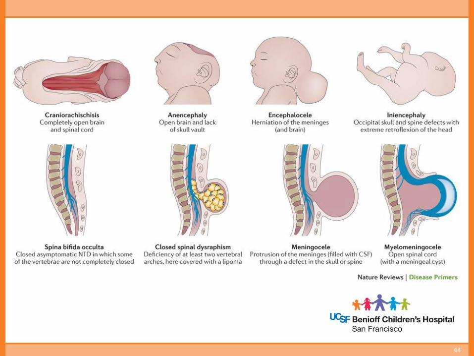
Neural Tube Defects:Overview
• Primary NTD
– Failure of neural tube closure or disruption of closed tube
– Occurs between 18-25 days of gestation
– Location of neural tube failure determines presentation
– Anencephaly, encephalocele, myelomeningocele
• Secondary NTD
– Abnormal development of the lower sacral or coccygeal segments during secondary neurulation
– Defects present primarily in lumbosacral spinal region
– Skin typically intact over lesion
– Meningocele, lipomeningocele, sacral agenesis/dysgenesis
43
44

Neural Tube Defects:Anencephaly
• Presentation
• Etiology
• Management
• Complications
• Outcome
45

Neural Tube Defects:Anencephaly
• Presentation
• Etiology
• Management
• Complications
• Outcome
Key Points:
1. Alpha-fetoprotein high in latter part of 1st trimester
2. Higher risk if you are: Female, White, poor, affected sibling
3. Failure of anterior neural tube closure
4. Occurs in 1st stage of neurodevelopment; primary neurulation
46

Neural Tube Defects:Encephalocele
• Presentation
• Etiology
• Management
• Complications
• Outcome
47
Neural Tube Defects:Encephalocele
• Presentation
• Etiology
• Management
• Complications
• Outcome
Key Points
1. May or may not contain meninges or brain parenchyma
2. Most occur in occipital region; most contain neural tissue
3. Treatment is surgical, 50% also have hydrocephalus
48
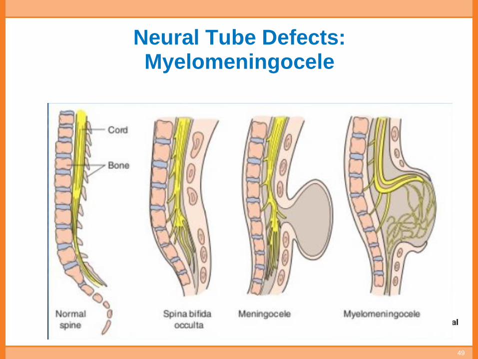
Neural Tube Defects:Myelomeningocele
49

Neural Tube Defects:Myelomeningocele
• Presentation
• Etiology
• Management
• Complications
• Outcome
Key Points:
1. Spinal cord and meninges are exposed
2. 95% will have some degree of hydrocephalus
3. Almost all infants with MM ALSO have Chiarimalformation
50

Chiari Malformation
• Brain tissue extends into the spinal canal
• Presents with reflux, laryngeal stridor and in severe forms, central apnea
51

Neural Tube Defects:Myelomeningocele
Key Points:
1. Failure of posterior neural tube to close
2. 80% occur in lumbar region
3. At birth, wrap defect in gauze soaked with NS and sterile feeding tube
4. Prone, kneeling position
5. Drape over buttocks below the lesion
6. 80% with normal intelligence
7. 85% ambulatory (with or without aid); ability to be ambulatory depends on location of lesion. < = S1 walk unaided, L4/5 walk with aid, > L2 wheelchair
52
Recommended further study:
• General brain anatomy
• Brain physiology
• Neuro Assessment
• Disorders not covered:
– Microcephaly
– Craniosynostosis
• Birth Injuries not covered:
• Cephalohematoma and Caput
• Skull fractures
53

Recommended further study:
• Intracranial Hemorrhage not covered:
– Subarachnoid
– Intracerubellar
• Meningitis
54