Embed Size (px)
Citation preview
Behavioural Neurology (1995),8,47-52 I CASE REPORT I
Neurobehavioral aspects of the delayed encephalopathy of carbon monoxide intoxication: case report and review
M.F. Mendez 1 and R.C. Doss 2
1 University of California at Los Angeles and Neurobehavior Unit, West Los Angeles Veterans Affairs Medical Center, Los Angeles, CA and 2St Paul-Ramsey Medical Center, USA
Correspondence to: M.F. Mendez, Neurobehavior Unit (691/116AF), West Los Angeles V.A. Medical Center, Wilshire & Sawtelle Blvds, Los Angeles, CA 90073, USA
We report the neurobehavioral aspects of the delayed encephalopathy of carbon monoxide (CO) intoxication in a 29 year old woman and review the literature. Four weeks after CO poisoning, the patient developed a frontal lobe syndrome, visuoperceptual impairment, and diffuse white matter lesions with an otherwise normal neurological examination. In contrast, patients with the classical syndrome also have a parkinsonian state or an akinetic-mute state. The delayed encephalopathy of CO poisoning usually results from demyelination of subcortical white matter, necrosis of the globus pallidus, or both. The clinical aspects, risk factors, neurobiological features, and therapy and prognosis are discussed.
Keywords: Carbon monoxide - Demyelination
INTRODUCTION
Carbon monoxide (CO) is a powerful toxin. CO is a colorless, tasteless gas produced in large amounts by automobile exhausts, inadequately vented furnaces and other heating equipment, fires, and methylene chloride from paint removers (Ellenhorn and Barceloux, 1988). Although the concentration of CO in the atmosphere is only about 0.1 p.p.m., these sources can increase the atmospheric CO over 1000%. Moreover, hemoglobin binds CO over 200 times more readily than it binds oxygen. It is not surprising that CO is a common cause of poisoning.
In addition to the effects of acute intoxication, a delayed encephalopathy may follow recovery from CO poisoning. After a lucid interval of 3 days to 6 weeks, up to 40% of survivors develop a neurobehavioral syndrome characterized by personality and cognitive changes, movement disorders, diffuse white matter changes, and lesions of the globus pallidus (Choi, 1983; Myers et ai., 1985). Clinicians may miss or misdiagnose this syndrome, particularly in the absence of known CO exposure. Furthermore, the clinical manifestations of the delayed encephalopathy of CO intoxication are variable and can be limited to an isolated and potentially subtle behavioral disorder (Jefferson, 1976). We review the literature on this syndrome and describe a previously healthy patient
© 1995 Rapid Communications of Oxford Ltd
with the delayed encephalopathy of CO presenting as a personality change in the absence of neuromotor findings.
CASE REPORT
A 28 year old woman had a toxic exposure to CO when her furnace malfunctioned. She was found comatose with an initial Glasgow Coma Scale of 3 and an arterial blood CO saturation of 11 %. After 90 min of hyperbaric oxygen therapy, her mental status cleared over the next 24 h. Five days after hospitalization, the patient went home fully recovered except for mild residual irritability.
Six weeks later, she was rehospitalized with "confusion". Her family described a personality change which began 4 weeks after recovery from her acute CO intoxication. The patient stopped engaging in her usual activities and would lie in bed unclothed. She would allow her small son to wander unattended and would greet strangers in an undressed state. Previously, she was "strong-willed", "uptight", and "analytical". Subsequently, she became "nonchalant" and "emotionally labile". Furthermore, the patient had trouble finding her way in her surroundings.
On examination, the patient was alert, oriented,
Behavioural Neurology. Vol 8 . 1995 47
M.F. MENDEZ AND R.C. DOSS
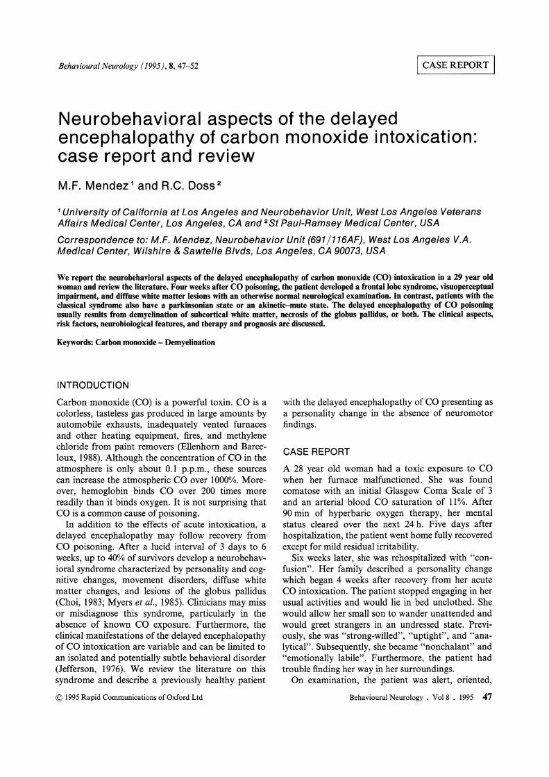
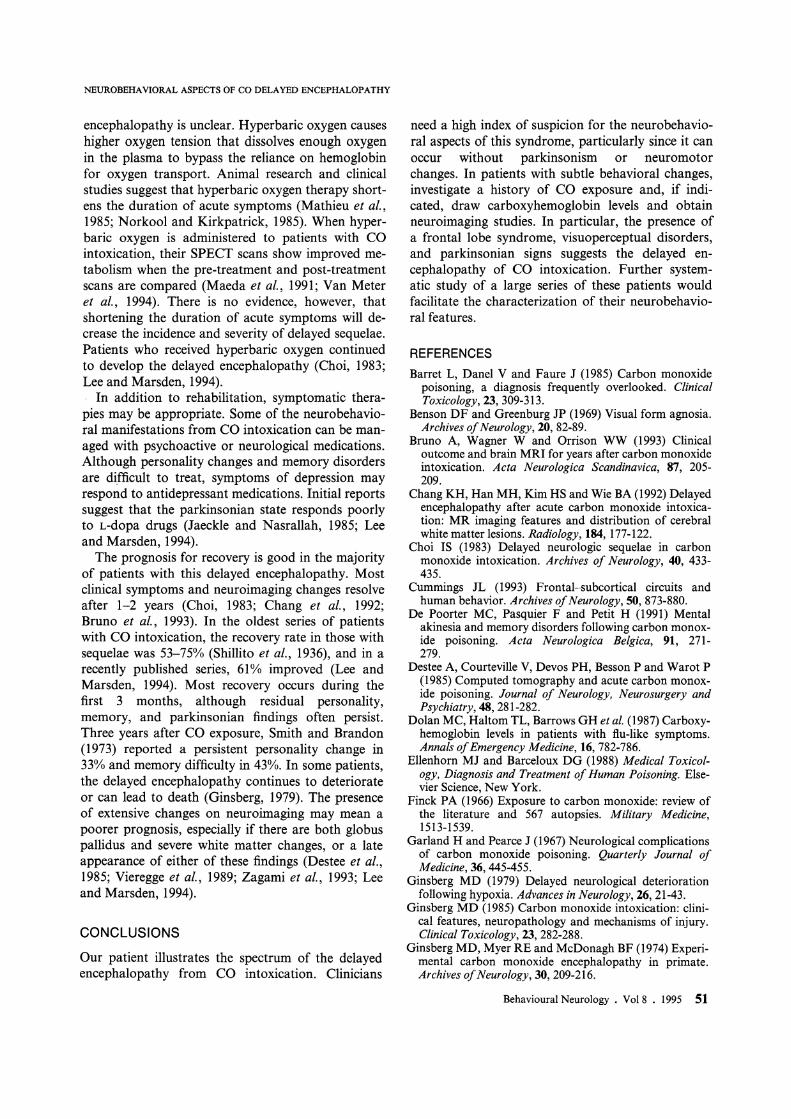
FIG. 1. Magnetic resonance images show extensive. confluent hyperintensities involving cerebral white matter on the long TR sequences. There may be slight involvement of middle cerebellar peduncle and posterior limbs of the internal capsules. The globi pallidi did not appear abnormal.
o ~D
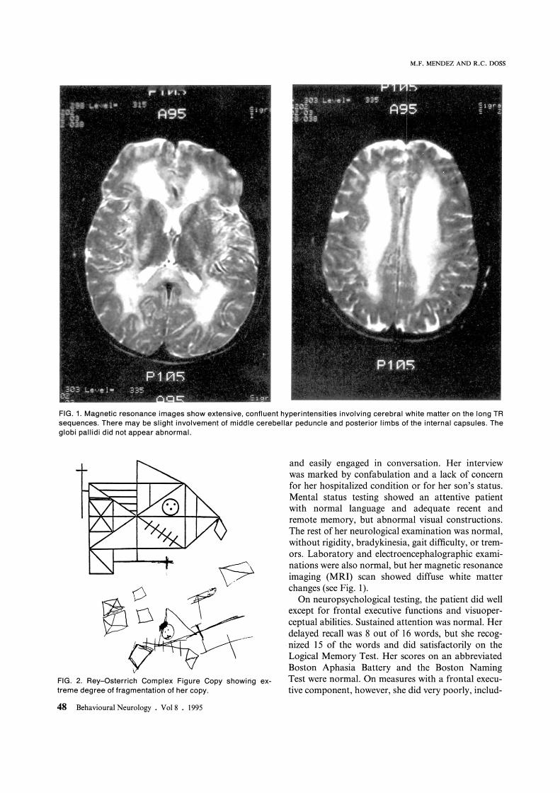
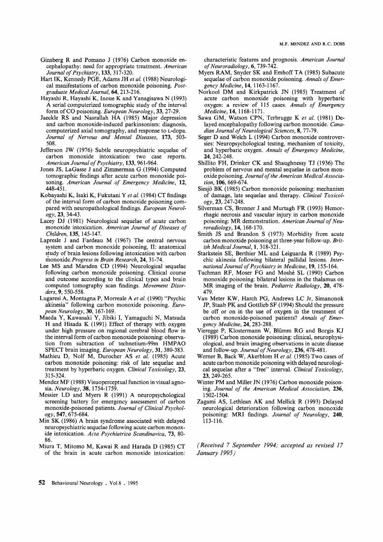
FIG. 2. Rey-Osterrich Complex Figure Copy showirig extreme degree of fragmentation of her copy.
48 Behavioural Neurology. Vol 8 . 1995
and easily engaged in conversation. Her interview was marked by confabulation and a lack of concern for her hospitalized condition or for her son's status. Mental status testing showed an attentive patient with normal language and adequate recent and remote memory, but abnormal visual constructions. The rest of her neurological examination was normal, without rigidity, bradykinesia, gait difficulty, or tremors. Laboratory and electroencephalographic examinations were also normal, but her magnetic resonance imaging (MRI) scan showed diffuse white matter changes (see Fig. I).
On neuropsychological testing, the patient did well except for frontal executive functions and visuoperceptual abilities. Sustained attention was normal. Her delayed recall was 8 out of 16 words, but she recognized 15 of the words and did satisfactorily on the Logical Memory Test. Her scores on an abbreviated Boston Aphasia Battery and the Boston Naming Test were normal. On measures with a frontal executive component, however, she did very poorly, incIud-
NEUROBEHAVIORAL ASPECTS OF CO DELAYED ENCEPHALOPATHY
ing a Porteus Mazes age of 4 with perseverations and scores ofless than the second percentile on the Stroop Color-Word Test. In addition, she was slow in recognizing complex pictures or drawings, and there was a breakdown of her constructions into smaller units (see Fig. 2). Her scores were less than second percentile on the Benton visual tests (Facial Discrimination, Line Orientation, Complex Figure Discrimination).
After an evaluation for rehabilitation, the patient went to a transitional care facility. On repeat neuropsychological testing 4 months after hospitalization, she continued to perform at less than the tenth percentile on the Porteus Mazes, Stroop Color-Word Test, and Benton visuoperceptual tasks. Follow-up MRI imaging at 6 months showed minimal change from her prior scans. At 1 year, her family reported the patient's behavior to be much improved, however, she remained disengaged and disinterested in performing housework or in returning to work.
CLINICAL FEATURES
This patient developed a persistent frontal lobe syndrome, visuoperceptual impairment, and white matter lesions following recovery from acute CO intoxication. Her behavioral change included apathy, disinhibition, confabulation, perseveration, poor judgement, and a dysexecutive syndrome. Similar to prior reports of apperceptive visual agnosia (Benson and Greenberg, 1969; Mendez, 1988), our patient could not immediately derive the global image from pictures or drawings and reverted to a slow, systematic analysis of visual details. She differed from most patients who develop the delayed encephalopathy of CO intoxication in the isolated behavioral presentation and the absence of Parkinsonian features or movement disorders (Choi, 1983; Lee and Marsden, 1994).
The immediate effects of acute CO intoxication are quite varied (Winter and Miller, 1976). Mild CO intoxication is frequently misdiagnosed as a flu-like illness, migraine headache, or other disorder (Barret et aI., 1985; Dolan et al., 1987) and can be associated with significant changes on neuropsychological tests (Messier and Myers, 1990). More severe CO exposure results in delirium, coma, or death. After an initial period of unconsciousness, a subgroup of patients develop an apathetic, akinetic-mute state which fails to improve. Among 31 patients with neuropsychiatric sequelae at 1 year, eight (26%) had a progressive course, and these were younger patients with an initial coma of about 2 days (Lee and Marsden, 1994).
The delayed encephalopathy is distinct from these acute CO effects. In the largest reported series of CO
intoxication, delayed encephalopathy occurred in 2.8% of 2369 patients and in 11.8% of the 549 patients that were hospitalized (Choi, 1983). The frequency of this complication is probably underreported given that, in the absence of gross deficits, subtle neuropsychiatric changes are usually missed (Smith and Brandon, 1973; Sawa et al., 1981; Myers et aI., 1985; Hart et at., 1988). Like our patient, most delayed encephalopathy patients are initially comatose, awaken within 24-48 h, and have a normal lucid interval averaging 2-40 days before they again deteriorate (Choi, 1983; Werner et at., 1985).
Delayed encephalopathy patients may develop apathy and akinetic mutism, irritability and other personality changes, memory difficulty, visuoperceptual problems, and parkinsonism or other movement disorders. Among 65 patients, Choi (1983) described a triad of mental deterioration (98%), gait disturbance (82%), and urinary incontinence (88%). In another series of 15 patients with delayed encephalopathy, mental dysfunction was the most common symptom, including memory difficulty, disorientation, and unspecified "abnormal behavior" (Chang et at., 1992). The mental problems were usually personality changes with apathy or poor impulse control (Smith and Brandon, 1973). Memory difficulty may persist years after the CO intoxication (Shillito et al.,1936; Lacey 1981; Choi 1983), and other patients have various forms of "psychic akinesia" (Lugaresi et at., 1991). There is also a spectrum of vi suo perceptual difficulties ranging from cortical blindness to aperceptive visual agnosia to mild residual perceptual deficits (Benson and Greenberg, 1969; Choi, 1983; Mendez, 1988). Most patients with the delayed encephalopathy have a parkinsonian state, but other movement disorders occur such as dystonias, tremors, chorea, myoclonus, and Giles de la Tourette's syndrome (Choi, 1983; Lee and Marsden, 1994). A few patients develop behavioral changes suggestive of the KliiverBucy syndrome with hyperoral behavior, a tendency to touch things, hypersexuality, and placidity (Starkstein et at., 1989; Lee and Marsden, 1994). Finally, there are reports of major depression following CO intoxication which may be a direct consequence of the brain disease (Jaeckle and Nasrallah, 1985; Myers et at., 1985; Bruno et at., 1993).
RISK FACTORS
No specific risk factors reliably predict the development of the delayed encephalopathy of CO intoxication. The severity of the initial acute intoxication is the most reliable. An initial comatose state correlates with the subsequent development of delayed encephalopa-
Behavioural Neurology. Vol 8 . 1995 49
thy and neurobehavioral symptoms, particularly if the coma is prolonged (Smith and Brandon, 1973; Choi, 1983; Min, 1986); however, loss of consciousness is not necessary for developing delayed symptoms. An age greater than 30 years is a second potential risk factor, although this apparent risk may emerge because of a better likelihood that younger people survive the acute intoxication. Third, individual patient susceptibilities are a consideration, such as the increased metabolic activity of children and pregnant women (Lacey, 1981; Seger and Welch, 1994). Fourth, an abnormal neuroimaging study at the time of acute CO poisoning may predict subsequent delayed encephalopathy, particularly with the presence of both white matter and globus pallidus changes (Miura et ai, 1985; Lee and Marsden, 1994). The association between specific clinical symptoms and neuroimaging is not robust, and MRI scans may show progressive changes in the absence of clinical symptoms (Bruno et al., 1993). Fifth, carboxyhemoglobin levels do not strongly correlate with either the amount of CO exposure or the risk of developing the delayed encephalopathy (Ginsberg, 1974; Mathieu et aI., 1985; Norkool and Kirkpatrick, 1985). Finally, other laboratory tests such as blood pH, pa02, and electroencephalograms fail to predict which patients will· develop the delayed encephalopathy (Ellenhorn and Barceloux, 1988; Vieregge et al., 1989).
NEUROBIOLOGICAL FEATURES
The most characteristic neuropathological changes are in the cerebral white matter and in the globus pallidus (Shillito et al.,1936; Finck, 1966; Kobayashi et al., 1984; Chang et al., 1992; Zagami et aI., 1993). Most of the lesions of the cerebral white matter reflect potentially reversible areas of symmetrical, periventricular demyelination which enlarge progressively during the latency period (Hayashi et al., 1993). If severe, there may be fragmentation of axis cylinders and extensive diffuse necrotic lesions. The lesions in the inner globus pallidus reflect ischemia and hemorrhagic necrosis (De Poorter et al., 1991; Chang et aI., 1992; Silverman et al., 1993). We do not understand why some patients develop white matter changes and others develop globus pallidus changes. In addition, there may be degeneration of the spongy cerebral cortex, necrotic lesions of the hippocampus and mesial temporal lobe, lesions in the thalamus, and Purkinje cell loss (Lapresle and Fardeau, 1967; Ginsberg, 1985; Tuchman et al., 1990).
The mechanism for this delayed neuropathology is unclear and may represent a maturation effect of the neuropathology (Siesjo, 1985). CO encephalopathy may result from hypoxia-ischemia in areas of poor
50 Behavioural Neurology. VolS . 1995
M.F. MENDEZ AND R.C. DOSS
anastomotic blood supply, watershed zones, and periventricular arterial distributions. The neuropathological changes of CO can be difficult to distinguish from changes after a cardiorespiratory arrest, and CO-induced neuropathological changes in primates may be indistinguishable from hypoxic-ischemic lesions (Ginsberg et al., 1974). Hypoxia-ischemia is not the whole story, however, particularly since white matter is not as vulnerable to hypoxia as cerebral gray matter, and globus pallidus lesions seldom occur with hypoxia. CO probably has an added neurotoxic effect as suggested by similar basal ganglia lesions from other poisonings, such as methanol and hydrogen sulfide poisoning. CO neurotoxicity could result form lipid peroxidation in brain by conversion of xanthine dehydrogenase to xanthine oxidase (Seger and Welch, 1994). Other theories include cellular toxicity secondary to cytochrome malfunction, increased leukocyte adherence to endothelia of brain microvasculature, and the effects of metabolic acidosis.
Recent advances in neurobiology and in neuroimaging have increased our understanding of frontal-subcortical circuits and their relationship to the basal ganglia (Cummings, 1993). Frontal lobe syndromes result from dysfunction of prefrontal connections in the subfrontal white matter and in the caudate. Similar to disorders such as multiple sclerosis and vascular dementia, the delayed encephalopathy of CO may develop a "frontal" lobe syndrome as seen in our patient. A midline frontal syndrome is also suggested from reports of akinetic mutism in nearly one-third of delayed CO encephalopathy patients (Choi, 1983; De Poorter et aI., 1991; Maeda et al., 1991; Chang et al., 1992; Hayashi et al., 1993). Prior studies indicate a frontal predominance pattern of demyelination in the delayed encephalopathy of CO which may correlate with "abnormal behavior" (Kobayashi et al., 1984; Chang et al., 1992). Subtraction single photon emission tomography (SPECT) studies in these patients show a diffuse, but frontal-dominant, hypoperfusion in gray and white matter (Maeda et al., 1991). In addition, hypoxia-ischemia may explain the vi suoperceptual changes due to bilateral parieto-occipital watershed ischemia, and the involvement of globus pallidus may be instrumental in producing the parkinsonism and other movement disorders.
THERAPY AND PROGNOSIS
The initial therapy of acute CO intoxication includes the administration of supplemental oxygen until carboxyhemoglobin levels are significantly reduced. The added efficacy of hyperbaric oxygen for the delayed
NEUROBEHAVIORAL ASPECTS OF CO DELAYED ENCEPHALOPATHY
encephalopathy is unclear. Hyperbaric oxygen causes higher oxygen tension that dissolves enough oxygen in the plasma to bypass the reliance on hemoglobin for oxygen transport. Animal research and clinical studies suggest that hyperbaric oxygen therapy shortens the duration of acute symptoms (Mathieu et ai., 1985; Norkool and Kirkpatrick, 1985). When hyperbaric oxygen is administered to patients with CO intoxication, their SPECT scans show improved metabolism when the pre-treatment and post-treatment scans are compared (Maeda et ai., 1991; Van Meter et ai., 1994). There is no evidence, however, that shortening the duration of acute symptoms will decrease the incidence and severity of delayed sequelae. Patients who received hyperbaric oxygen continued to develop the delayed encephalopathy (Choi, 1983; Lee and Marsden, 1994).
In addition to rehabilitation, symptomatic therapies may be appropriate. Some of the neurobehavioral manifestations from CO intoxication can be managed with psychoactive or neurological medications. Although personality changes and memory disorders are difficult to treat, symptoms of depression may respond to antidepressant medications. Initial reports suggest that the parkinsonian state responds poorly to L-dopa drugs (Jaeckle and Nasrallah, 1985; Lee and Marsden, 1994).
The prognosis for recovery is good in the majority of patients with this delayed encephalopathy. Most clinical symptoms and neuroimaging changes resolve after 1-2 years (Choi, 1983; Chang et ai., 1992; Bruno et ai., 1993). In the oldest series of patients with CO intoxication, the recovery rate in those with sequelae was 53-75% (Shillito et ai., 1936), and in a recently published series, 61% improved (Lee and Marsden, 1994). Most recovery occurs during the first 3 months, although residual personality, memory, and parkinsonian findings often persist. Three years after CO exposure, Smith and Brandon (1973) reported a persistent personality change in 33% and memory difficulty in 43%. In some patients, the delayed encephalopathy continues to deteriorate or can lead to death (Ginsberg, 1979). The presence of extensive changes on neuroimaging may mean a poorer prognosis, especially if there are both globus paUidus and severe white matter changes, or a late appearance of either of these findings (Destee et ai., 1985; Vieregge et ai., 1989; Zagami et ai., 1993; Lee and Marsden, 1994).
CONCLUSIONS
Our patient illustrates the spectrum of the delayed encephalopathy from CO intoxication. Clinicians
need a high index of suspicion for the neurobehavioral aspects of this syndrome, particularly since it can occur without parkinsonism or neuromotor changes. In patients with subtle behavioral changes, investigate a history of CO exposure and, if indicated, draw carboxyhemoglobin levels and obtain neuroimaging studies. In particular, the presence of a frontal lobe syndrome, visuoperceptual disorders, and parkinsonian signs suggests the delayed encephalopathy of CO intoxication. Further systematic study of a large series of these patients would facilitate the characterization of their neurobehavioral features.
REFERENCES Barret L, Danel V and Faure J (1985) Carbon monoxide
poisoning, a diagnosis frequently overlooked. Clinical Toxicology, 23, 309-313.
Benson DF and Greenburg JP (1969) Visual form agnosia. Archives of Neurology, 20, 82-89.
Bruno A, Wagner Wand Orrison WW (1993) Clinical outcome and brain MRI for years after carbon monoxide intoxication. Acta Neurologica Scandinavica, 87, 205-209.
Chang KH, Han MH, Kim HS and Wie BA (1992) Delayed encephalopathy after acute carbon monoxide intoxication: MR imaging features and distribution of cerebral white matter lesions. Radiology, 184, 177-122.
Choi IS (1983) Delayed neurologic sequelae in carbon monoxide intoxication. Archives of Neurology, 40, 433-435.
Cummings JL (1993) Frontal--subcortical circuits and human behavior. Archives of Neurology, 50, 873-880.
De Poorter MC, Pasquier F and Petit H (1991) Mental akinesia and memory disorders following carbon monoxide poisoning. Acta Neurologica Belgica, 91, 271-279.
Destee A, Courteville V, Devos PH, Besson P and Warot P (1985) Computed tomography and acute carbon monoxide poisoning. Journal of Neurology, Neurosurgery and Psychiatry, 48, 281-282.
Dolan MC, Haltom TL, Barrows GH et al. (1987) Carboxyhemoglobin levels in patients with fiu-like symptoms. Annals of Emergency Medicine, 16, 782-786.
Ellenhorn MJ and Barceloux DG (1988) Medical Toxicology, Diagnosis and Treatment of Human Poisoning. Elsevier Science, New York.
Finck PA (1966) Exposure to carbon monoxide: review of the literature and 567 autopsies. Military Medicine, 1513-1539.
Garland H and Pearce J (1967) Neurological complications of carbon monoxide poisoning. Quarterly Journal of Medicine, 36, 445-455.
Ginsberg MD (1979) Delayed neurological deterioration following hypoxia. Advances in Neurology, 26, 21-43.
Ginsberg MD (1985) Carbon monoxide intoxication: clinical features, neuropathology and mechanisms of injury. Clinical Toxicology, 23, 282-288.
Ginsberg MD, Myer RE and McDonagh BF (1974) Experimental carbon monoxide encephalopathy in primate. Archives of Neurology, 30, 209-216.
Behavioural Neurology. Vol 8 . 1995 51
Ginsberg Rand Pomano J (1976) Carbon monoxide encephalopathy: need for appropriate treatment. American Journal of Psychiatry, 133, 317-320.
Hart IK, Kennedy PGE, Adams JH et al. (1988) Neurological manifestations of carbon monoxide poisoning. Postgraduate Medical Journal, 64, 213-216.
Hayashi R, Hayashi K, Inoue K and Yanagisawa N (1993) A serial computerized tomographic study of the interval form of CO poisoning. European Neurology, 33, 27-29.
Jaeckle RS and Nasrallah HA (1985) Major depression and carbon monoxide-induced parkinsonism: diagnosis, computerized axial tomography, and response to L-dopa. Journal of Nervous and Mental Diseases, 173, 503-508.
Jefferson JW (1976) Subtle neuropsychiatric sequelae of carbon monoxide intoxication: two case reports. American Journal of Psychiatry, 133, 961-964.
Jones JS, LaGasse J and Zimmerman G (1994) Computed tomographic findings after acute carbon monoxide poisoning. American Journal of Emergency Medicine, 12, 448-451.
Kobayashi K, Isaki K, Fukutani Y et al. (1984) CT findings of the interval form of carbon monoxide poisoning compared with neuropathological findings. European Neurology, 23, 34-43.
Lacey DJ (1981) Neurological sequelae of acute carbon monoxide intoxication. American Journal of Diseases of Children, 135, 145-147.
Lapresle J and Fardeau M (1967) The central nervous system and carbon monoxide poisoning, II: anatomical study of brain lesions following intoxication with carbon monoxide. Progress in Brain Research, 24, 31-74.
Lee MS and Marsden CD (1994) Neurological sequelae following carbon monoxide poisoning. Clinical course and outcome according to the clinical types and brain computed tomography scan findings. Movement Disorders, 9, 550-558.
Lugaresi A, Montagna P, Morreale A et al. (1990) "Psychic akinesia" following carbon monoxide poisoning. European Neurology, 30, 167-169.
Maeda Y, Kawasaki Y, Jibiki I, Yamaguchi N, Matsuda Hand Hisada K (1991) Effect of therapy with oxygen under high pressure on regional cerebral blood flow in the interval form of carbon monoxide poisoning: observation from subtraction of technetium-99m HMP AO SPECT brain imaging. European Neurology, 31, 380-383.
Mathieu D, Nolf M, Durocher AS et al. (1985) Acute carbon monoxide poisoning: risk of late sequelae and treatment by hyperbaric oxygen. Clinical Toxicology, 23, 315-324.
Mendez MF (1988) Visuoperceptual function in visual agnosia. Neurology, 38, 1754-1759.
Messier LD and Myers R (1991) A neuropsychological screening battery for emergency assessment of carbon monoxide-poisoned patients. Journal of Clinical Psychology, 547, 675-684.
Min SK (1986) A brain syndrome associated with delayed neuropsychiatric sequelae following acute carbon monoxide intoxication. Acta Psychiatrica Scandinavica, 73, 80-86.
Miura T, Mitomo M, Kawai R and Harada D (1985) CT of the brain in acute carbon monoxide intoxication:
52 Behavioural Neurology. Vol 8 . 1995
M.F. MENDEZ AND R.C. DOSS
characteristic features and prognosis. American Journal of Neuroradiology, 6, 739-742.
Myers RAM, Snyder SK and Emhoff T A (1985) Subacute sequelae of carbon monoxide poisoning. Annals of Emergency Medicine, 14, 1163-1167.
Norkool DM and Kirkpatrick IN (1985) Treatment of acute carbon monoxide poisoning with hyperbaric oxygen: a review of 115 cases. Annals of Emergency Medicine, 14, 1168-1171.
Sawa GM, Watson CPN, Terbrugge K et al. (1981) Delayed encephalopathy following carbon monoxide. Canadian Journal of Neurological Sciences, 8,77-79.
Seger D and Welch L (1994) Carbon monoxide controversies: Neuropsychological testing, mechanism of toxicity, and hyperbaric oxygen. Annals of Emergency Medicine, 24, 242-248.
Shillito FH, Drinker CK and Shaughnessy TJ (1936) The problem of nervous and mental sequelae in carbon monoxide poisoning. Journal of the American Medical Association, 106, 669-674.
Siesjo BK (1985) Carbon monoxide poisoning: mechanism of damage, late sequelae and therapy. Clinical Toxicology, 23, 247-248.
Silverman CS, Brenner J and Murtagh FR (1993) Hemorrhagic necrosis and vascular injury in carbon monoxide poisoning: MR demonstration. American Journal of Neuroradiology, 14,168-170.
Smith JS and Brandon S (1973) Morbidity from acute carbon monoxide poisoning at three-year follow-up. British Medical Journal, 1, 318-321.
Starkstein SE, Berthier ML and Leiguarda R (1989) Psychic akinesia following bilateral pallidal lesions. International Journal of Psychiatry in Medicine, 19, 155-164.
Tuchman RF, Moser FG and Moshe SL (1990) Carbon monoxide poisoning: bilateral lesions in the thalamus on MR imaging of the brain. Pediatric Radiology, 20, 478-479.
Van Meter KW, Harch PG, Andrews LC Jr, Simanonok JP, Staab PK and Gottlieb SF (1994) Should the pressure be off or on in the use of oxygen in the treatment of carbon monoxide-poisoned patients? Annals of Emergency Medicine, 24, 283-288.
Vieregge P, Klostermann W, Bliimm RG and Borgis KJ (1989) Carbon monoxide poisoning: clinical, neurophysiological, and brain imaging observations in acute disease and follow-up. Journal of Neurology, 236, 478-481.
Werner B, Back W, Akerblom H et al. (1985) Two cases of acute carbon monoxide poisoning with deJayed neurological sequelae after a "free" interval. Clinical Toxicology, 23,249-265.
Winter PM and Miller IN (1976) Carbon monoxide poisoning. Journal of the American Medical Association, 236, 1502-1504.
Zagami AS, Lethlean AK and Mellick R (1993) Delayed neurological deterioration following carbon monoxide poisoning: MRI findings. Journal of Neurology, 240, 113-116.
(Received 7 September 1994; accepted as revised 17 January 1995)

Submit your manuscripts athttp://www.hindawi.com
Stem CellsInternational
Hindawi Publishing Corporationhttp://www.hindawi.com Volume 2014
Hindawi Publishing Corporationhttp://www.hindawi.com Volume 2014
MEDIATORSINFLAMMATION
of
Hindawi Publishing Corporationhttp://www.hindawi.com Volume 2014
Behavioural Neurology
EndocrinologyInternational Journal of
Hindawi Publishing Corporationhttp://www.hindawi.com Volume 2014
Hindawi Publishing Corporationhttp://www.hindawi.com Volume 2014
Disease Markers
Hindawi Publishing Corporationhttp://www.hindawi.com Volume 2014
BioMed Research International
OncologyJournal of
Hindawi Publishing Corporationhttp://www.hindawi.com Volume 2014
Hindawi Publishing Corporationhttp://www.hindawi.com Volume 2014
Oxidative Medicine and Cellular Longevity
Hindawi Publishing Corporationhttp://www.hindawi.com Volume 2014
PPAR Research
The Scientific World JournalHindawi Publishing Corporation http://www.hindawi.com Volume 2014
Immunology ResearchHindawi Publishing Corporationhttp://www.hindawi.com Volume 2014
Journal of
ObesityJournal of
Hindawi Publishing Corporationhttp://www.hindawi.com Volume 2014
Hindawi Publishing Corporationhttp://www.hindawi.com Volume 2014
Computational and Mathematical Methods in Medicine
OphthalmologyJournal of
Hindawi Publishing Corporationhttp://www.hindawi.com Volume 2014
Diabetes ResearchJournal of
Hindawi Publishing Corporationhttp://www.hindawi.com Volume 2014
Hindawi Publishing Corporationhttp://www.hindawi.com Volume 2014
Research and TreatmentAIDS
Hindawi Publishing Corporationhttp://www.hindawi.com Volume 2014
Gastroenterology Research and Practice
Hindawi Publishing Corporationhttp://www.hindawi.com Volume 2014
Parkinson’s Disease
Evidence-Based Complementary and Alternative Medicine
Volume 2014Hindawi Publishing Corporationhttp://www.hindawi.com