Embed Size (px)
Citation preview
Neural Pathways and Structures in SSAD Theory
The neural pathways and structures of SSAD theory are outlined in this section. Excerpts fromthe basic paper by Prescott (1970) are presented. The pioneering studies of Heath (1971) with hisgraphic model of neural pathways and brain structures are illustrated.http://www.violence.de/prescott/mp/article.html.
The comparative anatomy of frontal cortex and thalamofrontal connections provided by Akert(1964) document connections between Medialis Dorsalis (MD) and frontal cortex, specifically, the tri-partite structure of MD where Pars Magnocellularis thalamic projections defines the frontal orbital cortex
Berman, Berman and Prescott (1974) documented that paleocerebellar decortication but notneocerebellar decortication transformed an adult pathologically violent mother deprived monkey into apeaceful, social and inquisitive monkey. The paleocerebellum has primary connections with the brainstem and limbic system; the neocerebellum with the cerebral neocortex.http://www.violence.de/berman/article.html
Schwarz, Dietrich W.F. and Frederickson, John M. (1970) documented that there are minimalvestibular projection fields in the cerebral neocortex of the rhesus monkey. Science, 14 October 1970,Volume 172, p. 280f. http://www.violence.de/others/sci71ac.html
Prescott (1992) presents data that support sexual dimorphism in the developing human brain thatshows differential coupling of frontal-cerebellar connectivity in male and female brains. These data arebased upon NICHD supported research on cranial-facial growth and development, through lateral-skull X-rays, by the Krogman Growth Center, Children's Hospital, Philadelphia, PA. (Solomon Katz, PI andGeoffrey F. Walker, Biometrics Laboratory, University of Michigan).http://www.violence.de/archive.shtml
There are statistically significant differences between males and females where females show agreater neuronal interconnectivity between the cerebellum and frontal cortical areas throughoutdevelopment. Males show no brain maturational frontal-cerebellar connectivity. These findings suggest amore neurointegrative brain in the female than the male; a greater neural integration between cortical andsubcortical brain structures; and the observed greater nurturance and peaceful behaviors in the femalethan the male. The environment plays a major role in the structuring of these relationships and theunderlying biology.
Modern MRI and fMRI are needed to confirm these growth pattern differences, their sexualdimorphism and the implications that these findings have for the emotional-social-sexual and mentaldevelopment of the human male and female and the future of Homo sapiens.
Akert, K. (1964). Comparative anatomy of frontal cortex and thalamofrontal connections. In: Warrenand Akert, The Frontal Granular Cortex and Behavior. McGraw-Hill, New York.
Berman, A.J., Berman, D. and Prescott, J.W. (1974). The Effect of Cerebellar Lesions on EmotionalBehavior In The Rhesus Monkey. In The Cerebellum, Epilepsy and Behavior (Cooper, I.A., Riklan, M.and Snider, R.S., Eds). New York: Plenum Press, pp. 277-284.
Heath, R.G. (1972). Physiologic Basis of Emotional Expression: Evoked Potential and Mirror FocusStudies in Rhesus Monkeys. Biological Psychiatry 5(1): 15-31.
Prescott, J.W. (1971). Early Somatosensory Deprivation As An Ontogenetic Process In TheAbnormal Development of The Brain and Behavior. In Medical Primatology1970 (I.E.Goldsmith and J. Moor-Jankowski, Eds). S. Karger, Base, New York.
Prescott, J.W. (1983). Invited Address: The Quadrune Brain: Cerebellar Regulation of EmotionalBehaviors. European Seminar on Developmental Neurology. Institute fuer Kindesentwicklung,GmbH. Hamburg, Germany. February 14-17, 1983.
Prescott, J.W. (1992). Sexual Dimorphism in the Developing Human Brain: Evidence from LateralSkull X-Rays. Presented at the 35th Annual Meeting of the Society for the Scientific Study of Sex,November 12-15, 1992
Schwarz, Dietrich W.F. and Frederickson, John M. (1970). Rhesus Monkey Vestibular Cortex: ABimodal Primary Projection Field. SCIENCE, 14 October 1970, Volume 172, p. 280f
Click HERE for additional documentation.
EARLY SOMATOSENSORY DEPRIVATION AS ANONTOGENETIC PROCESS IN THE ABNORMAL
DEVELOPMENT OF THE BRAIN AND BEHAVIOR.
By James W. Prescott, Ph.D.
From Medical Primatology 1970 (I.E. Goldsmith and J. Moor-Jankowski, Eds.),pp. 356-375. S. Karger, Basel, New York
366 PRESOOTT
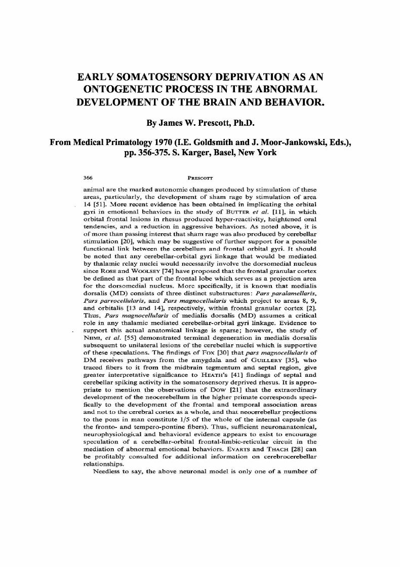
animal are the marked autonomic changes produced by stimulation of theseareas, particularly, the development t>f sham rage by stimulation of area14 [51]. More recent evidence has been, obtained in implicating the orbitalgyri in emotional behaviors in the study of BUTTER et al. [11], in whichorbital frontal lesions in rhesus produced hyper-reactivity, heightened oraltendencies, and a reduction in aggressive behaviors. As noted above, it isof more than passing interest that sham rage was also produced by cerebellarstimulation [20], which may be suggestive of further support for a possiblefunctional link between the cerebellum and frontal orbital gyri. It shouldbe noted that any cerebellar-orbital gyri linkage that would be mediatedby thalaxnic relay nuclei would necessarily involve the dorsomedial nucleussince ROSE and WQOLSEY [74] have proposed that the frontal granular cortexbe defined as that part of the frontal lobe which serves as a projection areafor the dorsomedial nucleus. More specifically, it is known that medial isdorsalis (MD) consists of three distinct substructures: Pars paralamettaris,Pars parvoceiiulariSf and Pars magnoceUularis which project to areas 8, 9,and orbitalis [13 and 14], respectively, within frontal granular cortex [2].Thus, Pars magnoceUularis of medialis dorsalis (MD) assumes a criticalrole in any thalamic mediated cerebellar-orbital gyri linkage. Evidence tosupport this actual anatomical linkage is sparse; however, the study ofNIIMI, et al, [55] demonstrated terminal degeneration in medialis dorsalissubsequent to unilateral lesions of the cerebellar nuclei which is supportiveof these speculations. The findings of Fox [30J that pars magnoceUularis ofDM receives pathways from the amygdala and of GUILLERY [35], whotraced fibers to it from the midbrain tegmentum and septal region, givegreater interpretative significance to HEATH'S [41] findings of septal andcerebellar spiking activity in the somatosensory deprived rhesus. It is appro-priate to mention the observations of Dow [21] that the extraordinarydevelopment of the neocerebellum in the higher primate corresponds speci-fically to the development of the frontal and temporal association areasand not to the cerebral cortex as a -whole, and that neocerebellar projectionsto the pons in man constitute 1/5 of the whole of the internal capsule (asthe fronto- and tempero-pontine fibers). Thus, sufficient neuronanatonical,neurophysiological and behavioral evidence appears to exist to encouragespeculation of a cerebellar-orbital frontal-limbic-reticular circuit in themediation of abnormal emotional behaviors. EVARTS and THACH [28] canbe profitably consulted for additional information on cerebrocerebellarrelationships.
Needless to say, the above neuronal model is only one of a number of
Biological Psychiatry, Vol. 5, No. 1, 1972
Physiologic Basis of Emotional Expression: EvokedPotential and Mirror Focus Studies in Rhesus Monkeys1
Robert G. Heath2
Received July 29, 1971-Revised November 21,1971
Data are reported which demonstrate functional connections between certainsensory relay nuclei (postero ventro lateral thalamus and deep nuclei of thecerebellum) and brain sites (primarily the septal region and hippocampus) wherephysiologic activity has previously been shown to be correlated with emotionalexpression. Using rhesus monkeys prepared with electrodes into relevant specificdeep brain sites and over the cortex, two procedures were performed: (i) Todemonstrate anatomic-physiologic connections between various nuclear sites,potentials were evoked by applying stimuli between leads of an implantedelectrode while recordings were obtained between leads of an electrode atanother site; (ii) To demonstrate the nature of the functional interrelationwithin the sites which the evoked potential studies had shown to be connected,cobalt was implanted into one nuclear mass and sequential electro-encephalograms were obtained to record the spread of epileptiform activity fromthe implanted site through other deep brain sites. By these techniques, thefastigius nucleus of the cerebellum and the postero ventro lateral thalamus wereshown not only to be directly connected to each other, but to have direct back
These studies were supported in part by National Institute of Health ContractPH-43-68-1412, and in part by funds from the It tie son Family Foundation, New York.New York.
1 This paper was presented in part at the Annual Meeting of the Society of BiologicalPsychiatry, San Francisco, California, May 1970.
2 Department of Psychiatry and Neurology, Tulane University School of Medicine, NewOrleans, Louisiana.
15
© 1972 Plenum Publishing Corporation, 227 West 17th Street, New York, N.Y. 10011.
16 Heath
and forth connections with the septal region and hippocampus. Indirectfpoly sy nap tic) connections were also shown to exist between these sensorynuclei and other sites, including the mesencephalic reticulum, hypothalamus,and orbital cortex, identified in pathways for emotional expression. Thesephysiologic findings support previously reported experimental data and clinicalobservations which suggest a close functional relation between sensory input andbehavioral phenomena (feelings, emotion, and awareness).
INTRODUCTION
In studies conducted in human subjects, rhesus monkeys, and cats duringthe past 20 years in the Tulane laboratories, designed to help clarify physiologiccorrelates of behavior, we identified the septal region (Heath, 19540) and its out*flow through the medial forebrain bundle to the interpeduncular nuclei, thehippocampi, and the amygdalae as key regions of the integrated brain inexpression of emotions and feelings and in awareness. Briefly summarized, ourprincipal observations were as follows:
1. The septal region, the medial forebrain bundle, and the region ofinterpeduncular nuclei constituted a pleasure and alerting system. When theirseptal regions were stimulated, patients prepared with deep and surfaceelectrodes became alerted and reported feelings of profound pleasure (Heath etal., 19540, 1968; Heath, 19640, 1964&), In contrast, when, during manifesta-tions of psychotic signs and symptoms, patients had reduced levels of awarenessand were dysphoric, electroencephalographic (EEC) abnormalities in the form ofspikes and slow waves were consistently recorded from the septal region.
2. Ablation of the septal region in animals lowered levels of awareness andproduced catatonic signs and impairment of emotional expression (Heath,1954&).
3. Correlations were observed between activity in sites closely inter-connected with the septal region—hippocampus and amygdala—and emotionalexpression (Heath and Gallant, 1964). With intense emotion, either painful(emergency states of fear and rage) or extremely pleasurable (states of joy orpleasurable recall or anticipation), changes, in the form of high-amplitudespindling, were consistently reflected in recordings from these sites. Whenfeelings of pleasure prevailed, the spindling was noted predominantly in theseptal region and at sites in the amygdala. Electrical stimulation of the amygdalaand the hippocampus consistently induced intense emergency emotions (fear orrage). Similar adversive or painful emergency responses were observed whenstimuli were directed to the midline (periventricular) hypothalamus and to themidline (periaqueductal) mesencephalic tegmentum. Stimulation of many otherbrain sites, including the basal ganglia, yielded neutral responses, that is, neitheradversive nor pleasurable.
Against the background of these extensive data, reports by Harlow and
30 Heath
The fastigius nucleus is conventionally considered part of the oldestcerebellum integrally associated with vestibular activity and without significantinfluence on higher centers. Our studies suggest that, through influences onstructures associated with expression of emotions, feelings, and levels ofawareness, vestibular and kinesthetic stimuli, as well as other somatosensoryinputs (via specific relay nuclei), profoundly affect the function of the highernervous system and behavior.
These physiologic data could provide an explanation for the behavioral data,derived from studies in animals and humans, which show that sensorydeprivation induces severe alterations in emotion and awareness, as well as forthe clinical observations that psychotic behavior with profound impairment ofemotional expression is associated with significant impairment of sensoryperception.
ACKNOWLEDGMENTS
The author wishes to acknowledge the technical assistance of Charles J.Fontana, John P. Wust, Jr., and Herbert J. Daigle.
REFERENCESAkert, K., and Hummel, P. (1968). The Limbic System-Anatomy and Physiology, Roche
Laboratories, Nutley, New Jersey.Anand, B. K., Malhotra, C. L., Singh, B., and Dua, S. (1959). Cerebellar projections to
limbic system./ Neurophysiol. 22: 451.Crosby, E. C., Humphrey, T., and Lauer, E. W. (1962). Cerebellum, in Correlative Anatomy
of the Nervous System, The Macmillan Company, New York, pp. 188-220.Guerrero-Figueroa, R., deBalbian Verster, F., and Heath, R. G. (1962). Mirror focus in
specific subcortical nuclei. Trans. Am. Neurol Assoc. 87: 207.Harlow, H F., and Harlow, M. (1962). Social deprivation in monkeys. Sci. Am. 207: 137.Harlow, H. F., and Harlow, M. (1965). The affectional systems, in Behavior of Nonhuman
Primates, Vol. 2, Schrier, A. M., Harlow, H. F., and Stollnitz, F. (eds.), Academic Press,New York, pp. 287-334
Harlow, H. F., Harlow, M. K.» and Suomi, S. J. (1971). From thought to therapy: Lessonsfrom a primate laboratory. Am. Sci. 59: 538.
Heath, R. G. (19540). Definition of the septal region, in Studies in Schizophrenia, HarvardUniversity Press, Cambridge, pp. 3-5.
Heath, R. G. (19546). Behavioral changes following destructive lesions in the subcorticalstructures of the forebrain in cats, in Studies in Schizophrenia, Harvard UniversityPress, Cambridge, pp. 83-84.
Heath, R. G. (19640). Developments toward new physiologic treatments in psychiatry. /.Neuropsychiat. 5: 318.
Heath, R. G. (1964ft). Pleasure response of human subjects to direct stimulation of thebrain: Physiologic and psychodynamic considerations, in The Role of Pleasure inBehavior, Heath, R. G. (ed.), Hoeber Medical Division, Harper and Row, New York,pp. 219-243.
Heath, R. G. (19720). Pleasure and brain activity in man: Deep and surface electroencephalo-grams during orgasm./ Nervous Mental Disease 154: 3.
Physiologic Basis of Emotional Expression (Heath 1972) 29
10 10
20 10 in 20
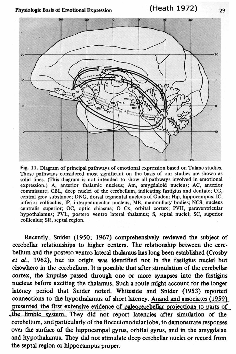
Fig. 11. Diagram ot principal pathways of emotional expression based on Tulane studies.Those pathways considered most significant on the basis of our studies are shown assolid lines. (This diagram is not intended to show all pathways involved in emotionalexpression.) A, anterior thalamic nucleus; Am, amygdaloid nucleus; AC, anteriorcommissure; CBL, deep nuclei of the cerebellum, indicating fastigius and dentate; CG,central grey substance; DNG, dorsal tegmental nucleus of Guden; Hip, hippocampus; 1C,inferior colliculus; IP, interpeduncular nucleus; MB, mammillary bodies; NCS, nucleuscentralis superior; OC, optic chiasma; O Cx, orbital cortex; PVH, paraventricularhypothalamus; PVL, postero ventro lateral thalamus; S, septal nuclei; SC, superiorcolliculus; SR, septal region.
Recently, Snider (1950; 1967) comprehensively reviewed the subject ofcerebellar relationships to higher centers. The relationship between the cere-bellum and the postero ventro lateral thalamus has long been established (Crosbyet al., 1962), but its origin was identified not in the fastigius nuclei butelsewhere in the cerebellum. It is possible that after stimulation of the cerebellarcortex, the impulse passed through one or more synapses into the fastigiusnucleus before exciting the thalamus. Such a route might account for the longerlatency period that Snider noted. Whiteside and Snider (1953) reportedconnections to the hypothalamus of short latency. Anand and associates (1959)presented the first extensive evidence of paleocerebellar projections to parts of
,thfi limhic system They did not report latencies after simulation of thecerebellum, and particularly of the flocculonodular lobe, to demonstrate responsesover the surface of the hippocampal gyrus, orbital gyrus, and in the amygdalaeand hypothalamus. They did not stimulate deep ceiebellar nuclei or record fromthe septal region or hippocampus proper.
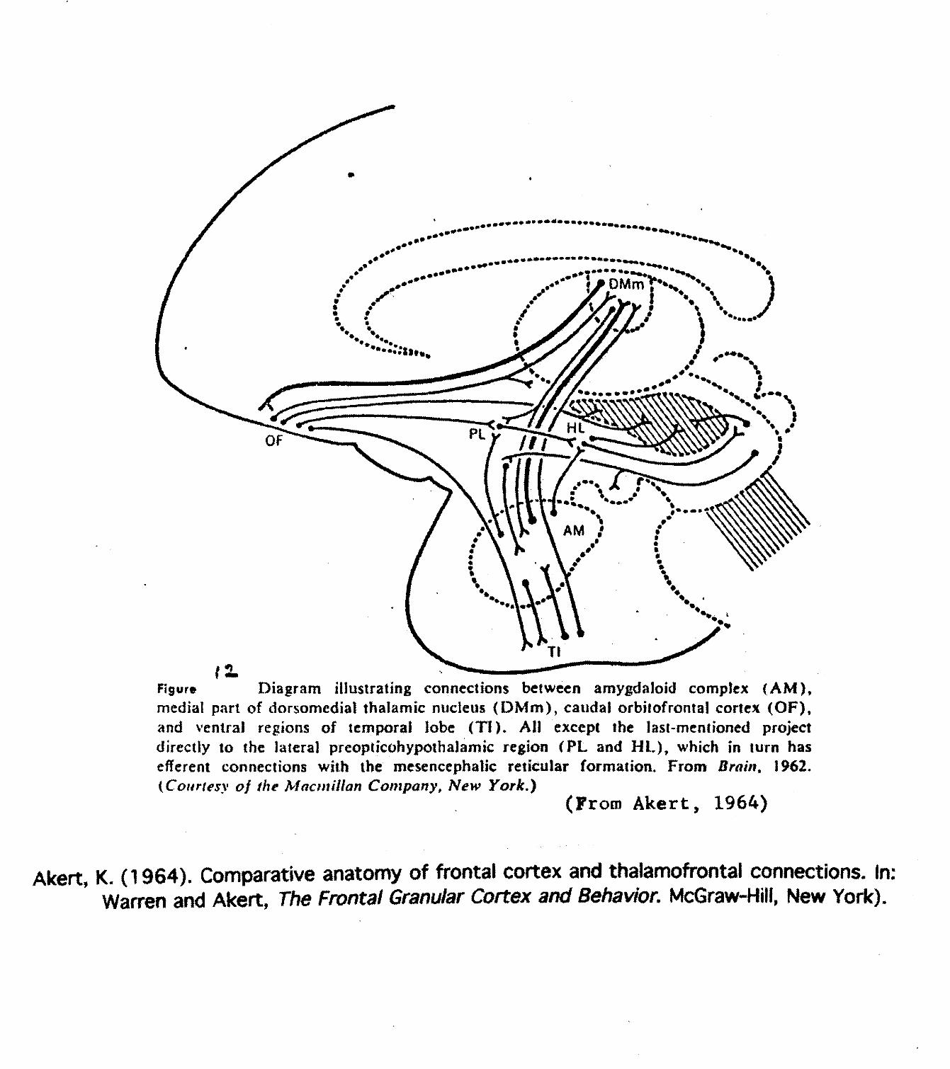
FJgur* Diagram illustrating connections between amygdaloid complex (AM),medial part of dorsomedial thalamic nucleus (DMm), caudal orbilofrental cortex (OF),and ventral regions of temporal lobe (Tl). All except the last-mentioned projectdirectly to the lateral preopticohypothalamic region (PL and HL), which in turn hasefferent connections with the mesencephalic reticular formation. From Brain, 1962.(Conrtfs\ of the Mactnillan Company, New York.)
(From Akert, 1964)
Akert, K. (1964). Comparative anatomy of frontal cortex and thalamofrontai connections. In;Warren and Akert, The Frontal Granular Cortex and Behavior. McGraw-Hill, New York).
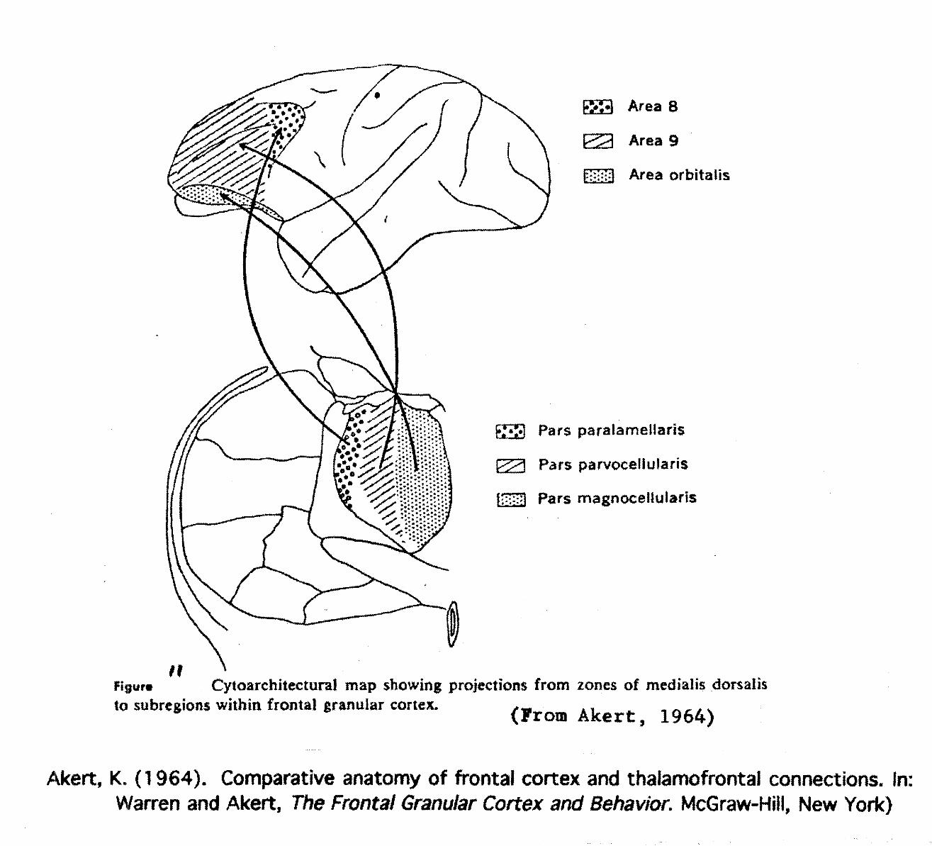
Area 8
Area 9
Area orbltalis
Pars paraiameHaris
Pars parvocellufaris
Pars magnoeeltularis
Cytoarchitectural map showing projections from zones of medtalis dorsalisto subregions within frontal granular cortex. >— ., . ^ _ ..& <Frora Akert, 1964)
Akert, K. (1964). Comparative anatomy of frontal cortex and thaiamofrontal connections. In:Warren and Akert, The Frontal Granular Cortex and Behavior. McGraw-Hill, New York)
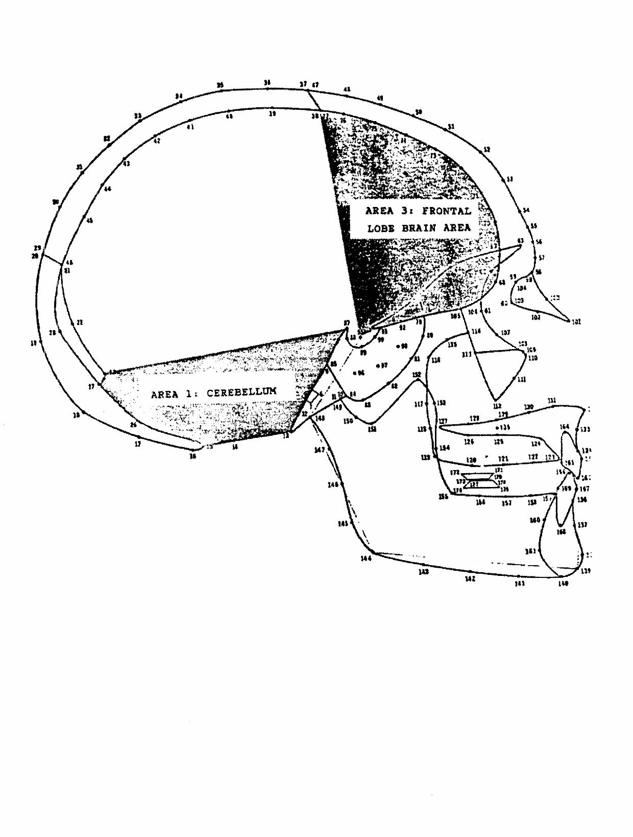
$4
ASEA 3c FRONTAL
LOBt BRAIN ABEA ™
*><k£^y.-f'«>i*B»4sv .1—-"T.4.^.... •S"4*̂ *̂ A^**. *JPfr< " -rf* »^ ^ N
B'^W'X^agB?^^ -7^IJB^feJ^Xt'OTS^fe-* /v8KBflP*xiji^ '̂ *\fej:^jEl^fc-vi***!"£r
S^ '̂:^^^^^^h§if
CEMBELLOM
I*
114
I4S
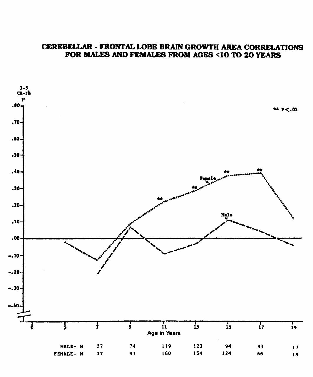
CEREBELLAR - FRONTAL LOBE BRAIN GROWTH AREA CORRELATIONSFOR MALES AND FEMALES FROM AGES <1O TO 2O TEARS
3-5ci-rfcr
.so-.
.70-
.60.
.30-
.40*
.20-
.10-
**
//
-.20-
//
4 i5
MALE- NFEMALE- N
27
37
7497
11 13Age in Years
119 123160 154
»15
94124
17
43
66
19
17
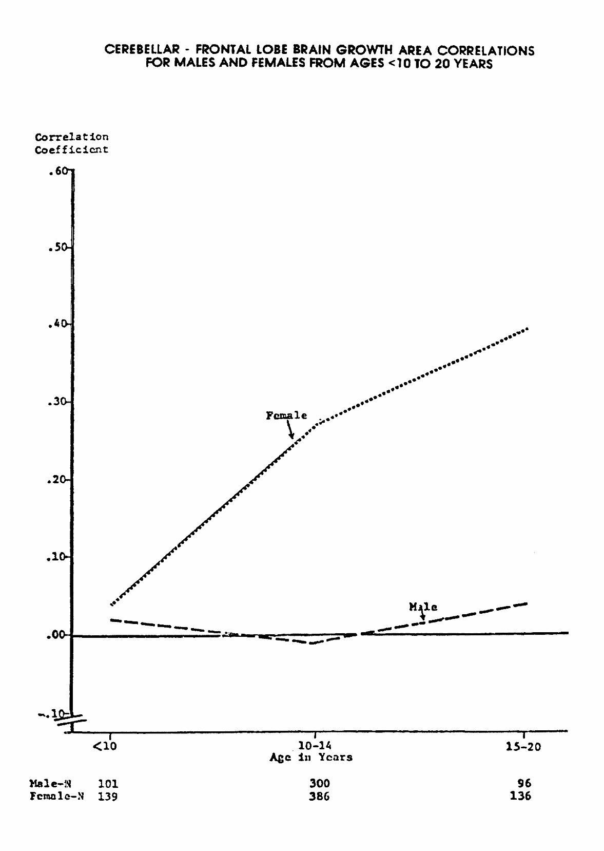
CEREBELLAR - FRONTAL LOBE BRAIN GROWTH AREA CORRELATIONSFOR MALES AND FEMALES FROM AGES <10 TO 20 YEARS
CorrelationCoefficient
.60-
.50-
.30-
.20-
,10-
,00-
....Female . .«•**
Male ^~~~~
10-1AIn Years
15-20
Female-N101139
300386
96136