Embed Size (px)
Citation preview

NeuroImage 174 (2018) 164–176
Contents lists available at ScienceDirect
NeuroImage
journal homepage: www.elsevier.com/locate/neuroimage
Neural and genetic determinants of creativity
Zhaowen Liu a,1, Jie Zhang b,**,1, Xiaohua Xie b,c,1, Edmund T. Rolls c,d,1, Jiangzhou Sun e,f,1,Kai Zhang g,1, Zeyu Jiao b,i, Qunlin Chen e,f, Junying Zhang a,****, Jiang Qiu e,f, j,***,Jianfeng Feng b,c,h,i,k,*
a School of Computer Science and Technology, Xidian University, Xi'an 710071, Shannxi, PR Chinab Institute of Science and Technology for Brain Inspired Intelligence, Fudan University, Shanghai, PR Chinac Department of Computer Science, University of Warwick, Coventry CV4 7AL, UKd Oxford Centre for Computational Neuroscience, Oxford UKe Key Laboratory of Cognition and Personality (SWU), Ministry of Education, Chongqing, PR Chinaf School of Psychology, Southwest University (SWU), Chongqing, PR Chinag Department of Computer and Information Sciences, Temple University, 1801 North Broad Street, Philadelphia, PA 19122, USAh Collaborative Innovation Center for Brain Science, Fudan University, Shanghai, 200433, PR Chinai Shanghai Center for Mathematical Sciences, Shanghai, 200433, PR Chinaj Southwest University Branch, Collaborative Innovation Center of Assessment Toward Basic Education Quality, Beijing Normal University, Beijing 100875, PR Chinak Zhongshan Hospital, Fudan University, Shanghai, 200433, PR China
A B S T R A C T
Creative thinking plays a vital role in almost all aspects of human life. However, little is known about the neural and genetic mechanisms underlying creative thinking.Based on a cross-validation based predictive framework, we searched from the whole-brain connectome (34,716 functional connectivities) and whole genome data(309,996 SNPs) in two datasets (all collected by Southwest University, Chongqing) consisting of altogether 236 subjects, for a better understanding of the brain andgenetic underpinning of creativity. Using the Torrance Tests of Creative Thinking score, we found that high figural creativity is mainly related to high functionalconnectivity between the executive control, attention, and memory retrieval networks (strong top-down effects); and to low functional connectivity between thedefault mode network, the ventral attention network, and the subcortical and primary sensory networks (weak bottom-up processing) in the first dataset (consisting of138 subjects). High creativity also correlates significantly with mutations of genes coding for both excitatory and inhibitory neurotransmitters. Combining the brainconnectome and the genomic data we can predict individuals' creativity scores with an accuracy of 78.4%, which is significantly better than prediction using singlemodality data (gene or functional connectivity), indicating the importance of combining multi-modality data. Our neuroimaging prediction model built upon the firstdataset was cross-validated by a completely new dataset of 98 subjects (r¼ 0.267, p¼ 0.0078) with an accuracy of 64.6%. In addition, the creativity–related functionalconnectivity network we identified in the first dataset was still significantly correlated with the creativity score in the new dataset (p<10�3). In summary, our researchdemonstrates that strong top-down control versus weak bottom-up processes underlie creativity, which is modulated by competition between the glutamate and GABAneurotransmitter systems. Our work provides the first insights into both the neural and the genetic bases of creativity.
Introduction
Creativity in humans is a complex cognitive behavior commonlydefined as the ability to generate responses that are novel, useful, andappropriate, (Sternberg and Lubart, 1996). Creative thinking plays animportant role in almost all aspects of our life, most prominently in thearts, science, and engineering.
Divergent thinking tests are so far the major type of psychometric
* Corresponding author. Department of Computer Science, University of Warwick** Corresponding author. Institute of Science and Technology for Brain Inspired In*** Corresponding author. Key Laboratory of Cognition and Personality (SWU), Min**** Corresponding author. School of Computer Science and Technology, Xidian Un
E-mail addresses: [email protected] (J. Zhang), [email protected] These authors contributed equally to the work.
https://doi.org/10.1016/j.neuroimage.2018.02.067Received 27 November 2017; Received in revised form 27 February 2018; Accepted 28 FebruAvailable online 5 March 20181053-8119/© 2018 Elsevier Inc. All rights reserved.
instrument in creativity testing (Gardner, 1988; Plucker and Runco,1998), including various verbal and figural tasks. Based on the divergentthinking tests, mounting evidence suggests that the default modenetwork and brain regions associated with cognitive control may beimportant for creativity. For example, regions of the default modenetwork, including the precuneus (Fink et al., 2014a, 2014b; Jauk et al.,2015; Jung et al., 2010; Takeuchi et al., 2010) and inferior parietal lobule(Takeuchi et al., 2011), and executive control network (ECN) (Ellamil
, Coventry CV4 7AL, UK.telligence, Fudan University, Shanghai, PR China.istry of Education, Chongqing, PR China.iversity, Xi’an 710071, Shannxi, PR China.(J. Zhang), [email protected] (J. Qiu), [email protected] (J. Feng).
ary 2018

Z. Liu et al. NeuroImage 174 (2018) 164–176
et al., 2012; Gonen-Yaacovi et al., 2013) have been implicated in neu-roimaging studies of divergent thinking tasks. The activation of thedefault mode network (DMN) in creative processes may reflect thespontaneous generation of candidate ideas, and/or the retrieval oflong-term memory (Beaty et al., 2016), while the control network mayserve to constrain cognition to meet specific goals of the tasks.
As a highly complex cognitive process, creativity relies on not onlythe activity of separate brain regions but also the interactions betweendifferent brain regions/networks (Bullmore and Sporns, 2009; Fox et al.,2005; van den Heuvel and Hulshoff Pol, 2010). Functional connectivity(FC) (the correlation between the BOLD signal of different brain regions)depicts the interaction between different brain regions, which is infor-mative for complex behaviors such as creativity and cognition. Forinstance, Beaty et al. (2014) found greater connectivity between theentire DMN and the left inferior frontal gyrus (IFG) within the ECN inhighly creative individuals. In addition, a recent study found that thestrength of connectivity between the DMN and the frontoparietalnetwork (FPN) was positively related to both visual and verbal creativityusing independent component analysis (Zhu et al., 2017). Such findingssuggest that creative thought may benefit from the cooperation of theDMN and ECN.
The genetic basis of divergent thinking has been previously exploredmainly by using candidate gene approaches. Several brain regions in thedopaminergic (DA) system have been found to be involved in creativitycognition (Flaherty, 2005; Heilman et al., 2003; Takeuchi et al., 2010).Thus, many genes in the dopamine pathway, like D2 Dopamine Receptor(DRD2) (de Manzano et al., 2010; Reuter et al., 2006), D4 DopamineReceptor (DRD4) (Mayseless et al., 2013), Tryptophan Hydroxylase(TPH1) (Reuter et al., 2006), Dopamine Transporter 1 (DAT1), andCatechol-O-Methyltransferase (COMT) (Zabelina et al., 2016), wereselected as candidate genes and were found to be associated withdivergent thinking. In addition, as several psychiatric illnesses are foundto have genetic links to creativity (Kyaga et al., 2011; Power et al., 2015),some genes involved in the pathology of psychosis, such as neuregulin 1(NRG1), were selected and were also found to be associated with crea-tivity (Keri, 2009).
Although previous studies have explored the relationship betweenbrain networks and creativity, most studies have focused on the regionspreviously implicated in creativity (ROI-based), or used correlationanalysis, i.e., correlation between neuroimaging metrics and behavioralscores, to search for the neural basis of creativity (correlation-based).One the one hand, the ROI-based analysis may limit the potential searchto only a small part of the whole brain, thus preclude the possibility ofnew findings (Xia and He, 2017). On the other hand, correlation-basedapproaches have the disadvantage of overfitting the data and often failto generalize to novel data (Shen et al., 2017). In addition, althoughseveral studies have explored creativity-related candidate genes, a searchfor the genetic basis of creativity at a genome-wide scale, especially fordivergent thinking, has not been described as far as we know. Thecorrelation-based methods have also been widely employed in the ge-netic analysis of divergent thinking which have the disadvantage of lowgeneralization ability.
Based on all the above, in the present study we used a large sample of236 individuals (which we separated into two non-overlapping datasets)in this research, which enabled statistical analyses of whole-brain FCs(34,716 FCs) and whole-genome genes (309,996 SNPs) that were relatedto creativity without a priori hypotheses, therefore not creating con-straints on the brain regions and genes to be investigated. Furthermore,we used a cross-validation based approach to identify the functionalbrain networks that are predictive of creativity, to ensure that the resultswere statistically firm and could be generalized to new populations. Ourapproach, IMuDP (Integrating Multi-scale Data for Prediction), isimproved from one that has been used to predict sustained attention inresting-state fMRI (Rosenberg et al., 2016), which can now deal with
165
multi-modal data. The prediction-based approach is held to providefMRI-derived statistics that relate to individual behaviors with moregeneralizability than traditional correlation-based analysis (Dubois andAdolphs, 2016). Furthermore, to understand the genetic underpinning ofcreative thinking, we also searched from the whole genome the SNPs(single nucleotide polymorphisms) whose mutations correlate mostclosely with the creativity score using the same prediction-based strat-egy. Part of the aim here was to investigate howmulti-modal data (in thiscase fMRI and genetic data) can be combined, and whether this helpsbetter predictions to be made. This research provides the firstwhole-brain functional network analysis for creativity with a corre-sponding whole-genome search for related SNPs, and is aimed to shedlight on both the neural and genetic underpinnings of creative thinking.
Materials and methods
Data
Ethics statementBoth the behavioral and MRI protocols were approved by the local
ethics committee of Southwest China University, Chongqing. Writteninformed consent was obtained from all participants prior to the study,which was approved by the Institutional Human Participants ReviewBoard of Southwest University Imaging Center for Brain Research. Themethods were conducted in accordance with approved guidelines.
ParticipantsThree hundred and fifteen subjects were recruited in this study (Li
et al., 2016). We firstly excluded 63 subjects without complete behav-ioral or other demographic data. After further fMRI head movementcontrol, 236 subjects were retained for further analysis. The retainedsubjects were then divided into two datasets. The first dataset consistedof 138 individuals, 49 males and 89 females (age 19.7�1.2 (mean� sd)),with both genetic and neuroimaging data, which we used to perform ourmultimodal prediction analysis. The second dataset consisted of 98 in-dividuals, 38 males and 60 females (age 20.2�1.3 (mean� sd)), withonly neuroimaging data, which we used as an external validation datasetto validate our prediction model generated from the first dataset.
All the resting-state fMRI data were collected in the Southwest Uni-versity Center for Brain Imaging, Chongqing, China, using a 3.0-TSiemens Trio MRI scanner (Siemens Medical, Erlangen, Germany). Eachsubject was required not to drink alcohol the day before the experiments,which was then confirmed right before the scanning by questionnaires.
Resting-state fMRIIn resting-state fMRI scanning, the subjects were instructed to rest
without thinking about a particular topic, and not to fall asleep or closetheir eyes. The 8-min scan of 242 contiguous whole-brain resting-statefunctional images was obtained using gradient-echo planar imaging (EPI)sequences with the following parameters: slices¼ 32, repetition time(TR)/echo time (TE)¼ 2000/30ms, flip angle¼ 90, field of view(FOV)¼ 220mm� 220mm, and thickness/slice gap¼ 3/1mm, voxelsize 3.4� 3.4� 3 mm3.
Structural MRIA magnetization-prepared rapid gradient echo (MPRAGE) sequence
was used to acquire high-resolution T1-weighted anatomical images(repetition time¼ 1900ms, echo time¼ 2.52ms, inversiontime¼ 900ms, flip angle¼ 90�, resolution matrix¼ 256� 256, sli-ces¼ 176, thickness¼ 1.0mm, voxel size¼ 1� 1� 1mm3).
Creativity assessmentWe adopted the Torrance Tests of Creative Thinking (TTCT) score
that measures divergent thinking, which is a central aspect of creativity

Z. Liu et al. NeuroImage 174 (2018) 164–176
(Huang et al., 2013; Sternberg and O'HARA, 1999). The TTCT containsverbal, figural and auditory tests (Huang et al., 2013). The Figural TTCTis supposed to be less biased than the verbal TTCT test (Kim, 2006), andthus is the focus of this paper. The figural test comprises of 3 tasks:picture construction, picture completion, and lines. To be specific, all theparticipants answer the same questions with a 10-min time limit for eachtask. The first activity requires subjects to draw a picture based on anellipse shape provided on the page. The second activity requires subjectsto use 10 incomplete figures to make an object or picture. The third ac-tivity requires subjects to draw as many as possible pictures or objects onthree pages of vertical lines. Participants were told to draw as many aspossible and as creatively as possible.
For each task the scoring comprised three components: fluency (thenumber of meaningful and relevant responses, which is associated withthe ability to generate and consider other possibilities), flexibility (thenumber of different categories of responses, which reflects the ability toshift between conceptual fields), and originality (the degree of originalityof the responses, which is associated with thinking “outside the box”)(Chavez-Eakle et al., 2007). More specifically, the score of fluency is thenumber of meaningful responses, the score of flexibility is the number ofdifferent categories of response, and the score of originality is the degreeof originality of the response with a four-point rating scale ranging from0 (“not original”) to 3 (“highly original”).
Three trained raters took part in the scoring. The raters displayed highinternal consistency in their ratings (Cronbach alpha¼ 0.90). The cur-rent study used the total creativity scores (sum of the fluency, flexibilityand originality scores) (de Souza et al., 2010) for each dimension whichwas highly correlated with the fluency, flexibility and originality scores(details in Fig. S1) on the basis of the findings of Heausler and Thompson(1988), who suggested that because of the high correlations between thethree subscales of the TTCT the subscales do not provide meaningfullydifferent data (Heausler and Thompson, 1988).
General intelligence assessmentThe participants' intellectual ability was examined using the Com-
bined Raven's Test (CRT). As the test is a highly reliable and valid in-telligence test, it is widely used (Tang et al., 2012). In our study, all the138 participants completed the test with the 72 items, including theRaven's standard progressive matrix (C, D, E sets) and Raven's coloredprogressive matrix (A, AB, B sets), revised by the Psychology Departmentof East China Normal University in 1989. The total number of correctanswers that an individual presented in 40min was used as a psycho-metric index of individual intelligence (Jaeggi et al., 2008).
GenotypingAmong all the participants in our study, 138 individuals were geno-
typed by using the DNA samples extracted from their whole blood. Foreach sample, approximately 200 ng of genomic DNA was used to geno-type on the Human Omni-Zhonghua chips (Illumina, San Diego, CA,USA). We processed all the samples following the Illumina Infinium assaymanual. Our main processing procedure are as follows: firstly, eachsample was amplified, fragment, precipitated, and resuspended in a well-designed hybridization buffer. Hybridization of the denatured sampleswere done on the Human Omni-Zhonghua chip for at least 16 h at 48 �C;Then, the beadchips were processed for the single-base extension reac-tion, stained, and imaged on the iScan Reader (Illumina, Inc.), The iScanemploys a laser to excite the red and green fluorophores and a laser-scanner to recode the high-resolution images of the light emitted bythe excited fluorophores. Next, the GenomeStudio software genotypingmodule (Illumina, Inc.) was used to analyze the raw intensity data (‘idat’file) that was transformed from the high-resolution images. It trans-formed the raw intensity data to the normalized bead intensity data andthen converted the normalized intensity data into SNP genotypes.Finally, only SNPs that were genotyped in more than 98% of samplesfrom the 138 subjects were retained.
166
Method
FMRI data preprocessing
All fMRI data were preprocessed using SPM8 (http://www.fil.ion.ucl.ac.uk/spm) and the Data Processing Assistant for Resting-State fMRI(DPARSF) (Chao-Gan and Yu-Feng, 2010). We first discarded the first 10EPI scans to suppress the equilibration effects and the remaining scanswere slice timing corrected, realigned and normalized to a standardtemplate (Montreal Neurological Institute) based on T1 images usinglinear transformation and resampled to 3� 3� 3mm3. Next, spatialsmoothing (8mm Full Width Half Maximum FWHM), band-pass tem-poral filtering (0.01–0.1 Hz), nuisance signal removal from the ventriclesand deep white matter, global mean signal removal, and 24 head motioncorrection parameters (Friston et al., 1996) were involved to remove thesources of spurious correlations. We also implemented additional carefulvolume censoring (“scrubbing”) movement correction suggested byPower et al. (Power et al., 2014) to remove the head-motion artifacts. Themean framewise displacement (FD) was computed with FD threshold fordisplacement being 0.2mm. In addition to the frame corresponding tothe displaced time point, 1 preceding and 2 succeeding time points werealso deleted to reduce the ‘spill-over’ effect of head movements. Subjectswith >10% displaced frames flagged were excluded from the analysis asit is likely that such high-level of movement would have had an influenceon several volumes.
SNP quality control
Single-nucleotide polymorphisms (SNPs) with call rate< 95%, minorallele frequency <5%, or deviation from the Hardy-Weinberg equilib-rium with p < 10�6, were excluded from the analysis. Individual samplesshowing an over- or under abundance of heterozygosity (>5 s d from themean) were excluded from the subsequent analysis. In order to preventcollinearity between SNPs, we further excluded SNPs in strong linkagedisequilibrium (R2 > 0:9) within a window of 50 kb using the –indepcommand implemented in PLINK. Finally, after quality control, we ob-tained 309,996 SNPs in the 138 participates.
Whole-brain functional network construction
A 264 putative functional area template that Power and colleaguesdefined was used to identify nodes in the whole-brain network (Poweret al., 2011). Their 264-region network definition has been shown toperform better than the 90-parcel AAL atlas and voxel-based graph inrepresenting some aspects of the functional organization of the brainwith fMRI data (Power et al., 2011). The 264 regional time series wereextracted by averaging voxel time series within each ROI. Then, for eachsubject, the Pearson cross-correlation between all pairs of regional BOLDsignals was calculated to reflect the FC between region pairs. A brainnetwork consisting of the 264 brain regions and 34,716 FCs that con-nected them was constructed.
Creativity-related functional brain network strength
We used a procedure similar to that presented by Rosenberg et al.(Rosenberg et al., 2016) to construct the functional brain network closelyrelated to the TTCT figural score and then calculate the network strength.We first calculated the Pearson correlation between each of the 34,716(in the 264 nodes network) FCs and the score with the age, sex, and theRaven's score being covariates of interest across all subjects. Then, thefunctional connectivities whose p-value was smaller than a predefinedthreshold (pthreshold) were selected, which were further separated intothose with FCs positively-related and negatively-related to the figuralTTCT score. Since these individual FC always share common nodes (brainregions), we call a set of such connectivity links a network. For example,

Z. Liu et al. NeuroImage 174 (2018) 164–176
the functional connectivities that were significantly pos-itively/negatively related to the TTCT score comprise thepositively-related/negatively-related network, and all FCs that weresignificantly related to the figural TTCT score comprise the overallnetwork. The sum of all FCs in the corresponding networks was definedas the network strength.
Creativity-related polygenic alliance
In order to identify SNPs that were significantly related to the TTCTscore, we used a linear regression model with the TTCT score being thephenotype and age, sex and the Raven's score as covariates. Similar to theidentification of the creativity-related functional brain network, wechose SNPs whose mutations correlated with the TTCT score significantly(p-value smaller than the pthreshold), and then divided them into thepositive and the negative group (according to their correlation with theTTCT score). Then, the sum of the mutation states (0, 1, 2) of all the SNPsin the positively-related polygenic alliance, negative-related polygenicalliance, and the whole polygenic alliance, were defined as the mutationstrengths, respectively.
Predicting creativity by brain connectome or genome
A leave-one-out cross-validation approach was then adopted to pre-dict the TTCT score of novel individuals, as follows, using the networkstrength of the creativity-related brain networks or the mutation strengthof the polygenic alliances and other covariances as predictors (see Fig. 1for details).
For each modality of data, we changed the pthreshold (0.0001, 0.0005,and 0.001 to 0.01 with a step of 0.0005). For a given pthreshold, we per-formed the leave-one-out cross-validation approach n times (n¼ numberof subjects), each time with one subject being the test set, and the rest ofthe subjects were used to construct the creativity-related brain networksor the polygenic alliances. The creativity-related network strength or themutation strength were calculated for individuals in the training set andthen fed into the linear regression models for prediction. We used fourkinds of prediction model with 1. the positive network/mutationstrength, 2. negative network/mutation strength, 3. overall network/mutation strength and 4. the positive network/mutation strength and thenegative/mutation strength, being used respectively in each model. Theage, sex, and the Raven's score were included in all the four models ascovariance terms. Then, the remaining test subject, who was not used inthe construction of the prediction models, was employed to test all 4models to achieve the best prediction of the TTCT score. The goodness ofprediction was assessed by the correlation between the predicted and realTTCT score. The optimal pthreshold and final regression model were theones with the best prediction.
Prediction of TTCT score combining multi-modal data
After we determined the optimal model and pthreshold for prediction ofcreativity for the neuroimaging and genetic data, respectively, we thencombined these two modalities using the elastic net linear regressionmodel to predict the creativity score of individuals (Zou and Hastie,2005). The elastic regression model was used here because the neuro-imaging and genetic data may be highly correlated. This model involvedthe network strength, the mutation strength, age, sex and the Raven'sscore as predictors. The network strength and the mutation strength weredetermined by the optimal prediction model identified in single modalitydata. The details of the regression model we used are as follows:
Y ¼ β0 þXi
β1iSi þXj
β2jTj þXk
β3kCk þ ε (1)
where Y is the TTCT figural score, Si is the ith network strength we usedin the best brain connectome prediction model, Tj is the jth mutation
167
strength we used in the best genome prediction model, Ck is the kthcovariance (age, sex or Raven's score), ε is the noise term, and β0; β1i;
and β2j are determined by the elastic-net approach that solves thefollowing problem:
minβ
1
2ðN � 1ÞXN�1
n¼1
yðnÞ � β0 �
Xi
β1isðnÞi �
Xj
β2jtðnÞj �
Xj
β3jcðnÞj
!2
þ λPαðβÞ!
(2)
where N is the number of samples, y(n), s(n),t(n) and c(n) represent theTTCT score, the network strength, the mutation strength and thecovariance of the nth sample, β ¼ ðβ0;β11;⋯;β21;⋯;β31;⋯Þ
PαðβÞ ¼X
k¼0;11;⋯21;⋯31;⋯
�ð1� αÞ2
β2k þ αjβkj�
(3)
Finally, a leave-one-out procedure was performed again to determinethe optimal α and λ (searching in [0,1] with a step of 0.01) with maximalcorrelation between the predicted TTCT score and the real TTCT. Thefinal α that we used was 0.80 and the final λ was 0.66.
Permutation analysis
To determine whether the prediction result is significantly better thanrandom, a non-parametric permutation analysis was performed. As ourprediction pipeline needs hours to run, a very large number of permu-tations time was not realizable. So, we randomly shuffled the TTCTscores across all the 138 individuals 1000 times and ran our predictionpipeline for each permutation run. The null distribution for the correla-tion between the predicted scores and the real scores was obtained. Then,based on the null distribution, the p value of our prediction result wasobtained.
Identification of the shared creativity network and polygenic allianceunderlying creativity
In the leave-one-out prediction of the creativity score, each trial hadidentified a different set of FCs or SNPs. To find the FCs or SNPs that wereshared among all individuals, we selected those FCs or SNPs thatappeared in each leave-one-out trial. As these FCs and SNPs are related tocreativity across all individuals, they may be closely related to thecreativity score. Further, the Pearson correlations between the TTCTscore, the network strength of the shared creativity network, and themutation strength of the shared polygenic score, were also calculatedwith the age, sex and the Raven's score as the covariance terms.
Independent validation: using the shared creativity network
We used the second dataset that consisted of 98 individuals with onlyfMRI data to validate our imaging prediction model. First, using all the138 samples in group 1, a linear model for the TTCT score was con-structed, using the network strength of the shared creativity network,sex, age, and Raven's score (This network consists of links that weidentified that were significantly correlated with the creativity andshared among all individuals by our predictive framework). Then, foreach of the 98 individuals in group 2 the network strength (i.e. the sum ofthe functional connectivities of the links) of the shared creativity networkidentified in group 1, with their sex, age, and Raven's score, were fed intothe linear model to predict the TTCT score of each individual in group 2.The Pearson correlation between the real TTCT scores and the predictedTTCT scores was calculated for individuals in group 2 to indicate thegeneralizability of our prediction model and its significance was deter-mined by performing non-parametric permutation analysis.

Fig. 1. Flowchart for predictions of the individual creativity score using whole brain functional-connectivity and genome information.Step 1: Data preprocessing: A functional network with 264 nodes is constructed with the resting state fMRI data for each individual. Different colors represent the nodein different networks such as the DMN and control network etc. Whole genome SNPs were also identified for each individual. Step 2: TTCT creativity score predictionby unimodal data. (a) First we set a threshold of p values for the correlation between functional connectivity/mutations of SNPS and TTCT score, and only FCs or SNPswith p< pthreshold are selected. (b) We then use a linear regression model and leave-one-out paradigm to predict TTCT score of each individual. We change pthreshold toselect the optimal one (p_opt) that achieves the best prediction. Step 3: TTCT creativity score prediction by multimodal data. We integrate fMRI and genome data in anelastic network in predicting the TTCT score, using the optimal pthreshold identified in Step 2. Results: (a) shows the correlation between the Shared networks, sharedSNPs, and TTCT score obtained in Step 2 and (b) shows the correlation between the predicted TTCT score and real score of the individuals.
Z. Liu et al. NeuroImage 174 (2018) 164–176
168

Z. Liu et al. NeuroImage 174 (2018) 164–176
Results
Prediction of the TTCT figural score
Using the whole brain functional connectome, the correlation be-tween the TTCT score predicted by the optimal prediction model with thereal TTCT score was 0.424 (p¼ 2:19� 10�7 , Fig. 2a). With wholegenome data, the correlation between the predicted and real TTCT scorewas 0.466 (p¼ 8:70� 10�9 , Fig. 2a). The mean absolute percentageerrors (MAPE) of the two prediction models were 23.9% and 22.5%,respectively (76.1% and 77.5% in terms of accuracy). With combinedneuroimaging and genomic data, the correlation between the predictedTTCT score and the real TTCT score increased to 0.524 (p ¼ 4:33�10�11 ), with the MAPE of the model being 21.6% (78.4% in terms ofaccuracy), see Fig. 2b. The absolute prediction error of the model thatcombined neuroimaging and genomic data is significantly smaller thanthe one that only used neuroimaging data or genetic data (paired twosample t-test, p¼ 0.002 and p¼ 0.042). The details of the final predictionmodels (the coefficients of the covariates in the linear regression model)and the optimal pthresholds we used can be found in the SupplementaryMaterial. In the functional neuroimaging regression analysis, the co-efficients for the neuroimaging data provided in the SupplementaryMaterial show that the coefficient for the average network strength madea contribution to the prediction that was significant at p¼ 6.19� 10�13,
Fig. 2. The results of predicting an individual's TTCT creativity score.(a) The results of predicting an individual's TTCT creativity score using whole genomscore combining whole-genome/connectome data. Here the scatter plot between this plotted.
169
and that the coefficients for age, gender, and the Raven's score made nosignificant contribution to the prediction. Thus, the predictions werebeing made from the neuroimaging data, and not from the covariates ofage, gender, and the Raven's score. In addition, for the prediction modelusing genomic data, the negative and positive mutation strength alsomade the main contribution to the prediction results with coefficientssignificant at p¼ 4.80� 10�14 and p¼ 1.34� 10�7, respectively. Toensure that our leave-one-out framework can provide results that aresignificantly better than random, a non-parametric permutation analysiswas performed to test the significance of the correlation between ourpredicted TTCT scores and real scores. One thousand times of randompermutation were performed and resulted in a correlation significant atp< 0.001 (see Fig. S2 for more details).
Creativity network and polygenic alliance shared among subjects
The TTCT-related FCs that were shared among all subjects are listedin Table 1, divided into positively-related (22 FCs) and negatively-relatedgroups (60 FCs). Most positively-related FCs (12) were associated withDMN, control networks and attention networks. 4 FCs connect ROIs indifferent task control networks and attention networks; 3 FCs connectedthe frontal-parietal task control network with the visual network.Another 4 FCs contained ROIs in the DMN, with 2 FCs within the DMNand 2 FCs connecting the DMN with the visual and ventral attentionnetwork.
e/connectome data. (b) The results of predicting an individual's TTCT creativitye predicted TTCT creativity score of each individual and the real TTCT score

Table 1Identified functional connectivities that are correlated with the TTCT figuralscore. As the 264 ROI template gives only the functional module that each ROIbelongs to (rather than the names of exact brain region), in the following, weprovide the names of the two functional modules for the two brain regionsassociated with each functional connectivity. Note that although the creativity-related functional connectivities are obtained by leave-one-out cross validation(i.e., the FCs common to each leave-one-out trial), the r and p value for thecorrelation are obtained using all 138 subjects. The network names used here arethose used by Power et al., 2011). In the tables, ROI refers to the area in thePower et al. atlas, and the Network name is the name that they used for thenetwork of which this ROI is a part. R refers to the correlation value of thefunctional connectivity, and p to its significance.
(a) Functional connectivities that are correlated positively with the TTCT figural score
Shared positive creativity network.
ROI Network ROI Network R P
208 Salience 73 Auditory 0.318 1.70E-04
53 Cingulo-opercularTask Control
235 Ventral attention 0.315 2.00E-04
29 Sensory/somatomotorHand
132 Uncertain 0.308 2.82E-04
235 Ventral attention 264 Dorsal attention 0.308 2.85E-04
56 Cingulo-opercularTask Control
181 Fronto-parietal TaskControl
0.302 3.77E-04
192 Fronto-parietal TaskControl
161 Visual 0.298 4.55E-04
212 Salience 225 Subcortical 0.292 5.89E-04
235 Ventral attention 62 Auditory 0.290 6.59E-04
212 Salience 234 Subcortical 0.285 8.14E-04
197 Fronto-parietal TaskControl
234 Subcortical 0.281 9.47E-04
235 Ventral attention 37 Sensory/somatomotor Hand
0.280 1.01E-03
48 Cingulo-opercularTask Control
264 Dorsal attention 0.276 1.18E-03
81 Default mode 91 Default mode 0.276 1.19E-03
124 Default mode 138 Ventral attention 0.276 1.20E-03
192 Fronto-parietal TaskControl
151 Visual 0.272 1.42E-03
77 Default mode 250 Uncertain 0.268 1.64E-03
53 Cingulo-opercularTask Control
136 Memory retrieval 0.267 1.73E-03
186 Fronto-parietal TaskControl
159 Visual 0.267 1.77E-03
97 Default mode 149 Visual 0.261 2.20E-03
182 Uncertain 204 Salience 0.258 2.48E-03
81 Default mode 88 Default mode 0.258 2.52E-03
(b) Functional connectivities that are correlated negatively with TTCT figural score
Shared negative creativity network
ROI Network ROI Network R P
49 Cingulo-opercularTask Control
149 Visual �0.339 5.77E-05
91 Default mode 225 Subcortical �0.339 5.80E-05
149 Visual 243 Cerebellar �0.332 8.57E-05
216 Salience 253 Uncertain �0.328 1.04E-04
91 Default mode 234 Subcortical �0.325 1.19E-04
191 Fronto-parietal TaskControl
227 Subcortical �0.323 1.32E-04
138 Ventral attention 151 Visual �0.320
Table 1 (continued )
(b) Functional connectivities that are correlated negatively with TTCT figural score
Shared negative creativity network
ROI Network ROI Network R P
1.54E-04
144 Visual 30 Sensory/somatomotor Hand
�0.318 1.76E-04
79 Default mode 235 Ventral attention �0.317 1.83E-04
230 Subcortical 253 Uncertain �0.311 2.46E-04
233 Subcortical 253 Uncertain �0.310 2.53E-04
136 Memory retrieval 132 Uncertain �0.309 2.61E-04
142 Uncertain 183 Uncertain �0.305 3.27E-04
91 Default mode 224 Subcortical �0.304 3.39E-04
126 Default mode 175 Fronto-parietal TaskControl
�0.302 3.67E-04
208 Salience 253 Uncertain �0.302 3.67E-04
77 Default mode 93 Default mode �0.297 4.78E-04
101 Default mode 235 Ventral attention �0.296 4.82E-04
53 Cingulo-opercularTask Control
151 Visual �0.295 5.09E-04
53 Cingulo-opercularTask Control
245 Cerebellar �0.295 5.20E-04
227 Subcortical 253 Uncertain �0.292 5.98E-04
111 Default mode 235 Ventral attention �0.290 6.44E-04
135 Memory retrieval 132 Uncertain �0.289 6.79E-04
53 Cingulo-opercularTask Control
158 Visual �0.288 7.09E-04
220 Salience 253 Uncertain �0.288 7.23E-04
138 Ventral attention 150 Visual �0.285 7.95E-04
86 Default mode 235 Ventral attention �0.285 8.00E-04
138 Ventral attention 173 Visual �0.283 8.78E-04
191 Fronto-parietal TaskControl
228 Subcortical �0.283 8.82E-04
125 Default mode 30 Sensory/somatomotor Hand
�0.283 8.95E-04
91 Default mode 73 Auditory �0.282 9.37E-04
191 Fronto-parietal TaskControl
233 Subcortical �0.281 9.63E-04
91 Default mode 223 Subcortical �0.280 1.02E-03
93 Default mode 19 Sensory/somatomotor Hand
�0.278 1.10E-03
228 Subcortical 253 Uncertain �0.278 1.12E-03
234 Subcortical 253 Uncertain �0.278 1.12E-03
138 Ventral attention 161 Visual �0.277 1.16E-03
53 Cingulo-opercularTask Control
146 Visual �0.276 1.19E-03
92 Default mode 121 Default mode �0.276 1.22E-03
227 Subcortical 31 Sensory/somatomotor Hand
�0.273 1.36E-03
67 Auditory 8 Uncertain �0.272 1.41E-03
133 Memory retrieval 224 Subcortical �0.271 1.47E-03
(continued on next page)
Z. Liu et al. NeuroImage 174 (2018) 164–176
170
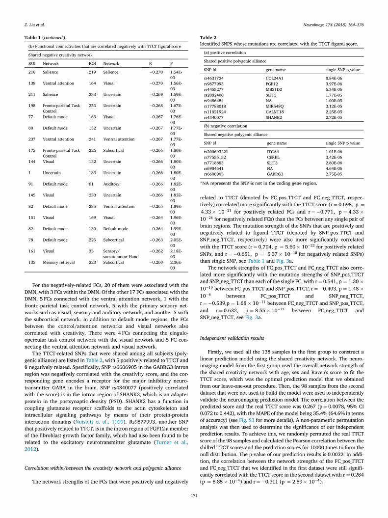
Table 1 (continued )
(b) Functional connectivities that are correlated negatively with TTCT figural score
Shared negative creativity network
ROI Network ROI Network R P
218 Salience 219 Salience �0.270 1.54E-03
138 Ventral attention 164 Visual �0.270 1.56E-03
211 Salience 253 Uncertain �0.269 1.59E-03
198 Fronto-parietal TaskControl
253 Uncertain �0.268 1.67E-03
77 Default mode 163 Visual �0.267 1.76E-03
80 Default mode 132 Uncertain �0.267 1.77E-03
237 Ventral attention 241 Ventral attention �0.267 1.77E-03
175 Fronto-parietal TaskControl
226 Subcortical �0.266 1.80E-03
144 Visual 132 Uncertain �0.266 1.80E-03
1 Uncertain 183 Uncertain �0.266 1.80E-03
91 Default mode 61 Auditory �0.266 1.82E-03
145 Visual 250 Uncertain �0.266 1.83E-03
82 Default mode 235 Ventral attention �0.265 1.89E-03
151 Visual 169 Visual �0.264 1.96E-03
82 Default mode 130 Default mode �0.264 1.99E-03
78 Default mode 225 Subcortical �0.263 2.05E-03
161 Visual 35 Sensory/somatomotor Hand
�0.262 2.18E-03
133 Memory retrieval 223 Subcortical �0.260 2.36E-03
Table 2Identified SNPS whose mutations are correlated with the TTCT figural score.
(a) positive correlation
Shared positive polygenic alliance
SNP id gene name single SNP p_value
rs4631724 COL24A1 8.84E-06rs9877993 FGF12 3.97E-06rs4455277 MB21D2 6.34E-06rs2082400 SLIT3 1.77E-05rs9486484 NA 1.00E-05rs17788018 MIR548Q 3.12E-05rs11021924 GALNT18 2.25E-05rs4340077 SHANK2 2.72E-05
(b) negative correlation
Shared negative polygenic alliance
SNP id gene name single SNP p_value
rs200693221 ITGA4 1.01E-06rs77555152 CERKL 3.42E-06rs7718883 SLIT3 2.80E-06rs6984541 NA 4.64E-06rs6606905 GABRG3 2.75E-05
*NA represents the SNP is not in the coding gene region.
Z. Liu et al. NeuroImage 174 (2018) 164–176
For the negatively-related FCs, 20 of them were associated with theDMN, with 3 FCs within the DMN. Of the other 17 FCs associatedwith theDMN, 5 FCs connected with the ventral attention network, 1 with thefronto-parietal task control network, 5 with the primary sensory net-works such as visual, sensory and auditory network, and another 5 withthe subcortical network. In addition to default mode regions, the FCsbetween the control/attention networks and visual networks alsocorrelated with creativity. There were 4 FCs connecting the cingulo-opercular task control network with the visual network and 5 FC con-necting the ventral attention network and visual network.
The TTCT-related SNPs that were shared among all subjects (poly-genic alliance) are listed in Table 2, with 5 positively related to TTCT and8 negatively related. Specifically, SNP rs6606905 in the GABRG3 intronregion was negatively correlated with the creativity score, and the cor-responding gene encodes a receptor for the major inhibitory neuro-transmitter GABA in the brain. SNP rs4340077 (positively correlatedwith the score) is in the intron region of SHANK2, which is an adapterprotein in the postsynaptic density (PSD). SHANK2 has a function incoupling glutamate receptor scaffolds to the actin cytoskeleton andintracellular signaling pathways by means of their protein-proteininteraction domains (Naisbitt et al., 1999). Rs9877993, another SNPthat positively related to TTCT, is in the intron region of FGF12 a memberof the fibroblast growth factor family, which had also been found to berelated to the excitatory neurotransmitter glutamate (Turner et al.,2012).
Correlation within/between the creativity network and polygenic alliance
The network strengths of the FCs that were positively and negatively
171
related to TTCT (denoted by FC_pos_TTCT and FC_neg_TTCT, respec-tively) correlated more significantly with the TTCT score (r¼ 0.698, p ¼4:33� 10�21 for positively related FCs and r¼�0.771, p ¼ 4:33�10�28 for negatively related FCs) than the FCs between any single pair ofbrain regions. The mutation strength of the SNPs that are positively andnegatively related to figural TTCT (denoted by SNP_pos_TTCT andSNP_neg_TTCT, respectively) were also more significantly correlatedwith the TTCT score (r¼ 0.704, p ¼ 5:60� 10�22 for positively relatedSNPs, and r¼�0.651, p ¼ 5:37� 10�18 for negatively related SNPs)than single SNP, see Table 1 and Fig. 3a.
The network strengths of FC_pos_TTCT and FC_neg_TTCT also corre-lated more significantly with the mutation strengths of SNP_pos_TTCTand SNP_neg_TTCT than each of the single FC, with r¼ 0.541, p ¼ 1:30�10�10 between FC_pos_TTCT and SNP_pos_TTCT, r¼�0.403, p ¼ 1:48�10�6 between FC_pos_TTCT and SNP_neg_TTCT,r¼�0.539,p ¼ 1:68� 10�11 between FC_neg_TTCT and SNP_pos_TTCT,and r¼ 0.632, p ¼ 8:55� 10�17 between FC_neg_TTCT andSNP_neg_TTCT, see Fig. 3a.
Independent validation results
Firstly, we used all the 138 samples in the first group to construct alinear prediction model using the shared creativity network. The neuro-imaging model from the first group used the overall network strength ofthe shared creativity network with age, sex and Raven's score to fit theTTCT score, which was the optimal prediction model that we obtainedfrom our leave-one-out procedure. Then, the 98 samples from the seconddataset that were not used to build the model were used to independentlyvalidate the neuroimaging prediction model. The correlation between thepredicted score and the real TTCT score was 0.267 (p¼ 0.0078, 95% CI0.072 to 0.442), with the MAPE of the model being 35.4% (64.6% in termsof accuracy) (see Fig. S3 for more details). A non-parametric permutationanalysis was then used to determine the significance of our independentprediction results. To achieve this, we randomly permuted the real TTCTscore of the 98 samples and calculated the Pearson correlation between theshifted TTCT scores and the prediction scores for 10000 times to form thenull distribution. The p-value of our prediction results is 0.0032. In addi-tion, the correlation between the network strengths of the FC_pos_TTCTand FC_neg_TTCT that we identified in the first dataset were still signifi-cantly correlated with the TTCT score in the second dataset with r¼ 0.284(p ¼ 8:85� 10�4) and r¼�0.311 (p ¼ 2:59� 10�4).

Fig. 3. Summary of the neural and genetic basis for figural creativity.(a) The neural and genetic basis for figural creativity. The shared positively/negatively-related FCs (with the TTCT score) are shown by red and blue on the left.Different sub-networks are represented by different colors in the left legend. The mutation of the shared positively-related/negatively-related (with the TTCT score)polygenic alliance are shown on the right, each row representing a SNP and each column represents an individual. Different colors denote different numbers of theminor allele. The correlations within/between the network strength of the shared positive/negative network and mutation strength of the shared positive/negativecreativity polygenic alliance are shown in (a), as well as their correlation with the TTCT figural score. (b) The creativity model we presented. The inner rectangleshows that both the increased top-down processing (in red arrows) and the decreasing bottom-up processing (in blue) relate to high creativity. The corresponding FCsrelevant for top-down/bottom-up processing are also shown. The outer rectangle demonstrates the genetic factors contributing to high creativity. The mutationstrength of the excitatory neurotransmitter related genes increases with creativity, and the mutation strength of the inhibitory neurotransmitter related genes de-creases with creativity. The competition between the two different kinds of neurotransmitters may influence the brain activity and further contribute to thehigh creativity.
Z. Liu et al. NeuroImage 174 (2018) 164–176
172

Z. Liu et al. NeuroImage 174 (2018) 164–176
Discussion
In this work, we use a cross-validation based predictive framework tosearch for the imaging and genetic correlates of creativity to providemore generalization than traditional correlation analyses. We found thatmodels using the network strength of the creativity network or the mu-tation strength of the creativity polygenic alliance could predict the in-dividual's performance with high accuracy, indicating that markers forcreativity are present in both the FC between brain regions and in thegenetic sequence. Further, the prediction model that combined neuro-imaging and genetic data achieved results significantly better than pre-diction with single modality data, indicating that complementaryinformation can be provided by different modalities, in this case func-tional neuroimaging and genetic modalities. Combining multi-modaldata helped us to obtain a fuller picture of the biological mechanismsunderlying creativity. We were able to validate our neuroimaging pre-diction model using a completely novel group containing 98 individuals.
We used the FCs obtained from the resting-state in our analysis, whichcan reflect patterns similar to those in cognitive task activation studies(Biswal et al., 1995; Smith et al., 2009; Tavor et al., 2016). The FCs in thecreativity-related brain networks associated with the DMN and areamong various higher level networks, such as the DMN, attentionnetwork and control network. These networks have all been implicated increative thought processes (Dietrich and Kanso, 2010; Fink et al., 2014a,2014b; Jung et al., 2010; Takeuchi et al., 2010, 2011). The DMN activityis associated with internally focused mental processes, such as mindwandering, perspective-taking, episodic future thinking and autobio-graphical retrieval (Andrews-Hanna et al., 2014). A growing number ofstudies reported that the DMN was recruited to generate novel ideas inboth domain-general and domain-specific creativity tasks (Liu et al.,2015b; Saggar et al., 2015). For example, individual differences in brainFC or structural characterizations within the DMN were able to predictindividual creativity (Chen et al., 2015; Takeuchi et al., 2012; Wei et al.,2014). Thus, the DMN activity contributes to the spontaneous generationof candidate ideas, which was considered as an initial and vital phaseduring creative thinking process. The control network and attentionnetworks, on the other hand, have been shown to be responsible for theevaluation of candidate ideas during creative cognitive processes (Die-trich, 2004; Dietrich and Kanso, 2010). As a further step, our findingshighlight the importance of the interactions among high level cognitivecontrol networks for creative thought processes. We found that the FCsbetween the DMN, control network and dorsal attention network/ventralattention network/memory retrieval network correlate positively withthe TTCT score. Our results suggest that creative thinking involves thefree generation of possible solutions as well as selection among theproduced alternatives (Campbell, 1960), that arise from the DMN andexecutive control networks, respectively (Dietrich A and R Kanso, 2010;Beaty RE et al., 2016). More importantly, these results provide supportfor the hypothesis that creative thought benefits from dynamic cooper-ation of the default mode and control networks to produce thoughts thatare both novel and appropriate. Thus, creative ideas depend onself-generated thought but also need control by a top-down goal-directedprocess (Beaty et al., 2016).
Our findings that the FCs between the DMN with the primary sensorynetworks (visual, auditory and sensory-motor) and ventral attentionnetworks are negatively correlated with the individuals' creativity score,indicate that high creativity individuals have the ability to inhibit con-spicuous task-relevant stimulation and shift toward task-irrelevantstimuli during creative thinking (Berkowitz and Ansari, 2010). This isin agreement with the finding that an individual's creativity level ispositively related to increased alpha power in the frontal cortex duringcreative ideation, which reflects more internally orientated attention thatis characterized by the absence of bottom-up stimulation (Fink andBenedek, 2014).
In addition to describing the creativity-related neural networks interms of the few networks used by Power et al., 2011), it is also useful to
173
consider in more detail the brain areas related to the creativity scoreusing a more detailed anatomical atlas such as a Brodmann parcellation(Jacobs, 2011). The MNI coordinates and Brodmann areas of the ROIs areshown in Table S1 for the functional connectivities with positive corre-lations with figural creativity, and in Table S2 for the functional con-nectivities with negative correlations with figural creativity.
First, we focus on the functional connectivities with positive corre-lations with figural creativity shown in Table S1 (see also Table 1). It isevident from Table S1 that many (11) of the 44 areas involved in FCrelated to figural creativity involved left area BA 40 (part of Wernicke'sarea), or right BA 45, 46 and 47 (contralateral to Broca's area). Alsorelated to this positive correlation with figural connectivity are the leftinferior prefrontal convexity/lateral orbitofrontal cortex areas, whichmay relate to processing in the nearby Broca's area. Increased FCs of someposterior cingulate areas (with e.g. temporal cortex areas) may reflect theinvolvement of these posterior cingulate areas in spatial processing(Kravitz et al., 2011) which is likely to be important for figural creativity.The findings described here are consistent with and thus supported by ameta-analysis of activations in 24 neuroimaging studies related to crea-tivity (Boccia et al., 2015), but the FC analyses described here take theanalysis an important step further, by providing evidence on which of thefunctional connectivities between these areas is related to figuralcreativity.
Second, we consider the functional connectivities with negative cor-relations with figural creativity shown in Table S2 (see also Table 1). Onebrain area with prominent negative correlations was the fusiform gyrus,with its functional connectivities with an inferior frontal gyrus/lateralorbitofrontal cortex region (Power et al. ROIs 175, 208, 211, and 198),with the anterior cingulate cortex (Power et al. ROIs 216), and with theputamen especially marked (Table S2). The fusiform gyrus has perceptualrepresentations, and the inferior frontal gyrus on the left includes part ofBroca's area and also non-reward/error systems (Rolls, 2016a, 2016b).Another brain area with prominent negative correlations with figuralcreativity was the auditory cortex/supramarginal gyrus (Power et al.ROIs 138) in especially its functional connectivities with secondary vi-sual cortical areas (Power et al. ROIs 164, 150, 173, 151, V3, V4 and V5).The reduced FCs between high order visual and auditory associationcortical areas that is associated with high figural creativity might suggestthat high figural creativity is associated with less visual/auditorybottom-up processing.
Finally, the right hemisphere has been shown to play a dominant rolein creative thinking (Bhattacharya and Petsche, 2005; Bowden andJung-Beeman, 2003; Falcone and Loder, 1984; Friedman and Forster,2005). It is interesting to note that although both within-hemisphere andbetween-hemisphere FCs correlate significantly with the figural crea-tivity score, the 4 FCs that connect ROIs in the task control network andattention network, and the FC between the cingular-opercular controlnetwork and the memory retrieval network, are all right-lateralized. Thissuggests that increased top-down control in the right hemisphere facili-tates a global thinking/context-dependent thinking style and figuralprocesses crucial for figural creativity (Mihov et al., 2010).
The creativity of the brain may be related, at least partly, to geneticand environmental interactions (Blunt, 2010). Although the geneticcauses of creativitymay be diverse, many heterogeneous genes have beenimplicated in creativity such as those affecting DA, 5HT or GABA meta-bolism (Blunt, 2010; Reuter et al., 2006), mostly belonging toneuro-transmitter systems. It has been shown that reduced task-induceddeactivation in the precuneus correlates with higher creativity in diver-gent thinking (Takeuchi et al., 2011). Considering also that individualswith a higher GABA/glutamate ratio tend to suppress ongoing neuralactivities of the precuneus more efficiently (Hu et al., 2013), these resultsindicate that the GABA/glutamate ratio may be closely related to thecreativity of a brain.
Our genetic results are concordant with these findings by showingthat mutations of genes related to both excitatory and inhibitory neu-rotransmitters are associated with creative thinking. We find that

Z. Liu et al. NeuroImage 174 (2018) 164–176
mutation of a SNP in GABRG3, the receptor for GABA, is negativelyrelated to the figural TTCT score, while several SNPs in genes related toglutamate (SHANK2, FGF12) correlate positively with the figural TTCTscore. The mutation of these SNPs relevant for neurotransmitter systemsmay disturb the balance between the two different kinds of neurotrans-mitter, i.e. glutamate and GABA. Since these mutations are found to besignificantly associated with both the creativity-related network and thefigural TTCT score, we therefore postulate that this difference in theglutamate and GABA transmitter may have an impact on the FCs asso-ciated with regions (including the precuneus) of the DMN, control, andattention networks. Moreover, it is interesting to note that among all the13 SNPs we found to be related to the figural TTCT score, 5 are associatedwith genes (ITGA4, SHANK2, MIR548, GABRG3 and FGF12) that havebeen reported to be relevant to autism (Ander et al., 2015; Buxbaumet al., 2002; Correia et al., 2009; Leblond et al., 2012; Vaccarino et al.,2009; Won et al., 2012). Autistic traits have been demonstrated to beassociated with high numbers of unusual responses on the divergentthinking tasks (Best et al., 2015).
The creativity-related functional brain networks and the genetic al-liances that we used to predict the creativity score in the first datasetcontained many different FCs and SNPs. Although we confirmed that thesum of all the FCs (strength of the networks) were significantly correlatedwith creativity score in all the two dataset and the FCs or SNPs as a whole(i.e., their sum) could predict the creativity score well, it should be notedthat not every FC/SNP in the brain networks/genetic alliances wasassociated with creativity score significantly (i.e., survived from correc-tion for multiple comparisons).
In our feature selection and prediction procedures, we took into ac-count factors that might have affected our prediction models. Forexample, for head motion, which could be a potential confounding factorfor neuroimaging data (Liu et al., 2015a; Yan et al., 2013; Zuo et al.,2013), we performed two further motion control procedures in the 138sample group to confirm that it did not confound our prediction models.Firstly, the correlation between the mean framewise displacement (FD)with the creativity score and our predicted score were calculated. Wefound that the mean FD did not correlate with the real TTCT score or thepredicted score (r¼ 0.116, p¼ 0.157 and r¼ 0.114 and p¼ 0.183).Secondly, we used the brain network with the mean FD regressed out tobuild a new prediction model using the network strength of thecreativity-related functional brain network. The correlation between thepredicted score (r¼ 0.420) and the prediction accuracy (72.7%) did notdiffer significantly from the results provided in the Results section.
The relationship between the individuals' TTCT figural creativity andtheir age and Raven's score were also analyzed. The procedure was to useage, sex and the Raven's score as covariates in the regression equation( Ck in eq. (1) above) that estimated the creativity score from the FCs.This ensured that the prediction was from the FCs with the effects of age,sex and the Raven's score regressed out. We found that the figural crea-tivity score was significantly though weakly (p¼ 0.037) negativelyrelated to age (from ~18 to ~22) (see Fig. S4). However, the Raven'sscore and sex were not significantly related to the figural creativity score,indicating that the figural creativity prediction is not confounded byindividuals' gender or general intelligence (see Fig. S5). In the regressionanalysis, the coefficients for the neuroimaging data (part A) show that thecoefficient for the average network strength made a contribution to theprediction that was significant at p¼ 6.19� 10�13, and that the co-efficients for age, gender, and the Raven's score made no significantcontribution to the prediction. In the Supplementary Material, in theprediction model from the genetic data (part B), there is a negative co-efficient for the contribution of age that is significant (p¼ 1.92� 10�5),and this is of interest.
In conclusion, we have for the first time investigated both the neuraland genetic correlates of figural creativity using whole-brain FCs and thewhole-genome. We find that we can predict figural connectivity signifi-cantly better from the FCs and the genome (78.4%) than from the FCsalone (76.1%) or the genetic data alone (77.5%). The FCs positively
174
related to creativity were associated with the “control networks” and“attention networks” (as identified by Power et al., 2011) typical oftop-down processes, while the FCs negatively related to creativity werebetween the DMN and the primary sensory networks that are associatedwith bottom-up processes. The implication is that figural creativity isassociated with strong top-down control versus weak bottom-up processin the resting state. The genes associated with figural connectivity areinvolved in glutamate and GABA functionality, and an implication is thatthe ratio of excitatory to inhibitory synaptic functionmay be important infigural creativity. The results described here provides the first insight intohow a combination of neural and gene-related factors are related tocreative thinking. The brain areas related to figural creativity includedincreased visual association cortex connectivity with inferior frontalgyrus areas related to Broca's area, and with Wernicke's area, which mayrelate to enhanced visual imagery in those with high figural creativity.
Funding
J. Feng is partially supported by the key project of Shanghai Science&Technology Innovation Plan (No. 15JC1400101 and No. 16JC1420402)and the National Natural Science Foundation of China (Grant No.71661167002 and No. 91630314). There search was also partially sup-ported by the Shanghai AI Platform for Diagnosis and Treatment of BrainDiseases (No. 2016-17). The research was also partially supported byBase for Introducing Talents of Discipline to Universities (No. B18015).Z. Liu and JY. Zhang are supported by National Science Foundation ofChina (NSFC 11674352 and 61571341), and the Fundamental ResearchFunds for the Central Universities of China (No. JBZ170301). J. Zhang issupported by National Science Foundation of China (NSFC 61573107),and special Funds for Major State Basic Research Projects of China (No.2015CB856003). J. Qiu was supported by the National Natural ScienceFoundation of China (NSFC 31571137 and 31771231), Project of theNational Defense Science and Technology Innovation Special Zone,Chang Jiang Scholars Program, National Outstanding Young People Plan,the Program for the Top Young Talents by Chongqing, Natural ScienceFoundation of Chongqing (No. cstc2015jcyjA10106), Fok Ying TungEducation Foundation (No. 151023), the Research Program Funds of theCollaborative Innovation Center of Assessment toward Basic EducationQuality at Beijing Normal University.
Acknowledgements
We would like to thank Prof. Keith M. Kendrick, Prof. Fengzhu Sun,and Jessie Liu for their helpful suggestions.
Appendix A. Supplementary data
Supplementary data related to this article can be found at https://doi.org/10.1016/j.neuroimage.2018.02.067.
References
Ander, B.P., Barger, N., Stamova, B., Sharp, F.R., Schumann, C.M., 2015. Atypical miRNAexpression in temporal cortex associated with dysregulation of immune, cell cycle,and other pathways in autism spectrum disorders. Mol. Autism 6, 37.
Andrews-Hanna, J.R., Smallwood, J., Spreng, R.N., 2014. The default network and self-generated thought: component processes, dynamic control, and clinical relevance.Ann. N. Y. Acad. Sci. 1316, 29–52.
Beaty, R.E., Benedek, M., Silvia, P.J., Schacter, D.L., 2016. Creative cognition and brainnetwork dynamics. Trends Cogn. Sci. 20, 87–95.
Beaty, R.E., Benedek, M., Wilkins, R.W., Jauk, E., Fink, A., Silvia, P.J., Hodges, D.A.,Koschutnig, K., Neubauer, A.C., 2014. Creativity and the default network: afunctional connectivity analysis of the creative brain at rest. Neuropsychologia 64,92–98.
Berkowitz, A.L., Ansari, D., 2010. Expertise-related deactivation of the righttemporoparietal junction during musical improvisation. Neuroimage 49, 712–719.
Best, C., Arora, S., Porter, F., Doherty, M., 2015. The relationship between subthresholdautistic traits, ambiguous figure perception and divergent thinking. J. Autism Dev.Disord. 45, 4064–4073.

Z. Liu et al. NeuroImage 174 (2018) 164–176
Bhattacharya, J., Petsche, H., 2005. Drawing on mind's canvas: differences in corticalintegration patterns between artists and non-artists. Hum. Brain Mapp. 26, 1–14.
Biswal, B., Yetkin, F.Z., Haughton, V.M., Hyde, J.S., 1995. Functional connectivity in themotor cortex of resting human brain using echo-planar MRI. Magn. Reson Med. 34,537–541.
Blunt, S., 2010. The creative brain: Fundamental features, associated conditions andunifying neural mechanisms. Neurol. music 31.
Boccia, M., Piccardi, L., Palermo, L., Nori, R., Palmiero, M., 2015. Where do bright ideasoccur in our brain? Meta-analytic evidence from neuroimaging studies of domain-specific creativity. Front. Psychol. 6, 1195.
Bowden, E.M., Jung-Beeman, M., 2003. Aha! Insight experience correlates with solutionactivation in the right hemisphere. Psychon. Bull. Rev. 10, 730–737.
Bullmore, E., Sporns, O., 2009. Complex brain networks: graph theoretical analysis ofstructural and functional systems. Nat. Rev. Neurosci. 10, 186.
Buxbaum, J., Silverman, J., Smith, C., Greenberg, D., Kilifarski, M., Reichert, J.,Cook Jr., E., Fang, Y., Song, C., Vitale, R., 2002. Association between a GABRB3polymorphism and autism. Mol. psychiatry 7, 311–316.
Campbell, D.T., 1960. Blind variation and selective retentions in creative thought as inother knowledge processes. Psychol. Rev. 67, 380–400.
Chao-Gan, Y., Yu-Feng, Z., 2010. DPARSF: a MATLAB toolbox for “pipeline” data analysisof resting-state fMRI. Front. Syst. Neurosci. 4, 13.
Chavez-Eakle, R.A., Graff-Guerrero, A., Garcia-Reyna, J.C., Vaugier, V., Cruz-Fuentes, C.,2007. Cerebral blood flow associated with creative performance: a comparativestudy. Neuroimage 38, 519–528.
Chen, Q.L., Xu, T., Yang, W.J., Li, Y.D., Sun, J.Z., Wang, K.C., Beaty, R.E., Zhang, Q.L.,Zuo, X.N., Qiu, J., 2015. Individual differences in verbal creative thinking arereflected in the precuneus. Neuropsychologia 75, 441–449.
Correia, C., Coutinho, A.M., Almeida, J., Lontro, R., Lobo, C., Miguel, T.S., Martins, M.,Gallagher, L., Conroy, J., Gill, M., Oliveira, G., Vicente, A.M., 2009. Association of thealpha4 integrin subunit gene (ITGA4) with autism. Am. J. Med. Genet. BNeuropsychiatr. Genet. 150B, 1147–1151.
de Manzano, O., Cervenka, S., Karabanov, A., Farde, L., Ullen, F., 2010. Thinking outsidea less intact box: thalamic dopamine D2 receptor densities are negatively related topsychometric creativity in healthy individuals. PLoS One 5 e10670.
de Souza, L.C., Volle, E., Bertoux, M., Czernecki, V., Funkiewiez, A., Allali, G., Leroy, B.,Sarazin, M., Habert, M.-O., Dubois, B., 2010. Poor creativity in frontotemporaldementia: a window into the neural bases of the creative mind. Neuropsychologia 48,3733–3742.
Dietrich, A., 2004. The cognitive neuroscience of creativity. Psychon. Bull. Rev. 11,1011–1026.
Dietrich, A., Kanso, R., 2010. A review of EEG, ERP, and neuroimaging studies ofcreativity and insight. Psychol. Bull. 136, 822–848.
Dubois, J., Adolphs, R., 2016. Building a science of individual differences from fMRI.Trends Cogn. Sci. 20, 425–443.
Ellamil, M., Dobson, C., Beeman, M., Christoff, K., 2012. Evaluative and generative modesof thought during the creative process. Neuroimage 59, 1783–1794.
Falcone, D.J., Loder, K., 1984. A modified lateral eye-movement measure, the righthemisphere and creativity. Percept. Mot. Ski. 58, 823–830.
Fink, A., Benedek, M., 2014. EEG alpha power and creative ideation. Neurosci. BiobehavRev. 44, 111–123.
Fink, A., Koschutnig, K., Hutterer, L., Steiner, E., Benedek, M., Weber, B., Reishofer, G.,Papousek, I., Weiss, E.M., 2014a. Gray matter density in relation to different facets ofverbal creativity. Brain Struct. Funct. 219, 1263–1269.
Fink, A., Weber, B., Koschutnig, K., Mathias, B., Reishofer, G., Ebner, F., Papousek, I.,Weiss, E.M., 2014b. Creativity and schizotypy from the neuroscience perspective.Cogn. Affect Behav. Neurosci. 14, 378–387.
Flaherty, A.W., 2005. Frontotemporal and dopaminergic control of idea generation andcreative drive. J. Comp. Neurol. 493, 147–153.
Fox, M.D., Snyder, A.Z., Vincent, J.L., Corbetta, M., Van Essen, D.C., Raichle, M.E., 2005.The human brain is intrinsically organized into dynamic, anticorrelated functionalnetworks. Proc. Natl. Acad. Sci. U S A 102, 9673.
Friedman, R.S., Forster, J., 2005. Effects of motivational cues on perceptual asymmetry:implications for creativity and analytical problem solving. J. Pers. Soc. Psychol. 88,263–275.
Friston, K.J., Williams, S., Howard, R., Frackowiak, R.S., Turner, R., 1996. Movement-related effects in fMRI time-series. Magn. Reson Med. 35, 346–355.
Gardner, H., 1988. Creativity: an interdisciplinary perspective. Creat. Res. J. 1, 8–26.Gonen-Yaacovi, G., de Souza, L.C., Levy, R., Urbanski, M., Josse, G., Volle, E., 2013.
Rostral and caudal prefrontal contribution to creativity: a meta-analysis of functionalimaging data. Front. Hum. Neurosci. 7, 10, 3389.
Heausler, N.L., Thompson, B., 1988. Structure of the torrance tests of creative thinking.Educ. Psychol. Meas. 48, 463–468.
Heilman, K.M., Nadeau, S.E., Beversdorf, D.O., 2003. Creative innovation: possible brainmechanisms. Neurocase 9, 369–379.
Hu, Y., Chen, X., Gu, H., Yang, Y., 2013. Resting-state glutamate and GABAconcentrations predict task-induced deactivation in the default mode network.J. Neurosci. 33, 18566–18573.
Huang, P., Qiu, L., Shen, L., Zhang, Y., Song, Z., Qi, Z., Gong, Q., Xie, P., 2013. Evidencefor a left-over-right inhibitory mechanism during figural creative thinking in healthynonartists. Hum. Brain Mapp. 34, 2724–2732.
Jacobs, K.M., 2011. Brodmann's areas of the cortex. In: Kreutzer, J.S., DeLuca, J.,Caplan, B. (Eds.), Encyclopedia of Clinical Neuropsychology. Springer New York,New York, NY, p. 459.
Jaeggi, S.M., Buschkuehl, M., Jonides, J., Perrig, W.J., 2008. Improving fluid intelligencewith training on working memory. Proc. Natl. Acad. Sci. U. S. A. 105, 6829–6833.
175
Jauk, E., Neubauer, A.C., Dunst, B., Fink, A., Benedek, M., 2015. Gray matter correlates ofcreative potential: a latent variable voxel-based morphometry study. Neuroimage111, 312–320.
Jung, R.E., Segall, J.M., Jeremy Bockholt, H., Flores, R.A., Smith, S.M., Chavez, R.S.,Haier, R.J., 2010. Neuroanatomy of creativity. Hum. Brain Mapp. 31, 398–409.
Keri, S., 2009. Genes for psychosis and creativity: a promoter polymorphism of theneuregulin 1 gene is related to creativity in people with high intellectualachievement. Psychol. Sci. 20, 1070–1073.
Kim, K.H., 2006. Can we trust creativity tests? a review of the torrance tests of creativethinking (TTCT). Creativ. Res. J. 18, 3–14.
Kravitz, D.J., Saleem, K.S., Baker, C.I., Mishkin, M., 2011. A new neural framework forvisuospatial processing. Nat. Rev. Neurosci. 12, 217–230.
Kyaga, S., Lichtenstein, P., Boman, M., Hultman, C., Langstrom, N., Landen, M., 2011.Creativity and mental disorder: family study of 300,000 people with severe mentaldisorder. Br. J. Psychiatry 199, 373–379.
Leblond, C.S., Heinrich, J., Delorme, R., Proepper, C., Betancur, C., Huguet, G.,Konyukh, M., Chaste, P., Ey, E., Rastam, M., Anckarsater, H., Nygren, G.,Gillberg, I.C., Melke, J., Toro, R., Regnault, B., Fauchereau, F., Mercati, O.,Lemiere, N., Skuse, D., Poot, M., Holt, R., Monaco, A.P., Jarvela, I., Kantojarvi, K.,Vanhala, R., Curran, S., Collier, D.A., Bolton, P., Chiocchetti, A., Klauck, S.M.,Poustka, F., Freitag, C.M., Waltes, R., Kopp, M., Duketis, E., Bacchelli, E.,Minopoli, F., Ruta, L., Battaglia, A., Mazzone, L., Maestrini, E., Sequeira, A.F.,Oliveira, B., Vicente, A., Oliveira, G., Pinto, D., Scherer, S.W., Zelenika, D.,Delepine, M., Lathrop, M., Bonneau, D., Guinchat, V., Devillard, F., Assouline, B.,Mouren, M.C., Leboyer, M., Gillberg, C., Boeckers, T.M., Bourgeron, T., 2012. Geneticand functional analyses of SHANK2 mutations suggest a multiple hit model of autismspectrum disorders. PLoS Genet. 8 e1002521.
Li, W., Yang, J., Zhang, Q., Li, G., Qiu, J., 2016. The association between restingfunctional connectivity and visual creativity. Sci. Rep. 6, 25395.
Liu, F., Guo, W., Fouche, J.P., Wang, Y., Wang, W., Ding, J., Zeng, L., Qiu, C., Gong, Q.,Zhang, W., Chen, H., 2015a. Multivariate classification of social anxiety disorderusing whole brain functional connectivity. Brain Struct. Funct. 220, 101–115.
Liu, S., Erkkinen, M.G., Healey, M.L., Xu, Y., Swett, K.E., Chow, H.M., Braun, A.R., 2015b.Brain activity and connectivity during poetry composition: toward amultidimensional model of the creative process. Hum. Brain Mapp. 36, 3351–3372.
Mayseless, N., Uzefovsky, F., Shalev, I., Ebstein, R.P., Shamay-Tsoory, S.G., 2013. Theassociation between creativity and 7R polymorphism in the dopamine receptor D4gene (DRD4). Front. Hum. Neurosci. 7, 502.
Mihov, K.M., Denzler, M., Forster, J., 2010. Hemispheric specialization and creativethinking: a meta-analytic review of lateralization of creativity. Brain Cogn. 72,442–448.
Naisbitt, S., Kim, E., Tu, J.C., Xiao, B., Sala, C., Valtschanoff, J., Weinberg, R.J.,Worley, P.F., Sheng, M., 1999. Shank, a novel family of postsynaptic density proteinsthat binds to the NMDA receptor/PSD-95/GKAP complex and cortactin. Neuron 23,569–582.
Plucker, J.A., Runco, M.A., 1998. The death of creativity measurement has been greatlyexaggerated: current issues, recent advances, and future directions in creativityassessment. Roeper Rev. 21, 36–39.
Power, J.D., Cohen, A.L., Nelson, S.M., Wig, G.S., Barnes, K.A., Church, J.A., Vogel, A.C.,Laumann, T.O., Miezin, F.M., Schlaggar, B.L., Petersen, S.E., 2011. Functionalnetwork organization of the human brain. Neuron 72, 665–678.
Power, J.D., Mitra, A., Laumann, T.O., Snyder, A.Z., Schlaggar, B.L., Petersen, S.E., 2014.Methods to detect, characterize, and remove motion artifact in resting state fMRI.Neuroimage 84, 320–341.
Power, R.A., Steinberg, S., Bjornsdottir, G., Rietveld, C.A., Abdellaoui, A., Nivard, M.M.,Johannesson, M., Galesloot, T.E., Hottenga, J.J., Willemsen, G., Cesarini, D.,Benjamin, D.J., Magnusson, P.K., Ullen, F., Tiemeier, H., Hofman, A., van Rooij, F.J.,Walters, G.B., Sigurdsson, E., Thorgeirsson, T.E., Ingason, A., Helgason, A., Kong, A.,Kiemeney, L.A., Koellinger, P., Boomsma, D.I., Gudbjartsson, D., Stefansson, H.,Stefansson, K., 2015. Polygenic risk scores for schizophrenia and bipolar disorderpredict creativity. Nat. Neurosci. 18, 953–955.
Reuter, M., Roth, S., Holve, K., Hennig, J., 2006. Identification of first candidate genes forcreativity: a pilot study. Brain Res. 1069, 190–197.
Rolls, E.T., 2016a. Cerebral Cortex: Principles of Operation.Rolls, E.T., 2016b. A non-reward attractor theory of depression. Neurosci. Biobehav Rev.
68, 47–58.Rosenberg, M.D., Finn, E.S., Scheinost, D., Papademetris, X., Shen, X., Constable, R.T.,
Chun, M.M., 2016. A neuromarker of sustained attention from whole-brain functionalconnectivity. Nat. Neurosci. 19, 165–171.
Saggar, M., Quintin, E.M., Kienitz, E., Bott, N.T., Sun, Z., Hong, W.C., Chien, Y.H., Liu, N.,Dougherty, R.F., Royalty, A., Hawthorne, G., Reiss, A.L., 2015. Pictionary-based fMRIparadigm to study the neural correlates of spontaneous improvisation and figuralcreativity. Sci. Rep. 5, 10894.
Shen, X.L., Finn, E.S., Scheinost, D., Rosenberg, M.D., Chun, M.M., Papademetris, X.,Constable, R.T., 2017. Using connectome-based predictive modeling to predictindividual behavior from brain connectivity. Nat. Protoc. 12, 506–518.
Smith, S.M., Fox, P.T., Miller, K.L., Glahn, D.C., Fox, P.M., Mackay, C.E., Filippini, N.,Watkins, K.E., Toro, R., Laird, A.R., Beckmann, C.F., 2009. Correspondence of thebrain's functional architecture during activation and rest. Proc. Natl. Acad. Sci. U. S.A. 106, 13040–13045.
Sternberg, R.J., Lubart, T.I., 1996. Investing in creativity. Am. Psychol. 51, 677.Sternberg, R.J., O'HARA, L.A., 1999. 13 Creativity and Intelligence. Handbook of
Creativity, vol. 251.Takeuchi, H., Taki, Y., Hashizume, H., Sassa, Y., Nagase, T., Nouchi, R., Kawashima, R.,
2011. Failing to deactivate: the association between brain activity during a workingmemory task and creativity. Neuroimage 55, 681–687.
Z. Liu et al. NeuroImage 174 (2018) 164–176
Takeuchi, H., Taki, Y., Hashizume, H., Sassa, Y., Nagase, T., Nouchi, R., Kawashima, R.,2012. The association between resting functional connectivity and creativity. Cereb.Cortex 22, 2921–2929.
Takeuchi, H., Taki, Y., Sassa, Y., Hashizume, H., Sekiguchi, A., Fukushima, A.,Kawashima, R., 2010. Regional gray matter volume of dopaminergic system associatewith creativity: evidence from voxel-based morphometry. Neuroimage 51, 578–585.
Tang, C., Li, A., Huang, H., Cheng, X., Gao, Y., Chen, H., Huang, Q., Luo, Y., Xue, Y.,Zuo, Q., 2012. Effects of lead pollution in SY River on children's intelligence. Life Sci.J. 9, 458–464.
Tavor, I., Parker Jones, O., Mars, R.B., Smith, S.M., Behrens, T.E., Jbabdi, S., 2016. Task-free MRI predicts individual differences in brain activity during task performance.Science 352, 216–220.
Turner, C.A., Watson, S.J., Akil, H., 2012. The fibroblast growth factor family:neuromodulation of affective behavior. Neuron 76, 160–174.
Vaccarino, F.M., Grigorenko, E.L., Smith, K.M., Stevens, H.E., 2009. Regulation ofcerebral cortical size and neuron number by fibroblast growth factors: implicationsfor autism. J. Autism Dev. Disord. 39, 511–520.
Van den Heuvel, M.P., Hulshoff Pol, H.E., 2010. Exploring the brain network: a review onresting-state fMRI functional connectivity. Eur. Neuropsychopharmacol. 20,519–534.
Wei, D., Yang, J., Li, W., Wang, K., Zhang, Q., Qiu, J., 2014. Increased resting functionalconnectivity of the medial prefrontal cortex in creativity by means of cognitivestimulation. Cortex 51, 92–102.
176
Won, H., Lee, H.R., Gee, H.Y., Mah, W., Kim, J.I., Lee, J., Ha, S., Chung, C., Jung, E.S.,Cho, Y.S., Park, S.G., Lee, J.S., Lee, K., Kim, D., Bae, Y.C., Kaang, B.K., Lee, M.G.,Kim, E., 2012. Autistic-like social behaviour in Shank2-mutant mice improved byrestoring NMDA receptor function. Nature 486, 261–265.
Xia, M., He, Y., 2017. Functional connectomics from a “big data” perspective. Neuroimage160, 152–167.
Yan, C.G., Cheung, B., Kelly, C., Colcombe, S., Craddock, R.C., Di Martino, A., Li, Q.,Zuo, X.N., Castellanos, F.X., Milham, M.P., 2013. A comprehensive assessment ofregional variation in the impact of head micromovements on functionalconnectomics. Neuroimage 76, 183–201.
Zabelina, D.L., Colzato, L., Beeman, M., Hommel, B., 2016. Dopamine and the creativemind: individual differences in creativity are predicted by interactions betweendopamine genes DAT and COMT. PLoS One 11 e0146768.
Zhu, W.F., Chen, Q.L., Xia, L.X., Beaty, R.E., Yang, W.J., Tian, F., Sun, J.Z., Cao, G.K.,Zhang, Q.L., Chen, X., Qiu, J., 2017. Common and distinct brain networks underlyingverbal and visual creativity. Hum. Brain Mapp. 38, 2094–2111.
Zou, H., Hastie, T., 2005. Regularization and variable selection via the elastic net. J. R.Stat. Soc. Ser. B Stat. Methodol. 67, 301–320.
Zuo, X.N., Xu, T., Jiang, L., Yang, Z., Cao, X.Y., He, Y., Zang, Y.F., Castellanos, F.X.,Milham, M.P., 2013. Toward reliable characterization of functional homogeneity inthe human brain: preprocessing, scan duration, imaging resolution andcomputational space. Neuroimage 65, 374–386.