Embed Size (px)
Citation preview
Dr. Poonam RaniZoology DepartmentB.Sc. Life Sciences 1 YearRoom No. A-210, A-214
Comparative Account of Nervous System of Vertebrates
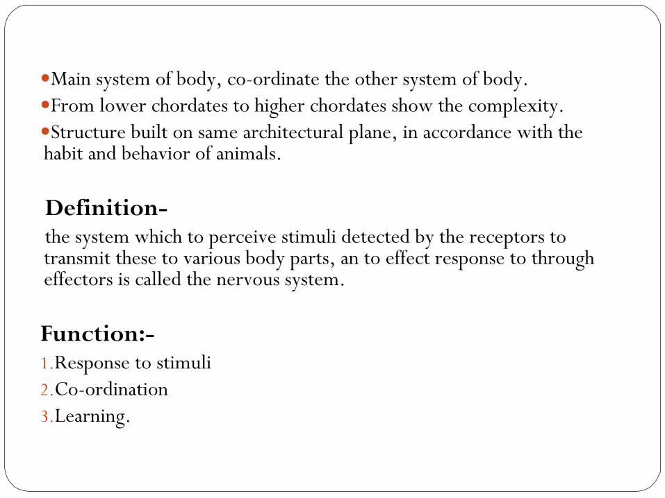
Main system of body, co-ordinate the other system of body.From lower chordates to higher chordates show the complexity.Structure built on same architectural plane, in accordance with the habit and behavior of animals.
Definition- the system which to perceive stimuli detected by the receptors to transmit these to various body parts, an to effect response to through effectors is called the nervous system.
Function:-1.Response to stimuli2.Co-ordination3.Learning.
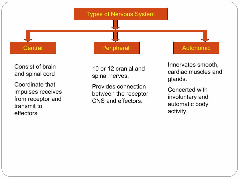
Types of Nervous System
Central AutonomicPeripheral
Consist of brain and spinal cord
Coordinate that impulses receives from receptor and transmit to effectors
10 or 12 cranial and spinal nerves.
Provides connection between the receptor, CNS and effectors.
Innervates smooth, cardiac muscles and glands.
Concerted with involuntary and automatic body activity.
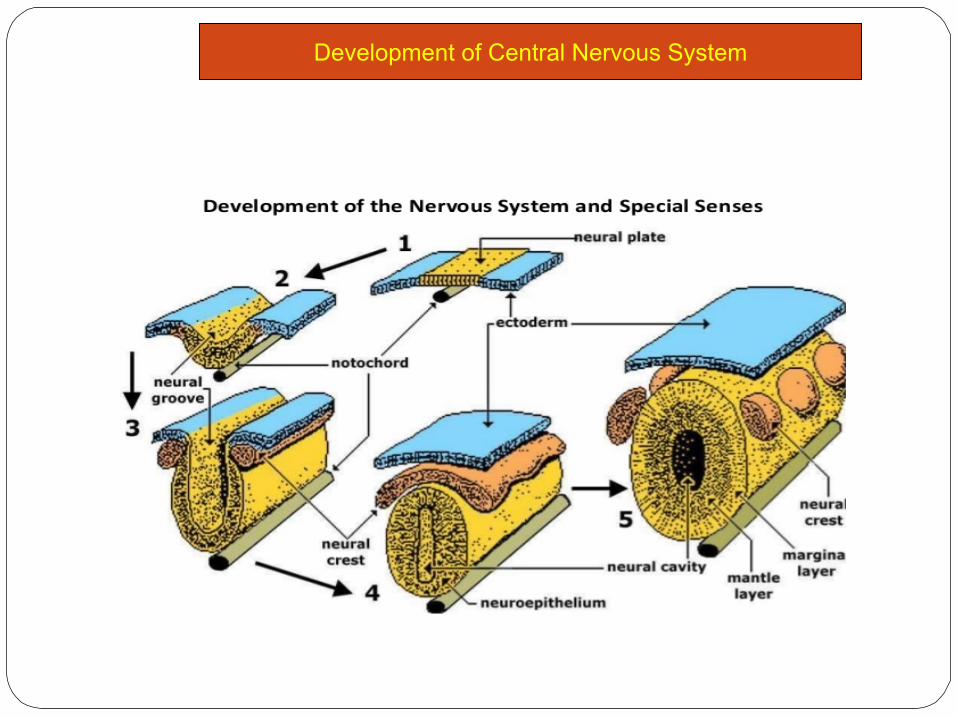
Development of Central Nervous System
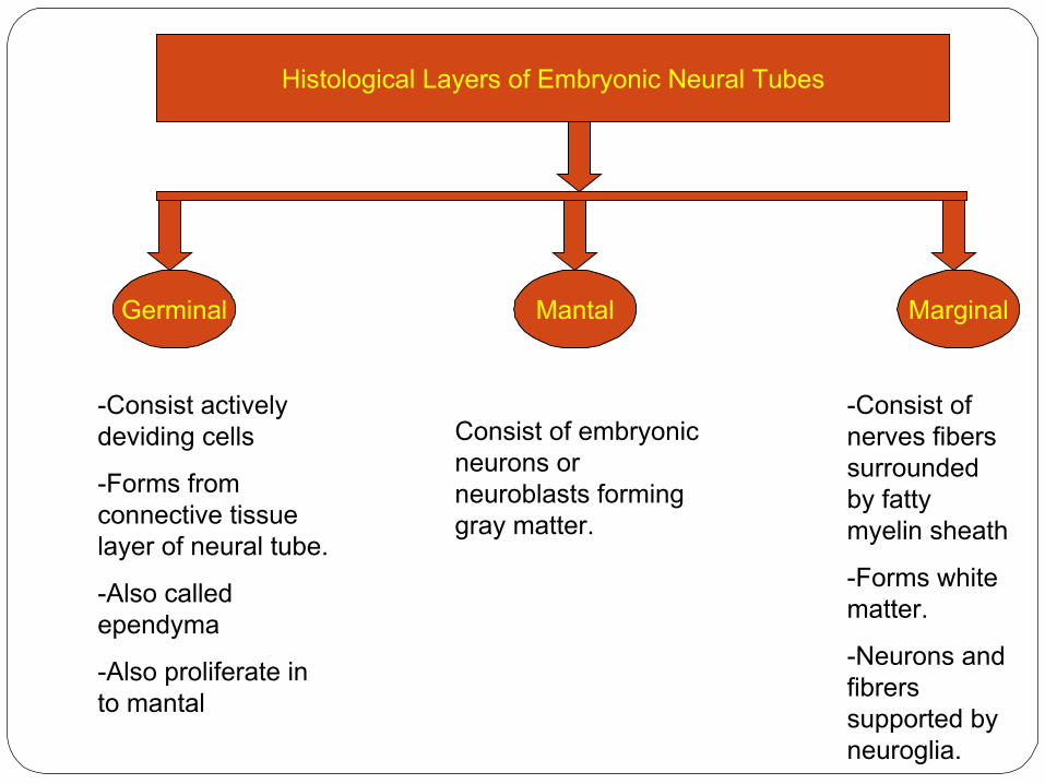
Histological Layers of Embryonic Neural Tubes
Germinal MarginalMantal
-Consist actively deviding cells
-Forms from connective tissue layer of neural tube.
-Also called ependyma
-Also proliferate in to mantal
Consist of embryonic neurons or neuroblasts forming gray matter.
-Consist of nerves fibers surrounded by fatty myelin sheath
-Forms white matter.
-Neurons and fibrers supported by neuroglia.
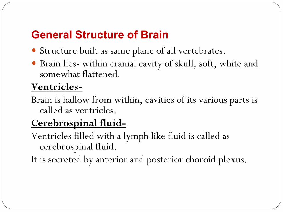
General Structure of Brain Structure built as same plane of all vertebrates. Brain lies- within cranial cavity of skull, soft, white and
somewhat flattened.Ventricles- Brain is hallow from within, cavities of its various parts is
called as ventricles.Cerebrospinal fluid-Ventricles filled with a lymph like fluid is called as
cerebrospinal fluid.It is secreted by anterior and posterior choroid plexus.
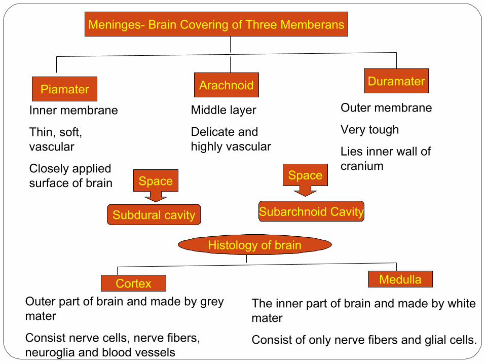
Meninges- Brain Covering of Three Memberans
Piamater Arachnoid Duramater
Inner membrane
Thin, soft, vascular
Closely applied surface of brain
Outer membrane
Very tough
Lies inner wall of cranium
Middle layer
Delicate and highly vascular
Space Space
Subdural cavity Subarchnoid Cavity
Histology of brain
Cortex Medulla
Outer part of brain and made by grey mater
Consist nerve cells, nerve fibers, neuroglia and blood vessels
The inner part of brain and made by white mater
Consist of only nerve fibers and glial cells.

rhinocoel
paracoel
iter
metacoel
Parts of brains1.forebrain2.midbrain3.hindbrain

Prosencephalon(forebrain)
Olphactory lobe DiencephalonCerebral hemisphere
Antriormost, paired ,small cup shaped, separate each other.
Continous beneth the frontal lobe of cerebrum as paired olfactory lobes connect hipocample lobe.
Cavity- frist venbtrical-rhinocoel
Function- sense of smell.
Paired separated by longitudinal median fissure.
Narrow in front and broad in behind and smooth.
Form 2/3 of brain and overlap to diancephalon.
Lateral sylvain fissure devides frontal and temporal lobe
Ventrally rinal fissure devides from olfactory lobs shows hippocample lobe.
Two hemisphere internally connect by band of nerve tissue called corpus collasum
Ventrical- second-paracoel and two paracoel connect form foramen of monro.
Function- though, memory, intellance, reasoing
along with the telencephalon (cerebrum)
Main structures of the diencephalon include the hypothalamus, thalamus, epithalamus (including the pineal gland), and subthalamus.
The diencephalon relays sensory information between brain regions and controls many autonomic functions of the peripheral nervous system.
Function-Directing Sense Impulses Autonomic Function ControlEndocrine Function ControlMotor Function ControlHomeostasisTouch Perception
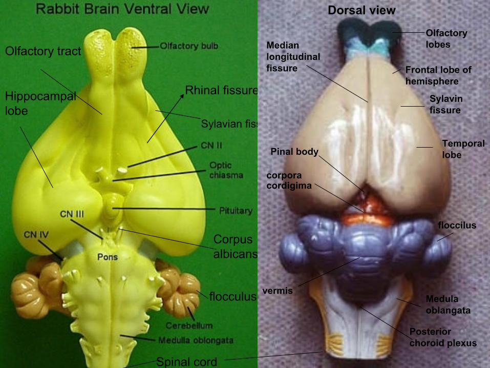
Rhinal fissure
Sylavian fissure
Hippocampal lobe
Olfactory tract
Corpus albicans
Spinal cord
flocculus
Olfactory lobes
Frontal lobe of hemisphere
Sylavin fissure
Temporal lobe
floccilus
vermisMedula oblangata
Posterior choroid plexus
Pinal body
corpora cordigima
Median longitudinal fissure
Dorsal view

Mesencephalon or midbrain Small middle part of brain, lies below the cerebral hemisphere. Dorsal surface has 4 rounded optic lobes called corpora cordigima. The anterior two lobes called as superior colliculi and concer with
the slight. The posterior two lobes are smaller called inferior colliculi and
associated with acute hearing. Cavity- narrow longitudinal passage-iter Its floor is thick fibres called crura cerebri, which link forebrain
and hindbrain. Function- slight and acute hearing.

Hindbrain or Rhombencephalon
Cerebellum Medula OblongataProns Varolii
Very well developed and transverse elongated.
Consist large median lobe called vermis and lateral lobe called as flocculus.
No cavity surface is much folded to increase the grey matter.
Surface folding forms elevation-gyri and grooves- sulci.
Function- equilibrium and co-ordination of voluntory muscles.
Ventral surface of hindbrain has stout transverse bands of fibers
Its connect right and left halves of the cerebrellum named prons varolli.
Last part of brain
Broad triangular anterior but taper in posterior
Continous to spinal cord.
Cavity- forth-metacoel
The roof of metacoel is non-nervous and vascular called the posterior choroid plexus.
Function- control the involuntary actions.
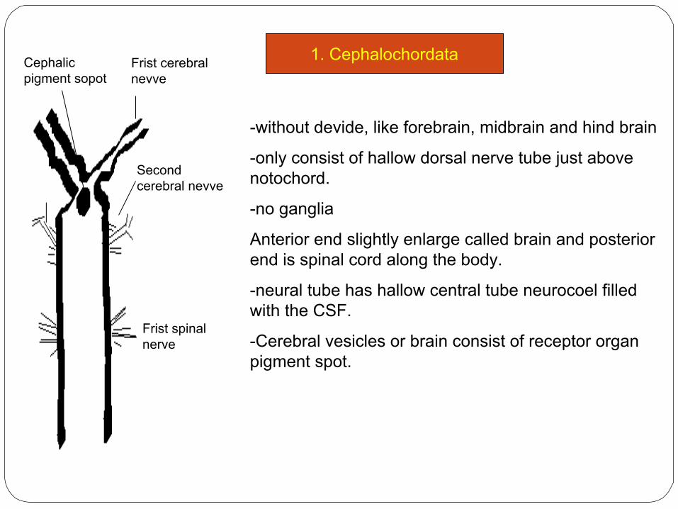
1. CephalochordataFrist cerebral nevve
Second cerebral nevve
Cephalic pigment sopot
Frist spinal nerve
-without devide, like forebrain, midbrain and hind brain
-only consist of hallow dorsal nerve tube just above notochord.
-no ganglia
Anterior end slightly enlarge called brain and posterior end is spinal cord along the body.
-neural tube has hallow central tube neurocoel filled with the CSF.
-Cerebral vesicles or brain consist of receptor organ pigment spot.

2. Cyclostoma Ex.Lampray
diancephalon
Habinulae ganglia
-brain is very primative, subdivision is not well marked, two olfactory lobes are prominent, cerebral hemisphere quite small.
Pineal apparatus and parapinal body is well developed but absent in myxin.
-Connected to pinal apperatus is epithalamus made by two habinulae ganglia.
-Optic lobs are imperfectly developed
-Medulla oblongata well developed but cerebellum is small transverse band.
-Well defined influndibulunm from hypothalamus of diencephalon bears a pituitary body.

3.Pisesa. Elesmobranch Fish Ex. Scoliodon
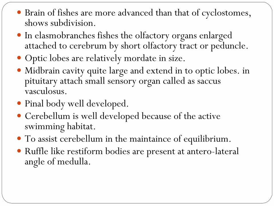
Brain of fishes are more advanced than that of cyclostomes, shows subdivision.
In elasmobranches fishes the olfactory organs enlarged attached to cerebrum by short olfactory tract or peduncle.
Optic lobes are relatively mordate in size. Midbrain cavity quite large and extend in to optic lobes. in
pituitary attach small sensory organ called as saccus vasculosus.
Pinal body well developed. Cerebellum is well developed because of the active
swimming habitat. To assist cerebellum in the maintaince of equilibrium. Ruffle like restiform bodies are present at antero-lateral
angle of medulla.
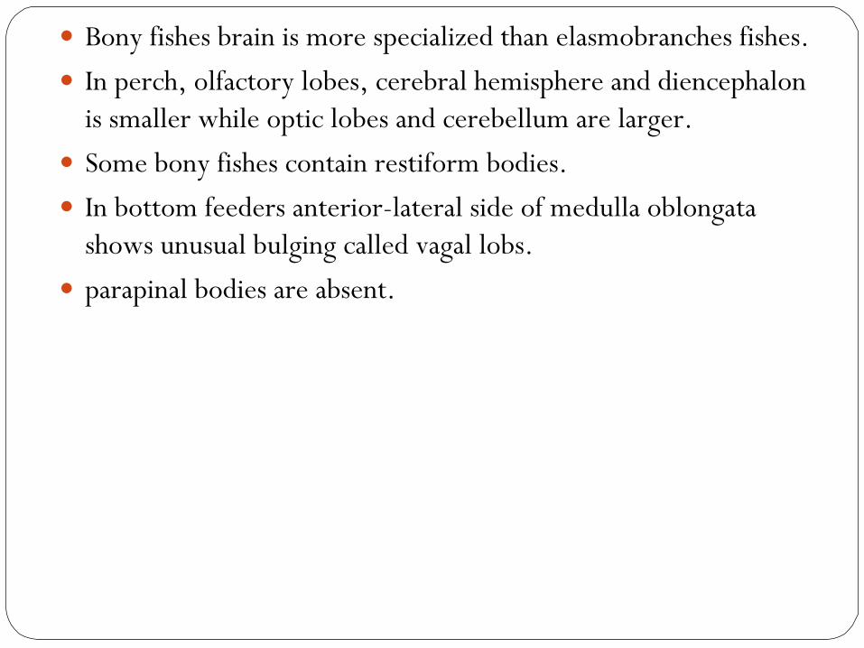
Bony fishes brain is more specialized than elasmobranches fishes. In perch, olfactory lobes, cerebral hemisphere and diencephalon
is smaller while optic lobes and cerebellum are larger. Some bony fishes contain restiform bodies. In bottom feeders anterior-lateral side of medulla oblongata
shows unusual bulging called vagal lobs. parapinal bodies are absent.

3. Amphibia Ex Frog
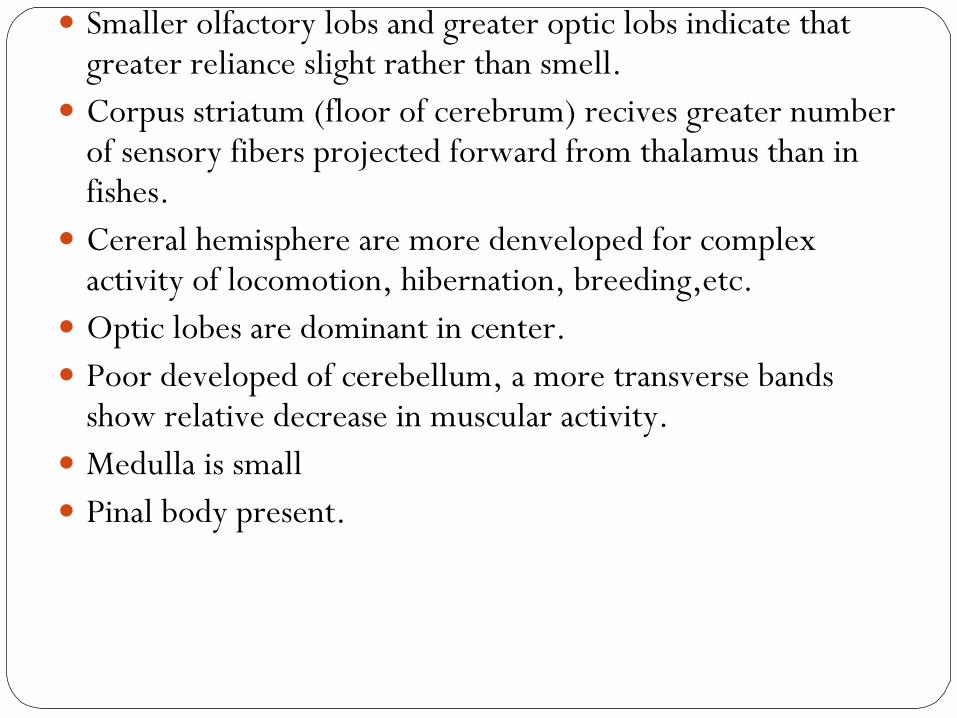
Smaller olfactory lobs and greater optic lobs indicate that greater reliance slight rather than smell.
Corpus striatum (floor of cerebrum) recives greater number of sensory fibers projected forward from thalamus than in fishes.
Cereral hemisphere are more denveloped for complex activity of locomotion, hibernation, breeding,etc.
Optic lobes are dominant in center. Poor developed of cerebellum, a more transverse bands
show relative decrease in muscular activity. Medulla is small Pinal body present.
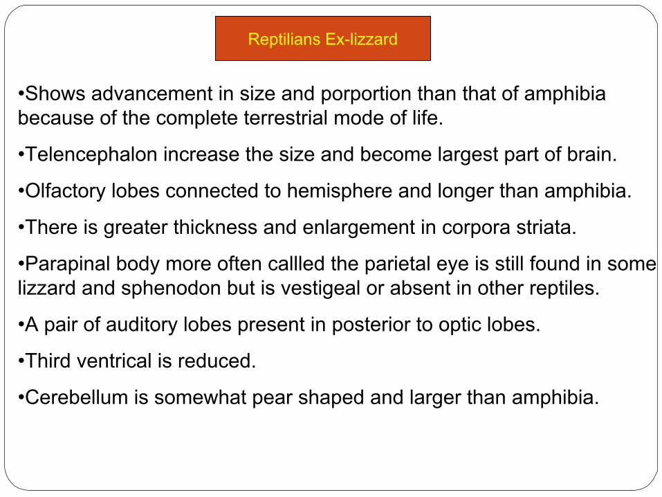
Reptilians Ex-lizzard
•Shows advancement in size and porportion than that of amphibia because of the complete terrestrial mode of life.
•Telencephalon increase the size and become largest part of brain.
•Olfactory lobes connected to hemisphere and longer than amphibia.
•There is greater thickness and enlargement in corpora striata.
•Parapinal body more often callled the parietal eye is still found in some lizzard and sphenodon but is vestigeal or absent in other reptiles.
•A pair of auditory lobes present in posterior to optic lobes.
•Third ventrical is reduced.
•Cerebellum is somewhat pear shaped and larger than amphibia.

Olfactory bulb
Olfactory peduncle
Cerebral hemisphere
Pinal body
Cerebellum
medulla
Spinal cord
Optic lobe
Optic chisma
influndiblum
hypophysis
Dorsal view Ventral view
Lizzard Brain
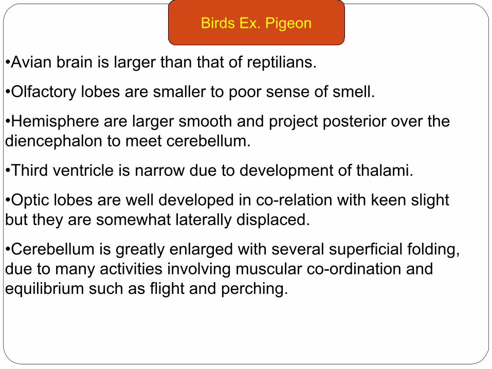
•Avian brain is larger than that of reptilians.
•Olfactory lobes are smaller to poor sense of smell.
•Hemisphere are larger smooth and project posterior over the diencephalon to meet cerebellum.
•Third ventricle is narrow due to development of thalami.
•Optic lobes are well developed in co-relation with keen slight but they are somewhat laterally displaced.
•Cerebellum is greatly enlarged with several superficial folding, due to many activities involving muscular co-ordination and equilibrium such as flight and perching.
Birds Ex. Pigeon

Olfactory lobes
Cerebral hemisphere
Pinal body
Optic lobe
Optic chisma
Optic tract
influndiblum
hypophysis
flocculus
vermis
medulla
Spinal cordDorsal view Ventral view
Pigeon Brain
Questions-1. Give an account of comparative structure of nervous system of vertebrates.2. Write the functions of different parts of brain.3.