Embed Size (px)
Citation preview

Animal physiology Lecture 5, 6
1
Nerves Physiology
The nervous system consists of nerve cells (neurons) and supporting cells. Neuron is the
structural and functional unit of the nervous system. A typical neuron consists of the soma or
cell body and two types of processes: the axon and dendrites. These cells functionally divided
to four zones:
1. Receptor Zone: It is the body cell and its dendrites. Dendrites provide a receptive area
that transmits electrical impulses to the cell body.
2. Impulse origin one: it is the axon hillock, the origin of the axon near the cell body.
Here, the nerve impulses originate.
3. Impulse transmission zone: it is the reign extends from the axon hillock to the
telodendria, the nerve ending. The nerve impulses transmit to synaptic buttons.
4. Neurotransmitter secretion zone: it is telodendria and its synaptic buttons which
responsible to transmit the impulses to other cell by secret the neurotransmitters.
Nerve fibers:
The nerve fibers divided to myelinated and unmyelinated. The myelin sheath formed by
oligo dendrocytes. All myelinated axons are surrounded by myelin sheath except Ranvier’s
nodes, which have 2000-12000 Na+ chanal/µm
2 axolemma, while in the first one of axone
350-500, in the cell body 50.75, in the myelin sheath 25, in the end of axon 20-75, and in the
unmyelinated axons 110. Unmyelinated are smaller than 2µm in diameter, whereas those that
are larger are likely to by myelinated. Myelinated axons conduct impulses more rapidly than
unmyelinated.

Animal physiology Lecture 5, 6
2
Resting membrane potential (RMP):
An electrical potential difference, or membrane potential, can be recorded across the
plasma membrane of living cells. The potential of unstimulated cells, or resting potential,
amounts to -9 to 100mV depend of the type of cell. A resting potential is caused by a slightly
unbalanced distribution of ions between intracellular fluid (ICF) and extracellular fluid (ECF).
The following factors are involved in establishing the membrane potential:
1. High K+
conductance: it is relatively easy for K+ ions to diffuse across the cell
membrane. Because of the steep concentration gradient, K+ ions diffuse from the ICF
to the ECF.
2. Maintenance of an unequal distribution of ions: the Na+-K
+ ATPase continuously
pumps Na+
out of the cell and K+ into it by active transport. As a result, the
intracellular K+ concentration is around 35 times higher and the intracellular Na
+
concentration is roughly 20 times lower than the extracellular concentration.
3. Cl- distribution: a passive distribution of Cl
- between intra- and extra- cellular spaces
exists only as long as there is no active Cl- uptake into the cell.
Membrane Action Potential (MAP):
An action potential is a signal passed on through an axon that influences other neurons or
induces somatic cell. Excitation of a neuron occurs if the membrane potential on the axon
hillock changes from its resting value -70mV to a less negative value -55mV, which called
threshold potential. This depolarization may be caused by neurotransmitter-induced opening
of postsynaptic action channels or by the transmission of stimuli from the surrounding.
If the membrane potential of a stimulated cell comes close to threshold potential, rapid
voltage-gated Na+ channels are activated. This results in increased Na
+ conductance, and
higher of Na+ into the cell. This is resulted a high potential value +35mV called Action
potential.
To understand the changes in the membrane potential, Cathode RAY Oscilloscope (CRO)
was used to record the electric evidences happened in nerve fiber by microelectrodes. One of
microelecrtodes was put on the extra-surface of cell membrane of nerve fiber and the other
was put in intra-surface. The excitation of nerve was begun by electric excitation. This
instrument records the changes by scheme. The excitation of nerve has four periods:

Animal physiology Lecture 5, 6
3
1. Latent (resting) period: in this period the membrane potential is stile -70mV (resting
potential) to a short or long time according the microelectrodes position.
2. Depolarization period: large numbers of Na+ channels are activated, and the influxing
Na+ accelerates depolarization. As a result, the membrane potential is slowly decreased
from -70 to -55mV, which called threshold or firing level. After that, the membrane
potential is rapidly decreased to (0mV), which called isopotential. The membrane
potential temporarily reaches positive levels (+35mV).
There are two types of stimuli:
a) Threshold stimuli: it is successful to produce active potential, which its intensity is
more than 15mV (-70mV, RMB, -55, threshold=15)
b) Sub-threshold stimuli: its intensity is less than 15mV, which cannot reach the
threshold so it cannot produce active potential.
3. Repolarization period: because the Na+ channels are inactivated, the potential reverses,
and restoration of the resting potential, the repolarization phase of the action potential,
begins. Depolarization has increased the open-probability of voltage gated K+ channels.
This has increased the potassium conductance, thereby accelerating repolarization.
4. Hyperpolarization period: in many cases, potassium conductance is still increased after
the original resting potential has been restored, resulting in a hyperpolarization
afterpotential. Increased Na+-K
+ ATPase pumping rates can contribute to this
afterpotential.

Animal physiology Lecture 5, 6
4
Nerve Impulse: Nerve impulse is an electrochemical phenomenon which includes:
1. Electrical, The movement of active potential by stimuli from stimulation point on the
long nerve fiber. This is like electrical flows through a cable when voltage is applied.
2. Chemical, neurotransmitter is released by regulated exocytosis of synaptic vesicles when
the action potential reached it to stimulate the adjacent cells.
Nerve impulse characteristics are:
1. All or None Low:
Action potential producing depends on intensity of stimulus and duration of stimulation.
All stimuli, which have threshod intensity and enough duration of stimulation, are success to
produce action potential. But none, which its intensity is less than threshold, can produce
action potential whatever its duration.
2. Refractory period:
During an action potential, the cell remains unresponsive to further stimuli. In absolute
refractory period, from firing point to repolarization period no other action potential can be
triggered, even by extremely strong stimuli, since Na+
channels in depolarized membranes
cannot be activated. This is flowed by a relative refractory period during which only action
potentials of smaller amplitudes and rates or rise can be generated, even by strong stimuli.
The refractory period ends once the membrane potential returns to its resting value.
3. Impulse conduction:
The start of an action potential is accompanied by a brief influx of Na+ into the nerve
fiber. The cell membrane that previously was inside negative now becomes positive, thus a
longitudinal potential difference with respect to adjacent, still unstimulated nerve segments.
This is followed by a passive electrotonic withdrawal of charge from the adjacent segment of

Animal physiology Lecture 5, 6
5
the nerve fiber, causing its depolarization. If it exceeds threshold, another action potential is
created in the adjacent segment dissipates.
Action potential normally run forward (thodromic conduction) because each segment of
nerve fiber becomes refractory when an action potential passes. If, however, the impulse are
conducted backwards (antidromic conduction) due, for example, to stimulation of nerve
fibers from an external source, they will terminate at the next synapse. Although the
continuous generation of action potentials in the immediately adjacent fiber segment
guarantees a refreshed signal, this process is rather time-consuming. The continuous
conduction or velocity conduction, in unmyelinated nerve fibers is only around 1m/s.
myelinated nerve fibers conduct much faster (up to 80m/s). In the Ranvier’s nodes, a myelin
sheath insulates the nerve fiber from the surroundings; thus, longitudinal current strong
enough to generate action potential can travels further down the axon. This results in more
rapid conduction because the action potential is generated only at the Ranvier’s nodes, where
there is a high density of Na+
channel. This results in rapid, jump-like passage of the action
potential from node to node (salutatory conduction).
4. Velocity of conduction: The conduction velocity depends on:
a. Myelination: the conduction velocity of such myelinated nerve fiber is much higher than
that of unmyelinated nerve fibers.
b. Diameter: the conduction velocity increases with the diameter of nerve fiber.
Classification of nerve fiber according its conduction velocity:
A) Type-A-nerve fiber:
Its conduction velocity is very high (2-80m/s) because it is myelinated and big
diameter (1-16µm). It is called fast fiber such as somatic nerve fiber which pressure and
touch sensation. It is can triggered the nerve impulse under anesthesia because of myeline
sheath, but cannot triggered the nerve impulse under compression because of the big
diameter which cause the paralysis.

Animal physiology Lecture 5, 6
6
B) Type-B-nerve fiber:
Their conduction velocity is less than types-A (3-15m/s) because it is myelinated but
smaller diameter (3µm). It is called moderate fiber such as visceral nerve fibers which
pressure and touch sensation like type A.
C) Type-C-nerve fiber:
Its conduction velocity is very low (0.25-1.5m/s) because it is unmyelinated and small
diameter (0.5-1.5µm).it is called slow fibers such as all nerve fiber which pain and
temperature sensation. It is cannot triggered the nerve impulse under anesthesia, because
it is unmyelinated; and under compression, because of the small diameter.
5. Compound action potential:
Synaptic transmission:
At the chemical synapse, the arrival of an action potential in the axon triggers the release of
transmitter form the presynaptic axon terminals (presynaptic membrane). The transmitter then
diffuses across the narrow synaptic cleft to bind postsynaptically to receptors in the subsynaptic
or postsynaptic membrane of a neuron or of glandular or muscle cell. Depending on the type of
transmitter and receptor involved, the effect on the postsynaptic membrane may either be
excitatory or inhibitory. Excitatory neurotransmitters such as Acetyl choline (Ach) and
Norepinephrine (NE) open Ca2+
channels leading to an increase in the cytosolic Ca2+
concentration, which increases the action potential. Inhibitory neurotransmitters such as Glycine
and Gamma Amino Butyric Acid (GABA) open K+ or Cl
- channels, as a result, excitatory
postsynaptic potential related depolarization is reduced and stimulation of postsynaptic neurons
is inhibited.
A signal excitatory postsynaptic potential normally is not able to generate a postsynaptic
(axonal) action potential, but requires the triggering of a large number of local depolarizations in
the dendrites. Their depolarizations are transmitted electrotonically across the soma and summed
on the axon hillock (spatial summation). Should the individual stimuli arrive at different times,
the prior depolarization will not have dissipated before the next one arrives, and summation will

Animal physiology Lecture 5, 6
7
make it easier to reach threshold. This type of temporal summation therefore increases the
excitability of the postsynaptic neuron.

Copyright © 2006 Pearson Education, Inc., publishing as Benjamin Cummings
The Digestive System
Dr. Ali Ebneshahidi

Copyright © 2006 Pearson Education, Inc., publishing as Benjamin Cummings
Functions of the Digestive System
ingestion – the oral cavity allows food to enter the digestive
tract and have mastication (chewing) occurs , and the resulting
food bolus is swallowed .
Digestion:
Mechanical digestion – muscular movement of the digestive
tract (mainly in the oral cavity and stomach) physically break
down food into smaller particles .
chemical digestion – hydrolysis reactions aided by
enzymes (mainly in the stomach and small intestine)
chemically break down food particles into nutrient
molecules , small enough to be absorbed . .

Copyright © 2006 Pearson Education, Inc., publishing as Benjamin Cummings
Secretion – enzymes and
digestive fluids secreted by
the digestive tract and its
accessory organs facilitate
chemical digestion .
Absorption – passage of the
end – products (nutrients) of
chemical digestion from the
digestive tract into blood or
lymph for distribution to
tissue cells .
Elimination – undigested
material will be released
through the rectum and anus
by defecation .

Copyright © 2006 Pearson Education, Inc., publishing as Benjamin Cummings
Organization of The Digestive System
Organs of the digestive system are divided into 2 main
group : the gastrointestinal tract (GI tract) and
accessory structures .
GI tract is a continuous tube extending through the
ventral cavity from the mouth to the anus – it consists
of the mouth , oral cavity , oropharynx , esophagus ,
stomach , small intestine , large intestine , rectum , and
anus .
Accessory structures include the teeth, tongue (in oral
cavity) , salivary glands , liver , gallbladder , and
pancreas .

Copyright © 2006 Pearson Education, Inc., publishing as Benjamin Cummings Figure 23.1

Copyright © 2006 Pearson Education, Inc., publishing as Benjamin Cummings
Muscular movement of the GI tract
Peristalsis – wavelike movement that occurs from the
oropharynx to the rectum , allowing GI tract to push
food particles toward the anus .
Mixing—mixing motion in the oral cavity and
stomach that allows the GI tract to repeatedly break
down food into smaller particles , using mechanical
digestion .
Segmentation – regions of the small intestine
contracting and relaxing independently , allowing the
small intestine to digestive and absorb more efficiently
.

Copyright © 2006 Pearson Education, Inc., publishing as Benjamin Cummings
Histology of the Alimentary Canal
Figure 23.6

Copyright © 2006 Pearson Education, Inc., publishing as Benjamin Cummings
Peristalsis and Segmentation
Figure 23.3

Copyright © 2006 Pearson Education, Inc., publishing as Benjamin Cummings
Regulation of GI Tract Activities
Autonomic nervous system
- parasympathetic nerves stimulate GI tract activities .
- sympathetic nerves inhibit GI tract activities .
Hormonal control
- hormones from endocrine gland and from GI tract itself
help regulate GI tract activities .
Reflex mechanism
- regions of the GI tract (especially the stomach and small
intestine) use reflexes to stimulate or inhibit one another .

Copyright © 2006 Pearson Education, Inc., publishing as Benjamin Cummings
Nervous Control of the GI Tract

Copyright © 2006 Pearson Education, Inc., publishing as Benjamin Cummings
Mouth & Oral Cavity
Food enters the GI tract by ingestion .
Food is broken down by mechanical digestion , using
mastication .
One chemical digestive process occur where amylase
enzyme in saliva breaks down polysaccharide into
disaccharides .
The tongue , made of skeletal muscle, manipulates the food
during mastication . it also contains taste buds to detect
taste sensations(intrinsic) .
Food particles are mixed with saliva during mastication ,
resulting in a moist lump called bolus for easier passage
into or pharynx .

Copyright © 2006 Pearson Education, Inc., publishing as Benjamin Cummings
Teeth Adapted for mechanical
digestion (mastication) in the
oral cavity .
20 deciduous or primary
teeth before the age of 6.
By age 7, 32 permanent or
secondary teeth are
developed & are divided into
4 types: incisors (for cutting)
, Canines (for tearing) ,
Premolars (for crushing),
and Molars (for grinding).
these teeth follow the human
dental formula of 2-1-2-3.

Copyright © 2006 Pearson Education, Inc., publishing as Benjamin Cummings
Salivary Glands
3 pairs of salivary glands called parotid , submandibular ,
and sublingual gland secrete most of the saliva in the oral
cavity , using salivary ducts .
Saliva helps moisten the food during mastication , dissolve
the food in forming the bolus , and help cleanse the teeth.
Saliva consists of 99.5% water , the remaining 0.5% is
dissolved substances including amylase enzyme (for
chemically digesting carbohydrate ), bicarbonate ion
(HCO3-; maintains pH of saliva at 6.5-7.5) , and many
electrolytes.

Copyright © 2006 Pearson Education, Inc., publishing as Benjamin Cummings
Salivary Glands
Figure 23.9a

Copyright © 2006 Pearson Education, Inc., publishing as Benjamin Cummings
Stomach
A pouch-like organ primarily designed for food storage (for 2-4
hours) , some mechanical and chemical digestion also occur .
Contains two sphincters at both ends to regulate food movement
– cardiac sphincter near the esophagus ,and pyloric sphincter
near the small intestine .
Divided into 4 regions : cardiac stomach (or cardiac), fundic
stomach (or funded) , body of stomach , and pyloric stomach
(or Pylorus).
Contain thick folds called rugae at its layer , for providing
larger surface area for expansion , secretion , digestion , and
some absorption.

Copyright © 2006 Pearson Education, Inc., publishing as Benjamin Cummings
Stomach

Copyright © 2006 Pearson Education, Inc., publishing as Benjamin Cummings
Gastric Secretory Cells
-Chief cells: secrete pepsinogen (an inactive enzyme).
-Parietal cells: secrete hydrochloric and (HCl) and
"intrinsic factor" (which helps absorption of vitamin
B12 in the intestines).
- Mucous cells: secrete mucus and alkaline substances
to help neutralize HCl in the gastric juice .
-G cells: secrete a hormone called gastrin , which
stimulates the parietal cells and overall gastric
secretion .

Copyright © 2006 Pearson Education, Inc., publishing as Benjamin Cummings
Gastric Cells

Copyright © 2006 Pearson Education, Inc., publishing as Benjamin Cummings
Chemical digestion & absorption in the stomach
- Carbohydrate digestion is continued with gastric amylase ,
resulting in disaccharides .
- Protein digestion begins with pepsin (activation of pepsinogen
by HCl) , resulting in peptides (small chains of protein).
- Lipid digestion begins with gastric lipases which can only
break down certain lipids such as butterfat , resulting in fatty
acids .
Absorption in the stomach is limited, where only small and fat-
soluble substances can be absorbed—water , alcohol, aspirin ,
and certain drugs .
The result of all these mixing , chemical digestion , secretion,
and absorption is a yellowish paste called chyme , which will
be passed on to the small intestine .

Copyright © 2006 Pearson Education, Inc., publishing as Benjamin Cummings
Regulation of Gastric Secretion
Regulation of gastric secretion and activities is by both nervous
and hormonal mechanisms – food moving along the oral cavity
and esophagus stimulates the parasympathetic nerves to
activate the secretion in gastric glands , the gastric hormone
from G cells in turn stimulates the gastric glands for more
activities ("positive feedback").
On the other hand , when food is emptying from the stomach ,
sympathetic nerves inhibit the gastric glands and gastric , and
a hormone called intestinal gastrin (released by small
intestine) inhibits other gastric activities.
The above regulations occur in 3 overlapping phases:
Cephalic Phase, Gastric Phase, & Intestinal Phase.

Copyright © 2006 Pearson Education, Inc., publishing as Benjamin Cummings
Cephalic phase
Cephalic phase: involves special senses detect food
and uses parasympathetic nerves in the vagus nerve to
stimulate gastric activities.
1. Sight, Smell , and Taste of food cause stimulation
of vagus nuclei in brain.
2. Vagus stimulates acid secretion.
a. Direct stimulation of parietal cells (major effect).
b. Stimulation of Gastrin secretion (lesser effect).

Copyright © 2006 Pearson Education, Inc., publishing as Benjamin Cummings
Gastric phase
Gastric phase involves the distention of stomach and
stimulates its own activities by the vagus nerve. Distension of
stomach (stretch - receptors) stimulates vagus nerve ; vagus
stimulates acid secretion .
Amino acids and peptides in stomach lumen srimulates acid
secretion (chemo - receptors)
Direct stimulation of parietal cells (lesser effect)
Stimulation of gastrin secretion ; gastrin stimulates acid
secretion (major effect)
Gastrin secretion inhibited when PH of gastric juice falls
below 2.5.

Copyright © 2006 Pearson Education, Inc., publishing as Benjamin Cummings
Intestinal Phase intestinal phase involves acidic chyme passing into the small
intestine which secretes intestinal gastrin hormone to inhibit gastric
activates.
Neural inhibition of gastric emptying and acid secretion. Arrival of
chyme in duodenum causes distension & an increase in osmotic
pressure. These stimuli activate a neural reflex that inhibits gastric
activity.
In response to fat in chyme , duodenum secretes the hormone,
secretin that inhibits gastric acid secretion.
The enterogastric reflex: This reflex begins in the small intestine
(entero) and ends in the stomach (gastro).
Duodenum fills with chyme. Sensory stretch receptors are stimulated.
Sensory nerve impulses travel to CNS. Nerve impulses from CNS
(vagus) inhibit peristalsis in stomach wall.

Copyright © 2006 Pearson Education, Inc., publishing as Benjamin Cummings
Stomach: Neural & Hormonal Mechanisms

Copyright © 2006 Pearson Education, Inc., publishing as Benjamin Cummings
Pancreas
Pancreas : most pancreatic enzymes are produced as inactivate
molecules , or zymogens , so that the risk of self – digestion within the
pancreas is minimized .
More than 98% of the pancreas mass is devoted to its exocrine
function: the secretion of pancreatic juice by the pancreatic acini and
their ductile cells. Ductile cells produce Sodium bicarbonate which
helps neutralize the acidic gastric contents .
Acinar cells of the exocrine pancreas produce a variety of digestive
enzymes to break down food substances into smaller absorbable
molecules .
Only 2% of pancreas mass is devoted to the islets of langerham , which
produce insulin and glucagon , hormones that regulate blood sugar
and carbohydrate metabolism (they have opposite effects) .

Copyright © 2006 Pearson Education, Inc., publishing as Benjamin Cummings

Copyright © 2006 Pearson Education, Inc., publishing as Benjamin Cummings
Major pancreatic Enzymes
-pancreatic amylase: digest polysaccharides into disaccharides
- pancreatic lipases digest triglycerides into fatty acids .
- pancreatic nucleases digest nucleic acids into nucleotides .
-Pancreatic proteinases (all secreted in their inactive forms) digest
peptides into amino acids:
Trypsinogen is activated by enterokinase (secreted by duodenum)
into trypsin , which in turn activates the other 3 enzymes – chymo-
trypsinogen becomes chymotrypisn , proaminopeptidase
becomes aminopeptidase, and procarboxypeptidase becomes
carboxypeptidase.

Copyright © 2006 Pearson Education, Inc., publishing as Benjamin Cummings
Activation of pancreatic proteases in the small intestine

Copyright © 2006 Pearson Education, Inc., publishing as Benjamin Cummings
Pancreatic Secretion
1.The parasympathetic nervous system increases
pancreatic secretion
2. Two duodenual hormones also influence
pancreatic secretion: Secretin and Cholecystokinin.
3. Food entering the small intestine stimulates the
secretion of both hormones.
4. Secretin stimulates the secretion of pancreatic
electrolyte – rich fluid , while CCK enhances the
enzymatic secretions of the pancreas .

Copyright © 2006 Pearson Education, Inc., publishing as Benjamin Cummings
Regulation of pancreatic Juice
1. Acidic chyme enters duodenum.
2. Secretin is released into blood stream from
intestinal mucosa.
3. Secretin stimulates pancreas.
4. Pancreas secretes pancreatic juice.
5. Pancreatic juice , high in bicarbonate ions ,
neutralizes acidic chyme.

Copyright © 2006 Pearson Education, Inc., publishing as Benjamin Cummings

Copyright © 2006 Pearson Education, Inc., publishing as Benjamin Cummings
Functions of The Liver Important in carbohydrate metabolism where hepatic cells conduct
glycogenesis (converting glucose into glycogen) , and
glycogenolysis (breaking glycogen down to glucose).
Also is critical in lipid metabolism where hepatic cells produce bile
(for fat emulsification), oxidize fatty acids , synthesize various
forms of lipids ,and convert glucose to fatty acids (lipogenesis) .
Other functions of the liver include :
- Storage of glycogen, iron , and vitamins A,D,B12.
-Contains phagocytes to destroy damaged erythrocytes and foreign
substances, using phagocytosis .
-Detoxifies harmful substances in the blood .
-Serves as a blood reservoir (contains 7% of blood volume).

Copyright © 2006 Pearson Education, Inc., publishing as Benjamin Cummings
Liver

Copyright © 2006 Pearson Education, Inc., publishing as Benjamin Cummings
Gall Bladder
A small sac located on the inferior , visceral surface of the liver.
Stores and concentrates bile secreted by the liver.
Regulation of Bile Release:
1. Chyme with fat enters small intestine.
2. Cells of intestinal mucosa secrete the hormone Cholecystokinin
(CCK) into the blood stream.
3. CCK stimulates muscular layer of gallbladder wall to contract.
4. Bile passes down the cystic duct and common bile duct to
duodenum .
5. Hepatopancreatic sphincter relaxes and bile enters duodenum.

Copyright © 2006 Pearson Education, Inc., publishing as Benjamin Cummings

Copyright © 2006 Pearson Education, Inc., publishing as Benjamin Cummings
Small Intestine
A long tube, with a small diameter (about 1 inch), extending from
pyloric sphincter to the ileocecal valve .
Divided into Duodenum, Jejunum, and ileum.
1. Secretions of small intestine:
a. Intestinal glands secrete a watery fluid that lack digestive
enzymes but provides a vehicle for moving chyme to villi
.Intestinal enzymes include : maltase digests maltose into glucose.
sucrose digests sucrose into glucose and fructose . lactase digests
sucrose into glucose and glucose. peptidases digest peptides into
amino acids . lipases digest triglycerides into fatty acids and
glycerol . Nucleases digest nucleotides into nitrogenous bases.
Enterokinase converts trypsinogen into trypsin.

Copyright © 2006 Pearson Education, Inc., publishing as Benjamin Cummings
b. Digestive enzymes embedded in the surfaces of microvilli split
molecules of sugars, proteins and fats .
c. Regulation of small intestine secretions: secretion is stimulated by
gastric juice , chyme , and reflex stimulated by distension of the
small intestinal wall .

Copyright © 2006 Pearson Education, Inc., publishing as Benjamin Cummings
d. Each villus contains blood capillaries to absorb water , glucose ,
amino acids , vitamins , minerals , and short-chain fatty acids , and also
contains lymphatic capillaries called lacteals to absorb long – chain
fatty acids in the forms of micelles .
e. Water is absorbed by osmosis , fatty acids are absorbed by diffusion
(since they are fat-soluble), and most other nutrients (glucose, amino
acids, & minerals) are absorbed by active transport.

Copyright © 2006 Pearson Education, Inc., publishing as Benjamin Cummings
Large intestine
The last segment of
the GI tract , with a
large diameter (2-3
inches) , extending
from the ileocecal
valve to the anus .
Divided into cecum ,
ascending colon ,
transverse colon ,
descending colon ,
sigmoid colon ,
rectum , anal canal
, and anus.

Copyright © 2006 Pearson Education, Inc., publishing as Benjamin Cummings
The large intestine has little or no digestive function , although it
secretes mucus. Its mucosa has no villa or microvillus , but cotains
numerous goblet cells for secreting mucus to aid in the formation
of feces and maintain an alkaline condition .
mechanical stimulation and parasympathetic impulses control the
rate of mucus secretion .
The large intestine only absorbs water, electrolytes and some
vitamins .
Many bacteria inhabit the large intestine , where they break down
certain indigestible substances and synthesize certain vitamins .
feces are formed and stored in the large intestine . Defecation
involves a reflex mechanism aided by voluntary contraction of the
diaphragm , abdominal muscles ,and the external anal sphincter .

Copyright © 2006 Pearson Education, Inc., publishing as Benjamin Cummings
Major Hormones of The Digestive Tract
1. Gastrin : (Gastric & intestinal) : released by Gastric cells , in
response to the presence of food. Causes Gastric glands to increase
their secretory activity.
2. Somatostatin : (Gastric inhibitory peptides - GIP): Inhibits
secretion of acid by parietal cells.
3. Cholecystokinin : released by intestinal wall cells , in response to
the presence of proteins and fats in the small intestine. It causes
gastric glands to decrease their secretory activity and inhibits gastric
motility ; stimulation of pancreas to secrete digestive enzyme;
stimulates gall – bladder to contract and release bile.
4. Secretin: released by cells in the duodenal wall, in response to
acidic chyme entering the small intestine.

Copyright © 2006 Pearson Education, Inc., publishing as Benjamin Cummings
Major Digestive Enzyme
Salivary enzyme: Begins carbohydrates digestion by breaking down
starch and glycogen to disaccharides
Gastric enzymes: Pepsin , from Gastric glands – Begins protein
digestion . Lipase, from Gastric glands – Begins fat digestion .
Pancreatic enzymes: Amylase , from pancreas – breaks down starch
and glycogen into disaccharides. Lipase, from pancreas – breaks down
fats into fatty acids and glycerol .
Proteolytic enzymes :
Trypsin, Chymotrypsin, and Carboxypeptidase from pancreas
breaks down peptides into amino acids . Nucleases, from pancreas-
breaks down nucleic acids into nucleotides.

Copyright © 2006 Pearson Education, Inc., publishing as Benjamin Cummings
Intestinal Enzymes: Peptidase, from mucosal cells, breaks down
peptides into amino acids. Sucrase, maltase, and lactase , from
mucosal cells, breaks down disaccharides into monosaccharides.
Lipase, from mucosal cells, breaks down fats into fatty acid and
glycerol. Enterokinase , from mucosal cells, (breaks down) converts
trypsinogen into trypsin .

Copyright © 2006 Pearson Education, Inc., publishing as Benjamin Cummings
Fat digestion & Absorption

Copyright © 2006 Pearson Education, Inc., publishing as Benjamin Cummings
Clinical Terms
Achalasia : failure of the smooth muscle to relax at some
junction in the digestive tube.
Cholecystitis : Inflammation of the gallbladder.
Chloelithiasis : stones in the gallbladder.
Cholestasis : Blockage in bile flow from the gallbladder.
Cirrhosis : liver cells degenerate and the surrounding
connective tissue thicken.
Diverticulitis : Inflammation of small pouches that sometimes
form in the lining and wall of the colon.
Dysentery : Intestinal infection.

Copyright © 2006 Pearson Education, Inc., publishing as Benjamin Cummings
Clinical terms
Dyspepsia: Indigestion
Dysphasia: Difficulty in swallowing
Enteritis: Inflammation of the intestine .

ECG INTERPRETATION:ECG INTERPRETATION: the basics
Damrong Sukitpunyaroj MDDamrong Sukitpunyaroj, MDPerfect Heart Institue, Piyavate Hospital

OverviewOverview• Conduction Pathways • Systematic Interpretation• Common abnormalities in Critical Care
– Supraventricular arrhythmiasVentric lar arrh thmias– Ventricular arrhythmias

Conduction PathwaysConduction Pathways

Conduction PathwaysConduction Pathways
P wave = atrial depolarisation.
PR Interval = impulse from atria to ventriclesto ventricles.
QRS complex = ventricular depolarisation.
ST segment = isoelectric - part of repolarisation.
T wave = usually same directionT wave = usually same direction as QRS - ventricular repolarisation.
QT Interval = This intervalQT Interval = This interval spans the onset of depolarisation to the completion of repolarization of the ventriclesof the ventricles.

InterpretationInterpretation
InterpretationInterpretation1. Rate = Number of P’s (atrial) R’s (ventricular) per
minute (6 second [30 squares] X 10 = minute rate).
P rate: 8 x 10 = 80 R rate: 8 x 10 = 80
2. Rhythm = Regular or irregular. Map P-P and R-R intervalsintervals.

Interpretation3 P t 1 QRS h d ti lt
Interpretation3. P wave = present, 1 per QRS, shape, duration, voltage.
4. P-R interval = length (0.12 - 0.2 sec = <1 big square), isoelectric.

InterpretationInterpretation5. QRS = duration (0.06 - 0.10 ), voltage, q or Q waves
6. ST Segment = shape, isoelectric with PR segment

InterpretationInterpretation7. T wave = shape, direction
8. QT interval = length (R-R/2 or QTc <0.40 sec)

Abnormalities: Supraventricular arrhythmias
• Atrial Fibrillation• Atrial Flutter • Supraventricular Tachycardia (SVT)
Abnormalities: V t i l h th i
• Premature Ventricular Complexes (PVCs)Ventricular arrhythmias
• Ventricular tachycardia (VT)

Conduction PathwaysConduction PathwaysSupraventricular Narrow QRS complex
V t i lVentricular Wide QRS complex

Abnormalities: atrial fibrillation
Rhythm: IrregularRate: A: 350 – 650; V: variesP: poorly definedP-R: N/AQRS: narrow complexS-T: normalT: normalQ-T: normal

Abnormalities: atrial flutter
Rhythm: Regular / IrregularRate: A: 220 – 430; V: <300 (2:1, 3:1 or sometimes 4:1)
P: Saw toothed appearance P-R: N/AQRS: narrow complexS-T: normalT: normalQ-T: normal

Abnormalities: supraventricular tachycardia (SVT)
Rhythm: RegularRate: >100P: not visibleP-R: not definedQRS: narrow complexS-T: depression (sometimes)T: normalQ-T: prolonged (sometimes)

Abnormalities: premature ventricular complexes

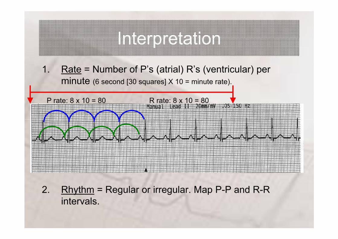
ExamplesExamples

ExamplesExamples

ECG INTERPRETATION:ECG INTERPRETATION: 12 Lead

OverviewOverview• Lead Placement• Axis• Common abnormalities in Critical Care
– Heart blockB ndle branch blocks– Bundle branch blocks
– Life threatening arrhythmias

Lead PlacementLead PlacementV1 4th ICS i ht tV1 = 4th ICS right sternumV2 = 4th ICS left sternumV3 = midway between V2V3 = midway between V2
and V4V4 = 5th ICS midclavicularV4 5th ICS midclavicularV5 = between V4 and V6
anterior auxiliary lineV6 = midauxillary line
lateral to V4 and V5

Lead PlacementLead Placement• Electrical activity towards = ↑• Electrical activity away = ↓

Lead PlacementLead Placement

AxisAxis• The direction of an ECG
waveform in the frontal plane measured inplane measured in degrees
• Represents the flow of pthe majority of electrical activity N ll h QRS• Normally the QRS complex is measured

AxisAxis• Each lead has its own axis

Lead PlacementLead PlacementCh t L dStandard Leads (bipolar)
• I - lateral wallChest Leads (unipolar)
• V1 - septal wall• V2 septal wall• II - inferior wall
• III - inferior wall
• V2 - septal wall• V3 - anterior wall• V4 - anterior wall
Augmented leads (unipolar)
• V4 - anterior wall• V5 - lateral wall• V6 - lateral wall
• aVR - no mans land• aVL - lateral wall
V6 lateral wall
• aVF - inferior wall

Lead PlacementLead PlacementNo-mans land, inferior, lateral, anterior, septal,

Abnormalities: bundle branch blocks
• QRS widened, greater than 0.12 secs• Change in axis • Difficult to interpret ECG• Right or Left• Normal P wave• Followed by a T wave

Abnormalities: right bundle branch blocks
• Indicates conduction problems in the right side of the heartthe heart
• May be normal in healthy peoplepeople
• R wave in V1, ie two R waves in V1in V1
• Q wave in V6 • Lead V1 cats earsLead V1 cats ears

Abnormalities: left bundle branch blocks
• Always indicates heart disease, usually of the left side of the heartside of the heart
• Hard to interpret an ECG with LBBBLBBB
• Lead V1 Q wave and an S wavewave
• Lead V6 an R wave followed by another R wavey
• Lead V6 Rabbit ears

Abnormalities: heart block
• SA block (exit block)• 1st degree AV block• 2nd degree AV block
– Wenckeback (type I)Mobit (t pe II)– Mobitz (type II)
• 3rd degree AV block

Abnormalities: heart block – SA block

Abnormalities: heart block – 1st degree AV

Abnormalities: heart block – 2nd degree AV
W k b kWenkeback
Mobitz

Abnormalities: heart block – 3rd degree AV

Abnormalities: life threatening arrhythmias
• Ventricular Tachycardia• Ventricular Fibrillation• Asystole

Abnormalities: life threatening arrhythmias - VT

Abnormalities: life threatening arrhythmias - VF

Abnormalities: life threatening arrhythmias – Asystole
ExamplesExamples

ExamplesExamples

Mechanoreception
Introduction
Hair cells : the basic mechanosensory unit
Hair cell structure
Inner ear and accessory organ structures
Vestibule
Otolith organs
Weberian ossicles
Lateral line
Lateral line structure
Receptor organs
Acoustic communication: sound production and reception
Sound production mechanisms
Locomotion and posture

IntroductionA mechanoreceptor is a sensory receptor that responds to mechanical
pressure or distortion. In fishes mechanoreception concerns the inner ear and the lateral line
system.
Hair cells are the UNIVERSAL MECHANOSENSORY TRANSDUCERS in both the lateral line and hearing systems.
The INNER EAR is responsible for fish EQUILIBRIUM, BALANCE and HEARING
LATERAL LINE SYSTEM detects DISTURBANCES in the water.

Hair cell structureEACH HAIR CELL CONSISTS OF TWO TYPES OF "HAIRS" OR
RECEPTOR PROCESSES:
Many microvillar processes called STEREOCILIA.
One true cilium called the KINOCILLIUM.
COLLECTIVELY, the cluster is called a CILIARY BUNDLE.
The NUMBER OF STEREOCILIA PER BUNDLE IS VARIABLE, and ranges from a 10s of stereocilia to more than a 100.
The STEREOCILIA PROJECT into a GELATINOUS CUPULA ON THE APICAL (exposed) SURFACE of the cell.
The cilium and villi are ARRANGED IN A STEPWISE GRADATION - the longest hair is the kinocillium, and next to it, the stereocilia are arranged in order of decreasing length.
These cells SYNAPSE WITH GANGLION CELLS.
They have DIRECTIONAL PROPERTIES - response to a stimulus depends on the direction in which the hairs are bent.
So, if the displacement causes the stereocilia to bend towards the kinocilium, the cell becomes DEPOLARIZED = EXCITATION.
If the stereocilia bend in the opposite direction, the cell becomes HYPERPOLARIZED = INHIBITION of the cell.
If the hair bundles are bent at a 90o angle to the axis of the kinocilium and stereocilia there will be no response.

The sensory hair cells are GROUPED TOGETHER TO FORM NEUROMAST ORGANS. These are situated on:
1. the body surface, 2. in the LATERAL LINE and HEAD. Here they
are buried in pits, canals and grooves, 3. in the inner organs of the ear and on the
pouches of otolith organs where they form LARGE FIELDS CALLED THE CRISTA AMPULLARIS
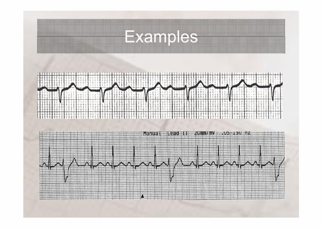
Inner ear and accessory organ structuresThe INNER EAR IS MADE UP OF 3 SEMI-CIRCULAR CANALS (the vestibule) and 3 otolith organs.
The otoliths are found in the UTRICLE, SACCULE AND LAGENA.
The inner ear is divided into the PARS SUPERIOR and the PARS INFERIOR.
The PARS SUPERIOR IS MADE UP OF THE SEMICIRCULAR CANALS AND THE UTRICLE in bony and cartilaginous fishes. The utricle contains the lapillus (=diminutive of stone)
The PARS INFERIOR IS MADE UP OF THE SACCULE AND THE LAGENA.
Many fishes have an additional organ, the MACULA NEGLECTA, a sensory structure located in Teleostomi in the utriculus of the inner ear near the opening of the ampulla of the posterior vertical semicircular canal, in selachians within a duct (posterior canal duct) through which the posterior vertical semicircular canal connects with the sacculus, while in the batoids it lies in the wall of the sacculus adjacent to the opening of the duct. It may have a neuromast associated with its sensory tissue. This structure has been demonstrated to be a sensitive vibration receptor in Raja. Also called crista neglecta, crista quarta, or papilla neglecta.
All these organs are INNERVATED BY BRANCHES OF CRANIAL NERVES.
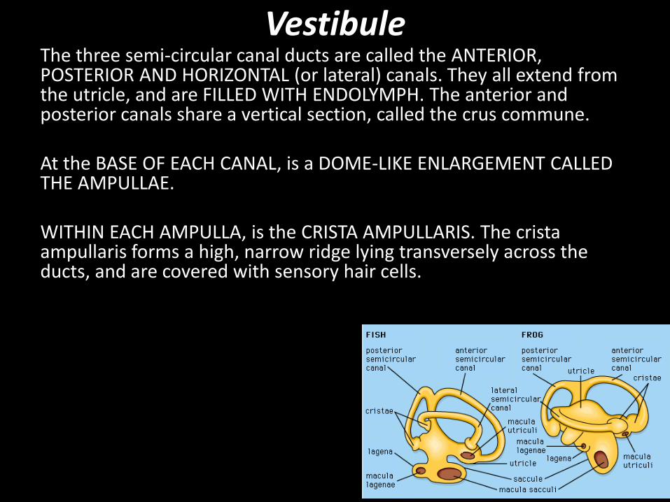
VestibuleThe three semi-circular canal ducts are called the ANTERIOR, POSTERIOR AND HORIZONTAL (or lateral) canals. They all extend from the utricle, and are FILLED WITH ENDOLYMPH. The anterior and posterior canals share a vertical section, called the crus commune.
At the BASE OF EACH CANAL, is a DOME-LIKE ENLARGEMENT CALLED THE AMPULLAE.
WITHIN EACH AMPULLA, is the CRISTA AMPULLARIS. The cristaampullaris forms a high, narrow ridge lying transversely across the ducts, and are covered with sensory hair cells.
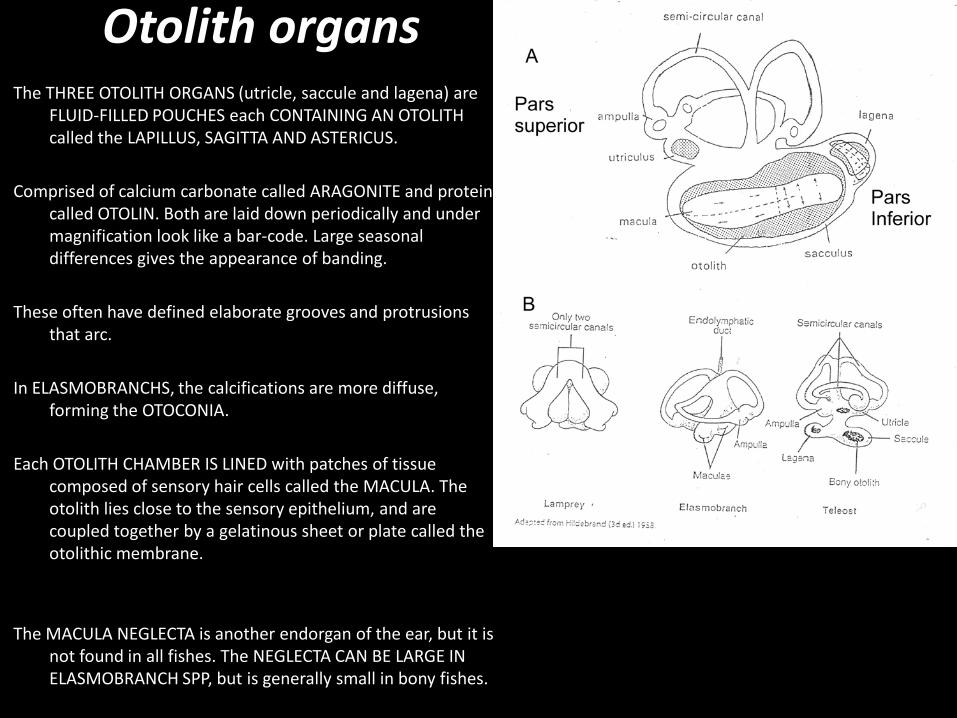
Otolith organsThe THREE OTOLITH ORGANS (utricle, saccule and lagena) are
FLUID-FILLED POUCHES each CONTAINING AN OTOLITH called the LAPILLUS, SAGITTA AND ASTERICUS.
Comprised of calcium carbonate called ARAGONITE and protein called OTOLIN. Both are laid down periodically and under magnification look like a bar-code. Large seasonal differences gives the appearance of banding.
These often have defined elaborate grooves and protrusions that arc.
In ELASMOBRANCHS, the calcifications are more diffuse, forming the OTOCONIA.
Each OTOLITH CHAMBER IS LINED with patches of tissue composed of sensory hair cells called the MACULA. The otolith lies close to the sensory epithelium, and are coupled together by a gelatinous sheet or plate called the otolithic membrane.
The MACULA NEGLECTA is another endorgan of the ear, but it is not found in all fishes. The NEGLECTA CAN BE LARGE IN ELASMOBRANCH SPP, but is generally small in bony fishes.

Weberian ossiclesThe OSTARIOPHYSIN fish (Characidae, Cyprinidae and Siluriformes) have
ACUTE HEARING AND PITCH DISCRIMINATION.
This is due to a SERIES OF SMALL BONES, called the WEBERIAN OSSICLES.
These bones physically CONNECT THE ANTERIOR END OF THE SWIM BLADDER WITH THE FLUID SYSTEM OF THE INNER EAR AT THE MIDLINE, BETWEEN THE SACCULES.
DEFLATION OF THE GAS BLADDER OR DISCONNECTION BETWEEN THE OSSICLES and the bladder causes DECREASED HEARING sensitivity in the fish.
A lateral view of the left side of the anterior portion of the vertebral region of an otophysan fish. The Weberian ossicles transmit sound vibrations from the swim bladder to the inner ear.

Lateral line systemThe LATERAL LINE SYSTEM consists of a CANAL SYSTEM ON
THE HEAD and a CANAL SYSTEM ON THE TRUNK.
There are often 3 major canals on the head, and a single major trunk canal running along the length of the body.
The CANALS IN TELEOSTS are WELL-OSSIFIED, PIERCED WITH PORES through which the FLUID-FILLED canal is linked to the external environment.

Figure 3.6: Four Types of head canal systems among teleost fishes: (a) narrow-simple canal system; (b) reduced canal system; (c) widened canal system; (d) branched canal system.
The canals on the head can be CLASSIFIED INTO FOUR TYPES: narrow simple, reduced, widened and branched.

There are EIGHT TRUNK CANAL PATTERNS are present in teleostfish.

Receptor organsThe lateral line canals are lined by a thin epithelium in which the NEUROMASTS are embedded.
Neuromasts can be classified into different types, depending on their location:
1) FREE OR SUPERFICIAL NEUROMASTS - forms patches on the skin, often in groups or lines called "stitches" or "pit lines".
2) CANAL NEUROMASTS - forms similar patches, but are located within the fluid-filled lateral line canals that lie under the skin.
The HAIR CELLS of the neuromasts are usually ORIENTED IN TWO OPPOSING DIRECTIONS.
The MOVEMENT OF THE FLUID within the canals STIMULATE THE HAIR CELLS, which in turn are innerved by lateral line nerves, specific to the different neuromast regions.
These nerves also have SPECIAL GANGLIA and specific projection sites in the HIND BRAIN, distinguishing them from other nerves.
Cross-section through the trunk of a minnow, showing the distribution and innervation of neuromast receptors and the location of the pores that connect the canal to the external environment. B) Each neuromast is composed of several hair cells, supporting cells and innervating sensory neurons. The apical kinocilia and stereocilia project into the cupula that overlies the entire neuromast.

Acoustic communication: sound production and reception
SOUND is a particularly USEFUL CHANNEL FOR COMMUNICATION in water.
Acoustic signals are NOT AFFECTED BY MURKINESS OR DARKNESS of the environment
SOUND TRAVELS 5 TIMES FASTER IN WATER THAN IN AIR (1500m/s as opposed to 300m/s).

Sound production mechanismsFishes from 50 different families are able to produce sound in a variety of
ways.
STRIDULATION by pharyngeal teeth or spines and fin rays (Gurnards, grunters).
The swim bladder often acts as a RESONATOR, it increases the amplitude of the sound wave (eg croakers/Kob).
Some marine catfish have special drumming muscles that vibrate the walls of the swim bladder (an ‘EXTRINSIC’ system)
In other fishes, sound is produced by an ‘INTRINSIC’ system, the muscles have their origin on the swim bladder, and the frequency of the sound depends on the rate of contraction of the muscle (toadfish, Opsanus).
http://www.dosits.org/audio/fishes/atlanticcroaker/
http://www.dosits.org/audio/fishes/barredgrunt/
http://www.dosits.org/audio/fishes/oystertoadfish/
http://www.dosits.org/audio/fishes/hhseacatfish/

Sound receptionSound reception, how do they hear?A sound produces TWO TYPES OF STIMULI within water.
Back-and-forth motion of the particles in the medium = PARTICLE DISPLACEMENTproduction of SOUND PRESSURE
Head of a FISH VIBRATES in a sound field OTOLITHS overlying the maculae will also VIBRATE.
Because OTOLITHS ARE DENSER than the surrounding tissue, the vibrations will be smaller than that of the surrounding tissue.
Causes hairs of the HAIR CELLS to BEND.
This bending will fire the hair cells if their polarity is appropriate to the direction of the vibration.
Figure 1 Otolith oscillating above the macular hair cells

The WEBERIAN APPARATUS is a particularly EFFECTIVE STRUCTURAL MODIFICATION that enhances the hearing of otophysan fishes(Cypriniformes, Characiformes, Siluroidei, and Gymnotoidei). Swim bladder pulsates in the SOUND PRESSURE FIELD AND ROCKS THE TRIPUS. Relayed through other bones to a sinus containing perilymph adjacent to the saccular macula.
CLUPEOIDS, such as anchovy, have an OTIC BULLA, which is closely associated with the UTRICULUS, on either side of the head.
The bulla ACTS AS A PRESSURE-DISPLACEMENT TRANSDUCTION MECHANISM.

Higgs, D. M. et al. J Exp Biol 2004;207:155-163
(A) Relationship of the prootic bulla to the inner ear of American shad, as demonstrated in a 12.5 mm TL
larva. The prootic bulla (arrow) sits just anterior to the utricle. (B) Diagram (modified from Denton and Gray,
1979) and (C) transverse section showing the relationship of the prootic bulla to the utricle in adult American shad. The bulla is connected to the middle macula (arrow) of the utricle by an `elastic thread' (as defined in Denton and Gray, 1979) connected to the
bullar membrane. et, elastic thread; bm, bullarmembrane. Sections through the utricle of (D) 12 mm
TL, (E) 16.5 mm TL, (F) 26 mm TL and (G) adult American shad. Arrows in B, C, E, F, G and H represent the middle utricular epithelium. Scale bar in A=1 mm,
in B=100 µm and in C,E–H=10 µm. Orientation of plates B–G is as shown in A (anterior is to the left and
dorsal up in all cases).

Locomotion and postureThe VESTIBULAR SYSTEM (pars superior, canals of the ear) is concerned with the MAINTENANCE OF
BODY ORIENTATION = control of posture and positioning and movement of the body during locomotion.
Changes in the ACCELERATION OR ORIENTATION will cause the ENDOLYMPH WITHIN THE VESTIBULAE TO MOVE. This causes a DISPLACEMENT OF THE CUPULA that encloses the cilia of the hair cells.
The downward pull of GRAVITY ON THE LAPILLUS (utricle otolith) triggers impulses from the sensory cells. This provides the fish with information regarding its VERTICAL ORIENTATION in the water.
Adaptations: Most fishes maintain their bodies in an upright position.
Flatfish: The VESTIBULAR SYSTEM IS AT 90° RELATIVE to other fishes. In fishes that are oriented “upwards”, the utricle is horizontal, and the saccule more vertical.
Upside-down catfish: Synodontus nigriventris often swims with its dorsal side down, while it feeds on the underside of floating vegetation. These fish also show NO STRUCTURAL MODIFICATIONS TO THE INNER EAR, and changes to the central nervous system have been inferred.
Tail-standers or head-standers: These fish can tilt their bodies by as much as 30° - SENSORY MACULA OF THE UTRICULAR OTOLITH IS TILTED.

The Lateral line and fish behaviourDETECTION OF MOVEMENT: Predator and prey
RESPONSE PROPERTIES: Functional differences exist between narrow and widened head canal systems. Widened canals have increased sensitivity and response time. The response properties of wide canals are also similar to that of superficial neuromasts and can explain the evolution of reduced canals, where superficial neuromasts predominate.
DETECTION OF OBSTACLES: When the water moves, a `flow field' occurs around a stationary object.
SCHOOLING: to maintains position and velocity relative to its nearest neighbour

Question - schooling or shoaling?School - a group of fish that swim in a synchronised manner, i.e. with similar
speeds and direction. They also display a consistent Nearest NeighbourDistance (NND), which means they maintain the same distance between all immediately adjacent fish. This NND is usually about 0.5 to 1 times the length of the fish.
Shoal - a group of fish that are randomly orientated within a group and exhibit a variable NND. Shoals of fish on the move nearly always form schools.


![Physiology of muscles and nerves - Al-Mustansiriya University10_09... · Describe rhythmicity of certain excitable tissues. ... have two essential properties: [1] ... Skeletal muscle](https://img.pdfslide.us/doc/110x75/5b2590fa7f8b9af7778b4871/physiology-of-muscles-and-nerves-al-mustansiriya-university-1009-describe.jpg)