Embed Size (px)
Citation preview

1
1
Human Anatomy and
Physiology ILaboratory
Gross Anatomy of the Brain and Cranial Nerves
This lab involves the exercise entitled “Gross Anatomy of the Brain and Cranial Nerves”. Complete the Review Sheet for the exercise and take the related quiz. There is also a video of the dissection of a sheep brain.Click on the sound icon for the audio file (mp3 format) for each slide. There is also a link to a dowloadable mp4 video which can be played on an iPod.

2
2
Central sulcus
Convolutions: Sulcus or fissure = a groove;
Gyrus = a raised area.
Convolutions: Sulcus or fissure = a groove;
Gyrus = a raised area.
Lateral sulcus
Frontal lobe
Parietal lobe
Temporal lobeOccipital lobe
Parieto-occipital fissure
The Cerebral CortexPre-central
gyrus Post-central gyrus

3
3
Lateral View of Brain
Cerebellum
Frontal lobe
Temporal lobe
Occipital lobeParietal lobe
Lateral sulcus

4
4
Cerebrum, Superior View
Central sulcus
Precentral gyrus
Postcentral gyrus
Longitudinal fissure
Pia mater – follows all convolutions, contains blood
vessels.

5
5
Arachnoid Granulations
Falxcerebri
The white arachnoidgranulations are where cerebrospinal fluid is reabsorbed.

6
6
Olfactory bulb
Optic nerve optic chiasma optic tract
Oculomotor n.
Trochlear n.
Trigeminal n.
Abducens n.
Facial n.
Vestibulocochlear n.
Glossopharyngeal n.
Vagus n.
Spinal Accessory n.
Hypoglossal n.
Ventral View of Brain w/
Cranial Nerves

60
The Cranial Nerves
On Old Olympus Towering Top A Finn And German Viewed Some Hops.
I Olfactory
II Optic
III Oculomotor
IV Trochlear
V Trigeminal
VI Abducens
VII Facial
VIII Acoustic
IX Glossopharyngeal
X Vagus
XI Spinal Accessory
XII Hypoglossal
a.k.a. vestibulocochlearand statoacoustic
This rhyme gives you the first letters of the twelve cranial nerves in order. There are other rhymes that work, take your pick. You must learn the names, numbers (always use Roman numerals), and functions. There is no need to learn a rhyme for whether they are motor or sensory. Knowing their functions will tell you if they are motor or sensory. And the fact is that, while some are sensory only, all of the motor nerves have sensory proprioceptivefibers, despite the rhyme and the table in Marieb.

7
7
Ventral Brain, Posterior View
Hypoglossal (12th) Nerve
Spinal Accessory (11th) Nerve
Pons
Medulla
Abducens n.

8
8
Ventral view of Brain with Cranial Nerves
Olfactory bulb
Optic n.
Optic chiasma
Oculomotor n.
Trochlear n.
Trigeminal n.
Abducens n.

9
9
Brain, Ventral View
1)Inferior Frontal Lobe, 2)Temporal Lobe, 3)Pons, 4)Medulla Oblongata, 5)Left CerebellarHemisphere, 6)Right CerebellarHemisphere
1
2
3 4
5
6

10
10
Sagittal Section of BrainCorpus callosum
Fornix
Septum pellucidum
Intermediate mass of thalamus
Hypothalamus
Pons
Medulla
Choroid plexus
Pineal body
Corpora quadrigemina
Third ventricleCerebellum
Arbor vitae
Cerebral aqueduct 4th ventricle

11
11
Sagittal Section of Human Brain
1) Cerebellum, 2) Pons, 3) Medulla, 4) Midbrain, 5) Mammillarybody, 6) Optic chiasma, 7) corpus callosum, 8) Septum pellucidum, 9) Cingulate gyrus, 10) Fornix, 11) Thalamus, 12) Hypothalamus.
1
87
9
5
2
3
46
10
12
11

12
12
Sagittal Section of Midbrain and Diencephalon
Corpus callosumSeptum pellucidumChoroid plexus
Fornix
Corpora quadrigemina
Thalamus
Hypothalamus
Cerebral aqueduct
4th
ventriclePons
IM

13
13
Sheep Brain Dissection: Removal of the Dura Mater

14
14
Dura Mater from the Sheep Brain
15
15
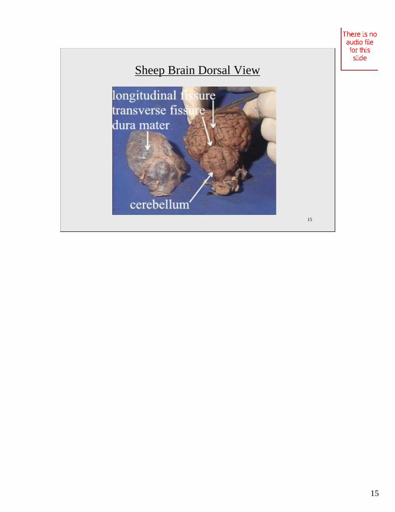
Sheep Brain Dorsal View

16
16
Sheep Brain Cerebellum

17
17
Sheep Brain: The Corpora Quadrigemia

18
18
Sheep Brain Ventral View
19
19
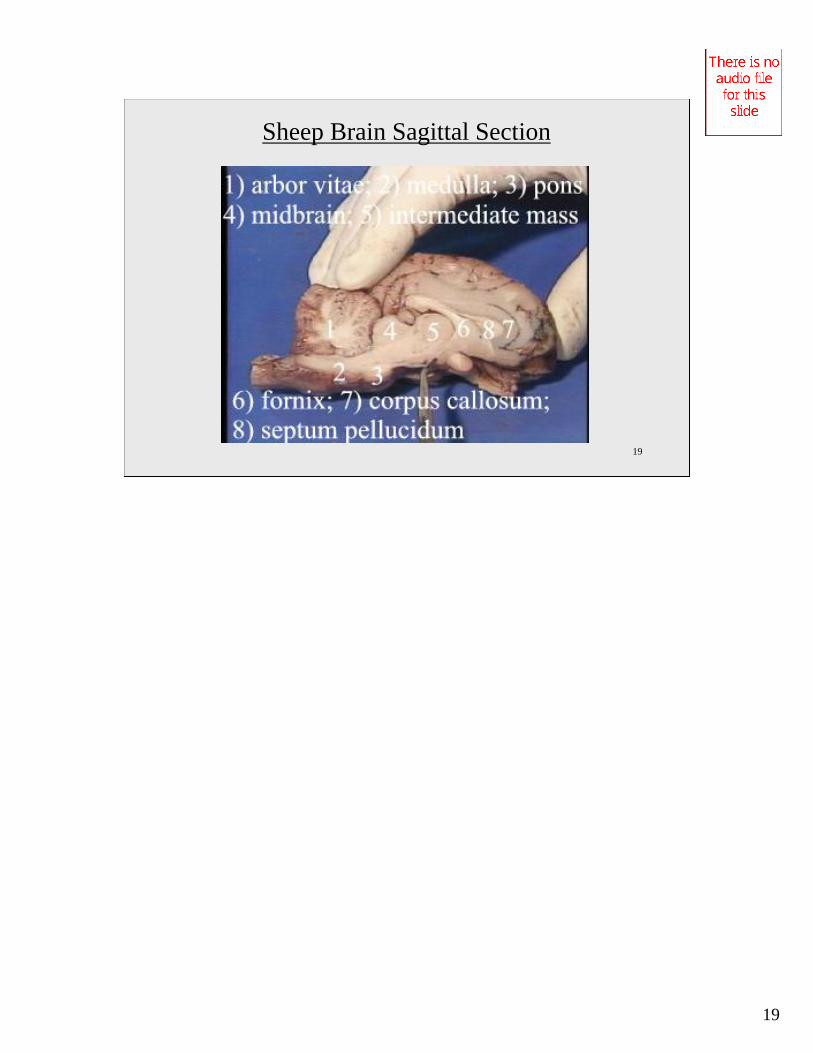
Sheep Brain Sagittal Section

20
20
Cerebellar Purkinje Cells
Interneuron cell bodies
Dendrites Axon to cerebellaroutflow
Purkinje cells are the largest and most distinguishing cells of the cerebellum. They have numerous dendrites and an axon which is the beginning of cerebellar outflow.

21
21
Alzheimer’s Tangle
This is a neurofibrillary "tangle" of Alzheimer's disease. The tangle appears as long pink filaments in the cytoplasm. They are composed of cytoskeletalintermediate filaments.

22
22
Alzheimer’s Tangle, Silver Stain
The characteristic microscopic findings of Alzheimer's disease include "senile plaques" which are collections of degenerative presynaptic endings along with astrocytes and microglia. These plaques are best seen with a silver stain, as seen here in a case with many plaques of varying size.

23
23
Lab Protocol for Spinal Nerves and Reflexes
1) Complete the Review Sheets for the exercise on the Gross Anatomyof the Brain and Cranial Nerves
2) Take the related quiz for the Brain and Cranial Nerves.
3) View the cadaver video showing dissection of the sheep brain.