Embed Size (px)
Citation preview

Five Things I Learned
about Nephrolithiasis in
Pregnancy
Nika Mehta, MD
Asst. Professor of Medicine
Warren Alpert Medical School of Brown University
Director of Ambulatory services,
Div. of Obstetrics and Consultative Medicine,
Women and Infants Hospital.


Nephrolithiasis in pregnancy is
commonly encountered
1.

Background:
Renal colic is the most common cause of
non-obstetric abdominal pain necessitating
hospitalization.
Incidence 1/200 to 1/1500 pregnancies,
same as the general population.
Majority occur in second or third trimester.
Multiparous greater than primiparous 3:1.
Incidence in General Population Stones affect 10% of general population.
Incidence increasing steadily over the last decade.
40% increase in hospital visits.
50% increase in annual expenditure from 1992-2000.
Urologic Diseases of America Project: urolithiasis. J. Urol. 173, 848–857 (2005).Time trends in reported prevalence of kidney stones in the United States: 1976–1994. Kidney Int. 63, 1817–1823 (2003).
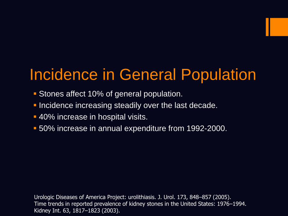
Stone Belt of America
Incidence:
In general population, incidence increasing
presumably from:
Obesity
Environmental changes
Increased incidence of comorbidities
like Diabetes Mellitus and Metabolic
Syndrome

Several Anatomic and
Physiologic Changes of
Pregnancy Affect Stone
Formation
2.

Anatomical Alterations in the
Urinary Tract in Pregnancy

Pregnancy Hydronephrosis
Physiologic hydronephrosis (Grade II) noted
in 90% of pregnancies

Hydronephrosis
90% of the right kidneys and 67% of the left kidneys.
Mechanical Effect:
Preferential compression of the right ureter due to dextrorotation of the uterus and compression by ovarian vein plexus.
Protection of the left ureter by the gas filled sigmoid colon.

Physiologic Hydronephrosis
Hormonal Effect:
Progesterone induced smooth muscle dilatation, resulting in reduced ureteral peristalsis and further dilatation.
Responsible for the hydronephrosis seen early in pregnancy, 6-10 weeks.

Physiologic Alterations in the
Urinary Tract in Pregnancy

Renal Physiology in Pregnancy
Increased GFR and renal plasma flow
due to:
Increased cardiac output.
Decreased renal vascular resistance.
Cr clearance increases by 50%.

Excretion of Stone Related
Substances in Pregnancy

SodiumIncreased circulating levels of natriuretic hormones: progesterone, HCG, aldosterone

Gestational Hypercalciuria
Increased GFR increased filtered load
of Calcium.
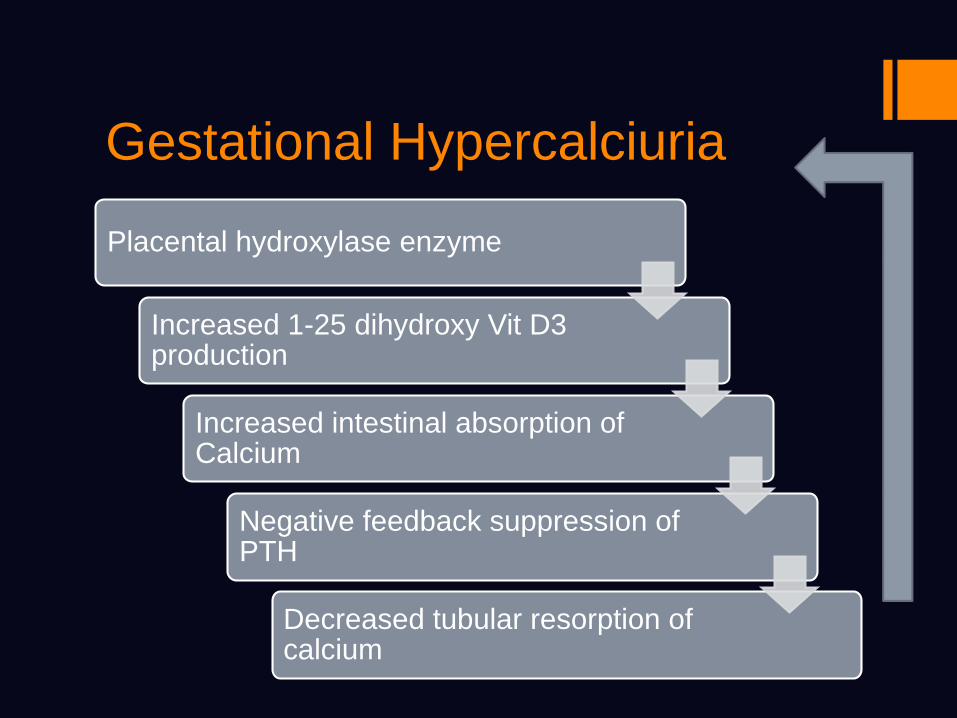
Gestational Hypercalciuria
Placental hydroxylase enzyme
Increased 1-25 dihydroxy Vit D3 production
Increased intestinal absorption of Calcium
Negative feedback suppression of PTH
Decreased tubular resorption of calcium

Gestational Glycosuria
Pregnancy is associated with a lowering of
the renal threshold for glucose excretion.
Increased GFR leads to increased glucose
load in the urine.
Reabsorption is compromised due to
overwhelming load.

Renal Physiology in Pregnancy
Increased renal filtration of potential stone
promoters
Glucose, Na, Ca and uric acid

Ureteral obstruction+ dilatation
Urinary stasis Longer contact
time between lithogenic factors
enhanced crystallization.

Stone Incidence in Pregnancy
Despite this, pregnancy is not associated
with an increased incidence of stone
formation
limited duration
Increased filtration of stone inhibitors:
Citrate, magnesium,
glycosaminoglycans
Nephrocalcin, uromodulin, thiosulfate

Stone Composition in
Pregnancy
Calcium phosphate stones predominant
stone type in pregnancy whereas oxalate
more common in general population.
Urol Res (2008) 36:99–102

USG is the Imaging Modality
of Choice
3.

X-ray Procedures during
Pregnancy: Key Points to
Review
No teratogenic effect of radiation if total
radiation exposure is kept below 5 rads
throughout gestation.
Almost all radiologic procedures involve far
less than 5 rads of radiation.
ACOG Committee opinion. Number 299, Sept 2004.

Radiation Exposure Associated With
Some Common Diagnostic Imaging
Type of Imaging Radiation Exposure (RADS)
Head CT <.001
Chest Xray <.001
Abdominal film (single view)
.01
Intravenous pyelogram
>1.0 rad
Abdominal CT >3.0 rad
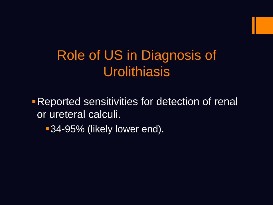
Role of US in Diagnosis of
Urolithiasis
Reported sensitivities for detection of renal
or ureteral calculi.
34-95% (likely lower end).

Differentiating Physiologic
Hydronephrosis from Obstructive
Ureteral Jets: Absence on affected side suggests obstruction. Reported sensitivity 100%, specificity 91%.
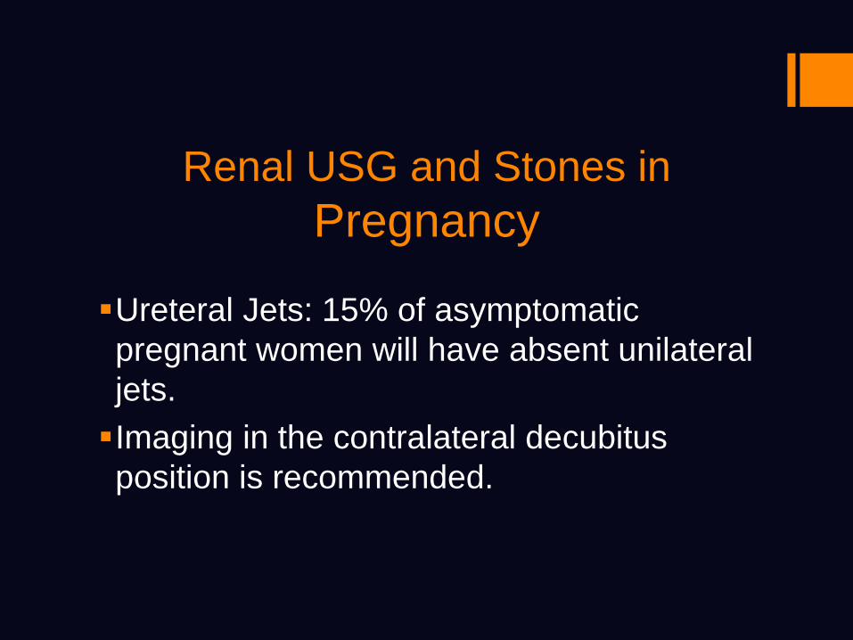
Renal USG and Stones in
Pregnancy
Ureteral Jets: 15% of asymptomatic
pregnant women will have absent unilateral
jets.
Imaging in the contralateral decubitus
position is recommended.
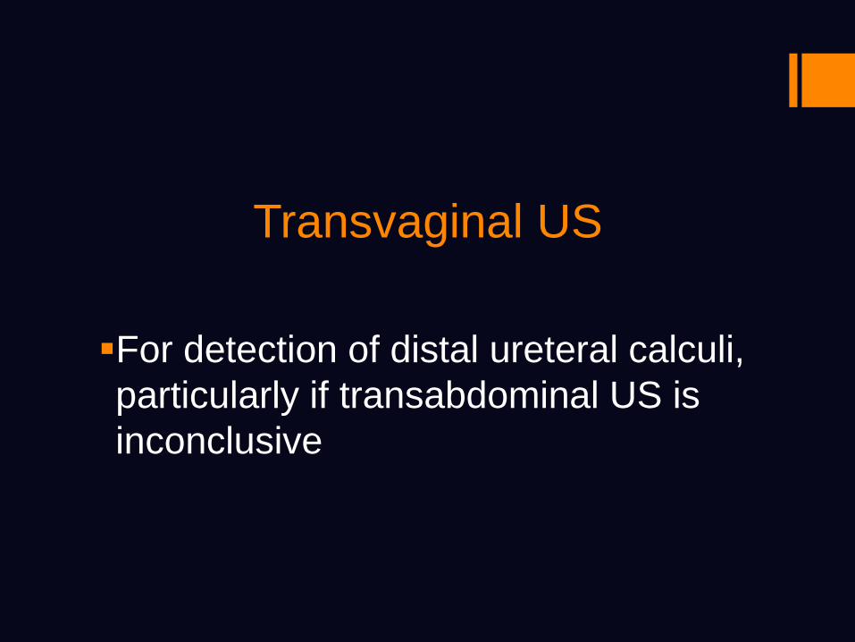
Transvaginal US
For detection of distal ureteral calculi,
particularly if transabdominal US is
inconclusive

Other Imaging Modalities
If diagnosis unclear from USG
T2-weighted half-fourier single-shot
turbo-spin echo (HASTE) MRU
(Magnetic Resonance Urography)

MR Urography
Second line test after US in pregnant pts
Relatively insensitive for direct detection
of small renal stones.
But can show secondary changes of
obstruction (Renal enlargement and
perirenal fluid).

MRU

Intravenous Pyelogram
Superior visualization of ureters compared
with US.
Less radiation than CT.
Limited 3 shot IVP: scout film, 30 second
and 20 minute films.
Single shot IVP: single film 5 minutes after
IV contrast injection.

IVP

IVP
Things to think about:
Iodinated contrast used in IVP can cross
placenta, has free iodide (fetal thyroid).
No fetal effects ever reported.
Superimposition of fetal skeleton can
obscure visualization of calculi.

CT
Low dose and ultra low dose CT stone
protocols have been developed.
Very low radiation dose comparable to the
‘3 shot’ IVP.
Not fully evaluated in pregnant women, but
small studies report 98% sensitivity and
95% specificity.

Algorithm for Imaging

Pregnancy outcomes are
not really affected by
nephrolithiasis
4.
Pregnancy Outcomes and
Renal Stones
Increased incidence of premature rupture
of membranes in pregnancies complicated
by renal stones.
Other pregnancy complications such as
preeclampsia, pregnancy loss are
unaffected.
J Reprod Med. 2003 Jan;48(1):28-32.

Most patients will only need
conservative management
5.
Management
Most stones < 1cm will pass spontaneously.
Rate of passage greater than general
population likely due to pregnancy
associated dilatation.
Of stones that do not pass in pregnancy, half
will pass after delivery.

Management
Trial of passage with adequate pain
management and aggressive hydration
Narcotics used for pain
NSAID’s not safe for use in pregnancy
Alpha blockers like Tamsulosin (FDA
category B for pregnancy safety) ok to
use

Management
Indications for aggressive approach:
Colic refractory to drug therapy
Sepsis/infection
Uncontrolled pain
Obstruction in single kidney
Renal dysfunction (normal creatinine low in
pregnancy)
Preterm labor
Psychosocial reasons

ManagementMore aggressive measures include:
Stents/ percutaneous nephrostomy tubes when
temporary drainage is needed.
Both need frequent changes to minimize risk of
encrustation.
Encrustation risk increased in pregnancy due
to hypercalciuria, hyperuricosuria and elevated
urine pH.
Ureteroscopy / Surgery for more definitive
treatment

Ureteroscopy
Has been successfully employed
throughout pregnancy, with no obstetric
complications reported.
May need GA, but has been performed with
sedation only.
Technically difficult in the third trimester.

Ureteroscopy
Potentially easier in pregnant women
because of smooth muscle dilatation.
Performed under fluoroscopy or US
guidance.
High success rates reported, obviating the
need for indwelling stent or nephrostomy
tube.

Ureteroscopy
Laser or pneumatic lithotripsy can be safely
performed in pregnancy after ureteroscopic
access obtained or stone can be retrieved
with a stone basket.

Open Surgery
Remains a viable alternative, especially in
symptomatic septic patients where
endourologic procedures have failed or are
unavailable.
Slight increased risk of preterm delivery.

Summary
Despite changes in pregnancy conducive to stone
formation, incidence of nephrolithiasis is not
increased.
USG is first-line imaging modality, but low dose
CT or MRU may be reasonable alternatives.
Most pregnant women respond to conservative
measures.
Stents, PCN, ureteroscopy may be performed
safely in pregnancy.

Questions?