Embed Size (px)
Citation preview
Nephrolithiasis
Cynthia Denu-Ciocca, M.D.

Epidemiology
Incidence 1:1000 per year Peak onset 20 - 35 years of age Male:Female 3 - 4 : 1

Epidemiology
In their lifetime2 - 5% of the Asian population
8 - 15% of North Americans and Europeans20% of Saudi
Arabians will develop a kidney stone
Epidemiology
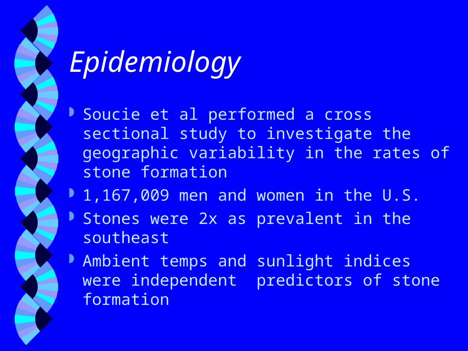
Soucie et al performed a cross sectional study to investigate the geographic variability in the rates of stone formation
1,167,009 men and women in the U.S. Stones were 2x as prevalent in the southeast Ambient temps and sunlight indices were
independent predictors of stone formation

Epidemiology
Curhan et al studied the influence of FH on stone formation
17.2% of men vs. 6.4% + family history 1986 - 1994 : 795 incident cases of stones RR of stone formation in men with +FH
2.57 (95% CI 2.19 - 3.02)

Epidemiology
Serio and Fraioli confirmed hereditary predisposition
22.5% of patients who developed stones in Italy between 1993 - 1994 had a positive FH in one or both of their parents

Natural HistoryRecurrence Rates 40 % in 2 - 3 years 55% in 5 - 7 years 75% in 7 - 10 years 100% in 15 - 20 years

Stone Formation
Urine saturation

Stone Formation
Urine saturation
Supersaturation

Stone Formation
Urine saturation
Supersaturation
Crystal nucleation
Stone Formation
Urine saturation
Supersaturation
Crystal nucleation
Aggregation

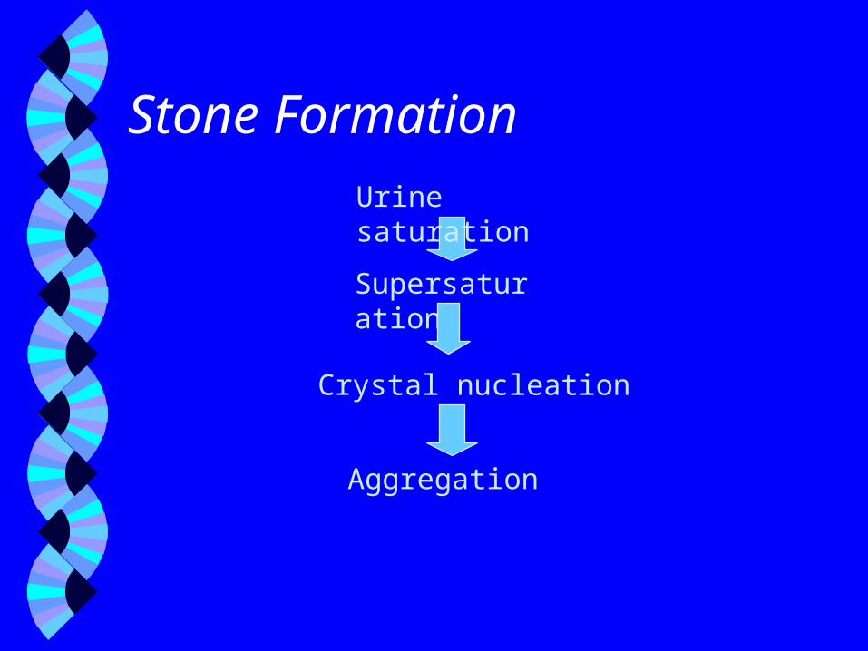
Stone Formation
Urine saturation
Supersaturation
Crystal nucleation
Aggregation
Retention and growth

Saturation
Saturation level - specific concentration of a salt = solubility product and no more salt can be dissolved
Crystals form in supersaturated urine Saturation is dependent on chemical free
ion activities of the components of a stone

Chemical free ion activities
concentration of the relevant ions urine pH complexation with substances in the urine

Nucleation
Homogeneous - supersaturation reaches a formation product and nuclei form in free solution
Heterogeneous - crystal growth occurs on the surface of dissimilar but complimentary crystal or foreign substances
Clinical Presentation
Renal colic begins suddenly and intensifies over 15 - 30 minutes
Associated with nausea & vomiting Pain passes from the flank anteriorly to the
groin At the ureterovesicular junction, urinary
frequency and dysuria may occur Microscopic hematuria> 75%, gross - 18%

Patient Evaluation: BasicSingle Stone Former Stone history Medical disease: skeletal disease, IBD,
UTI’s, HIV, granulomatous disease Meds: Lasix, glucocorticoids, theophylline,
calcium, vitamins A, C, and D

Patient Evaluation: BasicSingle Stone Former Family history Lifestyle/occupation Diet/fluids: protein, coffee, tea, dairy

Patient Evaluation: BasicSingle Stone Former Physical Examination Laboratory data
• Urinalysis, urine for cystine• Urine culture
-• Blood tests
• electrolytes, creatinine, calcium, phosphorous, uric acid, intact PTH if calcium elevated

Patient Evaluation: BasicSingle Stone Former Radiology
• KUB• IVP: anatomic abnormality, medullary sponge
kidney• Ultrasound• Unenhanced CT

Patient Evaluation: Complete
All patients with “metabolically active” stones, children, people in demographic groups that don’t usually form stones
Metabolically active stone: grow in size, number, or are passed within 1 year of f/up
Basic evaluation and 24 hour urine for volume, calcium, oxalate, sodium, phosphorous, uric acid, citrate, cystine

Patient Evaluation: Complete
Optimal values of 24-hr urine constituents:• volume > 2 - 2.5 L/day• calcium < 4 mg/kg or < 300 mg ,< 250 mg • uric acid < 800 mg , < 750 mg • citrate > 320 mg• sodium < 200 meq• phosphorous < 1100 mg• pH > 5.5 and < 7.0
Etiology
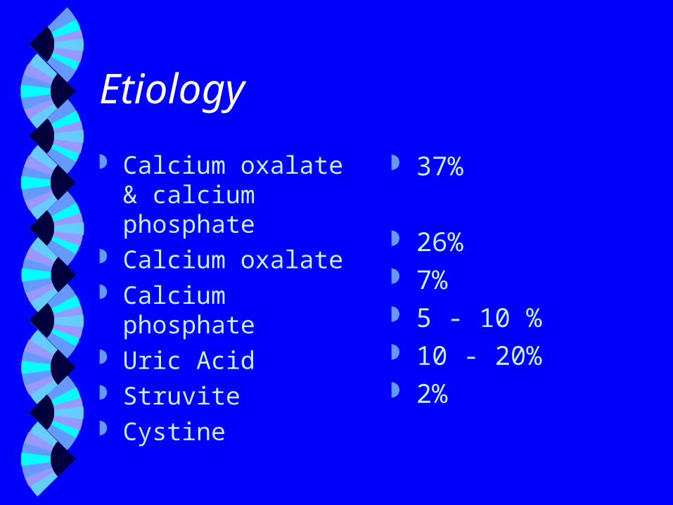
Calcium oxalate & calcium phosphate
Calcium oxalate Calcium phosphate Uric Acid Struvite Cystine
37%
26% 7% 5 - 10 % 10 - 20% 2%

Calcium Stones
70 - 75% of all stones Calcium oxalate - brown, gray, or tan Calcium oxalate monohydrate - dumbbell Calcium oxalate dihydrate - pyramidal Calcium phosphate - white or beige Calcium phosphate - elongated (brushite)
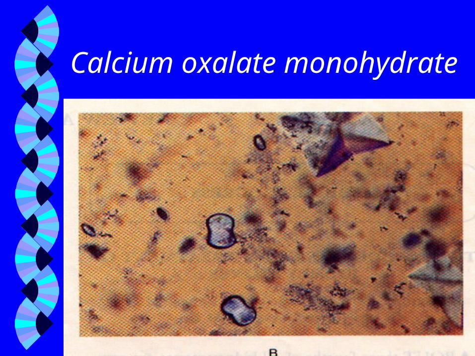
Calcium oxalate monohydrate

Calcium oxalate dihydrate

Calcium phosphate brushite

Calcium StonesPathophysiology Hypercalciuria Hypocitraturia Hyperoxaluria Unclassified
40 - 50% 20 - 30% 5% 25%

Calcium StonesHypercalciuria - Absorptive
Calcium Absorption
Serum calcium
PTH Urinary Calcium

Calcium StonesHypercalciuria - Absorptive Urine calcium exceeds 250 mg/day in
females, 300 mg/day in males Most common cause of hypercalciuria familial, 50% of 1st degree relatives, M=F Patients have a higher incidence of reduced
bone mineral density Caoxalate & Ca++phosphate stones

Calcium StonesHypercalciuria - Absorptive Pak et al have proposed two subtypes Type I - hypercalciuria on random and low
calcium diets Type II - normocalciuria on restricted diets Etiology unknown Some patients have high serum calcitriol
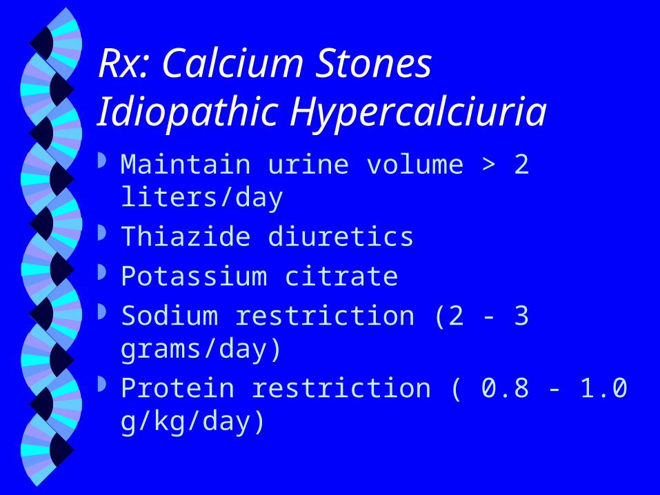
Rx: Calcium StonesIdiopathic Hypercalciuria Maintain urine volume > 2 liters/day Thiazide diuretics Potassium citrate Sodium restriction (2 - 3 grams/day) Protein restriction ( 0.8 - 1.0 g/kg/day)

HypercalciuriaDent’s Disease Rare, X-linked inheritance Tubular defect which causes renal
phosphate wasting(PO4 2.9 mg/dl) Stimulates vitamin D, intestinal calcium
absorption, urinary calcium Rx: Phosphorus replacement
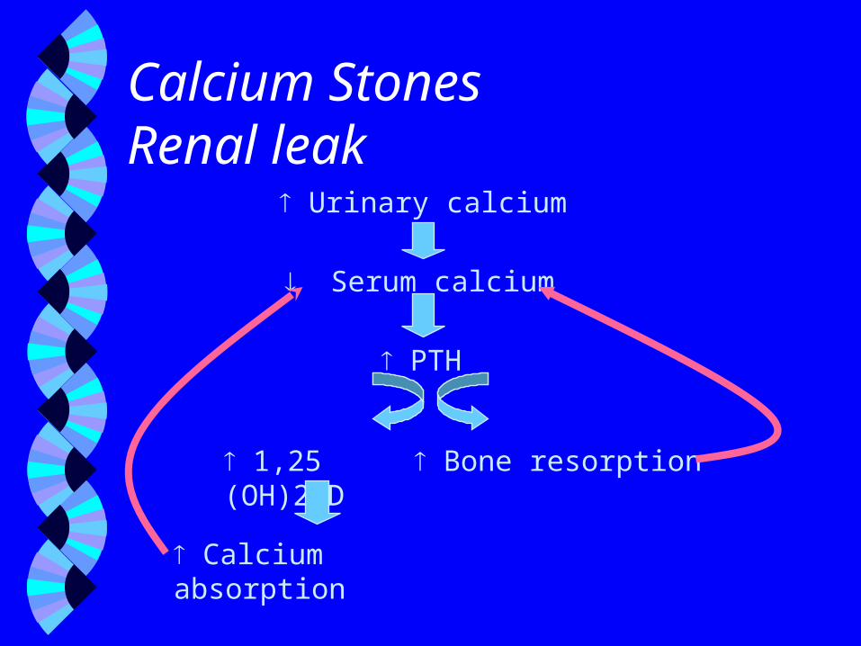
Calcium StonesRenal leak
Urinary calcium
Serum calcium
PTH
1,25 (OH)2 D Bone resorption
Calcium absorption

Calcium StonesHypercalciuria - Resorptive
PTH
1,25 (OH)2 D Bone Resorption
Calcium Absorption Serum Calcium
Urinary Calcium

Calcium StonesHyperparathyroidism 85% adenoma, 15% multigland hyperplasia Urine pH tends to be higher than in
idiopathic hypercalciuria so the fraction of calcium phosphate stones is higher
Rx: Parathyroidectomy

Calcium StonesHypocitraturia Occurs alone (10%) or with other
abnormalities (50%) More common in females May be idiopathic or secondary Acidosis reduces urinary citrate by tubular
reabsorption

Calcium StonesHypocitraturia Associated with distal RTA, metabolic
acidosis of diarrhea, & consumption of a diet rich in meat
Tend to form calcium oxalate stones (except Type I RTA)
Rx: dietary protein, alkali (K citrate or K bicarbonate), avoid sodium bicarbonate
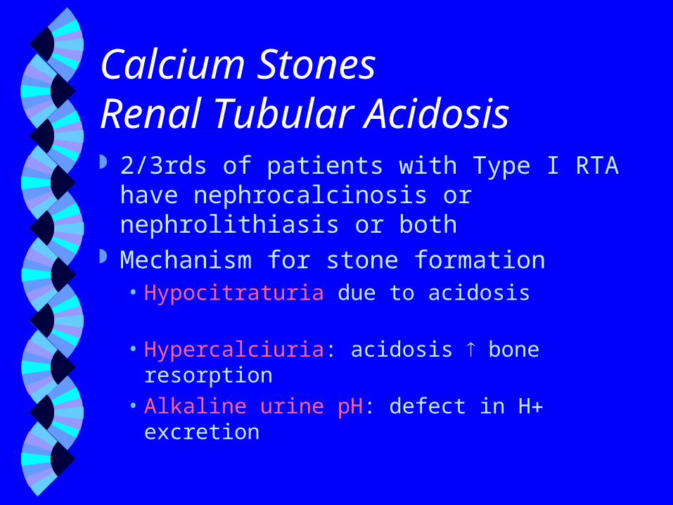
Calcium StonesRenal Tubular Acidosis 2/3rds of patients with Type I RTA have
nephrocalcinosis or nephrolithiasis or both Mechanism for stone formation
• Hypocitraturia due to acidosis • Hypercalciuria: acidosis bone resorption• Alkaline urine pH: defect in H excretion
Calcium StonesRenal Tubular Acidosis Form calcium phosphorus stones largely
due to the alkaline urine pH which decreases the solubility of calcium phosphate complexes
Rx: Alkali (calciuria, citraturia), thiazides if hypercalciuria persists

Calcium StonesHyperoxaluria Most patients with calcium oxalate stones
excrete normal urinary oxalate: <40 mg/day Excessive urinary oxalate
• Dietary hyperoxaluria• Endogenous hyperoxaluria• Enteric hyperoxaluria

Calcium StonesDietary Hyperoxaluria Normal people absorb < 5% of dietary
oxalate Foods rich in oxalate (spinach, chocolate,
beets, peanuts) can absorption 25 - 50% Low calcium diets urinary oxalate
excretion
Calcium StonesEndogenous hyperoxaluria Primary hyperoxaluria - enzymatic
deficiencies that result in massive oxaluria Widespread calcium oxalate deposition in
tissues ( bone marrow, blood vessels, heart, and renal parenchyma)
Rx: P.O. fluids, pyridoxine and orthophosphate urinary crystallization

Calcium
Calcium Oxalates
Calcium Soaps
Oxalic Acid
FattyAcids
NORMAL
A AB BS SO OR RP PT T I IO ON N
Calcium StonesEnteric Hyperoxaluria

Calcium StonesEnteric Hyperoxaluria
Calcium
Calcium Oxalates
Calcium Soaps
Oxalic Acid
FattyAcids
ENTERIC HYPEROXALURIA
A AB BS SO OR RP PT T I IO ON N

Calcium StonesEnteric Hyperoxaluria Treatment
• Decrease dietary oxalate and fat• Oral Calcium supplements• Cholestyramine• Increase fluid intake• Oral citrate

Calcium StonesHyperuricosuria Increased frequency of hyperuricosuria
(> 800 mg/day , > 750 mg/day) in patients who form calcium stones
Urate crystals serve as a nidus for calcium oxalate nucleation & comprise 4 - 8% of the cores of calcium stones
Rx: Allopurinol, Potassium citrate

Uric Acid Stones
Smooth, white or yellow-orange Radiolucent Crystals form various shapes: rhomboidal,
needle like, rosettes, amorphous

Uric acid
Uric Acid Stones
Main determinants of uric acid stone formation• Urinary pH < 5.5, urine volume
Hyperuricosuria• Genetic overproduction
• Myeloproliferative disorders
• High purine diet
• Drugs

Uric Acid Stones
Rx: fluid intake, purine diet, urinary alkalinization (pH 6.5 - 7.0) with potassium citrate , allopurinol to reduce 24 hour uric acid excretion

Struvite Stones - Magnesium Ammonium Phosphate More common in women than men Most common cause of staghorn calculi Grow rapidly, may lead to severe
pyelonephritis or urosepsis and renal failure Light brown or off white Gnarled and laminated on X-ray
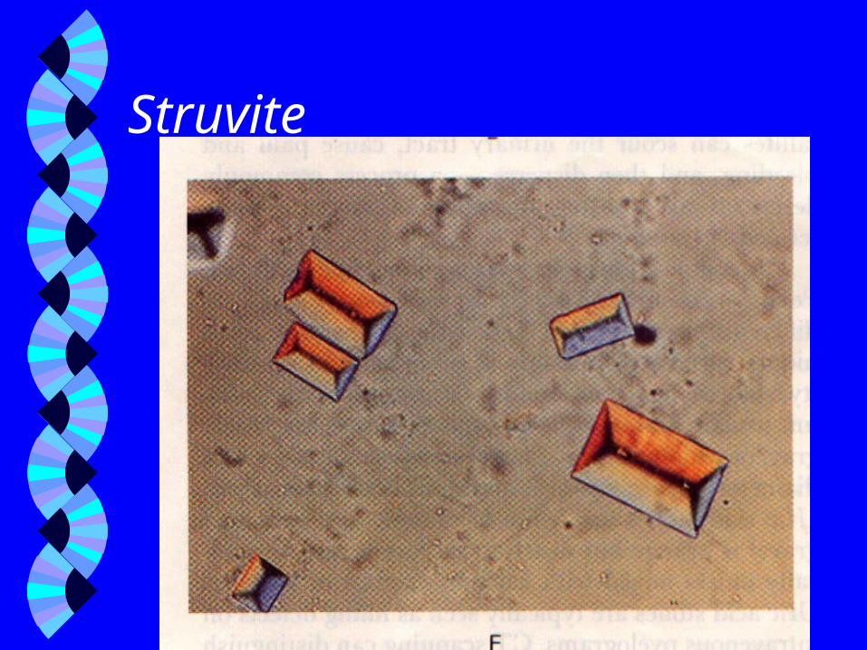
Struvite

Struvite StonesInfection Stones Caused in part by infections by organisms
with urease ( Proteus, Klebsiella, Pseudomonas, and Serratia)
Hydrolysis of urea yields ammonia & hydroxyl ions, consumes H+ & thusurine pH
urine pH increases saturation of struvite
Struvite StonesInfection Stones Rx: Remove existing stones (harbor
causative bacteria) with ESWL or PUL Prolonged antibiotics Acetohydroxamic acid (urease inhibitor) -
use limited because of side effects
Cystine Stones
Hereditary disorder caused by a tubular defect in dibasic amino acid transport, autosomal recessive
Excrete excessive amounts of cystine, ornithine, lysine and arginine

Cystine Stones
Cystine is soluble in the urine to a level of only 24 - 48 mg/dl
In affected patients, the excretion is 480 - 3500 mg/day
Nephrolithiasis usually occurs by the 4th decade

Cystine Stones
Hexagonal, radiopaque, greenish-yellow Often present as staghorn calculi or
multiple bilateral stones
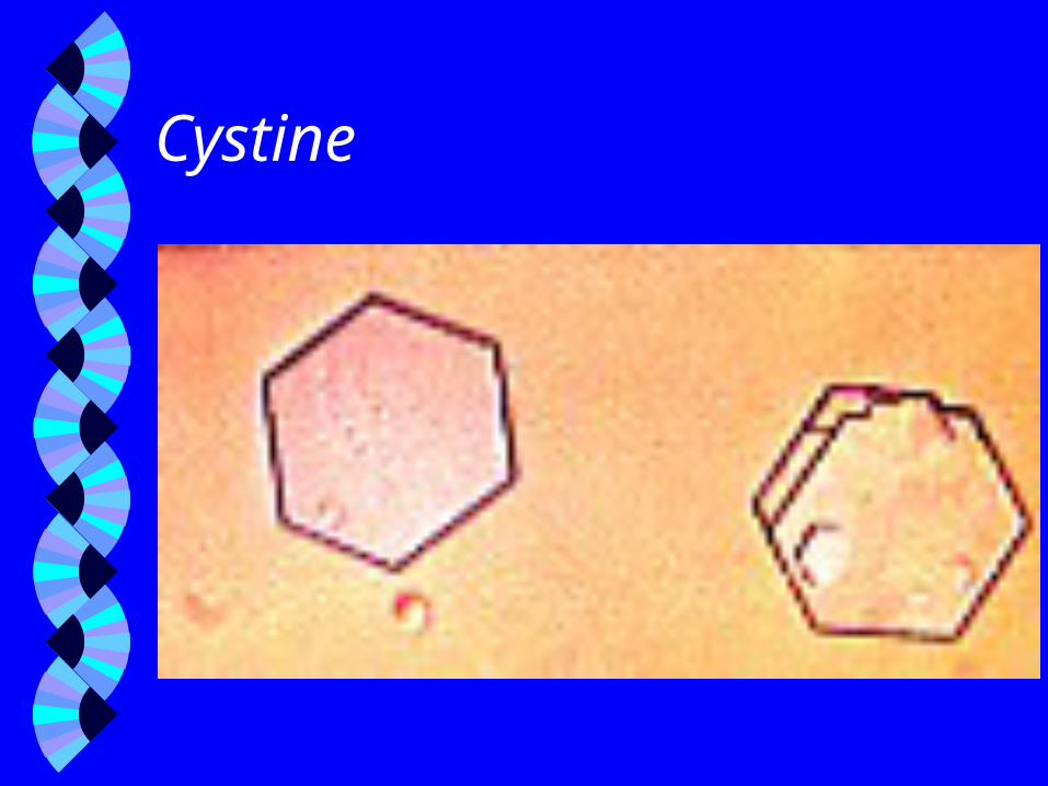
Cystine

Cystine

Cystine Stones
Treatment• estimate urine volume to maintain solubility
( 240 - 480 mg/l)• urine pH > 7.5• Restrict dietary sodium (60 meq/d)• Troponin or D-penicillamine bind cystine and
reduce urine supersaturation• Urological: removal difficult (2nd hardest) -PUL
often required

NephrolithiasisTreatment - Calcium Stones Peale, Roehborn and Pak performed a meta-
analysis to determine the efficacy of different drug therapies for stone disease
14 randomized, controlled trials, 6 drugs tx. Treatment arms: Thiazide diuretics (7),
allopurinol (4), magnesium (2), alkali citrate (3), phosphate (3), non-thiazide diuretic (1)

NephrolithiasisTreatment Significant reduction in stone recurrence
was found in 5/7 of the thiazide trials and the indapamide trial
Two remaining trials with no differences had mean follow up < 2 years
The phosphate and the magnesium trials showed no treatment benefit

NephrolithiasisTreatment Only 1/4 of the allopurinol studies showed a
significant benefit In that study however patients were selected
for hyperuricosuria and normocalcuria
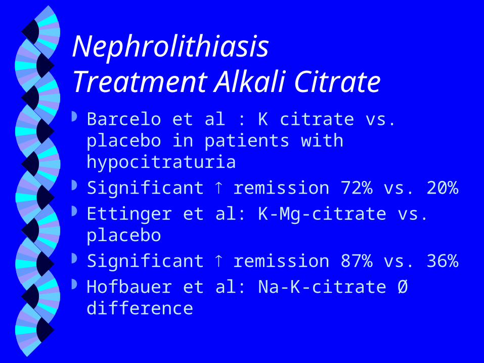
NephrolithiasisTreatment Alkali Citrate Barcelo et al : K citrate vs. placebo in
patients with hypocitraturia Significant remission 72% vs. 20% Ettinger et al: K-Mg-citrate vs. placebo Significant remission 87% vs. 36% Hofbauer et al: Na-K-citrate Ø difference

NephrolithiasisTreatment Meta-Analysis on 11/14 trials Risk difference of -22.6% for patient
receiving treatment 95% CI - 29.0 % to - 16 % , P < 0.001

NephrolithiasisTreatment Lee et al performed a retrospective analysis
to assess the efficacy of K citrate based tx 439 patients, 3 groups (regular prophylaxis,
intermittent prophylaxis, no prophylaxis) Stone recurrence was significantly in
group I (7.8% vs. 30% vs. 46%, p<0.001)

NephrolithiasisTreatment - Non-specific Curhan et al: prospective cohort study to
assess whether dietary ca++ and supplemental calcium risk of stones
dietary calcium relative risk = 0.65 (p = 0.005, 95%CI, 0.50 - 0.83)
Relative risk of supplemental calcium was 1.20 (p = 0.03)

NephrolithiasisTreatment Sodium and sucrose intake were associated
with an risk of stones Potassium and fluid were associated with
risk of stones ( RR 0.65 and 0.61 respectively)

Urological Treatment of Nephrolithiasis Extracorporeal shock wave lithotripsy
(ESWL)• Complications
• pain, steinstrasse (filling of the ureter with fragments of stone), bruising, perinephric hematoma, pancreatitis, urosepsis, BP
Percutaneous Urolithotomy Ureteroscopic lithotripsy or extraction
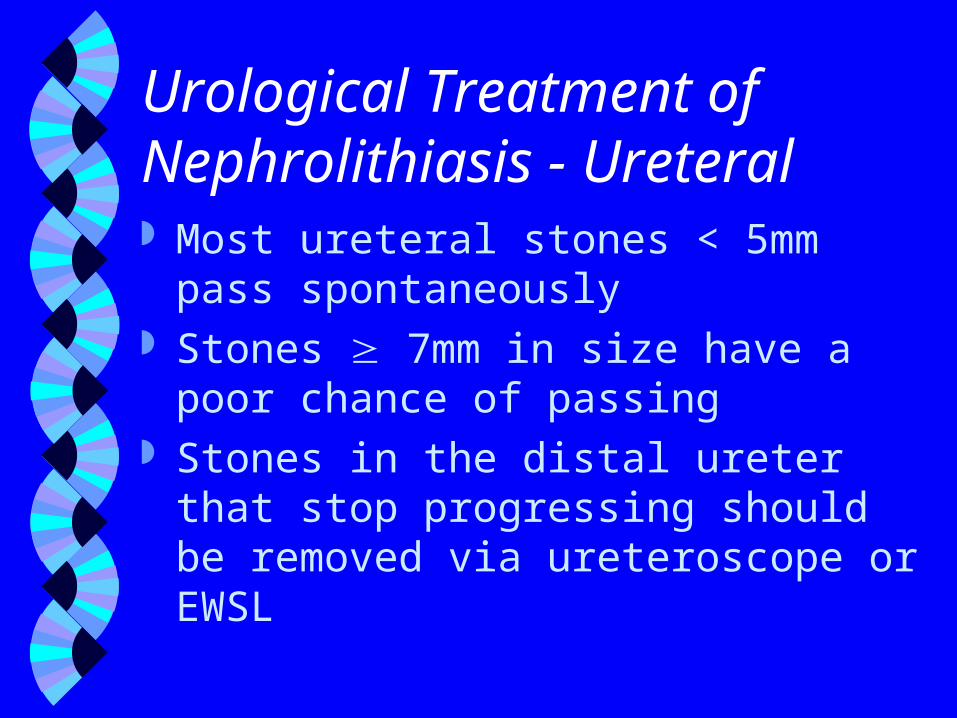
Urological Treatment of Nephrolithiasis - Ureteral Most ureteral stones < 5mm pass
spontaneously Stones 7mm in size have a poor chance
of passing Stones in the distal ureter that stop
progressing should be removed via ureteroscope or EWSL

Urological Treatment of Nephrolithiasis - Ureteral Stones in the proximal ureter that stop
progressing should be pushed upward into the renal pelvis, then disrupted with EWSL
If above fails, PUL

Urological Treatment of Nephrolithiasis - Renal Pelvis Stones that are < 2 cm and > 5 mm - EWSL Stones > 2 cm or exceed 1 cm and are in
the lower poles require PUL

Bibliography Monk RD. Clinical Approach in Adults. Semin Neph 1996; 16(5):375-388 Pak CYC. Kidney Stones. Lancet 1998; 351:1797-1801 Parks JH, Coe FL. Pathogenesis and Treatment of Calcium Stones. Semin Neph 1996; 16(5):398-411 Coe FL, Parks JH, Asplin JR. The Pathogenesis and Treatment of Kidney Stones. N Engl J Med 1992;
327(16):1141-1152 Soucie JM, Coates RJ, McClellan W, Austin H, Thun M. Relation between Geographic Variability in
Kidney Stone Prevalence and Risk Factors for Stones. Am J Epidemiol 1996; 143(5):487-495 Preminger GM. Renal Calculi: Pathogenesis, Diagnosis and Medical Therapy. Semin Neph 1992;
12(2):200-216 Serio A, Fraioli A. Epidemiology of Nephrolithiasis. Nephron 1999; 81:26-30 Mandel N. Mechanism of Stone Formation. Semin Neph 1996; 16(5):364-374 Pearl MS, Roehrborn CG, Pak CYC. Meta-Analysis of Randomized Trials for Medical Prevention of
Calcium Oxalate Nephrolithiasis. J Endo 1999; 13(9)679-685 Lee Y, Huang WC, Tsai JY, Huang JK. The Efficacy of Potassium Citrate Based Therapy. J Urol 1999;
161:1453-1457 Curhan GC, Willett WC et al. Comparison of Dietary Calcium with Supplemental Calcium and Other
Nutrients as Factors Affecting the Risk for Kidney Stones in Women. Ann Intern Med 1997; 126(7):497-504