Embed Size (px)
Citation preview
RESEARCH Open Access
Neorickettsia risticii surface-exposed proteins:proteomics identification, recognition bynaturally-infected horses, and strain variationsKathryn E Gibson, Gabrielle Pastenkos, Susanne Moesta and Yasuko Rikihisa*
Abstract
Neorickettsia risticii is the Gram-negative, obligate, and intracellular bacterial pathogen responsible for Potomachorse fever (PHF): an important acute systemic disease of horses. N. risticii surface proteins, critical for immunerecognition, have not been thoroughly characterized. In this paper, we identified the 51-kDa antigen (P51) as amajor surface-exposed outer membrane protein of older and contemporary strains of N. risticii through massspectrometry of streptavidin-purified biotinylated surface-labeled proteins. Western blot analysis of sera fromnaturally-infected horses demonstrated universal and strong recognition of recombinant P51 over otherNeorickettsia recombinant proteins. Comparisons of amino acid sequences for predicted secondary structures ofP51, as well as Neorickettsia surface proteins 2 (Nsp2) and 3 (Nsp3) among N. risticii strains from horses with PHFduring a 26-year period throughout the United States revealed that the majority of variations among strains wereconcentrated in regions predicted to be external loops of their b-barrel structures. Large insertions or deletionsoccurred within a tandem-repeat region in Ssa3. These data demonstrate patterns of geographical association forP51 and temporal associations for Nsp2, Nsp3, and Ssa3, indicating evolutionary trends for these Neorickettsiasurface antigen genes. This study showed N. risticii surface protein population dynamics, providing groundwork fordesigning immunodiagnostic targets for PHF.
IntroductionDiscovered in 1984, Neorickettsia (formerly Ehrlichia) ris-ticii is an obligate intracellular bacterium and the causativeagent of Potomac horse fever (PHF) [1-3]. The bacteriumuses a digenetic trematode to survive and proliferate in itsnatural lifecycle [4-9]. It is through accidental ingestionof the metacercarial stage of the digenetic trematodewithin its insect host that the horse becomes infected withN. risticii and develops PHF [6]. PHF is an acute, severe,and potentially fatal disease of horses, normally contractedduring the summer months in North America when aqua-tic insect larvae infested with N. risticii-infected digenetictrematodes molt and emerge (hatch) from the water asadults [6,10]. Clinical signs range from mild (anorexia,fever, lethargy, and depression) to life-threatening (lamini-tis, abortion, and diarrhea followed by severe dehydration)[10,11]. The administration of tetracyclines at the early
stage of infection is effective, in part by inhibiting bacterialprotein synthesis and facilitating lysosome fusion withinclusions containing N. risticii [12-15]. Diagnosis of thisdisease is mainly done by indirect fluorescent-antibody(IFA) test based on N. risticii-infected cells and by nestedpolymerase chain reaction (PCR) on blood samples[5,16-22]. The only available vaccines are bacterins usingthe 1984 N. risticii type strain, which demonstrate inade-quate efficacy [23,24].It was determined that N. risticii has similar genetic,
antigenic, and morphologic characteristics to Neorickett-sia helminthoeca [25,26], which were the major reasonsit, as well as Neorickettsia (formerly Rickettsia, Ehrlichia)sennetsu, was regrouped into the genus Neorickettsia[27]. In addition, the bacterial parasite, known as the Stel-lantchasmus falcatus (SF) agent, isolated from metacer-cariae in fish from Japan and Oregon [28-30] belongs tothis group. N. risticii also consists of a variety of strains,based on PCR and sequencing of 16S RNA and groEL,Western blot analyses using purified bacteria as antigen,and morphology [20,22,24,31].
* Correspondence: [email protected] of Veterinary Biosciences, The Ohio State University College ofVeterinary Medicine, 1925 Coffey Rd, Columbus, OH 43210, USA
Gibson et al. Veterinary Research 2011, 42:71http://www.veterinaryresearch.org/content/42/1/71 VETERINARY RESEARCH
© 2011 Gibson et al; licensee BioMed Central Ltd. This is an Open Access article distributed under the terms of the Creative CommonsAttribution License (http://creativecommons.org/licenses/by/2.0), which permits unrestricted use, distribution, and reproduction inany medium, provided the original work is properly cited.

Little is known about N. risticii surface-exposed pro-teins, and this missing information is crucial in the under-standing of bacterium-host cell interactions. Antigenic andpotential surface proteins ranging between 28 and110-kDa in mass were previously detected by Westernblotting, but these proteins were not identified [32].Immunoprecipitation of N. risticii labeled with I125 and N.risticii immune mouse sera revealed potential surface pro-teins ranging from 25 to 62-kDa in mass, although theseproteins were not identified [33]. Antigenic proteins of 70,55, 51, and 44-kDa masses have been demonstrated utiliz-ing recombinant proteins; again the proteins were notidentified [34]. Two highly-immunodominant proteins intwo N. risticii strains were identified as GroEL and the 51-kDa antigen (P51) [35], but it was not shown whetherthese proteins were surface exposed. Strain-specific anti-gen (Ssa) was suggested as a surface immunogenic proteinwith potential use in vaccine production, although it wasnot determined to be bacterial surface exposed [24,36].The identification of Neorickettsia proteins is now achiev-
able with the availability of whole genome sequencing dataon both the type strain (Miyayama) of N. sennetsu [37] andthe type strain (Illinois) of N. risticii [38]. In this paper, wedetermined 1) major surface proteins by proteomics analy-sis on N. risticii, 2) horse immune recognition of N. risticiisurface proteins, and 3) strain variations in alignedsequences of these major surface proteins with respect totheir predicted secondary structures.
Materials and methodsCulturing and isolation of N. risticii strainsN. risticii IllinoisT [3] and a Pennsylvania strain (PA-1) [6]were cultured in P388D1 cells in 75-cm2 flasks containingRPMI 1640 (Mediatech, Inc., Herdon, VA, USA) supple-mented with 5-10% fetal bovine serum (FBS) (U.S. Bio-technologies, Inc., Pottstown, PA, USA) and 4-6 mML-glutamine (Invitrogen, Carlsbad, CA, USA) at 37°Cunder 5% CO2. N. risticii was isolated from highly-infectedP388D1 cells as previously described for N. sennetsuMiyayamaT [39].
Biotinylation and streptavidin-affinity purification of N.risticii surface proteinsBiotinylation of purified N. risticii Illinois and PA-1 fromtwenty-five 75-cm2 flasks using EZ Link Sulfo-NHS-SS-Biotin (Pierce Biotechnology, Rockford, IL, USA) and sub-sequent bacterial lysis and collection of solubilized bacter-ial proteins were performed as previously described [39].Streptavidin purification of Sulfo-NHS-SS-BiotinylatedN. risticii proteins was then performed, followed by SDS-polyacrylamide gel electrophoresis (PAGE) and fixationand GelCode blue (Pierce) staining of the gel [39]. Proteinsfrom seven bands from N. risticii Illinois and proteinsfrom four bands or band collections from PA-1 were
identified by capillary-liquid chromatography-nanospraytandem mass spectrometry (Nano-LC/MS/MS) as pre-viously described [40].
Western blotting using recombinant proteinsRecombinant P51 (rP51, 57 kDa), cloned from N. risticiiIllinois (NRI_0235), and rNsp2 (35 kDa) and rNsp3 (28kDa), cloned from N. sennetsu Miyayama (NSE_0873 andNSE_0875, respectively), were expressed by transformedBL21(DE3) cells using isopropyl-b-D-thiogalactopyrano-side induction and His-tag purified as described previously[30,39]. Recombinant GroEL (55 kDa), derived fromN. sennetsu Miyayama (NSE_0642), was acquired fromstored aliquots [41]. Fifty μg of each recombinant proteinwere separated by SDS-PAGE, transferred to nitrocellulosemembranes, and cut into strips. Western blotting was thenperformed on these strips using 1:500 dilutions of knownpositive horse sera samples as determined by IFA [16,21].The membrane was subsequently incubated with a 1:1000dilution of horseradish peroxidase-conjugated goatanti-horse (Kirkegaard & Perry Laboratories, Inc.,Gaithersburg, MD, USA) as secondary antibody. Enhancedchemiluminescence (ECL) LumiGLO chemiluminescentreagent (Pierce) and a LAS3000 image documentation sys-tem (FUJIFILM Medical Systems USA, Stamford, CT,USA) were used to visualize the protein bands with 300 sexposure. Bands were aligned using Precision Plus pre-stained protein standards (Bio-Rad Laboratories, Hercules,CA, USA).
Polymerase chain reaction, sequencing, and sequencealignmentDNA was purified from buffy coats of PHF-positive horsesor cultures of N. risticii in P388D1 cells using the DNeasyBlood and Tissue Kit (QIAGEN, Valencia, CA, USA),according to manufacturer’s instructions. PCR amplifica-tion was then performed using either Phusion or TaqDNA polymerase (New England BioLabs, Ipswich, MA,USA) and primers designed for conserved regions throughalignment of multiple Neorickettsia spp. and/or N. risticiistrains (see Additional file 1). Sequencing was performedby The Ohio State University Plant-Microbe GenomicsFacility. Sequences containing whole genes or gene frag-ments were translated and aligned mainly through theCLUSTAL W (slow/accurate) method in the MegAlignprogram of DNAStar (DNAStar, Madison, WI, USA); P51was first aligned by CLUSTAL V (PAM250) method, andSsa3 was aligned both by CLUSTAL W and manually.External loops were also aligned separately by CLUSTALW for both P51 and Nsp3. Amino acid (aa) variations inN. risticii strains and other Neorickettsia spp. for all pro-teins were determined in relation to N. risticii Illinois. Pro-tein alignments of the same size (including deletions asdashes) were analyzed by PHYLIP (v3.66) to obtain
Gibson et al. Veterinary Research 2011, 42:71http://www.veterinaryresearch.org/content/42/1/71
Page 2 of 14

bootstrap values for 1000 replicates (using the programsSeqBoot, Protdist, Neighbor, and Consense) and to createdendrograms (using the programs Protdist, Neighbor, andDrawgram) [42]. Protein properties, including antigenicityprofiles and b-sheet predictions were determined usingthe Protean program (DNAStar). Gene and proteinsequence homologies were also demonstrated using BasicLocal Alignment Search Tool (BLAST) algorithms, includ-ing blastn, protein-protein blastp, and blastp [43,44].
Prediction of secondary structuresPredictions for Nsp2 and Nsp3 were based on a combi-nation of the programming algorithm in the PRED-TMBB web server [45], hydrophobicity and hydrophobicmovement profiles [46], and DNAStar MegAlign(DNAStar, Madison, WI, USA) alignment and analysesof all available N. risticii strain and Neorickettsia spp.sequences.
GenBank Accession NumbersGenBank accession numbers of all sequences deter-mined in this study are shown in Table 1. P51sequences previously deposited in GenBank used in thisstudy are listed in Table 2. Nsp2 sequences include N.risticii Illinois (NRI_0839, YP_003082043) and N. sen-netsu Miyayama (YP_746740). Previously-depositedNsp3 sequences include N. risticii Illinois (NRI_0841,YP_003082045) and N. sennetsu Miyayama (YP_506742).Ssa3 sequences include N. risticii Illinois (NRI_0872,YP_003082075) and N. sennetsu Miyayama (NSE_0908,YP_506773). The Ssa1 sequence is from N. risticii Illi-nois (NRI_0870, YP_003082073), and other Ssas arefrom 25-D (AAC31427) and 90-12 (AAC31428).
ResultsNano-LC/MS/MS of streptavidin-affinity purified surfaceproteinsGiven that only the N. risticii Illinois genome (NC_013009)has been sequenced [38], these data were used for proteo-mic analyses. Four N. risticii proteins in N. risticii Illinois(1984 isolate) and five N. risticii proteins (with conservedpeptide sequences in relation to N. risticii Illinois) in PA-1(2000 isolate) contained two or more peptide queries iden-tified by Nano-LC/MS/MS (Table 3). Proteins identifiedfor N. risticii Illinois were P51, GroEL (NRI_0614), Nsp3,and a conserved hypothetical protein (NRI_0567). The lar-gest protein coverage and the largest number of peptidesidentified were both from P51. Proteins identified in PA-1also included P51 and GroEL; the largest number of pep-tides was from P51. Minor proteins identified in PA-1strain were DnaK (NRI_0017), ATP synthase F1, alpha sub-unit (AtpA, NRI_0132), and strain-specific antigen 3 (Ssa3,NRI_0872).
Immune recognition of major surface antigens by PHF-positive horse seraBacterial surface-exposed proteins are generally majorantigens [47]. Though only Nsp3 was detected on the sur-face of N. risticii Illinois by nano-LC/MS/MS, rNsp2 wasincluded in the Western blotting studies because bothNsp3 and Nsp2 from N. sennetsu Miyayama are significantsurface proteins (Figure 1, Table 4) [39]. All 15 PHF-posi-tive samples demonstrated recognition of rP51, with 11out of 15 sera having strong recognition. N. sennetsuMiyayama GroEL is 98% identical to N. risticii IllinoisGroEL, and antisera to rGroEL of N. sennetsu cross-reactswith GroEL from multiple species of Rickettsiales, includ-ing N. risticii [41]. Six out of 15 PHF-positive serum sam-ples demonstrated strong reactivity to rGroEL, with therest having weak to no reactivity. Nsp2 and Nsp3 fromN. sennetsu Miyayama are 83% and 84% identical to Nsp2and Nsp3 from N. risticii Illinois, respectively, using pro-tein-protein blastp. Only one serum sample reactedstrongly to rNsp2, with the rest having weak to no reactiv-ity. Three sera reacted strongly to rNsp3, with the resthaving weak to no reactivity. All negative controls did notrecognize any of the recombinant proteins.
Sequence variation in P51P51 sequences are known to be strain variable [5,30].Since P51 was found to be the major target of horseimmune recognition, we examined in which part of theP51 molecule sequence variations occur. N. sennetsu P51was predicted to have 18 transmembrane b-barrel proteinswith nine external loops [39]. N. sennetsu and the SFagent, which are closely-related to N. risticii [28,30,48]were included for comparison. P51 alignments of a total of52 sequences and sequence fragments from N. risticii dur-ing a 26-year period throughout the United States revealedhigh variability within regions corresponding to externalloops 2 and 4 (Figure 2). Forty-three P51 sequence frag-ments (aa 136-176) containing most of external loop 2 (aa120-176), and 36 P51 sequence fragments (aa 259-286)containing the entire external loop 4 were analyzed usingPHYLIP (Figure 3a and 3b). Both loops 2 and 4 createdpatterns of clustering for sequences from states in theEastern and Midwestern United States (East/Midwest US)and sequences from Japan, Malaysia, and US states bor-dering the Pacific Ocean (Pacific coast). The Californiastrain Doc and the Ohio strain 081 did not follow this pat-tern, both being in East/Midwest US for external loop 2and in Pacific coast for external loop 4. In external loop 2,N. risticii Illinois was only loosely associated with theother East/Midwest US sequences; in external loop 4, N.risticii Illinois tightly clustered with several East/MidwestUS sequences. External loop 4 of 081 clustered with theSF agent strains rather than with other N. risticii strains.
Gibson et al. Veterinary Research 2011, 42:71http://www.veterinaryresearch.org/content/42/1/71
Page 3 of 14

Table 1 Sequences amplified for Neorickettsia
Sample IDa Location/Year Fragment size (bp)b Gene(s) amplifiedc Accession no.
PA-1 Pennsylvania/2000 2091 nsp2, nsp3 HQ857586
765 ssa1 (p) HQ857584
1812 ssa3 HQ857585
Herodia Pennsylvania/1999 673 p51 (p) HQ857589
2133 nsp2, nsp3 HQ857588
1460 ssa3 HQ857587
081 Ohio/1991 2420 nsp2, nsp3 HQ857591
717 ssa3 (p) HQ857590
MN Minnesota/2002 676 p51 (p) HQ857594
2156 nsp2, nsp3 HQ857593
1029 ssa3 (p) HQ857592
OV Kentucky/1993 2103 nsp2, nsp3 HQ857596
863 ssa3 HQ857595
IA03-1 Iowa/2003 1550 nsp2 (p), nsp3 HQ875741
IL01-1 Illinois/2001 623 nsp2 (p) HQ875742
489 nsp3 (p) HQ875743
IN01-1 Indiana/2001 1879 nsp2 (p), nsp3 HQ875744
IN02-1 Indiana/2002 2052 nsp2 (p), nsp3 HQ875745
IN02-2 Indiana/2002 542 p51 (p) HQ875747
733 nsp3 (p) HQ875746
IN03-1 Indiana/2003 542 p51 (p) HQ906674
2110 nsp2, nsp3 HQ906673
IN03-2 Indiana/2003 1361 nsp2, nsp3 (p) HQ906675
KY03-1 Kentucky/2003 673 p51 (p) HQ906678
594 p51 (p) HQ906679
306 p51 (p) HQ906680
2095 nsp2, nsp3 HQ906677
1129 ssa3 (p) HQ906676
KY03-2 Kentucky/2003 1398 nsp2, nsp3 (p) HQ906681
KY03-3 Kentucky/2003 1042 nsp2 (p), nsp3 (p) HQ906682
OH07-1 Ohio/2007 259 p51 (p) HQ906685
721 ssa1 (p) HQ906683
1739 ssa3 HQ906684
OH07-2 Ohio/2007 259 p51 (p) HQ906686
OH07-3 Ohio/2007 1558 nsp2 (p), nsp3 (p) HQ906688
995 ssa3 (p) HQ906687
OH07-4 Ohio/2007 654 p51 (p) HQ906691
1118 nsp2 (p), nsp3 (p) HQ906690
1029 ssa3 (p) HQ906689
OH10-1 Ohio/2010 768 ssa3 (p) HQ906692
OH10-2 Ohio/2010 660 p51 (p) HQ906693
TN02-1 Tennessee/2002 676 p51 (p) HQ906695
622 p51 (p) HQ906696
1893 nsp2 (p), nsp3 HQ906694
SF Oregon Oregon/2004 1171 nsp2 HQ906697
842 nsp3 HQ906698
370 ssa3 (p) HQ906699aAll samples, except for PA-1 and SF Oregon are from naturally-infected horses. PA-1 is an isolate from an experimental equine infection utilizing N. risticii-infected insects from Pennsylvania [6]. Both 081 and OV are strains of N. risticii previously described and with unique morphologies and sequences [5,20,22]. SFOregon is a strain of the Stellantchasmus falcatus agent [30].bThe largest fragment size acquired containing the given gene(s) is shown. Multiple fragments may be present for a sample.cp, partial sequence for the given gene was obtained.
Gibson et al. Veterinary Research 2011, 42:71http://www.veterinaryresearch.org/content/42/1/71
Page 4 of 14
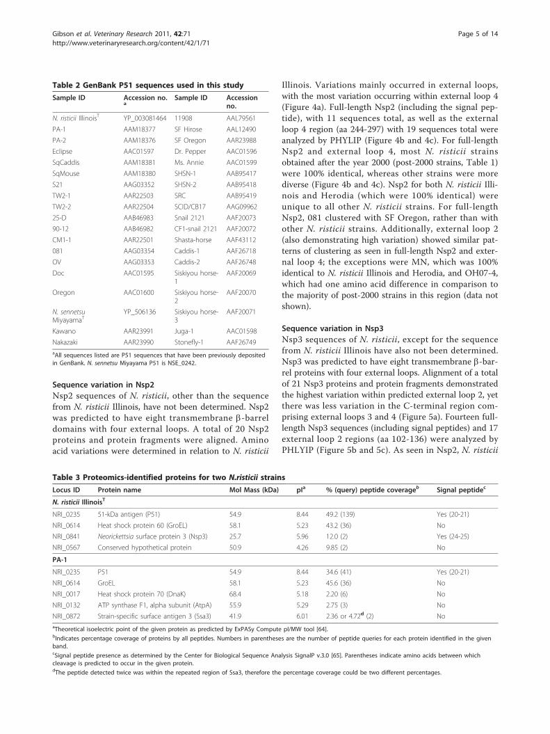
Sequence variation in Nsp2Nsp2 sequences of N. risticii, other than the sequencefrom N. risticii Illinois, have not been determined. Nsp2was predicted to have eight transmembrane b-barreldomains with four external loops. A total of 20 Nsp2proteins and protein fragments were aligned. Aminoacid variations were determined in relation to N. risticii
Illinois. Variations mainly occurred in external loops,with the most variation occurring within external loop 4(Figure 4a). Full-length Nsp2 (including the signal pep-tide), with 11 sequences total, as well as the externalloop 4 region (aa 244-297) with 19 sequences total wereanalyzed by PHYLIP (Figure 4b and 4c). For full-lengthNsp2 and external loop 4, most N. risticii strainsobtained after the year 2000 (post-2000 strains, Table 1)were 100% identical, whereas other strains were morediverse (Figure 4b and 4c). Nsp2 for both N. risticii Illi-nois and Herodia (which were 100% identical) wereunique to all other N. risticii strains. For full-lengthNsp2, 081 clustered with SF Oregon, rather than withother N. risticii strains. Additionally, external loop 2(also demonstrating high variation) showed similar pat-terns of clustering as seen in full-length Nsp2 and exter-nal loop 4; the exceptions were MN, which was 100%identical to N. risticii Illinois and Herodia, and OH07-4,which had one amino acid difference in comparison tothe majority of post-2000 strains in this region (data notshown).
Sequence variation in Nsp3Nsp3 sequences of N. risticii, except for the sequencefrom N. risticii Illinois have also not been determined.Nsp3 was predicted to have eight transmembrane b-bar-rel proteins with four external loops. Alignment of a totalof 21 Nsp3 proteins and protein fragments demonstratedthe highest variation within predicted external loop 2, yetthere was less variation in the C-terminal region com-prising external loops 3 and 4 (Figure 5a). Fourteen full-length Nsp3 sequences (including signal peptides) and 17external loop 2 regions (aa 102-136) were analyzed byPHLYIP (Figure 5b and 5c). As seen in Nsp2, N. risticii
Table 2 GenBank P51 sequences used in this study
Sample ID Accession no.a
Sample ID Accessionno.
N. risticii IllinoisT YP_003081464 11908 AAL79561
PA-1 AAM18377 SF Hirose AAL12490
PA-2 AAM18376 SF Oregon AAR23988
Eclipse AAC01597 Dr. Pepper AAC01596
SqCaddis AAM18381 Ms. Annie AAC01599
SqMouse AAM18380 SHSN-1 AAB95417
S21 AAG03352 SHSN-2 AAB95418
TW2-1 AAR22503 SRC AAB95419
TW2-2 AAR22504 SCID/CB17 AAG09962
25-D AAB46983 Snail 2121 AAF20073
90-12 AAB46982 CF1-snail 2121 AAF20072
CM1-1 AAR22501 Shasta-horse AAF43112
081 AAG03354 Caddis-1 AAF26718
OV AAG03353 Caddis-2 AAF26748
Doc AAC01595 Siskiyou horse-1
AAF20069
Oregon AAC01600 Siskiyou horse-2
AAF20070
N. sennetsuMiyayamaT
YP_506136 Siskiyou horse-3
AAF20071
Kawano AAR23991 Juga-1 AAC01598
Nakazaki AAR23990 Stonefly-1 AAF26749aAll sequences listed are P51 sequences that have been previously depositedin GenBank. N. sennetsu Miyayama P51 is NSE_0242.
Table 3 Proteomics-identified proteins for two N.risticii strains
Locus ID Protein name Mol Mass (kDa) pIa % (query) peptide coverageb Signal peptidec
N. risticii IllinoisT
NRI_0235 51-kDa antigen (P51) 54.9 8.44 49.2 (139) Yes (20-21)
NRI_0614 Heat shock protein 60 (GroEL) 58.1 5.23 43.2 (36) No
NRI_0841 Neorickettsia surface protein 3 (Nsp3) 25.7 5.96 12.0 (2) Yes (24-25)
NRI_0567 Conserved hypothetical protein 50.9 4.26 9.85 (2) No
PA-1
NRI_0235 P51 54.9 8.44 34.6 (41) Yes (20-21)
NRI_0614 GroEL 58.1 5.23 45.6 (36) No
NRI_0017 Heat shock protein 70 (DnaK) 68.4 5.18 2.20 (6) No
NRI_0132 ATP synthase F1, alpha subunit (AtpA) 55.9 5.29 2.75 (3) No
NRI_0872 Strain-specific surface antigen 3 (Ssa3) 41.9 6.01 2.36 or 4.72d (2) NoaTheoretical isoelectric point of the given protein as predicted by ExPASy Compute pI/MW tool [64].bIndicates percentage coverage of proteins by all peptides. Numbers in parentheses are the number of peptide queries for each protein identified in the givenband.cSignal peptide presence as determined by the Center for Biological Sequence Analysis SignalP v.3.0 [65]. Parentheses indicate amino acids between whichcleavage is predicted to occur in the given protein.dThe peptide detected twice was within the repeated region of Ssa3, therefore the percentage coverage could be two different percentages.
Gibson et al. Veterinary Research 2011, 42:71http://www.veterinaryresearch.org/content/42/1/71
Page 5 of 14

Illinois had marked differences to other sequences, inparticular to most post-2000 strains (Table 1). TN02-1and IL01-1 had the highest similarity to N. risticii Illinois.
Sequence variation in Ssa3Ssa3 sequences of N. risticii, other than that of N. risticiiIllinois have not been ascertained. Ssa3 was included inthe analysis, since unknown Ssas were previouslyreported as major N. risticii surface antigens in the 1984Maryland strain 25-D and the 1990 Maryland strain 90-12 [31], and a small amount Ssa3 was detected in bothN. risticii PA-1 in this study and in N. sennetsuMiyayama [39]. There was no signal peptide predictedfor Ssa3 [38], and Ssa3 was not predicted to have a b-bar-rel structure. It was originally shown that ssas contain awide variety of mainly small repeats of 10-55 bp in size[31]. Tandem repeats ranging in size from 63-156 bp arepresent in ssa1, ssa2, and ssa3 of N. risticii Illinois [38].In particular, the N terminus of Ssa3 contains 2.2 copiesof a 52-aa (156 bp) tandem repeat in N. risticii Illinois (aa53-196) [38]. Thirteen Ssa3 proteins and protein frag-ments were aligned and compared (Figure 6a). Withinthis N-terminal repeated region, Neorickettsia spp.
consisted of anywhere from zero to four repeated 52-aapeptides arranged in tandem followed by a terminal 40-aa peptide similar to the 52-aa repeats (for N. risticii Illi-nois: 50% identical, E-value = 6 × 10⁻⁸, using protein-pro-tein blastp). It appears that the number of 52-aa repeatsincreases over time; six post-2000 strains (Table 1) havefour repeats. There is further variety in the form of pointmutations within the 52-aa repeats and terminal 40-aapeptide. In addition, the terminal 40-aa peptide in SFOregon was truncated by 9 aa (31 aa in length, withthe downstream sequence aligning with the other Neor-ickettsia sequences downstream of their terminal 40-aapeptides). Of note, there are b-sheets predicted toencompass most of the repeated region (aa 40-67; 76-119; 128-167) and scattered within the C-terminal region(aa 235-433).
Sequence variation in Ssa1Ssa1 sequences of N. risticii, other than that of N. risticiiIllinois have not been determined. Given the strongestsimilarities between ssa1 of N. risticii Illinois and theunknown ssas from N. risticii strains 25-D (isolated in1984) and 90-12 (isolated in 1990) [38], two ssa1 fragments
Figure 1 Western blotting against rP51, rGroEL, rNsp2, and rNsp3 using PHF-positive equine sera. Recombinant P51, rGroEL, rNsp2, andrNsp3 were separated by SDS-PAGE and probed with 1:500 dilution of PHF-positive horse sera (PHF sera, 1-15) and negative sera (Neg sera, N1-N3). Molecular masses are shown for each recombinant protein. Information regarding the sera samples is given in Table 4.
Gibson et al. Veterinary Research 2011, 42:71http://www.veterinaryresearch.org/content/42/1/71
Page 6 of 14

were amplified, sequenced, and translated from PA-1 andOH07-1. PA-1 (aa 11-249) and OH07-1 (aa 1-239) Ssa1fragments were aligned with corresponding regions fromN. risticii Illinois Ssa1 (aa 246-469) and the Ssas from 25-D(aa 287-507) and 90-12 (aa 579-817). Ssa1 fragments fromPA-1 and OH07-1, which are both post-2000 strains, clus-tered with the 90-12 Ssa, rather than with the 1980s iso-lates N. risticii Illinois Ssa1 and 25-D Ssa, suggesting achronological trend (Figure 6b).
DiscussionThe genes p51, nsp2, nsp3, and ssa3 are uniquely evolvedin Neorickettsia spp. The gene p51 is a single copy geneand demonstrates only loose associations with other pro-teins of the family Anaplasmataceae [37,38]. The nspsand ssas are both potential operons, consisting of threegenes tandemly arranged [38]. The nsps belong topfam01617, and similar to Ehrlichia chaffeensis omp-1(p28) genes (also from pfam01617) [49], the proteins
Table 4 PHF-positive sera from naturally-infected horses and negative sera
Horse IDa Clinical signsb Location Year IFA titer
1 (OH10-1) A, F, De, Deh, C Johnstown, OH 2010 > 1:10,240
2 (OH10-2) A, F, De, C, L, Et, EUTH Grove City, OH 2010 > 1:10,240
3 A, F, De, Deh, L, Et, EUTH Richwood, OH 2010 > 1:10,240
4 A, De, F Galloway, OH 2010 > 1:10,240
5 A, De, Deh, F, C, L Dayton, OH 2010 > 1:10,240
6 A, F, C, L, EUTH Loveland, OH 2010 > 1:10,240
7 U Indiana 2010 1:5120
8 A, Di, De, Deh, F, L Troy, OH 2008 1:1280
9 U Kentucky 2008 1:1280
10 U Indiana 2008 1:1280
11 A, F, Di, De, Deh Columbus, OH 2008 1:1280
12 A, F, Di Cattaraugus, NY 2010 1:640
13 U Indiana 2008 1:640
14 A, F, C Oak Hill, OH 2008 1:80
15 A, F Utica, OH 2008 1:80
N1 U New Jersey 2010 < 1:20
N2 U Ohio 2010 < 1:20
N3 U New Jersey 2010 < 1:20aSera 1 and 2 are from the same horses as buffy coats OH10-1 and OH10-2, respectively, as identified in Table 1.bA, anorexia; F, fever; De, depression; Deh, dehydration; C, colic; L, laminitis; Et, endotoxemia; EUTH, euthanized; U, Unknown; Di, diarrhea.
Figure 2 P51 amino acid sequence variations. Amino acids different from N. risticii Illinois, including insertions and deletions are divided by thenumber of sequences plotted for each amino acid position (# aa diffs). The horizontal axis displays P51 amino acid positions (aa position) includingthe signal peptide and all detected amino acid insertions (515 aa total). SP, signal peptide. E, external loop; and TM, transmembrane domain arebased on the predicted secondary structure [39]. The number of sequences available at each amino acid position on P51 (# seqs) is shown below.
Gibson et al. Veterinary Research 2011, 42:71http://www.veterinaryresearch.org/content/42/1/71
Page 7 of 14

encoded by nsp2 and nsp3 were strain variable. As seenin the ssas, other members of the family Anaplasmata-ceae have genes encoding proteins containing strain-vari-able tandem repeats (involving amino acid variation andchanges in the numbers of tandem repeats), includingTrp120 (formerly gp120), Trp47 (formerly gp47), andVLPT (variable-length PCR target) from E. chaffeensisand Trp140 (formerly gp140), Trp36 (formerly gp36),and gp19 from Ehrlichia canis [50-52]. Of note, the pro-teins encoded by the ssas are not homologous to any pro-teins of the family Anaplasmataceae by blastp. Amongp51, the nsps, and the ssas, there have been no signs of
intragenomic recombination events, which are seen inthe Anaplasma p44/msp2 expression locus [53,54].Proteomics results performed on two strains of N. risticii
established that P51 is a dominant surface-expressed pro-tein. The recognition of recombinant P51 by PHF horsesera, even by 1:80 IFA titer sera suggests P51 is expressedand highly recognized within the present day naturally-infected horses. Despite P51 amino acid sequence varia-tion among N. risticii strains, this strong universal recogni-tion by horse immune sera suggests rP51 may serve as adefined serodiagnostic antigen. Furthermore, the studysuggests that there are immunodominant conserved
Figure 3 P51 amino acid sequence variations among Neorickettsia sequences. Dendrograms of P51 from (A) a 41-aa fragment (counting allinsertions) including the majority of predicted external loop 2 with 43 sequences and (B) a 31-aa fragment (counting all insertions) including theentire predicted external loop 4 with 36 sequences are shown with bootstrap values greater than 50.0% for 1000 replicates. *, bootstrap value of90.0% or greater. East/Midwest US, sequences from states in the Eastern and Midwestern US. Pacific coast, sequences from Japan, Malaysia, andUS states bordering the Pacific Ocean. GenBank accession numbers for P51 sequences are listed in Tables 1 and 2.
Gibson et al. Veterinary Research 2011, 42:71http://www.veterinaryresearch.org/content/42/1/71
Page 8 of 14

peptide sequences within P51 which might serve as evenmore specific PHF diagnostic antigens.Sequence comparison of these surface-exposed pro-
teins of N. risticii strains, with respect to the predictedprotein secondary structure, the majority of which are
clinical isolates, indicates there are hot spots within thegenes with greater strain divergence. These includeexternal loops 2 and 4 in P51, external loop 4 in Nsp2,external loop 2 in Nsp3, and the repeated region ofSsa3. P51 showed strong geographical association; and
Figure 4 Nsp2 amino acid sequence variations. (A) Amino acids different from N. risticii Illinois, including insertions and deletions are dividedby the number of sequences plotted for each amino acid position (# aa diffs). The horizontal axis displays Nsp2 amino acid positions (aaposition) including the signal peptide and all detected amino acid insertions (309 aa total). SP, signal peptide. E, external loop; and TM,transmembrane domain are based on the predicted secondary structure. The number of sequences available at each amino acid position onNsp2 (# seqs) is shown below. (B) Dendrograms of Nsp2 from the full-length protein, including the signal peptide (12 sequences total) and (C)the predicted external loop 4 (55 aa, including all insertions; 19 sequences total) are shown with bootstrap values greater than 50.0% for 1000replicates. *, bootstrap value of 90.0% or greater. Post-2000 sequences are shown in the shaded area. GenBank accession numbers of newsequences are listed in Table 1.
Gibson et al. Veterinary Research 2011, 42:71http://www.veterinaryresearch.org/content/42/1/71
Page 9 of 14
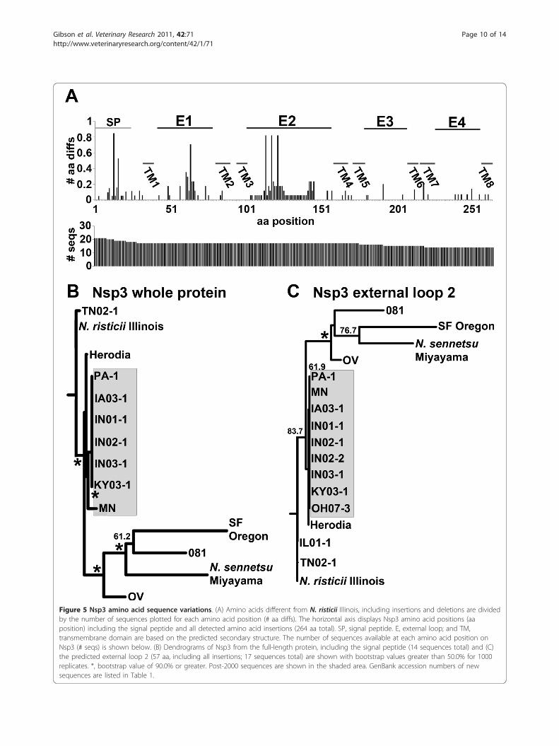
Figure 5 Nsp3 amino acid sequence variations. (A) Amino acids different from N. risticii Illinois, including insertions and deletions are dividedby the number of sequences plotted for each amino acid position (# aa diffs). The horizontal axis displays Nsp3 amino acid positions (aaposition) including the signal peptide and all detected amino acid insertions (264 aa total). SP, signal peptide. E, external loop; and TM,transmembrane domain are based on the predicted secondary structure. The number of sequences available at each amino acid position onNsp3 (# seqs) is shown below. (B) Dendrograms of Nsp3 from the full-length protein, including the signal peptide (14 sequences total) and (C)the predicted external loop 2 (57 aa, including all insertions; 17 sequences total) are shown with bootstrap values greater than 50.0% for 1000replicates. *, bootstrap value of 90.0% or greater. Post-2000 sequences are shown in the shaded area. GenBank accession numbers of newsequences are listed in Table 1.
Gibson et al. Veterinary Research 2011, 42:71http://www.veterinaryresearch.org/content/42/1/71
Page 10 of 14
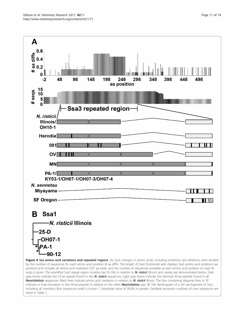
Figure 6 Ssa amino acid variations and repeated regions. (A) Ssa3 changes in amino acids, including insertions and deletions were dividedby the number of sequences for each amino acid position (# aa diffs). The length of Ssa3 (horizontal axis) displays Ssa3 amino acid positions (aaposition) and includes all amino acid insertions (537 aa total), and the number of sequences available at each amino acid position on Ssa3 (#seqs) is given. The amplified Ssa3 repeat region location (aa 53-196, in relation to N. risticii Illinois) and variety are demonstrated below. Darkgray boxes indicate the 52-aa repeats found in the N. risticii sequences. Light gray boxes indicate the terminal 40-aa peptide found in allNeorickettsia sequences. Black lines indicate amino acid variations in relation to N. risticii Illinois. The box containing diagonal lines in SFindicates a 9-aa truncation in the 40-aa peptide in relation to the other Neorickettsia spp. (B) The dendrogram of a 241-aa fragment of Ssa1,including all insertions (five sequences total) is shown. *, bootstrap value of 90.0% or greater. GenBank accession numbers of new sequences arelisted in Table 1.
Gibson et al. Veterinary Research 2011, 42:71http://www.veterinaryresearch.org/content/42/1/71
Page 11 of 14

Nsp2, Nsp3, and Ssa3 showed temporal association.Importantly, N. risticii Illinois (upon which vaccines forPHF are produced) is distinct from most East/MidwestUS strains (P51) and most post-2000 strains (Nsp2,Nsp3, and Ssa3), which may be a contributing factor inPHF vaccine failure [24,55].There are outlier strains which do not fit the geogra-
phical and temporal patterns. These include 081 [20,22],the Kentucky strain OV [22], and the Kentucky strainHerodia. Unique sequences in other N. risticii strains,such as TN02-1 (P51, Nsp2, and Nps3), KY03-3 (Nsp2),IL01-1 (Nsp3), and OH10-1 (Ssa3), suggest that varia-tion contrary to the popular geographical and temporalinfluences may be more widespread. When additionalcontemporary sequences and sequences from more var-ied geographic regions become available, these analysesare expected to improve.Possible explanations for extensive DNA sequence var-
iation within Neorickettsia include the defective DNArepair systems in both N. risticii and N. sennetsu [37,38].This would result in higher mutation rates for Neorickett-sia [56], which would agree with the temporal changesand the production of outlier strains of N. risticii. P51variation showed substantial geographical association,suggesting these variations were selected under localenvironmental pressures. It is possible that geographicalassociation of N. risticii sequence variation is due to N.risticii strains being selected within essential reservoirtrematode populations. In addition, diverse N. risticiistrains may have emerged due to selective pressuresinflicted on the infected trematodes and/or on the trema-todes’ hosts [4-9,57-59]. Humoral immunity would thusnot play any direct role in creating genetic diversitywithin N. risticii populations. Since Neorickettsia spp. areknown (N. risticii and N. helminthoeca) and suspected tobe vertically transmitted within their trematode hosts[8,13,60], mammalian infection is not expected to berequired for maintaining Neorickettsia in the naturalenvironment.Regardless the cause, this genetic variation would result
in increased N. risticii survival as a species. N. risticii sur-face protein genetic diversity revealed in the present studywill help in understanding variations in PHF virulence andclinical signs. It may also be possible to use this new mole-cular knowledge for vaccine development. It would, how-ever necessitate taking into account that the pathogen isan obligate intracellular pathogen, indicating that not onlyhumoral immune responses, but also cell-mediated immu-nity would play an active role in preventing bacterial infec-tion [61-63].Genes encoding the two original Ssas, called P85 (90-12)
and P50 (25-D) are most related to ssa1 from N. risticiiIllinois [24,31,38,55], but they also show similarities tossa2 and the non-coding region between ssa1 and ssa2
using blastn. Although both are Maryland isolates, the25-D strain was isolated six years earlier than the 90-12strain [31], suggesting both temporal variation and thepotential development of chimeras of multiple Ssas andnon-coding regions in P50, P85, and post-2000 Ssa1 (dueto the similarities of PA-1 and OH07-1 Ssa1 fragments toP85). It is possible that the high variability of Ssa1 mayhave prevented PA-1 Ssa1 from being identified by proteo-mics. However, there is the obvious lack of large numbersof peptides identified by proteomics for Ssas in N. risticiiIllinois using the isogenic Illinois strain sequence data andin N. sennetsu using Miyayama isogenic strain data [39]. Itis likely that Ssas are not a dominant surface protein inmammalian cells.In conclusion, our data demonstrate the variety present
within major surface proteins of N. risticii, and they sug-gest conservation among geographical regions and timeperiods. In addition, P51 is implicated as the major sur-face antigen of N. risticii. These data will be valuable indeveloping better diagnostic methods and may help inthe development of more efficacious vaccines.
Additional material
Additional file 1: Supplemental Table 1. Primers utilized for PCRamplification. Word document demonstrating primers utilized for PCRamplification of p51, nsp2, nsp3, ssa1, and ssa3.
AcknowledgementsWe would like to thank the members of the Mass Spectrometry andProteomics Facility, including Dr Kari Green-Church for their expertise andassistance. We would like to thank Dr Mingqun Lin for his technical adviceand assistance and Dr Koshiro Miura for his assistance in training GP. Thiswork was funded by grant R01AI047885 from the National Institutes ofHealth. KEG was partially supported by T32 RR0070703, and GP and SM werefunded by T35 RR021310 from the National Institutes of Health.
Authors’ contributionsKEG drafted the manuscript, designed primers, performed PCR and overallsequence analyses, and created secondary structures and dendrograms. GPdesigned primers, performed PCR, and performed preliminary sequenceanalyses. SM performed all SDS-PAGE and Western blotting experiments andgathered clinical data. YR edited the manuscript and supervised all research.All authors read and approved the final manuscript.
Competing interestsThe authors declare that they have no competing interests.
Received: 11 March 2011 Accepted: 2 June 2011 Published: 2 June 2011
References1. Rikihisa Y, Perry B: Causative agent of Potomac horse fever. Vet Rec 1984,
115:554-555.2. Rikihisa Y, Perry B, Cordes D: Rickettsial link with acute equine diarrhoea.
Vet Rec 1984, 115:390.3. Holland CJ, Ristic M, Cole AI, Johnson P, Baker G, Goetz T: Isolation,
experimental transmission, and characterization of causative agent ofPotomac horse fever. Science 1985, 227:522-524.
4. Barlough JE, Reubel GH, Madigan JE, Vredevoe LK, Miller PE, Rikihisa Y:Detection of Ehrlichia risticii the agent of Potomac horse fever in
Gibson et al. Veterinary Research 2011, 42:71http://www.veterinaryresearch.org/content/42/1/71
Page 12 of 14

freshwater stream snails (Pleuroceridae Juga spp.) from northernCalifornia. Appl Environ Microbiol 1998, 64:2888-2893.
5. Kanter M, Mott J, Ohashi N, Fried B, Reed S, Lin YC, Rikihisa Y: Analysis of16S rRNA and 51-kilodalton antigen gene and transmission in mice ofEhrlichia risticii in virgulate trematodes from Elimia livescens snails inOhio. J Clin Microbiol 2000, 38:3349-3358.
6. Mott J, Muramatsu Y, Seaton E, Martin C, Reed S, Rikihisa Y: Molecularanalysis of Neorickettsia risticii in adult aquatic insects in Pennsylvania, inhorses infected by ingestion of insects, and isolated in cell culture. J ClinMicrobiol 2002, 40:690-693.
7. Chae JS, Pusterla N, Johnson E, Derock E, Lawler SP, Madigan JE: Infectionof aquatic insects with trematode metacercariae carrying Ehrlichia risticii,the cause of Potomac horse fever. J Med Entomol 2000, 37:619-625.
8. Gibson KE, Rikihisa Y, Zhang C, Martin C: Neorickettsia risticii is verticallytransmitted in the trematode Acanthatrium oregonense and horizontallytransmitted to bats. Environ Microbiol 2005, 7:203-212.
9. Gibson KE, Rikihisa Y: Molecular link of different stages of the trematodehost of Neorickettsia risticii to Acanthatrium oregonense. Environ Microbiol2008, 10:2064-2073.
10. Knowles RC, Anderson C, Shipley W, Whitlock R, Perry B, Davidson J: AcuteEquine Diarrhea Syndrome (AEDS): A Preliminary Report. In Proceedingsof the 29th annual convention of the American Association of EquinePractitioners. Edited by: Milne FJ. Las Vegas, Nevada: American Associationof Equine Practitioners; 1984:353-357.
11. Long MT, Goetz TE, Kakoma I, Whiteley HE, Lock TE, Holland CJ,Foreman JH, Baker GJ: Evaluation of fetal infection and abortion inpregnant ponies experimentally infected with Ehrlichia risticii. Am J VetRes 1995, 56:1307-1316.
12. Rikihisa Y, Jiang BM: In vitro susceptibilities of Ehrlichia risticii to eightantibiotics. Antimicrob Agents Chemother 1988, 32:986-991.
13. Rikihisa Y, Dumler JS, Dasch GA: Neorickettsia. In The Proteobacteria, Part C,Bergey’s Manual of Systematic Bacteriology. Volume 2. 2 edition. Edited by:Garrity GM, Brenner DJ, Krieg NR, Staley JT. New York, NY Springer;2005:132-137.
14. Rikihisa Y: Rickettsial Diseases. In Equine Internal Medicine.. 2 edition. Editedby: Reed SM, Bayly WM, Sellon DC. Philadelphia, PA: W. B. Saunders;2004:96-109.
15. Wells MY, Rikihisa Y: Lack of lysosomal fusion with phagosomescontaining Ehrlichia risticii in P388D1 cells: abrogation of inhibition withoxytetracycline. Infect Immun 1988, 56:3209-3215.
16. Mott J, Rikihisa Y, Zhang Y, Reed SM, Yu CY: Comparison of PCR andculture to the indirect fluorescent-antibody test for diagnosis ofPotomac horse fever. J Clin Microbiol 1997, 35:2215-2219.
17. Ristic M, Holland CJ, Dawson JE, Sessions J, Palmer J: Diagnosis of equinemonocytic ehrlichiosis (Potomac horse fever) by indirectimmunofluorescence. J Am Vet Med Assoc 1986, 189:39-46.
18. Rikihisa Y, Reed SM, Sams RA, Gordon JC, Pretzman CI: Serosurvey ofhorses with evidence of equine monocytic ehrlichiosis. J Am Vet MedAssoc 1990, 197:1327-1332.
19. Rikihisa Y: The tribe Ehrlichieae and ehrlichial diseases. Clin Microbiol Rev1991, 4:286-308.
20. Chaichanasiriwithaya W, Rikihisa Y, Yamamoto S, Reed S, Crawford TB,Perryman LE, Palmer GH: Antigenic, morphologic, and molecularcharacterization of new Ehrlichia risticii isolates. J Clin Microbiol 1994,32:3026-3033.
21. Pretzman CI, Rikihisa Y, Ralph D, Gordon JC, Bech-Nielsen S: Enzyme-linkedimmunosorbent assay for Potomac horse fever disease. J Clin Microbiol1987, 25:31-36.
22. Wen B, Rikihisa Y, Fuerst PA, Chaichanasiriwithaya W: Diversity of 16S rRNAgenes of new Ehrlichia strains isolated from horses with clinical signs ofPotomac horse fever. Int J Syst Bacteriol 1995, 45:315-318.
23. Palmer JE: Prevention of Potomac horse fever. Cornell Vet 1989,79:201-205.
24. Dutta SK, Vemulapalli R, Biswas B: Association of deficiency in antibodyresponse to vaccine and heterogeneity of Ehrlichia risticii strains withPotomac horse fever vaccine failure in horses. J Clin Microbiol 1998,36:506-512.
25. Rikihisa Y, Stills H, Zimmerman G: Isolation and continuous culture ofNeorickettsia helminthoeca in a macrophage cell line. J Clin Microbiol1991, 29:1928-1933.
26. Pretzman C, Ralph D, Stothard DR, Fuerst PA, Rikihisa Y: 16S rRNA genesequence of Neorickettsia helminthoeca and its phylogenetic alignmentwith members of the genus Ehrlichia. Int J Syst Bacteriol 1995, 45:207-211.
27. Dumler JS, Barbet AF, Bekker CP, Dasch GA, Palmer GH, Ray SC, Rikihisa Y,Rurangirwa FR: Reorganization of genera in the families Rickettsiaceaeand Anaplasmataceae in the order Rickettsiales: unification of somespecies of Ehrlichia with Anaplasma, Cowdria with Ehrlichia and Ehrlichiawith Neorickettsia, descriptions of six new species combinations anddesignation of Ehrlichia equi and ‘HGE agent’ as subjective synonyms ofEhrlichia phagocytophila. Int J Syst Evol Microbiol 2001, 51:2145-2165.
28. Fukuda T, Yamamoto S: Neorickettsia-like organism isolated frommetacercaria of a fluke, Stellantchasmus falcatus. Jpn J Med Sci Biol 1981,34:103-107.
29. Wen B, Rikihisa Y, Yamamoto S, Kawabata N, Fuerst PA: Characterization ofthe SF agent, an Ehrlichia sp. isolated from the fluke Stellantchasmusfalcatus, by 16S rRNA base sequence, serological, and morphologicalanalyses. Int J Syst Bacteriol 1996, 46:149-154.
30. Rikihisa Y, Zhang C, Kanter M, Cheng Z, Ohashi N, Fukuda T: Analysis ofp51, groESL, and the major antigen P51 in various species ofNeorickettsia, an obligatory intracellular bacterium that infectstrematodes and mammals. J Clin Microbiol 2004, 42:3823-3826.
31. Biswas B, Vemulapalli R, Dutta SK: Molecular basis for antigenic variationof a protective strain-specific antigen of Ehrlichia risticii. Infect Immun1998, 66:3682-3688.
32. Dutta SK, Mattingly BL, Shankarappa B: Antibody response to Ehrlichiaristicii and antibody reactivity to the component antigens in horses withinduced Potomac horse fever. Infect Immun 1989, 57:2959-2962.
33. Kaylor PS, Crawford TB, McElwain TF, Palmer GH: Passive transfer ofantibody to Ehrlichia risticii protects mice from ehrlichiosis. Infect Immun1991, 59:2058-2062.
34. Dutta SK, Shankarappa B, Mattingly-Napier BL: Molecular cloning andanalysis of recombinant major antigens of Ehrlichia risticii. Infect Immun1991, 59:1162-1169.
35. Vemulapalli R, Biswas B, Dutta SK: Cloning and molecular analysis ofgenes encoding two immunodominant antigens of Ehrlichia risticii.Microb Pathog 1998, 24:361-372.
36. Vemulapalli R, Biswas B, Dutta SK: Studies with recombinant proteins ofEhrlichia risticii: identification of strain-specific antigen as a protectiveantigen. Vet Parasitol 1998, 76:189-202.
37. Hotopp JC, Lin M, Madupu R, Crabtree J, Angiuoli SV, Eisen J, Seshadri R,Ren Q, Wu M, Utterback TR, Smith S, Lewis M, Khouri H, Zhang C, Niu H,Lin Q, Ohashi N, Zhi N, Nelson W, Brinkac LM, Dodson RJ, Rosovitz MJ,Sundaram J, Daugherty SC, Davidsen T, Durkin AS, Gwinn M, Haft DH,Selengut JD, Sullivan SA, et al: Comparative genomics of emerginghuman ehrlichiosis agents. PLoS Genet 2006, 2:e21.
38. Lin M, Zhang C, Gibson K, Rikihisa Y: Analysis of complete genomesequence of Neorickettsia risticii: causative agent of Potomac horse fever.Nucleic Acids Res 2009, 37:6076-6091.
39. Gibson K, Kumagai Y, Rikihisa Y: Proteomic analysis of Neorickettsiasennetsu surface-exposed proteins and porin activity of the majorsurface protein P51. J Bacteriol 2010, 192:5898-5905.
40. Ge Y, Rikihisa Y: Surface-exposed proteins of Ehrlichia chaffeensis. InfectImmun 2007, 75:3833-3841.
41. Zhang Y, Ohashi N, Lee EH, Tamura A, Rikihisa Y: Ehrlichia sennetsu groEoperon and antigenic properties of the GroEL homolog. FEMS ImmunolMed Microbiol 1997, 18:39-46.
42. Felsenstein J: PHYLIP – Phylogeny Inference Package (Version 3.2).Cladistics 1989, 5:164-166.
43. Zhang Z, Schwartz S, Wagner L, Miller W: A greedy algorithm for aligningDNA sequences. J Comput Biol 2000, 7:203-214.
44. Altschul SF, Madden TL, Schaffer AA, Zhang J, Zhang Z, Miller W,Lipman DJ: Gapped BLAST and PSI-BLAST: a new generation of proteindatabase search programs. Nucleic Acids Res 1997, 25:3389-3402.
45. Bagos PG, Liakopoulos TD, Spyropoulos IC, Hamodrakas SJ: PRED-TMBB: aweb server for predicting the topology of beta-barrel outer membraneproteins. Nucleic Acids Res 2004, 32:W400-404.
46. Jeanteur D, Lakey JH, Pattus F: The bacterial porin superfamily: sequencealignment and structure prediction. Mol Microbiol 1991, 5:2153-2164.
47. Brogden KA: Virulence mechanisms of bacterial pathogens. 4 edition.Washington, D.C.: ASM Press; 2007.
Gibson et al. Veterinary Research 2011, 42:71http://www.veterinaryresearch.org/content/42/1/71
Page 13 of 14

48. Rikihisa Y: Cross-reacting antigens between Neorickettsia helminthoecaand Ehrlichia species, shown by immunofluorescence and Westernimmunoblotting. J Clin Microbiol 1991, 29:2024-2029.
49. Miura K, Rikihisa Y: Virulence potential of Ehrlichia chaffeensis strains ofdistinct genome sequences. Infect Immun 2007, 75:3604-3613.
50. Sumner JW, Childs JE, Paddock CD: Molecular cloning andcharacterization of the Ehrlichia chaffeensis variable-length PCR target:an antigen-expressing gene that exhibits interstrain variation. J ClinMicrobiol 1999, 37:1447-1453.
51. Zhang X, Luo T, Keysary A, Baneth G, Miyashiro S, Strenger C, Waner T,McBride JW: Genetic and antigenic diversities of major immunoreactiveproteins in globally distributed Ehrlichia canis strains. Clin VaccineImmunol 2008, 15:1080-1088.
52. Doyle CK, Cardenas AM, Aguiar DM, Labruna MB, Ndip LM, Yu XJ,McBride JW: Molecular characterization of E. canis gp36 and E.chaffeensis gp47 tandem repeats among isolates from differentgeographic locations. Ann N Y Acad Sci 2005, 1063:433-435.
53. Lin Q, Rikihisa Y, Ohashi N, Zhi N: Mechanisms of variable p44 expressionby Anaplasma phagocytophilum. Infect Immun 2003, 71:5650-5661.
54. Barbet AF, Meeus PF, Belanger M, Bowie MV, Yi J, Lundgren AM,Alleman AR, Wong SJ, Chu FK, Munderloh UG, Jauron SD: Expression ofmultiple outer membrane protein sequence variants from a singlegenomic locus of Anaplasma phagocytophilum. Infect Immun 2003,71:1706-1718.
55. Vemulapalli R, Biswas B, Dutta SK: Pathogenic, immunologic, andmolecular differences between two Ehrlichia risticii strains. J Clin Microbiol1995, 33:2987-2993.
56. LeClerc JE, Li B, Payne WL, Cebula TA: High mutation frequencies amongEscherichia coli and Salmonella pathogens. Science 1996, 274:1208-1211.
57. Pusterla N, Johnson EM, Chae JS, Madigan JE: Digenetic trematodes,Acanthatrium sp. and Lecithodendrium sp., as vectors of Neorickettsiaristicii, the agent of Potomac horse fever. J Helminthol 2003, 77:335-339.
58. Park BK, Kim MJ, Kim EH, Kim MS, Na DG, Chae JS: Identification oftrematode cercariae carrying Neorickettsia risticii in freshwater streamsnails. Ann N Y Acad Sci 2003, 990:239-247.
59. Pusterla N, Madigan JE, Chae JS, DeRock E, Johnson E, Pusterla JB:Helminthic transmission and isolation of Ehrlichia risticii, the causativeagent of Potomac horse fever, by using trematode stages fromfreshwater stream snails. J Clin Microbiol 2000, 38:1293-1297.
60. Nyberg PA, Knapp SE, Millemann RE: “Salmon poisoning” disease. IV.Transmission of the disease to dogs by Nanophyetus salmincola eggs.J Parasitol 1967, 53:694-699.
61. Park J, Rikihisa Y: L-arginine-dependent killing of intracellular Ehrlichiaristicii by macrophages treated with gamma interferon. Infect Immun1992, 60:3504-3508.
62. Park J, Rikihisa Y: Inhibition of Ehrlichia risticii infection in murineperitoneal macrophages by gamma interferon, a calcium ionophore,and concanavalin A. Infect Immun 1991, 59:3418-3423.
63. Rikihisa Y: Protection against murine potomac horse fever by aninactivated Ehrlichia risticii vaccine. Vet Microbiol 1991, 27:339-350.
64. Gasteiger E, Hoogland C, Gattiker A, Duvaud S, Wilkins MR, Appel RD,Bairoch A: Protein identification and analysis tools on the ExPASy server.In The proteomics protocols handbook. Edited by: Walker JM. Totowa, N.J:Humana Press; 2005:571-607.
65. Bendtsen JD, Nielsen H, von Heijne G, Brunak S: Improved prediction ofsignal peptides: SignalP 3.0. J Mol Biol 2004, 340:783-795.
doi:10.1186/1297-9716-42-71Cite this article as: Gibson et al.: Neorickettsia risticii surface-exposedproteins: proteomics identification, recognition by naturally-infectedhorses, and strain variations. Veterinary Research 2011 42:71.
Submit your next manuscript to BioMed Centraland take full advantage of:
• Convenient online submission
• Thorough peer review
• No space constraints or color figure charges
• Immediate publication on acceptance
• Inclusion in PubMed, CAS, Scopus and Google Scholar
• Research which is freely available for redistribution
Submit your manuscript at www.biomedcentral.com/submit
Gibson et al. Veterinary Research 2011, 42:71http://www.veterinaryresearch.org/content/42/1/71
Page 14 of 14