Embed Size (px)
Citation preview

Near-continuously synthesized leading strands inEscherichia coli are broken by ribonucleotide excisionGlen E. Cronana, Elena A. Kouzminovaa, and Andrei Kuzminova,1
aDepartment of Microbiology, University of Illinois at Urbana–Champaign, Urbana, IL 61801
Edited by Thomas A. Kunkel, National Institute of Environmental Health Sciences, NIH, Research Triangle Park, NC, and accepted by Editorial Board MemberKiyoshi Mizuuchi December 10, 2018 (received for review August 23, 2018)
In vitro, purified replisomes drive model replication forks to synthe-size continuous leading strands, even without ligase, supporting thesemidiscontinuous model of DNA replication. However, nascentreplication intermediates isolated from ligase-deficient Escherichiacoli comprise only short (on average 1.2-kb) Okazaki fragments. Itwas long suspected that cells replicate their chromosomal DNA bythe semidiscontinuous mode observed in vitro but that, in vivo, thenascent leading strand was artifactually fragmented postsynthesisby excision repair. Here, using high-resolution separation of pulse-labeled replication intermediates coupledwith strand-specific hybrid-ization, we show that excision-proficient E. coli generates leading-strand intermediates >10-fold longer than lagging-strand Okazakifragments. Inactivation of DNA-repair activities, including ribonucle-otide excision, further increased nascent leading-strand size to∼80 kb, while lagging-strand Okazaki fragments remained unaf-fected. We conclude that in vivo, repriming occurs ∼70× less fre-quently on the leading versus lagging strands, and that DNAreplication in E. coli is effectively semidiscontinuous.
replication intermediates | Okazaki fragments | the leading strand |ligase mutant | ribonucleotide excision repair
The two strands of a duplex DNA are paired in antiparallelfashion, as first proposed by Watson and Crick (1), perhaps
to ensure that optimal base pairing is sequence-independent (2).Early studies on the mechanism of cellular DNA replicationrevealed that simultaneous replication of both parental strandsof chromosomal DNA happens at Y-shaped structures (3, 4)called “replication forks” (5, 6). Contemporaneous in vivo ex-periments with replication intermediates (7) and in vitro char-acterization of various DNA polymerases (8–10) showed DNAsynthesis only in the 5′→3′ direction, necessitating that in anti-parallel DNA duplexes at least one of the strands at the repli-cation fork must be synthesized discontinuously (11).Indeed, discontinuous DNA replication was discovered by
Okazaki and others, using experiments where nascent DNA waspulse-labeled, denatured, and separated by size using alkaline-sucrose (AS) gradients (12–14). Unexpectedly, Okazaki’s resultsshowed that all nascent DNA in bacteria is originally synthesizedas low molecular weight (LMW) 1- to 2-kb-long replication inter-mediates (RIs) (11, 13, 15–19), which hybridized to both parentalDNA strands (17, 20). Furthermore, pulse–chase experimentsconfirmed that these initially small fragments were later joinedtogether to form high molecular weight (HMW), chromosome-length DNA strands (12, 18, 19). Fellow researchers coined theterm “Okazaki fragments” (21, 22) to describe these LMW RIs,and similar findings of fully discontinuous DNA replication (Fig.1A, Top) were promptly confirmed without exception in diverseexperimental systems, including mammalian cells (23–25), bacteria(20, 21, 26, 27), and lower eukaryotes (28, 29).Importantly, due to the rapidity of RI joining in unicellular
organisms, isolation of their Okazaki fragments necessitates in-hibition of RI maturation (joining) activities. In bacteria, LMWRIs (Okazaki fragments) mature into full-length DNA strands bya two-enzyme system, composed of a repair DNA polymerase(DNA pol I, gppolA) and DNA ligase (gpligA). Polymerase I
removes the RNA primer of the downstream fragment whilesimultaneously extending the current fragment, thus generatingan all-DNA nick, which is then closed by DNA ligase (8). Thismaturation process ensures that inactivation of DNA ligasepreserves Okazaki fragments (albeit lacking their RNA primers),as first reported by Okazaki himself (13, 18). In fact, nochromosomal-size HMW RIs have ever been observed in ligase-minus conditions (21, 26, 28–34).However, inactivation of ligase also prevents joining of nicks
arising from excision-repair processes. Replicative DNA poly-merases readily incorporate various base analogs such as hypo-xanthine into the growing chain, from where they are quicklyremoved by specialized excision enzymes (35, 36). Incorporation–excision reactions of this type may fragment the nascent DNA,with the resulting shorter pieces appearing similar in size to trueRNA-primed Okazaki fragments (Fig. 1A). This potential forexcision-driven formation of LMW RIs was confirmed for uracil-DNA incorporation using Escherichia coli dutmutants that, due tothe defective dUTPase, frequently incorporate dU into their na-scent DNA (37, 38). This coupling of incorporation and frag-mentation was further characterized using dut and ung mutants(deficient in DNA-uracil excision) in a cell-free system (39, 40),and subsequently in thyA mutant Bacillus subtilis (which synthe-sized only LMW RIs), where a dramatic increase in RI size wasnoticed after inactivation of uracil excision (41). However, a thyAmutant of E. coli synthesized mostly HMW DNA, which was un-affected by ung inactivation (42). Importantly, the ung mutant
Significance
The antiparallel orientation of strands in a DNA duplex andunidirectional polarity of DNA polymerases necessitate thatreplication forks operate semidiscontinuously. That is, thestrand replicated codirectionally to replication-fork movement,termed the “leading” strand, is synthesized continuously, whilethe oppositely oriented “lagging” strand is synthesized in short“Okazaki” fragments, later joined by ligase. Semidiscontinuousreplication is observed in vitro, even in reactions lacking ligase.However, in vivo, in ligase (lig) mutants, nascent DNA fromboth strands is found only in small pieces. Here, we report thecontinuous synthesis of leading strands in lig mutant Escher-ichia coli lacking all DNA excision-repair activities and, using animproved sucrose-gradient system, identify misincorporated ribo-nucleotides as the major driver of leading-strand fragmentation.
Author contributions: G.E.C., E.A.K., and A.K. designed research; G.E.C. and E.A.K. per-formed research; G.E.C., E.A.K., and A.K. analyzed data; and G.E.C. and A.K. wrotethe paper.
The authors declare no conflict of interest.
This article is a PNAS Direct Submission. T.A.K. is a guest editor invited by the EditorialBoard.
Published under the PNAS license.1To whom correspondence should be addressed. Email: [email protected].
This article contains supporting information online at www.pnas.org/lookup/suppl/doi:10.1073/pnas.1814512116/-/DCSupplemental.
Published online January 7, 2019.
www.pnas.org/cgi/doi/10.1073/pnas.1814512116 PNAS | January 22, 2019 | vol. 116 | no. 4 | 1251–1260
BIOCH
EMISTR
Y
Dow
nloa
ded
by g
uest
on
Feb
ruar
y 15
, 202
2
E. coli continued to synthesize exclusively LMW RIs undermaturation-deficient conditions (30, 34, 43), indicating thaturacil excision was not a source of nascent leading-strandfragmentation.That the leading strand could be synthesized by a continuous
mechanism in the complete absence of ligase was amply dem-onstrated in various in vitro replication systems using highlypurified replisomes (44–46). The resulting semidiscontinuousDNA-replication model (Fig. 1A, Bottom) became a fixture oftextbooks (47), even though it contradicted the bulk of in vivoresults, and persisted despite our lack of understanding of thesupposed fragmenting activities (22). Several attempts to identifythe in vivo DNA-repair activities responsible for the putative
postsynthesis fragmentation in E. coli were unsuccessful (30, 33,34). The most recent study went so far as to inactivate all eightindividual DNA glycosylases (eliminating base-excision repair),in addition to blocking nucleotide-excision repair (uvrA), mis-match removal (mutS), and alternative excision repair (nfi), yeteven this dodecuple mutant [including the ligA251(Ts) defect]showed no increase in RI size (31). Still, those authors wereaware of the possibility that the tails of intermediate molecularweight (IMW) species could represent the leading-strand RIs(31). Besides, other work showed that only ∼50% of Okazakifragments from wild-type E. coli harbored the 5′-RNA moietyindicative of priming, suggesting that the other half was generatedby excision (48).
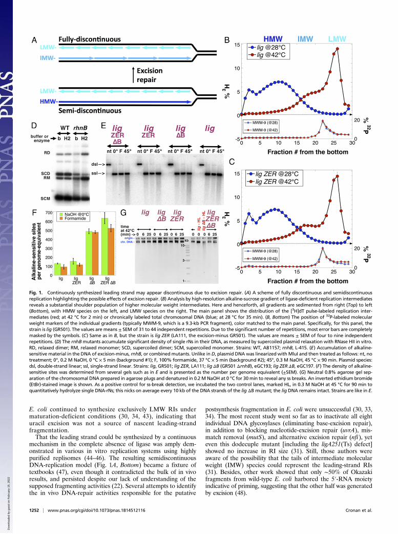
Fig. 1. Continuously synthesized leading strand may appear discontinuous due to excision repair. (A) A scheme of fully discontinuous and semidiscontinuousreplication highlighting the possible effects of excision repair. (B) Analysis by high-resolution alkaline-sucrose gradient of ligase-deficient replication intermediatesreveals a substantial shoulder population of higher molecular weight intermediates. Here and henceforth, all gradients are sedimented from right (Top) to left(Bottom), with HMW species on the left, and LMW species on the right. The main panel shows the distribution of the [3H]dT pulse-labeled replication inter-mediates (red; at 42 °C for 2 min) or chronically labeled total chromosomal DNA (blue; at 28 °C for 35 min). (B, Bottom) The position of 32P-labeled molecularweight markers of the individual gradients (typically MWM-9, which is a 9.3-kb PCR fragment), color matched to the main panel. Specifically, for this panel, thestrain is lig (GR501). The values are means ± SEM of 31 to 44 independent repetitions. Due to the significant number of repetitions, most error bars are completelymasked by the symbols. (C) Same as in B, but the strain is lig ZER (LA111, the excision-minus GR501). The values are means ± SEM of four to nine independentrepetitions. (D) The rnhBmutants accumulate significant density of single rNs in their DNA, as measured by supercoiled plasmid relaxation with RNase HII in vitro.RD, relaxed dimer; RM, relaxed monomer; SCD, supercoiled dimer; SCM, supercoiled monomer. Strains: WT, AB1157; rnhB, L-415. (E) Accumulation of alkaline-sensitive material in the DNA of excision-minus, rnhB, or combinedmutants. Unlike inD, plasmid DNAwas linearized with MluI and then treated as follows: nt, notreatment; 0°, 0.2 M NaOH, 0 °C × 5 min (background #1); F, 100% formamide, 37 °C × 5 min (background #2); 45°, 0.3 M NaOH, 45 °C × 90 min. Plasmid species:dsl, double-strand linear; ssl, single-strand linear. Strains: lig, GR501; lig ZER, LA111; lig ΔB (GR501 ΔrnhB), eGC193; lig ZER ΔB, eGC197. (F) The density of alkaline-sensitive sites was determined from several gels such as in E and is presented as the number per genome equivalent (±SEM). (G) Neutral 0.8% agarose gel sep-aration of the chromosomal DNA prepared in agarose plugs and denatured in 0.2 M NaOH at 0 °C for 30min to reveal any ss breaks. An inverted ethidium bromide(EtBr)-stained image is shown. As a positive control for ss-break detection, we incubated the two control lanes, marked HL, in 0.3 M NaOH at 45 °C for 90 min toquantitatively hydrolyze single DNA-rNs; this nicks on average every 10 kb of the DNA strands of the lig ΔB mutant; the lig DNA remains intact. Strains are like in E.
1252 | www.pnas.org/cgi/doi/10.1073/pnas.1814512116 Cronan et al.
Dow
nloa
ded
by g
uest
on
Feb
ruar
y 15
, 202
2
Two recent developments prompted our reevaluation of thisconundrum. First, development of a higher-resolution AS-gradient system, buttressed by MW markers from 1 to 170 kb,revealed a distinct IMW (5- to 25-kb) shoulder adjoining themain LMW (∼1.2-kb) Okazaki fragment peak formed underligase-deficient (lig) conditions (Fig. 1B). Furthermore, the ex-cision mutant used in previous work, deficient in all DNA-onlyexcision-repair systems (now called lig ZER; zero excision re-pair), that previously produced exactly the same RI distributionas excision-proficient cells in regular AS gradients (31), nowdeveloped a clear separation of the two RI peaks in the im-proved AS-gradient system (Fig. 1C). Thus, this high-resolutionanalysis confirmed the presence of two distinct RI populations ofsignificantly different molecular weight. Second, a new DNAcontaminant was identified: Monoribonucleotides (rNs) werefound to misincorporate into the chromosomal DNA of eu-karyotes at high frequency (49, 50), even though in B. subtilis thedensity of a single DNA-rN misincorporation initially appearedqualitatively low (51). Recently, the density of single DNA-rNs inthe rnhB mutant E. coli was found to be one in 14,000 nt (52)(Fig. 1D). As DNA-rNs are efficiently excised (52), one chainbreak per 14,000 nt in the leading strand could produce frag-ments similar in size to the observed IMW fragments (Fig. 1 Band C). Unexpectedly, this significant accumulation of DNA-rNswas without any effect in E. coli, as rnhB mutants showed es-sentially wild-type behavior (52).To test DNA-rNs as a potential source of RI breakage, we
added an rnhB defect in ribonucleotide excision repair to our ligZER strain (31). The resulting lig ZER ΔB mutant, in addition tothe ligA251 temperature-sensitive ligase deficiency, carries 12additional mutations completely inactivating all known excision-repair pathways in E. coli, including ribonucleotide-excision repair.By preventing the excision of all known “incorrect” bases, wehoped to increase the stability of nascent DNA and preservethe true size of RIs for examination. However, AS gradientsappeared chemically inadequate for the task of separating theribonucleotide-containing RIs from rnhB mutants, as the stronglybasic pH of AS gradients, required to denature RIs from theirtemplate strands, efficiently hydrolyzes mono-rNs in DNA (52)and also destabilizes abasic sites (53). Therefore, we developed apH-neutral denaturing gradient system to separate RIs withoutinducing breakage at alkaline-sensitive sites. Employing this gradientsystem, in concert with traditional alkaline-sucrose methods andstrand-specific hybridization of DNA from gradient fractions, wedemonstrate that the lig ZER ΔB mutant of E. coli replicates itsleading strand by an essentially continuous mechanism.
ResultsThe Density of Alkaline-Sensitive Sites in Nascent DNA. Our basicprotocol for RI detection follows the classic outline (15) andincludes shifting exponentially growing cultures of ligA251(Ts)mutant strains from 28 to 42 °C for 4 min to inactivate DNAligase, followed by addition of a [3H]thymidine label, and con-tinuing incubation at 42 °C for 2 additional minutes to label RIs.Following RI labeling, cellular metabolism is stopped instantly,the cells are lysed to release their genomic DNA, and the DNA isthen purified and sedimented on alkaline-sucrose gradients. Wenever exceeded 6 min as the total incubation time of the ligA251(Ts) mutant at 42 °C, avoiding chromosome fragmentation thatbegins after 20 min at the nonpermissive temperatures in thismutant (54) (SI Appendix, Fig. S1). The basic pH of alkalinesucrose denatures labeled RIs, freeing them from the unlabeledtemplate strands, so they can sediment as single-stranded DNAsto be separated according to their length (molecular weight)(15). Importantly, the same alkaline pH at 0°C treatment of thechromosomal DNA of all our experimental strains [all ligA(Ts)mutants] after incubation at 42 °C for up to 25 min reveals no
accumulation of single-strand breaks in bulk genomic DNA (Fig.1G and SI Appendix, Fig. S1).In this work, resolution of AS gradients was optimized, par-
ticularly for the IMW size range of 5 to 50 kb, by two modifi-cations to the previous methodology: (i) The significantlyreduced total applied g force maintained the majority of chro-mosomal DNA within the gradient instead of sedimenting it intothe cushion at the bottom of the tube; and (ii) the apparatusesused for the pouring and fractionation of gradients were com-pletely reworked (Material and Methods). A 9.3-kb 32P-labeledDNA was included in every gradient, as both an MWmarker andpositional control (Fig. 1 B and C, Bottom). In some experi-ments, virion DNAs from phages lambda (48.5 kb) and T4D(∼170 kb) were also used as, correspondingly, IMW and HMWmarker DNAs, and to build an MW standard curve (SI Appendix,Fig. S5) (55). Additionally, the MW marker DNAs served asinternal controls against nonspecific DNA hydrolysis.As stated in the Introduction, it became apparent that our
previous conclusions concerning LMWRIs in the ligA251mutantat 42 °C (30, 31) should be revisited, as the IMW shoulder in theRI distribution of the excision+ lig strain clearly shifted to highermolecular weight in the lig ZER mutant (Fig. 1B versus Fig. 1C,compare fractions 16 to 21; for quantification, see SI Appendix,Fig. S5D). Meanwhile, in vitro plasmid relaxation by RNase HIIindicated a significant density of single ribonucleotides in theDNA of rnhB mutants (Fig. 1D) (52). Since for this project it wasmore relevant to assess the density of alkaline-sensitive sites inDNA, we treated linearized plasmid DNA with alkali (0.3 MNaOH, 90 min at 45 °C), analyzing the intactness of the strandsin neutral agarose gels (Fig. 1E). We found alkaline-induced nickdensities in the lig ΔB and lig ZER ΔB mutants of one per8,000 to 9,000 nt, translating into 500 to 600 alkaline-sensitivesites per genome equivalent (Fig. 1 E and F). The increase ofalkaline-sensitive sites in the DNA of the lig ZER mutant com-pared with the lig mutant (both strains RnhB+) was insignificant(Fig. 1 E and F). Therefore, the density of DNA-rNs in the rnhBmutants pointed to ribonucleotides as the major genome con-taminant, suggesting that in AS gradients, alkaline hydrolysis ofDNA-rNs masks the true size of RIs. This prompted us to de-velop a pH-neutral denaturing sucrose-gradient system for sep-aration of rN-containing DNA.
Formamide-Urea Gradients Reveal HMW Replication Intermediates.Formamide and urea are known to destabilize duplex DNAwithout breaking strands at rNs or other modified bases (56–58).In fact, formamide-sucrose gradients were used before to sepa-rate RIs, to avoid the instability of potential abasic sites in alkali(33) and to preserve RNA-primed lagging strands (59). Afterexperimentation with various concentrations of formamide, urea,and sucrose, we settled on gradients of 5 to 40% sucrose in 70%formamide and 1 M urea, with sample loading in 50% form-amide and ∼5 M urea. In these formamide-urea-sucrose (FUS)gradients, the sedimentation patterns of HMW chromosomalDNA (ligase+ conditions), or LMW RIs (ligase– conditions),were similar to those obtained in AS gradients (Fig. 2A). Besidesthe almost coincidental positions of major peaks, a significantshoulder of IMW RIs can clearly be seen in both systems. Similarto AS gradients, the distribution of IMW RIs from the lig ZERstrain shifts to higher molecular weights and develops into aseparate peak in FUS gradients (Fig. 2B), confirming that longerRIs are fragmented into shorter ones by excision repair in na-scent DNA of WT cells.Most importantly, FUS gradients allowed us to experiment
with the rnhB mutants without inducing artifactual rN hydrolysis.As expected, we found that inactivation of RNase HII in theoriginal ligA mutant shifted IMW RIs even further towardHMWs (Fig. 2B, lig ΔB @42 °C), showing that “contamination”of nascent DNA by ribonucleotides exceeded contamination by
Cronan et al. PNAS | January 22, 2019 | vol. 116 | no. 4 | 1253
BIOCH
EMISTR
Y
Dow
nloa
ded
by g
uest
on
Feb
ruar
y 15
, 202
2

all other DNA modifications removed by all-DNA excision-repairsystems inactivated in the lig ZER mutant. A crucial finding camefrom introduction of the rnhB defect into the lig ZER strain, toyield the lig ZER ΔB mutant deficient in all known initiators ofexcision repair in E. coli. In the FUS gradients, RIs from the ligZER ΔB strain form two well-separated peaks: the standard LMWpeak of Okazaki fragments and a now fully separate HMW peaksimilar in size to the chronically labeled chromosome (Fig. 2C).Importantly, in all these mutants (Fig. 2; also, the four gradientsare overlaid in Fig. 3B), while the IMW peak gradually becomesHMW, the LMW peak does not shift to higher molecular weights,confirming that it is insensitive to repair activities and thereforelikely represents true Okazaki fragments.The MW standards for FUS gradients show a uniform sep-
aration between Okazaki fragments (1.2 kb), the 9.3-kb PCRfragment, lambda chromosome (48.5 kb), and T4 chromosome(∼170 kb) (Fig. 2D). This allowed us to estimate the size of theHMW RI peak as stretching from ∼20 kb to 300 kb, with themajor fraction found between 50 and 150 kb (Fig. 2 C and D;also see SI Appendix, Figs. S4 and S5). We conclude that whenall known excision-repair activities are eliminated (in the ligZER ΔB mutant), and alkaline hydrolysis of DNA-rNs is prevented(in FUS gradients), RI distribution becomes strictly bimodal,with the HMW fraction appearing to result from continuoussynthesis.
Return to Alkaline-Sucrose Gradients Facilitates Testing StrandIdentity of RIs. Continuous synthesis of half of the RIs is consis-tent with the semidiscontinuous model of the replication fork(Fig. 1A), which makes a strong prediction about strand identity:According to the semidiscontinuous model, LMW RIs representthe lagging strand, whereas HMW RIs come from the leadingstrand. In previous studies, LMW RIs from maturation-deficient(both excision-proficient and -deficient) mutants hybridizedequally to both strands (17, 20, 27, 31, 33, 60). Unique replica-tion origin and terminus of bacterial chromosomes (61), by en-suring a fixed direction of movement of replication forks throughspecific loci, facilitate unequivocal determination of the leadingversus lagging source of RIs. Would hybridization of lig ZER ΔBmutant RIs reveal a mixture of leading and lagging strands withineach peak or their partitioning between the two peaks?To answer this question, we sought to isolate different pop-
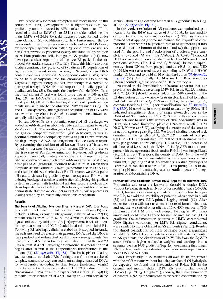
ulations of RIs directly from gradient fractions and to query theirstrand identity by strand-specific hybridization. In so doing,technical considerations forced us to return to the alkaline-gradient system, which is more rapid, taking 1 d of centrifugationinstead of 2 for FUS gradients, and whose lower viscosity greatlyincreased the speed with which fractions could be centrifugallydialyzed. We managed to dramatically reduce the problem ofDNA-rN hydrolysis in alkaline pH due to our previous seren-dipitous finding that rN-containing DNA oligos become stable atalkaline pH if kept on ice (ref. 52; Fig. 1E, lanes 0° for lig ZERΔB and lig ΔB strains). As a proof of concept, rRNA stability in0.2 M NaOH was greatly increased at 0 °C, compared with 16 °Cand especially 37 °C (Fig. 3A, lines c to e), while the same ma-terial incubated in the FUS loading buffer was stable at alltemperatures (Fig. 3A, lines k to m). Calculations of the rate ofhydrolysis of rRNA at 0 °C (SI Appendix, section 10) show that asimilarly slow hydrolysis of sparse DNA-rNs should have only aminor effect on the final size of the HMW distribution.We estimated the degree of alkaline hydrolysis in AS(4 °C)
gradients using the DNA species most sensitive to breakage, theHMW (chromosomal) DNA. Separation of chromosomal DNAsfrom the RnhB+ strain (lig) and its rnhB mutant sibling (lig ΔB)that accumulate DNA-rNs in AS(4 °C) gradients revealed adifference of two fractions between the peaks of HMW material(Fig. 3C), consistent with a minor instability of single DNA-rNsunder these conditions. Subsequent experiments confirmed these
Fig. 2. Formamide-urea-sucrose gradients reveal HMW replication inter-mediates in the lig ZER ΔB mutant. (A) The separation pattern of FUS gra-dients for the lig mutant (GR501). The temperature of [3H]dT labeling isindicated. The corresponding alkaline-sucrose gradients from Fig. 1B areshown in faded colors for comparison. (B) The effect of removal of DNA-rNexcision (lig ΔB) versus all other excision-repair systems (lig ZER) in FUSgradients. Strains: lig ZER, LA111; lig ΔB, eGC193. (C) The effect of the re-moval of both DNA-rN excision and all other excision-repair systems in onestrain (lig ZER ΔB, eGC197) in FUS gradients. (D) FUS-gradient separation ofour standard molecular weight markers: the 9.3-kb PCR fragment, phagelambda (48.5 kb), and phage T4 (∼170 kb), relative to the Okazaki fragmentspeak (∼1.1 kb) from the lig mutant (GR501).
1254 | www.pnas.org/cgi/doi/10.1073/pnas.1814512116 Cronan et al.
Dow
nloa
ded
by g
uest
on
Feb
ruar
y 15
, 202
2
predictions in ligase-minus conditions. If DNA-rNs were signif-icantly hydrolyzed during AS-gradient centrifugation at 4 °C, thedistribution of RIs from the lig ΔB mutant would sedimentsimilar to those from its lig (RnhB+) parent. This is clearly notthe case. Comparison of the four RI profiles from the FUSgradients (Fig. 3B), with identical material sedimented throughAS gradients at 4 °C (Fig. 3D), clearly demonstrates conservationof the relative sedimentation patterns between the two systems.In particular, a large gap between the HMW and LMW speciesof RIs from the lig ZER ΔB mutant was essentially unchanged inAS gradients (Fig. 3D), allowing for a clean isolation of the twomolecular weight populations with minimal cross-contamination.
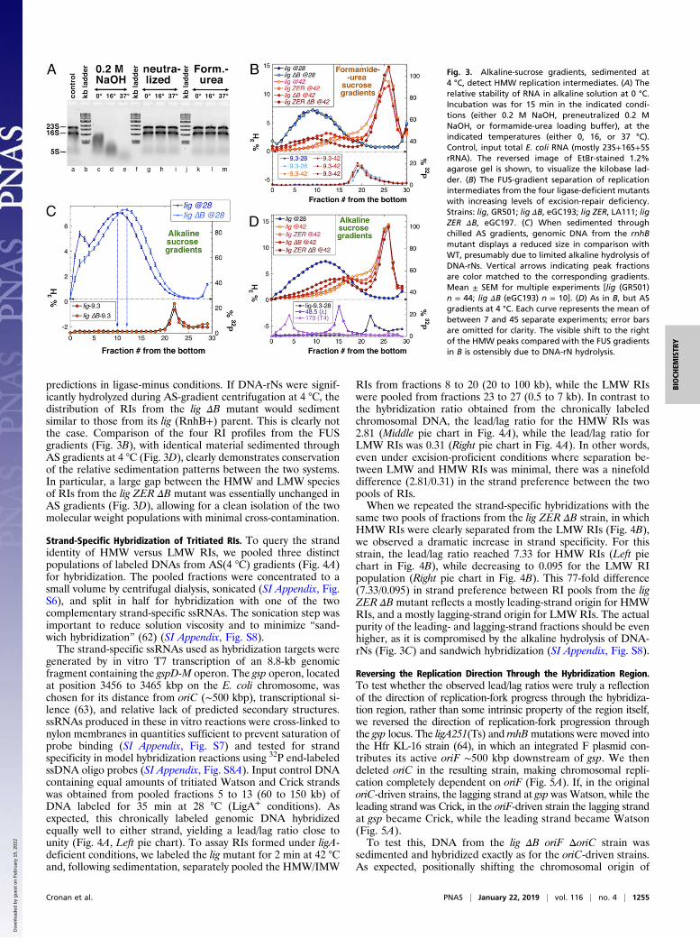
Strand-Specific Hybridization of Tritiated RIs. To query the strandidentity of HMW versus LMW RIs, we pooled three distinctpopulations of labeled DNAs from AS(4 °C) gradients (Fig. 4A)for hybridization. The pooled fractions were concentrated to asmall volume by centrifugal dialysis, sonicated (SI Appendix, Fig.S6), and split in half for hybridization with one of the twocomplementary strand-specific ssRNAs. The sonication step wasimportant to reduce solution viscosity and to minimize “sand-wich hybridization” (62) (SI Appendix, Fig. S8).The strand-specific ssRNAs used as hybridization targets were
generated by in vitro T7 transcription of an 8.8-kb genomicfragment containing the gspD-M operon. The gsp operon, locatedat position 3456 to 3465 kbp on the E. coli chromosome, waschosen for its distance from oriC (∼500 kbp), transcriptional si-lence (63), and relative lack of predicted secondary structures.ssRNAs produced in these in vitro reactions were cross-linked tonylon membranes in quantities sufficient to prevent saturation ofprobe binding (SI Appendix, Fig. S7) and tested for strandspecificity in model hybridization reactions using 32P end-labeledssDNA oligo probes (SI Appendix, Fig. S8A). Input control DNAcontaining equal amounts of tritiated Watson and Crick strandswas obtained from pooled fractions 5 to 13 (60 to 150 kb) ofDNA labeled for 35 min at 28 °C (LigA+ conditions). Asexpected, this chronically labeled genomic DNA hybridizedequally well to either strand, yielding a lead/lag ratio close tounity (Fig. 4A, Left pie chart). To assay RIs formed under ligA-deficient conditions, we labeled the lig mutant for 2 min at 42 °Cand, following sedimentation, separately pooled the HMW/IMW
RIs from fractions 8 to 20 (20 to 100 kb), while the LMW RIswere pooled from fractions 23 to 27 (0.5 to 7 kb). In contrast tothe hybridization ratio obtained from the chronically labeledchromosomal DNA, the lead/lag ratio for the HMW RIs was2.81 (Middle pie chart in Fig. 4A), while the lead/lag ratio forLMW RIs was 0.31 (Right pie chart in Fig. 4A). In other words,even under excision-proficient conditions where separation be-tween LMW and HMW RIs was minimal, there was a ninefolddifference (2.81/0.31) in the strand preference between the twopools of RIs.When we repeated the strand-specific hybridizations with the
same two pools of fractions from the lig ZER ΔB strain, in whichHMW RIs were clearly separated from the LMW RIs (Fig. 4B),we observed a dramatic increase in strand specificity. For thisstrain, the lead/lag ratio reached 7.33 for HMW RIs (Left piechart in Fig. 4B), while decreasing to 0.095 for the LMW RIpopulation (Right pie chart in Fig. 4B). This 77-fold difference(7.33/0.095) in strand preference between RI pools from the ligZER ΔB mutant reflects a mostly leading-strand origin for HMWRIs, and a mostly lagging-strand origin for LMW RIs. The actualpurity of the leading- and lagging-strand fractions should be evenhigher, as it is compromised by the alkaline hydrolysis of DNA-rNs (Fig. 3C) and sandwich hybridization (SI Appendix, Fig. S8).
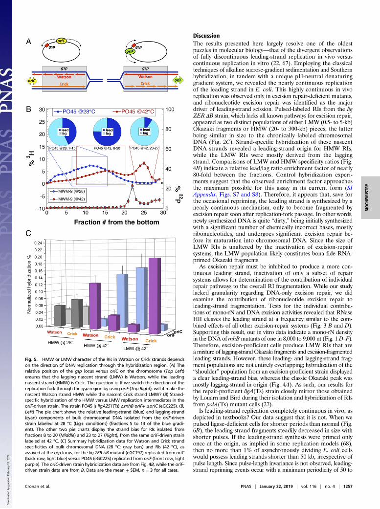
Reversing the Replication Direction Through the Hybridization Region.To test whether the observed lead/lag ratios were truly a reflectionof the direction of replication-fork progress through the hybridiza-tion region, rather than some intrinsic property of the region itself,we reversed the direction of replication-fork progression throughthe gsp locus. The ligA251(Ts) and rnhBmutations were moved intothe Hfr KL-16 strain (64), in which an integrated F plasmid con-tributes its active oriF ∼500 kbp downstream of gsp. We thendeleted oriC in the resulting strain, making chromosomal repli-cation completely dependent on oriF (Fig. 5A). If, in the originaloriC-driven strains, the lagging strand at gsp was Watson, while theleading strand was Crick, in the oriF-driven strain the lagging strandat gsp became Crick, while the leading strand became Watson(Fig. 5A).To test this, DNA from the lig ΔB oriF ΔoriC strain was
sedimented and hybridized exactly as for the oriC-driven strains.As expected, positionally shifting the chromosomal origin of
Fig. 3. Alkaline-sucrose gradients, sedimented at4 °C, detect HMW replication intermediates. (A) Therelative stability of RNA in alkaline solution at 0 °C.Incubation was for 15 min in the indicated condi-tions (either 0.2 M NaOH, preneutralized 0.2 MNaOH, or formamide-urea loading buffer), at theindicated temperatures (either 0, 16, or 37 °C).Control, input total E. coli RNA (mostly 23S+16S+5SrRNA). The reversed image of EtBr-stained 1.2%agarose gel is shown, to visualize the kilobase lad-der. (B) The FUS-gradient separation of replicationintermediates from the four ligase-deficient mutantswith increasing levels of excision-repair deficiency.Strains: lig, GR501; lig ΔB, eGC193; lig ZER, LA111; ligZER ΔB, eGC197. (C) When sedimented throughchilled AS gradients, genomic DNA from the rnhBmutant displays a reduced size in comparison withWT, presumably due to limited alkaline hydrolysis ofDNA-rNs. Vertical arrows indicating peak fractionsare color matched to the corresponding gradients.Mean ± SEM for multiple experiments [lig (GR501)n = 44; lig ΔB (eGC193) n = 10]. (D) As in B, but ASgradients at 4 °C. Each curve represents the mean ofbetween 7 and 45 separate experiments; error barsare omitted for clarity. The visible shift to the rightof the HMW peaks compared with the FUS gradientsin B is ostensibly due to DNA-rN hydrolysis.
Cronan et al. PNAS | January 22, 2019 | vol. 116 | no. 4 | 1255
BIOCH
EMISTR
Y
Dow
nloa
ded
by g
uest
on
Feb
ruar
y 15
, 202
2

replication neither influenced the 1:1 hybridization ratio for thecontrol ligase-proficient conditions (Fig. 5B, Left pie chart), nordid it alter the bimodal nature of the ligase-deficient RI distri-bution (Fig. 5B, red 3H profile). However, 42 °C RI profiles fromthe oriF strain did display an altered ratio of areas under theHMW and LMW RI curves, with ∼70% of total RIs now foundunder the HMW peak (Fig. 5B). The reason for this alteredproportion of label in the HMW peak is unknown, but we sus-pect residual activity of the LigA251(Ts) enzyme at 42 °C in thisbackground, possibly reflecting strain-specific variations in theprotein-degradation levels (65, 66).Nevertheless, the oriF strains produced broadly similar hy-
bridization results to those obtained for the oriC strains. Thelead/lag ratio was 3.1 for the HMW RIs (Middle pie chart in Fig.5B), while it was 0.1 for LMW RIs (Right pie chart in Fig. 5B),yielding a more than 30-fold difference in the relative strandpreference between the two pools of RIs. When hybridizationresults from the oriC and oriF strains were plotted side by side,the chronically labeled DNA showed an even distribution of la-bel between the two strands, regardless of the replication origintested (Fig. 5C, gray bars). Importantly, shifting the origin ofreplication relative to the hybridization region reversed the ratiosfor pulse-labeled fractions: If, in the oriC strain, the Watsonstrand signal was low in a particular MW pool, in the oriF strainthat pool showed increased signal, and vice versa (Fig. 5C),confirming the theoretical considerations (Fig. 5A). We concludethat, for at least the gsp locus of the chromosome, the leadingstrands are synthesized as HMW species, while the laggingstrands are synthesized as LMW species (Okazaki fragments),independent of the direction of replication.
Periodic Repriming of the Leading Strand. Are the nascent leadingstrands truly continuous with the rest of the chromosome, or arethey periodically reprimed? To detect LMW RIs, the Okazakigroup pulse-labeled growing cells with [3H]dT for several seconds(11, 19). They later found that, under ligase-deficient conditions,
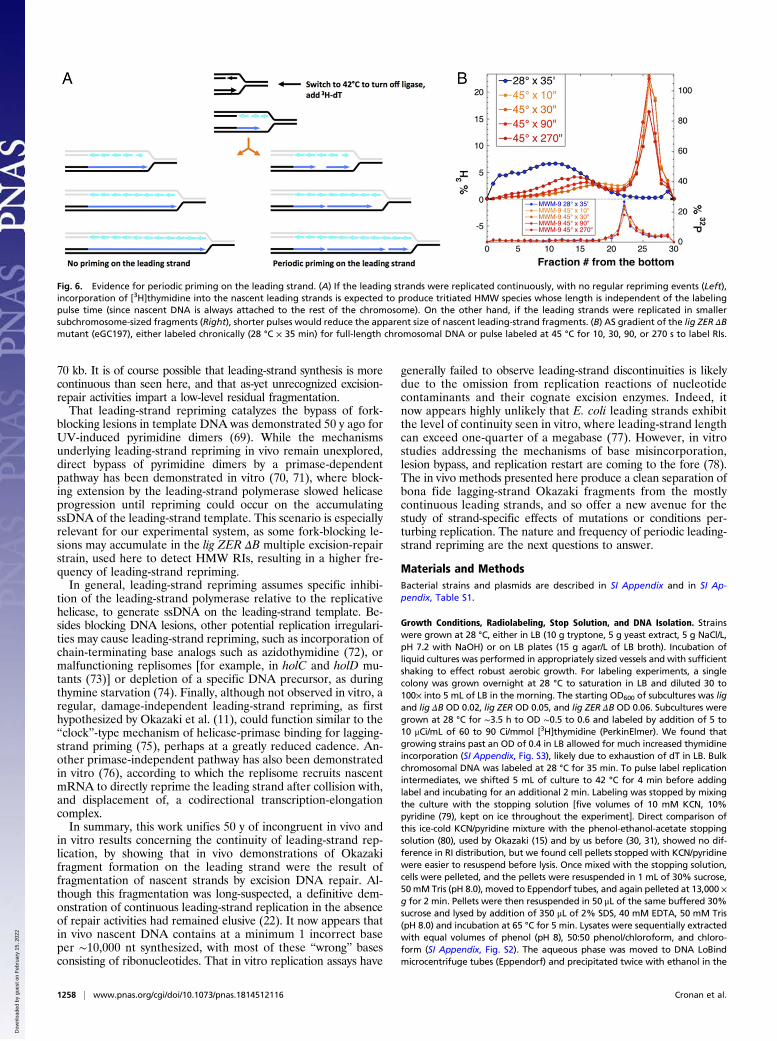
even with increased pulse length, LMW RIs continued accu-mulating linearly over at least 1 min without significant increasein length (18). In our hands, with the ligA251(Ts) mutant, incu-bation at 42 °C up to 3 min did not change the overall RI distri-bution (31). To maximize the yield of material for hybridization, wetypically labeled our lig mutants at 42 °C for 2 min. Under this longperiod of labeling, HMW RIs from the ZER mutant appearedsimilar in length to the chromosomal DNA (Fig. 3 B and D), aswould be expected if the leading strands were synthesized con-tinuously, without periodic repriming, and therefore were contig-uous with the rest of the chromosome (Fig. 6A, Left) (within the∼100-kb size limit of intact DNA strands due to our cell-lysisprocedure; SI Appendix, Fig. S2). At the same time, incubationat 42 °C for 2 min allowed for polymerization of 120 to 150 kb ofnascent DNA (54), making it possible that this long pulse timecould obscure periodic repriming events (Fig. 6A, Right).To detect possible periodic repriming on the leading strand,
we labeled the lig ZER ΔB mutant under ligase-deficient condi-tions for times between 10 and 270 s. Since this experiment isexquisitely sensitive to any traces of remaining ligase activity, thelig ZER ΔB mutant was labeled at an elevated temperature of45 °C, 3 °C above normal (but similar results were also obtainedat 42 °C). There is little to no active ligase present in these cells,as can be seen from the labeling-time insensitivity of the positionand height of the Okazaki fragment peak (Fig. 6B). While theOkazaki (LMW) peak remained unchanged, increased labelingtime caused the position of the (very broad) leading-strand dis-tribution to shift toward higher molecular weights, while thetrough between the two distributions deepened (Fig. 6B). Spe-cifically, the leading-strand distribution was centered at ∼20 kbfollowing a 10-s pulse of [3H]dT, but shifted to ∼50 kb following270 s of labeling. This result is consistent with the periodicrepriming of the leading strand, at least every ∼50 kb, althoughthis value is likely an underestimate due to the limited hydrolysisof rNs in alkaline sucrose (Fig. 3 C and D).
Fig. 4. Strand-specific hybridization of the HMW versus LMW replication intermediates. Strand-specific hybridizations were performed on tritiated DNAisolated directly from alkaline sucrose-gradient fractions. Portions of all gradient fractions were counted to generate the gradient profiles, while the re-mainder of selected fractions (colored rectangles) were pooled and hybridized against complementary strand-specific ssRNA targets. The specificity (lead/lag)ratios found in the hybridization experiments are shown as pie charts (Top), above the corresponding boxes indicating the pooled fractions used for hy-bridization. Results are shown as the mean of three independent experiments; error bars represent SEM (see Fig. 5C for hybridization SEMs; nonnormalizedhybridization data are presented in SI Appendix, Fig. S9). (A) Excision repair-proficient strain lig (GR501) was labeled with tritiated thymidine either at 28 °Cfor 35 min (bulk genomic DNA, blue curve) or 42 °C for 2 min (replication intermediates, red curve). Fractions 5 to 13 from the 28 °C gradient were hybridizedto generate the specificity ratio shown in the Left pie chart, while fractions 8 to 20 and 23 to 27 from the 42 °C labeling were hybridized to produce the Centerand Right pie charts, respectively. (B) As in A, but the excision repair-deficient strain lig ZER ΔB (eGC197) was labeled at 42 °C for 2 min, and the resultingreplication intermediates were pooled from gradient fractions 8 to 20 and 23 to 27 and hybridized to produce the strand-specificity pie charts (shown,correspondingly, Center and Right).
1256 | www.pnas.org/cgi/doi/10.1073/pnas.1814512116 Cronan et al.
Dow
nloa
ded
by g
uest
on
Feb
ruar
y 15
, 202
2
DiscussionThe results presented here largely resolve one of the oldestpuzzles in molecular biology—that of the divergent observationsof fully discontinuous leading-strand replication in vivo versuscontinuous replication in vitro (22, 67). Employing the classicaltechniques of alkaline sucrose-gradient sedimentation and Southernhybridization, in tandem with a unique pH-neutral denaturinggradient system, we revealed the nearly continuous replicationof the leading strand in E. coli. This highly continuous in vivoreplication was observed only in excision repair-deficient mutants,and ribonucleotide excision repair was identified as the majordriver of leading-strand scission. Pulsed-labeled RIs from the ligZER ΔB strain, which lacks all known pathways for excision repair,appeared as two distinct populations of either LMW (0.5- to 5-kb)Okazaki fragments or HMW (20- to 300-kb) pieces, the latterbeing similar in size to the chronically labeled chromosomalDNA (Fig. 2C). Strand-specific hybridization of these nascentDNA strands revealed a leading-strand origin for HMW RIs,while the LMW RIs were mostly derived from the laggingstrand. Comparisons of LMW and HMW specificity ratios (Fig.4B) indicate a relative lead/lag ratio enrichment factor of nearly80-fold between the fractions. Control hybridization experi-ments suggest that the observed enrichment factor approachesthe maximum possible for this assay in its current form (SIAppendix, Figs. S7 and S8). Therefore, it appears that, save forthe occasional repriming, the leading strand is synthesized by anearly continuous mechanism, only to become fragmented byexcision repair soon after replication-fork passage. In other words,newly synthesized DNA is quite “dirty,” being initially synthesizedwith a significant number of chemically incorrect bases, mostlyribonucleotides, and undergoes significant excision repair be-fore its maturation into chromosomal DNA. Since the size ofLMW RIs is unaltered by the inactivation of excision-repairsystems, the LMW population likely constitutes bona fide RNA-primed Okazaki fragments.As excision repair must be inhibited to produce a more con-
tinuous leading strand, inactivation of only a subset of repairsystems allows for determination of the contribution of individualrepair pathways to the overall RI fragmentation. While our studylacked granularity regarding DNA-only excision repair, we didexamine the contribution of ribonucleotide excision repair toleading-strand fragmentation. Tests for the individual contribu-tions of mono-rN and DNA excision activities revealed that RNaseHII cleaves the leading strand at a frequency similar to the com-bined effects of all other excision-repair systems (Fig. 3 B and D).Supporting this result, our in vitro data indicate a mono-rN densityin the DNA of rnhBmutants of one in 8,000 to 9,000 nt (Fig. 1D–F).Therefore, excision-proficient cells produce LMW RIs that areamixture of lagging-strandOkazaki fragments and excision-fragmentedleading strands. However, these leading- and lagging-strand frag-ment populations are not entirely overlapping; hybridization of the“shoulder” population from an excision-proficient strain displayeda clear leading-strand bias, whereas the classic Okazaki peak wasmostly lagging-strand in origin (Fig. 4A). As such, our results forthe repair-proficient ligA(Ts) strain closely mirror those obtainedby Louarn and Bird during their isolation and hybridization of RIsfrom polA(Ts) mutant cells (27).Is leading-strand replication completely continuous in vivo, as
depicted in textbooks? Our data suggest that it is not. When wepulsed ligase-deficient cells for shorter periods than normal (Fig.6B), the leading-strand fragments steadily decreased in size withshorter pulses. If the leading-strand synthesis were primed onlyonce at the origin, as implied in some replication models (68),then no more than 1% of asynchronously dividing E. coli cellswould possess leading strands shorter than 50 kb, irrespective ofpulse length. Since pulse-length invariance is not observed, leading-strand repriming events occur with a minimum periodicity of 50 to
Fig. 5. HMW or LMW character of the RIs in Watson or Crick strands dependson the direction of DNA replication through the hybridization region. (A) Therelative position of the gsp locus versus oriC on the chromosome (Top Left)ensures that the lagging nascent strand (LMW) is Watson, while the leadingnascent strand (HMW) is Crick. The question is: If we switch the direction of thereplication fork through the gsp region by using oriF (Top Right), will it make thenascent Watson strand HMW while the nascent Crick strand LMW? (B) Strand-specific hybridization of the HMW versus LMW replication intermediates in theoriF-driven strain. The strain PO45 is ligA251(Ts) ΔrnhB oriF+ ΔoriC (eGC225). (B,Left) The pie chart shows the relative leading-strand (blue) and lagging-strand(cyan) components of bulk chromosomal DNA isolated from the oriF-drivenstrain labeled at 28 °C (Lig+ conditions) (fractions 5 to 13 of the blue gradi-ent). The other two pie charts display the strand bias for RIs isolated fromfractions 8 to 20 (Middle) and 23 to 27 (Right), from the same oriF-driven strainlabeled at 42 °C. (C) Summary hybridization data for Watson and Crick strandspecificities of bulk chromosomal DNA (28 °C; gray bars) and RIs (42 °C), asassayed at the gsp locus, for the lig ZER ΔBmutant (eGC197) replicated from oriC(back row, light blue) versus PO45 (eGC225) replicated from oriF (front row, lightpurple). The oriC-driven strain hybridization data are from Fig. 4B, while the oriF-driven strain data are from B. Data are the mean ± SEM, n = 3 for all cases.
Cronan et al. PNAS | January 22, 2019 | vol. 116 | no. 4 | 1257
BIOCH
EMISTR
Y
Dow
nloa
ded
by g
uest
on
Feb
ruar
y 15
, 202
2
70 kb. It is of course possible that leading-strand synthesis is morecontinuous than seen here, and that as-yet unrecognized excision-repair activities impart a low-level residual fragmentation.That leading-strand repriming catalyzes the bypass of fork-
blocking lesions in template DNA was demonstrated 50 y ago forUV-induced pyrimidine dimers (69). While the mechanismsunderlying leading-strand repriming in vivo remain unexplored,direct bypass of pyrimidine dimers by a primase-dependentpathway has been demonstrated in vitro (70, 71), where block-ing extension by the leading-strand polymerase slowed helicaseprogression until repriming could occur on the accumulatingssDNA of the leading-strand template. This scenario is especiallyrelevant for our experimental system, as some fork-blocking le-sions may accumulate in the lig ZER ΔB multiple excision-repairstrain, used here to detect HMW RIs, resulting in a higher fre-quency of leading-strand repriming.In general, leading-strand repriming assumes specific inhibi-
tion of the leading-strand polymerase relative to the replicativehelicase, to generate ssDNA on the leading-strand template. Be-sides blocking DNA lesions, other potential replication irregulari-ties may cause leading-strand repriming, such as incorporation ofchain-terminating base analogs such as azidothymidine (72), ormalfunctioning replisomes [for example, in holC and holD mu-tants (73)] or depletion of a specific DNA precursor, as duringthymine starvation (74). Finally, although not observed in vitro, aregular, damage-independent leading-strand repriming, as firsthypothesized by Okazaki et al. (11), could function similar to the“clock”-type mechanism of helicase-primase binding for lagging-strand priming (75), perhaps at a greatly reduced cadence. An-other primase-independent pathway has also been demonstratedin vitro (76), according to which the replisome recruits nascentmRNA to directly reprime the leading strand after collision with,and displacement of, a codirectional transcription-elongationcomplex.In summary, this work unifies 50 y of incongruent in vivo and
in vitro results concerning the continuity of leading-strand rep-lication, by showing that in vivo demonstrations of Okazakifragment formation on the leading strand were the result offragmentation of nascent strands by excision DNA repair. Al-though this fragmentation was long-suspected, a definitive dem-onstration of continuous leading-strand replication in the absenceof repair activities had remained elusive (22). It now appears thatin vivo nascent DNA contains at a minimum 1 incorrect baseper ∼10,000 nt synthesized, with most of these “wrong” basesconsisting of ribonucleotides. That in vitro replication assays have
generally failed to observe leading-strand discontinuities is likelydue to the omission from replication reactions of nucleotidecontaminants and their cognate excision enzymes. Indeed, itnow appears highly unlikely that E. coli leading strands exhibitthe level of continuity seen in vitro, where leading-strand lengthcan exceed one-quarter of a megabase (77). However, in vitrostudies addressing the mechanisms of base misincorporation,lesion bypass, and replication restart are coming to the fore (78).The in vivo methods presented here produce a clean separation ofbona fide lagging-strand Okazaki fragments from the mostlycontinuous leading strands, and so offer a new avenue for thestudy of strand-specific effects of mutations or conditions per-turbing replication. The nature and frequency of periodic leading-strand repriming are the next questions to answer.
Materials and MethodsBacterial strains and plasmids are described in SI Appendix and in SI Ap-pendix, Table S1.
Growth Conditions, Radiolabeling, Stop Solution, and DNA Isolation. Strainswere grown at 28 °C, either in LB (10 g tryptone, 5 g yeast extract, 5 g NaCl/L,pH 7.2 with NaOH) or on LB plates (15 g agar/L of LB broth). Incubation ofliquid cultures was performed in appropriately sized vessels and with sufficientshaking to effect robust aerobic growth. For labeling experiments, a singlecolony was grown overnight at 28 °C to saturation in LB and diluted 30 to100× into 5 mL of LB in the morning. The starting OD600 of subcultures was ligand lig ΔB OD 0.02, lig ZER OD 0.05, and lig ZER ΔB OD 0.06. Subcultures weregrown at 28 °C for ∼3.5 h to OD ∼0.5 to 0.6 and labeled by addition of 5 to10 μCi/mL of 60 to 90 Ci/mmol [3H]thymidine (PerkinElmer). We found thatgrowing strains past an OD of 0.4 in LB allowed for much increased thymidineincorporation (SI Appendix, Fig. S3), likely due to exhaustion of dT in LB. Bulkchromosomal DNA was labeled at 28 °C for 35 min. To pulse label replicationintermediates, we shifted 5 mL of culture to 42 °C for 4 min before addinglabel and incubating for an additional 2 min. Labeling was stopped by mixingthe culture with the stopping solution [five volumes of 10 mM KCN, 10%pyridine (79), kept on ice throughout the experiment]. Direct comparison ofthis ice-cold KCN/pyridine mixture with the phenol-ethanol-acetate stoppingsolution (80), used by Okazaki (15) and by us before (30, 31), showed no dif-ference in RI distribution, but we found cell pellets stopped with KCN/pyridinewere easier to resuspend before lysis. Once mixed with the stopping solution,cells were pelleted, and the pellets were resuspended in 1 mL of 30% sucrose,50mM Tris (pH 8.0), moved to Eppendorf tubes, and again pelleted at 13,000 ×g for 2 min. Pellets were then resuspended in 50 μL of the same buffered 30%sucrose and lysed by addition of 350 μL of 2% SDS, 40 mM EDTA, 50 mM Tris(pH 8.0) and incubation at 65 °C for 5 min. Lysates were sequentially extractedwith equal volumes of phenol (pH 8), 50:50 phenol/chloroform, and chloro-form (SI Appendix, Fig. S2). The aqueous phase was moved to DNA LoBindmicrocentrifuge tubes (Eppendorf) and precipitated twice with ethanol in the
Fig. 6. Evidence for periodic priming on the leading strand. (A) If the leading strands were replicated continuously, with no regular repriming events (Left),incorporation of [3H]thymidine into the nascent leading strands is expected to produce tritiated HMW species whose length is independent of the labelingpulse time (since nascent DNA is always attached to the rest of the chromosome). On the other hand, if the leading strands were replicated in smallersubchromosome-sized fragments (Right), shorter pulses would reduce the apparent size of nascent leading-strand fragments. (B) AS gradient of the lig ZER ΔBmutant (eGC197), either labeled chronically (28 °C × 35 min) for full-length chromosomal DNA or pulse labeled at 45 °C for 10, 30, 90, or 270 s to label RIs.
1258 | www.pnas.org/cgi/doi/10.1073/pnas.1814512116 Cronan et al.
Dow
nloa
ded
by g
uest
on
Feb
ruar
y 15
, 202
2

presence of 150 mM KCl. The final DNA pellet was resuspended in 100 μL ofTE buffer (10 mM Tris HCl pH 8.0, 1 mM EDTA).
Plasmid Assay for RNA Density and Alkaline-Liable Sites. Assays of RNase HIIsensitivity and alkali sensitivity of plasmid DNA were performed as describedpreviously (52), but the plasmid was pCY566 (81). For linear plasmid speciestreated with formamide or NaOH, the average number of strand breaks wasderived from the zero class of the Poisson distribution, taking into account thebackground level of nicks in parental molecules (82), by the equation −ln(Ftreated/Funtreated), where Ftreated is the fraction of the full-length ssDNA in 0.3 M NaOH,45 °C × 90-min treatment, and Funtreated is the fraction of the full-length ssDNA in0.2 M NaOH, 0 °C × 5-min treatment or dissolution in 100% formamide. Theaverage density of the nicks was determined by dividing the total single-strandedplasmid length in nucleotides by the average number of nicks per molecule.
Alkaline-Sucrose Gradients.Alkaline-sucrose gradients weremade essentially asdescribed (15). Briefly, 15.6-mL linear gradients of 5 to 20% sucrose in 0.1 MNaOH, 0.9 M NaCl, 1 mM EDTA were formed in 16 × 102-mm polypropyleneultracentrifuge tubes (Beckman; 337986) using an SG15 gradient maker(Hoefer). Gradients were poured from the bottom up by pumping the solutionat ∼1.5 mL/min through a length of tubing capped with a glass capillary tube.During pouring, just as the final 20% of solution exited the mixing chamber,flow was stopped, and a 1.2-mL shelf of 80% sucrose, in the same buffer, wasadded and pumped to the bottom of the tube. Finished gradients were bal-anced and chilled at 4 °C for at least 1 h before loading. Our DNA-treatmentprotocol includes, for historical reasons, a mock alkali treatment step: To each100-μL DNA solution was added 100 μL of 0.6 M NaCl, and the mixture wasincubated at 45 °C for 1 h. Following this mock treatment, a few microliters of[32P]marker DNA was added, and the mixture was chilled in an ice/water bathfor 20 min. To denature the DNA, 200 μL of ice-cold 0.4 M NaOH was addedand the mixture was maintained on ice for 20 min. The centrifuge, rotor, andbuckets were all precooled to 4 °C. Denatured DNA samples were slowlypipetted onto the gradients. If phage DNA was included, it was liberated fromconcentrated phage particle stocks (>1012 PFU/mL) by mixing a small volume(1 to 4 μL) with an equal volume of 0.4 M Na3PO4 (55, 83) on a spoon-typespatula and then layering this mixture on top of the sample layer. Gradientswere centrifuged in a Beckman SW28.1 Ti rotor at 19,000 rpm for 20 h at 4 °C.For fractionation, the centrifuge tube was immobilized on a makeshift stand,and its bottom was punctured with a 32-gauge needle. Fractions were col-lected through the needle, with the flow rate controlled by a peristaltic pumpconnected to the top of the tube by a stopper. It took ∼20 min to fractionateeach 16.8-mL gradient into 29 fractions of ∼550 μL. Fifty to 100 μL of eachfraction was added directly to 4 mL of Bio-Safe II scintillation fluid (RPI) andcounted in a liquid scintillation counter. Fraction zero, representing tritiatedDNA that had penetrated the 80% sucrose cushion, was obtained by rinsingthe empty centrifuge tube twice with 100% ethanol, removing the tubebottom with scissors, and placing it in a tube with scintillant. To identify theposition of lambda or T4 phage DNA in a gradient, 200 μL of each fraction wasdot blotted to a nylon membrane and hybridized with a phage-specific probeusing standard methods (84).
Formamide-Urea-Sucrose Gradients. Pouring, fractionation, scintillation counting,and phage-marker hybridization procedures were identical to those detailed foralkaline sucrose. Formamide was from Fisher (99%, molecular biology grade),
and anhydrous urea was from Sigma (≥99%, ACS grade). Formamide, if notfreshly opened, was deionized immediately before use by stirring for 1 h with5% (wt/vol) bed-bed ion-exchange resin [Bio-Rex MSZ-501(D); Bio-Rad] (85).Linear 16.4-mL 5 to 40% sucrose gradients, without a shelf, were formed in70% formamide, 1 M urea, 1 M NaCl, 10 mM Tris (pH 7.5), 1 mM EDTA. Sampleswere prepared as for alkaline sucrose but, after mock alkali treatment, 50 μL of100 mM Tris (pH 7.5) was added to each DNA sample, to bring the total volumeto 250 μL. Two hundred and fifty microliters of 99% formamide was thenadded, followed by 360 mg of dry urea, and the samples were gently rocked todissolve the urea. [32P]marker DNA and phage particles were added and dis-persed by gentle inversion. Just before loading, DNA solutions were denaturedby incubation at 80 °C for 20 min and immediately but slowly pipetted onto thegradients that were kept at 4 °C, and centrifuged in a Beckman SW28.1 Ti rotorat 20,500 rpm for 44 h at 4 °C.
ssRNA Preparation and Strand-Specific Hybridization. The two pGEM-4::gspD-Mplasmids described above, which contain the gsp operon in either of the twoorientations, were linearized with BamHI for use as templates for production of8.8-kb stand-specific ssRNAs using an AmpliScribe T7-Flash Kit (Epicenter)according to the manufacturer’s instructions. For hybridization, ssRNAs weredenatured in formamide/formaldehyde as described (84), and applied toHybond-XL nylon membrane (GE Healthcare) in 3-mm-diameter circles using adot-blot vacuum manifold (Bio-Rad). Twenty micrograms of ssRNA (SI Appen-dix, Fig. S7) was applied per spot and immobilized by UV cross-linking (UVPScientific). Membranes were cut into 8 × 16-mm pieces containing individualspots of RNA and placed singly into screwcap 1.5-mL microcentrifuge tubes.One milliliter of hybridization buffer (8% SDS, 0.5 M NaPO4, 10 mM EDTA, pH7.2) was added and the tubes were rotated end over end at 55 °C for at least1 h. Gradient fractions for hybridization were pooled and concentrated to 200μL in TE buffer by centrifugal dialysis (Amicon Ultra-4, 10K NMWL; Millipore).Dialysates were moved to screwcap Eppendorf tubes and sonicated (SI Ap-pendix, Fig. S6) at 75% power, 100% duty cycle, in the “cup horn” attachmentof a Misonix S-4000 sonicator chilled to 4 °C, and then brought to a total vol-ume of 1.5 mL in 1× hybridization buffer. Hybridization mixtures were boiledfor 5 min and kept at 65 °C before each was split into two tubes containing themembrane-attached Watson or Crick gsp ssRNA targets. Hybridization pro-ceeded overnight at 55 °C, after which membranes were removed to trays andwashed successively for 30-min intervals with 5 mL each of 2× SSC, 0.5× SSC, and0.1× SSC, each containing 0.5% SDS. To improve the efficiency of tritiumcounting, membranes were moved to fresh screwcap Eppendorf tubes andstripped by boiling in 250 μL of 0.2 M NaOH. Stripping solution was thenneutralized by addition of 250 μL of 0.2 M acetic acid, 100 mM Tris (pH 7.5),added to 4 mL of scintillation fluid (Bio-Safe II; RPI), and counted to 2% error ina liquid scintillation counter.
ACKNOWLEDGMENTS. We thank Stuart Shuman (Sloan-Kettering Institute)for reminding us of the importance of rnhB inactivation for the quest tounderstand the nature of RIs in ligase-deficient conditions, and LucianaAmado Bustamante (Universidad Peruana de Ciencias) for critical readingof the manuscript. We are grateful to John E. Cronan for encouraging thedevelopment of this project and for helpful discussions on alternativesucrose-gradient solvents. We are grateful to all members of the A.K. labo-ratory for their enthusiastic support of this project. This work was supportedby National Institutes of Health Grant GM 073115.
1. Watson JD, Crick FHC (1953) Molecular structure of nucleic acids; a structure for de-
oxyribose nucleic acid. Nature 171:737–738.2. Shchyolkina AK, et al. (2000) Parallel-stranded DNAwithmixed AT/GC composition: Role of
trans G·C base pairs in sequence dependent helical stability. Biochemistry 39:10034–10044.3. Levinthal C, Crane HR (1956) On the unwinding of DNA. Proc Natl Acad Sci USA 42:436–438.4. Freese E (1958) The arrangement of DNA in the chromosome. Cold Spring Harb Symp
Quant Biol 23:13–18.5. Cairns J (1963) The chromosome of Escherichia coli. Cold Spring Harbor Symp Quant
Biol 28:43–46.6. Cairns J (1963) The bacterial chromosome and its manner of replication as seen by
autoradiography. J Mol Biol 6:208–213.7. Okazaki T, Okazaki R (1969) Mechanism of DNA chain growth. IV. Direction of syn-
thesis of T4 short DNA chains as revealed by exonucleolytic degradation. Proc Natl
Acad Sci USA 64:1242–1248.8. Kornberg A (1974) DNA Synthesis (W.H. Freeman, San Francisco).9. Gefter ML, Molineux IJ, Kornberg T, Khorana HG (1972) Deoxyribonucleic acid syn-
thesis in cell-free extracts. 3. Catalytic properties of deoxyribonucleic acid polymerase
II. J Biol Chem 247:3321–3326.10. Kornberg T, Gefter ML (1972) Deoxyribonucleic acid synthesis in cell-free extracts. IV.
Purification and catalytic properties of deoxyribonucleic acid polymerase III. J Biol
Chem 247:5369–5375.
11. Okazaki R, Okazaki T, Sakabe K, Sugimoto K, Sugino A (1968) Mechanism of DNAchain growth. I. Possible discontinuity and unusual secondary structure of newly
synthesized chains. Proc Natl Acad Sci USA 59:598–605.12. Yudelevich A, Ginsberg B, Hurwitz J (1968) Discontinuous synthesis of DNA during
replication. Proc Natl Acad Sci USA 61:1129–1136.13. Okazaki R, et al. (1968) In vivo mechanism of DNA chain growth. Cold Spring Harb
Symp Quant Biol 33:129–143.14. Bird RE, Lark KG (1970) Chromosome replication in Escherichia coli 15T− at different
growth rates: Rate of replication of the chromosome and the rate of formation of
small pieces. J Mol Biol 49:343–366.15. Okazaki R (1974) Short-chain intermediates. DNA Replication, Methods in Molecular
Biology, ed Wickner RB (Marcel Dekker, New York), Vol 7, pp 1–32.16. Okazaki R, ArisawaM, Sugino A (1971) Slow joining of newly replicated DNA chains in
DNA polymerase I-deficient Escherichia coli mutants. Proc Natl Acad Sci USA 68:
2954–2957.17. Sugimoto K, Okazaki T, Imae Y, Okazaki R (1969) Mechanism of DNA chain growth. 3.
Equal annealing of T4 nascent short DNA chains with the separated complementary
strands of the phage DNA. Proc Natl Acad Sci USA 63:1343–1350.18. Sugimoto K, Okazaki T, Okazaki R (1968) Mechanism of DNA chain growth, II. Ac-
cumulation of newly synthesized short chains in E. coli infected with ligase-defectiveT4 phages. Proc Natl Acad Sci USA 60:1356–1362.
Cronan et al. PNAS | January 22, 2019 | vol. 116 | no. 4 | 1259
BIOCH
EMISTR
Y
Dow
nloa
ded
by g
uest
on
Feb
ruar
y 15
, 202
2

19. Sakabe K, Okazaki R (1966) A unique property of the replicating region of chromo-somal DNA. Biochim Biophys Acta 129:651–654.
20. Kurosawa Y, Okazaki R (1975) Mechanism of DNA chain growth. XIII. Evidence fordiscontinuous replication of both strands of P2 phage DNA. J Mol Biol 94:229–241.
21. Pauling C, Hamm L (1969) Properties of a temperature-sensitive, radiation-sensitivemutant of Escherichia coli. II. DNA replication. Proc Natl Acad Sci USA 64:1195–1202.
22. Okazaki T (2017) Days weaving the lagging strand synthesis of DNA—A personalrecollection of the discovery of Okazaki fragments and studies on discontinuousreplication mechanism. Proc Jpn Acad Ser B Phys Biol Sci 93:322–338.
23. Nuzzo F, Brega A, Falaschi A (1970) DNA replication in mammalian cells. I. The size ofnewly synthesized helices. Proc Natl Acad Sci USA 65:1017–1024.
24. Horwitz MS (1971) Intermediates in the synthesis of type 2 adenovirus deoxy-ribonucleic acid. J Virol 8:675–683.
25. Gautschi JR, Clarkson JM (1975) Discontinuous DNA replication in mouse P-815 cells.Eur J Biochem 50:403–412.
26. Dermody JJ, Robinson GT, Sternglanz R (1979) Conditional-lethal deoxyribonucleicacid ligase mutant of Escherichia coli. J Bacteriol 139:701–704.
27. Louarn J-M, Bird RE (1974) Size distribution and molecular polarity of newly repli-cated DNA in Escherichia coli. Proc Natl Acad Sci USA 71:329–333.
28. Nasmyth KA (1977) Temperature-sensitive lethal mutants in the structural gene forDNA ligase in the yeast Schizosaccharomyces pombe. Cell 12:1109–1120.
29. Johnston LH, Nasmyth KA (1978) Saccharomyces cerevisiae cell cycle mutant cdc9 isdefective in DNA ligase. Nature 274:891–893.
30. Amado L, Kuzminov A (2006) The replication intermediates in Escherichia coli are notthe product of DNA processing or uracil excision. J Biol Chem 281:22635–22646.
31. Amado L, Kuzminov A (2013) Low-molecular-weight DNA replication intermediates inEscherichia coli: Mechanism of formation and strand specificity. J Mol Biol 425:4177–4191.
32. Konrad EB, Modrich P, Lehman IR (1974) DNA synthesis in strains of Escherichia coliK12 with temperature-sensitive DNA ligase and DNA polymerase I. J Mol Biol 90:115–126.
33. Wang T-CV, Chen S-H (1994) Okazaki DNA fragments contain equal amounts oflagging-strand and leading-strand sequences. Biochem Biophys Res Commun 198:844–849.
34. Wang T-CV, Smith KC (1989) Discontinuous DNA replication in a lig-7 strain ofEscherichia coli is not the result of mismatch repair, nucleotide-excision repair, or thebase-excision repair of DNA uracil. Biochem Biophys Res Commun 165:685–688.
35. Bradshaw JS, Kuzminov A (2003) RdgB acts to avoid chromosome fragmentation inEscherichia coli. Mol Microbiol 48:1711–1725.
36. Budke B, Kuzminov A (2010) Production of clastogenic DNA precursors by the nu-cleotide metabolism in Escherichia coli. Mol Microbiol 75:230–245.
37. Tye B-K, Lehman IR (1977) Excision repair of uracil incorporated in DNA as a result of adefect in dUTPase. J Mol Biol 117:293–306.
38. Tye BK, Nyman PO, Lehman IR, Hochhauser S, Weiss B (1977) Transient accumulationof Okazaki fragments as a result of uracil incorporation into nascent DNA. Proc NatlAcad Sci USA 74:154–157.
39. Olivera BM (1978) DNA intermediates at the Escherichia coli replication fork: Effect ofdUTP. Proc Natl Acad Sci USA 75:238–242.
40. Olivera BM, Manlapaz-Ramos P, Warner HR, Duncan BK (1979) DNA intermediates atthe Escherichia coli replication fork. II. Studies using dut and ung mutants in vitro.J Mol Biol 128:265–275.
41. Tamanoi F, Okazaki T (1978) Uracil incorporation into nascent DNA of thymine-requiring mutant of Bacillus subtilis 168. Proc Natl Acad Sci USA 75:2195–2199.
42. Tamanoi F, Machida Y, Okazaki T (1979) Uracil incorporation into nascent DNA ofBacillus subtilis and Escherichia coli. Cold Spring Harb Symp Quant Biol 43:239–242.
43. Tye B-K, Chien J, Lehman IR, Duncan BK, Warner HR (1978) Uracil incorporation: Asource of pulse-labeled DNA fragments in the replication of the Escherichia colichromosome. Proc Natl Acad Sci USA 75:233–237.
44. Cha TA, Alberts BM (1989) The bacteriophage T4 DNA replication fork. Only DNAhelicase is required for leading strand DNA synthesis by the DNA polymerase holo-enzyme. J Biol Chem 264:12220–12225.
45. Nakai H, Richardson CC (1988) Leading and lagging strand synthesis at the replicationfork of bacteriophage T7. Distinct properties of T7 gene 4 protein as a helicase andprimase. J Biol Chem 263:9818–9830.
46. Wu CA, Zechner EL, Marians KJ (1992) Coordinated leading- and lagging-strandsynthesis at the Escherichia coli DNA replication fork. I. Multiple effectors act tomodulate Okazaki fragment size. J Biol Chem 267:4030–4044.
47. Kornberg A (1980) DNA Replication (W.H. Freeman, San Francisco).48. Machida Y, Okazaki T, Miyake T, Ohtsuka E, Ikehara M (1981) Characterization of
nascent DNA fragments produced by excision of uracil residues in DNA. Nucleic AcidsRes 9:4755–4766.
49. Williams JS, Kunkel TA (2014) Ribonucleotides in DNA: Origins, repair and conse-quences. DNA Repair (Amst) 19:27–37.
50. Williams JS, Lujan SA, Kunkel TA (2016) Processing ribonucleotides incorporatedduring eukaryotic DNA replication. Nat Rev Mol Cell Biol 17:350–363.
51. Yao NY, Schroeder JW, Yurieva O, Simmons LA, O’Donnell ME (2013) Cost ofrNTP/dNTP pool imbalance at the replication fork. Proc Natl Acad Sci USA 110:12942–12947.
52. Kouzminova EA, Kadyrov FF, Kuzminov A (2017) RNase HII saves rnhA mutantEscherichia coli from R-loop-associated chromosomal fragmentation. J Mol Biol 429:2873–2894.
53. Tamm C, Shapiro HS, Lipshitz R, Chargaff E (1953) Distribution density of nucleotideswithin a desoxyribonucleic acid chain. J Biol Chem 203:673–688.
54. Kouzminova EA, Kuzminov A (2012) Chromosome demise in the wake of ligase-deficient replication. Mol Microbiol 84:1079–1096.
55. Abelson J, Thomas CA (1966) The anatomy of the T5 bacteriophage DNA molecule.J Mol Biol 18:262–288.
56. Marmur J, Ts’o PO (1961) Denaturation of deoxyribonucleic acid by formamide.Biochim Biophys Acta 51:32–36.
57. Chomczynski P (1992) Solubilization in formamide protects RNA from degradation.Nucleic Acids Res 20:3791–3792.
58. Hegedüs E, Kókai E, Kotlyar A, Dombrádi V, Szabó G (2009) Separation of 1–23-kbcomplementary DNA strands by urea-agarose gel electrophoresis. Nucleic Acids Res37:e112.
59. Bouché JP, Zechel K, Kornberg A (1975) dnaG gene product, a rifampicin-resistantRNA polymerase, initiates the conversion of a single-stranded coliphage DNA to itsduplex replicative form. J Biol Chem 250:5995–6001.
60. Sternglanz R, Wang HF, Donegan JJ (1976) Evidence that both growing DNA chains ata replication fork are synthesized discontinuously. Biochemistry 15:1838–1843.
61. Kuzminov A (2014) The precarious prokaryotic chromosome. J Bacteriol 196:1793–1806.
62. Dunn AR, Hassell JA (1977) A novel method to map transcripts: Evidence for ho-mology between an adenovirus mRNA and discrete multiple regions of the viralgenome. Cell 12:23–36.
63. Francetic O, Belin D, Badaut C, Pugsley AP (2000) Expression of the endogenous type IIsecretion pathway in Escherichia coli leads to chitinase secretion. EMBO J 19:6697–6703.
64. Low B (1968) Formation of merodiploids in matings with a class of Rec- recipientstrains of Escherichia coli K12. Proc Natl Acad Sci USA 60:160–167.
65. Van Dyk TK, Gatenby AA, LaRossa RA (1989) Demonstration by genetic suppression ofinteraction of GroE products with many proteins. Nature 342:451–453.
66. Tokuriki N, Tawfik DS (2009) Chaperonin overexpression promotes genetic variationand enzyme evolution. Nature 459:668–673.
67. Wang TC (2005) Discontinuous or semi-discontinuous DNA replication in Escherichiacoli? BioEssays 27:633–636.
68. Baker TA, Wickner SH (1992) Genetics and enzymology of DNA replication in Es-cherichia coli. Annu Rev Genet 26:447–477.
69. Rupp WD, Howard-Flanders P (1968) Discontinuities in the DNA synthesized in anexcision-defective strain of Escherichia coli following ultraviolet irradiation. J Mol Biol31:291–304.
70. Heller RC, Marians KJ (2006) Replication fork reactivation downstream of a blockednascent leading strand. Nature 439:557–562.
71. Yeeles JT, Marians KJ (2013) Dynamics of leading-strand lesion skipping by the re-plisome. Mol Cell 52:855–865.
72. Cooper DL, Lovett ST (2011) Toxicity and tolerance mechanisms for azidothymidine, areplication gap-promoting agent, in Escherichia coli. DNA Repair (Amst) 10:260–270.
73. Duigou S, Silvain M, Viguera E, Michel B (2014) ssb gene duplication restores the vi-ability of ΔholC and ΔholD Escherichia coli mutants. PLoS Genet 10:e1004719.
74. Kuong KJ, Kuzminov A (2012) Disintegration of nascent replication bubbles duringthymine starvation triggers RecA- and RecBCD-dependent replication origin de-struction. J Biol Chem 287:23958–23970.
75. Tougu K, Marians KJ (1996) The interaction between helicase and primase sets thereplication fork clock. J Biol Chem 271:21398–21405.
76. Pomerantz RT, O’Donnell M (2008) The replisome uses mRNA as a primer after col-liding with RNA polymerase. Nature 456:762–766.
77. Graham JE, Marians KJ, Kowalczykowski SC (2017) Independent and stochastic actionof DNA polymerases in the replisome. Cell 169:1201–1213.e17.
78. Marians KJ (2018) Lesion bypass and the reactivation of stalled replication forks. AnnuRev Biochem 87:217–238.
79. Jacobson MK, Lark KG (1973) DNA replication in Escherichia coli: Evidence for twoclasses of small deoxyribonucleotide chains. J Mol Biol 73:371–396.
80. Manor H, Goodman D, Stent GS (1969) RNA chain growth rates in Escherichia coli.J Mol Biol 39:1–29.
81. Cronan JE (2013) Improved plasmid-based system for fully regulated off-to-on geneexpression in Escherichia coli: Application to production of toxic proteins. Plasmid 69:81–89.
82. Kouzminova EA, Kuzminov A (2006) Fragmentation of replicating chromosomestriggered by uracil in DNA. J Mol Biol 355:20–33.
83. Freifelder D, Davison PF (1963) Physicochemical studies on the reaction betweenformaldehyde and DNA. Biophys J 3:49–63.
84. Sambrook J, Fritsch EF, Maniatis T (1989) Molecular Cloning: A Laboratory Manual(Cold Spring Harbor Lab Press, Cold Spring Harbor, NY).
85. Samanta HK, Engel D (1987) Deionization of formamide with Biorad AG501-X(D).J Biochem Biophys Methods 14:261–266.
1260 | www.pnas.org/cgi/doi/10.1073/pnas.1814512116 Cronan et al.
Dow
nloa
ded
by g
uest
on
Feb
ruar
y 15
, 202
2