Embed Size (px)
Citation preview

�
RESEARCH ARTICLE
NDST1 Missense Mutations in Autosomal RecessiveIntellectual Disability
Miriam S. Reuter,1* Luciana Musante,2 Hao Hu,2 Stefan Diederich,3 Heinrich Sticht,4 Arif B. Ekici,1Steffen Uebe,1 Thomas F. Wienker,2 Oliver Bartsch,3 Ulrich Zechner,3 Cornelia Oppitz,5
Krystyna Keleman,5,8 Rami Abou Jamra,1 Hossein Najmabadi,6,7 Susann Schweiger,3 Andre Reis,1
and Kimia Kahrizi6**1Institute of Human Genetics, Friedrich-Alexander-Universitat Erlangen-Nurnberg, Erlangen, Germany2Max-Planck-Institute for Molecular Genetics, Dept. Human Molecular Genetics, Berlin, Germany3Institute of Human Genetics, University Medical Center of the Johannes Gutenberg University Mainz, Mainz, Germany4Institute of Biochemistry, Friedrich-Alexander-Universitat Erlangen-Nurnberg, Erlangen, Germany5Research Institute of Molecular Pathology, Vienna, Austria6Genetics Research Center, University of Social Welfare and Rehabilitation Sciences, Tehran, Iran7Kariminejad-Najmabadi Pathology & Genetics Center Tehran, Tehran, Iran8Janelia Farm Research Campus, Howard Hughes Medical Institute, Ashburn, Virginia
Manuscript Received: 7 March 2014; Manuscript Accepted: 9 July 2014
How to Cite this Article:Reuter MS, Musante L, Hu H, Diederich S,
Sticht H, Ekici AB, Uebe S, Wienker TF,
Bartsch O, Zechner U, Oppitz C, Keleman
K, Jamra RA, Najmabadi H, Schweiger S,
Reis A, Kahrizi K. 2014. NDST1 missense
mutations in autosomal recessive
intellectual disability.
Am J Med Genet Part A. 9999:1–11.
Miriam S. Reuter and Luciana Musante contributed equally.
Grant sponsor: MRNET consortium, the EU FP 7 project GENCODYS;
Grant numbers: 241995, 92/801/T/1/7829.
NDST1was recently proposed as a candidate gene for autosomal
recessive intellectual disability in two families. It encodes a
bifunctional GlcNAc N-deacetylase/N-sulfotransferase with im-
portant functions inheparan sulfate biosynthesis. Inmice,Ndst1
is crucial for embryonic development and homozygous null
mutations are perinatally lethal. We now report on two addi-
tional unrelated families with homozygous missense NDST1
mutations. All mutations described to date predict the substitu-
tion of conserved amino acids in the sulfotransferase domain,
and mutation modeling predicts drastic alterations in the local
protein conformation. Comparing the four families, we noticed
significant overlap in the clinical features, including both dem-
onstrated and apparent intellectual disability, muscular hypoto-
nia, epilepsy, and postnatal growth deficiency. Furthermore, in
Drosophila, knockdown of sulfateless, the NDST ortholog,
impairs long-term memory, highlighting its function in cogni-
tion.Our data confirmNDST1mutations as a cause of autosomal
recessive intellectual disability with a distinctive phenotype, and
support an important function of NDST1 in human develop-
ment. � 2014 Wiley Periodicals, Inc.
Key words: autosomal recessive intellectual disability; heparan
sulfate biosynthesis; NDST1; sulfateless
�Correspondence to:Miriam Reuter, Schwabachanlage 10, 91054 Erlangen, Germany
E-mail: [email protected]��Correspondence to:
Kimia Kahrizi, Kodakyar St, Daneshjo Ave, Tehran, Iran
E-mail: [email protected]
Article first published online in Wiley Online Library
(wileyonlinelibrary.com): 00 Month 2014
DOI 10.1002/ajmg.a.36723
INTRODUCTION
Intellectual disability (ID) comprises a large and heterogeneous
group of neurodevelopmental disorders, characterized by signifi-
cant early-onset cognitive impairment and limitations of learning,
2014 Wiley Periodicals, Inc.
adaptive behavior and skills [Salvador-Carulla et al., 2011]. In
outbred populations, the most frequent cause of severe sporadic
ID is chromosomal abnormalities, de novo point mutations and
small insertions/deletions [de Vries et al., 2005; Vissers et al., 2010].
Nonetheless, in affected children from consanguineous families,
autosomal recessive inheritance is common [Musante and
1
2 AMERICAN JOURNAL OF MEDICAL GENETICS PART A
Ropers, 2014], and identifying the underlying genetic cause is an
important issue in clinical genetics. By means of homozygosity
mapping and next-generation sequencing approaches, a large
number of candidate genes for autosomal recessive intellectual
disability (ARID) have been identified, and althoughmany of them
share common pathways, there is a large degree of genetic hetero-
geneity [Najmabadi et al., 2007;Najmabadi et al., 2011]. Identifying
multiple families with potentially pathogenic variants in the same
gene is important to provide convincing evidence for disease
causality of candidate genes and for delineating the associated
phenotype.
NDST1 was previously suggested as a candidate gene for ARID
based on a homozygous missense mutation in one family with ID
[Najmabadi et al., 2011]. A further variant in this genewas reported
in a second family in the supplementary material of this report
although no interpretation was given. NDST1 encodes the bifunc-
tional GlcNAc N-deacetylase/N-sulfotransferase NDST1, which
has an important function in the generation of sulfated ligand-
binding sites during heparan sulfate biosynthesis [Esko and
Selleck, 2002]. Heparan sulfate interacts with growth factors and
morphogens and is crucial for embryonic differentiation
and development. Mice lacking Ndst1 function die perinatally
and exhibit variable developmental defects of the forebrain, fore-
brain-derived and facial structures [Ringvall et al., 2000; Grobe
et al., 2005; Pallerla et al., 2007].
We report on two additional families with NDST1 missense
mutations in the sulfotransferase domain and provide protein
modeling based evidence for the pathogenicity of all mutations.
We also provide detailed clinical presentation in two previously
published families [Najmabadi et al., 2011]. In addition, we show
that brain specific knockdown of the Drosophila NDST ortholog,
sulfateless, causes a long-term memory deficit in flies. Our data
support the role of NDST1 in human development and provide
insight into the clinical phenotype resulting from variants affecting
the sulfotransferase domain.
PATIENTS AND METHODS
PatientsPatients were recruited either in Germany or in collaboration with
local genetic counselors in Iran. Written informed consent was
obtained from all participants or their respective guardians. The
study was approved by the respective local ethic committees.
Genomic DNA was extracted from blood samples using standard
protocols. Tandem mass spectrometry screening for metabolic
disorders was performed for at least one patient per family.
SNP Genotyping and Homozygosity MappingMolecular karyotyping using a high density Affymetrix Genome-
Wide Human SNP Array was performed for selected individuals
from each family and did not identify any relevant copy number
variants. Homozygosity mapping using the computer program
PLINK [Purcell et al., 2007] or parametric multipoint linkage
analysis with MERLIN were performed assuming an autosomal
recessive mode of inheritance and complete penetrance [Abecasis
et al., 2002].
Exon Enrichment and High-ThroughputSequencingFor Patient 3.1, the SureSelect Human All Exon 50Mb Kit (Agilent
Technologies) was used for enrichment. Paired-end sequencing
(65/10 bp) was carried out on a SOLiD 5500xl instrument (Life
Technologies/Applied Biosystems). For Patient 4.1, the so-called
MPIMG-1-Test, a TruSeq Custom Enrichment Kit (Illumina Inc.,
San Diego, USA), targeting 1,222 genes linked to recessive child-
hood disorders or intellectual disability, was used for enrichment.
2� 250 bp paired-end sequencing (IlluminaMiSeqReagentKit v2)
was carried out on an Illumina MiSeq system.
Sequence Alignment, Variant Calling,Annotation, and VerificationFor Individual 3.1, read alignment to the human reference genome
(hg 19, GRCH37) was performed with LifeScope version 2.5.
Average target coverage was 74, while 82% of the target sequence
was covered at least five times. Single-nucleotide variants and small
indels were called with LifeScope version 2.5, GATK 2 [McKenna
et al., 2010], and SAMtools/BCFtools [Li et al., 2009]. Data were
compiled from a variety of public and in-house databases and
variant annotation was performed with Annovar [Wang
et al., 2010]. Assuming an autosomal recessivemode of inheritance,
only variants in identical-by-descent segments were considered for
further analysis. Computer-based analysis was performed with
SIFT, PolyPhen-2, and MutationTaster. For Individual 4.1, read
alignmentwasperformedwithSOAPversion2.2.More than91%of
the enriched exons were covered by at least ten reads. An improved
version of the Medical Re-sequencing Analysis Pipeline (MERAP)
developed by H. Hu (submitted) was used to check all detected
variants against dbSNP137, the 1000 Genomes Project, the Exome
Variant Server (NHLBI GO Exome Sequencing Project, http://evs.
gs.washington.edu/EVS/), the OMIM catalog (http://www.ncbi.
nlm.nih.gov/omim), and the Human Gene Mutation Database
(http://www.hgmd.org/).
For exclusion of technical artifacts and segregation testing of all
variants, PCR and Sanger sequencing were performed according to
standard protocols. Each variant was excluded in 100 to 340
chromosomes of ethnically matched controls.
Mutation ModelingTo further evaluate the effect of the amino acid substitutions on a
molecular level, mutations were modeled in silico based on known
crystal structures. The structure of the sulfotransferase domain of
human NDST1 was retrieved from the protein data bank (PDB
code: 1NST; [Kakuta et al., 1999]). The location of the substrate was
deduced from the homologous structure of 3-O-sulfotransferase
complexed with heparan sulfate (PDB code: 3UAN; [Moon
et al., 2012]). A hexasaccharide was modeled into the large open
substrate-binding site of NDST1 where it could be accommodated
without any steric clashes. Mutations were modeled using Swiss-
PDB Viewer [Guex et al., 1999] and the lowest-energy rotamer was
selected for each mutated residue. RasMol [Sayle and Milner-
White, 1995] was used for structural analysis and visualization.

REUTER ET AL. 3
Drosophila Behavioral TestingThe uas-RNAi lines (5070, 108109) were obtained from Vienna
Drosophila RNAi center (http://stockcenter.vdrc.at/control/main).
Flies were raised on semi-defined medium at 25˚C in a 12 hr dark-
light cycle. Virgin males were collected at eclosion and aged
individually for five days before training. Canton-S premated
females were aged for four days in groups of 50–100 with Can-
ton-S males collected at the same time.
Males were assayed for courtship conditioning as described
[Siwicki and Ladewski, 2003]. In brief, for training, individual
males were placed in food chambers either with (trained) or
without (naıve) a single premated female for seven hours. After
training, each male was recovered, transferred to a fresh food
chamber, kept in isolation and tested for long-term memory after
24 hr. Tests were performed in a 10mm diameter courtship cham-
ber and videotaped for 10min (JVChandycam, 30GBHD). Videos
were scoredwith automated software (C. Schusterreiter, C.Macha-
cek, B.Dickson, unpublished) for courtship index (CI), which is the
fraction of time eachmale spent courting during the test. Mean CIs
were used to calculate the learning index (LI): CInaive-CI trained/
CInaive� 100. Data represent average values� the standard error of
the mean (SEM) of performance indices. Learning indices were
analyzed using a MATLAB script by permutation test [Kamyshev
et al., 1999]. Briefly, the entire set of courtship indices for both the
naıve and trainedflieswere pooled and then randomly assorted into
simulated naıve and trained sets of the same size as in the original
data. A LIp was calculated for each of 100,000 randomly permuted
data sets, and P-values were estimated as the fraction for which
LIp> LI (to test H0, LI¼ 0).
RESULTS
Clinical DescriptionWe describe eight patients from four unrelated families, who were
initially referred for developmental delay or intellectual disability.
Medical histories, diagnostic findings, and pedigrees were obtained
in a clinical setting in Iran or Germany, and are summarized in
Table I, Figure 1 and Figure 2.
Family 1Patient 1.1 is the first child of consanguineous healthy parents from
Eastern Iran.After anuneventful pregnancy, hewasborn at 38weeks
of gestation. Birth weight was unavailable, but said to be high for
gestational age, length was not documented, and occipitofrontal
circumference (OFC)was 34 cm (�0.2 SD). Psychomotor delaywas
noted in early childhood. At age 2.5 years, he started to walk and
spoke first words. Fine motor development was not documented.
Generalized seizures in childhood could be controlled by antiepi-
leptic medication. Short stature and non-progressive ataxia were
noted around age 15 years. The patient suffered from polyuria, but
urine analysis and renal ultrasoundwere normal and further studies
were not performed. At age 36 years, height was 153 cm (�3.7 SD)
andOFCwas55 cm(�1.3 SD).Parentalheightwasnormal (�1 SD).
The patient exhibited muscular hypotonia and showed good eye
contact with his parents. He didn’t have ability to communicate nor
to follow simple commands. Testing of cognitive function using the
WechslerAdult IntelligenceScale (WAIS-IV) showedan intelligence
quotient (IQ) of 30 in the range of severe intellectual disability.
Aggressive social behavior, agitation, and sleep disturbances were
confirmed by psychiatric assessment using the Diagnostic Assess-
ment for the Severely Handicapped (DASH-II). Cranial magnetic
resonance imaging (MRI) and electroencephalography (EEG) were
normal, facial features are shown in Figure 2A.
Patient 1.2 is the younger sister of Patient 1.1. She was born at
term after an uneventful pregnancy. Birth weight was unavailable,
but said to be large for gestational age and OFC was 35.5 cm
(þ0.2 SD). She started to walk and spoke first words at age three
years. She had a history of generalized seizures in childhood that
could be controlled by antiepileptic medication. Short stature was
noted from age 16 years.When evaluated at age 32 years, height was
145 cm (�3.1 SD) andOFCwas 54 cm (�1 SD).Hearing and visual
testing were normal except strabismus. IQ testing at age 32 years by
WAIS-IV revealed an IQ of 26. The parents reported a generally
quiet temperament with sometimes increased irritability, which
was not formally tested.
Patient 1.3 is the youngest brother of patient 1.1. He was born at
term after an uneventful pregnancy. Birthweightwas again high for
gestational age and OFC was 35 cm (þ0.1 SD). He started to walk
and spoke first words at age 2.5 years. Muscular hypotonia and flat
feet were first described in adolescence, ataxia was not noted. At age
28 years, height was 150 cm (�4.1 SD) and OFC was 55 cm
(�1.3 SD). An IQ of 26 was revealed by WAIS-IV. Aggressive
and self-injurious behavior was noted by the family and was
confirmed by psychiatric assessment using the DASH-II checklist.
Hearing and vision were normal.
Family 2Patient 2.1 is the first child of healthy consanguineous parents (first
cousins), originating from North Eastern Iran. The patient was
born at term after an uneventful pregnancy. Birth weight was
4,200 g (þ2.4 SD), length and OFC were not documented. She
was breast-fedwithout problems for 12months. She started to walk
and spoke first words at age three years. Generalized seizures first
occurred at age 12 years, and could be controlled by antiepileptic
medication. No history of ataxia or muscular hypotonia was
reported. Her body height at age 18 years was 155 cm (�1.4 SD),
weight was 52 kg (BMI 22), and OFC was 55 cm (�0.4 SD). Facial
appearance was non-dysmorphic and no physical malformation
was observed. She had good eye contact with her parents, but
showed very little interest inher environment and spoke less than20
words. Speech perception was apparently better, and hearing was
tested normal. Cognitive evaluation at age 18 years (WAIS-IV)
yielded an IQ of 37 in the range of moderate intellectual disability.
Aggressive behavior was assessed by the Achenbach Child Behavior
Checklist (CBCL/6–18). A cranial MRI obtained at age 22 years
showed normal brain anatomy.
Patient 2.2 is the younger brother of patient 2.1. He was born at
term after an uneventful pregnancy. Birth weight was 4,000 g
(þ1.6 SD), length and OFC were not documented. He was
breast-fed without problems for 11 months. Global developmental
delay was noted in childhood, he started to walk and spoke first
words at age 2.5 years. Generalized seizures and ataxic gait were

TABLE
I.Summarized
Clinical
Findings
intheAffected
Individuals
Patient1.1
�Patient1.2
�Patient1.3
�Patient2.1
��Patient2.2
��Patient3.1
Patient4.1
Patient4.2
NDST1mutation
(NM_001543)
c.2126G>A
(p.Arg709Gln)
c.2126G>A
(p.Arg709Gln)
c.2126G>A
(p.Arg709Gln)
c.1926G>T
(p.Glu642Asp)
c.1926G>T
(p.Glu642Asp)
c.1918T>C
(p.Phe640Leu)
c.1831G>A
(p.Gly611Ser)
c.1831G>A
(p.Gly611Ser)
Gender
male
female
male
female
male
female
male
female
Parental
consanguinity
first
cousins
first
cousins
first
cousins
first
cousins
first
cousins
first
cousins
likelyendogamy
likelyendogamy
Ethnicorigin
Iranian
Iranian
Iranian
Iranian
Iranian
Turkish
Turkish
Turkish
Birth
38w
40w
40w
38w
38w
41w,afterpreterm
labor
inthe
seventh
month
ofpregnancy,
cesarean
section
atterm
,vacuum
extraction
with
subsequentcaput
succedaneum
and
cephalhematom
a
atterm
BW
high
for
gestational
age
high
for
gestational
age
high
for
gestational
age
4200g
(þ2.4
SD)
4000g(þ
1.6
SD)
4390g(þ
1.9
SD)
3850g(þ
0.5
SD)
ND
BL
ND
ND
ND
ND
ND
51cm
(�0.6
SD)
52cm
(�0.2
SD)
ND
OFC
34cm (�
0.2
SD)
35cm (þ
0.1
SD)
35cm
(þ0.1
SD)
ND
ND
35.5cm
(þ0.2
SD)
ND
ND
Ageat
last
exam
ination
36y
32y
28y
18y
12y
11y9m
12y
4y2m
Postnatal
grow
thWeightat
last
exam
ination
normal
for
ageandheight
normal
for
ageandheight
normal
for
ageandheight
52kg (B
MI22)
55kg
(BMI34)
40kg
(BMI18)
normal
forage
andheight
22kg
(BMI20)
Heightat
last
exam
ination
153cm (�3.7
SD)
145cm (�3.1
SD)
150cm (�4.1
SD)
155cm (�1.4
SD)
126cm
(�3.1
SD)
149cm
(�0.1
SD)
147.5cm
(�1.2
SD)
105cm
(0.9
SD)
OFC
atlast
exam
ination
55cm (�
1.3
SD)
54cm
(�1SD)
55cm (�
1.3
SD)
55cm (�
0.4
SD)
53cm
(�1SD)
53.5cm
(�0.7
SD)
60.5cm
(þ3.3
SD)
54cm
(þ2.4
SD)
Psychomotor
developm
ent
Gross
motor
developm
ent
delayed
delayed
delayed
delayed
delayed
mildly
delayed
delayed
delayed
Finemotor
developm
ent
ND
ND
ND
delayed
delayed
delayed
delayed
delayed
Language
expressive
delayed
expressive
delayed,
slurredspeech
expressive
delayed
expressive
delayed
expressive:
delayed,
perceptive:
mildly
delayed
expressive:delayed,
perceptive:
mildly
delayed
profoundexpressive
speech
delay;
speech
perception
better
severe
expressive
speech
delay
Social developm
ent
aggression
(DASH-II)
increased
irritability,
generally
quiet
temperament
(parentreport)
aggression,self-
injurious
behavior
(DASH-II)
aggression
(CBCL/6-18)
aggression,
self-injurious
behavior
(parentreport)
aggression,
self-injurious
behavior,
genderdysphoria
(parentreport)
lowactivity,quiet
temperament
(parentreport)
well-balanced,
kind
temperament
(parentreport)
Intellectual
disability
severe(IQ30;WAIS-IV)
severe(IQ26;WAIS-IV)
severe(IQ26;WAIS-IV)
moderate
(IQ37;WAIS-IV)
moderate-severe
(estimated)
mild
(IQ64;HAW
IK-IV)
severe
(IQ20–35;
estimated)
moderate(estimated)
Epilepsy
Seizures
(onset)
þ(childhood)
þ(childhood)
þ(childhood)
þ(14y)
þ(14y)
��
�Therapyresponse
þþ
þþ
þ�
��
Motor
system
Ataxia
þ�
��
þ�
��
Muscular
hypotonia
þ�
þ�
�þ
þþ
Eyes
Visual
function
normal
normal
normal
normal
myopia
normal
normal
normal
Strabism
us�
þ�
�þ
��
not
when
rested,
exotropia
whentired
Nystagm
us�
��
�þ
��
�Hearing
normal
normal
normal
normal
normal
normal
normal
normal
Joints
normal
normal
normal
normal
normal
hyperm
obility
ofwrist
and
fingerjoints
normal
normal
Physical
malform
ations
��
��
��
��
Facial dysm
orphism
��
��
�protrudingchin,
synophrys,gaps
betweenteeth
largeprotrudingears
facial
hypotonia,
epicanthal
folds,
mild
ptosis
MRI(age)
normal
ND
ND
normal
(22y)
normal
(10y)
normal
(7.5y)
symmetrically
enlarged
lateral
ventricles,less
dilated3rd
ventricle,
otherwisenormal
brainanatom
y(8y)
ND
EEG
normal
ND
ND
ND
normal
normal
normal
ND
Other
anom
alies
sleepdisturbance,polyuria
flatfeet
sleepdisturbance
sleepdisturbance,
frequent
constipation
flatfeet
Abbreviationsareas
follows:y,years;m,months;w,weeks;ND,nodata
available;BW,birthweight;BL,birthlength;OFC,occipito
frontalhead
circum
ference;IQ,intelligence
quotient.
� Najmabadiet
al.[2011],Family
M8600277.
��Najmabadiet
al.[2011],Family
M161.
4 AMERICAN JOURNAL OF MEDICAL GENETICS PART A

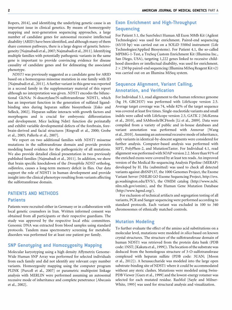
FIG. 1. Pedigrees and electropherograms of the four families. Black fill defines individuals with homozygous NDST1 mutations, gray fill defines
sibling with glycogen storage disease type 1b. (A) Family 1 (M8600277), homozygous NDST1 mutation c.2126G> A (p.Arg709Gln). (B)
Family 2 (M161), homozygous NDST1 mutation c.1926G> T (p.Glu642Asp). (C) Family 3 (ER44462), homozygous NDST1 mutation
c.1918T> C (p.Phe640Leu). (D) Family 4 (MZ-778/12), homozygous NDST1 mutation c.1831G> A (p.Gly611Ser).
REUTER ET AL. 5
noted at age 12 years. At this age, height was 126 cm (�3.1 SD),
weight was 55 kg (BMI 35), and head circumference was 53.5 cm
(�1 SD). IQ was not formally tested but he was unable to count
money, nor was he able to memorize simple family names or
degrees of relationships. The parents reported aggressive, self-
injurious behavior, sleep disturbances, impaired verbal expres-
sion and communication problems. Visual testing showed myo-
pia, strabismus, and horizontal nystagmus with no other
abnormality. Facial appearance was normal and a cranial MRI
obtained at age ten years was normal.

FIG. 2. Photographs of affected individuals from families 1 and 4. (A) Patient 1.1. (B) Patient 1.3. (C) Patient 1.2. (D) Patient 4.1 aged
12 years, exhibiting large protruding ears. (E) Patient 4.2 aged 4 years 3 months, exhibiting mild ptosis of eyelids, epicanthal folds, and
facial hypotonia.
6 AMERICAN JOURNAL OF MEDICAL GENETICS PART A
Family 3Patient 3.1 is the first child of healthy consanguineous parents (first
cousins), originating from Turkey. The pregnancy was uneventful
except preterm labor in the seventh month of pregnancy. The girl
was born by cesarean at 41 weeks of gestation. Her weight was
4,390 g (þ1.9 SD), length 51 cm (�0.6 SD), OFC 35.5 cm
(�0.4 SD), APGAR 10/10, umbilical cord pH 7.28. Development
initially appeared normal; she walked and spoke first words at age
12 months. However, in follow-up examinations global develop-
mental delay was noted, with building of two-word phrases at age
three years, delayed fine motor development, and muscular hypo-
tonia. Formal developmental testing at age 7.5 years by Wechsler
Intelligence Scale for Children (HAWIK-IV) showed an IQ of 64.
Thepatient attended a special needs school.When last seen at age 11
years 9 months, the parents reported difficulties concentrating,
hyperactivity, reduced memorization and social interaction prob-
lems (aggression, self-injuriousbehavior, genderdysphoria), aswell
as enuresis, encopresis, sleep disturbances, and frequent constipa-
tion. No history of seizures or deterioration in neurological func-
tions was reported. Growth parameters were all normal at age 11
years 9 months. Her height was 149 cm (�0.1 SD), weight 40 kg
(BMI 18), OFC 53.5 cm (�0.7 SD). She spoke only short sentences
with limited vocabulary (Turkish and German), but receptive
language appeared better. Balance, coordination, and deep tendon
reflexes were normal. Hypermobility of the wrist and finger joints
resulted in a Beighton score of 4/9. Minor dysmorphic features
included a prominent protruding chin, synophrys, wide gaps
between the teeth, and a compact trunk. Ophthalmologic and
audiometric examinations were normal. Cranial MRI and EEG
examinations at age 7.5 years were normal.
Family 4Patient 4.1 is the first child of a healthy 27-year-oldmother and 29-
year-old father. His parents were of Turkish origin and not known
to be consanguineous, but likely endogamic originating from the
same religiously isolated village in Bulgaria. After an uneventful
pregnancy, he was born at term by vacuum extraction, with
subsequent caput succedaneum, and cephalhematoma. His weight
was 3,850 g (þ0.5 SD), length 52 cm (�0.2 SD), but his OFC was
not documented. Developmental delay and muscular hypotonia
were noted in the first year of life; hewalked and spoke first words at

REUTER ET AL. 7
age three years. A full neuropediatric evaluation was performed in
Germany at age eight years and five months. He had good eye
contact with his parents, but showed very little interest in his
environment, played stereotypically, and spoke no word. Parents
reported a total vocabulary of less than 10–20 words. Receptive
language appeared better, and hearing loss was excluded. Cognitive
testing at age eight years and five months, using the Snijders-
Oomen non-verbal intelligence test (SON-R 2 1/2–7) was not
possible due to lack of participation. IQwas estimated within range
20–35. At age 12 years, parents still reported a total vocabulary of
maximum 10–20 words and a quiet temperament. He attended a
special needs school for the severely intellectually disabled. Growth
parameters at 8.5 years were: height 125 cm (�1 SD), weight 31 kg
(BMI 20), OFC 59 cm (þ3.1 SD); 12 years: height 147.5 cm
(�1.2 SD), OFC 60.5 cm (þ3.3 SD); parental OFCs were
within normal ranges. Minor dysmorphic features included large
protruding ears (Fig. 2D) and broad, flat feet. A cranial MRI
obtained at age 8 years showed symmetrically enlarged lateral
ventricles and a less dilated third ventricle, but otherwise normal
brain anatomy.
Patient 4.2, the younger sister of patient 4.1, was born at term
after an uneventful pregnancy. Developmental delay and muscular
hypotonia became obvious in the second year of life. She walked at
age 18–24 months and expressive speech was delayed. Non-verbal
cognitive testing using the SON-R 2 1/2 - 7 was attempted at age
four years and two months, but she was unable to follow the
instructions, neither in German language nor with a Turkish
interpreter. Moderate intellectual disability was diagnosed and
her developmental age was estimated at less than two years. She
attended a special needs kindergarten. The parents described a
happy, well-balanced, and kind temperament. At age four years two
months, weight and OFC exceeded normal values. Her height was
105 cm (0.9 SD), weight 22 kg (BMI 20), OFC 54 cm (þ2.4 SD).
Minor dysmorphic features included frontal bossing, facial hypo-
tonia,mild ptosis, epicanthal folds, a single crease on the right hand,
short fingers, and a sacral dimple. Hearing tests were repeatedly
performed with likely normal results.
MutationsUsing homozygosity mapping and next-generation sequenc-
ing approaches, missense mutations in the NDST1 gene
(MIM�600853) couldbe identified in two furtherunrelated families
with intellectual disability. In patient 3.1, an apparently homozy-
gous mutation c.1918T>C (NM_001543; chr5:149922481T>C;
p.Phe640Leu) was identified by exome sequencing, and in pa-
tient 4.1, an apparently homozygous mutation c.1831G>A
(NM_001543; chr5:149921213G>A; p.Gly611Ser) was identified
by custom-designed targeted sequencing (Fig. 1C and D). Direct
Sanger sequencing analysis was performed for all available family
members, and demonstrated segregation in agreement with a
recessive mode of inheritance. The mutations have not been
reported in dbSNP 137, 1000 Genomes and the NHLBI Exome
Variant Server, were absent in 100 to 340 chromosomes of ethni-
cally matched controls, and affected highly conserved amino acids.
Analyses with SIFT, PolyPhen-2, andMutationTaster predicted the
amino acid substitutions to be damaging.
In silico mutation modeling suggested that all four mutations
alter the sulfotransferase domain of NDST1. Phenylalanine and
glutamic acid at positions 640 and 642, respectively, are located in
a loop of the substrate-binding site and establish tight contacts
with the substrate (Fig. 3A and C). These interactions are lost,
when the respective residue is substituted by an amino acid with a
shorter side chain (leucine or aspartic acid) (Fig. 3B and D).
Arginine at position 709 forms direct interactions with cysteine at
position 751, as well as water-mediated interactions with gluta-
mine at position 613 and arginine at position 750 (Fig. 3E).
These interactions cannot be maintained by the shorter and
uncharged glutamine at position 709 in the mutant (Fig. 3F),
which is predicted to cause a drastic destabilization of the three-
dimensional fold of NDST1. Glycine at position 611 is located
in a loop in spatial proximity to the 30-phosphoadenosine50-phosphate (PAP) binding site (Fig. 3G). It adopts an unusual
backbone conformation (F¼ 97˚,C¼� 147˚), exclusively feasi-
ble for the small amino acid glycine lacking a side chain. Conse-
quently, a replacement by the larger serine results in steric clashes
with adjacent amino acids, in particular proline at position 612
(Fig. 3H). These clashes will result in a destabilization of the
structure and conformational rearrangement of the loop, which
most likely also affects PAP binding. In summary, all four amino
acid substitutions are predicted to change the substrate–binding
capability and/or three-dimensional structure of the sulfotrans-
ferase domain of NDST1.
Drosophila Behavioral TestingIn order to evaluate NDST1 function in the brain, the Drosophila
ortholog of the NDST family, sulfateless (sfl) [Aikawa et al., 2001],
was knocked down pan-neuronally by driving the RNAi expression
with elev-Gal4. To strengthen the knockdown efficiency, we co-
expressed dicer2 [Dietzl et al., 2007]. Given the fact that brain
knockdown had no impact on survival or morphology, and loco-
motion appeared normal in negative geotaxis assay (data not
shown), we investigated long-term memory as a higher cognitive
function by the courtship-conditioning paradigm [Siwicki and
Ladewski, 2003]. In this paradigm, male flies learn to adapt their
courtship behavior. In response to rejection by a previously mated
unreceptive female during training, the male fly will reduce its
courtship attempts when exposed again to another unreceptive
female during the test phase. This was shown for control flies
(control-RNAi/elav-Gal4 and uas-sflRNAi/þ) by significantly dif-
ferent courtship indices CI (Fig. 4A) and high learning indices LI
(Fig. 4B). In contrast, CI of naıve and trained sfl knockdown flies
were not significantly different (Fig. 4A), resulting in a severely
reduced LI of 5% (Fig. 4B), indicating that long-termmemory of sfl
knockdown flies was severely impaired. Levels of naıve courtship
howeverwere similar for all genotypes indicating that sensorimotor
functions did not influence the memory phenotype observed in sfl
knockdown (Fig. 4A).
DISCUSSION
Autosomal recessive intellectual disability is extremely heteroge-
neous in terms of its underlying genetic causes, and the total

FIG. 3. Structural effect of the mutations in NDST1. (A) Location of F640 (space-filled) in the substrate binding site of NDST1. A
hexasaccharide substrate (Sub) is shown in stick presentation and colored according to the atom type. The backbone of NDST1 is indicated as
green ribbon. (B) Effect of the F640L mutation revealing that the lid formed by F640 on the substrate cannot be formed by the smaller
leucine sidechain. (C) Location of E642 (stick presentation) in the substrate-binding site of NDST1. A hydrogen bond formed with a hydroxyl
group of one N-acetylglucosamine unit is indicated as blue dotted line. (D) The hydrogen bond cannot be formed by the shorter sidechain in
the E642D mutation. (E) R709 forms hydrogen bonds (blue dotted lines) with Q613, R750, and C751. Two of these interactions are mediated
by a water molecule (W; shown as brown ball). (F) Effect of the R709Q mutation, showing that several stabilizing interactions are lost
compared to the wildtype. (G) G611 is located in a turn close to the PAP binding site (stick presentation). G611 and P612 (cyan) are shown
in space-filled representation. G611 and PAP are colored according to the atom type. (H) Effect of the G611S mutation resulting in steric
clashes between S611 and P612 (magenta arrow).
8 AMERICAN JOURNAL OF MEDICAL GENETICS PART A

FIG. 4. Brain knockdown of sulfateless results in long-term
memory deficit in Drosophila. (A) Mean courtship indices (CIs)
of trained (þ) and naıve (�) flies are displayed. Learning
ability was assayed using a non-parametric permutation (ran-
domization) test. P-values are indicated for H0, LI¼ 0 and thus
show the probability that flies did not learn-a low P-value
signifies that flies learned. Naıve and trained control flies (uas-
controlRNAi/elav-Gal4 and uas-sflRNAi/þ) show a significant
difference (���, P¼ 0.00051 and ��, P¼ 0.00388), whereas (þ)
and (�) Sfl-KD (uas-sflRNAi/elav-Gal4) flies show no difference
(NS, P¼ 0.29232), indicating impairment in learning ability.
Numbers in the boxplot depict sample sizes (n) for each
genotype and condition. (B) The relative difference between
naıve and trained CIs, called learning index (LI), is shown. LI of
sfl-KD was significantly lower than LI of control (uas-controlR-
NAi/elav-Gal4) (��, P¼ 0.03228), while the two controls were
not significantly different. Asterisks denote significance (�,P< 0.05; ��, P< 0.01; ���, p< 0.001).
REUTER ET AL. 9
number of ARID genes is estimated to run into the thousands
[Musante and Ropers, 2014]. Complicating the identification of
novel disease-related genes is the fact, that offspring of consan-
guineous parents typically carry numerous rare and difficult-to-
assess homozygous variants. A detection of mutations in the same
gene in unrelated patients is therefore important to provide further
evidence for its disease causality.
Two missense mutations in NDST1 were initially reported by
Najmabadi et al., [2011], each in one consanguineous family with
autosomal recessive intellectual disability. We now identified two
additional families with NDST1 mutations and provide a first
characterizationof thehumanphenotype, in a total of eight patients
from four unrelated families, including two families previously
reported by Najmabadi et al. [2011]. Their clinical features exhib-
ited remarkable overlap (Table I), supporting the pathogenic
impact of the described NDST1 missense mutations. The main
symptom was an impairment of motor and cognitive functions. In
addition, there was evidence for a frequent occurrence of muscular
hypotonia and postnatal growth deficiency, affecting particularly
adult height, but not OFC. Some patients developed seizures in
childhood or adolescence. Malformations of the brain or other
organs were not detectable (apart from enlarged lateral ventricles in
onepatient). In addition,mutationmodelingperformed for all four
mutations also argues for a functional relevance of the amino acid
substitutions.
In mouse models and murine cells, the role of Ndst1 in differ-
entiation and development is well established. Ndst1 is one in a
family of four bifunctional GlcNAcN-deacetylase/N-sulfotransfer-
ase enzymes (Ndsts), catalyzing the first and essential modifying
steps (N-deacetylation/N-sulfation ofGlcNAc residues) in heparan
sulfate (HS)biosynthesis.Different formsofHSarepresent onmost
cell membranes and in extracellular matrix proteoglycans, and by
interaction with growth factors and morphogens are crucial for
embryonic differentiation and development [Bernfield et al., 1999;
Esko and Lindahl, 2001]. An important role of HS in the develop-
ment andmaintenance of the central nervous system is indicated by
studies in conditional knockout mice and cell cultures [Inatani
et al., 2003; Irie et al., 2012].
The expression of HS sulfotransferase genes is spatiotemporally
regulated [Yabe et al., 2005]. Ndst1 is expressed ubiquitously, and
purified protein has higherN-sulfotransferase activity compared to
the other three isozymes [Aikawa et al., 2001]. Mice lacking Ndst1
function die perinatally and exhibit variable developmental defects
of the forebrain- and neural crest-derived structures (telencepha-
lon, diencephalon, eyes, neuro- andviscerocranium) and, to a lower
extent, the neural tube [Grobe et al., 2005; Pallerla et al., 2007]. The
developmental defects were attributed to impaired Wnt, sonic
hedgehog and fibroblast growth factor signaling [Pallerla et al.,
2007]. Mouse embryonic stem cells deficient of both Ndst1 and
Ndst2 resulted in undersulfation of heparan sulfate and a differen-
tiation block at the primitive ectoderm/endoderm stage, while
mesodermal differentiation appeared to be normal [Forsberg
et al., 2012].
Heparan sulfate also exhibits crucial functions in Drosophila
development [Perrimon and Bernfield, 2000] and the NDST
ortholog sulfateless is required for the development of the
stomatogastric nervous system [Lin and Perrimon, 1999]. Genes,

10 AMERICAN JOURNAL OF MEDICAL GENETICS PART A
pathways and regulatory networks are well conserved between
human and Drosophila [Bellen et al., 2010]. Increasing evidence
indicates that flies represent a powerful animal model to gain
insights into ID/ARID gene functions [Abbasi-Moheb et al.,
2012; Gatto and Broadie, 2011]. Our data indicate that sfl brain-
specific knockdown results in long-term memory deficits in flies,
strongly suggesting a crucial role for sfl in higher cognitive func-
tions.We speculate, that a regulatory role of sfl in theWnt receptor
signaling pathway [Kamimura et al., 2011]might be responsible for
the observed long-term memory phenotype, as Wnt signaling has
recently been shown tobe required for long-termolfactorymemory
formation in Drosophila [Tan et al., 2013].
Our data support an important function of NDST1 also in
human development. Sibs carrying the same missense mutation
inNDST1 exhibited a strikingly similar clinical phenotype, whereas
certain differences were apparent between the families. It is unclear
at this stage, whether the variability is attributable to the different
NDST1 mutations or to additional genetic or non-genetic factors.
All so far described homozygousmutations aremissensemutations
clustering in the sulfotransferase domain, and are predicted to
incapacitate the enzymatic function. Whether amino acid substi-
tutions in other protein domains cause a similar phenotype, and if a
complete loss of the enzymatic activity would be compatible with
life inhumans, remainsunclear at thispoint.Descriptionsof further
patients will be necessary to elucidate correlations between partic-
ular mutant alleles and consistent clinical phenotypes. In addition,
further analyses regarding NDST1 activity and the extent and
pattern of sulfation in patients’ cell lines will be required to test
the functional consequences of specific missense mutations.
ACKNOWLEDGMENTS
We thank the patients and their families for participation in this
study. We thank Dr. Mohammad Reza Khodaei, who performed
behavioral and cognitive assessment of Families 1 and 2, and
Dr. Uwe Jakob, Dr. Helmut Peters, Dr. Bettina Weßner and their
medical and psychological co-workers for providing detailed clini-
cal data on Patients 4.1 and 4.2, as well as Mrs. Julia Weber, Mrs.
Anna Siegl and co-workers fromMedinetzMainz e.V., a non-profit
charity organization for children, for their continuous medical
support of Family 4 inGermany.Thisworkwas inpart supportedby
the MRNET consortium, the EU FP 7 project GENCODYS, grant
no 241995 and grant no 92/801/T/1/7829.
REFERENCES
Abbasi-Moheb L,Mertel S, GonsiorM, Nouri-Vahid L, Kahrizi K, Cirak S,WieczorekD,MotazackerMM,Esmaeeli-NiehS,CremerK,WeissmannR,TzschachA,GarshasbiM,Abedini SS,NajmabadiH,RopersHH,Sigrist SJ,Kuss AW. 2012. Mutations in NSUN2 cause autosomal-recessive intel-lectual disability. Am J Hum Genet 90:847–855.
Abecasis GR, Cherny SS, Cookson WO, Cardon LR. 2002. Merlin–rapidanalysis of dense genetic maps using sparse gene flow trees. Nat Genet30:97–101.
Aikawa J, Grobe K, Tsujimoto M, Esko JD. 2001. Multiple isozymes ofheparan sulfate/heparinGlcNAcN-deacetylase/GlcNN-sulfotransferase.
Structure and activity of the fourth member, NDST4. J Biol Chem276:5876–5882.
BellenHJ, Tong C, TsudaH. 2010. 100 years of Drosophila research and itsimpact on vertebrate neuroscience: A history lesson for the future. NatRev Neurosci 11:514–522.
BernfieldM,GotteM, Park PW,ReizesO, FitzgeraldML, Lincecum J, ZakoM. 1999. Functions of cell surface heparan sulfate proteoglycans. AnnuRev Biochem 68:729–777.
de Vries BB, Pfundt R, Leisink M, Koolen DA, Vissers LE, Janssen IM,Reijmersdal S, Nillesen WM, Huys EH, Leeuw N, Smeets D, SistermansEA, Feuth T, van Ravenswaaij-Arts CM, van Kessel AG, SchoenmakersEF, Brunner HG, Veltman JA. 2005. Diagnostic genome profiling inmental retardation. Am J Hum Genet 77:606–616.
Dietzl G, Chen D, Schnorrer F, Su KC, Barinova Y, Fellner M, Gasser B,KinseyK,Oppel S, Scheiblauer S, CoutoA,MarraV, KelemanK,DicksonBJ. 2007. A genome-wide transgenic RNAi library for conditional geneinactivation in Drosophila. Nature 448:151–156.
Esko JD, Lindahl U. 2001. Molecular diversity of heparan sulfate. J ClinInvest 108:169–173.
Esko JD, Selleck SB. 2002. Order out of chaos: Assembly of ligand bindingsites in heparan sulfate. Annu Rev Biochem 71:435–471.
ForsbergM, Holmborn K, Kundu S, Dagalv A, Kjellen L, Forsberg-NilssonK. 2012. Undersulfation of heparan sulfate restricts differentiationpotential of mouse embryonic stem cells. J Biol Chem 287:10853–10862.
Gatto CL, Broadie K. 2011. Drosophila modeling of heritable neuro-developmental disorders. Curr Opin Neurobiol 21:834–841.
GrobeK, InataniM, Pallerla SR,Castagnola J, Yamaguchi Y, Esko JD. 2005.Cerebral hypoplasia and craniofacial defects in mice lacking heparansulfate Ndst1 gene function. Development 132:3777–3786.
Guex N, Diemand A, Peitsch MC. 1999. Protein modelling for all. TrendsBiochem Sci 24:364–367.
Inatani M, Irie F, Plump AS, Tessier-Lavigne M, Yamaguchi Y. 2003.Mammalian brain morphogenesis and midline axon guidance requireheparan sulfate. Science 302:1044–1046.
Irie F, Badie-Mahdavi H, Yamaguchi Y. 2012. Autism-like socio-commu-nicative deficits and stereotypies in mice lacking heparan sulfate. ProcNatl Acad Sci U S A 109:5052–5056.
Kakuta Y, Sueyoshi T, Negishi M, Pedersen LC. 1999. Crystal structure ofthe sulfotransferase domain of human heparan sulfate N-deacetylase/N-sulfotransferase 1. J Biol Chem 274:10673–10676.
Kamimura K, Maeda N, Nakato H. 2011. In vivo manipulation of heparansulfate structure and its effect onDrosophila development. Glycobiology21:607–618.
Kamyshev NG, Iliadi KG, Bragina JV. 1999. Drosophila conditionedcourtship: Two ways of testing memory. Learn Mem 6:1–20.
Li H, Handsaker B, Wysoker A, Fennell T, Ruan J, Homer N, Marth G,Abecasis G, Durbin R. Genome Project Data Processing S. 2009. Thesequence alignment/map format and SAMtools. Bioinformatics 25:2078–2079.
Lin X, Perrimon N. 1999. Dally cooperates with Drosophila Frizzled 2 totransduce Wingless signalling. Nature 400:281–284.
McKenna A, Hanna M, Banks E, Sivachenko A, Cibulskis K, Kernytsky A,Garimella K, Altshuler D, Gabriel S, Daly M, DePristo MA. 2010. TheGenome Analysis Toolkit: A MapReduce framework for analyzing next-generation DNA sequencing data. Genome Res 20:1297–1303.
Moon AF, Xu Y, Woody SM, Krahn JM, Linhardt RJ, Liu J, Pedersen LC.2012. Dissecting the substrate recognition of 3-O-sulfotransferase for thebiosynthesis of anticoagulant heparin. Proc Natl Acad Sci U S A109:5265–5270.

REUTER ET AL. 11
Musante L, Ropers HH. 2014. Genetics of recessive cognitive disorders.Trends Genet 30:32–39.
Najmabadi H, Hu H, Garshasbi M, Zemojtel T, Abedini SS, Chen W,Hosseini M, Behjati F, Haas S, Jamali P, Zecha A,MohseniM, PuttmannL, Vahid LN, Jensen C, Moheb LA, Bienek M, Larti F, Mueller I,Weissmann R, Darvish H, Wrogemann K, Hadavi V, Lipkowitz B,Esmaeeli-Nieh S, Wieczorek D, Kariminejad R, Firouzabadi SG, CohenM, Fattahi Z, Rost I, Mojahedi F, Hertzberg C, Dehghan A, Rajab A,Banavandi MJ, Hoffer J, Falah M, Musante L, Kalscheuer V, Ullmann R,Kuss AW, Tzschach A, Kahrizi K, Ropers HH. 2011. Deep sequencingreveals 50 novel genes for recessive cognitive disorders.Nature 478:57–63.
NajmabadiH,MotazackerMM,GarshasbiM,Kahrizi K, TzschachA,ChenW, Behjati F, Hadavi V, Nieh SE, Abedini SS, Vazifehmand R, Firouza-badi SG, Jamali P, Falah M, Seifati SM, Gruters A, Lenzner S, Jensen LR,Ruschendorf F, Kuss AW, Ropers HH. 2007. Homozygosity mapping inconsanguineous families reveals extreme heterogeneity of non-syn-dromic autosomal recessive mental retardation and identifies 8 novelgene loci. Hum Genet 121:43–48.
Pallerla SR, Pan Y, Zhang X, Esko JD, Grobe K. 2007. Heparan sulfate Ndst1gene function variably regulatesmultiple signaling pathways duringmousedevelopment. Dev Dyn 236:556–563.
Perrimon N, Bernfield M. 2000. Specificities of heparan sulphate proteo-glycans in developmental processes. Nature 404:725–728.
Purcell S, Neale B, Todd-Brown K, Thomas L, Ferreira MA, Bender D,Maller J, Sklar P, deBakkerPI,DalyMJ, ShamPC.2007. PLINK:A tool setfor whole-genome association and population-based linkage analyses.Am J Hum Genet 81:559–575.
Ringvall M, Ledin J, Holmborn K, van Kuppevelt T, Ellin F, Eriksson I,Olofsson AM, Kjellen L, Forsberg E. 2000. Defective heparan sulfatebiosynthesis and neonatal lethality in mice lacking N-deacetylase/N-sulfotransferase-1. J Biol Chem 275:25926–25930.
Salvador-Carulla L, Reed GM, Vaez-Azizi LM, Cooper SA, Martinez-LealR, BertelliM,AdnamsC,Cooray S,Deb S, Akoury-Dirani L, Girimaji SC,Katz G, KwokH, Luckasson R, Simeonsson R,Walsh C,Munir K, SaxenaS. 2011. Intellectual developmental disorders: towards a new name,definition and framework for “mental retardation/intellectual disability”in ICD-11. World Psychiatry 10:175–180.
Sayle RA,Milner-White EJ. 1995. RASMOL: Biomolecular graphics for all.Trends Biochem Sci 20:374.
Siwicki KK, Ladewski L. 2003. Associative learning and memory in Dro-sophila: Beyond olfactory conditioning. Behav Processes 64:225–238.
TanY,YuD,BustoGU,WilsonC,DavisRL. 2013.Wnt signaling is requiredfor long-term memory formation. Cell Rep 4:1082–1089.
Vissers LE, de Ligt J, Gilissen C, Janssen I, Steehouwer M, de Vries P, vanLier B, Arts P,WieskampN, del RosarioM, van Bon BW,Hoischen A, deVries BB,BrunnerHG,Veltman JA. 2010.Adenovoparadigm formentalretardation. Nat Genet 42:1109–1112.
WangK, LiM,HakonarsonH. 2010. ANNOVAR: Functional annotationof genetic variants from high-throughput sequencing data. NucleicAcids Res 38:e164.
Yabe T, Hata T, He J, Maeda N. 2005. Developmental and regionalexpression of heparan sulfate sulfotransferase genes in the mouse brain.Glycobiology 15:982–993.