Embed Size (px)
Citation preview

Copyright © 2004 by the American Society for Biochemistry and Molecular Biology, Inc.
1092 Journal of Lipid Research
Volume 45, 2004
This article is available online at http://www.jlr.org
Natural occurrence of cancer-preventive geranylgeranoic acid in medicinal herbs
Yoshihiro Shidoji
1
and Hiroko Ogawa
Laboratory of Cellular Biochemistry, Graduate School of Human Health Sciences, Siebold University of Nagasaki, Nagayo, Nagasaki 851-2195, Japan
Abstract Geranylgeranoic acid (GGA; all
-trans
3,7,11,15-tetramethyl-2,6,10,14-hexadecatetraenoic acid) has beenshown to induce apoptosis in a human hepatoma-derivedcell line, HuH-7. We aimed not only to confirm the apopto-genic properties of GGA and its derivatives, but also tosearch for natural GGA in medicinal herbs. GGA inducedapoptosis in human hepatoma-derived cell lines, HuH-7,PLC/PRF-5, and mouse transformed hepatocyte-derivedcell line, MLE-10, in a dose- and time-dependent manner,but failed to induce cell death in human hepatoblastoma-derived HepG-2 and mouse primary hepatocytes in thesame condition. Besides GGA, 4,5-didehydro GGA, 14,15-dihydro GGA, and 2,3-dihydro GGA were also active toinduce cell death in HuH-7 cells, while 4,5-didehydro-10,11,14,15-tetrahydro GGA, 4,5,8,9-tetrahydro GGA, farnesoicacid, and geranylgeraniol were inert. By using liquid chro-matography/mass spectrometry, we found natural GGA as anegative ion of
m/z
303.4 in a Chinese herb,
Schisandra chin-ensis,
and Schisandra GGA was identified by derivatizationwith both mild methylation and catalytic hydrogenation.Some other GGAs hydrogenated in the different degrees,including phytanic acid (perhydro GGA), were also found in
S. chinensis
. GGA and phytanic acid were detected in 24out of 25 herbs tested. The present study is the first re-port of natural GGA in medicinal herbs.
—Shidoji, Y., andH. Ogawa.
Natural occurrence of cancer-preventive gera-nylgeranoic acid in medicinal herbs.
J. Lipid Res.
2004.
45:
1092–1103.
Supplementary key words
apoptosis
•
cancer chemoprevention
•
he-patoma
The efficacy of a synthetic 20-carbon polyprenoic acid
(all
-trans
3,7,11,15-tetramethyl-2,4,6,10,14-hexadecapen-taenoic acid) on prevention of second primary hepatomahas been proven in a double-blinded and randomizedphase II clinical trial with postoperative hepatoma pa-tients with few side effects (1), and, recently, it was re-vealed that the polyprenoic acid significantly increased a5-year survival rate after a radical therapy of primary
hepatoma in these patients (2). We have also shown thatthe polyprenoic acid binds to cellular retinoic acid bind-ing protein (CRABP) (3) as well as to nuclear retinoidreceptors (4), exerts transcriptional activation of somehepatocyte-specific genes in hepatoma cells (5), and haspreventive actions in chemical and spontaneous hepato-carcinogenesis (6,7). We named this compound “acyclicretinoid” (8).
Although acyclic retinoid shares characteristics of natu-ral retinoids in vitro and in vivo (6), acyclic retinoid ap-parently differs from natural retinoids such as all
-trans
and9-
cis
retinoic acids in the following respects:
1)
acyclic reti-noid upregulated the cellular level of albumin mRNA inhuman hepatoma-derived cell lines, HuH-7 and PLC/PRF-5, while all
-trans
retinoic acid downregulated the ex-pression (5);
2)
acyclic retinoid induced apoptosis inthese cell lines, whereas neither all
-trans
nor 9-
cis
retinoicacid did (9); and
3)
acyclic retinoid showed no growth-promoting activity in vitamin A-deficient animals (unpub-lished observation). Therefore, we have speculated thatacyclic retinoid may have a different metabolism from ret-inoic acid in cells and animals and may mimic other bio-logical components such as acyclic diterpenoids, for ex-ample, geranylgeranoic acid (GGA), from which it differsfor the additional double bond at a position of C4 and C5.
From a chemical structural point of view, GGA belongsto isoprenoids or terpenoids, which are the most function-ally and structurally varied group of plant metabolites.Plants have two distinct biosynthetic routes for the forma-tion of isopentenyl diphosphate (IPP; C5), either in themevalonic acid pathway or in the methylerythritol phos-phate pathway (10) (
Scheme 1
). IPP represents the acti-vated monomer building block for all other isoprenoids.
Abbreviations: Chl, chlorophyll; CHL-P, geranylgeranyl reductase;CRABP, cellular retinoic acid binding protein; GGA, geranylgeranoicacid; GGOH, geranylgeraniol; GGPP, geranylgeranyl diphosphate; IPP,isopentenyl diphosphate; LC/MS, liquid chromatography/massspectrometry; phytylPP, phytyl diphosphate; PPAR
�
, peroxisome prolif-erator-activated receptor
�
; RT, retention time; SIR, selected ion re-cording.
1
To whom correspondence should be addressed.e-mail: [email protected]
Manuscript received 8 December 2003 and in revised form 24 February 2004.
Published, JLR Papers in Press, April 1, 2004.DOI 10.1194/jlr.M300502-JLR200
by guest, on July 3, 2019w
ww
.jlr.orgD
ownloaded from

Shidoji and Ogawa
Natural geranylgeranoic acid in herbs 1093
Condensation of dimethylallyl diphosphate with one IPPin a head-to-tail fashion generates geranyl diphosphate(C10); addition of a second IPP produces farnesyl diphos-phate (C15); a third IPP gives rise to geranylgeranyldiphosphate (GGPP; C20); and so on. From this trunkroute, the branched isoprenoid biosynthetic pathway pro-duces a myriad of cellular products and plays a pivotalrole in plant life. This pathway supplies primary metabo-lites for normal growth and development and synthesizesa vast array of secondary metabolites in response to bioticand abiotic environmental stress (11). GGPP is one of thekey isoprenoids to be allocated to the synthesis of vari-ous end products necessary for plant growth and de-fense, such as gibberellins, carotenoids, quinones, chloro-phylls (Chls), tocopherol, or geranylgeranylated proteinsin plants (12).
To our knowledge, GGA (C20) has so far been recog-nized as only a synthetic substance, and there has been noreport of the existence of natural GGA in plants or ani-mals. Although a variety of cyclic diterpenoids have beendiscovered in nature, there is limited information avail-able on acyclic diterpenoids, except for phytanic acid(3,7,11,15-tetramethyl hexadecanoic acid). Phytanic acid(C20) is an isoprenoid-derived 3-methyl branched-chainfatty acid, originating from the phytol (C20) side chain ofplant Chl. Although phytanic acid is easily found in nor-mal human plasma, there has been no report of the exist-ence of phytanic acid in plants except for some dry lichenspecies from
Lecanorales
(13) and walnuts (14). Microor-ganisms, which are present in the rumen of ruminants,are thought to release phytol from Chl, after which phytolis converted into phytanic acid (15). Because humans arenot capable of releasing phytol from Chl (16), all phytanicacid is considered to enter the human body via the diet. Inparticular, ruminant fats, fish, and dairy products are richsources of phytanic acid (17).
Based upon the chemical structural similarity of GGA tophytanic acid, it seems reasonable for us to assume thatGGA can be enzymatically derived from geranylgeraniol(GGOH; C20), which is a common precursor of all natu-ral diterpenoids hydrolyzed from GGPP by a phosphataseenzyme (Scheme 1). If this metabolic pathway exists inplant cells, we should be able to find GGA as a naturalcomponent of traditional medicinal herbs, especially liverdisease-preventive herbs.
In the first part of the present study, we show that exog-enous GGA is a micromolar inducer of apoptosis in hu-man hepatoma-derived cells. In the second and main part,we searched for naturally occurring acyclic diterpenoidacids, including GGA in plants. Liquid chromatography/mass spectrometry (LC/MS) analysis was performed witha variety of traditional medicinal plants mainly used asliver tonics to clarify whether these plants contain GGA asnatural substances. We carried out a screening of 25 dryherbs and some fresh herbs. The identification of GGAwas performed by chemical modification through bothmethylation and hydrogenation. Here we show a repre-sentative LC/MS analysis of GGA in herbal extracts from
Schisandra chinensis
.
MATERIALS AND METHODS
Materials
All-
trans
GGA and 4,5-didehydro GGA were obtained from Nik-ken Chemicals Co. (Saitama, Japan), and other GGA derivativeswere provided by Eisai Co. (Tokyo), and all-
trans
GGA and all-
trans
GGOH for HPLC or LC/MS analysis were prepared byKuraray Co. (Okayama, Japan). Phytanic acid, arachidonic acid,arachidic acid, 11,14-eicosadienoic acid, 8,11,14-eicosatrienoicacid, 5,8,11-eicosatrienoic acid, and platinum (IV) oxide werepurchased from Sigma-Aldrich (St. Louis, MO). Methanol,n-hexane, chloroform (HPLC grade), ethanol, dehydrated di-ethyl ether, and 5 M sodium hydroxide solution were from WakoPure Chemical Industries (Osaka, Japan). Ammonium acetatewas from Fluka Bio Chemika (Switzerland), and N-methyl-N-nitro-N-nitrosoguanidine was from GL Sciences (Tokyo, Japan). Allother chemicals were of reagent grade.
Cell culture
Human hepatoma-derived cell lines, PLC/PRF-5 and HuH-7,human hepatoblastoma-derived cell line, HepG-2, and mousec-H-
ras
transformed hepatocyte-derived cell line, MLE-10, whichwas kindly supplied by Dr. T. Kitagawa (Cancer Institute, Tokyo)(18), were all maintained with Dulbecco’s-MEM containing 10%fetal bovine serum (FBS). Hepatocytes were prepared from liv-ers of 8-week-old male Balb/c mice after digestion with collagenaseperfusion. The isolated hepatocytes were cultured with Williams’medium E supplemented with 50 ng/ml epidermal growth fac-tor, 10
�
7
M insulin, 10
�
6
M dexamethasone, and 10% FBS.
Treatment of cells with isoprenoids
Twenty four hours before the treatment of cells in 6-well plateswith isoprenoids, the media were replaced by FBS-free Dul-becco’s-MEM. After addition of the isoprenoids at the indicatedconcentrations, viable cells were counted by the trypan blue dye-exclusion method at the indicated time points.
DNA fragmentation assay
After the treatment with isoprenoids, the detached and theloosely attached cells were collected by flushing with phosphatebuffered saline, and the cell suspension was centrifuged at 400
g
for 10 min. The pelleted cells were lysed with 0.5% SDS in 10mM Tris-HCl buffer, pH 8.0, containing 25 mM EDTA and 0.1 MNaCl, and digested with 0.1 mg/ml proteinase K at 50
�
C over-night. The DNA was extracted twice with a solvent mixture ofphenol:chloroform:isoamyl alcohol (25:24:1; v/v/v). After pre-cipitation with ethanol and digestion with 10
�
g/ml RNase A,the washed DNA was loaded onto a 1.5% agarose gel in 89 mMTris, 80 mM boric acid, and 0.2 mM EDTA, pH 8.0. The frag-mented DNA was stained with ethidium bromide after electro-phoresis (100 V, 1.5 h) with a 100-bp DNA ladder maker (Invitro-gen, CA).
Preparation of plant extracts
The dried fruits of
S. chinensis
(Schisandra; China) were ho-mogenized in methanol with Polytron-MR3100 (probe type:3012/2, KINEMATIKA, Switzerland) and extracted with meth-anol/chloroform (1:1; v/v) at 4
�
C overnight. The resultantmonophasic extracts were evaporated to dryness and the resi-dues were dissolved in ethanol and then partitioned with H
2
O/ethanol/n-hexane (1:2:4; v/v/v). The upper organic phase wasevaporated to dryness, and the residues were dissolved in etha-nol. The ethanolic solution was analyzed by LC/MS. The otherdried herbs were also extracted in the same procedure.
Cur-cuma longa
(turmeric, rhizome; China),
Glycyrrhiza uralensis
(lico-rice, root; China),
Rheum palmatum
(rhubarb, root; China),
Saus-
by guest, on July 3, 2019w
ww
.jlr.orgD
ownloaded from

1094 Journal of Lipid Research
Volume 45, 2004
surea lappa
(saussurea, root; China),
Gentiana scabra
(gentian,root; China),
Bupleurum falcatum
(bupleurum, root; China),
As-tragalus membranaceus
(astragalus, root; China),
Trichosanthes kir-ilowii
(snake gourd, root; China),
Coptis chinensis
(coptis, root;China),
Scutellaria baicalensis
(skullcap, root; China),
Cornus offici-nalis
(cornelian cherry, fruit; China),
Cinnamomum cassia
(cinna-mon, bark; China),
Magnolia obovata
(magnolia, bark; Japan),
Commiphora abyssinica
(myrrh, resin; China), and
Ganoderma luci-dum
(reishi, whole plant; China) were purchased from a localpharmacy.
Rosa canina
(rosehip, fruit; Chile),
Cynara scolymus
(artichoke, stem and leaves; Germany),
Echinacea augustifolia
(echinacea, root; Egypt),
Taraxacum officinale
(dandelion, root;Hungary), and
Citrus aurantium
(orange peel, peel; Egypt) werefrom a local herb shop.
Picrorrhiza kurroa
(hellebore, rhizome; In-dia),
Bauhinia variegata
(mountain ebony, bark; India), and puri-fied
Commiphora mukul
(Indian bdellium tree, resin; India) weregenerously provided by Dr. S. P. Sardeshmukh (Ayurveda Hospi-tal and Research Center of Baratiya Sanskriti Darshan Trust,India).
Emblica officinalis
was from the Institute of TraditionalOriental Medicine (Tokyo, Japan). Other plants analyzed in ad-ditional experiments are the fresh herbs
Aloe barbadensis
(aloe,gel) and
Vinca rosea
(rose periwinkle, whole plant), which wereobtained from a local private garden, and marine algae, Nori(sun-dried laver) and Kajime (dried seaweed), which were pur-chased from a local seafood store.
HPLC/MS analysis of herbal extracts
HPLC was performed using a Waters 2690 separations module(Waters, MA) equipped with a semimicro Capcell-Pak UG80 re-verse-phase C18 column (2.0 mm
�
50 mm, 3
�
m; Shiseido FineChemicals, Tokyo). The mobile phase consisted of methanol 50mM ammonium acetate aqueous solution (90:10; v/v) at a flowrate of 0.1 ml/min. One microliter of a sample was injected intothe column by the automatic injector. Inline MS was performedby using a Waters ZMD 2000 (Micromass, UK) with an electro-
spray interface in the negative or positive ion mode, specificallywith a capillary voltage of 3 kV and a cone voltage of 30 V. Dehy-drated nitrogen was used for both cone gas (56 l/h) and desolva-tion gas (496 l/h), with the source and desolvation temperaturesbeing held at 130
�
C and 350
�
C, respectively. The data acquisitionprogram (MassLynx NT, version 3.5, Micromass) was set to scanin the total ion current at
m/z
200 to
m/z
350 negative ions, or inthe selected ion mode at
m/z
303.4,
m/z
311.4 of negative ion,and
m/z
319.5 of positive ion.
Methylation with diazomethane
The methylation with diazomethane was conducted with au-thentic GGA and herbal extracts in the same procedure. A 100
�
l aliquot of samples was dried under nitrogen, and the residueswere treated with diazomethane. To this end, diazomethane wasreleased from 130 mg of N-methyl-N-nitro-N-nitrosoguanidine byaddition of 0.5 ml of water and 0.6 ml of 5 M NaOH, and thegenerated gas was trapped in 3.0 ml of ice-cold ether in a diazo-methane generator (millimole size, GL Sciences, Tokyo). A suf-ficient amount (0.5 ml) of the diazomethane-ether solution wasadded to the dry residues, and the mixture was left for 30 min atroom temperature. After the reaction, the solvent was removedunder nitrogen, and the methylated residues were redissolvedin 0.25 ml of ethanol. The methylated herbal extracts werecompared with the behavior of a methylated GGA standard onLC/MS.
Catalytic hydrogenation
The catalytic hydrogenation was conducted with authenticGGA and herbal extracts in the same procedure. A 0.1 ml aliquotof samples was diluted in 5 ml of ethanol and stirred under H
2
gas in the presence of 10 mg of Pt
2
O catalyst for 3 h. After the re-action, the spent Pt
2
O was removed by centrifugation and the su-pernatant was evaporated to dryness. The hydrogenated residueswere redissolved in 0.1 ml of ethanol. The perhydrogenated
Scheme 1. Schematic diagram of the isoprenoid pathways via the acetate/mevalonate [mevalonic acid(MVA)] or the GAP/pyruvate [methylerythritol phosphate (MEP)] in plants. The representative primaryand secondary metabolites are shown. The details are described in the text. CHL-P, geranylgeranyl reductase;DMAPP, dimethylallyl diphosphate; FDPase, farnesyl diphosphate; FOH, farnesol; FPP, farnesyl diphosphate;GDPase, geranyl diphosphatase; GGDPase, geranylgeranyl diphosphatase; GPP, geranyl diphosphate; HMG-CoA, hydroxymethylglutaryl-CoA.
by guest, on July 3, 2019w
ww
.jlr.orgD
ownloaded from

Shidoji and Ogawa
Natural geranylgeranoic acid in herbs 1095
GGA standard and herbal extracts were compared with the be-havior of authentic phytanic acid on LC/MS.
Quantitative measurement of GGA by selectedion recording
The standard curve for quantification of GGA was made frommass peak areas of ion plots in the selected ion recording (SIR) at
m/z
303.4 of the negative ion using MassLynx NT software, version3.5. GGA standard solution (0.2
�
l) was injected into the reverse-phase column ranging in amounts from 50 pg to 0.6 ng and de-tected by LC/MS. The concentrations of GGA in herbal extractswere calculated from the mean mass area by SIR-LC/MS chro-matogram with triplicate injection based upon the standard curve.
RESULTS
Cytokilling effect of GGA and its derivatives on hepatoma-derived cell lines
We have reported that 4,5-didehydro GGA inducedapoptosis in HuH-7 cells via downregulation of autocrinetransforming growth factor-
�
expression (9). Here, we re-port that the exogenously added all-
trans
GGA inducedcell death in HuH-7, PLC/PRF-5, and MLE-10 cells atmicromolar concentrations (
Fig. 1A
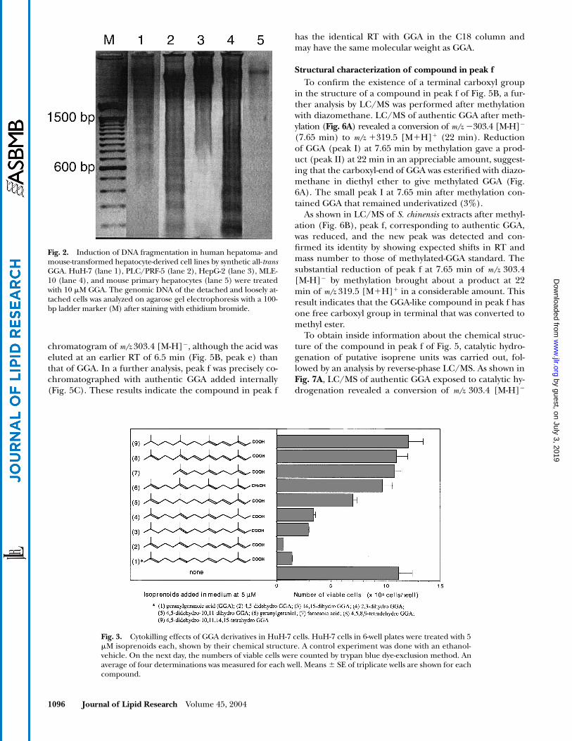
) and in 16 h (Fig.1B) but not in HepG-2 cells and mouse primary-culturedhepatocytes. Cell death was due to apoptosis as revealedby DNA stepladder formation (
Fig. 2
). GGA-induced ap-optosis was also morphologically evidenced by chroma-tin condensation stained with Hoechst 33258 (data notshown).
We next searched what structure in GGA was requiredto induce apoptosis. For this we used several synthetic iso-prenoids. As shown in
Fig. 3
, 4,5-didehydration did not re-duce the apoptosis-inducing activity of GGA, and either
�
-or
�
-saturation slightly decreased the activity, but neither4,5,8,9-tetradehydration nor 4,5-didehydro-10,11,14,15-tet-rahydration of GGA kept the activity. The reduction of a
terminal carboxylic group to an alcohol group abolishedthe cell killing activity and so did the reduction in thenumber of isoprene units to farnesoic acid.
LC/MS analysis of herbal extracts
Inasmuch as GGA showed apoptosis-inducing activity inhepatoma cells, it was worthwhile to search for naturalGGA in a hepatoma-prevention strategy with GGA-deriva-tives. A synthetic GGA has a molecular mass of 304 corre-sponding to four isoprene units with a carboxyl group interminal. In order to detect GGA in the extracts from
S.chinensis
, a reverse-phase LC/MS analysis was performedin the negative ion mode of electrospray ionization, be-cause authentic GGA gave a proton-deleted molecularion, [M-H]
�
at
m/z
303.4, without an appreciable amountof positive ion signals.
Figure 4A
shows the LC/MS chro-matogram of total negative ions from
m/z
200 to
m/z
350 in
S. chinensis
extracts. Four major peaks were eluted, andeach peak was relatively pure as evidenced by their massspectra (Fig. 4B), indicating that the herbal extracts con-tained free fatty acids such as linolenic acid (
m/z
277.3[M-H]
�
) in peak 1, linoleic acid (
m/z
279.3 [M-H]
�
) inpeak 2, palmitic acid (
m/z
255.3 [M-H]
�
) in peak 3, andoleic acid (
m/z
281.3 [M-H]�) in peak 4. The prominentpeaks of these four fatty acids were observed as a typicalchromatogram for all herbal extracts, as described in Ma-terials and Methods.
In total negative ion mode, no peaks were foundaround at a retention time (RT) of 7.65 min correspond-ing to authentic GGA. However, as shown in Fig. 5, a GGA-like component (Fig. 5B, peak f) was unveiled as a promi-nent peak with the same RT as authentic GGA (Fig. 5A) inSIR for m/z 303.4 [M-H]�, which is a base ion of GGA (onemass unit lower than the molecular mass). Because arachi-donic acid has the same molecular mass as GGA, it waspossible to mistake this popular fatty acid for GGA in a
Fig. 1. Induction of cell death in human hepatoma- and mouse-transformed hepatocyte-derived cell linesby synthetic all-trans geranylgeranoic acid (GGA). Concentration dependence (A) and time course (B) ofthe cytokilling effects of GGA were measured in HuH-7 (open circle), PLC/PRF-5 (closed circle), MLE-10(closed diamond), HepG-2 (open square), and mouse primary hepatocytes (open triangle). Viable cells werecounted by trypan blue dye-exclusion method 24 h after GGA treatment (A) or after treatment with 5 �MGGA (B). An average of four determinations was calculated for each well. Means � SE of triplicate wells areshown for each point.
by guest, on July 3, 2019w
ww
.jlr.orgD
ownloaded from
1096 Journal of Lipid Research Volume 45, 2004
chromatogram of m/z 303.4 [M-H]�, although the acid waseluted at an earlier RT of 6.5 min (Fig. 5B, peak e) thanthat of GGA. In a further analysis, peak f was precisely co-chromatographed with authentic GGA added internally(Fig. 5C). These results indicate the compound in peak f
has the identical RT with GGA in the C18 column andmay have the same molecular weight as GGA.
Structural characterization of compound in peak fTo confirm the existence of a terminal carboxyl group
in the structure of a compound in peak f of Fig. 5B, a fur-ther analysis by LC/MS was performed after methylationwith diazomethane. LC/MS of authentic GGA after meth-ylation (Fig. 6A) revealed a conversion of m/z �303.4 [M-H]�
(7.65 min) to m/z 319.5 [MH] (22 min). Reductionof GGA (peak I) at 7.65 min by methylation gave a prod-uct (peak II) at 22 min in an appreciable amount, suggest-ing that the carboxyl-end of GGA was esterified with diazo-methane in diethyl ether to give methylated GGA (Fig.6A). The small peak I at 7.65 min after methylation con-tained GGA that remained underivatized (3%).
As shown in LC/MS of S. chinensis extracts after methyl-ation (Fig. 6B), peak f, corresponding to authentic GGA,was reduced, and the new peak was detected and con-firmed its identity by showing expected shifts in RT andmass number to those of methylated-GGA standard. Thesubstantial reduction of peak f at 7.65 min of m/z 303.4[M-H]� by methylation brought about a product at 22min of m/z 319.5 [MH] in a considerable amount. Thisresult indicates that the GGA-like compound in peak f hasone free carboxyl group in terminal that was converted tomethyl ester.
To obtain inside information about the chemical struc-ture of the compound in peak f of Fig. 5, catalytic hydro-genation of putative isoprene units was carried out, fol-lowed by an analysis by reverse-phase LC/MS. As shown inFig. 7A, LC/MS of authentic GGA exposed to catalytic hy-drogenation revealed a conversion of m/z 303.4 [M-H]�
Fig. 2. Induction of DNA fragmentation in human hepatoma- andmouse-transformed hepatocyte-derived cell lines by synthetic all-transGGA. HuH-7 (lane 1), PLC/PRF-5 (lane 2), HepG-2 (lane 3), MLE-10 (lane 4), and mouse primary hepatocytes (lane 5) were treatedwith 10 �M GGA. The genomic DNA of the detached and loosely at-tached cells was analyzed on agarose gel electrophoresis with a 100-bp ladder marker (M) after staining with ethidium bromide.
Fig. 3. Cytokilling effects of GGA derivatives in HuH-7 cells. HuH-7 cells in 6-well plates were treated with 5�M isoprenoids each, shown by their chemical structure. A control experiment was done with an ethanol-vehicle. On the next day, the numbers of viable cells were counted by trypan blue dye-exclusion method. Anaverage of four determinations was measured for each well. Means � SE of triplicate wells are shown for eachcompound.
by guest, on July 3, 2019w
ww
.jlr.orgD
ownloaded from

Shidoji and Ogawa Natural geranylgeranoic acid in herbs 1097
(7.65 min) to 311.4 m/z [M-H]� (13.9 min). Reduction ofGGA (peak I) at 7.65 min by hydrogenation gave rise to aproduct (peak III) at 13.9 min in an appreciable amountat the same RT as authentic phytanic acid, suggesting thatfour double-bonds in GGA were fully saturated throughincorporation of eight hydrogen atoms. No partiallyhydrogenated GGA was detected. The small peak I at7.65 min after hydrogenation contained GGA that re-
mained underivatized (1.8%). Further, an additional ex-periment of arachidonic acid with catalytic hydrogenationconfirmed that perhydro-arachidonic acid eluted approxi-mately 5 min later than perhydro GGA at the same RT asarachidic acid (20:0n-6) at m/z 311.4 [M-H]� (results notshown).
LC/MS of S. chinensis extracts after hydrogenation (Fig.7B) showed the substantial reduction in peak f at 7.65 min
Fig. 4. Reverse-phase liquid chromatography/tandem mass spectrometry (LC/MS) analysis of Schisandra chinensis extracts. A: LC/MS of theextracts was performed by electrospray ionization and monitoring of total negative ions from m/z 200 to m/z 350. B: The mass spectra of peaks1 to 4 in panel (A) are shown: m/z 277.3 corresponding to 18:3 (C18H29O2
�, linolenoate ion); m/z 279.3 corresponding to 18:2 (C18H31O2�,
linoleate ion); m/z 255.3 corresponding to 16:0 (C16H31O2�, palmitate ion); and m/z 281.3 corresponding to 18:1 (C18H33O2
�, oleate ion).
by guest, on July 3, 2019w
ww
.jlr.orgD
ownloaded from

1098 Journal of Lipid Research Volume 45, 2004
of m/z 303.4 [M-H]� and the significant enhancement of13.9-min peak at m/z 311.4 [M-H]�. Prior to hydrogena-tion (Fig. 7B, upper panel), the herbal extracts were sug-gested to contain endogenous phytanic acid. These results
indicate that the GGA-like compound in peak f had fourisoprenyl double-bonds, as evidenced by incorporation ofeight hydrogen atoms after hydrogenation, and the hydro-genated peak f was identical to phytanic acid.
However, the calculated amount of the 13.9-min peak(phytanic acid) produced after hydrogenation was morethan the expected area of the reduced peak f. To explorethe presence of possible sources for phytanic acid otherthan GGA or peak f, GGA derivatives saturated to the dif-ferent extents, such as di- (m/z 305.4), tetra- (m/z 307.4),and hexa- (m/z 309.4) hydrogenation, were surveyed onLC/MS before and after hydrogenation. As shown inFig. 8, the prominent components eluted at m/z 303.4,305.4, 307.4, and 309.4 prior to hydrogenation decreasedor disappeared after being subjected to hydrogenation,whereas peak j corresponding to phytanic acid signifi-cantly rose. These components were demonstrated toelute at clearly different RTs from common straight-chainfatty acids with the same molecular weight, such as 20:4n-6(arachidonic acid, m/z 303.4), 20:3n-6 (dihomo--lino-lenic acid, m/z 305.4), 20:3n-9 (mead acid, m/z 305.4),20:2n-6 (11,14-eicosadienoic acid, m/z 307.4), and 20:0(arachidic acid, m/z 311.4), respectively, except that an au-thentic standard corresponding to 20:1 at m/z 309.4 wasnot available (Fig. 8). Therefore, it is strongly suggestedthat peaks g, h, and i might correspond to di-, tetra- andhexa-hydro GGAs, respectively. These results indicate thatthe significant amount of peak j was produced during thehydrogenation, possibly arising from partially saturatedcompounds in peaks f, g, h, and i. The similar elution pro-file of putative GGA derivatives, including phytanic acid,was observed in all herbal extracts as described in Materi-als and Methods.
Fig. 5. Selected ion chromatograph (negative ions at m/z 303.4)of authentic GGA and S. chinensis extracts by LC/MS. A: AuthenticGGA was eluted at the retention time (RT) of 7.65 min. B: A promi-nent component (peak f) in the herbal extracts was eluted at thesame RT as authentic GGA. C: Cochromatography of peak f withauthentic GGA.
Fig. 6. LC/MS elution profiles of authentic GGA and S. chinensis extracts before and after methylation with diazomethane. A: Selected iontracings of authentic GGA (peak I) for m/z 303.4 [M-H]� and methylated GGA (peak II) for m/z 319.5 [MH]. B: Selected ion tracings ofthe herbal extracts for m/z 303.4 [M-H]� and m/z 319.5 [MH] are shown. MA, mass area.
by guest, on July 3, 2019w
ww
.jlr.orgD
ownloaded from

Shidoji and Ogawa Natural geranylgeranoic acid in herbs 1099
Amounts of GGA in herbsAs a practical example, the standard curve was estab-
lished in order to quantify GGA contents in herbs usingLC/MS under conditions as described in Materials andMethods. A linear relationship between the amount of in-jected GGA and the peak area was confirmed, ranging inamounts from 50 pg to 0.6 ng, with the regression valuesfor R2 � 0.9834. The amounts of unesterified GGA inherbal extracts were calculated from the mean mass areawith triplicate injection by LC/MS chromatogram basedupon the standard curve. The recovery of GGA in the ex-tract was checked using the method of extraction as de-scribed in Materials and Methods by addition of knownamounts of the GGA standard to herbal homogenates andwas greater than 98% throughout the process.
GGA was detected for almost all dry herbs in thisscreening. GGA concentration in herbs ranged from 0.1�g/g to 2.9 �g/g (Table 1), in decreasing order (�1 �g/g):turmeric (root), schisandra (fruit), licorice (root), In-dian gooseberry (fruit), rhubarb root (root), hellebore(root), saussurea (root), cornelian cherry (fruit), androsehip (fruit). There was no significant difference inGGA content according to the plant part used.
In addition to a discovery of natural GGA, the existenceof free phytanic acid, fully saturated GGA, was confirmed inLC/MS chromatograms for all dry herbs as shown in an ex-
ample of S. chinensis (Figs. 7, 8). Without quantitative deter-mination of phytanic acid, a simple peak area ratio of GGAto phytanic acid in total negative ion chromatogram waslisted in Table 1. The ratio ranged widely from 0.1 to 6.0, andin a higher ratio of GGA (�2.0): turmeric (root) 6.0, schisan-dra (fruit) 4.4, hellebore (bark) 3.5, snake gourd root(root) 2.9, rhubarb root (root) 2.8, and artichoke (stem,leaves) 2.3; inversely, in a higher ratio of phytanic acid( 0.5): magnolia (bark) 0.1, Indian bdellium tree (resin)0.2, licorice (root) 0.4, coptis (root) 0.5, Indian gooseberry(fruit) 0.5, and echinacea (root) 0.5. The relationship be-tween the amounts of GGA and phytanic acid significantlycorrelated in Chinese herbs, but not in Ayurvedic herbs.
Aside from dry herbs, the same LC/MS analysis was per-formed with fresh herbs, including aloe (A. barbadensis;gel) and rose periwinkle (V. rosea; whole plant), and ma-rine algae, including Nori (sun-dried laver) and Kajime(dried seaweed), which had no significant amount ofGGA (data not shown).
DISCUSSION
In the present study, first we showed that GGA was a mi-cromolar inducer of apoptosis in human hepatoma- andmouse-transformed hepatocyte-derived cell lines, but not
Fig. 7. LC/MS elution profiles of authentic GGA and S. chinensis extracts before and after catalytic hydrogenation. Catalytic hydrogenationwas performed with PtO2 and H2 as described in the Materials and Methods section. A: Selected ion tracings of authentic GGA (peak I) form/z 303.4 [M-H]� and perhydro GGA (peak III) for m/z 311.4 [M-H]� after being subjected to hydrogenation. Perhydro GGA was eluted atthe same RT of 13.9 min as authentic phytanic acid. B: Selected ion tracings of the herbal extracts for m/z 303.4 and m/z 311.4 [M-H]� be-fore and after being subjected to hydrogenation.
by guest, on July 3, 2019w
ww
.jlr.orgD
ownloaded from

1100 Journal of Lipid Research Volume 45, 2004
in human hepatoblastoma-derived cell line and mouseprimary hepatocytes. Second, and most importantly inthis paper, a natural occurrence of cancer-preventive GGAin medicinal herbs was demonstrated by LC/MS analysis,and we also suggested the existence of some other GGAderivatives, including phytanic acid, in herbal extracts.
Previous studies from our group showed that GGA and4,5-didehydro GGA were both potent ligands for CRABP(3) and nuclear retinoid receptors (4), so we called theseacids “acyclic retinoids.” Consistent with our findings anddefinition of acyclic retinoids, some other acyclic com-pounds have been reported, such as methoprene acid
Fig. 8. Selected negative ion tracings for m/z 303.4, 305.4, 307.4, 309.4, and 311.4 in S. chinensis extracts be-fore and after hydrogenation. Before hydrogenation, prominent components f, g, h, and j eluted well sepa-rately from fatty acids such as 20:4n-6 (arachidonic acid), 20:3n-6 (dihomo--linolenic acid), 20:3n-9 (meadacid), 20:2n-6 [11,14-eicosadienoic acid (EDA)], and 20:0 (arachidic acid), respectively.
TABLE 1. Concentrations of geranylgeranoic acid and its relative amounts to phytanic acid in herbs
HerbsGGA/Phytanic
AcidCommon Name Latin Name Part Used Origin GGA
�g/g dry wt mass area ratio
Turmeric Curcuma longa rhizome China 2.9 6.0Schisandra Schisandra chinensis fruit China 2.4 4.4Licorice Glycyrrhiza uralensis root China 2.3 0.4Indian gooseberry Emblica officinalis fruit India 2.1 0.5Rhubarb root Rheum palmatum root China 1.7 2.8Hellebore Picrorrhiza kurroa rhizome China 1.4 3.5Saussurea Saussurea lappa root China 1.1 1.8Cornelian cherry Cornus officinalis fruit China 1.0 0.9Myrrh Commiphora abyssinica resin China 1.0 0.7Rosehip Rosa canina fruit China 1.0 1.6Cinnamon Cinnamomum cassia bark China 0.9 1.3Artichoke Cynara scolymus stem, leaves Germany 0.8 2.3Gentian Gentiana scabra root China 0.7 1.1Bupleurum Bupleurum falcatum root China 0.7 1.8Orange peel Citrus aurantium peel Egypt 0.7 0.8Echinacea Echinacea augustifolia root Egypt 0.7 0.5Astragalus Astragalus membranaceus root China 0.5 1.4Indian bdellium tree Commiphora mukul resin India 0.5 0.2Mountain ebony Bauhinia variegata bark India 0.5 1.5Magnolia Magnolia obovata bark Japan 0.4 0.1Snake gourd root Trichosanthes kirilowii root China 0.4 2.9Reishi Ganoderma lucidum whole China 0.4 0.6Dandelion Taraxacum officinale root Hungary 0.3 0.8Coptis Coptis chinensis root China 0.1 0.5Skullcap Scutellaria baicalensis root China 0.01 0.01
GGA, geranylgeranoic acid.
by guest, on July 3, 2019w
ww
.jlr.orgD
ownloaded from

Shidoji and Ogawa Natural geranylgeranoic acid in herbs 1101
(19) and phytanic acid (20), as ligands for retinoid-X re-ceptor. At the initial point of our study, we thought thatapoptosis-inducing activity of 4,5-didehydro GGA was at-tributable to the retinoidal function; however, natural ret-inoids such as all-trans and 9-cis retinoic acids were unex-pectedly unable to induce apoptosis in HuH-7 cells (9).Henceforth, 4,5-didehydro GGA was expected to mimicother natural isoprenoids such as GGA rather than retin-oids in terms of apoptosis induction in hepatoma cells.In fact, GGA itself actively induced apoptosis in humanhepatoma- (21) and mouse hepatocyte-derived cell lines(Figs. 1, 2). Furthermore, 2,3-dihydro GGA possessed thisactivity (Fig. 3), even though the �-saturation of GGA to-tally destroyed the ligand activity for retinoid receptors(4), suggesting that the induction of apoptosis by GGAmay not be mediated through retinoid receptors. Re-cently, it was reported that phytanic acid transactivatedperoxisome proliferator-activated receptor � (PPAR�), aswell as polyunsaturated fatty acids (22). GGA may well bea potent ligand for PPAR� and for some other orphan re-ceptors. We cannot even exclude the possibility that GGAmay induce apoptosis through a hypothetical GGA-spe-cific receptor, which is now under investigation in our lab-oratory by using a technique of RNAi with a whole geno-mic siRNA expression library.
Inasmuch as GGA and its derivatives were shown to in-duce apoptosis in human hepatoma cell lines (Fig. 3), ournext question was whether GGA is present in nature.Isoprenoids are synthesized in all organisms but areespecially abundant and diverse in plants, with tens ofthousands of compounds reported to date (23). To ourknowledge, however, there has been no report of the ex-istence of the acyclic diterpenoid GGA in the isoprenoidbiosynthetic pathway in plants or animals.
The present characterization of a GGA-like compoundin herbal extracts revealed its structure by LC/MS analy-sis. The compound, coeluted with authentic GGA, wasconverted to a product corresponding to GGA methyl-ester after its methylation with diazomethane and was con-verted to a product incorporated with eight hydrogenatoms corresponding to phytanic acid by catalytic hydro-genation. These results strongly support a conclusion thatthe GGA-like compound is structurally identical withGGA, which has four isoprenyl double-bonds and a car-boxyl group in terminal with a molecular mass of 304, al-though further isolation analysis is required to warrantthe existence of natural GGA.
The amounts of GGA in herbs were in the range of mi-crograms/grams (Table 1), suggesting that GGA may berelevant to micronutrients such as vitamins. GGA and itsderivatives are active to induce apoptosis in hepatoma-derived HuH-7 cells at concentrations of 1�10 �M (Fig. 1).On the supposition that all of the ingested GGA will ap-pear in blood, several hundred grams of herbs will be re-quired to reach the effective concentrations. It is alsoworthwhile to mention that the ingested GGA may beselectively taken up and accumulated in tumor cells,because tumor cells show the enhanced expression ofCRABP, to which GGA may be bound (3,4). Ratios of GGA
to phytanic acid contents varied by 60-fold in our samplesof herbs. What does determine a proportion of GGA tophytanic acid in the dry herbs? Chl, the main constituentof the photosynthetic apparatus in plants, consists of twomoieties, chlorophyllide and phytol. The hydrogenationof GGPP is catalyzed to phytyl diphosphate (phytylPP)by a geranylgeranyl reductase (CHL-P), as indicated inScheme 1. It is reported that CHL-P uses GGPP and gera-nylgeranylated chlorophyllide as substrates and directsphytylPP to the tocopherol- and Chl-synthesizing pathway(24). CHL-P catalyzes a stepwise reduction of geranylgera-nyl derivatives to phytyl derivatives. This reductase broadspecific for geranylgeranyl moiety could catalyze GGA tophytanic acid conversion in a stepwise mode. Therefore,once GGA is produced from GGOH in plants, a propor-tion of phytanic acid to GGA contents would be attribut-able to CHL-P activity.
Furthermore, the finding of other GGA derivatives withonly subtle differences on numbers of double bonds inherbal extracts is informative for supporting the existenceof GGA and CHL-P activity. We could demonstrate thatthese peaks did not represent the well-known straight-chain fatty acids, and that these were all converted to phy-tanic acid by hydrogenation. In the present study, the ex-act positions of the double bonds for m/z 305.4, 307.4, and309.4 [H-M]� were not assigned because authentic stan-dards were not available. However, based upon a mode ofCHL-P enzyme action (25), it is reasonable to speculatethat these may be 6,7-dihydro (for m/z 305.4), 6,7,10,11-tetrahydro (for m/z 307.4), and 6,7,10,11,14,15-hexahydro(for m/z 309.4) GGAs. Obviously, further structural analy-sis is required of each isolated compound and additionalexamination of biological activity for these GGA deriva-tives found in herbs.
In terms of its distribution, interestingly, GGA and phy-tanic acid were observed in almost all dry herbs examinedin this screening. In additional experiments of otherplants, however, fresh herbs, including aloe (A. barbaden-sis; gel) and rose periwinkle (V. rosea; whole plant), used asliver tonics had no detectable GGA and its derivatives. Ma-rine algae, including Nori (sun-dried laver) and Kajime(dried seaweed), contained only GGA derivatives, includ-ing phytanic acid, with no significant amount of GGA. Al-though little is known about the presence of phytanic acidin higher plants (14), most of the medicinal dry herbsused in the present study contained both GGA and phy-tanic acid as a characteristic common to all, independentof their own specific effects, such as liver tonics. The accu-mulation of these compounds is supposed to be involvedwith a certain type of plants, such as “dried” plants, be-cause it has been reported that phytanic acid was notfound in any food consisting of nondried vegetable (26).
The information that dry herbs, not fresh plants, con-tain GGA and its derivatives could provide a new vista onthe biological significance of GGA in plants. A large vari-ety of products are derived from isoprenoids in plants fortheir growth and response to environmental stress, whichis closely associated with plant defense mechanisms (11).For example, Nah, Song, and Back (27) have reported
by guest, on July 3, 2019w
ww
.jlr.orgD
ownloaded from

1102 Journal of Lipid Research Volume 45, 2004
that farnesyl disphosphatase and geranylgeranyl disphos-phatase (Scheme 1) were induced in rice seedlings whenthey were exposed to UVC. Unlike mammals, the iso-prenoid metabolism in plants is a cell autonomous pro-cess (28). It can, therefore, be presumed that the reac-tions to environmental stress, such as drying or UV rays,occur in the plant body as plant defense mechanisms evenafter collecting herbs off the soil. In this context, the sec-ondary metabolites in the isoprenoid pathway, such asGGOH-derived diterpenoids in response to environmen-tal stress, can be synthesized and accumulated duringpreparation of dry herbs.
The drying process in plants may be a sort of “dying”process that results in cell death. Though it can be abroad interpretation, it is intriguing for us to speculatethat GGA may be involved in apoptosis as a defense mech-anism in plants. Apoptosis has now been recognized as anindispensable facet of development, defense responses,and tissue sculpturing in both animal and plant, thoughlittle is known about the molecular mechanisms of apop-tosis in plant cells (29). As the present study demonstratesthe possibility that GGA is a natural component of dryherbs, it may also be one of the putative intracrine regula-tors for apoptosis in plant cells. Traditional medicine hasutilized dry herbs for thousands of years, which is not onlybecause of preservation, but may be because of naturallyoccurring substances induced by a defense mechanism fordrying, which may be taken into account empirically in or-der to obtain the optimal efficacy.
It is interesting that we found the same substance in na-ture as the one that has been chemically synthesized forcancer chemoprevention of hepatoma. GGA and its 4,5-didehydro derivative were already reported to both inhibitexperimental hepato-carcinogenesis and induce differen-tiation and apoptosis in human hepatoma-derived celllines. Further, a one-year intake of 4,5-didehydro GGA al-ready has been proven effective for prevention of secondprimary hepatoma and increased the 5-year survival rateby a phase II double-blinded, placebo-controlled clinicaltrial with relatively low toxicity (1,2).
Our research suggests that cancer-preventive GGA andits derivatives could be synthesized in the isoprenoid bio-synthetic pathway in plants. With regard to the practicaluse of GGA contained in herbs in order to prevent secondprimary hepatoma, we have not obtained yet a clear inter-pretation of the relation between the amount of naturalGGA and its effect from the perspective of cancer preven-tion. However, the presence of natural GGA in herbs is in-formative for supporting the therapeutic efficacy of 4,5-didehydro GGA in the phase II clinical trial (1) and alsowill provide concrete information on whether naturalGGA and its derivatives can be expected to play an impor-tant role in improving primary health care besides cancerprevention (30).
The authors express their gratitude to Dr. Luigi M. De Luca(NCI, NIH, Bethesda) for his kind advice during preparationof this manuscript. The authors also greatly thank Dr. Yasutoshi
Muto (Professor Emeritus of Gifu University) for his warm sup-port and encouragement during this study.
REFERENCES
1. Muto, Y., H. Moriwaki, M. Ninomiya, S. Adachi, A. Saito, K.Takasaki, T. Tanaka, K. Tsurumi, M. Okuno, E. Tomita, T. Naka-mura, and T. Kojima. 1996. Prevention of second primary tumorsby an acyclic retinoid, polyprenoic acid, in patients with hepatocel-lular carcinoma. N. Engl. J. Med. 334: 1561–1567.
2. Muto, Y., H. Moriwaki, and A. Saito. 1999. Prevention of secondprimary tumors by an acyclic retinoid in patients with hepatocellu-lar carcinoma. N. Engl. J. Med. 340: 1046–1047.
3. Muto, Y., H. Moriwaki, and M. Omori. 1981. In vitro binding affin-ity of novel synthetic polyprenoids (polyprenoic acids) to cellularretinoid-binding proteins. Jpn. J. Cancer Res. 72: 974–977.
4. Araki, H., Y. Shidoji, Y. Yamada, H. Moriwaki, and Y. Muto. 1995.Retinoid agonist activities of synthetic geranylgeranoic acid deriva-tives. Biochem. Biophys. Res. Commun. 209: 66–72.
5. Yamada, Y., Y. Shidoji, Y. Fukutomi, T. Ishikawa, T. Kaneko, H. Nak-agama, M. Imawari, H. Moriwaki, and Y. Muto. 1994. Positive andnegative regulations of albumin gene expression by retinoids inhuman hepatoma cell lines. Mol. Carcinog. 10: 151–158.
6. Muto, Y., and H. Moriwaki. 1984. Antitumor activity of vitamin Aand its derivatives. J. Natl. Cancer Inst. 73: 1389–1393.
7. Fukutomi, Y., M. Omori, Y. Muto, M. Ninomiya, M. Okuno, and H.Moriwaki. 1990. Inhibitory effects of acyclic retinoid (polyprenoicacid) and its hydroxy derivative on cell growth and on secretion ofalpha-fetoprotein in human hepatoma-derived cell line (PLC/PRF/5). Jpn. J. Cancer Res. 81: 1281–1285.
8. Muto, Y., and H. Moriwaki. 1991. Acyclic retinoids and cancerchemoprevention. Pure Appl. Chem. 63: 157–160.
9. Nakamura, N., Y. Shidoji, H. Moriwaki, and Y. Muto. 1996. Apopto-sis in human hepatoma cell line induced by 4,5-didehydro gera-nylgeranoic acid (acyclic retinoid) via down-regulation of trans-forming growth factor-alpha. Biochem. Biophys. Res. Commun. 219:100–104.
10. Lichtenthaler, H. K. 2000. Non-mevalonate isoprenoid biosynthe-sis: enzymes, genes and inhibitors. Biochem. Soc. Trans. 28: 785–789.
11. Sacchettini, J. C., and C. D. Poulter. 1997. Creating isoprenoid di-versity. Science. 277: 1788–1789.
12. Chappell, J. 1995. Biochemistry and molecular biology of the iso-prenoid bio-synthetic pathway in plants. Annu. Rev. Plant Physiol.Plant Mol. Biol. 46: 521–547.
13. Dembitsky, V. M., T. Rezanka, and I. Bychek. 1992. Fatty acids andphospholipids from lichens of the order lecanorales. Phytochemis-try. 31: 851–853.
14. Brown, P. J., G. Mei, F. B. Gibberd, D. Burston, P. D. Mayne, J. E.McClinchy, and M. Sidey. 1993. Diet and Refsum’s disease. The de-termination of phytanic acid and phytol in certain foods and theapplication of this knowledge to choice of suitable conveniencefoods for patients with Refsum’s disease. J. Hum. Nutr. Diet. 6: 295–305.
15. Patton, S., and A. A. Benson. 1966. Phytol metabolism in the bo-vine. Biochim. Biophys. Acta. 125: 22–32.
16. Baxter, J. H. 1968. Absorption of chlorophyll phytol in normalman and in patients with Refsum’s disease. J. Lipid Res. 9: 636–641.
17. Steinberg, D. 1995. Refsum disease. In Metabolic and MolecularBasis of Inherited Disease. 7th edition. C. R. Scriver, A. L. Beaudet,W. S. Sly, et al., editors. McGraw-Hill, New York. 2351–2369.
18. Kanda, H., G. H. Lee, K. Nomura, K. Ohtake, and T. Kitagawa.1993. Malignant transformation of a mouse liver epithelial cellline by transfection of an activated c-H-ras gene with a point muta-tion at codon 12. Carcinogenesis. 14: 1061–1063.
19. Harmon, M., M. Boehm, R. Heyman, and D. Mangelsdorf. 1995.Activation of mammalian retinoid X receptors by the insectgrowth regulator methoprene. Proc. Natl. Acad. Sci. USA. 92: 6157–6160.
20. Lemotte, P. K., S. Keidel, and C. M. Apfel. 1996. Phytanic acid is aretinoid X receptor ligand. Eur. J. Biochem. 236: 328–333.
21. Shidoji, Y., N. Nakamura, H. Moriwaki, and Y. Muto. 1997. Rapidloss in the mitochondrial membrane potential during geranylgera-noic acid-induced apoptosis. Biochem. Biophys. Res. Commun. 230:58–63.
by guest, on July 3, 2019w
ww
.jlr.orgD
ownloaded from
Shidoji and Ogawa Natural geranylgeranoic acid in herbs 1103
22. Lee, C. H., P. Olson, and R. M. Evans. 2003. Minireview: Lipidmetabolism, metabolic diseases, and peroxisome proliferator-acti-vated receptors. Endocrinology. 144: 2201–2207.
23. Chappell, J. 1995. The biochemistry and molecular biology of iso-prenoid metabolism. Plant Physiol. 107: 1–6.
24. Tanaka, R., U. Oster, E. Kruse, W. Rudiger, and B. Grimm. 1999.Reduced activity of geranylgeranyl reductase leads to loss of chlo-rophyll and tocopherol and to partially geranylgeranylated chloro-phyll in transgenic tobacco plants expressing antisense RNA forgeranylgeranyl reductase. Plant Physiol. 120: 695–704.
25. Addlesee, H. A. A., and C. N. Hunter. 1999. Physical mapping andfunctional assignment of the geranylgeranyl-bacteriochlorophyllreductase gene, bachP, of Rhodobacter sphaeroides. J. Bacteriol.181: 7248–7255.
26. Muralidharan, F. N., and V. B. Muralidharan. 1985. In vitro conver-
sion of phytol to phytanic acid in rat liver: subcellular distributionof activity and chemical characterization of intermediates using anew bromination technique. Biochim. Biophys. Acta. 835: 36–40.
27. Nah, J., S. J. Song, and K. Back. 2001. Partial characterization offarnesyl and geranylgeranyl diphosphatases induced in rice seed-lings by UV-C irradiation. Plant Cell Physiol. 42: 864–867.
28. Gan, S., and R. M. Amasino. 1997. Making sense of senescence.Plant Physiol. 113: 313–319.
29. Lam, E., N. Kato, and M. Lawton. 2001. Programmed cell death,mitochondria and the plant hypersensitive response. Nature. 411:848–853.
30. Wang, X., J. Wu, Y. Shidoji, Y. Muto, N. Ohishi, K. Yagi, S. Ikegami, T.Shinki, N. Udagawa, T. Suda, and Y. Ishimi. 2002. Effects of gera-nylgeranoic acid in bone: induction of osteoblast differentiationand inhibition of osteoclast formation. J. Bone Miner. Res. 17: 91–100.
by guest, on July 3, 2019w
ww
.jlr.orgD
ownloaded from