Embed Size (px)
Citation preview

ISBN: 978-81-931973-6-3National Conference on “Emerging Trends in Management of Infectious Diseases and Public Health, 4 – 5 Feb 16
MOHAMED SATHAK COLLEGE OF ARTS AND SCIENCE(Affiliated to the University of Madras, Accredited with NAAC)PG AND RESEARCH DEPARTMENT OF BIOTECHNOLOGYSHOLINGANALLUR, CHENNAI- 600 119PROCEEDINGS
NATIONAL CONFERENCE ON EMERGING TRENDS INMANAGEMENT OF INFECTIOUS DISEASES AND
PUBLIC HEALTH (ETMIDPH 2016)
Organised byPG AND RESEARCH DEPARTMENT OF BIOTECHNOLOGY
DATE: 4 - 5 FEBRUARY 2016
ISBN: 978-81-931973-6-3
ISBN: 978-81-931973-6-3National Conference on “Emerging Trends in Management of Infectious Diseases and Public Health, 4 – 5 Feb 16
PROCEEDINGSOF
NATIONAL CONFERENCE ON EMERGINGTRENDS IN MANAGEMENT OF INFECTIOUS
DISEASES AND PUBLIC HEALTH(ETMIDPH - 2016)
4-5 FEBRUARY 2016MOHAMED SATHAK COLLEGE OF ARTS AND SCIENCE(Affiliated to the University of Madras, Accredited with NAAC)SHOLINGANALLUR, CHENNAI- 600 119
ORGANISED BY
PG AND RESEARCH DEPARTMENT OFBIOTECHNOLOGY
ISBN: 978-81-931973-6-3
ISBN: 978-81-931973-6-3National Conference on “Emerging Trends in Management of Infectious Diseases and Public Health, 4 – 5 Feb 16

ISBN: 978-81-931973-6-3National Conference on “Emerging Trends in Management of Infectious Diseases and Public Health, 4 – 5 Feb 16
ISBN: 978-81-931973-6-3National Conference on “Emerging Trends in Management of Infectious Diseases and Public Health, 4 – 5 Feb 16
ISBN: 978-81-931973-6-3National Conference on “Emerging Trends in Management of Infectious Diseases and Public Health, 4 – 5 Feb 16
ISBN: 978-81-931973-6-3National Conference on “Emerging Trends in Management of Infectious Diseases and Public Health, 4 – 5 Feb 16
ISBN: 978-81-931973-6-3National Conference on “Emerging Trends in Management of Infectious Diseases and Public Health, 4 – 5 Feb 16
ISBN: 978-81-931973-6-3National Conference on “Emerging Trends in Management of Infectious Diseases and Public Health, 4 – 5 Feb 16
ISBN: 978-81-931973-6-3National Conference on “Emerging Trends in Management of Infectious Diseases and Public Health, 4 – 5 Feb 16
ISBN: 978-81-931973-6-3National Conference on “Emerging Trends in Management of Infectious Diseases and Public Health, 4 – 5 Feb 16
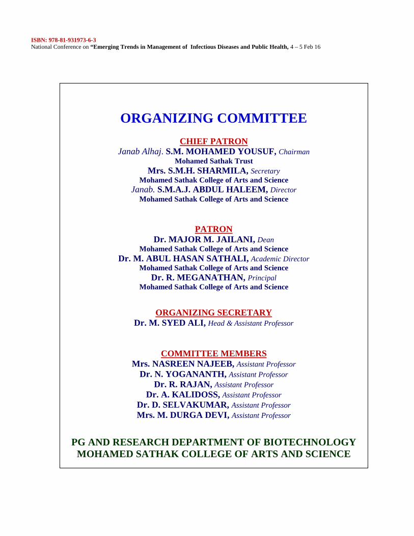
ORGANIZING COMMITTEE
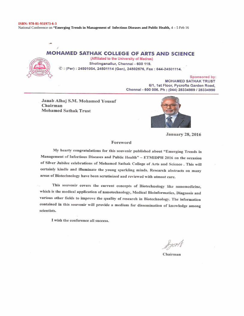
CHIEF PATRONJanab Alhaj. S.M. MOHAMED YOUSUF, Chairman
Mohamed Sathak TrustMrs. S.M.H. SHARMILA, Secretary
Mohamed Sathak College of Arts and ScienceJanab. S.M.A.J. ABDUL HALEEM, Director
Mohamed Sathak College of Arts and Science
PATRONDr. MAJOR M. JAILANI, Dean
Mohamed Sathak College of Arts and ScienceDr. M. ABUL HASAN SATHALI, Academic Director
Mohamed Sathak College of Arts and ScienceDr. R. MEGANATHAN, Principal
Mohamed Sathak College of Arts and Science
ORGANIZING SECRETARYDr. M. SYED ALI, Head & Assistant Professor
COMMITTEE MEMBERSMrs. NASREEN NAJEEB, Assistant Professor
Dr. N. YOGANANTH, Assistant ProfessorDr. R. RAJAN, Assistant Professor
Dr. A. KALIDOSS, Assistant ProfessorDr. D. SELVAKUMAR, Assistant ProfessorMrs. M. DURGA DEVI, Assistant Professor
PG AND RESEARCH DEPARTMENT OF BIOTECHNOLOGYMOHAMED SATHAK COLLEGE OF ARTS AND SCIENCE
ISBN: 978-81-931973-6-3National Conference on “Emerging Trends in Management of Infectious Diseases and Public Health, 4 – 5 Feb 16
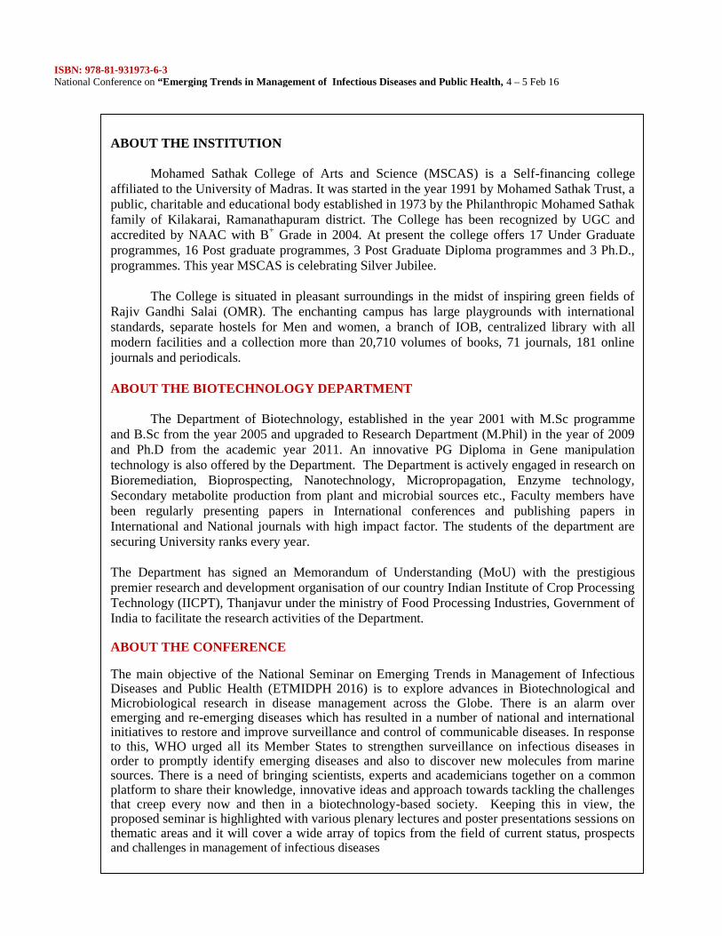
ABOUT THE INSTITUTION
Mohamed Sathak College of Arts and Science (MSCAS) is a Self-financing collegeaffiliated to the University of Madras. It was started in the year 1991 by Mohamed Sathak Trust, apublic, charitable and educational body established in 1973 by the Philanthropic Mohamed Sathakfamily of Kilakarai, Ramanathapuram district. The College has been recognized by UGC andaccredited by NAAC with B+ Grade in 2004. At present the college offers 17 Under Graduateprogrammes, 16 Post graduate programmes, 3 Post Graduate Diploma programmes and 3 Ph.D.,programmes. This year MSCAS is celebrating Silver Jubilee.
The College is situated in pleasant surroundings in the midst of inspiring green fields ofRajiv Gandhi Salai (OMR). The enchanting campus has large playgrounds with internationalstandards, separate hostels for Men and women, a branch of IOB, centralized library with allmodern facilities and a collection more than 20,710 volumes of books, 71 journals, 181 onlinejournals and periodicals.
ABOUT THE BIOTECHNOLOGY DEPARTMENT
The Department of Biotechnology, established in the year 2001 with M.Sc programmeand B.Sc from the year 2005 and upgraded to Research Department (M.Phil) in the year of 2009and Ph.D from the academic year 2011. An innovative PG Diploma in Gene manipulationtechnology is also offered by the Department. The Department is actively engaged in research onBioremediation, Bioprospecting, Nanotechnology, Micropropagation, Enzyme technology,Secondary metabolite production from plant and microbial sources etc., Faculty members havebeen regularly presenting papers in International conferences and publishing papers inInternational and National journals with high impact factor. The students of the department aresecuring University ranks every year.
The Department has signed an Memorandum of Understanding (MoU) with the prestigiouspremier research and development organisation of our country Indian Institute of Crop ProcessingTechnology (IICPT), Thanjavur under the ministry of Food Processing Industries, Government ofIndia to facilitate the research activities of the Department.
ABOUT THE CONFERENCE
The main objective of the National Seminar on Emerging Trends in Management of InfectiousDiseases and Public Health (ETMIDPH 2016) is to explore advances in Biotechnological andMicrobiological research in disease management across the Globe. There is an alarm overemerging and re-emerging diseases which has resulted in a number of national and internationalinitiatives to restore and improve surveillance and control of communicable diseases. In responseto this, WHO urged all its Member States to strengthen surveillance on infectious diseases inorder to promptly identify emerging diseases and also to discover new molecules from marinesources. There is a need of bringing scientists, experts and academicians together on a commonplatform to share their knowledge, innovative ideas and approach towards tackling the challengesthat creep every now and then in a biotechnology-based society. Keeping this in view, theproposed seminar is highlighted with various plenary lectures and poster presentations sessions onthematic areas and it will cover a wide array of topics from the field of current status, prospectsand challenges in management of infectious diseases
ISBN: 978-81-931973-6-3National Conference on “Emerging Trends in Management of Infectious Diseases and Public Health, 4 – 5 Feb 16
PREFACE
Infectious diseases are disorders caused by microorganisms- such as bacteria, viruses,fungi or parasites. Many organisms live in and on our bodies. They are normally harmless or evenhelpful, but under certain conditions, some organisms may cause disease. Some infectiousdiseases can be passed from person to person. Infectious diseases, including HIV/AIDS,tuberculosis, malaria, polio, and several neglected tropical diseases (NTDs) are easily spreadthrough personal contact, water, and air, (many NTDs are vector borne transmitted bymosquitoes, flies, etc) and are a particularly significant problem in developing countries. In thepast, infectious diseases have been widespread in developing countries and chronic diseases werefound primarily in high income countries. However, the global pattern of disease burden isshifting.
Viral Hepatitis, Influenza, and Tuberculosis (TB) remain among the leading causes ofillness and death in the United States and account for substantial spending on the relatedconsequences of infection. The infectious disease public health infrastructure, which carries outdisease surveillance at the Federal, State, and local levels, is an essential tool in the fight againstnewly emerging and re-emerging infectious diseases. Other important defenses against infectiousdiseases include: Proper use of vaccines, Antibiotics, Screening and testing guidelines, scientificimprovements in the diagnosis of infectious disease-related health concerns.
Today’s infectious disease challenges are broader and more complex than they were in1998, when CDC last issued a comprehensive plan to guide national efforts to prevent and controlemerging infectious threats. Since then, new microbes or new forms of old ones have beendiscovered nearly every year, and infectious disease outbreaks triggering international responseshave been reported on nearly every continent. While our changing, globalized world has providedincreased opportunities for emergence and spread of infectious diseases, it has also broughtsignificant advances toward their control. The ID Framework takes into account many of thescientific, demographic, technological, and economic developments currently modifying efforts toprotect public health, challenging us to rethink our processes and strategies and take advantage ofnew ways to prevent disease and improve health. Emerging infectious diseases may be consideredof public health importance based on a variety of criteria, including their designation as aemerging disease. It may be considered of public health importance based on a variety of criteria,including their designation as an emerging disease by international, federal, and/or provincialhealth authorities; their potential for preventability or public health action; and the seriousness oftheir impact on the health of the population and potential spread.
With this background, the present conference will definitely help to come to know theadvances and various strategies used to tackle the challenge of infectious diseases.
ISBN: 978-81-931973-6-3National Conference on “Emerging Trends in Management of Infectious Diseases and Public Health, 4 – 5 Feb 16
SL.NO TITLE OF THE PAPER PAGE1. DR. S. RAVIKUMAR
BIO-PROSPECTING OF MARINE MICROORGANISMS FORFOOD AND MEDICINE FOR FUTURE PROSPERIT
17
2. S. SABESANVECTOR BORNE DISEASES: DEVELOPMENT OF RISKPREDICTION MODELS, USING RS - GIS TECHNOLOGIES
18
3. DR. M. MUNIARAJINSECTS AND MICROBES: FRIENDS OR FOES?
19
4. DR. C. P. GIRISH KUMAR PHDLEPROSY - A NEGLECTED TROPICAL DISEASE
21
5. R. KALPANA, DR. R. DHAMOTHARANPHYTOPHARMACOLOGICAL PROFILE AND BRINE SHRIMPLETHALITY ASSAY OF THE METHANOLIC ANDETHANOLIC EXTRACTS OF THE LEAF AND BARK OFSYMPLOCOS COCHINCHINENSIS.
22
6. P. NIRMALA1* AND R. KRISHNAVENI2
ANALYSIS OF SOIL PHYSICO- CHEMICALCHARACTERISTICS OF ORGANIC FARMING ANDCONVENTIONAL FARMING PRACTICES.
37
7. D. SELVAKUMAR. AND K. DHEVENDARANSTUDIES ON NUTRITIONAL GROUPING OFSTREPTOMYCETES FROM FISHES OF THREEENVIRONMENTAL BIOTOPES, THEIR ANTIBIOGRAMAGAINST VIBRIO CHOLERAE.
44
8. R. MADHANAGOPAL, M.R. ASIYA MARIYAM AND A.KALIDOSSANTIPLASMODIAL PHYTOTHERAPY AND DRUGDISCOVERY- AN OVERVIEW
56
9. R.SYEDA ASHFIANAZ*, A.THANGARAJ
GREEN SYNTHESIS OF SILVER NANOPARTICLES USINGCITRUS MEDICA(RUTACEAE) LEAF EXTRACT AND ITSANTIBACTERIAL EFFICACY
68
10. Sewali Ghosh, K.P Sanjayan & S.K.M. Habeeb*
INHIBITION OF THE BIOSYNTHESIS OF EPICATECHININ TEA, CAMELLIA SINENSIS (L. KUNTZE) BYINSECTICIDES DEPLOYED FOR THE CONTROL OF THETEA MOSQUITO BUG, HELOPELTIS THEIVORAWATERHOUSE.
75
11. ANJANA.E , RAVICHANDRAN.VA PILOT STUDY ON PHYTOCHEMICAL SCREENING,ANTIOXIDANT AND ANTIINFLAMMATORY ACTIVITIESOF PUNICA GRANATUM SEED EXTRACTS
80
12. N.K. UdayaPrakash., R. Caroline., D.S. Arjun Krishnan., N.Sripriya., N. Ashwin Karthick and S. BhuvaneswariANTIBACTERIAL EVALUATION OF METALLIC SALTS
91
ISBN: 978-81-931973-6-3National Conference on “Emerging Trends in Management of Infectious Diseases and Public Health, 4 – 5 Feb 16
AGAINST INFECTIOUS BACTERIA13. X. ASBIN MARY AND M. SYED ALI
EVALUATION OF CAULERPA SCALPELLIFORMIS FOR THEPRESENCE OF PHYTOCHEMICALS AND ITSANTIBACTERIAL ACTIVITY
96
14. Praveena. A1, Anuradha. V2
MOLECULAR EVOLUTIONARY ANALYSIS ON INSECTACETYLCHOLINESTERAS
102
15. MAHALAKSHMI R, BALACHANDAR C, ANTONYPRABHU J, MELCHIAS GPRELIMINARY STUDY ON ISOLATION ANDCHARACTERIZATION OF DYE DECOLORIZING BACTERIAFROM LIME STONE SOILS.
108
16. Pandiyarajan G., Mary Swarnalatha SEFFICIENT MANAGEMENT STRATEGIES OFCOMBATTING FUNGAL INFECTIONS IN COPRA (Cocosnucifera).
115
17. S. Bhuvaneswari., S. Deepa.,HimaAishwarya., N. AshwinKarthick and N.K. UdayaPrakashDETERMINATION OF FUNGAL QUALITY OF MARKETEDHONEY SAMPLES
120
18. D. Kishore Ram Kumara, J.Arockia John Paulb,*, B.KarunaiSelvic
CYTOTOXIC EFFECT OF SEMECARPUSANACARDIUMNUTEXTRACT IN B16F10 MELANOMA CELL LINES
128
19. V. Saravanan, M. Gnanadesigan, S. RavikumarHerbo-mineral salts from Marine halophytes against human bacterialpathogens
134
20. C Kiruba and K ManjulaBiosynthesis and characterization of Alkaline protease from Bacillussubtilis
141
21. R.Swaminathan1, M.Syed Ali*1 V.Anuradha, N.YogananthModulatory effect of Polydatin on in vivo carbohydrate metabolismin streptozocin-induced type II diabetic rats.
151
22. B.Bharathi* and Kavitha.SMOLECULAR DIAGNOSIS OF ISONIAZID DRUG RESISTANTMycobacterium tuberculosis IN HIV PATIENTS
160
23. X.Asbin Mary, Robert LothaPHYTOCHEMICAL SCREENING AND ANTICANCEROUSACTIVITY OF RHIZOME EXTRACT COLEUS FORSKHOLI ONHEP G2 CELL LINE
167
24. N. Sivakumar., K. Krishnapriya., P. Nirmala., A. Chandhru.STUDIES ON PHYSICO – CHEMICAL PROPERTIES,NODULATION PATTERN AND RHIZOBIAL POPULATIONOF BLACK GRAM CULTIVATED SOIL.
173
25. R. Selvi and N. YogananthEVALUATION OF PHYTOCHEMICAL SCREENING ANDIN VITRO BIOACTIVITY OF LEAF AND STEMEXTRACTS OF SOLANUM XANTHOCARPUM SCHRADAND WENDL. (SOLANACEAE)
181
26. Shenbagam.R, Dr.Sheela Sasikumar, Dr.Thendral Hepisba 184
ISBN: 978-81-931973-6-3National Conference on “Emerging Trends in Management of Infectious Diseases and Public Health, 4 – 5 Feb 16
PHYTOCHEMICAL SCREENING AND ANTI-INFLAMMATORY ACTIVITY OF CITRUS LIMETTAPEEL. OIL EXTRACT
27. S.KRISHNAKUMAR, R. DIVYA, N.R. KANCHANADEVI, G. KEERTHANA AND A. ANCY JUDIGALACTOSE MEDIATED SYNTHESIS OF SILVERNANOPARTICLES (AGNPS) AND ITS ANTIMICROBIALACTIVITY
189
28. Valli. S. and Jaganathan. SANTIMICROBIAL ACTIVITY OF BOUGAINVILLEASPECTABILIS AND IDENTIFICATION OF ITS ACTIVECOMPOUNDS BY GC- MS
194
29. Praveena. A, Anuradha. VEFFECT OF MONOCROTOPHOS AND AZADIRACTINON GERMINATION OF PISUM SATIVUM AND ZEA MAYS
201
ISBN: 978-81-931973-6-3National Conference on “Emerging Trends in Management of Infectious Diseases and Public Health, 4 – 5 Feb 16
17
EDITED BY
Dr. M. SYED ALI, Head & Assistant ProfessorMrs. NASREEN NAJEEB, Assistant ProfessorDr. N. YOGANANTH, Assistant ProfessorDr. R. RAJAN, Assistant ProfessorDr. A. KALIDOSS, Assistant ProfessorDr. D. SELVAKUMAR, Assistant ProfessorMrs. M. DURGA DEVI, Assistant Professor
PUBLISHED BY
DARSHAN PUBLISHERS,NO: 8/173, VENGAYAPALAYAM, KAKKAVERI, RASIPURAM,NAMAKKAL, TAMIL NADU,INDIA – 637406.www.darshanpublishers.come-mail:[email protected]
ISBN: 978-81-931973-6-3National Conference on “Emerging Trends in Management of Infectious Diseases and Public Health, 4 – 5 Feb 16
18
BIO-PROSPECTING OF MARINE MICROORGANISMS FOR FOOD
AND MEDICINE FOR FUTURE PROSPERITY
DR. S. RAVIKUMARProfessor
School of Marine SciencesAlagappa University
Thondi campus 623 409, Ramnad District, Tamil Nadu
Diverse group of marine microorganisms are yet to be explored due to lack ofisolation and identification efforts by the marine biologists. The photosynthetic and nonphotosynthetic microorganism particularly, the associated microorganism fromseaweeds, seagrass and sponges have recently been identified as a potential source oforganisms for the exploitation of bioactive principles. Technologies available for theextraction of bioactive compounds from marine microorganisms are not the same formarine microorganism as they are all the pure marine forms. Standardisation ofmethodologies for the isolation and identification of microorganism from marineresources, extraction of bioactive compounds, hurdles for the mass production ofbioactive compounds by continuous and batch fermentation process will be discussed.Standardisation of drugs from marine microorganisms for drug development will bediscussed. Besides that, a modified method of extraction of herbal salt from the marinesalt intruders (mangrove plants) over traditional Indian methods will also be highlightedand their bioactive potential for the drug development will also be discussed. Moreoverthe bio potential of extremophilic microorganisms such as Halobacteria and solar salternCyanobacteria for the treatment of wastes from coconut retting waste water and waterdischarge from the industries with cyanide and metal contamination and the methodsstandardized for the mathematical kinetic modeling will be discussed. Moreover recenteffort on the “Impact of ocean acidification on the marine Drug Loss”, the currentresearch on the identification of potential drugs for the treatment of Malaria and furtherthe enhancement of some beneficial microorganisms in the open ocean through bioinoculation with the mangrove root associated microorganism will also be discussed indetail at that time of conference.
ISBN: 978-81-931973-6-3National Conference on “Emerging Trends in Management of Infectious Diseases and Public Health, 4 – 5 Feb 16
19
VECTOR BORNE DISEASES: DEVELOPMENT OF RISK
PREDICTION MODELS, USING RS - GIS TECHNOLOGIES
S. SABESANVector Control Research Centre
(Indian Council of Medical Research)Puducherry – 605 006
While on the one side our country is proud of major achievements in science andtechnology including space and our march towards a knowledge society, it is true on theother side that a large proportion of our population has no access to even safe drinkingwater, to cite one of the major problems facing the country. The continued practice ofopen drainage system, indiscriminate disposal of water and industrial effluents intowater bodies, and added to this, the increased migration of people from rural areas haveresulted in large slums in our urban centres creating an environment unsuitable forhealthy living, thus aggravating the spread of water borne diseases. Yet another area ofconcern to the country is the spread of vector-borne diseases (VBDs) such as malaria,filariasis, Japanese encephalitis and dengue to newer areas with mosquitoes, the vectorscarrying these diseases breeding in water bodies. The World Health Organization(WHO) and other international bodies highlight the threat posed by these VBDs to theworld’s population in general and to India in particular. It is said that in India alone, overtwo million cases of VBDs are reported every year. VBDs are spreading to newer areasdue to increased risk of transmission fuelled by changing climatic conditions,developmental activities, more specifically the urbanization and industrialization alongwith demographic changes to name a few possible causes. With advanced knowledge onthe principles underlying the disease transmission dynamics, the prediction of areas ofhealth risk is possible based on geo-environmental factors. Modern tools like RemoteSensing (RS) and Geographical Information Systems (GIS) have now come in handy toaddress the issues on health risk, and predicting the trend in disease prevalence forundertaking intervention measures. Mosquito vectors thrive on water, vegetation anddwellings (with the availability of vertebrate host). Study of mosquito populationdynamics with the change of environmental variables helps in understanding thecriticality of those variables. Satellite remote sensing technique along with GIS enablesurveillance of environmental conditions for vector development and diseasetransmission providing information on epidemiology of a region, viz. favourableecological conditions, habitat types providing breeding sites and their characterization,prevailing disease, past history of epidemics and environment and social and economicfactors associated with the epidemics. Major factors such as climate, landscape anddevelopmental activities responsible for risky conditions are being studied with the helpof remotely sensed data analysis. The satellite imagery is being used to explain thesevariables on a desired spatial and temporal scale, and GIS facilitates acceptance ofsatellite information, fit it to a vector mosquito model and produce imagery, indicatingthe areas of risk of transmission of VBDs. Our responsibility in the immediate futureshould be to provide technical information on these, facilitate formulation of policystatement, preparation of strategic plan, ease advocacy steps at different stages and fostereffective linkages with all partners.
ISBN: 978-81-931973-6-3National Conference on “Emerging Trends in Management of Infectious Diseases and Public Health, 4 – 5 Feb 16
20
INSECTS AND MICROBES: FRIENDS OR FOES?DR. M. MUNIARAJ
Scientist “C”Centre for Research in Medical Entomology (ICMR),
4- Sarojini Street, Chinna Chokkikulam, Madurai – 625 002Telephone: 0452 – 2520565, 2530746; FAX: 0452- 2530660;
E mail: [email protected]
Insects (Phylum Arthropoda; Class Insecta) are one of the most diverse groups ofliving creatures on Earth. First appeared in the late Carboniferous period about 300million years ago, they have subsequently radiated into a group that now includes about850,000 to several million species. Some 80–95% of insect species have yet to becollected, named and described, most of them living in the tropics. Even for the 850,000-plus species that have been named, we know little about how they are distributed or whatthey feed on. Equally diverse are the feeding habits and behaviors exhibited by the groupas a whole, and there is virtually no terrestrial food source that escapes exploitation byone or more species. However one property shared by all insects is their commonassociation with microorganisms. The associations range from loose and nonspecificones, in which the insect merely serves as an inadvertent carrier and distributor ofmicrobes, to much tighter, highly interdependent and remarkably regulated symbioticinteractions, and they include literally all gradations in between these extremes.Microbes on the other hand still more diversified than insects and the only group on theearth which can surpass the insects in diversity. Microorganisms include prokaryotes (acell characterized by the lack of a distinct membrane-bound nucleus) such as bacteriaand archea and eukaryotes (cells whose chromosomes are contained within a membrane-bound nucleus) such as fungi and protozoa. For over 3.8 billion years, these organismshave formed the foundation of the biosphere, surviving in extremes of heat, cold,radiation, pressure, salt, acidity, and darkness. For 2 billion years microbes were the onlyforms of life on Earth. During this long history, all of the basic biochemistries of lifeevolved, and all life forms have developed from these microbial ancestors. With theirability to harvest energy in almost any form, and thrive with or without oxygen,microbes have spent over a billion years making nitrogen available to plants whiletransforming the atmosphere with oxygen. Microbes are found throughout the entireplanetary ecosystem including niches where higher animal species are rare or absent(e.g. the ocean depths, the planet’s subsurface, thermal and polar environments, andoxygen-free environments). This wide ecological range reflects their vast metaboliccapabilities that allow different microbial species to inhabit different environments.
Current evidence suggests that perhaps 1.5 million species of fungi exist yet only5 % are described. For bacteria there may be 300,000 to 1 million species on earth yetonly 5000 bacteria are described. A gram of typical soil contains about 1 billion bacteria,but only 1 % of those can be cultured. Hence, most microbes remain to be discovered.Estimates suggest that up to 99% of microbes could not be cultured in a laboratory usingconventional methods. Detection and characterization could be achieved only byCulture-independent techniques, including sequencing of the 16S rRNA gene, a
ISBN: 978-81-931973-6-3National Conference on “Emerging Trends in Management of Infectious Diseases and Public Health, 4 – 5 Feb 16
21
relatively recent methodology adopted by microbial taxonomists. Microbes provide thefundamental underpinning of all ecosystems. Without microorganisms, alllife on earthwould cease to exist. Insects have a delicate and intricate set of relationships with amicrobial world of astonishing diversity. All insect species are known to harbour a richand complex community of microorganisms in their guts and other body regions. Thismicrobiota participates in many types of interactions ranging from prey & predator andpathogenesis to obligate mutualism. One reason for the microbial diversity is thatdifferent groups of insects have different feeding habits; this results in different gutstructures and functions and promotes the establishment of different phylotypes. Inrecent years there has been renewed interest in the understanding of insect gutmicroorganisms for two reasons. First, this diverse microbiota is a potential source ofnovel bioactive compounds such as antimalarial, antiviral and antitumour peptides,enzymes and novel metabolites. Second, manipulating these microbial symbionts isthought to be an effective strategy for controlling the spread of pathogens that useinsects as hosts.
Early studies revealed the often striking anatomical and behavioral adaptationsof insects to harbor the microbial partners in, on, or around them and to ensuretransmission of microbial symbionts to their offspring. The distinct microbial symbiontsor communities of symbionts were common and often essential in insects that feed onrestricted and or relatively refractory food resources. Such diets are often deficient innutrients such as amino acids (eg. Plant sap) or vitamins (eg. Animal blood), andlignocellulosic plant material is not only poor in nitrogenous compounds, vitamins, andsterols, but it is also difficult to digest. Symbiotic microbes are providing such missingnutrients or digestive enzymes to the insect host. However, microbial symbionts are alsoinvolved in numerous other aspects of insect biology eg. Detoxification of plantdefensive secretions; production of insect behavior modifying compounds; protectionagainst microbial pathogens and pest; and alteration of host reproductive patterns.Moreover, the microbial biomass itself is consumed by a large group of insects as wellas its larval forms as their main source of food. On the other hand, natural population ofinsects is kept under check by the activities of parasites and predators. Several species ofviruses, bacteria, fungi, protozoa and nematodes are known to cause infection in insects.The positive and negative association of insect and microbes will be discussed in detail.
ISBN: 978-81-931973-6-3National Conference on “Emerging Trends in Management of Infectious Diseases and Public Health, 4 – 5 Feb 16
22
LEPROSY - A NEGLECTED TROPICAL DISEASEDR. C. P. GIRISH KUMAR PHD
Scientist DNational Institute of Epidemiology (ICMR), Chennai 600077
Email Id: [email protected]
Leprosy or Hansen’s disease is a Neglected Tropical Disease (NTD) caused byMycobacterium leprae and is the leading infectious cause of permanent physicaldisability. A chronic disease, Leprosy continues to be a major challenge to public healthin several countries of the world including India. WHO recommended multi drugtherapy is an effective intervention strategy against leprosy and its implementation inIndia through the National Leprosy Eradication Programme (NLEP) has immenselycontributed to significant decline in trend of leprosy prevalence and annual new casedetection rates (ANCDR). The presentation will encompass important aspects ofhistorical and current research on leprosy in India.
ISBN: 978-81-931973-6-3National Conference on “Emerging Trends in Management of Infectious Diseases and Public Health, 4 – 5 Feb 16
23
Phytopharmacological profile and Brine Shrimp lethality assay ofthe Methanolic and Ethanolic extracts of the leaf and bark of
Symplocos cochinchinensis.*R. Kalpana, 1Dr. R. Dhamotharan
1Dept. of plant Biology and Biotechnology, Presidency College, Chennai*Corresponding author: R. Kalpana, Research Scholar, Dept. of Plant Biology
and Biotechnology, Presidency College, ChennaiEmail: [email protected]
AbstractCancer is one of the deadliest diseases in the world. Researchers all over the worldare in constant search for new drugs to cure cancer. Traditional medicinal practiceshave utilized various plant sources for the treatment of cancer. Medical scientists allover the world have started exploring the traditional medicinal practices to preparenew formulations to treat cancer. The present study is one such approach. Thecytotoxicity of the methanolic and ethanolic extracts of the leaf and stem powder ofSymplocos cochinchinensis against brine shrimp larvae is done for the first time. Itwas found that the ethanolic extracts of the leaf showed 100% mortality at 2 mg/mlconcentration in the 24th hour. The methanolic and ethanolic extracts of the leaf andbark showed 100% mortality at 3 mg/ml concentration in the 24th hour. Themethanolic and ethanolic extracts of Symplocos cochinchinensis showed the presenceof various phytoconstituents like phenols, flavonoids, tannins, saponins andglycosides. The methanolic extract of the leaf has the maximum phytoconstituentsviz. Total phenolic content of 13.67+0.29 mgTAE/g, Total flavonoid content6.34+0.4 µgQE/g. The phytopharmacological profile of the two extracts of the leafand bark of S. cochinchinensis are also analysed. This study proves that themethanolic and ethanolic extracts of the leaf and bark of S. cochinchinensis arepotential candidates for further research.
IntroductionCancer is one of the deadliest diseases worldwide. Every year millions of
people are diagnosed with various types of cancer, millions die and millions areunder treatment. Cancer still remains to be an aggressive killer.
The International agency for Research on Cancer estimates of the incidenceof mortality and prevalence from major types of cancer, at national level, for 184countries of the world revealed that there were 14.1 million new cancer cases, 8.2million cancer deaths and 32.6 million people living with cancer worldwide. (1). By2030, it is projected that there will be 26 million new cancer cases and 17 millioncancer deaths per year (2).
The development of novel synthetic drugs or novel drug delivery systems hasnot been successful in the curing of cancer. So, much focus is on the development ofnew, effective and affordable anticancer drugs (3). Natural products have been
ISBN: 978-81-931973-6-3National Conference on “Emerging Trends in Management of Infectious Diseases and Public Health, 4 – 5 Feb 16
24
receiving increasing attention over the past 30 years for their potential as novelcancer preventive and therapeutic agents (4,5)
The favorite source is the compounds from the medicinal plants. There aremillions of medicinal plants worldwide which are unexplored till date. Many of theseplants may contain a number of active compounds which can be potential drugsagainst cancer. Tannins are naturally occurring water soluble phenolic compounds(6) A review of tannins and human health had been carried out by Chung et al. (7).Condensed tannins of higher molecular weight are commonly described asphlobatannins (8). They are formed due to the aging of tissues (9) or due toenzymatic action on dead cells (10) Flavonoids and phenolics are the most importantgroup of secondary metabolites and bioactive compounds in plants (11).They possessdiverse biological activities such as anti ulcer and anti inflammatory (12) and as ananti oxidant (13). Antidiabetic activity and anti cancer activity of flavonoids havebeen reviewed (14,15)
Bioactive compounds are often toxic to shrimp larvae. Hence, invitrolethality to shrimp larvae can be used as a rapid and simple preliminary monitor forbioactive compounds during the isolation of natural products. Cytotoxicity to brineshrimp, Artemia salina, larvae is a rapid, inexpensive, in house, general bioassaywhich has been developed for screening, fractionation and monitoring ofphysiologically active natural products. (16)
Plant species belonging to many genera have medicinal properties.(17) One suchgenus is Symplocos. This genus is widespread all over the world but only a very fewspecies from this genus have been extensively studied for their medicinal properties.A species little explored in this genus is Symplocos cochinchinensis which isdistributed in tropical and sub tropical Asia. It is a small evergreen tree reaching aheight upto 7 m with thin, smooth, light grey bark and white wood.(18)
S. cochinchinensis has many uses in the indigenous system of medicine. Thebark is astringent, acrid, ophthalmic, expectorant, anti inflammatory, depurative,febrifuge, haemostatic and stomachic. According to the Ayurveda system ofmedicine, it is useful in vitiated conditions of pitta and kapha, asthma, bronchitis,dropsy, arthritis, ulcers, leprosy, skin diseases, ulmeorrhagia, dyspepsia andgonorrhea (19). Its bark is described as bitter and pungent which is used as anaphrodisiac and in menorrhagia, the diseases of “raktpitta” and against the disease ofthe eyes. (20). Bark is used in ophthalmia and in threatened abortion (21)
The “Sarabendra vaidya muraigal” (a text generated by many ayurvedic,siddha and unani physicians at the period of King Sarfoji II) reports the use ofSymplocos cochichinensis (Lour.)S.Moore. to treat Diabetes mellitus (22,23). Ved(24) reported the use of three species of Symplocos viz Symplocos racemosa,Symplocos paniculata and Symplocos cochinchinensis as “lodhra” for treatingdiabetes mellitus. The decoction of the leaves is valued in Indian medicines to treatdiabetes. Paste of the leaves, boiled in oil is used for application in the scalp diseases(21). The leaves impart a yellow dye which is used as a mordant. The fruits andseeds are strung into rosaries (21). The wood is white, soft and even grained. It is
ISBN: 978-81-931973-6-3National Conference on “Emerging Trends in Management of Infectious Diseases and Public Health, 4 – 5 Feb 16
25
used for making temporary rafts (21) and as fuel (26) and is used for match splints(21).
Materials and methodsPlant source and preparation of the plant extracts:
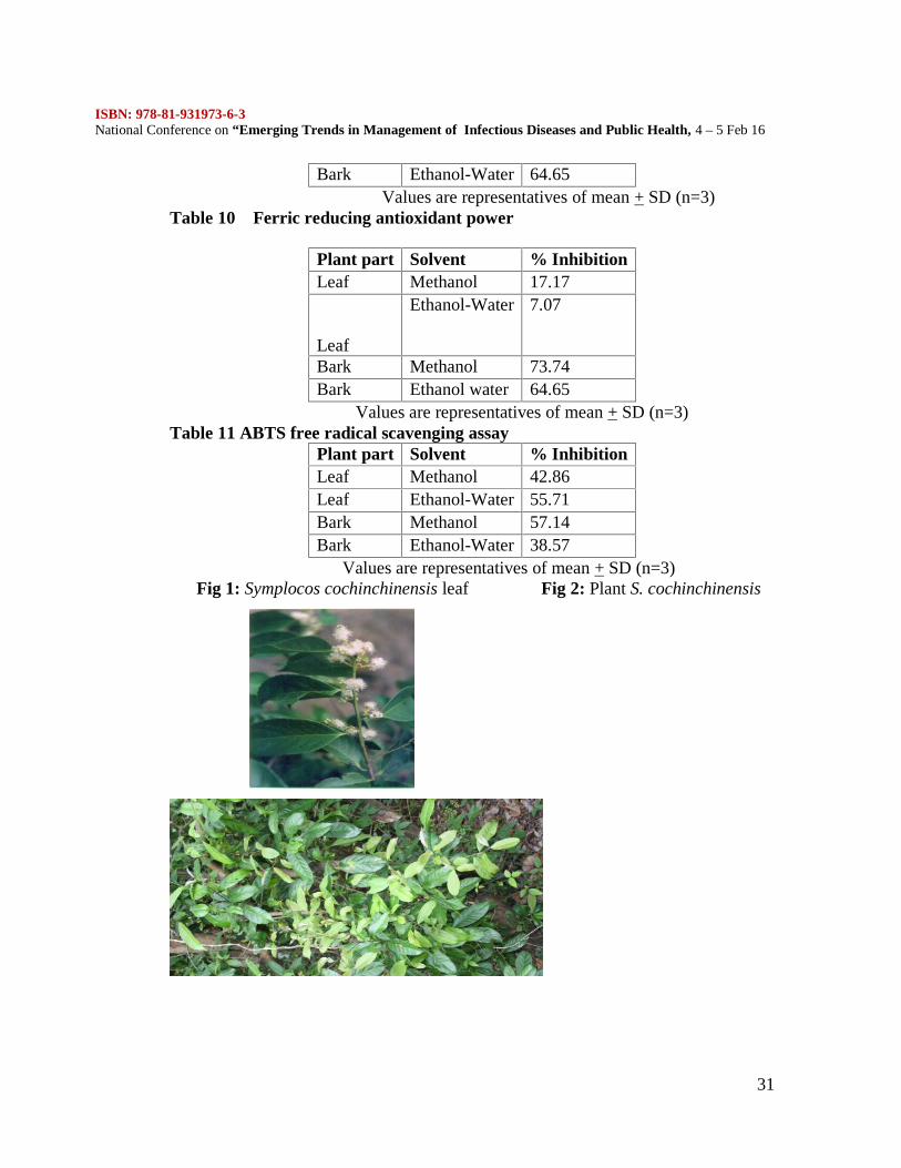
The leaves and barks of Symplocos cochinchinensis (Figures 1,2) werecollected from the Western Ghats, Nilgiris, India in the month of June 2015. Theplant parts were authenticated by Dr.Chelladurai, Research officer, Central Councilfor Research in Ayurveda and Siddha. The leaves and bark were shade dried andpowdered. The methanolic and ethanolic extracts of the dried powders wereprepared. 10 g of the dried powder was dissolved in 100 ml of methanol and 1:2 ratioof ethanol – water. The contents were stirred well and left for 48 hours at roomtemperature. The filtrate collected after cold percolation was used for furtheranalysis.
Phytochemical analysisThe methanolic and ethanolic extracts obtained from the dried leaves and
bark powder of Symplocos cochinchinensis were tested for the presence ofphytochemicals – Tannins, Phlobatannins, Flavonoids, Terpenoids, Cardiacglycosides and Steroids following the methodology described by Evans.(8)Tests for Tannins
To 5 ml of the extracts, a few drops of 0.1% of Ferric chloride were added.The presence of brownish green or blue black colour indicated that the plant materialpossessed Tannins.Tests for Phlobatannins
10 ml of the plant extracts were boiled with 1% HCl in a test tube. Thepresence of Phlobatannins was confirmed by the deposition of red precipitate in thetest tube.Tests for Saponins
To 10 ml of the extracts, 3 ml of distilled water was added and shaken well,so as to obtain froth. To the froth formed, a few drops of olive oil were added. Theformation of emulsion indicates the presence of saponins.Test for Flavonoids
A few drops of 1% liquid ammonia were taken in test tubes, to which themethanolic and ethanolic extracts were added. Yellow colouration of the solutionconfirmed the presence of FlavonoidsTest for Terpenoids
Around 2 ml of chloroform and 3 ml of concentrated sulphuric acid wereadded consecutively to 5 ml of the plant extracts. A reddish brown interface in thesolution denoted the presence of Terpenoids.Test for Cardiac Glycosides:
To 5 ml of the plant extracts, 2 ml of glacial acetic acid containing a drop ofFerric chloride was added. This was followed by the addition of 1 ml of concentratedSulphuric acid. The brown ring thus obtained yield positive result for the test.
ISBN: 978-81-931973-6-3National Conference on “Emerging Trends in Management of Infectious Diseases and Public Health, 4 – 5 Feb 16
26
Test for SteroidsA couple of grams of plant powder were mixed with 10 ml of chloroform
followed by boiling and filtration. To the above 2 ml of the filtrate 2 ml of aceticanhydride and a few drops of concentrated sulphuric acid were added. Stablepresence of blue green ring in the solution confirms the presence of steroids.Determination of Total phenolic content
Folin- ciocalteau method was followed for the determination of the totalphenolic content. Distilled water (500µl) and Folin –ciocalteau reagent (500µl) wereadded to 100 µl of the plant extracts and incubated at room temperature for 6minutes. The final volume was made up to 3 ml with 7% sodium carbonate solution.The absorbance was measured at 760 nm using UV-visible spectrophotometer afteran incubation period of 90 min. The total phenolic content was expressed asmilligrams of tannic acid equivalents per gram of dry weight (mg TAE/gDW) of theplant using a standard plot of tannic acid.(9)Determination of Total flavonoid content
The total flavonoid content of the plant was determined by the methodadopted by Moussa et al. (2011). The plant extracts were taken in test tubes and thesolvent were allowed to evaporate. To the residue 5 ml of 0.1 M Aluminum chloridewas added and shaken well. This was followed by incubation for 40 minutes at roomtemperature and the absorbance value was measured at 415 nm using UV -Visspectrophotometer. A standard plot of quercetin at varying concentration was used toevaluate the total flavonoid content, expressed as milligrams of Quercetin equivalentper gram dry weight (mg QE/gDW) of the plant material.(10)Determination of the Total antioxidant activity
The total antioxidant activity was estimated by phosphomolybdenum method.To the plant extract (0.5 ml), 4.5 ml of the reagent solution (0.6 M sulphuric acid, 28mm sodium phosphate and 4 mM of ammonium molybdate) was added. The solutionwas maintained in a boiling water bath at 95 C for 90 min. The solution was cooledto room temperature and the absorbance was measured at 695 nm using UV- visiblespectrophotometer. The total antioxidants in the plant were expressed as mg TAE/gDW of the plant material (11).2, 2 – Diphenyl-1- picryl hydrazyl (DPPH) free radical scavenging assay
The two plant extracts were taken at various concentrations (10,20,30,40,50µg/ml) in small test tubes and made up to 1 ml using methanol. 1 ml of 0.01 mMDPPH dissolved in methanol was added to all the test tubes and maintained in darkfor 30 minutes, at room temperature. The absorbance of the solutions was read at 517nm. The percentage inhibition and IC 50 values were calculated with DPPH as thecontrol and butylated hydroxyl anisole (BHA) as the reference. The concentration inµg of dry material per ml of solvent (µg/ml) that inhibits the formation of DPPHradicals by 50% is defined as the IC 50 value.(12)
% inhibition = (absorbance of control (Ac) - Absorbance of the sample (As)) x 100-----------------------------------------------------------------------------------
Absorbance of the control (Ac)
ISBN: 978-81-931973-6-3National Conference on “Emerging Trends in Management of Infectious Diseases and Public Health, 4 – 5 Feb 16
27
Ferric thiocyanate (FTC) assayThe assay involves the addition of 120 µl of 98% ethanol, 100 µl of 2.5 %
linoleic acid and 9 ml of 40 mM phosphate buffer (pH 7) to 100 µl of the plantextract. To 100 µl of the mixture, 9.7 ml of 75% ethanol, 100 µl of 30% ammoniumthiocyanate and 100 µl of 20 mM FeCl3 in 3.5% HCl were added after maintainingthe solution in the dark, at 40 C. The absorbance of the solution was measured at500 nm using UV-visible spectrophotometer after 3 min. The percentage ofinhibition was calculated with Tannic acid as the standard (Deepa et al., 2013). (13)
% inhibition = (absorbance of control (Ac) - Absorbance of the sample (As)) x 100-----------------------------------------------------------------------------------
Absorbance of the control (Ac)Thiobarbituric acid (TBA) assay
Equal volume (200 µl) of 20% trichloroacetic acid and 0.67% thiobarbituricacid were mixed with 1 ml of 2.51% linoleic acid and 1 ml of plant extract. Thesolution was maintained in boiling water bath for 10 min; cooled to roomtemperature and centrifuged at 3000 rpm. The supernatant was subjected to UV–visible spectrophotometric analysis at 532 nm. The percentage inhibition of the plantagainst the secondary products of lipid peroxidation was evaluated with reference tothe standard solution of butylated hydroxyl toluene (BHT) (Deepa et al., 2013) (13)% inhibition = (absorbance of control (Ac) - Absorbance of the sample (As)) x 100
-----------------------------------------------------------------------------------Absorbance of the control (Ac)
Ferric reducing antioxidant power (FRAP) assay1 ml of plant extract, 2.5 ml phosphate buffer (of 0.2 M, pH 7) and 1%
potassium ferricyanide (2.5 ml) were mixed and incubated at 50 C for 30 min. Tothe solution, 2.5 ml of 10% trichloroacetic acid was added and centrifuged at 6500rpm for 10 min. Distilled water (2.5 ml) and 0.5 ml of 0.1% FeCl3 were added to 2.5ml of the supernatant. The absorbance of the solution was measured at 700 nm usingUV–visible spectrophotometer. The reducing ability of the plant was evaluated interms of percentage by relating to the standard, FeSO4 (Kalita et al., 2013) (14).% inhibition = (absorbance of control (Ac) - Absorbance of the sample (As)) x 100
-----------------------------------------------------------------------------------Absorbance of the control (Ac)
[2,2_ -Azino-bis(3-ethylbenzothiazoline-6-sulphonic acid)] ABTS assayA solution of 7 mM ABTS [2,2 -azino-bis(3-ethylbenzothiazoline-6-
sulphonic acid)] and 2.45 mM potassium persulphate was incubated in the dark for12–16 h, after which the solution was diluted with ethanol till the absorbancereached 0.7 ± 0.02 at 734 nm. 1 ml of the diluted solution was mixed with 100 µl ofplant extract and the absorbance was evaluated at 734 nm after 6 min. Thepercentage reduction against ABTS was calculated with reference to the standard,Tannic acid (Deepa et al., 2013) (13).
ISBN: 978-81-931973-6-3National Conference on “Emerging Trends in Management of Infectious Diseases and Public Health, 4 – 5 Feb 16
28
Larvicidal activityCulture of larvae
The Artemia salina seeds were obtained from Marina labs, India. The seedswere incubated in marine water for 48 hours for hatching in a small water tank.Aeration was provided with an aerator pump. Required light was provided withPhilips 40 Watts lamp for 12 hours cycle . After 48 hours, the larvae were removedand used for the experiments. The hatched larvae were used in the nauplii stage (26).Bioassay
Larvae of Artemia salina were taken in different test tubes containing theextracts of Symplocos cochinchinensis, leaf and bark powder at differentconcentrations. Then five concentrations (1,2,3,4.5 ml) of each extract were added to10 ml of sea water and 20 larvae were added to each of the test tubes. After 24 and48 hours the viability of the larvae was recorded. The test tubes were maintained intriplicates. At the end of the experimental period, the numbers of mobile and deadlarvae in each test tube were checked using a hand lens. Nauplii were considereddead when they were immobile and stayed at the bottom of the test tube. (26)
RESULTThe present study was done to find out the phytochemicals, anti oxidant
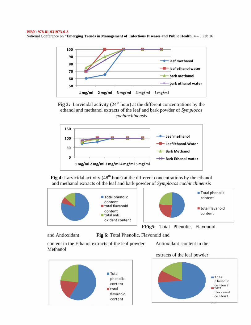
activity, free radical scavenging activity and the toxicity assay of the methanolic andethanolic extracts of the bark and leaf powder of Symplocos cochinchinensis. It wasfound that the ethanolic extracts of the leaf and bark showed the presence ofphytochemicals like flavonoids, tannins and saponins. (Table 1) Both the extractshad significant larvicidal activity. The % mortality of the ethanolic extract of the leafpowder at the concentration of 2 mg/ml was 100 in the 24th hour whereas themethanolic leaf extract showed 100% mortality at 24th hour at the concentration of 3mg/ml.
Similarly methanolic and ethanolic extracts of the bark showed 100%mortality at 3 mg/ml in the 24th hour.(Table 2, Figure 3) At the 48th hour ethanolicextracts of both the leaf and bark showed 100% mortality at 2 mg/ml whereas themethanolic extracts showed the same at 3 mg/ml.(Table 3 , Figure 4) The ethanolwater extracts of the leaf and the bark showed maximum larvicidal activity at 24th
and 48th hour.The total phenolic, total flavonoid, total antioxidant content were estimated.
The total phenolic content was expressed as milligrams of tannic acid equivalents pergram of dry weight(mg TAE/gDW)of the plant using a standard plot of tannic acid.The ethanol water leaf extract had the maximum phenolic content of 13.81+0.21.(Table 4, Figure 5,6,7,8)
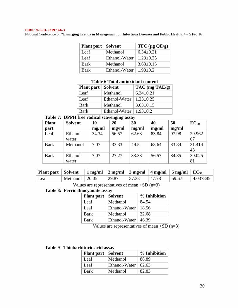
The total flavonoid content is expressed as milligrams of Quercetinequivalent per gram dry weight (mg QE/gDW) of the plant material. The methanolextract of leaf had the maximum flavonoid content with 6.34 +0.21.(Table 5, Figure5,6,7,8)
ISBN: 978-81-931973-6-3National Conference on “Emerging Trends in Management of Infectious Diseases and Public Health, 4 – 5 Feb 16
29
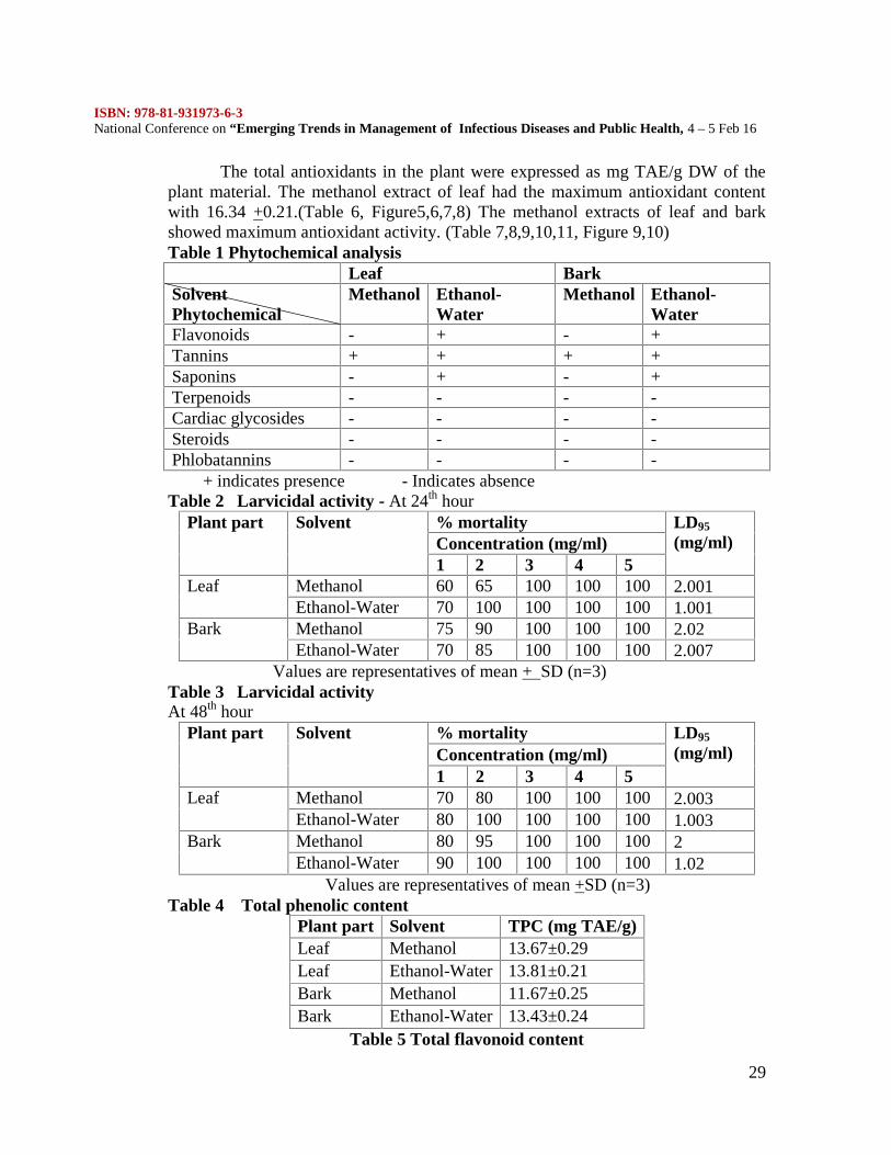
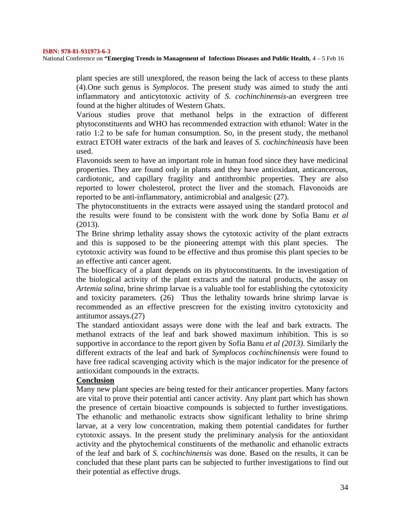
The total antioxidants in the plant were expressed as mg TAE/g DW of theplant material. The methanol extract of leaf had the maximum antioxidant contentwith 16.34 +0.21.(Table 6, Figure5,6,7,8) The methanol extracts of leaf and barkshowed maximum antioxidant activity. (Table 7,8,9,10,11, Figure 9,10)Table 1 Phytochemical analysis
Leaf BarkSolventPhytochemical
Methanol Ethanol-Water
Methanol Ethanol-Water
Flavonoids - + - +Tannins + + + +Saponins - + - +Terpenoids - - - -Cardiac glycosides - - - -Steroids - - - -Phlobatannins - - - -
+ indicates presence - Indicates absenceTable 2 Larvicidal activity - At 24th hour
Plant part Solvent % mortality LD95
(mg/ml)Concentration (mg/ml)1 2 3 4 5
Leaf Methanol 60 65 100 100 100 2.001Ethanol-Water 70 100 100 100 100 1.001
Bark Methanol 75 90 100 100 100 2.02Ethanol-Water 70 85 100 100 100 2.007
Values are representatives of mean + SD (n=3)Table 3 Larvicidal activityAt 48th hour
Plant part Solvent % mortality LD95
(mg/ml)Concentration (mg/ml)1 2 3 4 5
Leaf Methanol 70 80 100 100 100 2.003Ethanol-Water 80 100 100 100 100 1.003
Bark Methanol 80 95 100 100 100 2Ethanol-Water 90 100 100 100 100 1.02
Values are representatives of mean +SD (n=3)Table 4 Total phenolic content
Plant part Solvent TPC (mg TAE/g)Leaf Methanol 13.67±0.29Leaf Ethanol-Water 13.81±0.21Bark Methanol 11.67±0.25Bark Ethanol-Water 13.43±0.24
Table 5 Total flavonoid content
ISBN: 978-81-931973-6-3National Conference on “Emerging Trends in Management of Infectious Diseases and Public Health, 4 – 5 Feb 16
30
Plant part Solvent TFC (µg QE/g)Leaf Methanol 6.34±0.21Leaf Ethanol-Water 1.23±0.25Bark Methanol 3.63±0.15Bark Ethanol-Water 1.93±0.2
Table 6 Total antioxidant contentPlant part Solvent TAC (mg TAE/g)Leaf Methanol 6.34±0.21Leaf Ethanol-Water 1.23±0.25Bark Methanol 3.63±0.15Bark Ethanol-Water 1.93±0.2
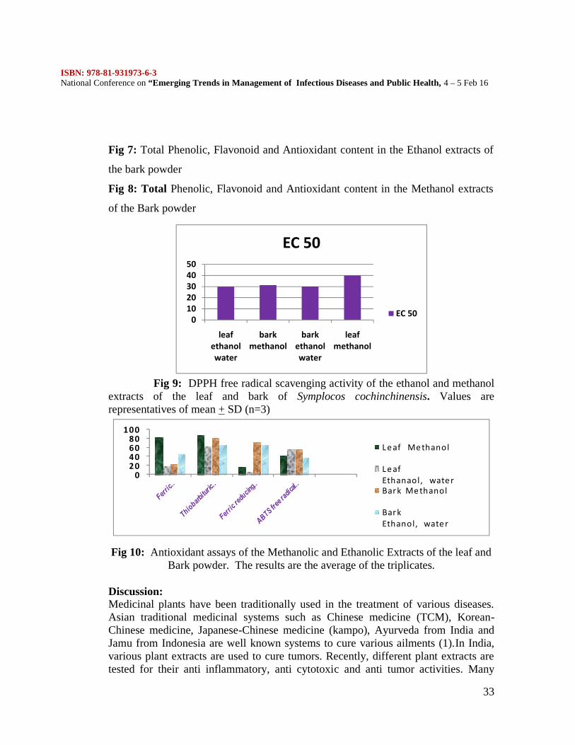
Table 7: DPPH free radical scavenging assayPlantpart
Solvent 10mg/ml
20mg/ml
30mg/ml
40mg/ml
50mg/ml
EC50
Leaf Ethanol-water
34.34 56.57 62.63 83.84 97.98 29.96267
Bark Methanol 7.07 33.33 49.5 63.64 83.84 31.41443
Bark Ethanol-water
7.07 27.27 33.33 56.57 84.85 30.02581
Plant part Solvent 1 mg/ml 2 mg/ml 3 mg/ml 4 mg/ml 5 mg/ml EC50
Leaf Methanol 20.05 29.87 37.33 47.78 59.67 4.037885Values are representatives of mean +SD (n=3)
Table 8: Ferric thiocyanate assayPlant part Solvent % InhibitionLeaf Methanol 84.54Leaf Ethanol-Water 18.56Bark Methanol 22.68Bark Ethanol-Water 46.39
Values are representatives of mean +SD (n=3)
Table 9 Thiobarbituric acid assayPlant part Solvent % InhibitionLeaf Methanol 88.89Leaf Ethanol-Water 62.63Bark Methanol 82.83
ISBN: 978-81-931973-6-3National Conference on “Emerging Trends in Management of Infectious Diseases and Public Health, 4 – 5 Feb 16
31
Bark Ethanol-Water 64.65Values are representatives of mean + SD (n=3)
Table 10 Ferric reducing antioxidant power
Plant part Solvent % InhibitionLeaf Methanol 17.17
Leaf
Ethanol-Water 7.07
Bark Methanol 73.74Bark Ethanol water 64.65
Values are representatives of mean + SD (n=3)Table 11 ABTS free radical scavenging assay
Plant part Solvent % InhibitionLeaf Methanol 42.86Leaf Ethanol-Water 55.71Bark Methanol 57.14Bark Ethanol-Water 38.57
Values are representatives of mean + SD (n=3)Fig 1: Symplocos cochinchinensis leaf Fig 2: Plant S. cochinchinensis
ISBN: 978-81-931973-6-3National Conference on “Emerging Trends in Management of Infectious Diseases and Public Health, 4 – 5 Feb 16
32
50
60
70
80
90
100
1 mg/ml 2 mg/ml 3 mg/ml 4 mg/ml 5 mg/ml
leaf methanol
leaf ethanol water
bark methanol
bark ethanol water
Fig 3: Larvicidal activity (24th hour) at the different concentrations by theethanol and methanol extracts of the leaf and bark powder of Symplocos
cochinchinensis
0
50
100
150
1 mg/ml 2 mg/ml 3 mg/ml 4 mg/ml 5 mg/ml
Leaf methanol
Leaf Ethanol-Water
Bark Methanol
Bark Ethanol water
Fig 4: Larvicidal activity (48th hour) at the different concentrations by the ethanoland methanol extracts of the leaf and bark powder of Symplocos cochinchinensis
Total phenoliccontent
total flavanoidcontent
FFig5: Total Phenolic, Flavonoid
and Antioxidant Fig 6: Total Phenolic, Flavonoid and
content in the Ethanol extracts of the leaf powder Antioxidant content in theMethanol
extracts of the leaf powder
Total phenolicc ontenttotal flavanoidc ontenttotal antioxidant content
Totalphenoliccontenttotalflavanoidcontent
T o t a lp h e n o l icco n te n tt o ta lf l av a n o idco n te n t
ISBN: 978-81-931973-6-3National Conference on “Emerging Trends in Management of Infectious Diseases and Public Health, 4 – 5 Feb 16
33
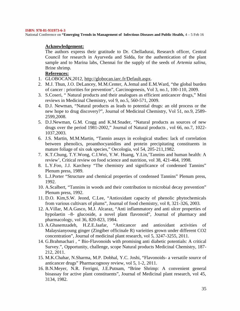
Fig 7: Total Phenolic, Flavonoid and Antioxidant content in the Ethanol extracts of
the bark powder
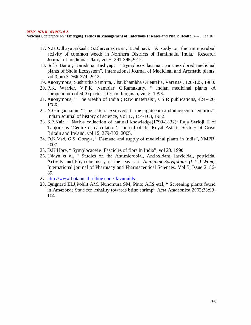
Fig 8: Total Phenolic, Flavonoid and Antioxidant content in the Methanol extracts
of the Bark powder
01020304050
leafethanolwater
barkmethanol
barkethanolwater
leafmethanol
EC 50
EC 50
Fig 9: DPPH free radical scavenging activity of the ethanol and methanolextracts of the leaf and bark of Symplocos cochinchinensis. Values arerepresentatives of mean + SD (n=3)
020406080
100
L e af Me thanol
L e afEthanaol, wate rBark Me thanol
BarkEthanol, wate r
Fig 10: Antioxidant assays of the Methanolic and Ethanolic Extracts of the leaf andBark powder. The results are the average of the triplicates.
Discussion:Medicinal plants have been traditionally used in the treatment of various diseases.Asian traditional medicinal systems such as Chinese medicine (TCM), Korean-Chinese medicine, Japanese-Chinese medicine (kampo), Ayurveda from India andJamu from Indonesia are well known systems to cure various ailments (1).In India,various plant extracts are used to cure tumors. Recently, different plant extracts aretested for their anti inflammatory, anti cytotoxic and anti tumor activities. Many
ISBN: 978-81-931973-6-3National Conference on “Emerging Trends in Management of Infectious Diseases and Public Health, 4 – 5 Feb 16
34
plant species are still unexplored, the reason being the lack of access to these plants(4).One such genus is Symplocos. The present study was aimed to study the antiinflammatory and anticytotoxic activity of S. cochinchinensis-an evergreen treefound at the higher altitudes of Western Ghats.Various studies prove that methanol helps in the extraction of differentphytoconstituents and WHO has recommended extraction with ethanol: Water in theratio 1:2 to be safe for human consumption. So, in the present study, the methanolextract ETOH water extracts of the bark and leaves of S. cochinchineasis have beenused.Flavonoids seem to have an important role in human food since they have medicinalproperties. They are found only in plants and they have antioxidant, anticancerous,cardiotonic, and capillary fragility and antithrombic properties. They are alsoreported to lower cholesterol, protect the liver and the stomach. Flavonoids arereported to be anti-inflammatory, antimicrobial and analgesic (27).The phytoconstituents in the extracts were assayed using the standard protocol andthe results were found to be consistent with the work done by Sofia Banu et al(2013).The Brine shrimp lethality assay shows the cytotoxic activity of the plant extractsand this is supposed to be the pioneering attempt with this plant species. Thecytotoxic activity was found to be effective and thus promise this plant species to bean effective anti cancer agent.The bioefficacy of a plant depends on its phytoconstituents. In the investigation ofthe biological activity of the plant extracts and the natural products, the assay onArtemia salina, brine shrimp larvae is a valuable tool for establishing the cytotoxicityand toxicity parameters. (26) Thus the lethality towards brine shrimp larvae isrecommended as an effective prescreen for the existing invitro cytotoxicity andantitumor assays.(27)The standard antioxidant assays were done with the leaf and bark extracts. Themethanol extracts of the leaf and bark showed maximum inhibition. This is sosupportive in accordance to the report given by Sofia Banu et al (2013). Similarly thedifferent extracts of the leaf and bark of Symplocos cochinchinensis were found tohave free radical scavenging activity which is the major indicator for the presence ofantioxidant compounds in the extracts.ConclusionMany new plant species are being tested for their anticancer properties. Many factorsare vital to prove their potential anti cancer activity. Any plant part which has shownthe presence of certain bioactive compounds is subjected to further investigations.The ethanolic and methanolic extracts show significant lethality to brine shrimplarvae, at a very low concentration, making them potential candidates for furthercytotoxic assays. In the present study the preliminary analysis for the antioxidantactivity and the phytochemical constituents of the methanolic and ethanolic extractsof the leaf and bark of S. cochinchinensis was done. Based on the results, it can beconcluded that these plant parts can be subjected to further investigations to find outtheir potential as effective drugs.
ISBN: 978-81-931973-6-3National Conference on “Emerging Trends in Management of Infectious Diseases and Public Health, 4 – 5 Feb 16
35
Acknowledgement:The authors express their gratitude to Dr. Chelladurai, Research officer, CentralCouncil for research in Ayurveda and Sidda, for the authentication of the plantsample and to Marina labs, Chennai for the supply of the seeds of Artemia salina,Brine shrimp.References:
1. GLOBOCAN,2012, http://globocan.iarc.fr/Default.aspx.2. M.J. Thun, J.O. DeLancey, M.M.Center, A.Jemal and E.M.Ward, “the global burden
of cancer : priorities for prevention”, Carcinogenesis, Vol 3, no.1, 100-110, 2009.3. S.Coseri, “ Natural products and their analogues as efficient anticancer drugs,” Mini
reviews in Medicinal Chemistry, vol 9, no.5, 560-571, 2009.4. D.J. Newman, “Natural products as leads to potential drugs: an old process or the
new hope to drug discovery?”, Journal of Medicinal Chemistry, Vol 51, no.9, 2589-2599,2008.
5. D.J.Newman, G.M. Cragg and K.M.Snader, “Natural products as sources of newdrugs over the period 1981-2002,” Journal of Natural products , vol 66, no.7, 1022-1037,2003.
6. J.S. Martin, M.M.Martin, “Tannin assays in ecological studies: lack of correlationbetween phenolics, proanthocyanidins and protein precipitating constituents inmature foliage of six oak species,” Oecologia, vol 54, 205-211,1982.
7. K.T.Chung, T.Y.Wong, C.I.Wei, Y.W. Huang, Y.Lin,’Tannins and human health: Areview’, Critical review on food science and nutrition, vol 38, 421-464, 1998.
8. L.Y.Foo, J.J. Karchesy “The chemistry and significance of condensed Tannins”Plenum press, 1989.
9. L.J.Porter “Structure and chemical properties of condensed Tannins” Plenum press,1992.
10. A.Scalbert, “Tannins in woods and their contribution to microbial decay prevention”Plenum press, 1992.
11. D.O. Kim,S.W. Jeond, C.Lee, “Antioxidant capacity of phenolic phytochemicalsfrom various cultivars of plums”, Journal of food chemistry, vol 8, 321-326, 2003.
12. A.Villar, M.A.Gasco, M.J. Alcaraz, “Anti inflammatory and anti ulcer properties ofhypolaetin –8- glucoside, a novel plant flavonoid”, Journal of pharmacy andpharmacology, vol 36, 820-823, 1984.
13. A.Ghasemzadeh, H.Z.E.Jaafar, “Anticancer and antioxidant activities ofMalaysianyoung ginger (Zingiber officinale R) varieities grown under different CO2concentration”, Journal of medicinal plant research, vol 5, 3247-3255, 2011.
14. G.Brahmachari , “ Bio-Flavonoids with promising anti diabetic potentials: A criticalSurvey.”, Opportunity, challenge, scope Natural products Medicinal Chemistry, 187-212, 2011.
15. M.K.Chahar, N.Sharma, M.P. Dobhal, Y.C. Joshi, “Flavonoids- a versatile source ofanticancer drugs” Pharmacognosy review, vol 5, 1-2, 2011.
16. B.N.Meyer, N.R. Ferrigni, J.E.Putnam, “Brine Shrimp: A convenient generalbioassay for active plant constituents”, Journal of Medicinal plant research, vol 45,3134, 1982.
ISBN: 978-81-931973-6-3National Conference on “Emerging Trends in Management of Infectious Diseases and Public Health, 4 – 5 Feb 16
36
17. N.K.Udhayaprakash, S.Bhuvaneshwari, B.Jahnavi, “A study on the antimicrobialactivity of common weeds in Northern Districts of Tamilnadu, India,” ResearchJournal of medicinal Plant, vol 6, 341-345,2012.
18. Sofia Banu , Karishma Kashyap, “ Symplocos laurina : an unexplored medicinalplants of Shola Ecosystem”, International Journal of Medicinal and Aromatic plants,vol 3, no 3, 366-374, 2013.
19. Anonymous, Sushrutha Samhita, Chaukhambha Orientalia, Varanasi, 120-125, 1980.20. P.K. Warrier, V.P.K. Nambiar, C.Ramakutty, “ Indian medicinal plants -A
compendium of 500 species”, Orient longman, vol 5, 1996.21. Anonymous, “ The wealth of India ; Raw materials”, CSIR publications, 424-426,
1986.22. N.Gangadharan, “ The state of Ayurveda in the eighteenth and nineteenth centuries”,
Indian Journal of history of science, Vol 17, 154-163, 1982.23. S.P.Nair, “ Native collection of natural knowledge(1798-1832): Raja Serfoji II of
Tanjore as ‘Centre of calculation’, Journal of the Royal Asiatic Society of GreatBritain and Ireland, vol 15, 279-302, 2005.
24. D.K.Ved, G.S. Goraya, “ Demand and supply of medicinal plants in India”, NMPB,2007.
25. D.K.Hore, “ Symplocaceae: Fascicles of flora in India”, vol 20, 1990.26. Udaya et al, “ Studies on the Antimicrobial, Antioxidant, larvicidal, pesticidal
Activity and Phytochemistry of the leaves of Alangium Salvifolium (L.f .) Wang,International journal of Pharmacy and Pharmaceutical Sciences, Vol 5, Issue 2, 86-89.
27. http://www.botanical-online.com/flavonoids.28. Quignard ELJ,Pohlit AM, Nunomura SM, Pinto ACS etal, “ Screening plants found
in Amazonas State for lethality towards brine shrimp” Acta Amazonica 2003;33:93-104

ISBN: 978-81-931973-6-3National Conference on “Emerging Trends in Management of Infectious Diseases and Public Health, 4 – 5 Feb 16
37
ANALYSIS OF SOIL PHYSICO- CHEMICAL CHARACTERISTICS OF ORGANICFARMING AND CONVENTIONAL FARMING PRACTICES
P. Nirmala1* and R. Krishnaveni2
1*Assistant Professor, PG and Research Department of Microbiology, Mohamed sathakCollege of Arts and Science, Sholinganallur, Chennai.
2 Research Scholar, PG and Research Department of Microbiology, Mohamed sathakCollege of Arts and Science, Sholinganallur, Chennai.
Abstract
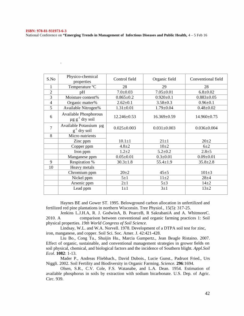
With an increasing awareness about the harmful effects of agrochemicals, thedemand for technologies and product based on biological processes such as organic farminghas been increasing steadily worldwide. The objective of our research was to evaluate theimpact of organic and conventional management strategies on grower fields in Chennai onsoil physico-chemical characteristics. Physico-chemical properties such as temperature, pH,moisture content, organic matter, macronutrients (Nitrogen, Phosphorous and Potassium),micronutrients (Zinc, Manganese, Iron and Copper), respiration and heavy metals weredetermined. There wasn’t much difference in the temperature measurement. Soil pH wasslightly higher in organic field than control and conventional field. Moisture content washigher in organic field than conventional and control field. The results showed that organicfarming practices showed higher organic matter, respiration, micronutrients,macronutrients except potassium which was followed by conventional field and control field.Heavy metals content was higher in conventional fields than the organic fields. Henceorganically managed soils establish ecological systems that are able to sustain biologicalproductivity as well as agricultural productivity in the long-term.
Key words: Organic farming, conventional farming, soil, physic-chemical characteristics.
IntroductionIn the past decades and up to the present, high crop yields have been achieved
through conventional farming, i.e., the use of high-yielding varieties (HYVS), agrochemicalinputs (inorganic fertilizers and pesticides), farm machineries that facilitate land preparation,and irrigation that relives the crop from any yield-depressing effect of water deficit. Thelong-term ecological consequences of this production approach have since been recognized,i.e., soil and water pollution, loss of biodiversity, genetic erosion, receding undergroundwater etc. The intensive uses of agrochemicals and monocropping have resulted in thedegradation of the environment.
High yields continue to be the most important indicator of assessing the success orfailure of crop farming. But if high yields are obtained with negative consequences, then it isimperative to re-think about continuing to practice much more supporting or subsidingconventional farming. As early as the 1980’s, various organizations started the campaign fororganic and sustainable agriculture. So there is a need to promote chemical-free agricultureor environment-friendly pest management techniques and/or organic farming. Now there is agrowing realization that the adoption of ecological and sustainable farming practices canonly reverse the declining trend in the global productivity and environment protection.
ISBN: 978-81-931973-6-3National Conference on “Emerging Trends in Management of Infectious Diseases and Public Health, 4 – 5 Feb 16
38
Organic agriculture is the oldest form of agriculture on earth. The system avoids applicationsof synthetic fertilizers and pesticides, use of organic inputs and recycling for nutrient supplyand emphasizes cropping system design and biological processes for pest management. In along-term field trial, microbial biomass was higher in soils from organic plots (Gelsomino etal., 2004; Tu et al., 2005; Liu et al., 2007). Other researchers have shown that incorporationof organic amendments increased soil microbial activity, microbial diversity (Girvan et al.,2004; Grayston et al., 2004), densities of bacteria (Bruggen and Semenov, 2000) andnematodes (Abawi and Widmer, 2000).
So organic agriculture is gaining worldwide acceptance. Here was a surprising lackof research regarding a variety of aspects of organic agriculture. The number of studiescomparing organic and conventional systems is also limited to India. Most of the researchrelated to these studies was carried out on foreign countries like North Carolina (Liu et al.,2007), New Zealand (Condron et al., 2000), Brazil (Araujo et al., 2009), England (Jenkins etal., 2010), China (Hu and Zhiping Cao, 2007). The objectives of our research were toevaluate the impact of organic and conventional management strategies on grower fields inChennai on soil physical and chemical characteristics. The results will help to provide aplatform for future research into the impacts of organic farming.
Materials and MethodsSelection of field for experimental study
Six soil samples were collected with a history of organic and conventional cropproduction farms in and around Chennai. Three farms were certified organic and did not usesynthetic fertilizers or pesticides. They were located in Wallajabadh, Redhills andChengelpet. Three of the farms were classified as conventional farms where chemicalfertilizers and pesticides were used. They were located in Porur, Kundrathur and Kovur.Control field also set up without the addition of any fertilizers (biofertilizers and chemicalfertilizers).
COLLECTION OF SOIL SAMPLEWell mixed composite soil samples of about 20g were taken at a depth of 20cm from
four corners and from the middle of the farm field. All the soil samples were mixedthoroughly to get a homogenous mixture. Stones and crop residues are discarded. The soilsamples were stored at 4°C for microbial analysis and the remaining were air dried andsieved for the determination of physico-chemical properties of soil.
PHYSICO-CHEMICAL ANALYSIS OF SOIL SAMPLESTEMPERATURE
Soil temperature was noted by using soil thermometer at the time of samplecollection.
MEASUREMENT OF SOIL pHTo 40ml of distilled water, 20g of soil was added and stirred thoroughly with a glass
rod. It was kept undisturbed for 15min. The electrode of pH meter was dipped into thesuspension and the pH was noted. For standardization, buffer solutions of pH 4.0, 7.0 and9.2 were used.
MOISTURE CONTENTThe moisture content of the soil samples were determined gravimetrically by
weighing, drying in a hot air oven at 105°C for 24 hours and then reweighing.
ISBN: 978-81-931973-6-3National Conference on “Emerging Trends in Management of Infectious Diseases and Public Health, 4 – 5 Feb 16
39
ORGANIC MATTERSoil organic matter was found by rapid titration method of Walkely and Black
(1934).
MACRO AND MICRONUTRIENTSMacronutrients such as Nitrogen (Sankaran, 1966), Phosphorus (Olsen et al., 1954)
and Potassium (Sankaran, 1966) and Micronutrients (Lindsay and Norwell, 1978) like Zinc,Manganese, Copper and Iron were also analyzed.
RESPIRATION100g of soil sample was dissolved with distilled water and its Water Holding
Capacity (WHC) was adjusted to 33%. The soil sample was kept in a 1000ml flask in whicha test tube containing 10 ml of freshly prepared N/10 NaOH was hung using a thread.Another flask devoid of soil sample was used as control. The flask was incubated at 30°C for2-3 weeks. At weekly intervals NaOH was taken out, to which 2-3 drops of phenolphthaleinwas added and titrated against N/10 HCl solution (Haynes And Gower, 1995).
HEAVY METALSThe heavy metals which were present in all the three soil samples were determined
by using X-ray fluorescence spectroscopy (XRF).
STATISTICAL ANALYSISAll the experiments were conducted in triplicates and data shown in results are
means and standard deviation.
Results and DiscussionThe physico-chemical analysis of soil sample was carried out by studying the
temperature, pH, moisture content, organic matter, macronutrients (Nitrogen, Phosphorousand Potassium), micronutrients (Zinc, Copper, Iron and Manganese), respiration and heavymetal content of all the three fields (Organic, Conventional and Control field). The resultswere tabulated (Table.1).
There wasn’t much difference in the temperature measurement. Soil pH was slightlyhigher in organic field (7.05±0.01) than control (7.0±0.03) and conventional field (6.8±0.02).The results were correlated with the findings of Padmavathy and Poyyamoli, (2011), whoreported that pH of the organic field (7.6±0.1), was slightly higher than conventional field(7.2±0.2). There were some significant differences in the soil pH which was agreed byPoudel et al., (2002) studies of soils from California. However other researchers have shownthat pH was not significantly different between organically and conventionally managedsoils (Mader et al., 2002; Diepeningen et al., 2006). The change of pH in soil samplegenerally occurred as a consequence of mineralization of organic compound.
Moisture content was higher in organic field (0.920±0.1%) than conventional(0.883±0.05%) and control field (0.865±0.2%). This is in accordance to the study done byChauhan et al., (2011) who reported that moisture content was higher in organic field(45.460±1.44%) than conventional field (44.360±1.31%). The result was also correlated withthe findings of Liu et al., (2007).
The highest organic matter was observed in organically treated plots (3.58±0.3%)and the least in control (2.62±0.1%) and conventional fields (0.96±0.1%).This result iscorrelated with the study done by Chauhan et al., (2011) which showed higher organiccontent in organic fields (2.320±0.04%) than conventional field (2.290±0.04%). Soil from
ISBN: 978-81-931973-6-3National Conference on “Emerging Trends in Management of Infectious Diseases and Public Health, 4 – 5 Feb 16
40
the organic field showed an increase in organic matter, compared to other fields; this mightbe due to the addition of organic contents as they are the sources of nitrogen and carbon tosoils. A positive effect of organic fertilizers on the microbial biomass nitrogen and theorganic matter content in the soil was also observed by Cerny et al., (2008). The greateramounts of organic matter contribute to the better soil structure observed in organic field.
The total Nitrogen content was higher in organic field (1.79±0.04%) than other twofields (control-1.31±0.01%, conventional-0.48±0.02%). This is in accordance with the studydone by Padmavathy and Poyyamoli, (2011) who reported that the total Nitrogen content inorganic fields were higher than the conventional fields, due to the external chemical inputseffects microbial population in conventional fields. This result is also correlated with thefindings of Condron et al., (2000), who reported that the higher level of microbiologicalactivity in the organic soil leads to the increase in nitrogen content of soil. The present resultwas contrast to the results showed by Chauhan et al., (2011), where his study showed higherNitrogen content in conventional field (0.570±0.05%) than organic (0.550±0.03%) andcontrol field (0.440±0.31%).
The phosphorous content was significantly higher in organic field (16.369±0.59 µg g-1drysoil) than conventional field (14.960±0.75 µg g-1dry soil). This was agreed to the study doneby Padmavathy and Poyyamoli, (2011) who reported higher phosphorous content in organicfield (17±2 kg ha-1) than conventional field (8±1 kg ha-1). But there was a contrast in resultsshowed in the study made by Condron et al., (2000). His study showed higher phosphorouscontent in conventional field (66.2 mg kg-1) than organic field (45.7 mg kg-1). This is alsocorrelated with the results of Chauhan et al., (2011) because conventional fields amendedwith the mixture of urea showed highest content in phosphorous.
The potassium content was lower in organic field (0.031±0.003 µg g-1dry soil) thanconventional field (0.036±0.004 µg g-1dry soil). The result is correlated with the findings ofCondron et al., (2000) who reported that the potassium content was higher in conventionalfield (1.00 cmol kg-1) than organic field (0.97 cmol kg-1). Mader et al., (2002) reported thatthe lower nutrient inputs in organic fields cause lower amount of potassium. But thepotassium level was more variable among farms from year to year (Liu et al., 2007).
Soil micronutrients were higher in organic field (Zn-21±1 ppm, Cu-10±2 ppm, Fe-5.2±0.2 ppm, Mn-0.3±0.01 ppm) than conventional (Zn-20±1 ppm, Cu-6±2 ppm, Fe-2.8±5ppm, Mn-0.09±0.01 ppm) and control field (Zn-10.1±1 ppm, Cu-4.8±2 ppm, Fe-1.2±2 ppm,Mn-0.05±0.01 ppm). This was in accordance with to Liu et al., (2007) who reported that themicronutrient content was higher in organic field than conventional field. The present studywas also supported by Bending et al., (2000). He reported that the increase in microbialnumbers leads to the higher content of micronutrients in soil. The experimental study carriedout by Padmavathy and Poyyamoli (2011) also showed higher micronutrient content inorganically treated plots (Zn-26±1 ppm, Cu-9±2 ppm, Fe-4.2±0.2 ppm, Mn-0.3±0.01 ppm)than convetional plots (Zn-15±1 ppm, Cu-7±2 ppm, Fe-3.2±0.2 ppm, Mn-0.1±0.01 ppm).
Soil microbial respiration rates were higher in soils from organic fields (55.4±1.9%)than conventional field (35.8±2.8). This was in accordance with Liu et al., (2007) whoshowed higher respiration rates in organic field (96.7 mg/kg) than conventional field (11.0mg/kg). The results suggested that largest activity of soil microbes existed in soil organicfarms leads to higher respiration rates (Mader et al., 2002; Diepeningen et al., 2006).
Heavy metals content was higher in conventional fields (Cr-101±3 ppm, Ni-28±4ppm, Ar-14±2 ppm, Pb-13±2 ppm) than the organic fields (Cr-45±5 ppm, Ni-11±2 ppm, Ar-5±3 ppm, Pb-3±1 ppm). This was supported by the study done by Padmavathy and
ISBN: 978-81-931973-6-3National Conference on “Emerging Trends in Management of Infectious Diseases and Public Health, 4 – 5 Feb 16
41
Poyyamoli (2011) where they stated that the usage of chemical fertilizers leads to the higherrate of heavy metal content in conventional field (Cr-89±2 ppm, Ni-20±4 ppm, Ar-6±2 ppm,Pb-6±2 ppm).
On comparing the Physico-chemical analysis of three soil samples (Organic,Conventional and Control field), the results showed that organic farming practices showedhigher organic matter, respiration, micronutrients, macronutrients except potassium whichwas followed by conventional field and control field. Hence organically managed soilsestablish ecological systems that are able to sustain biological productivity as well asagricultural productivity in the long-term.
ReferencesAraújo S.F.A., Luiz F.C. Leite., Valdinar B. Santos and Romero F.V. Carneiro. 2009.Soil Microbial Activity in Conventional and Organic Agricultural Systems.Sustainability. 1:268-276
Abawi G.S. and T.L. Widmer. 2000. Impact of soil health management practices onsoil borne pathoge-,nematodes and root diseases of vegetable crops. Appl. Soil Ecol. 15: 37-47.
Bending G.D., Putland C., Rayns F. 2000. Changes in microbial communitymetabolism and labile organic matter fractions as early indicators of the impact ofmanagement on soil biological quality. Biol.Fertil.Soils. 31:78–84.
Bruggen V.A.H.C. and A.M. Semenov. 2000. In search of biological indicators forplant health and disease suppression. Appl. Soil Ecol. 15: 13-24.
Cerny J., J. Balik., M. Kulhanek and V. Neded. 2008. Plant Soil Environ. 54: 212-218.
Chauhan P.K., V. Singh., Vinod Kumar Dhatwalia and Abhishek. 2011. Physico-chemical and Microbial activity of soil under Conventional and Organic AgriculturalSystems. J. Chem. Pharm. Res. 3(3):799-804.
Condron L. M., K. C. Cameron., H. J. Di., T. J. Clough., E. A.Forbes., R. G.McLaren & R. G. Silva. 2000. A comparison of soil and environmental quality underorganic and conventional farming systems in New Zealand. New Zealand Journal ofAgricultural Research. 43(4): 443-466.
ISBN: 978-81-931973-6-3National Conference on “Emerging Trends in Management of Infectious Diseases and Public Health, 4 – 5 Feb 16
42
.
Haynes BE and Gower ST. 1995. Belowground carbon allocation in unfertilized andfertilized red pine plantations in northern Wisconsin. Tree Physiol., 15(5): 317-25.
Jenkins L.J.H.A, R. J. GodwinA, B. PearceB, R SakrabaniA and A. WhitmoreC.2010. A comparison between conventional and organic farming practices 1: Soilphysical properties. 19th World Congress of Soil Science.
Lindsay, W.L. and W.A. Norvell. 1978. Development of a DTPA soil test for zinc,iron, manganese, and copper. Soil Sci. Soc. Amer. J. 42:421-428.
Liu Bo., Cong Tu., Shuijin Hu., Marcia Gumpertz., Jean Beagle Ristaino. 2007.Effect of organic, sustainable, and conventional management strategies in grower fields onsoil physical, chemical, and biological factors and the incidence of Southern blight. Appl.SoilEcol. 1082: 1-13.
Mader P., Andreas Fliebbach., David Dubois., Lucie Gunst., Padruot Fried., UrsNiggli. 2002. Soil Fertility and Biodiversity in Organic Farming. Science. 296:1694.
Olsen, S.R., C.V. Cole, F.S. Watanabe, and L.A. Dean. 1954. Estimation ofavailable phosphorus in soils by extraction with sodium bicarbonate. U.S. Dep. of Agric.Circ. 939.
S.NoPhysico-chemical
propertiesControl field Organic field Conventional field
1 Temperature ºC 28 29 282 pH 7.0±0.03 7.05±0.01 6.8±0.023 Moisture content% 0.865±0.2 0.920±0.1 0.883±0.054 Organic matter% 2.62±0.1 3.58±0.3 0.96±0.15 Available Nitrogen% 1.31±0.01 1.79±0.04 0.48±0.02
6Available Phosphorous
µg g-1 dry soil12.246±0.53 16.369±0.59 14.960±0.75
7Available Potassium µg
g-1 dry soil0.025±0.003 0.031±0.003 0.036±0.004
8 Micro nutrients10.1±1 21±1 20±2Zinc ppm
Copper ppm 4.8±2 10±2 6±2Iron ppm 1.2±2 5.2±0.2 2.8±5
Manganese ppm 0.05±0.01 0.3±0.01 0.09±0.019 Respiration % 30.3±1.8 55.4±1.9 35.8±2.8
10 Heavy metals20±2 45±5 101±3Chromium ppm
Nickel ppm 5±1 11±2 28±4Arsenic ppm 2±1 5±3 14±2
Lead ppm 1±1 3±1 13±2
ISBN: 978-81-931973-6-3National Conference on “Emerging Trends in Management of Infectious Diseases and Public Health, 4 – 5 Feb 16
43
Padmavathy A and G Poyyamoli. 2011. Effects of conventional and organicmanagement strategies on soil quality and biodiversity in agriculture fields of Bahour,Puducherry, India. American-Eurasian J. Agric. And Environ. Sci. 10(4):644-652.
Poudel D.D., Horwarth W.R., Lanini W.T., Temple S.R., vanBruggen A.H.C. 2002.Comparison of soil N availability and leaching potential, crop yields and weeds in organic,low-input and conventional farming systems in northern California. Agric. Ecosyst. Environ.90:125–137.
Sankaran, A. 1966. Laboratory manual for Agricultural Chemistry. Madras: AsiaPublishing House.Tu C., Frank J. Louws., Nancy G. Creamer., J. Paul Mueller., Cavell Brownie.,Ken Fager., Melissa Bell., Shuijin Hu. 2005. Responses of soil microbial biomass and Navailability to transition strategies from conventional to organic farming systems.Agriculture, Ecosystems and Environment. 113(1-4):206-215.
Walkley, A.; Black, I.A. 1934. An examination of Degtjareff method fordetermining soil organic matter, and proposed modification of the chromic acid tritationmethod. Soil Science 37:29-38.
ISBN: 978-81-931973-6-3National Conference on “Emerging Trends in Management of Infectious Diseases and Public Health, 4 – 5 Feb 16
44
STUDIES ON NUTRITIONAL GROUPING OF STREPTOMYCETES FROMFISHES OF THREE ENVIRONMENTAL BIOTOPES, THEIR
ANTIBIOGRAM AGAINST VIBRIO CHOLERAE
Selvakumar Dharmaraj1*, Kandasamy Dhevendaran2
1PG and Research Department of Biotechnology, Mohamed Sathak College of Artand Science, Sholinganallur 600116, Tamil Nadu, India, *Corresponding author:
[email protected] of Chemical and Biotechnology, SASTRA University, Thanjavur-613401,
Tamil Nadu, India
Abstract
For thousands of years, natural products have played an important role throughout the worldin treating and preventing human diseases. Marine, estuarine and fresh-water organismsproduce many of the pharmaceutically active natural compounds (drugs). Natural productsare the carbon compounds isolated from diverse living things. These compounds may bederived from primary or rather secondary metabolism of living organisms. The primarymetabolites (polysaccharides, nucleic and fatty acids) are common in all biological systems.The secondary metabolites are, however, of low molecular weight (MW<3000), chemicallyand taxonomically extremely diverse compounds with obscure function, characteristicmainly to some specific and distinct types of organisms. The practical importance ofantibiotics and other secondary metabolites is tremendous. They are widely used in thehuman therapy, veterinary, agriculture, scientific research and in countless other areas.Microbial sources are one among them synthesizing antibiotics and other secondarymetabolites. Considering the above facts, we attempted to isolate antibiotics fromStreptomyces that were isolated from the gut of fishes from three environmental biotopes –marine (Epinephelus diacanthus [Grouper]), estuarine (Oreochromis mossambicus [Tilapia])and fresh-water (Cyprinus carpio [Common carp]). From the initial screening, around 121cultures of Streptomyces were obtained and from these, 23 cultures were selected for thestudy on the basis of their anti-microbial activity. Four cultures showed positive antibiogramagainst Vibrio cholerae. TLC and U.V spectral analysis results also supported thisobservation, by revealing an Rf value of the compounds to be in the range of 0.40 to 0.78 andMaximum absorbance peaks ranged from 215 to 300 nm, which corresponds high bioactivityand polyene nature. These strains produced either a broad spectrum antibacterial compoundor several compounds with different activities against Vibrio cholerae.
Key words: Nutritional grouping, microbes, Streptomyces, Vibrio cholera
INTRODUCTION
Natural product medicines have come from various source materials includingterrestrial plants, terrestrial microorganisms, marine organisms and terrestrialvertebrates and invertebrates (Newman DJ et al, 2000). Marine, estuarine and fresh-water organisms produce many of the pharmaceutically active natural compounds
ISBN: 978-81-931973-6-3National Conference on “Emerging Trends in Management of Infectious Diseases and Public Health, 4 – 5 Feb 16
45
(drugs). Drug discovery research from Marine, estuarine and fresh-water organismshas been accelerating and now involves interdisciplinary research includingbiochemistry, biology, ecology, organic chemistry, and pharmacology. An importantpart of the natural products, the group of small molecular secondary metabolites ofmicroorganisms, usually exhibits some kinds of biological activities, and theircompounds, the bioactive secondary microbial metabolites. Most characteristicfeature of secondary metabolites is their incredible array of unique chemicalstructures and their very frequent occurrence and versatile bioactivities. Thesecondary metabolites isolated from microbe’s exhibits either anti microbial(antibacterial, antifungal, anti protozoan) anti tumor and antiviral activities) used tobe called as “antibiotics”. Various antibiotics were mainly isolated from bacteria ofdifferent species, the search for new bioactive substances has been remarkablysuccessful and approximately two third of naturally occurring antibiotics includingmany medical importance have been isolated from actinomycetes (Okami andOkazaki, T. 1976) and majority from the genus Streptomyces. Compared toterrestrial forms, aquatic Streptomyces are important sources for the discovery ofnovel antibiotics. The light of our accumulated knowledge and in the statistical data,however, the potency of Streptomyces spp should not be under estimated. Theircapacity to produce promising new compounds will certainly be unsurpassed and fora long time and they still have been producing the majority of thechemotherapeutically applied antibiotics. Most Streptomyces and otheractinomycetes produce a diverse array of antibiotics including amino-glycosides,anthracyclins, glycopeptides, -lactams, macrolides, nucleosides, peptides, polyenes,polyethers and tetracycline’s (Good Fellow et.al., 1988). Considering the above factswhich leads to studies on (1) isolation and nutritional grouping of Streptomyces sppfrom gut of fishes of three environmental biotopes – marine (Epinephelus diacanthus[Grouper]), estuarine (Oreochromis mossambicus [Tilapia]) and fresh-water(Cyprinus carpio [Common carp]) and (2) their antibiogram against V.cholerae andits antibiotic spectrum.
MATERIALS AND METHODS
In the present study we aimed to isolate and enumerate the nutritional grouping ofStreptomyces spp from the gut of fishes includes of three environmental biotopes –marine (Epinephelus diacanthus[grouper]) Vizhinjam, estuarine (Oreochromismossambicus [Tilapia]) Veli lake and fresh-water (Cyprinus carpio [Common carp])CARE (Centre for Aquatic and Research Extension). Then the samples weretransported to the laboratory within the minimum possible time to avoid the externalmicrobial contamination. After transportation to the laboratory fish gut wereremoved. Normally for nutritional grouping of microorganisms they use basal mediafor isolation but instead of that used selective media like Glycerol asparagine agarand some additional growth factors like amino acids, vitamins, yeast extract,sediment extract, for enrichment as well as isolation of more number of Streptomycescolonies. The selective medium used is Glycerol asparagine agar, the best medium
ISBN: 978-81-931973-6-3National Conference on “Emerging Trends in Management of Infectious Diseases and Public Health, 4 – 5 Feb 16
46
allowing good development of actinomycetes micelles while suppressing bacterialgrowth which were containing starch or glucose as the carbon sources with casein &asparagines or nitrate as the nitrogen source, which is enriched with amino acids,vitamins, and yeast, sediment extracts. Totally 7 different media were prepared. 1.GA + C, 2. GA + AA, 3. GA + V, 4. GA + AA + V, 5.GA + Y,6. GA + S, 7.GA +Y + S .[GA- Glycerol asparagine agar,C- Control, AA-Amino acid (Alanine) -0.100g in 100ml, V-Vitamin (Ascorbic acid) – 0.005g in 100ml, Y-Yeast extract –0.025g in 100ml, S-Sediment extract – 25ml in 75 ml of media].
Enumeration and Maintenance of Cultures
In each selective media, numbers of colonies of Streptomyces spp were found. Thereselective colonies of Streptomyces species are sub cultured in slants by using Glycerolasparagine agar medium enriched with nutrients. Later they were kept in refrigerator (40C)till further analysis was to be carried out.
Characterization of selected Streptomyces spp
The characterization of the selected Streptomyces spp were carried out according to themethods employed by collaborator in International Streptomyces Project (ISP) (Shirling andGottlieb, 1966).
Colour Determination
For colour determination cultures were streaked on different media such as Glycerolasparagine agar and they were observed after 7 days. Aerial and substrate mycelialcolours were recorded in a simple way (Grey, white, red, yellow, orange, green,cream etc).
Antibacterial Activity of Selected Streptomyces spp
A loop full of Streptomyces strains was inoculated into Glycerol asparagine brothand incubated for 280C at 120h done in shaker. Then it was centrifuged at 5000 rpmfor 15 minutes and the cells are separated. 2ml of the cells was transferred tofermentation broth (100ml) and incubated at 280C for 120h in shaker at 105 rpm.After growth it was centrifuged at 10,000 rpm for 20 minutes to separate themycelial biomass. Then the supernatant was mixed with ethyl acetate, hexane, andtoluene in 1:1 proportion. Then it was shaken for 2hrs in shaker and transferred toseparating funnel, solvent was separated. Again it was centrifuged at 5000 rpm for15 minutes to remove the traces from fermentation broth. Then the solvent was driedin water bath of 80-900C and residue was weighed. The residue is mixed andconcentrated with little ethanol. Then it was impregnated with filter paper disk(6mm diameter) and dried. Then it was placed in microbes cultivated plates and keptin 370C for 48h. Inhibition zone was noted and measured.
ISBN: 978-81-931973-6-3National Conference on “Emerging Trends in Management of Infectious Diseases and Public Health, 4 – 5 Feb 16
47
TLC and UV Vis Spectral Analysis
Quantitative analysis of antibacterial compounds in the experimental sample wascarried out by using Thin Layer Chromatography (TLC). Then, applying slurry madeby silica gel G for TLC grade and applied over the glass slides, TLC slides weremade. This was dried at 60°C for an hour of period. The dried slides were pre-activation base was drawn on the TLC slides 1.0 cm away from the base line on theportion of the TLC slides.
After that sample were spotted on the baseline of the TLC slides at 1.0 cm and thenallowed to dry at room temperature. Then sample applied TLC slides were placed inpre-saturated TLC chamber contains mobile phase of two combination solvents likeethyl acetate (3.5 ml) : chloroform (1.5 ml) for marine fish, ethyl acetate(1.5 ml) :benzene(3.5 ml) for estuarine fish, ethyl acetate (2 ml) : benzene (3 ml) for fresh-water fish. Then the chromatogram was developed up to a mark. Then the slideswere taken out dried for few minutes. Using iodine vapour the slides were stainedand spots were marked. The distances traveled by each spot in base line and relativeR f values were calculated. The residue mixed and diluted with ethanol was taken forspectral analysis at 200-400nm using UV spectrophotometer.RESULTS AND DISCUSSION
The present investigation was an attempt to understand the distribution pattern ofStreptomyces spp in the micro-environment of gut regions of fishes of threeenvironmental biotopes – marine (Epinephelus diacanthus [grouper]), estuarine(Oreochromis mossambicus [Tilapia]) and fresh-water (Cyprinus carpio [commoncarp]). The primary isolation of Streptomyces was carried out under selective medialike Glycerol asparagine agar (Dhevendaran and Annie, 1999a). This is the first timeby choosing selective media instead of basal media and other six nutritional enrichedmedia for isolation of Streptomyces was attempted successively. Although thenutritional classification of terrestrial bacteria has been established ( Lochhead,1952; Rouaft, 1968) a systematic approach to understand. The original nutritionalgrouping of microorganisms in the normal gut of three environmental biotopes fisheshas not so far been made. It is interesting to note that in some fishes nutritionalenriched media are essential to activate the inactive genes and to harbour morenumber of diversified Streptomyces spp.
Tables 1, 2 and 3 showed the number of microbial colonies in selectivemedia from gut of fishes of three environmental biotopes – marine (Epinephelusdiacanthus[grouper]), estuarine (Oreochromis mossambicus [Tilapia]) and fresh-water (Cyprinus carpio [common carp]). In that bacterial and fungal colonies wereminimum in numbers because of these selective media (Glycerol asparagine agar)have glycerol in the medium which inhibit the growth of bacterial and fungalpopulation (Zobel, 1946; 1963). But maximal growths of Streptomyces spp were
ISBN: 978-81-931973-6-3National Conference on “Emerging Trends in Management of Infectious Diseases and Public Health, 4 – 5 Feb 16
48
found particularly in the selective media enriched with amino acids and vitamins.This maximal occurrence of Streptomyces colonies inhibited the growth of bacteriabecause it has already been proved that marine Streptomyces synthesized antibiotics,anticancer agents, L-asparaginase enzyme as reported earlier (Nishino et al., 1999).
Tables-4 showed mycelial colour characteristics of selected strains ofStreptomyces in different selective media like Glycerol Asparagine agar. Thecolouration pattern of aerial and substrate mycelia were totally different in eachmedia with enrichment this may be due to that the enriched media provides certainnutrients for triggering of genes for the conversion as well as expression of othermetabolic products. Which in turn lead to different aerial and substrate mycelialcolourations (Figure 1). This colouration difference may be due to other secondarymetabolites production provided by the enriched media (Dhevendaran and Annie,1999; Dhevendaran et al., 2004; Dharmaraj 2011). These secondary metabolites ofdifferent colouration are the rich source of certain compounds like amino acids,sugars, fatty acids, terpenes,etc.
Table 5 exhibited the antibacterial activity. The antibacterial activities ofsome Streptomyces strains were found to be active against test organism like Vibriocholerae. The maximum inhibition zone (5-8mm) occurred in strains obtained fromgut of fishes (Figure 2). Antagonistic Streptomyces spp are famous for theirantibiotic production against bacteria, fungi and virus (Annie Mathew, 1995 andDhevendaran and Annie, 1999b). Suja Devan (2000) screened Streptomyces fromVeli Lake for antagonistic property but in the present study the components wereisolated antibacterial from fermentation media and were tested for antagonismagainst Vibrio cholerae. The inhibition zone proves that these strains showedeffective antagonism against particular pathogen. Further purification of componentscan be done and be studied for individual compound’s activity against any otherpathogens (Sahin and Ugur, 2003; Augustine et al., 2005). Our findings arecorroborated the above observations.
Then TLC was carried out for the ethyl acetate, hexane, and petroleum etherextract samples of the selected strains. The Rf values ranged from 0.40 to 0.78(Table- 6; Figure 3). The similar results were observed earlier (Illic et al .,2005;Dharmaraj and Sumantha 2009) and their bioactive regions were detected on TLCplates and the Rf values were ranges from 0.70 to0.88.
The U.V spectral data for the ethanol extract of selected active strains fromfermented broth are shown in Figure 4. Maximum absorbance peaks ranged from215 to 300nm. The range of peaks was observed from 200 to 400nm and thecharacteristics of absorption peaks indicate a highly polyene nature. These strainsproduced either a broad spectrum antibacterial compound or several compounds withdifferent activities. The spectral data are consistent with those reported earlier(Swaadoun et al., 1999; Sheeja et al., 2011; Sanjana et al., 2014). It is quite obvious
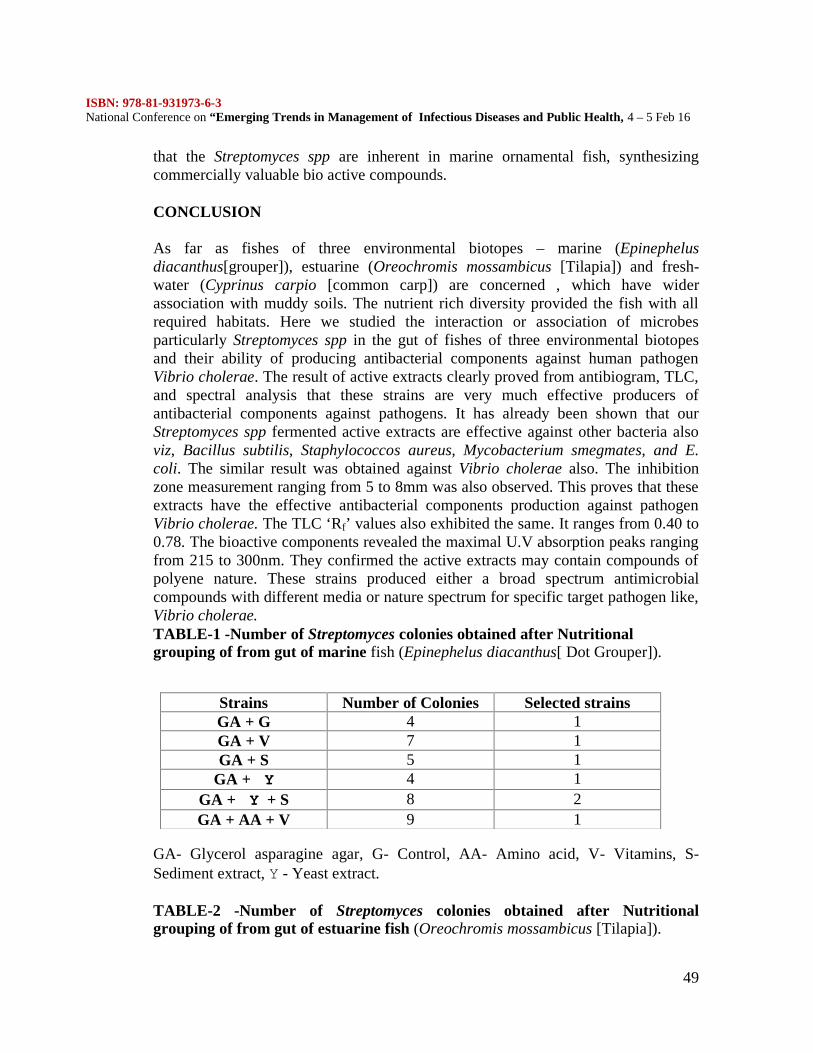
ISBN: 978-81-931973-6-3National Conference on “Emerging Trends in Management of Infectious Diseases and Public Health, 4 – 5 Feb 16
49
that the Streptomyces spp are inherent in marine ornamental fish, synthesizingcommercially valuable bio active compounds.
CONCLUSION
As far as fishes of three environmental biotopes – marine (Epinephelusdiacanthus[grouper]), estuarine (Oreochromis mossambicus [Tilapia]) and fresh-water (Cyprinus carpio [common carp]) are concerned , which have widerassociation with muddy soils. The nutrient rich diversity provided the fish with allrequired habitats. Here we studied the interaction or association of microbesparticularly Streptomyces spp in the gut of fishes of three environmental biotopesand their ability of producing antibacterial components against human pathogenVibrio cholerae. The result of active extracts clearly proved from antibiogram, TLC,and spectral analysis that these strains are very much effective producers ofantibacterial components against pathogens. It has already been shown that ourStreptomyces spp fermented active extracts are effective against other bacteria alsoviz, Bacillus subtilis, Staphylococcos aureus, Mycobacterium smegmates, and E.coli. The similar result was obtained against Vibrio cholerae also. The inhibitionzone measurement ranging from 5 to 8mm was also observed. This proves that theseextracts have the effective antibacterial components production against pathogenVibrio cholerae. The TLC ‘Rf’ values also exhibited the same. It ranges from 0.40 to0.78. The bioactive components revealed the maximal U.V absorption peaks rangingfrom 215 to 300nm. They confirmed the active extracts may contain compounds ofpolyene nature. These strains produced either a broad spectrum antimicrobialcompounds with different media or nature spectrum for specific target pathogen like,Vibrio cholerae.TABLE-1 -Number of Streptomyces colonies obtained after Nutritionalgrouping of from gut of marine fish (Epinephelus diacanthus[ Dot Grouper]).
GA- Glycerol asparagine agar, G- Control, AA- Amino acid, V- Vitamins, S-Sediment extract, Y - Yeast extract.
TABLE-2 -Number of Streptomyces colonies obtained after Nutritionalgrouping of from gut of estuarine fish (Oreochromis mossambicus [Tilapia]).
Strains Number of Colonies Selected strainsGA + G 4 1GA + V 7 1GA + S 5 1
GA + Y 4 1GA + Y + S 8 2GA + AA + V 9 1
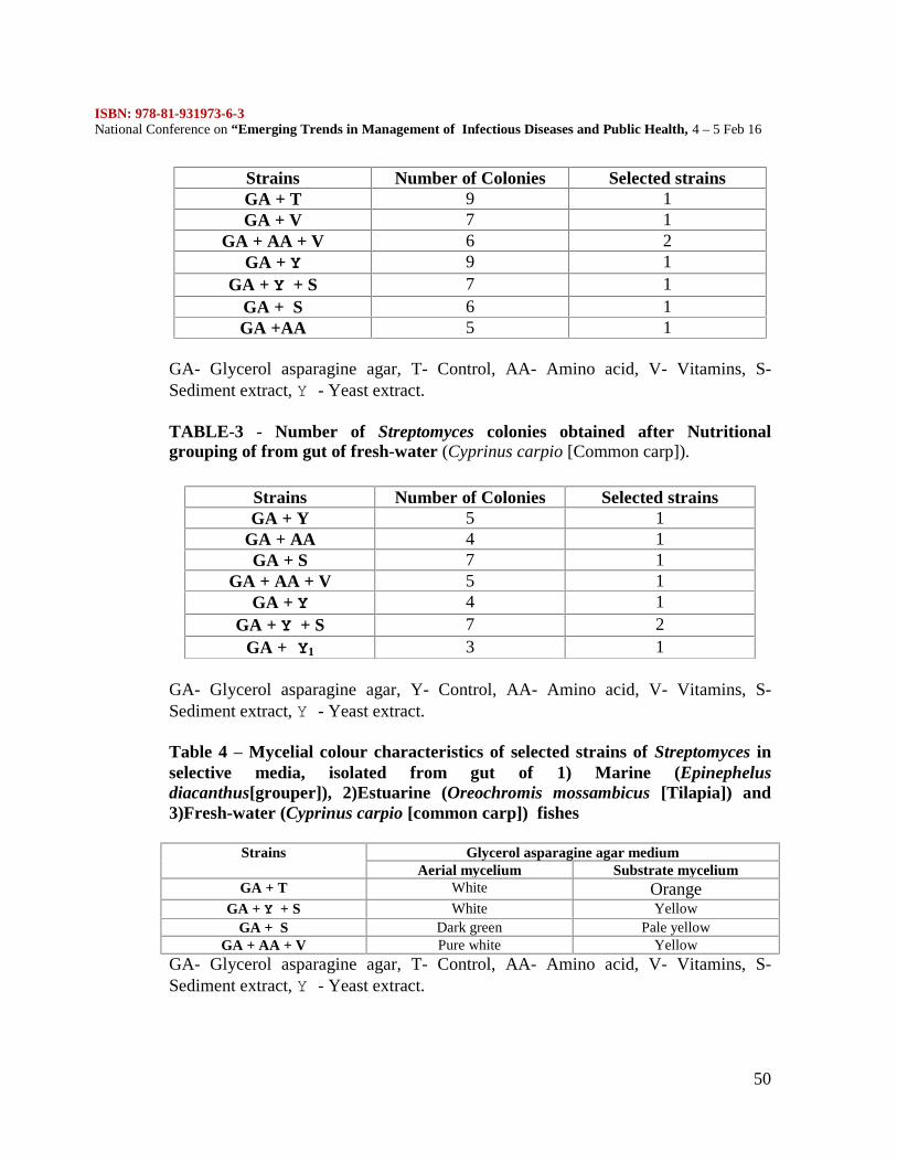
ISBN: 978-81-931973-6-3National Conference on “Emerging Trends in Management of Infectious Diseases and Public Health, 4 – 5 Feb 16
50
GA- Glycerol asparagine agar, T- Control, AA- Amino acid, V- Vitamins, S-Sediment extract, Y - Yeast extract.
TABLE-3 - Number of Streptomyces colonies obtained after Nutritionalgrouping of from gut of fresh-water (Cyprinus carpio [Common carp]).
GA- Glycerol asparagine agar, Y- Control, AA- Amino acid, V- Vitamins, S-Sediment extract, Y - Yeast extract.
Table 4 – Mycelial colour characteristics of selected strains of Streptomyces inselective media, isolated from gut of 1) Marine (Epinephelusdiacanthus[grouper]), 2)Estuarine (Oreochromis mossambicus [Tilapia]) and3)Fresh-water (Cyprinus carpio [common carp]) fishes
Strains Glycerol asparagine agar mediumAerial mycelium Substrate mycelium
GA + T White OrangeGA + Y + S White Yellow
GA + S Dark green Pale yellowGA + AA + V Pure white Yellow
GA- Glycerol asparagine agar, T- Control, AA- Amino acid, V- Vitamins, S-Sediment extract, Y - Yeast extract.
Strains Number of Colonies Selected strainsGA + T 9 1GA + V 7 1
GA + AA + V 6 2GA + Y 9 1
GA + Y + S 7 1GA + S 6 1GA +AA 5 1
Strains Number of Colonies Selected strainsGA + Y 5 1
GA + AA 4 1GA + S 7 1
GA + AA + V 5 1GA + Y 4 1
GA + Y + S 7 2GA + Y1 3 1
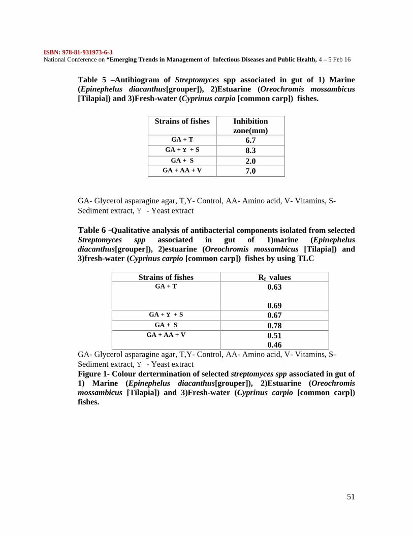
ISBN: 978-81-931973-6-3National Conference on “Emerging Trends in Management of Infectious Diseases and Public Health, 4 – 5 Feb 16
51
Table 5 –Antibiogram of Streptomyces spp associated in gut of 1) Marine(Epinephelus diacanthus[grouper]), 2)Estuarine (Oreochromis mossambicus[Tilapia]) and 3)Fresh-water (Cyprinus carpio [common carp]) fishes.
GA- Glycerol asparagine agar, T,Y- Control, AA- Amino acid, V- Vitamins, S-Sediment extract, Y - Yeast extract
Table 6 -Qualitative analysis of antibacterial components isolated from selectedStreptomyces spp associated in gut of 1)marine (Epinephelusdiacanthus[grouper]), 2)estuarine (Oreochromis mossambicus [Tilapia]) and3)fresh-water (Cyprinus carpio [common carp]) fishes by using TLC
Strains of fishes Rf valuesGA + T 0.63
0.69GA + Y + S 0.67
GA + S 0.78GA + AA + V 0.51
0.46GA- Glycerol asparagine agar, T,Y- Control, AA- Amino acid, V- Vitamins, S-Sediment extract, Y - Yeast extractFigure 1- Colour dertermination of selected streptomyces spp associated in gut of1) Marine (Epinephelus diacanthus[grouper]), 2)Estuarine (Oreochromismossambicus [Tilapia]) and 3)Fresh-water (Cyprinus carpio [common carp])fishes.
Strains of fishes Inhibitionzone(mm)
GA + T 6.7GA + Y + S 8.3
GA + S 2.0GA + AA + V 7.0
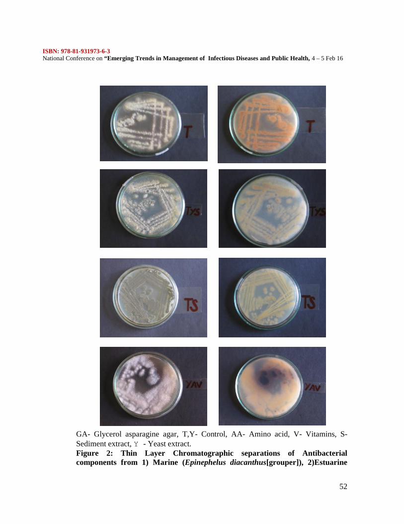
ISBN: 978-81-931973-6-3National Conference on “Emerging Trends in Management of Infectious Diseases and Public Health, 4 – 5 Feb 16
52
GA- Glycerol asparagine agar, T,Y- Control, AA- Amino acid, V- Vitamins, S-Sediment extract, Y - Yeast extract.Figure 2: Thin Layer Chromatographic separations of Antibacterialcomponents from 1) Marine (Epinephelus diacanthus[grouper]), 2)Estuarine
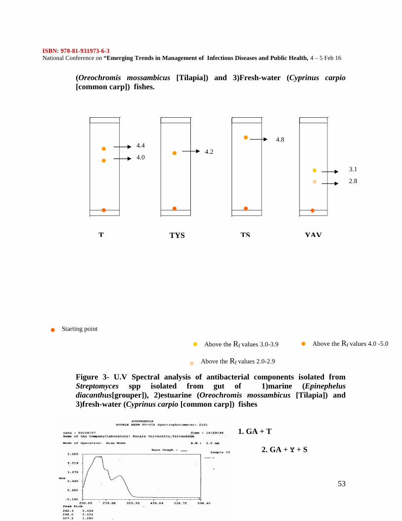
ISBN: 978-81-931973-6-3National Conference on “Emerging Trends in Management of Infectious Diseases and Public Health, 4 – 5 Feb 16
53
(Oreochromis mossambicus [Tilapia]) and 3)Fresh-water (Cyprinus carpio[common carp]) fishes.
Figure 3- U.V Spectral analysis of antibacterial components isolated fromStreptomyces spp isolated from gut of 1)marine (Epinephelusdiacanthus[grouper]), 2)estuarine (Oreochromis mossambicus [Tilapia]) and3)fresh-water (Cyprinus carpio [common carp]) fishes
1. GA + T
2. GA + Y + S
4.4
4.0
T
4.2
TYS
4.8
TS
2.8
3.1
YAV
Starting point
Above the Rf values 2.0-2.9
Above the Rf values 3.0-3.9 Above the Rf values 4.0 -5.0
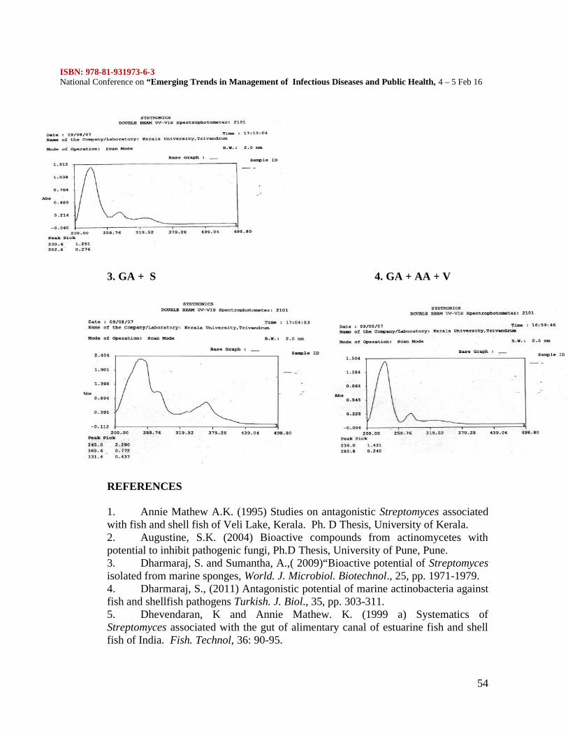
ISBN: 978-81-931973-6-3National Conference on “Emerging Trends in Management of Infectious Diseases and Public Health, 4 – 5 Feb 16
54
3. GA + S 4. GA + AA + V
REFERENCES
1. Annie Mathew A.K. (1995) Studies on antagonistic Streptomyces associatedwith fish and shell fish of Veli Lake, Kerala. Ph. D Thesis, University of Kerala.2. Augustine, S.K. (2004) Bioactive compounds from actinomycetes withpotential to inhibit pathogenic fungi, Ph.D Thesis, University of Pune, Pune.3. Dharmaraj, S. and Sumantha, A.,( 2009)“Bioactive potential of Streptomycesisolated from marine sponges, World. J. Microbiol. Biotechnol., 25, pp. 1971-1979.4. Dharmaraj, S., (2011) Antagonistic potential of marine actinobacteria againstfish and shellfish pathogens Turkish. J. Biol., 35, pp. 303-311.5. Dhevendaran, K and Annie Mathew. K. (1999 a) Systematics ofStreptomyces associated with the gut of alimentary canal of estuarine fish and shellfish of India. Fish. Technol, 36: 90-95.
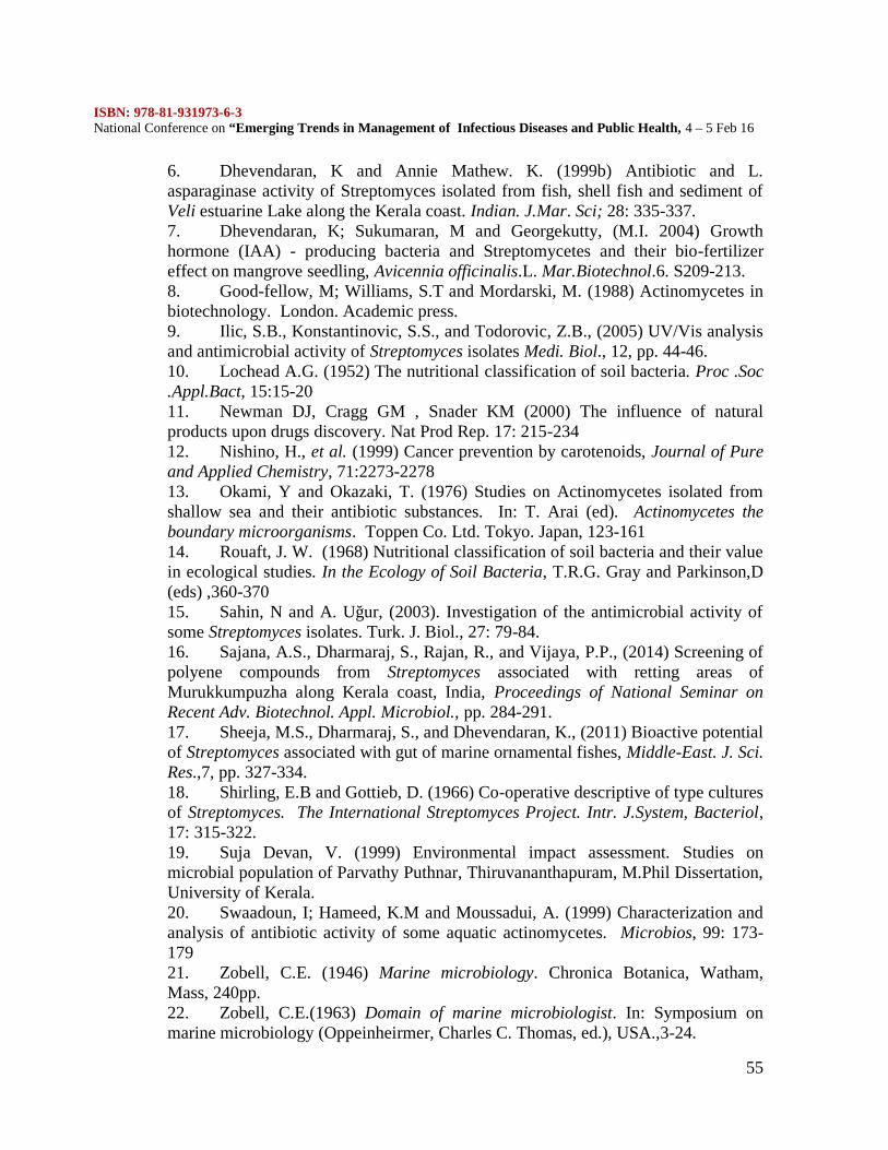
ISBN: 978-81-931973-6-3National Conference on “Emerging Trends in Management of Infectious Diseases and Public Health, 4 – 5 Feb 16
55
6. Dhevendaran, K and Annie Mathew. K. (1999b) Antibiotic and L.asparaginase activity of Streptomyces isolated from fish, shell fish and sediment ofVeli estuarine Lake along the Kerala coast. Indian. J.Mar. Sci; 28: 335-337.7. Dhevendaran, K; Sukumaran, M and Georgekutty, (M.I. 2004) Growthhormone (IAA) - producing bacteria and Streptomycetes and their bio-fertilizereffect on mangrove seedling, Avicennia officinalis.L. Mar.Biotechnol.6. S209-213.8. Good-fellow, M; Williams, S.T and Mordarski, M. (1988) Actinomycetes inbiotechnology. London. Academic press.9. Ilic, S.B., Konstantinovic, S.S., and Todorovic, Z.B., (2005) UV/Vis analysisand antimicrobial activity of Streptomyces isolates Medi. Biol., 12, pp. 44-46.10. Lochead A.G. (1952) The nutritional classification of soil bacteria. Proc .Soc.Appl.Bact, 15:15-2011. Newman DJ, Cragg GM , Snader KM (2000) The influence of naturalproducts upon drugs discovery. Nat Prod Rep. 17: 215-23412. Nishino, H., et al. (1999) Cancer prevention by carotenoids, Journal of Pureand Applied Chemistry, 71:2273-227813. Okami, Y and Okazaki, T. (1976) Studies on Actinomycetes isolated fromshallow sea and their antibiotic substances. In: T. Arai (ed). Actinomycetes theboundary microorganisms. Toppen Co. Ltd. Tokyo. Japan, 123-16114. Rouaft, J. W. (1968) Nutritional classification of soil bacteria and their valuein ecological studies. In the Ecology of Soil Bacteria, T.R.G. Gray and Parkinson,D(eds) ,360-37015. Sahin, N and A. Uğur, (2003). Investigation of the antimicrobial activity ofsome Streptomyces isolates. Turk. J. Biol., 27: 79-84.16. Sajana, A.S., Dharmaraj, S., Rajan, R., and Vijaya, P.P., (2014) Screening ofpolyene compounds from Streptomyces associated with retting areas ofMurukkumpuzha along Kerala coast, India, Proceedings of National Seminar onRecent Adv. Biotechnol. Appl. Microbiol., pp. 284-291.17. Sheeja, M.S., Dharmaraj, S., and Dhevendaran, K., (2011) Bioactive potentialof Streptomyces associated with gut of marine ornamental fishes, Middle-East. J. Sci.Res.,7, pp. 327-334.18. Shirling, E.B and Gottieb, D. (1966) Co-operative descriptive of type culturesof Streptomyces. The International Streptomyces Project. Intr. J.System, Bacteriol,17: 315-322.19. Suja Devan, V. (1999) Environmental impact assessment. Studies onmicrobial population of Parvathy Puthnar, Thiruvananthapuram, M.Phil Dissertation,University of Kerala.20. Swaadoun, I; Hameed, K.M and Moussadui, A. (1999) Characterization andanalysis of antibiotic activity of some aquatic actinomycetes. Microbios, 99: 173-17921. Zobell, C.E. (1946) Marine microbiology. Chronica Botanica, Watham,Mass, 240pp.22. Zobell, C.E.(1963) Domain of marine microbiologist. In: Symposium onmarine microbiology (Oppeinheirmer, Charles C. Thomas, ed.), USA.,3-24.
ISBN: 978-81-931973-6-3National Conference on “Emerging Trends in Management of Infectious Diseases and Public Health, 4 – 5 Feb 16
56
ANTIPLASMODIAL PHYTOTHERAPY AND DRUG DISCOVERY- ANOVERVIEW
R. MADHANAGOPAL, M.R. ASIYA MARIYAM AND A. KALIDOSS*
P.G and Research Department of BiotechnologyMohamed Sathak College of Arts and Science
Sholinganallur, Chennai- 600 119
*Corresponding Author: [email protected]
ABSTRACT
Malaria is the most destructive and dangerous parasitic infection in many tropical andsubtropical countries. The burden of this disease is getting worse, mainly due to theincreasing resistance of Plasmodium falciparum against the widely available antimalarialdrugs. There is an urgent need for new, more affordable and accessible antimalarial agentspossessing original modes of action. Natural products have played a dominant role in thedrug discovery to treat human diseases and this fact anticipates that new antimalarial drugsmay certainly emerge from tropical plant sources. This present review covers a list ofantimalarial plants, natural compounds from plants with antiplasmodial and antimalarialproperties, besides the majority of papers describing antiplasmodial crude extracts publishedin the last five years and prospecting of natural compounds. In addition, some perspectivesand remarks on the development of new drugs and phytomedicines for malaria.
INTRODUCTION
Malaria is a foremost health problem in tropical, subtropical world and to the world whichare socioeconomically backward. In India, 95% population resides in malaria endemicregions and it is major public health problem in most part of the country. Malaria is aparasitic protozoan disease, caused by parasites of Plasmodium genus. About 92% malariacases and 97% of deaths due to malaria is reported from North-eastern states ,Chhattisgarh,Jharkhand, Madhya Pradesh, Orrissa, Andhra Pradesh, Maharashtra, Gujarat, Rajasthan,West Bengal and Karnataka (1). Around 2.5 million malaria cases are reported annuallyfrom South East Asian Nations (2) of which India alone contributes 76% of in the globalmalaria incidences (WHO, 2012). Its general symptoms are high fever come with chills,intermittent fever, and very often headache as well as trembling as major symptom. Overmillennia, its victims have included Neolithic dwellers, early Chinese and Greeks, princesand paupers. In the 20th century alone, malaria claimed between 150 million and 300 millionlives, accounting for 2 to 5 percent of all deaths (3). Although its chief sufferers today arethe poor of sub Saharan Africa, Asia, the Amazon basin, and other tropical regions, 40percent of the world's population still lives in areas where malaria is transmitted. Malaria iscaused by single-celled protozoan parasites called Plasmodium and transmitted to manthrough the Anopheles mosquito. It is one of the major fatal diseases in the world, especiallyin the tropics, and is endemic in some 102 countries, with more than half of the worldpopulation at risk with fatality rates being extremely high among young children below 5years of age.(4) The World Health Organization estimates that there are between 300 and
ISBN: 978-81-931973-6-3National Conference on “Emerging Trends in Management of Infectious Diseases and Public Health, 4 – 5 Feb 16
57
500 million new cases of malaria worldwide, every year, mostly in Africa, Asia, SouthPacific Islands and South America, which causes at least 1 million deaths annually. In spiteof control programs in many countries, there has been very little improvement in the controlof malaria, and infections can reduce the effectiveness of labor and can lead to botheconomic and human losses. Control of malaria is complex because of the appearance ofdrug resistant strains of Plasmodium and with the discovery that man becomes infested withspecies of simian (monkey) malaria (4) At the same time, the Anopheles mosquitoes havedeveloped resistance to many insecticides(5).
Malaria is an infectious disease endemic throughout tropical countries. Malaria is alsoprevalent in subtropical areas, where the disease is contagious affecting both indigenouspopulation and travelers (6). Malaria is caused by Plasmodium parasites that are transmittedthrough the bite of Anopheles mosquitoes and have a life cycle in mosquito and human hosts(6). Of all parasite types, Plasmodium falciparum (P. falciparum) is the most dangerousPlasmodium, causing human malaria with a mortality of 1–2 million people annually.According to surveys conducted between 1900 and 2008 in 2,366 locations in Indonesia,four species of Plasmodium may infect humans, P. falciparum, P. vivax, P. malariae and P.ovale. P. falciparum is the most common parasite that is contagious in Indonesia, withprevalence rates of 33% in Papua, 29% in Lesser Sundas and 21% in Sumatra (7). Findingsof studies performed in other parts of Indonesia, including the Thousand Island district (8),Nias Island (9), Sumba Island (10) and Aceh (11), have shown that P. falciparum was themost frequent parasite that caused malaria.
APPLICATION OF PLANTS IN THE TREATMENT OF MALARIA
Apart from the pharmacological treatment, various options are being used since ancienttimesfor many health ailments. Nearly 80% of the global population still depends uponthe herbal drugs for their health care. In India, the use of several medicinal plants to curespecific ailments has been practiced since ancient times. Various cultural traditions areassociated with use of wild plants as medicinal herbs. This medico-lore is passed overgenerations traditionally all over the world. Reliance on plants is primarily due to theirsafety, effectiveness, cultural preferences, Inexpensiveness and abundant availabilityall the time. The medicinal virtues of plants are identified by instinct/intuition or bytrial and errors. Globally, traditional healers are using various medicinal plants for thetreatment of malaria; however, this practice is not really completely recognized bymodern medical Science. Knowledge about traditional medicinal practices and plants iscurrently transmitted from generation to generation principally by word of mouth. Largenumber of plant species has been identified as anti-malarial medicinal plants.
Medicinal plants have been used as traditional medicines for hundreds of years. The firstantimalarial agents, including quinine as well as the next generation of antimalarial agents,such as lapachol and artemisinin, have been isolated from plants (12,13). Traditionalmedicines have a high potential as novel drug candidates, can provide valuable clues to findnovel drugs and may shift the drug discovery paradigm from ‘finding newentity drugs’ to‘combining existing agents’ (14,15). Certain approaches in finding novel drugs use plantsthat are consumed by particular groups (16). Primates have a close similarity with humanphysiology and also have similar characteristics of disease as humans. Humans use drugs to
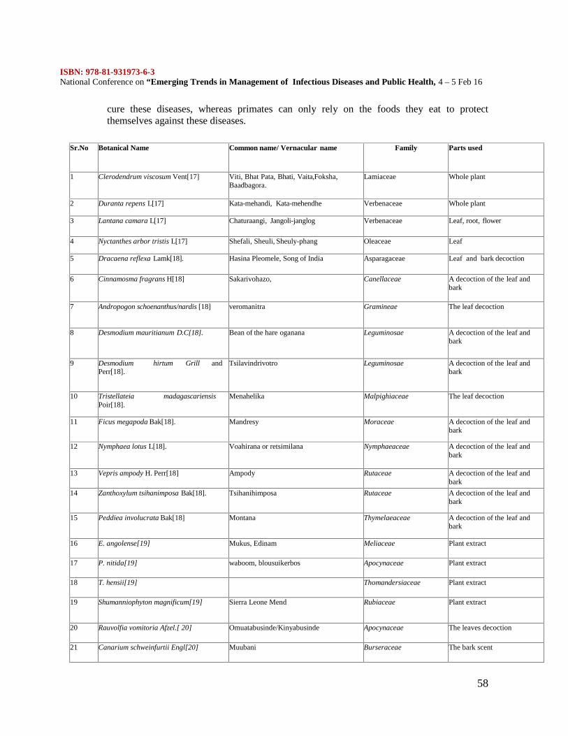
ISBN: 978-81-931973-6-3National Conference on “Emerging Trends in Management of Infectious Diseases and Public Health, 4 – 5 Feb 16
58
cure these diseases, whereas primates can only rely on the foods they eat to protectthemselves against these diseases.
Sr.No Botanical Name Common name/ Vernacular name Family Parts used
1 Clerodendrum viscosum Vent[17] Viti, Bhat Pata, Bhati, Vaita,Foksha,Baadbagora.
Lamiaceae Whole plant
2 Duranta repens L[17] Kata-mehandi, Kata-mehendhe Verbenaceae Whole plant
3 Lantana camara L[17] Chaturaangi, Jangoli-janglog Verbenaceae Leaf, root, flower
4 Nyctanthes arbor tristis L[17] Shefali, Sheuli, Sheuly-phang Oleaceae Leaf
5 Dracaena reflexa Lamk[18]. Hasina Pleomele, Song of India Asparagaceae Leaf and bark decoction
6 Cinnamosma fragrans H[18] Sakarivohazo, Canellaceae A decoction of the leaf andbark
7 Andropogon schoenanthus/nardis [18] veromanitra Gramineae The leaf decoction
8 Desmodium mauritianum D.C[18]. Bean of the hare oganana Leguminosae A decoction of the leaf andbark
9 Desmodium hirtum Grill andPerr[18].
Tsilavindrivotro Leguminosae A decoction of the leaf andbark
10 Tristellateia madagascariensisPoir[18].
Menahelika Malpighiaceae The leaf decoction
11 Ficus megapoda Bak[18]. Mandresy Moraceae A decoction of the leaf andbark
12 Nymphaea lotus L[18]. Voahirana or retsimilana Nymphaeaceae A decoction of the leaf andbark
13 Vepris ampody H. Perr[18] Ampody Rutaceae A decoction of the leaf andbark
14 Zanthoxylum tsihanimposa Bak[18]. Tsihanihimposa Rutaceae A decoction of the leaf andbark
15 Peddiea involucrata Bak[18] Montana Thymelaeaceae A decoction of the leaf andbark
16 E. angolense[19] Mukus, Edinam Meliaceae Plant extract
17 P. nitida[19] waboom, blousuikerbos Apocynaceae Plant extract
18 T. hensii[19] Thomandersiaceae Plant extract
19 Shumanniophyton magnificum[19] Sierra Leone Mend Rubiaceae Plant extract
20 Rauvolfia vomitoria Afzel.[ 20] Omuatabusinde/Kinyabusinde Apocynaceae The leaves decoction
21 Canarium schweinfurtii Engl[20] Muubani Burseraceae The bark scent
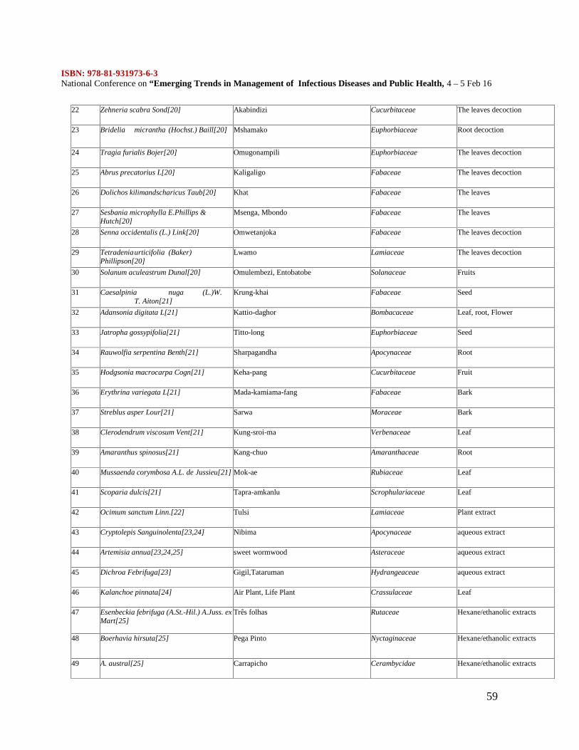
ISBN: 978-81-931973-6-3National Conference on “Emerging Trends in Management of Infectious Diseases and Public Health, 4 – 5 Feb 16
59
22 Zehneria scabra Sond[20] Akabindizi Cucurbitaceae The leaves decoction
23 Bridelia micrantha (Hochst.) Baill[20] Mshamako Euphorbiaceae Root decoction
24 Tragia furialis Bojer[20] Omugonampili Euphorbiaceae The leaves decoction
25 Abrus precatorius L[20] Kaligaligo Fabaceae The leaves decoction
26 Dolichos kilimandscharicus Taub[20] Khat Fabaceae The leaves
27 Sesbania microphylla E.Phillips &Hutch[20]
Msenga, Mbondo Fabaceae The leaves
28 Senna occidentalis (L.) Link[20] Omwetanjoka Fabaceae The leaves decoction
29 Tetradeniaurticifolia (Baker)Phillipson[20]
Lwamo Lamiaceae The leaves decoction
30 Solanum aculeastrum Dunal[20] Omulembezi, Entobatobe Solanaceae Fruits
31 Caesalpinia nuga (L.)W.T. Aiton[21]
Krung-khai Fabaceae Seed
32 Adansonia digitata L[21] Kattio-daghor Bombacaceae Leaf, root, Flower
33 Jatropha gossypifolia[21] Titto-long Euphorbiaceae Seed
34 Rauwolfia serpentina Benth[21] Sharpagandha Apocynaceae Root
35 Hodgsonia macrocarpa Cogn[21] Keha-pang Cucurbitaceae Fruit
36 Erythrina variegata L[21] Mada-kamiama-fang Fabaceae Bark
37 Streblus asper Lour[21] Sarwa Moraceae Bark
38 Clerodendrum viscosum Vent[21] Kung-sroi-ma Verbenaceae Leaf
39 Amaranthus spinosus[21] Kang-chuo Amaranthaceae Root
40 Mussaenda corymbosa A.L. de Jussieu[21] Mok-ae Rubiaceae Leaf
41 Scoparia dulcis[21] Tapra-amkanlu Scrophulariaceae Leaf
42 Ocimum sanctum Linn.[22] Tulsi Lamiaceae Plant extract
43 Cryptolepis Sanguinolenta[23,24] Nibima Apocynaceae aqueous extract
44 Artemisia annua[23,24,25] sweet wormwood Asteraceae aqueous extract
45 Dichroa Febrifuga[23] Gigil,Tataruman Hydrangeaceae aqueous extract
46 Kalanchoe pinnata[24] Air Plant, Life Plant Crassulaceae Leaf
47 Esenbeckia febrifuga (A.St.-Hil.) A.Juss. exMart[25]
Três folhas Rutaceae Hexane/ethanolic extracts
48 Boerhavia hirsuta[25] Pega Pinto Nyctaginaceae Hexane/ethanolic extracts
49 A. austral[25] Carrapicho Cerambycidae Hexane/ethanolic extracts
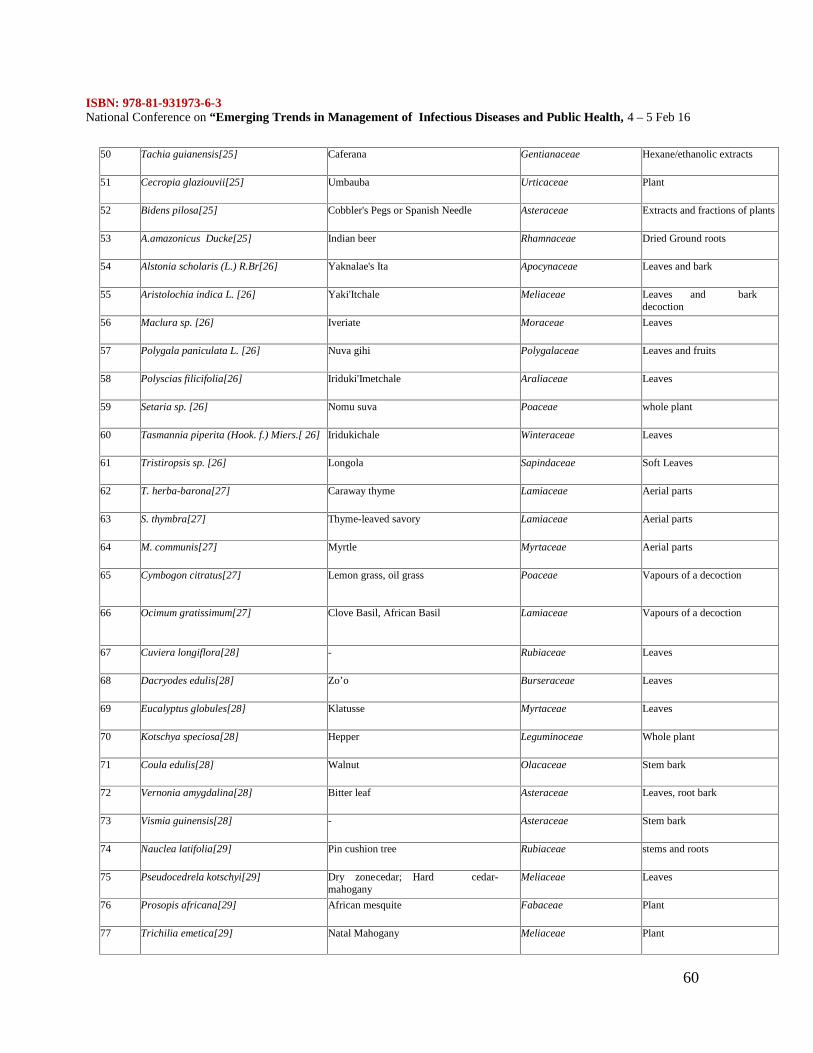
ISBN: 978-81-931973-6-3National Conference on “Emerging Trends in Management of Infectious Diseases and Public Health, 4 – 5 Feb 16
60
50 Tachia guianensis[25] Caferana Gentianaceae Hexane/ethanolic extracts
51 Cecropia glaziouvii[25] Umbauba Urticaceae Plant
52 Bidens pilosa[25] Cobbler's Pegs or Spanish Needle Asteraceae Extracts and fractions of plants
53 A.amazonicus Ducke[25] Indian beer Rhamnaceae Dried Ground roots
54 Alstonia scholaris (L.) R.Br[26] Yaknalae's Ita Apocynaceae Leaves and bark
55 Aristolochia indica L. [26] Yaki'Itchale Meliaceae Leaves and barkdecoction
56 Maclura sp. [26] Iveriate Moraceae Leaves
57 Polygala paniculata L. [26] Nuva gihi Polygalaceae Leaves and fruits
58 Polyscias filicifolia[26] Iriduki'Imetchale Araliaceae Leaves
59 Setaria sp. [26] Nomu suva Poaceae whole plant
60 Tasmannia piperita (Hook. f.) Miers.[ 26] Iridukichale Winteraceae Leaves
61 Tristiropsis sp. [26] Longola Sapindaceae Soft Leaves
62 T. herba-barona[27] Caraway thyme Lamiaceae Aerial parts
63 S. thymbra[27] Thyme-leaved savory Lamiaceae Aerial parts
64 M. communis[27] Myrtle Myrtaceae Aerial parts
65 Cymbogon citratus[27] Lemon grass, oil grass Poaceae Vapours of a decoction
66 Ocimum gratissimum[27] Clove Basil, African Basil Lamiaceae Vapours of a decoction
67 Cuviera longiflora[28] - Rubiaceae Leaves
68 Dacryodes edulis[28] Zo’o Burseraceae Leaves
69 Eucalyptus globules[28] Klatusse Myrtaceae Leaves
70 Kotschya speciosa[28] Hepper Leguminoceae Whole plant
71 Coula edulis[28] Walnut Olacaceae Stem bark
72 Vernonia amygdalina[28] Bitter leaf Asteraceae Leaves, root bark
73 Vismia guinensis[28] - Asteraceae Stem bark
74 Nauclea latifolia[29] Pin cushion tree Rubiaceae stems and roots
75 Pseudocedrela kotschyi[29] Dry zonecedar; Hard cedar-mahogany
Meliaceae Leaves
76 Prosopis africana[29] African mesquite Fabaceae Plant
77 Trichilia emetica[29] Natal Mahogany Meliaceae Plant
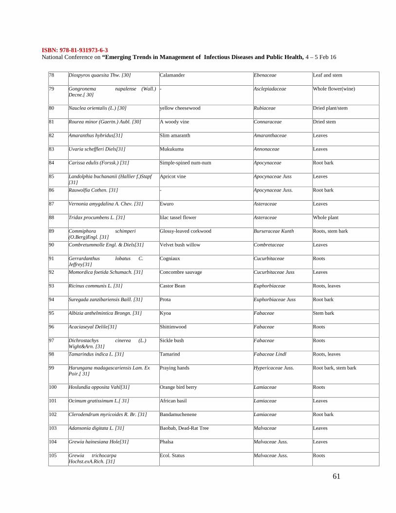
ISBN: 978-81-931973-6-3National Conference on “Emerging Trends in Management of Infectious Diseases and Public Health, 4 – 5 Feb 16
61
78 Diospyros quaesita Thw. [30] Calamander Ebenaceae Leaf and stem
79 Gongronema napalense (Wall.)Decne.[ 30]
- Asclepiadaceae Whole flower(wine)
80 Nauclea orientalis (L.) [30] yellow cheesewood Rubiaceae Dried plant/stem
81 Rourea minor (Gaertn.) Aubl. [30] A woody vine Connaraceae Dried stem
82 Amaranthus hybridus[31] Slim amaranth Amaranthaceae Leaves
83 Uvaria scheffleri Diels[31] Mukukuma Annonaceae Leaves
84 Carissa edulis (Forssk.) [31] Simple-spined num-num Apocynaceae Root bark
85 Landolphia buchananii (Hallier f.)Stapf[31]
Apricot vine Apocynaceae Juss Leaves
86 Rauwolfia Cothen. [31] - Apocynaceae Juss. Root bark
87 Vernonia amygdalina A. Chev. [31] Ewuro Asteraceae Leaves
88 Tridax procumbens L. [31] lilac tassel flower Asteraceae Whole plant
89 Commiphora schimperi(O.Berg)Engl. [31]
Glossy-leaved corkwood Burseraceae Kunth Roots, stem bark
90 Combretummolle Engl. & Diels[31] Velvet bush willow Combretaceae Leaves
91 Gerrardanthus lobatus C.Jeffrey[31]
Cogniaux Cucurbitaceae Roots
92 Momordica foetida Schumach. [31] Concombre sauvage Cucurbitaceae Juss Leaves
93 Ricinus communis L. [31] Castor Bean Euphorbiaceae Roots, leaves
94 Suregada zanzibariensis Baill. [31] Prota Euphorbiaceae Juss Root bark
95 Albizia anthelmintica Brongn. [31] Kyoa Fabaceae Stem bark
96 Acaciaseyal Delile[31] Shittimwood Fabaceae Roots
97 Dichrostachys cinerea (L.)Wight&Arn. [31]
Sickle bush Fabaceae Roots
98 Tamarindus indica L. [31] Tamarind Fabaceae Lindl Roots, leaves
99 Harungana madagascariensis Lam. ExPoir.[ 31]
Praying hands Hypericaceae Juss. Root bark, stem bark
100 Hoslundia opposita Vahl[31] Orange bird berry Lamiaceae Roots
101 Ocimum gratissimum L.[ 31] African basil Lamiaceae Leaves
102 Clerodendrum myricoides R. Br. [31] Bandamuchenene Lamiaceae Root bark
103 Adansonia digitata L. [31] Baobab, Dead-Rat Tree Malvaceae Leaves
104 Grewia hainesiana Hole[31] Phalsa Malvaceae Juss. Leaves
105 Grewia trichocarpaHochst.exA.Rich. [31]
Ecol. Status Malvaceae Juss. Roots
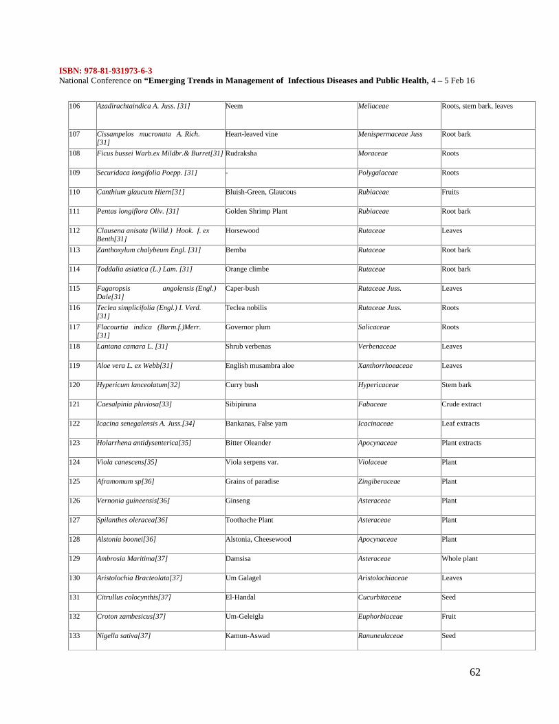
ISBN: 978-81-931973-6-3National Conference on “Emerging Trends in Management of Infectious Diseases and Public Health, 4 – 5 Feb 16
62
106 Azadirachtaindica A. Juss. [31] Neem Meliaceae Roots, stem bark, leaves
107 Cissampelos mucronata A. Rich.[31]
Heart-leaved vine Menispermaceae Juss Root bark
108 Ficus bussei Warb.ex Mildbr.& Burret[31] Rudraksha Moraceae Roots
109 Securidaca longifolia Poepp. [31] - Polygalaceae Roots
110 Canthium glaucum Hiern[31] Bluish-Green, Glaucous Rubiaceae Fruits
111 Pentas longiflora Oliv. [31] Golden Shrimp Plant Rubiaceae Root bark
112 Clausena anisata (Willd.) Hook. f. exBenth[31]
Horsewood Rutaceae Leaves
113 Zanthoxylum chalybeum Engl. [31] Bemba Rutaceae Root bark
114 Toddalia asiatica (L.) Lam. [31] Orange climbe Rutaceae Root bark
115 Fagaropsis angolensis (Engl.)Dale[31]
Caper-bush Rutaceae Juss. Leaves
116 Teclea simplicifolia (Engl.) I. Verd.[31]
Teclea nobilis Rutaceae Juss. Roots
117 Flacourtia indica (Burm.f.)Merr.[31]
Governor plum Salicaceae Roots
118 Lantana camara L. [31] Shrub verbenas Verbenaceae Leaves
119 Aloe vera L. ex Webb[31] English musambra aloe Xanthorrhoeaceae Leaves
120 Hypericum lanceolatum[32] Curry bush Hypericaceae Stem bark
121 Caesalpinia pluviosa[33] Sibipiruna Fabaceae Crude extract
122 Icacina senegalensis A. Juss.[34] Bankanas, False yam Icacinaceae Leaf extracts
123 Holarrhena antidysenterica[35] Bitter Oleander Apocynaceae Plant extracts
124 Viola canescens[35] Viola serpens var. Violaceae Plant
125 Aframomum sp[36] Grains of paradise Zingiberaceae Plant
126 Vernonia guineensis[36] Ginseng Asteraceae Plant
127 Spilanthes oleracea[36] Toothache Plant Asteraceae Plant
128 Alstonia boonei[36] Alstonia, Cheesewood Apocynaceae Plant
129 Ambrosia Maritima[37] Damsisa Asteraceae Whole plant
130 Aristolochia Bracteolata[37] Um Galagel Aristolochiaceae Leaves
131 Citrullus colocynthis[37] El-Handal Cucurbitaceae Seed
132 Croton zambesicus[37] Um-Geleigla Euphorbiaceae Fruit
133 Nigella sativa[37] Kamun-Aswad Ranuneulaceae Seed
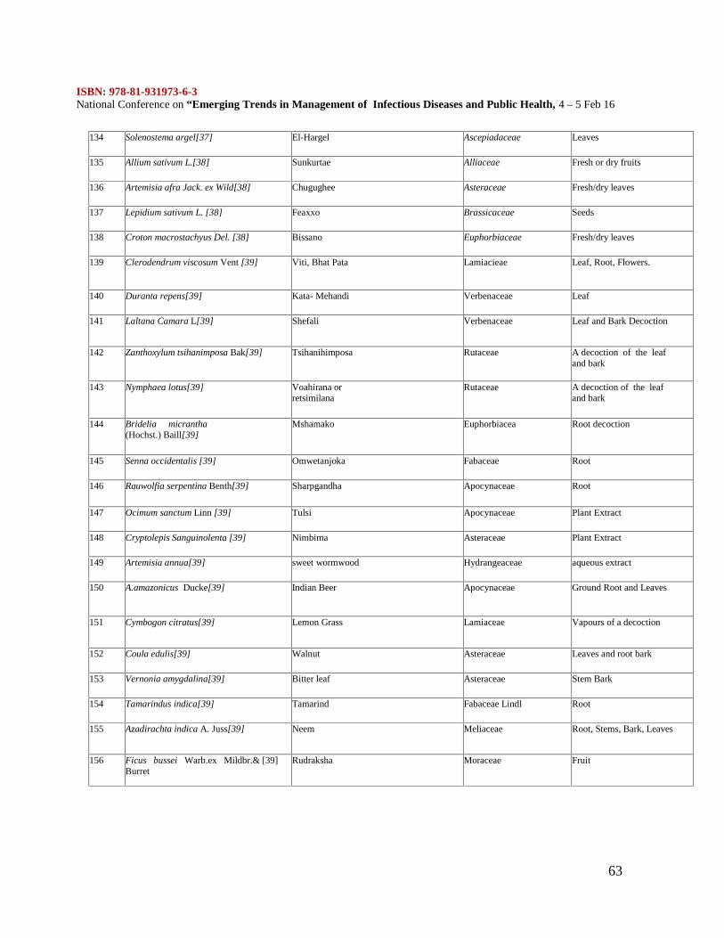
ISBN: 978-81-931973-6-3National Conference on “Emerging Trends in Management of Infectious Diseases and Public Health, 4 – 5 Feb 16
63
134 Solenostema argel[37] El-Hargel Ascepiadaceae Leaves
135 Allium sativum L.[38] Sunkurtae Alliaceae Fresh or dry fruits
136 Artemisia afra Jack. ex Wild[38] Chugughee Asteraceae Fresh/dry leaves
137 Lepidium sativum L. [38] Feaxxo Brassicaceae Seeds
138 Croton macrostachyus Del. [38] Bissano Euphorbiaceae Fresh/dry leaves
139 Clerodendrum viscosum Vent [39] Viti, Bhat Pata Lamiacieae Leaf, Root, Flowers.
140 Duranta repens[39] Kata- Mehandi Verbenaceae Leaf
141 Laltana Camara L[39] Shefali Verbenaceae Leaf and Bark Decoction
142 Zanthoxylum tsihanimposa Bak[39] Tsihanihimposa Rutaceae A decoction of the leafand bark
143 Nymphaea lotus[39] Voahirana orretsimilana
Rutaceae A decoction of the leafand bark
144 Bridelia micrantha(Hochst.) Baill[39]
Mshamako Euphorbiacea Root decoction
145 Senna occidentalis [39] Omwetanjoka Fabaceae Root
146 Rauwolfia serpentina Benth[39] Sharpgandha Apocynaceae Root
147 Ocimum sanctum Linn [39] Tulsi Apocynaceae Plant Extract
148 Cryptolepis Sanguinolenta [39] Nimbima Asteraceae Plant Extract
149 Artemisia annua[39] sweet wormwood Hydrangeaceae aqueous extract
150 A.amazonicus Ducke[39] Indian Beer Apocynaceae Ground Root and Leaves
151 Cymbogon citratus[39] Lemon Grass Lamiaceae Vapours of a decoction
152 Coula edulis[39] Walnut Asteraceae Leaves and root bark
153 Vernonia amygdalina[39] Bitter leaf Asteraceae Stem Bark
154 Tamarindus indica[39] Tamarind Fabaceae Lindl Root
155 Azadirachta indica A. Juss[39] Neem Meliaceae Root, Stems, Bark, Leaves
156 Ficus bussei Warb.ex Mildbr.& [39]Burret
Rudraksha Moraceae Fruit
ISBN: 978-81-931973-6-3National Conference on “Emerging Trends in Management of Infectious Diseases and Public Health, 4 – 5 Feb 16
64
LIST OF PLANTS IN APPLIED AS ANTIMALARIAL AGENTSPLANT SOURCE FOR ANTIMALARIAL DRUG
One can make an argument that biologically derived secondary metabolites and syntheticcompounds derived from them perform better as drugs than do randomly synthesizedcompounds. Primary and secondary metabolites, receptors, enzymes, transporters andregulatory proteins originated from a limited number of parent molecules. These originatingmolecules were present in primitive life forms and therefore coevolved to interact with oneanother, thus granting direct ecological benefit to the producing organism, whether incompetition for resources, avoiding predation or combating pathogens. Although adivergence in function and structure has subsequently occurred, some structural relationshipstill remains that make natural products, on average, better ligands for biological targets thanrandomly synthesized compounds [39, 40]. Biosynthesis uses a very parsimonious set ofbuilding blocks. It diversifies by taking its limited battery of building blocks and distributesthem into many different pathways. Nature has a tendency to form oxides by hydrolysis orabstraction of oxygen from organic compounds by enzymes that delicately achieve siteselective C–H activation to introduce oxygen and discriminate between numerous functionalgroups at different oxidation levels.
In contrast, medicinal chemistry focuses on nitrogen and often includes additional atomssuch as sulfur and halogens that are relatively rare in nature [41]. Generally, naturalmetabolites have a high ‘sterical complexity’. This is an evident outcome of the spatialdimensionality and the chirality of the enzymes involved in biosynthesis, their moleculartargets and the metabolites themselves [42]. Bioactive compounds share pharmacophoricfeatures, and natural products provide a rich source of potentially attractive scaffolds andmolecular building blocks for synthesis. Since natural compounds have not evolved astherapeutic agents, sometimes their chemical structure must be further improved, whether interms of efficacy and selectivity for the target or achieving optimal pharmacokinetic andpharmacodynamic properties. The numerous ring system scaffolds have no orthologs in thedrug synthesis database. Hence, natural products could provide new starting points in drugdiscovery. The vastly unexplored flora and fauna (90 % of total species) could provide othernew leads and drugs for chemotherapy, for the isolation of certain natural products in largeamounts, total synthesis by chemical approaches, or limited scope for chemical modification.
PROSPECTING FOR NATURAL COMPOUNDS
The validation of traditional remedies can be problematic because of the lack of sufficientinformation, documentation and of standardization of extracts to be evaluated, but theseremedies deserve deep and thorough consideration [43]. In spite of these problems, suchmaterials serve as a valuable source for novel compounds. They also conceal an abundantcombination of secondary metabolites which might act in concert to enhance the therapeuticeffect [44, 45]. Because the quantitative and qualitative composition of secondarymetabolites in a plant is notoriously varied, standardization is obligatory. Modern agronomicand plant genetic methods could be harnessed to domesticate some of the most promisingmedicinal plants, so as to ensure a reproducible qualitative and quantitative production ofactive ingredients, in spite of changes in biotic and abiotic constraints. Achieving such goalsmay provide indigenous populations in malarious areas with new commercial crops. Inaddition, the road to antimalarial development could be shortened considerably, since it is
ISBN: 978-81-931973-6-3National Conference on “Emerging Trends in Management of Infectious Diseases and Public Health, 4 – 5 Feb 16
65
much easier to approve the use of extracts than that of their active ingredients. This does notmean that the use of such extracts does not have to pass through rigorous clinical test toassess that toxicity, teratogenicity, mutagenicity etc are not a problem.
CONCLUSION
In this review, the rationale for using natural compounds for drug development has beenpresented with specific consideration of antimalarial drugs. The underlying principles forusing drug combination and the benefits of using multicomponent plant extracts have beenargued. It seems that the scientific and pharmacological justification of these principlescould provide the basis for further development of ethnic/traditional medicine as a valid,cheap and locally available means to treat malaria.
REFERENCE
1. Malaria situation in India. Delhi: National Vector Borne Disease ControlProgramme, Ministry of Health and Family Welfare, Govt. of India. Availablefrom:http://www.nvbdcp.gov.in/Doc/malaria1.pdf2. World Health Organization, World Malaria Report 2012, Genevahttp://www.who.int/malaria/publications/world_malaria_report_2012/en/3. Carter R, Mendis KN. 2002. Evolutionary and historical aspects of the burden ofmalaria. Clinical Microbiology Reviews 15(4):564594.
4. Symth JD. Animal Parasitology. Cambridge, England: Cambridge University Press;1994.5. Srisilam K, Veersham C. Antimalarials of plant origin. In: Khanum A, Khan IA,editors. Role of Biotechnology in Medicinal and aromatic plants. Vol. 7. Hyderabad India:Ukaaz Publishers; 2003. pp. 17–47.6. Hyde JE. Drugresistant malaria an insight. FEBS J. 2007;274:4688–4698.7. Elyazar IR, Hay SI, Baird JK. Malaria distr ibution, prevalence, drug resistance andcontrol in Indonesia. Adv Parasitol. 2011;74:41–175.8. Maguire JD, Tuti S, Sismadi P, et al. Endemic coastal malaria in the ThousandIslands District, near Jakarta, Indonesia. Trop Med Int Health. 2005;10:489–496.9. Syafruddin D, Asih PB, Wahid I, et al. Malaria prevalence in Nias District, NorthSumatra Province, Indonesia. Malar J. 2007;6:116.10. Asih PB, Rogers WO, Susanti AI, et al. Seasonal distribution of antimalarial drugresistance alleles on the island of Sumba, Indonesia. Malar J. 2009;8:222.11. Asih PB, Rozi IE, Herdiana, et al. The baseline distribution of malaria in the initialphase of elimination in Sabang Municipality, Aceh Province, Indonesia. Malar J.2012;11:291.12. Wells TN. Natural products as starting points for future antimalarial therapies: goingback to our roots? Malar J. 2011;10(Suppl 1):S3.13. Ginsburg H, Deharo E. A call for using natural compounds in the development ofnew antimalarial treatments an introduction. Malar J. 2011;10(Suppl 1):S1.14. Kong DX, Li XJ, Zhang HY. Where is the hope for drug discovery? Let history tellthe future. Drug Discov Today. 2009;14:115–119.15. Wagner H, Ulrich Merzenich G. Synergy research: approaching a new generation ofphytopharmaceuticals. Phytomedicine. 2009;16:97–110.
ISBN: 978-81-931973-6-3National Conference on “Emerging Trends in Management of Infectious Diseases and Public Health, 4 – 5 Feb 16
66
16. Koshimizu K, Murakami A, Hayashi H, Ohigashi H, Subarnas A, Gurmaya KJ, AliA. Biological activities of edible and medicinal plants from Indonesia and Malaysia.Proceedings of The Tokyo International Forum on Conservation and Sustainable Use ofTropical Bioresources; Tokyo: Japan Bioindustry Association; 1998. pp.203–208.17. WHO (2010). Guidelines for the treatment of Malaria- Second edition. Availablefrom: http://whqlibdoc.who.int/publications/2010/9 789241547925_eng.pdf. Last accessedon 18th Jan 201318. Rahmatullah et al. Folk medicinal uses of verbenaceae family plants in Bangladesh. Afr J Tradit Complement Altern Med 2011; 8(S):53- 65.19. Milijaona Randrianarivelojosia et al. Plants traditionally prescribed to treat tazo(malaria) in the eastern region of Madagascar. Malaria Journal 2003, 2:25.20. Jean Bickii, Guy Raymond Feuya Tchouya, Jean Claude Tchouankeu and EtienneTsamo. Antimalarial activity in crude extracts of some Cameroonian medicinal plants. Afr.J. Trad. CAM. 2007; 4(1):107-1121. Moshi et al. Ethnomedicine of the Kagera Region, north western Tanzania. Part 3:plants used in traditional medicine in Kikuku village, Muleba District. Journal ofEthnobiology and Ethnomedicine 2012, 8:14.22. Mohammed Rahmatullah et. al. Medicinal Plants Used by Various Tribes ofBangladesh for Treatment of Malaria. Malaria Research and Treatment. Volume 2012,Article ID 371798: 1- 5.23. Priyabrata Pattanayak et.al. Ocimum sanctum Linn. A reservoir plant fortherapeutic applications: An Overview. Pharmacogn Rev. 2010 Jan-Jun; 4(7): 95–105.24. Alaíde B. Oliveira et al. Plant-derived antimalarial agents: new leads and efficientphythomedicines. An Acad Bras Cienc 2009;81(4):715-40.25. Merlin L Willcox, Gerard Bodeker. Traditional herbal medicines for malaria. BMJ2004; 329:1156-59.26. Antoniana U Krettli et al. The Search for New Antimalarial Drugs from PlantsUsed to Treat Fever and Malaria or Plants Ramdomly Selected: a Review. Mem InstOswaldo Cruz, Rio de Janeiro .November 2001; 96(8):1033-42.27. Louis R Barrows.et.al. An ethnobotanical survey of medicinal plants used in theeastern highlands of Papua New guinea. Journal of Ethnobiology and Ethnomedicine 2012,8:47.28. Dell’Agli et al. Anti-plasmodial and insecticidal activities of the essential oils ofaromatic plants growing in the Mediterranean area Malaria Journal 2012, 11:219.29. Denis Zofou et al. In Vitro Antiplasmodial Activity and Cytotoxicity of Extracts ofSelectedMedicinal Plants Used by Traditional Healers of Western Cameroon. MalariaResearch and Treatment; Volume 2011.30. Tchacondo et al., Herbal remedies and their adverse effects in tem tribe traditionalmedicine in Togo. Afr J Tradit Complement Altern Med. 2011.8(1):45-60.31. Soejarto et al. An ethnobotanical survey of medicinal plants of Laos toward thediscovery of bioactive compounds as potential candidates for pharmaceutical development.Pharm Biol. 2012 ; 50(1): 42-60.32. Ngutaetal.et.al.Ethnodiagnostic skills of the Digo community for malaria: a leadto traditional bioprospecting. FrontiersinPharmacology. June2011;2: Article 30.33. Zofou et al. Hypericum lanceolatum (Hypericaceae) as a potential source of newanti-malarial agents: a bioassay-guided fractionation of the stem bark. Malaria Journal2011;10:167.
ISBN: 978-81-931973-6-3National Conference on “Emerging Trends in Management of Infectious Diseases and Public Health, 4 – 5 Feb 16
67
34. Kayano et al. In vitro and in vivo assessment of the antimalarial activity ofCaesalpinia pluviosa. Malaria Journal 2011;10:112.35. Sarr et al. Icacina senegalensis (Icacinaceae), traditionally used for the treatment ofmalaria, inhibits in vitro Plasmodium falciparum growth without host cell toxicity. MalariaJournal 2011;10:85.36. Verma et al. Anti-malarial activity of Holarrhena antidysenterica and Violacanescens, plants traditionally used against malaria in the Garhwal region of north-westHimalaya .Malaria Journal 2011;10:20.37. Fokunang et al. Traditional medicine: past, present and future research anddevelopment prospects and integration in the national health system Of cameroon. Afr JTradit Complement Altern Med 2011; 8(3):284-95.38. Ahmed et al. Antiplasmodial Activity of Some Medicinal Plants Used in SudaneseFolk- medicine. Environmental Health Insights 2010; 4:1-6.39. Ganesan A: The impact of natural products upon modern drug discovery. CurrOpin Chem Biol 2008, 12:306317.40. Clardy J, Walsh C: Lessons from natural molecules. Nature 2004, 432:829837.41. Feher M, Schmidt JM: Property distributions: differences between drugs, naturalproducts, and molecules from combinatorial chemistry. J Chem Inf Comput Sci 2003,43:218227.42. Grabowski K, Baringhaus KH, Schneider G: Scaffold diversity of natural products:inspiration for combinatorial library design. Nat Prod Rep 2008, 25:892904.43. Corson TW, Crews CM: Molecular understanding and modern application oftraditional medicines: triumphs and trials. Cell 2007, 130:769774.44. Keith CT, Borisy AA, Stockwell BR: Multicompo nent therapeutics for networkedsystems. Nat Rev Drug Discov 2005, 4:7178.45. Li XJ, Zhang HY: Synergy in natural medicines: implications for drug discovery.Trends Pharmacol Sci 2008, 29:331332.46. Bahekar, Satish and Kale, Ranjana, “ Herbal Plants Used For the Treatment ofMalaria” Journal of Pharmacognosy and Phytochemistry 1, (6), 123-149.
ISBN: 978-81-931973-6-3National Conference on “Emerging Trends in Management of Infectious Diseases and Public Health, 4 – 5 Feb 16
68
Green Synthesis of Silver Nanoparticles using Citrus Medica(Rutaceae) leaf extractand its Antibacterial Efficacy
R.Syeda Ashfianaz*, A.Thangaraj,PG and Research Department of Biochemistry, Mohamed Sathak College of Arts and
Science, Sholinganallur, Chennai-119, Tamilnadu. India.Corresponding Author Email-id : [email protected]
AbstractNanoparticles behave differently than other similarly sized particles. It is therefore necessaryto develop specialized approaches to testing and monitoring their effects. It was observed thatto use natural processes such as use of plants based biological systems makes a fast andconvenient method for the synthesis of silver nanoparticles(AgNPs) and can reduce silver ionsinto AgNPs within minutes of reaction time without using any severe conditions.The presentstudy describes a rapid and green eco-friendly method for the synthesis of AgNPs fromaqueous solution of silver nitrate using Citrus Medica (Rutaceae) leaf extract. UV–visiblespectrum of the aqueous medium containing silver ion showed a peak at 420 nmcorresponding to the plasmon absorbance of AgNPs. Scanning electron microscopy (SEM)micrograph showed formation of well-dispersed crystalline AgNPs in the range of 50-63nm.Electron dispersive spectroscopy (EDX) reveals the elemental composition of producedAgNPs. X-ray diffraction (XRD) spectrum of the AgNPs exhibited 2θ values corresponding tothe AgNPs of crystalline nature. The process of reduction being extracellular and very fastmay lead to the development of an easy bioprocess for synthesis of silver nanoparticles.Antimicrobial activity of the produced silver bionanoparticles was performed by well diffusionmethod against Pseudomonas aeruginosa and Escherichia coli. Further, the produced silvernanoparticles showed an effective antibacterial activity towards the pathogenic bacteria.
Key words: Green synthesis, Citrus Medica, SEM, XRD, EDX, AgNPs, Antibacterialefficacy.
IntroductionNanoparticles are of great scientific interest as they are effectively a bridge between bulkmaterials and atomic molecular structures. The uses for nano sized particles are evenmore remarkable. Different types of nanomaterials like copper, zinc, titanium [1],magnesium, gold [2], alginate [3] and silver have come up but AgNPs have proved to bemost effective as it has good antimicrobial efficacy against bacteria, viruses and othereukaryotic microorganisms [4]. As the AgNPs are playing a major role in the field ofnanotechnology and nanomedicine. The use of nanoparticles derived from noble metalshas spread too many areas including, medical fields, and electronics. The application ofnanoparticles as delivery vehicles for bactericidal agents represents a new paradigm inthe design of antibacterial therapeutics. Numerous microorganisms and plant extractshave been applied to synthesize inorganic nanostructures either intracellularly orextracellularly [5-7].The use of microbial cells for the synthesis of nanosized materialshas recently emerged as a novel approach for the synthesis of metal nanoparticles. Moreand more research inputs are rendered for this and there has been upsurge of interest inthe biological synthesis of nanomaterials by using, microorganisms, several plants in thepast few years, microbes like bacteria [8-9], fungi [10-11] and plant extract. In recent
ISBN: 978-81-931973-6-3National Conference on “Emerging Trends in Management of Infectious Diseases and Public Health, 4 – 5 Feb 16
69
times, AgNPs have been synthesized using various plant extracts such as Hibiscus rosasinensis [12], Svensonia hyderabadensis [13], Trianthema decandra [14], Dioscoreabatatas [15], Catharanthus roseus [16], Bacopa monniera [17], Citrus limon [18],Crossandra infundibuliformis [19], Ficus benghalensis [20], Mentha piperita [21], Piperbetle [22] etc. The metal nanoparticles are extensively used in many biomedical andbioelectrochemical applications due to their extraordinary electrocatalytic activity.Although metal is a poor catalyst in bulk form, nanometer-sized particles can exhibitexcellent catalytic activity due to their relative high surface area to volume ratio andtheir interface dominated properties [23 & 21], which drastically differ from those of thebulk material.
Here, we report an inexpensive, versatile, and very reproducible method for the large-scale synthesis of AgNPs by reduction process using leaf extract of Citrus Medica. Thisleaf extract acts both as reducing and stabilizing agent. Citrus Medica is an importantmedicinal plant, belongs to the family Rutaceae. It is a common constituent of dietworldwide in various forms with considerable effects as potent antibiotic agent, etc.
Materials and methodsCollection and preparation of leaf extractThe collected Citrus Medica leaves were washed 2–3 times with de-ionized water.Aqueous extract of citrus medica leaf was prepared using freshly collected leaves of 20gwere boiled with 100ml of de-ionizeds water at 100°C for 30 minutes.Biosynthesis of silver nanoparticles(AgNPs)In Erlenmeyer flask 2.5 ml of ammonium solution was added with 5 ml of 1 mM
AgNO3 solution, followed by addition of leaf extract 1–10 ml and the final volume wasadjusted to 50 ml by adding the appropriate amount of de-ionized water. A control set upwas also maintained without leaf extract. The Erlenmeyer flasks were incubated in darkconditions at 37 °C under agitation (200 rpm) for 24 h. For the synthesis of AgNPs, thesolution containing the Citrus Medica leaf extract turned from yellow to dark brown.Characterization of silver nanoparticlesTo determine the time point of maximum production of AgNPs, the absorption spectraof the samples were taken at 300–540 nm, using UV–vis spectrophotometer (HITACHI,Model U-2800 spectrophotometer). The sample of AgNPs produced from Citrus Medicaleaves were air-dried and allowed to be characterized by SEM and XRD patterns. Inaddition, presence of metals in the sample was analysed by EDX.Antimicrobial activity by well diffusion methodThe AgNPs produced from aqueous leaf extract of Citrus Medica were subjected to theirantimicrobial activity by well diffusion method against pathogenic organisms like P.aeruginosa and E. coli. The pure cultures of organism were sub cultured on Muller–Hinton broth at 35 °C on rotary shaker at 200 rpm. Each strain was swabbed uniformlyon the individual plates using sterile cotton swab. Wells of size 6 mm have been madeon Muller–Hinton agar plates using gel puncture. Using micropipette the samples ofnanoparticles, plant extract and antibiotics were poured into wells on all plates. Afterincubation at 37°C for 24 h, the different levels of zone of inhibition were measured.Results and discussion
ISBN: 978-81-931973-6-3National Conference on “Emerging Trends in Management of Infectious Diseases and Public Health, 4 – 5 Feb 16
70
The detailed study on biosynthesis of AgNPs by natural plant extract of Citrus Medicahas carried out in this work. The aqueous Ag+ ions were reduced to AgNPs when addedto the natural leaf extract of Citrus Medica. It was observed that the color of the solutionturned from yellow to bright yellow and then to dark brown after 1- 24 h of the reactionindicated the formation of silver nanoparticles.UV-Vis Spectra analysisThe formation and stability of the reduced AgNPs in the colloidal solution wasmonitored by UV–Vis spectrophotometer analysis by taking readings at regular timeintervals. The strong surface plasmon resonance band positioned at 420 nm wasobserved for AgNPs Fig.1. which increased in intensity as a function of time of reactionand attained its maximum. The observation indicates the reduction of the Ag+ ions tookplace extracellularly. It is reported earlier that absorbance at around 430 nm for silver isacharacteristic of these nobel metal nanoparticles [24].SEM analysisFig .2. Shows the scanning electron micrograph of the Citrus Medica leaf extract treatedwith 1 mM silver nitrate solution. SEM exhibited the cubic structure of well dispersedAgNPs as measured to be in the range of 10-63nm in size.EDAXCitrus Medica leaf extract reduced silver solutions were dried, drop coated on to carbonfilm. The EDAX pattern thus clearly shows that the AgNPs are crystalline in nature bythe reduction of silver ions by using leaf broth. It exposes strong signal in the silverregion and confirms the formation of AgNPs. Metallic silver nanocrystals generallyshow typical optical absorption peak approximately at 3 keV due to surface plasmonresonance [25]. Other elemental (O, Cl) signals are recorded possibly due to elementsfrom enzymes or proteins present within the flower extract Fig.3.XRD patternThe crystalline nature of AgNPs produced from the aqueous leaf extract of citrus medicawas confirmed by XRD analysis. Fig.4. shows six different clear peaks in the range of10-100 with 2θ values. The mean particle diameter of AgNPs was calculated from theXRD pattern according to the line width of the plane, refraction peak using theScherrer’s equation [26].Antimicrobial activityAs shown in the fig.5. The AgNPs synthesized by citrus medica leaf extract was found tohave highest antimicrobial activity against P. aeruginosa (30 mm) and E. coli (25 mm),compare to the activity of leaf extract without the silver ion and the antibioticsrespectively.The AgNPs showed efficient antimicrobial property compared to other compounds dueto their extremely large surface area, which provides better contact with microorganisms.As the mechanism involved is nanoparticles get attached to the cell membrane and alsopenetrated inside the bacteria. The bacterial membrane contains sulfur containingproteins and the silver nanoparticles interact with these proteins in the cell as well aswith the phosphorus containing compounds like DNA. When AgNPs enter the bacterialcell it forms a low molecular weight region in the center of the bacteria to which thebacteria conglomerates thus, protecting the DNA from the silver ions. The nanoparticlespreferably attack the respiratory chain, cell division finally leading to cell death.The
ISBN: 978-81-931973-6-3National Conference on “Emerging Trends in Management of Infectious Diseases and Public Health, 4 – 5 Feb 16
71
nanoparticles release silver ions in the bacterial cells, which enhance their bactericidalactivity [27 & 28].The results of present study indicates that the plant extract is found to be more efficientin the production of AgNPs, this may be due to mass of young cells with different ploidyin the plants which can be metabolically active to produce various types of chemicalsresponsible for the reduction of silver ions. Many researchers have reported thebiosynthesis of nanoparticles with plants extracts. Synthesis of quasi spherical AgNPsusing purified apiin compound, extracted from henna leaf at ambient conditions [29].Using green tea, C. sinensis extract as reducing and stabilizing agents gold nanoparticlesand silver nanostructures could be produced in aqueous solution at ambient conditions[24]. Plant extracts from live alfalfa, the broths of lemongrass, geranium leaves andothers have served as green reactants in AgNPs synthesis [30 - 32]. The reaction ofaqueous AgNO3 with an aqueous extract of leaves of a common ornamental geraniumplant, P. graveolens gave AgNPs after 24 h of incubation. Biosynthesis of AgNPs wasalso conducted using Cycas leaf extract [33]. Plants extract from Ocimum tenuiflorum,Solanum tricobatum, Syzygium cumini, Centella asiatica and Citrus sinensis was usedfor the synthesis of AgNPs from silver nitrate solution [34]. Evaluated study onantimicrobial activity of synthesised AgNPs using Millingtonia hortensis shows the bestpossibility of using plant materials [35].
ConclusionThe silver nanoparticles have been produced by Citrus Medica leaf extract is a greeneco-friendly, economical and efficient process. UV–vis spectrophotometer, XRD andSEM techniques have confirmed the reduction of silver nitrate to silver nanoparticles ofcrystalline nature. The zones of inhibition were formed in the antimicrobial screeningtest indicated that the AgNPs synthesized from the aqueous leaf extract of Citrus Medicahas the efficient antimicrobial activity against pathogenic bacteria. The biologicallysynthesized silver nanoparticles could be of immense use in medical field for theirefficient antimicrobial function.AcknowledgementAuthors gratefully acknowledge the Mohamed Sathak Trust for providing variousfacilities. We acknowledge our beloved Dean Sir Dr.Major.M.Jailani for rendering fullsupport throughout the work with constant encouragement.References[1]. Retchkiman-Schabes, P.S., Canizal, G., Becerra-Herrera, R., Zorrilla, C., Liu, H.B.,
Ascencio, J.A., (2006). Biosynthesis and characterization of Ti/Ni bimetallicnanoparticles.
Opt. Mater. 29, 95–99.[2]. Gu, H., Ho, P.L., Tong, E., Wang, L., Xu, B., (2003). Presenting vancomycin on
nanoparticles to enhance antimicrobial activities. Nano Lett. 3 (9), 1261–1263.[3]. Ahmad, Z., Pandey, R., Sharma, S., Khuller, G.K. (2005). Alginate nanoparticles as
antituberculosis drug carriers: formulation development, pharmacokinetics andtherapeutic
potential. Ind. J. Chest Dis. Allied Sci. 48, 171–176.
ISBN: 978-81-931973-6-3National Conference on “Emerging Trends in Management of Infectious Diseases and Public Health, 4 – 5 Feb 16
72
[4]. Gong, P., Li, H., He, X., Wang, K., Hu, J., Tan, W. (2007). Preparation andantibacterial
activity of Fe3O4@Ag nanoparticles. Nanotechnology 18, 604–611.[5]. A.B Bazylinski and B.R. Frankel. (2004). Magnetosome formation inProcayotes,Nature
reviews-Microbiology. 2, 213-230.[6]. F.M. Lengke E.M Fleet., G. Southam. (2007). Biosynthesis of silver nanoparticlesby
filamentous cyanobacteria a from a silver (I) nitrate complex, Langmuir. 23, 2694-2699.[7]. V.Parashar., R. Prashar., B. Sharma., & A.C. Pandey. (2009). Parthenium leafextract
Mediated synthesis of silver nanoparticles: a novel approach towards weedutilization,
Digest journal of nanomaterials and biostructures. 4(1), 45-50.[8]. M. Gericke and A.Pinches. (2006). Microbial Production of Gold Nanoparticles,Gold
Bulletin.1-39.[9]. M. Saravanan., A.K. Vemu., S.K.Barik. (2011). Rapid biosynthesis of silvernanoparticles
from Bacillus megaterium (NCIM 2326) and their antibacterial activity on multidrug
resistant clinical pathogens, Colloids and Surfaces B: Biointerfaces. 88, 325– 331.[10]. M. Sastry., A. Ahmad., M.I. Khan and R. Kumar. (2003). Biosynthesis of metal
nanoparticles using fungi and actinomycete, Current science. 85, 2- 25.[11]. K.T Dhermendra., J. Behari1., and P. Sen. (2008). Time and dose-dependentantimicrobial
potential of Ag nanoparticles synthesized by top-down Approach, current science.95, 5-10.[12]. D. Philip. (2010).Green synthesis of gold and silver nanoparticles using Hibiscusrosa
sinensis, Physica E. 42, 1417–1424.[13]. M. L. Rao and N. Savithramma. (2011). Biological Synthesis of SilverNanoparticles using
Svensonia Hyderabadensis Leaf Extract and Evaluation of their AntimicrobialEfficacy, J.
Pharm. Sci. & Res.3, 1117-1121.[14]. R.Geethalakshmi and D.V.L. Sarada. (2010). Synthesis of plant-mediated silver
nanoparticles using Trianthema decandra extract and evaluation of their antimicrobial
activities, International Journal of Engineering Science and Technology. 2, 970-975.[15]. P.C.Nagajyothi and K. D. Lee. (2011). Synthesis of Plant-Mediated SilverNanoparticles
ISBN: 978-81-931973-6-3National Conference on “Emerging Trends in Management of Infectious Diseases and Public Health, 4 – 5 Feb 16
73
Using Dioscorea batatas Rhizome Extract and Evaluation of Their AntimicrobialActivities,
Journal of Nanomaterials.10, 1155-2011.[16]. K.S. Mukunthan., E.K. Elumalai., T. N. Patel., V. R. Murty. (2011). Catharanthusroseus: a
natural source for the synthesis of silver nanoparticles, Asian Pacific Journal ofTropical
Biomedicine.270-274.[17].B. Mahitha., B. Deva., P. raju., G.R. Dillip., C.M. reddy., K. Mallikaruna., I.manoj.,
S.Priyanka., K.J. rao., N.J. sushma. (2011). Biosynthesis, Charecterization andantimicrobial studies of AgNPs extract from Bacopa monniera whole plant, Digest
Journalof Nanomaterials and Biostructures . 6, 135-142.
[18]. T.C. Prathna., N. Chandrasekaran., A. M. Raichur., A. Mukherjee. (2011).Biomimetic
synthesis of silver nanoparticles by Citrus limon (lemon) aqueous extract andtheoretical
prediction of particle size, Colloids and Surfaces B: Biointerfaces. 82, 152–159.[19]. S. Kaviya., J. Santhanalakshmi., B. Viswanathan.( 2011). Biosynthesis of silvernano-flakes
by Crossandra infundibuliformis leaf extract, Materials Letters. 3, 12709.[20]. A. Saxena., R.M. Tripathi., F. Zafar., P.Singh. (2011). Green synthesis of silver
nanoparticles using aqueous solution of Ficus benghalensis leaf extract andcharacterization
of their antibacterial activity. Materials Letters. 4, 12724.[21]. D. M. Ali, N. Thajuddin., K. Jeganathan., M. Gunasekaran. (2011). Plant extractmediated
synthesis of silver and gold nanoparticles and its antibacterial activity againstclinically
isolated pathogens, Colloids and Surfaces B: Biointerfaces. 85, 360–365.[22]. P. U. Rani and P. Rajasekharreddy. (2011). Green synthesis of silver-protein (core–shell)
nanoparticles using Piper betel L. leaf extract and its ecotoxicological studies onDaphnia
magna, Colloids and Surfaces A: Physicochemical and Engineering Aspects.7,17211.[23]. S. Thangavel., R. Ramaraj. (2008). The need for monitoring the actual nitric oxide
concentration in tumors, J. Phys. Chem. 112, 19825.[24]. Nestor, A.R.V., Mendieta, V.S., Lopez, M.A.C., Espinosa, R.M.G., Lopez,M.A.C.,
Alatorre, J.A.A. (2008). Solventless synthesis and optical properties of Au and Agnanoparticles using Camielliasinensis extract. Mater. Lett. 62, 3103–3105.
ISBN: 978-81-931973-6-3National Conference on “Emerging Trends in Management of Infectious Diseases and Public Health, 4 – 5 Feb 16
74
[25]. P. Magudapatty., P. Gangopadhyayrans., B.K. Panigrahi., K.G.M.Nair., S. Dhara.(2001).
Electrical transport studies of Ag nanoparticles embedded in glass matrix, PhysicaB. 299,
142–146.[26]. Balaji, D.S., Basavaraja, S., Deshpande, R., Bedre Mahesh, D., Prabhakar, B.K.,
Venkataraman, A. (2009). Extracellular biosynthesis of functionalized silvernanoparticles
by strains of Cladosporium cladosporioides fungus. Colloids Surf. B Biointerf. 68,88–92.[27]. Sondi, I., Salopek-Sondi, B. (2004). Silver nanoparticles as antimicrobial agent: acase
study on E. coli as a model for Gram-negative bacteria. J. Colloids Interface Sci.275, 177–
182.[28]. Morones, J.R., Elechiguerra, J.L., Camacho, A., Holt, K., Kouri, J.B., Ramfrez,J.T.,
Yacaman, M.J. (2005). The bactericidal effect of silver nanoparticles.Nanotechnology 16,
2346–2353.[29]. Kasthuri, J., Veerapandian, S., Rajendiran, N. (2009). Biological synthesis of silverand
gold nanoparticles using apiin as reducing agent. Colloids Surf. B: Biointerf. 68,55–60.[30]. Torresdey, J.L.G., Gomez, E., Videa, J.R.P., Parsons, J.G., Troiani, H., Yacaman,M.J.,
(2003). Alfalfa sprouts: a natural source for the synthesis of silver nanoparticles.Langmuir
19, 1357–1361.[31]. Shankar, S.S., Ahmad, A., Sastry, M. (2003). Geranium leaf assisted biosynthesisof silver nanoparticles. Biotechnol. Prog. 19, 1627–1631.[32]. Shankar, S.S., Rai, A., Ahmad, A., Sastry, M. (2005). Controlling the opticalproperties of lemongrass extract synthesized gold nanotriangles and potentialapplication in infrared- absorbing optical coatings. Chem. Mater. 17, 566–572.[33]. Jha, K.A., Prasad, K. (2010). Green synthesis of silver nanoparticles using Cycasleaf. Int. J. Green Nanotechnol. Phy. Chem. 1 (2), 110–117.[34]. Peter Logeswari., Sivagnanam Silambarasan., Jayanthi Abraham. (2012). Synthesisof silver nanoparticles using plants extract and analysis of their antimicrobialproperty. Journal of Saudi Chemical Society. 10.1016.[35].Gnanadhas Gnanajobitha., Mahendran Vanaja., Kanniah Paulkumar., ShanmugamRajeshkumar., Chelladurai Malarkodi., Gurusamy Annadurai., Cellapandian Kannan.(2013). Green Synthesis of Silver Nanoparticles using Millingtonia hortensis andEvaluation of their Antimicrobial Efficacy. International Journal of Nanomaterials andBiostructures. 2277-3851.
ISBN: 978-81-931973-6-3National Conference on “Emerging Trends in Management of Infectious Diseases and Public Health, 4 – 5 Feb 16
75
Inhibition of the Biosynthesis of Epicatechin in tea, Camellia sinensis(L. Kuntze) by insecticides deployed for the control of the tea
mosquito bug, Helopeltis theivora Waterhouse
1Sewali Ghosh, 1K.P Sanjayan & 2S.K.M. Habeeb*
1Department of Advanced Zoology and Biotechnology, Guru Nanak College, Chennai 600042.
2Department of Bioinformatics, School of Bioengineering, SRM University, Kattakulathur,Tamilnadu – 603 203.
AbstractHerbal medicines have vital role in prevention and treatment of cancer. Most drugs forcancer care have originated from natural products. Epicatechin, a known anti-cancerouscompound derived from tea (Camellia sinensis (L. Kuntze)) is produced due to the action ofthe enzyme anthocyanidin reductase (1.3.1.77) on its substrate cyanidin. Helopeltistheivora Waterhouse (Heteroptera: Miridae) or the tea mosquito bug (TMB) is a majorsucking pest of tea (Camellia sinensis L.) in most tea-producing countries. Pesticides are theroutine approaches to control this pest. Application of pesticides may be successful incontrolling this pest, but could also cause an indirect loss to the production of vitalsecondary metabolites. Here, we have studied the effect of pesticides that are commonlyused in the tea fields of India such as clothianidin, bifenthrin, cypermethrin, deltamethrin,endosulfan, imidacloprid, phosalone, profenofos, quinolphos, thiacloprid, thiamethoxam,dimethoate on the production of Epicatechin using molecular modeling approaches. Thisstudy reveals that the pesticides studied share the same binding site as the substrate,indicating competitive means of inhibition of Epicatechin biosynthesis by pesticides.
Introduction:
Herbal medicine is being practiced since ancient cultures and recent popularity can be tied tothe belief that herbs can provide benefit over and above allopathic medicines. Recentsurveys suggest one in three Americans use dietary supplements daily and the rate of usageis much higher in cancer patients (in some cases, up to 50% of patients treated in cancercenters) (1). Tea (Camellia sinensis) is one of the most consumed beverages in the world.Green tea is derived from Camellia sinensis, an evergreen shrub of the Theaceae family; hasbeen reported to have antioxidant, anti-inflammatory, and anticancer properties (2, 3 & 4).Green tea and its extracts are a rich source of flavonoids which contain flavon-3-ols,commonly known as catechins, which include (–)-epigallocatechin-3-gallate (EGCG), (–)-epigallocatechin (EGC), (–)-epicatechin- 3-gallate (ECG) and (–)-epicatechin (EC) (5 & 6).Anthocyanidin reductase (ANR) is an enzyme that participates in flavonoid biosynthesis. Inthe flavonoid biosynthesis pathway the substrates cyanidin and delphidin get converted toepicatechin and epigallocatechin respectively via ANR by using reduced nicotinamideadenine dinucleotide phosphate (NADPH) as a coenzyme (7). Helopeltis theivora is a majorpest of tea in India and Asia. It causes severe crop loss to the tune of up to 25% (8). Thenymphs and adults of this pest suck the sap of the young leaves and tender stem and canproduce 100 spots within 24 hr (9). Pesticides are the routine approaches to control this pest.
ISBN: 978-81-931973-6-3National Conference on “Emerging Trends in Management of Infectious Diseases and Public Health, 4 – 5 Feb 16
76
Most often used pesticides to control this pest include clothianidin, bifenthrin, cypermethrin,deltamethrin, endosulfan, imidacloprid, phosalone, profenofos, quinolphos, thiacloprid, andthiamethoxam (10). Integrated Pest Management (IPM) talks about sustainable agriculture,meaning no loss to non target flora and fauna. However, application of pesticides might besuccessful in controlling this pest, but could also cause an indirect loss to the production ofvital secondary metabolites. Studies have indicated this irreparable situation quite often. Oneof the concerns includes the effect of these pesticides on the production of vital anti-cancerous secondary metabolites. Therefore, we studied the effect of these commonly usedpesticides in the tea fields of India on the production of flavonoids using molecular modelingapproaches (11).
Materials and Methods
The Kegg pathway with the accession number map00941 (flavonoid biosynthesis) providedthe basis for the understanding of biosynthetic pathway of catechins. Anthocyanidinreductase (ANR; EC: 1.3.1.77) are involved in the synthesis of five vital secondarymetabolites proven to have anti-cancerous properties quercitin, (+)-gllocatechin and (+)-catechin, and (-)-epigallocatechin and (-)-epicatechin respectively.
Sequence Retrieval and Structure Modeling:
The enzyme sequences were retrieved from the Uniprot database and the 3D structures werepredicted using the swissmodel server. The predicted models were tested for their accuracyby the Ramachandran plot analysis and procheck analysis. The binding site of these enzymeswas predicted by 3D Ligand site server.
In order to understand the most commonly used pesticides in tea fields, the Indian tearesearch institutes UPASI, Coonoor, Tamilnadu and Tea Research Foundation, Tocklai,Jorhat, Assam in India were visited. The pesticide list was compiled based on theinteractions had with the experts at these places. These compounds were downloaded fromNCBI’s Pubchem database and were further pruned to the required format using OpenBabelapplication.
Molecular Docking:
Docking was performed using Autodock 4.0 version. Grid was generated by taking thebinding site residues into due consideration. We employed the Lamarckian genetic algorithm(LGA) for ligand conformational searching, which is a hybrid of a genetic algorithm and alocal search algorithm. For every docking experiment ten different poses were generated andbest one was chosen based on the binding energy. Binding energies that are reportedrepresent the sum of the total intermolecular energy, total internal energy and torsional freeenergy minus the energy of the unbound system.
Results and Discussion
Molecular Docking Studies:
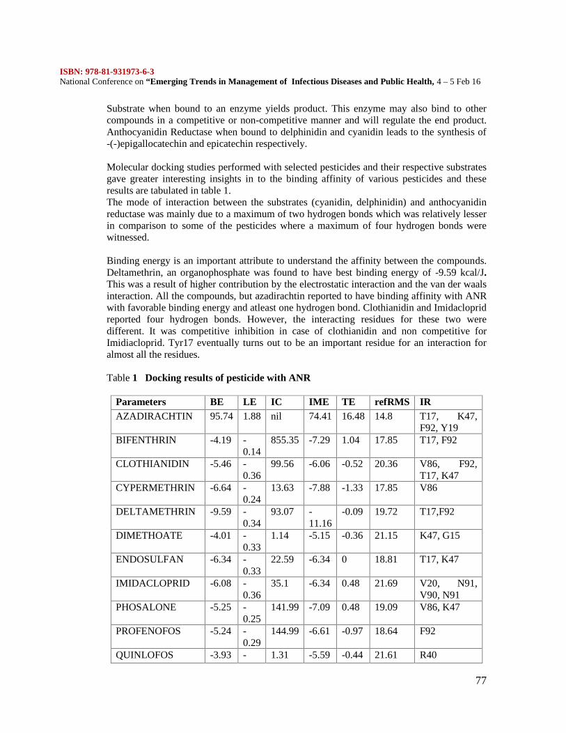
ISBN: 978-81-931973-6-3National Conference on “Emerging Trends in Management of Infectious Diseases and Public Health, 4 – 5 Feb 16
77
Substrate when bound to an enzyme yields product. This enzyme may also bind to othercompounds in a competitive or non-competitive manner and will regulate the end product.Anthocyanidin Reductase when bound to delphinidin and cyanidin leads to the synthesis of-(-)epigallocatechin and epicatechin respectively.
Molecular docking studies performed with selected pesticides and their respective substratesgave greater interesting insights in to the binding affinity of various pesticides and theseresults are tabulated in table 1.The mode of interaction between the substrates (cyanidin, delphinidin) and anthocyanidinreductase was mainly due to a maximum of two hydrogen bonds which was relatively lesserin comparison to some of the pesticides where a maximum of four hydrogen bonds werewitnessed.
Binding energy is an important attribute to understand the affinity between the compounds.Deltamethrin, an organophosphate was found to have best binding energy of -9.59 kcal/J.This was a result of higher contribution by the electrostatic interaction and the van der waalsinteraction. All the compounds, but azadirachtin reported to have binding affinity with ANRwith favorable binding energy and atleast one hydrogen bond. Clothianidin and Imidaclopridreported four hydrogen bonds. However, the interacting residues for these two weredifferent. It was competitive inhibition in case of clothianidin and non competitive forImidiacloprid. Tyr17 eventually turns out to be an important residue for an interaction foralmost all the residues.
Table 1 Docking results of pesticide with ANR
Parameters BE LE IC IME TE refRMS IRAZADIRACHTIN 95.74 1.88 nil 74.41 16.48 14.8 T17, K47,
F92, Y19BIFENTHRIN -4.19 -
0.14855.35 -7.29 1.04 17.85 T17, F92
CLOTHIANIDIN -5.46 -0.36
99.56 -6.06 -0.52 20.36 V86, F92,T17, K47
CYPERMETHRIN -6.64 -0.24
13.63 -7.88 -1.33 17.85 V86
DELTAMETHRIN -9.59 -0.34
93.07 -11.16
-0.09 19.72 T17,F92
DIMETHOATE -4.01 -0.33
1.14 -5.15 -0.36 21.15 K47, G15
ENDOSULFAN -6.34 -0.33
22.59 -6.34 0 18.81 T17, K47
IMIDACLOPRID -6.08 -0.36
35.1 -6.34 0.48 21.69 V20, N91,V90, N91
PHOSALONE -5.25 -0.25
141.99 -7.09 0.48 19.09 V86, K47
PROFENOFOS -5.24 -0.29
144.99 -6.61 -0.97 18.64 F92
QUINLOFOS -3.93 - 1.31 -5.59 -0.44 21.61 R40
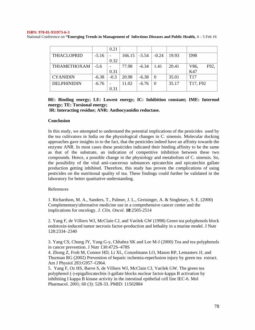
ISBN: 978-81-931973-6-3National Conference on “Emerging Trends in Management of Infectious Diseases and Public Health, 4 – 5 Feb 16
78
0.21
THIACLOPRID -5.16 -0.32
166.15 -5.54 -0.24 19.93 D98
THIAMETHOXAM -5.6 -0.31
77.98 -6.34 1.41 20.41 V86, F92,K47
CYANIDIN -6.38 -0.3 20.98 -6.38 0 35.01 T17
DELPHINIDIN -6.76 -0.31
11.02 -6.76 0 35.17 T17, F92
BE: Binding energy; LE: Lowest energy; IC: Inhibition constant; IME: Intermolenergy; TE: Torsional energy;IR: Interacting residue; ANR: Anthocyanidin reductase.
Conclusion
In this study, we attempted to understand the potential implications of the pesticides used bythe tea cultivators in India on the physiological changes in C. sinensis. Molecular dockingapproaches gave insights in to the fact, that the pesticides indeed have an affinity towards theenzyme ANR. In most cases these pesticides indicated their binding affinity to be the sameas that of the substrate, an indication of competitive inhibition between these twocompounds. Hence, a possible change in the physiology and metabolism of C. sinensis. So,the possibility of the vital anti-cancerous substances epicatechin and epicatechin gallateproduction getting inhibited. Therefore, this study has proven the complications of usingpesticides on the nutritional quality of tea. These findings could further be validated in thelaboratory for better quatitative understanding.
References
1. Richardson, M. A., Sanders, T., Palmer, J. L., Greisinger, A. & Singletary, S. E. (2000)Complementary/alternative medicine use in a comprehensive cancer center and theimplications for oncology. J. Clin. Oncol. 18:2505-2514
2. Yang F, de Villiers WJ, McClain CJ, and Varilek GW (1998) Green tea polyphenols blockendotoxin-induced tumor necrosis factor-production and lethality in a murine model. J Nutr128:2334–2340
3. Yang CS, Chung JY, Yang G-y, Chhabra SK and Lee M-J (2000) Tea and tea polyphenolsin cancer prevention. J Nutr 130:472S–478S4. Zhong Z, Froh M, Connor HD, Li XL, Conzelmann LO, Mason RP, Lemasters JJ, andThurman RG (2002) Prevention of hepatic ischemia-reperfusion injury by green tea extract.Am J Physiol 283:G957–G964.5. Yang F, Oz HS, Barve S, de Villiers WJ, McClain CJ, Varilek GW. The green teapolyphenol (-)-epigallocatechin-3-gallate blocks nuclear factor-kappa B activation byinhibiting I kappa B kinase activity in the intestinal epithelial cell line IEC-6. MolPharmacol. 2001; 60 (3): 528-33. PMID: 11502884
ISBN: 978-81-931973-6-3National Conference on “Emerging Trends in Management of Infectious Diseases and Public Health, 4 – 5 Feb 16
79
6. Yang GY, Liao J, Kim K, Yurkow EJ, Yang CS. Inhibition of growth and induction ofapoptosis in human cancer cell lines by tea polyphenols. Carcinogenesis. 1998; 19 (4): 611-6. PMID: 96003457. Characterisation of anthocyanidin reductase from Shuchazao green tea.Zhang X1, Liu Y, Gao K, Zhao L, Liu L, Wang Y, Sun M, Gao L, Xia T.
8. Prasad, S.: Infestation of Helopeltis. Tea Times Sandoz (India) Ltd. Bombay. 1-6 (1992).9. Banerjee, B.: Annual Scientific Report of Tocklai Exptt Stn for 1977-78. p. 50 (1978).10. A K Barooah. Present status of use of agrochemicals in tea industry of eastern India andfuture directions.11. R. J. Rabindra. Sustainable pest management in tea: Prospects and challenges. Two and aBud 59:1-10, 2012
ISBN: 978-81-931973-6-3National Conference on “Emerging Trends in Management of Infectious Diseases and Public Health, 4 – 5 Feb 16
80
A PILOT STUDY ON PHYTOCHEMICAL SCREENING, ANTIOXIDANTAND ANTIINFLAMMATORY ACTIVITIES OF PUNICA GRANATUM SEED
EXTRACTS
ANJANA.E 1, RAVICHANDRAN.V 2
1 Department of Biotechnology, Alpha Arts and Science College, University of Madras,India, Asst.Professor, [email protected]
2 Department of Biotechnology, Alpha Arts and Science College, University of Madras,India, II MSc Student, [email protected]
ABSTRACT
Punica granatum L. belongs to the family Puniaceae which includes only one genus and twospecies, the other one, little known, being P. protopunica Balf. peculiar to the island ofSocotra. The pomegranate is considered to have originated from Iran to northern India andhas been cultivated since ancient times throughout the Mediterranean region. Many researchactivities on P. garanatum leaves, flowers, fruits have been carried out. The number of seedsin pomegranate vary from 200 to about 1400 in number. Natural products sometimes havepharmacological or biological activity that can be of therapeutic benefit in treating diseases.As such, natural products are the active components not only of most traditionalmedicines but also many modern medicines. Furthermore, because the structural diversity ofnatural products exceeds that readily achievable bychemical synthesis, and synthetic analogscan be prepared with improved potency and safety, natural products are often used asstarting points for drug discovery. In fact, natural products are the inspiration forapproximately one half of U.S. Food and Drug Administration-approved drugs. Though thenutritional content and nutritional value of pomegranate seeds have been studied extensively,their medicinal and therapeutic properties are yet to be well documented. A preliminaryphytochemical screening can be done to reveal the phytoconstituents present within theseeds and antioxidant and anti-inflammatory studies can also be performed to gain an insightinto the therapeutic properties of the pomegranate seeds extracts.
Keywords:- Punica granatum, seed extracts, phytochemical, antioxidant, anti-inflammatory
IntroductionNatural products have contributed significantly towards the development of modern medicalscience. Recently traditional medicine worldwide is being re-evaulated by extensive researchon different plant species and their active therapeutic principles. The rich wealth of plantkingdom can represent a novel source of newer compounds with significant anti-inflammatory activities. The major merits of herbal medicine seem to be their perceivedefficiency, low incidence of serious adverse effects and low cost.
Antioxidant activity is one of the most important medical advances of the last decades of thetwentieth century. It is a new understanding of the way in which a wide range of disease andother agencies cause damage to the human body. The essence of all research conducted inthe past is that the damage to our cells and tissues at the root of most diseases is caused by
ISBN: 978-81-931973-6-3National Conference on “Emerging Trends in Management of Infectious Diseases and Public Health, 4 – 5 Feb 16
81
highly active and dangerous chemical groups called “free radicals”. Free radicals areconstantly being formed in the body as a result of basic disease processes. They are formedfollowing exposure to cigarette smoke, car exhaust, industrial fumes, and almost all forms ofradiation. Antioxidants are now recognized as being of considerable importance to health.Several reports concern the antioxidant activities of natural compounds in fruits, vegetables,dietary and rhizomatous plants with medicinal properties (Stajneret al., 1998; Moreno et al.,1999; Mukherjee, 2002; Liu et al., 2003, Hu and Kitts, 2000).
Inflammation is a pathophysiological response to injury, infection or destructioncharacterized by heat, redness, pain, swelling and diet distributed functions. Inflammation isa normal protective response to tissue injury caused by physical trauma, noxious chemical ormicrobial agents. It is the body response to inactive or destroy the invading organisms, toremove the irritant and set the stage of tissue repair. It is triggered by the release of chemicalmediators from injured tissue and migrating cells (Anonymous; 2005). The most commonlyused drug for management of inflammatory condition are non-steroidal anti-inflammatorydrugs (NSAIDs), which have several adverse effects especially gastric irritation leading toformation of gastric ulcers.(Tripathi K.D. 2008, Bennett P.N., Brown M.J. 2005).
Inflammation involves a complex array of enzyme activation, mediator release, fluidextravasations, cell migration, tissue breakdown and repair (Vane et al., 1995).It is acomplex process, which is frequently associated with pain and involves occurrences such as:the increase in vascular permeability, increase of protein denaturation and membranealterations (Umapathy et al., 2010). Harmful stimuli including pathogens, irritants ordamaged cells initiate response of vascular tissue as inflammation. Inflammation is aprotective attempt by the organism to remove injurious stimuli as well as initiate the healingprocess for the tissue (Denko.1992). However, if inflammation is not treated it leads to onsetof diseases like vasomotor rhinorrhoea, rheumatoid arthritis and atherosclerosis (Henson etal., 1989).
The pomegranate (Punica granatum L.) is one of the oldest edible fruits and is widely grownin many tropical and subtropical countries [Salaheddin, 1984]. It is an important commercialfruit in Iran with a total production of 665,000 tons in 2003. Pomegranate juice and peelcontain substantial amounts of polyphenols such as ellagic tannins, ellagic acid and gallicacid [Loren et al., 2005]. An earlier study has reported the use of leaf and bark extracts ofPunica granatum as a good antibacterial agent [Egharevba et al., 2010]. It has been used inthe preparation of tinctures, cosmetic, therapeutic formula and food recipes [Finkel et al.,2000] and in this regard pomegranate peel is a good source of antioxidants [Singh et al.,2001]. The synergistic action of the pomegranate constituents appears to be superior to thatof single constituents. In the past decade, numerous studies on the antioxidant,anticarcinogenic, and anti-inflammatory properties of pomegranate constituents have beenpublished, focusing on treatment and prevention of cancer, cardiovascular disease, diabetes,dental conditions, erectile dysfunction, bacterial infections and antibiotic resistance, andultraviolet radiation-induced skin damage (Jurenka, 2008).
The present study is aimed at revealing the phytoconstituents present within the Punicagranatum seed extracts by a preliminary phytochemical screening technique and also tostudy their antioxidant and anti-inflammatory properties.
ISBN: 978-81-931973-6-3National Conference on “Emerging Trends in Management of Infectious Diseases and Public Health, 4 – 5 Feb 16
82
MATERIALS AND METHODS
I.Fruit collection
Healthy fruits of Punica granatum were collected 2013 December from Chennai, TamilNadu, India and were maintained and shaded conditions for drying process at PoongaBiotech Research centre.
II.Preparation of the seed extract
Preparation of the extracts was done according to a combination of the methods used byJanarthanam et al., 2010. About 1g of fleshy dried powder of Punica granatum plant seedmaterials were extracted with 20 mL ethanol 75%, acetone, chloroform, aqueous andpetroleum ether (Merck, extra pure) for 1 min using an Ultra Turax mixer (13,000 rpm) andsoaked overnight at room temperature. The sample was then filtered through Whatman No. 1paper in a Buchner funnel. The filtered solution was evaporated under vacuum in a rota-vator at 40 °C to a constant weight and then dissolved in methanol ethanol and water. Thedissolving rate of the crude extracts was approximately 100 %. The solution was stored at 18°C until use.
III.Phytochemical Screening from seed extracts of Punica granatum
The phytochemical screening of seed extracts were assessed by standard method asdescribed by Brinda et al., 1981; Siddiqui and Ali, 1997 and Savithramma et al., 2011.Phytochemical screening was carried out on the leaf extracts using different solvents toidentify the major natural chemical groups such as tannins, saponins, flavonoids, phenols,terpenoids, alkaloids, glycosides, cardiac glycosides, coumarins and steroids. Generalreactions in these analysis will reveal the presence or absence of these compounds in the leafextracts tested.
IV.Qualitative Antioxidant Assay
Antioxidant assay Ozkan et al., (2004) and George et al., (1996) on plant seed dried powderextracts of Punica granatum plant were estimated for their free radical scavenging activityby using DPPH (1,1-Diphenyl-2,Picryl-Hydrazyl) free radicals.
The radical of sample is calculated by the following formula,
PrincipleDPPH (1,1-Diphenyl-2-picrylhydrazyl) is a stable free radial with purple colour (absorbed at517nm). If free radials have been scavenged, DPPH will degenerate to yellow colour. Thisassay uses this character to show free radical scavenging activity.
Inhibition = [(Absorbance of control (Ac 517) – Absorbance of sample (As517)(Absorbance control (Ac517)] X 100
ISBN: 978-81-931973-6-3National Conference on “Emerging Trends in Management of Infectious Diseases and Public Health, 4 – 5 Feb 16
83
Screening method
50µL of seed dried powder extracts of Punica granatum medicinal plant were taken in themicrotiter plate. 100µL of 0.1% methanolic DPPH was added over the samples andincubated for 30 minutes in dark condition. The samples were then observed fordiscoloration; from purple to yellow and pale pink were considered as strong and weakpositive respectively. The antioxidant positive samples were subjected for furtherquantitative analysis.
V.Quantitative assay of antioxidant activity
Selected samples of 100µl from qualitative assay were mixed with 2.7ml of methanol andthen 200µl of 0.1 % methanolic DPPH was added. The suspension was incubated for 30minutes in dark condition. Subsequently, at every 5 min interval, the absorption maxima ofthe solution were measured using a UV double beam spectra scan (Chemito, India) at517nm. The antioxidant activity of the sample was compared with known synthetic standardof (0.16%) of Butylated Hydroxy Toluene (BHT).
VI.Estimation of Total phenol content in Punica granatum
Total phenolic content in the ethanolic seed extracts was determined by the Folin Ciocalteaucolorimetric method (Slinkard and Singleton, 1984). For the analysis, 0.5 ml of drypowdered ethanolic seed extracts were added to 0.1 ml of Folin- Ciocalteau reagent (0.5N)and the contents of the flask were mixed thoroughly. Later 2.5 ml of Sodium carbonate(Na2CO3) was added and the mixture was allowed to stand for 30 min after mixing. Theabsorbance was measured at 760 nm in a UV-Visible Spectrophotometer. The total phenolicscontents were expressed as mg gallic acid equivalents (GAE)/g extract.
VII. ASSESSMENT OF INVITRO ANTI-INFLAMMATORY ACTIVITYVII a. INHIBITION OF ALBUMIN DENATURATION
The reaction mixture (5 ml) consisted of 0.2 ml of egg albumin (from fresh hen’s egg), 2.8ml of phosphate buffered saline (PBS, pH 6.4) and 2 ml of varying concentrations of the testextract (Punica granatum seed extract) so that final concentrations become 1.56, 3.125,6.25, 12.5, 25.0, 50.0 mg/ml. Similar volume of double-distilled water served as control.Then the mixtures were incubated at 37±2ºC in a incubator for 15 minutes and then heated at50ºC for 5 minutes. After cooling, their absorbance was measured at 660 nm (SHIMADZU,UV 1800) by using vehicle as blank. Diclofenac sodium at the final concentration of(78.125, 156.25, 312.5, 625, 1250 µg/ml) was used as reference drug and treated similarlyfor determination of absorbance (Dey et al., 2011; Chandra et al., 2012). The percentageinhibition of protein denaturation was calculated by using the following formula:
% Inhibition of Protein Denaturation = 100 x [Vc – Vt / Vc]Where, Vt = absorbance of test sample, Vc = absorbance of control.The extract/drug concentration for 50% inhibition (IC50) was determined form the doseresponse curve by plotting percentage inhibition with respect to control againsttreatment concentration.
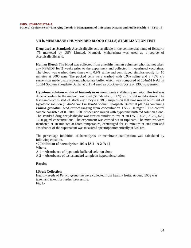
ISBN: 978-81-931973-6-3National Conference on “Emerging Trends in Management of Infectious Diseases and Public Health, 4 – 5 Feb 16
84
VII b. MEMBRANE ( HUMAN RED BLOOD CELLS) STABILIZATION TEST
Drug used as Standard: Acetylsalicylic acid available in the commercial name of Ecosprin-75 marketed by USV Limited, Mumbai, Maharashtra was used as a source ofAcetylsalicylic acid.
Human Blood: The blood was collected from a healthy human volunteer who had not takenany NSAIDS for 2 weeks prior to the experiment and collected in heparinzed vacutainer.The blood was washed three times with 0.9% saline and centrifuged simultaneously for 10minutes at 3000 rpm. The packed cells were washed with 0.9% saline and a 40% v/vsuspension made using isotonic phosphate buffer which was composed of 154mM NaCl in10mM Sodium Phosphate Buffer at pH 7.4 used as Stock erythrocyte or RBC suspension.
Hypotonic solution –induced haemolysis or membrane stabilizing activity: This test wasdone according to the method described (Shinde et al., 1999) with slight modifications. Thetest sample consisted of stock erythrocyte (RBC) suspension 0.030ml mixed with 5ml ofhypotonic solution (154mM NaCl in 10mM Sodium Phosphate Buffer at pH 7.4) containingPunica granatum seed extract ranging from concentration 1.56 - 50 mg/ml. The controlsample consisted of 0.030ml RBC suspension mixed with hypotonic buffered solution alone.The standard drug acetylsalicylic was treated similar to test at 78.125, 156.25, 312.5, 625,1250 µg/ml concentrations. The experiment was carried out in triplicate. The mixtures wereincubated at 10 minutes at room temperature, centrifuged for 10 minutes at 3000rpm andabsorbance of the supernatant was measured spectrophotometrically at 540 nm.
The percentage inhibition of haemolysis or membrane stabilization was calculated byfollowing equation.% Inhibition of haemolysis = 100 x [A 1 –A 2 /A 1]Where:A 1 = Absorbance of hypotonic buffered solution aloneA 2 = Absorbance of test /standard sample in hypotonic solution.
Results
I.Fruit CollectionHealthy seeds of Punica granatum were collected from healthy fruits. Around 100g wastaken and taken for further processing.Fig 1:-
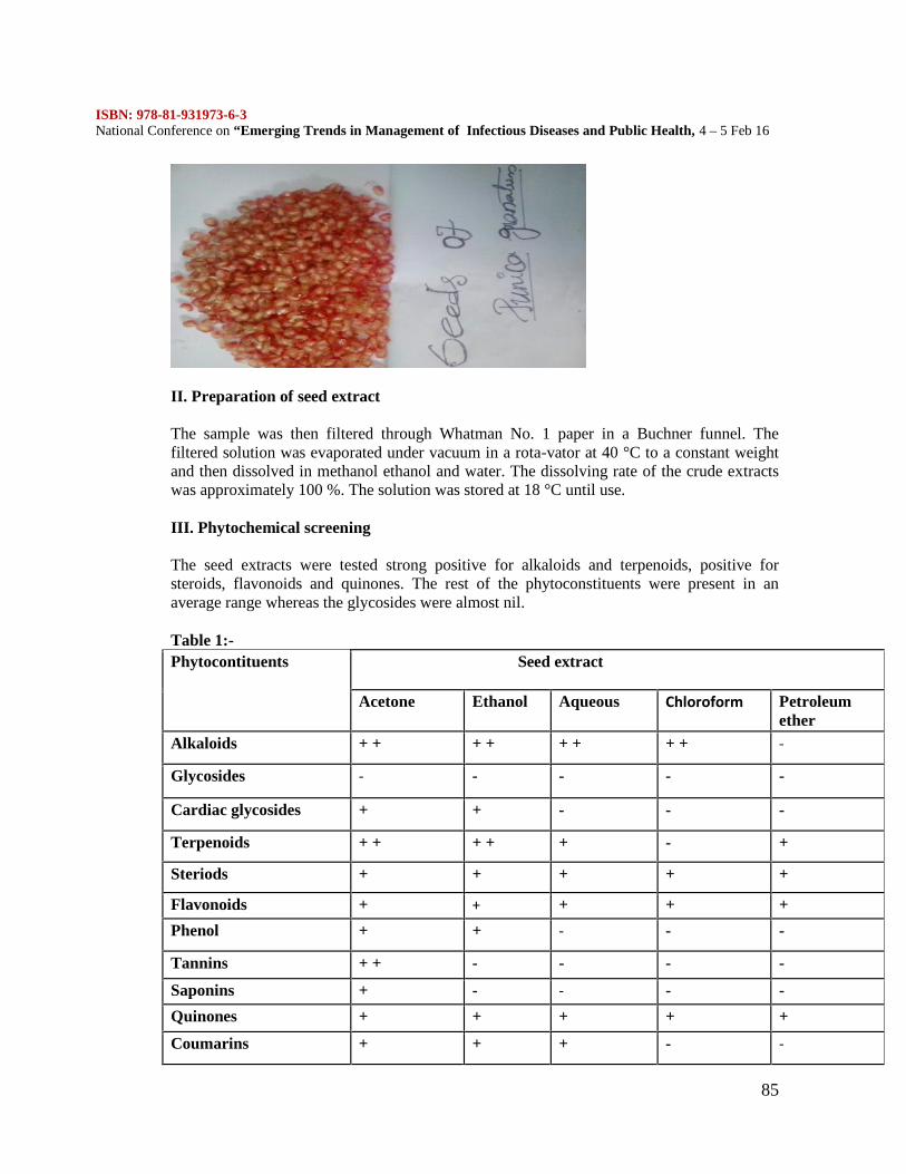
ISBN: 978-81-931973-6-3National Conference on “Emerging Trends in Management of Infectious Diseases and Public Health, 4 – 5 Feb 16
85
II. Preparation of seed extract
The sample was then filtered through Whatman No. 1 paper in a Buchner funnel. Thefiltered solution was evaporated under vacuum in a rota-vator at 40 °C to a constant weightand then dissolved in methanol ethanol and water. The dissolving rate of the crude extractswas approximately 100 %. The solution was stored at 18 °C until use.
III. Phytochemical screening
The seed extracts were tested strong positive for alkaloids and terpenoids, positive forsteroids, flavonoids and quinones. The rest of the phytoconstituents were present in anaverage range whereas the glycosides were almost nil.
Table 1:-Phytocontituents Seed extract
Acetone Ethanol Aqueous Chloroform Petroleumether
Alkaloids + + + + + + + + -
Glycosides - - - - -
Cardiac glycosides + + - - -
Terpenoids + + + + + - +
Steriods + + + + +
Flavonoids + + + + +
Phenol + + - - -
Tannins + + - - - -
Saponins + - - - -
Quinones + + + + +
Coumarins + + + - -
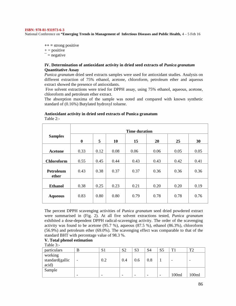
ISBN: 978-81-931973-6-3National Conference on “Emerging Trends in Management of Infectious Diseases and Public Health, 4 – 5 Feb 16
86
++ = strong positive+ = positive¯ = negative
IV. Determination of antioxidant activity in dried seed extracts of Punica granatumQuantitative AssayPunica granatum dried seed extracts samples were used for antioxidant studies. Analysis ondifferent extraction of 75% ethanol, acetone, chloroform, petroleum ether and aqueousextract showed the presence of antioxidants.Five solvent extractions were tried for DPPH assay, using 75% ethanol, aqueous, acetone,
chloroform and petroleum ether extract.The absorption maxima of the sample was noted and compared with known syntheticstandard of (0.16%) Butylated hydroxyl toluene.
Antioxidant activity in dried seed extracts of Punica granatumTable 2:-
The percent DPPH scavenging activities of Punica granatum seed dried powdered extractwere summarised in (Fig. 2). At all five solvent extractions tested, Punica granatumexhibited a dose-dependent DPPH radical-scavenging activity. The order of the scavengingactivity was found to be acetone (95.7 %), aqueous (87.5 %), ethanol (86.3%), chloroform(56.9%) and petroleum ether (69.0%). The scavenging effect was comparable to that of thestandard BHT with percentage value of 98.3 %.V. Total phenol estimationTable 3:-particulars B S1 S2 S3 S4 S5 T1 T2workingstandard(gallicacid)
- 0.2 0.4 0.6 0.8 1 - -
Sample- - - - - - 100ml 100ml
SamplesTime duration
0 5 10 15 20 25 30
Acetone 0.33 0.12 0.08 0.06 0.06 0.05 0.05
Chloroform 0.55 0.45 0.44 0.43 0.43 0.42 0.41
Petroleumether
0.43 0.38 0.37 0.37 0.36 0.36 0.36
Ethanol 0.38 0.25 0.23 0.21 0.20 0.20 0.19
Aqueous 0.83 0.80 0.80 0.79 0.78 0.78 0.76
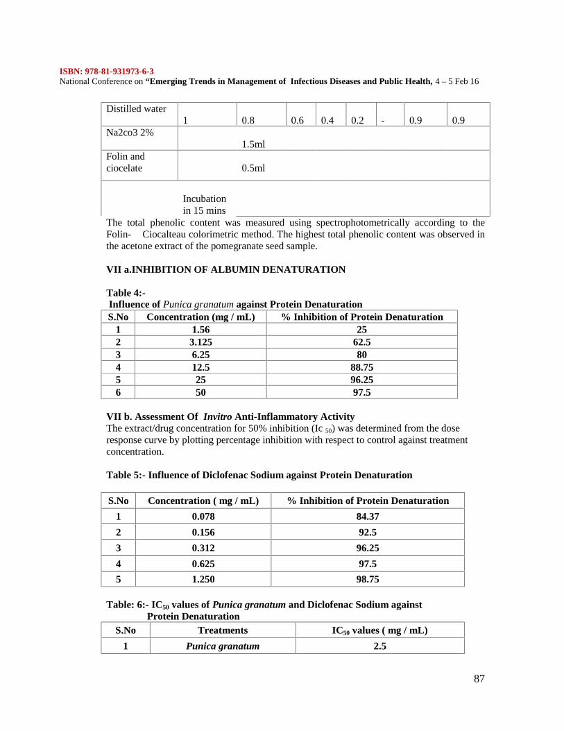
ISBN: 978-81-931973-6-3National Conference on “Emerging Trends in Management of Infectious Diseases and Public Health, 4 – 5 Feb 16
87
Distilled water1 0.8 0.6 0.4 0.2 - 0.9 0.9
Na2co3 2%1.5ml
Folin andciocelate 0.5ml
Incubationin 15 mins
The total phenolic content was measured using spectrophotometrically according to theFolin- Ciocalteau colorimetric method. The highest total phenolic content was observed inthe acetone extract of the pomegranate seed sample.
VII a.INHIBITION OF ALBUMIN DENATURATION
Table 4:-Influence of Punica granatum against Protein DenaturationS.No Concentration (mg / mL) % Inhibition of Protein Denaturation
1 1.56 252 3.125 62.53 6.25 804 12.5 88.755 25 96.256 50 97.5
VII b. Assessment Of Invitro Anti-Inflammatory ActivityThe extract/drug concentration for 50% inhibition (Ic 50) was determined from the doseresponse curve by plotting percentage inhibition with respect to control against treatmentconcentration.
Table 5:- Influence of Diclofenac Sodium against Protein Denaturation
S.No Concentration ( mg / mL) % Inhibition of Protein Denaturation
1 0.078 84.37
2 0.156 92.5
3 0.312 96.25
4 0.625 97.5
5 1.250 98.75
Table: 6:- IC50 values of Punica granatum and Diclofenac Sodium againstProtein Denaturation
S.No Treatments IC50 values ( mg / mL)
1 Punica granatum 2.5
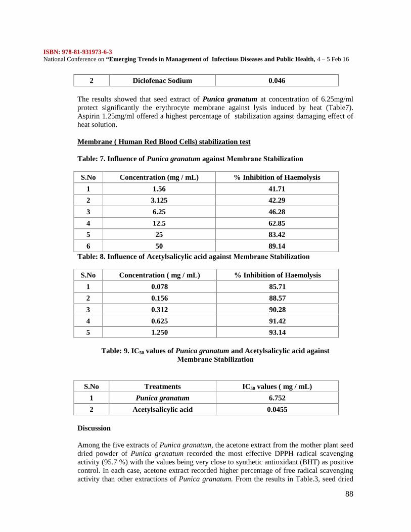
ISBN: 978-81-931973-6-3National Conference on “Emerging Trends in Management of Infectious Diseases and Public Health, 4 – 5 Feb 16
88
2 Diclofenac Sodium 0.046
The results showed that seed extract of Punica granatum at concentration of 6.25mg/mlprotect significantly the erythrocyte membrane against lysis induced by heat (Table7).Aspirin 1.25mg/ml offered a highest percentage of stabilization against damaging effect ofheat solution.
Membrane ( Human Red Blood Cells) stabilization test
Table: 7. Influence of Punica granatum against Membrane Stabilization
S.No Concentration (mg / mL) % Inhibition of Haemolysis
1 1.56 41.71
2 3.125 42.29
3 6.25 46.28
4 12.5 62.85
5 25 83.42
6 50 89.14Table: 8. Influence of Acetylsalicylic acid against Membrane Stabilization
S.No Concentration ( mg / mL) % Inhibition of Haemolysis
1 0.078 85.71
2 0.156 88.57
3 0.312 90.28
4 0.625 91.42
5 1.250 93.14
Table: 9. IC50 values of Punica granatum and Acetylsalicylic acid againstMembrane Stabilization
S.No Treatments IC50 values ( mg / mL)
1 Punica granatum 6.752
2 Acetylsalicylic acid 0.0455
Discussion
Among the five extracts of Punica granatum, the acetone extract from the mother plant seeddried powder of Punica granatum recorded the most effective DPPH radical scavengingactivity (95.7 %) with the values being very close to synthetic antioxidant (BHT) as positivecontrol. In each case, acetone extract recorded higher percentage of free radical scavengingactivity than other extractions of Punica granatum. From the results in Table.3, seed dried
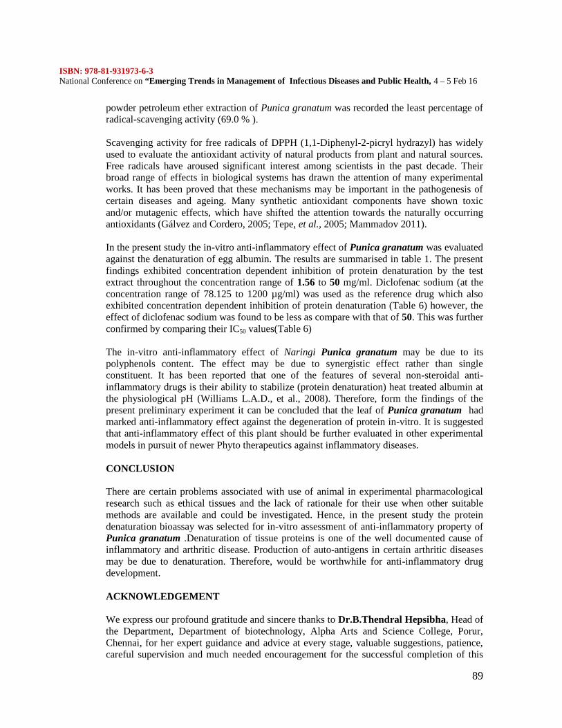
ISBN: 978-81-931973-6-3National Conference on “Emerging Trends in Management of Infectious Diseases and Public Health, 4 – 5 Feb 16
89
powder petroleum ether extraction of Punica granatum was recorded the least percentage ofradical-scavenging activity (69.0 % ).
Scavenging activity for free radicals of DPPH (1,1-Diphenyl-2-picryl hydrazyl) has widelyused to evaluate the antioxidant activity of natural products from plant and natural sources.Free radicals have aroused significant interest among scientists in the past decade. Theirbroad range of effects in biological systems has drawn the attention of many experimentalworks. It has been proved that these mechanisms may be important in the pathogenesis ofcertain diseases and ageing. Many synthetic antioxidant components have shown toxicand/or mutagenic effects, which have shifted the attention towards the naturally occurringantioxidants (Gálvez and Cordero, 2005; Tepe, et al., 2005; Mammadov 2011).
In the present study the in-vitro anti-inflammatory effect of Punica granatum was evaluatedagainst the denaturation of egg albumin. The results are summarised in table 1. The presentfindings exhibited concentration dependent inhibition of protein denaturation by the testextract throughout the concentration range of 1.56 to 50 mg/ml. Diclofenac sodium (at theconcentration range of 78.125 to 1200 µg/ml) was used as the reference drug which alsoexhibited concentration dependent inhibition of protein denaturation (Table 6) however, theeffect of diclofenac sodium was found to be less as compare with that of 50. This was furtherconfirmed by comparing their IC50 values(Table 6)
The in-vitro anti-inflammatory effect of Naringi Punica granatum may be due to itspolyphenols content. The effect may be due to synergistic effect rather than singleconstituent. It has been reported that one of the features of several non-steroidal anti-inflammatory drugs is their ability to stabilize (protein denaturation) heat treated albumin atthe physiological pH (Williams L.A.D., et al., 2008). Therefore, form the findings of thepresent preliminary experiment it can be concluded that the leaf of Punica granatum hadmarked anti-inflammatory effect against the degeneration of protein in-vitro. It is suggestedthat anti-inflammatory effect of this plant should be further evaluated in other experimentalmodels in pursuit of newer Phyto therapeutics against inflammatory diseases.
CONCLUSION
There are certain problems associated with use of animal in experimental pharmacologicalresearch such as ethical tissues and the lack of rationale for their use when other suitablemethods are available and could be investigated. Hence, in the present study the proteindenaturation bioassay was selected for in-vitro assessment of anti-inflammatory property ofPunica granatum .Denaturation of tissue proteins is one of the well documented cause ofinflammatory and arthritic disease. Production of auto-antigens in certain arthritic diseasesmay be due to denaturation. Therefore, would be worthwhile for anti-inflammatory drugdevelopment.
ACKNOWLEDGEMENT
We express our profound gratitude and sincere thanks to Dr.B.Thendral Hepsibha, Head ofthe Department, Department of biotechnology, Alpha Arts and Science College, Porur,Chennai, for her expert guidance and advice at every stage, valuable suggestions, patience,careful supervision and much needed encouragement for the successful completion of this
ISBN: 978-81-931973-6-3National Conference on “Emerging Trends in Management of Infectious Diseases and Public Health, 4 – 5 Feb 16
90
study. We thank all the Staff members of the Poonga Biotech Research Centre, PlantBiotechnology Division, Choolaimedu, Chennai for the help rendered during this projectwork. We are also extremely thankful to the Management (Alpha Group of Institutions) andthe Principal (Alpha Arts and Science College) for their support without which this projectwould not have materialized. Above all we thank God Almighty for the successfulcompletion of our work.REFERENCES
Benjamin Jeya Rathna Kumar P and Christopher Patrick Kiladi S,Preliminary Phytochemical and Pharmacognostic Studies of Holoptelea integrifolia Roxb.,2009, Ethnobotanical Leaflets 13: 1222-31 Galvez M, Martin-Cordero C, Houghton PJ and Ayuso MJ (2005). Antioxidantactivity of methanol extracts obtained from Plantago species. J. Agric. Food Chem., 53:1927-1933. George, J.A., Burke, W.D., Eickbush, T.H. (1996). Analysis of the 5'junctions of R2 insertions with the 28S gene: Implications for non-LTRretrotransposition. Genetics 142(3): 853--863. Janarthanam B & Sumathi E, 2010. Antimicrobial activity of Gymnema sylvestreleaf and callus extracts. Journal of Tropical Medicinal Plants, 11(2):143-147. Mammadov R, Ili P, Ertem Vaizogullar H, 2011. Antioxidant activity and totalphenolic content of Gagea fibrosa and Romulea ramiflora . Iran... J. Chem. Chem. Eng. 30. Marina Gálvez, Carmen Martín-Cordero,Peter J. Houghton and María Jesús AyusoAntioxidant activity of Plantago bellardii All. Phytotherapy Research December 2005Volume 19, Issue 12, pages 1074–1076, Mukherjee, A., 2006. Deeper Groundwater Chemistry and Flow in the ArsenicAffected Western Bengal Basin, West Bengal, India. Dissertation. University of Kentucky,Lexington. 248 pp N. Savithramma, M. Linga Rao and G. Bhum, Phytochemical screening ofThespesia populnea (L.) Soland and Tridax procumbens L. J. Chem. Pharm. Res., 2011,3(5):28-34 Nguelefack TB, Fotio LA, Watcho P, Wansi S, Dimo T, Kamanyi A. Analgesicactivities of aqueous and ethanolic extracts of the leaves of Kalanchoe crenata(Crassulaceae). Phytother Res 2004;18:385-8. Tepe B, Sokmen M, Akpulat HA and Sokmen A (2005). In vitro antioxidantactivities of the methanol extracts of four Helichrysum species from Turkey. Food Chem.,90: 685-689. Toker G, Küpeli E, Memisoðlu M and Yesilada E. Flavonoids with antinociceptiveand anti-inflammatory activities from the leaves of Tilia argentea (silver linden) JEthnopharmacol 2004;95:393-97.
ISBN: 978-81-931973-6-3National Conference on “Emerging Trends in Management of Infectious Diseases and Public Health, 4 – 5 Feb 16
91
ANTIBACTERIAL EVALUATION OF METALLIC SALTS AGAINSTINFECTIOUS BACTERIA
N.K. UdayaPrakash1., R. Caroline1., D.S. Arjun Krishnan1., N. Sripriya1.,N. Ashwin Karthick1 and S. Bhuvaneswari2
1. R and D, Marina Labs, 14, Kavya Gardens, N.T. Patel Road,Nerkundram, Chennai 600107
2. Department of Plant Biology and Plant Biotechnology,LoganathaNarayanaswamy Government Arts College, Ponneri
ABSTRACT
The bacteria, Bacillussubtilis, Enterococcus sp., Escherichia coli, Klebsiella sp., Proteus sp.,Pseudomonas aeruginosa, Salmonella typhi, Staphylococcus aureus, Staphylococcusepidermidis, Streptococcus sp., Vibrio choleraeand Vibrio parahaemolyticuswhich werefound to be either pathogen or opportunistic pathogen were evaluated for their sensitivitytowards metallic salts. The antibacterial potency of the metallic salt solutions of Chromium,Cobalt, Mercury, Nickel, Lead, Cadmium and Zinc were evaluated. The study wasconducted using Kirby-Bauer’s disc diffusion method. The results in detail will be discussed.
INTRODUCTION
The prevalence of microbial infections leading to an increased rate of mortality is on thehigh for the past two decades, causing it a major concern globally (Tatli and Akdemir,2005). Though the fourth generation of antibiotics have been discovered, the incidence ofantibiotic resistance strains has increased owing to the misuse or overuse of the drugs andantibiotics (Diab, 2002; WHO 2015). Infection by such drug resistant strains leads todifficulties in treatment. In India, about 58,000 deaths of neonatal sepsis were attributed tomicrobial resistance to drugs (Laxminarayaet al., 2013). Subsequently, there is an increasingurge to identify potential antimicrobial agents from varying sources. Several reports inter-relate drug resistance and metal tolerance (Silver, 1996; Mgbemenaet al., 2012).
Metals such as copper, mercury, silver and arsenic have been conventionally used asantimicrobial agents since ancient times, until they were replaced by antibiotics. Due to thedeclining efficiency of antibiotics, the use of metals as nanostructures or alloys orcomplexes, especially those of silver, are re-emerging for their use as an antimicrobialagents. Their efficiency is supported by recent studies that indicate the oxidative stress,protein dysfunction or membrane impairment to microbial cells caused by different metals,eventually leading to cell death (Franciet al., 2015). Thus, the present study is focused on theantibacterial activity of different metallic salts against pathogens/opportunistic pathogens,which are found to be associated with diseases such as bacteremia, urinary tract infections,respiratory infections, etc.
MATERIALS AND METHODSBacterial cultures
ISBN: 978-81-931973-6-3National Conference on “Emerging Trends in Management of Infectious Diseases and Public Health, 4 – 5 Feb 16
92
Laboratory cultures of 12pathogenic bacterial strains were used for the study, of which 7areGram negative and 5 are Gram positive.The Gram positive bacteriaincludes,Bacillussubtilis,Enterococcus sp., Staphylococcus aureus, Staphylococcus epidermidis and Streptococcus sp.while the Gram negative strains areEscherichia coli, Klebsiella sp., Proteus sp.,Pseudomonas aeruginosa, Salmonella typhi, Vibrio choleraeand Vibrioparahaemolyticus.The strains of bacteria mentioned above are of infectious nature and theirsource and infection differ according to the strain.
Kirby-Bauer disc diffusion method
The salts used for the study are sulphate salt of Chromium, nitrate salts of Cobalt, Nickel,Lead, Cadmium and Zinc and Mercuric chloride. The antibacterial activity of the metallicsalts is performed by Kirby-Bauer disc diffusion method where nutrient agar plates arespread with 100 µl of 24 hour cultures of bacteria. Sterile discs (5mm diameter) loaded with15, 20 and 25 µl of the respective metallic salt solutions corresponding to 100, 150 and 200ppm were placed over the plates. After an incubation period of 24 h, at 37°C, the plates wereobserved for the zone of inhibition against the bacterial strains.
RESULTS
Among the metallic salts studied, mercuric chloride was found to be the most efficientantibacterial agent which showed zone of inhibition against all the species of bacteriastudied. This was followed by the nitrate salt of Cadmium which recorded zone of inhibitionagainst all the bacteria except that of Escherichia coli. The nitrate salt of Cobalt too showedsimilar trend in recording antibacterial nature against all the bacteria studied except that ofEscherichia coli and Vibrio parahaemolyticus. It is noticed that the sulphate salt ofChromium has not recorded zone of inhibition against any of the bacteria studied.
The salt of lead recorded zone of inhibition only against the following bacteria, i.e. Bacillussubtilis, Proteus sp. and Vibrio cholerae. All other bacteria showed resistance against thesalt of lead even at the concentration of 200ppm. Although, the salt of Nickel has showedwide spectrum of antibiotic against 9 out of 13 bacterial strains studied, the zone ofinhibition was not so significant when compared to other metallic salts. Similar trend wasobserved with that of the salt of Zinc. The bacteria, Bacillus subtilis, Enterococcus sp. andEscherichia coli have showed resistance towards the nitrate salt of Zinc.
Mercury, cadmium, cobalt and zinc exhibited high antibacterial activity against S. typhi, withthe zone of 24, 21, 20 and 20 mm respectively, at 200 ppm of respective salts, but in case oflead, a zone of only 7 mm was recorded at 200 ppm. For most of the strains, nickel and leadshowed marginal antibacterial activity only at higher concentrations while cobalt, cadmiumand zinc showed moderate action. Zinc was found to be the most toxic to S. typhiand Proteussp., with zones of 12, 16, 20 and 17, 17, 19 mm respectively at 100, 150 and 200 ppm of thesalt. B. subtilis and E. coli had tolerance towards zinc.In addition to chromium and zinc, E.coli had gained resistance against nickel, lead and cadmium, while low susceptibility, with azone of 6 mm was recorded against cobalt. The pathogen, S. aureus, had gained resistanceagainst nickel and lead.Enterococcus sp., Klebsiellasp. and S. epidermidis showed toleranceagainst lead whereas V. parahaemolyticushad tolerance towards cobalt and nickel at all the
ISBN: 978-81-931973-6-3National Conference on “Emerging Trends in Management of Infectious Diseases and Public Health, 4 – 5 Feb 16
93
concentrations studied.The zones of inhibition recorded for individual metallic salts againstthe bacterial strains are tabulated in Table 1.
DISCUSSION
Heavy metals, at lower concentrations, are found to be crucial for the growth and metabolicprocesses of microbes while at higher concentrations, they are toxic (Appenroth, 2010). Inthe present study, Mercuric chloride was found to be the most toxic of the metallic saltsstudied, followed by Zinc, Cadmium, Cobalt and Nickel, while Lead was toxic to very fewof the strains studied.
Among the metallic salts studied, Chromium sulphatewas non-toxic to all the bacterialstrains evaluated. Further,B. subtilis had acquired tolerance towards Zinc while S. aureusand V. parahaemolyticus were resistant to nickel and lead. E. coli was found to resistant toall the salts studied except mercury and cobalt. This can be correlated with a report byCenter for Disease Dynamics, Economics & Policy, revealing the resistance exhibited by13% of E. coli against carbapenems in India by the year 2013 (CDDEP 2015).
The resistance of the bacteria against the metals could be attributed to the mobilization ofmetals in the environment by anthropogenic sources, which has eventually led to thedevelopment of resistance factors in bacteria. The major mechanisms by which microbestolerate heavy metals include exclusion by permeability barrier, intra- and extra-cellularsequestration, active transport efflux pumps, enzymatic detoxification and reduction in thesensitivity of cellular targets to metal ions (Bruins et al., 2000), which might be encoded intheir plasmids (Mgbemenaet al., 2012).
It is proposed to study the other parameters that influence the toxic nature of metals such asthe form of metals used, effect of media components, hydrogen ion concentration, etc., arerequired.It is of concern that the salts of metals studied belongs to that of heavy metals, thequestion of toxicity is raised. So, to avoid toxicity of these metallic salts conversion of theminto non-ionic form from the ionic form will be of helpful in nature. Thus, conversion ofthese metallic salts to that of the oxides of metals or as nanoparticles is recommended andusage of these as an antimicrobial agent is highly proposed to combat the infectious bacteria.
CONCLUSION
The present study reveals the antibacterial activity of the metallic salts of Chromium, Cobalt,Mercury, Nickel, Lead, Cadmium and Zinc against 12 bacterial pathogens. The action ofeach metal differs with each bacterium. It is evident that the metallic salts are a potentialagent in combating the infectious bacteria as most of them showed zone of inhibition againstthe infectious bacteria studied. As, the metallic salts used are reported to be that of heavymetals, it is proposed to use the metallic salts in their oxide forms or as nanostructures incombatting infectious bacteria.
REFERENCES
1. Antimicrobial resistance – Factsheet. WHO. 2015
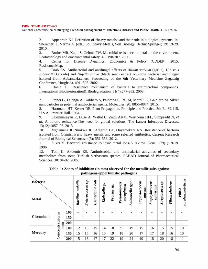
ISBN: 978-81-931973-6-3National Conference on “Emerging Trends in Management of Infectious Diseases and Public Health, 4 – 5 Feb 16
94
2. Appenroth KJ. Definition of “heavy metals” and their role in biological systems. In:Sherameti I., Varma A. (eds.) Soil heavy Metals, Soil Biology. Berlin: Springer; 19: 19-29.2010.3. Bruins MR, Kapil S, Oehme FW. Microbial resistance to metals in the environment.Ecotoxicology and environmental safety. 45: 198-207. 2000.4. Center for Disease Dynamics, Economics & Policy (CDDEP). 2015.ResistanceMap.5. Diab AS. Antibacterial and antifungal effects of Allium sativum (garlic), Hibiscussubdariffa(karkade) and Nigella sativa (black seed) extract on some bacterial and fungalisolated from AbbassaHatchert, Proceeding of the 6th Veterinary Medicine ZagaazigConference, Hurghada. 491- 505. 2002.6. Cloete TE. Resistance mechanism of bacteria to antimicrobial compounds.International Biodeterioration& Biodegradation. 51(4):277-282. 2003.
7. Franci G, Falanga A, Galdiero S, Palomba L, Rai M, Morelli G, Galdiero M. Silvernanoparticles as potential antibacterial agents. Molecules. 20: 8856-8874. 2015.8. Hartmann HT, Kester DE. Plant Propagation, Principle and Practice. 5th Ed 89:115,U.S.A, Prentice Hall. 1964.9. Laxminarayan R, Duse A, Wattal C, Zaidi AKM, Wertheim HFL, Sumpradit N, etal. Antibiotic resistance-The need for global solutions. The Lancet Infectious Diseases,13(12):1057–98. 2013.10. Mgbemena IC,Nnokwe JC, Adjeroh LA, Onyemekara NN. Resistance of bacteriaisolated from Otamiririverto heavy metals and some selected antibiotics. Current ResearchJournal of Biological Sciences. 4(5): 551-556. 2012.11. Silver S. Bacterial resistance to toxic metal ions-A review. Gene. 179(1): 9-19.1996.12. Tatli II, Akdemir ZS. Antimicrobial and antimalarial activities of secondarymetabolites from some Turkish Verbascum species. FABAD Journal of PharmaceuticalSciences. 30: 84-92. 2005.
Table 1 : Zones of inhibition (in mm) observed for the metallic salts againstpathogens/opportunistic pathogens
Bacteria
Metal
Bac
illus
sub
tilis
Ent
eroc
occu
ssp
.
Esc
heri
chia
col
i
Kle
bsie
llasp
.
Pro
teus
sp.
Pse
udom
onas
aeru
gino
sa
Salm
onel
la ty
phi
Stap
hylo
cocc
usau
reus
Stap
hylo
cocc
usep
ider
mis
Stre
ptoc
occi
sp.
Vib
rio
chol
erae
Vib
rio
para
haem
olyt
icus
Chromium
Con
cent
rati
on in
ppm
100 - - - - - - - - - - - -
150 - - - - - - - - - - - -
200 - - - - - - - - - - - -
Mercury100 12 13 15 14 18 9 19 15 16 15 15 10
150 15 15 16 15 19 18 20 17 17 18 16 10
200 15 16 17 17 22 19 24 19 18 20 18 11
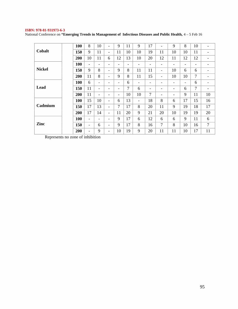
ISBN: 978-81-931973-6-3National Conference on “Emerging Trends in Management of Infectious Diseases and Public Health, 4 – 5 Feb 16
95
Cobalt100 8 10 - 9 11 9 17 - 9 8 10 -
150 9 11 - 11 10 10 19 11 10 10 11 -
200 10 11 6 12 13 10 20 12 11 12 12 -
Nickel100 - - - - - - - - - - - -
150 9 8 - 9 8 11 11 - 10 6 6 -
200 11 8 - 9 8 11 15 - 10 10 7 -
Lead100 6 - - - 6 - - - - - 6 -
150 11 - - - 7 6 - - - 6 7 -
200 11 - - - 10 10 7 - - 9 11 10
Cadmium100 15 10 - 6 13 - 18 8 6 17 15 16
150 17 13 - 7 17 8 20 11 9 19 18 17
200 17 14 - 11 20 9 21 20 10 19 19 20
Zinc100 - - - 9 17 6 12 6 6 9 11 6
150 - 6 - 9 17 8 16 7 8 10 16 7
200 - 9 - 10 19 9 20 11 11 10 17 11
Represents no zone of inhibition
ISBN: 978-81-931973-6-3National Conference on “Emerging Trends in Management of Infectious Diseases and Public Health, 4 – 5 Feb 16
96
EVALUATION OF CAULERPA SCALPELLIFORMIS FOR THE PRESENCE OFPHYTOCHEMICALS AND ITS ANTIBACTERIAL ACTIVITY
X.Asbin Mary1 and M. Syed Ali2
1 Assistant Professor, Department of Biotechnology, Alpha Arts and Science College,Chennai.2Department of Biotechnology, Mohamed Sathak College of Arts and Science,Sholinganallur, Chennai
ABSTRACT
Marine algae were considered as the source of various bioactive compounds as they areable to produce variety of secondary metabolites. The study reveals the presence ofphytochemical, and antibacterial activity of Caulerpa scalpelliformis. By direct extractionmethod the algae material was extracted with methanol. The phytochemical analysis ofCaulerpa scalpelliformis extract reveals the presence of terpenoids, glycosides, and steroids.But the extract shows the absence tannins, phlobatannins, alkaloids, saponins, flavonoids.The antibacterial activity was carried out by using Disc Diffusion Method against thepathogenic organisms such as Escherichia coli, Bacillus subtilis, Staphlococcus aureus,Klebsiella pneumonia, Pseudomonas aeruginosa.
KEYWORDS: Caulerpa scalpelliformis, phytochemicals, Antibacterial.1. INTRODUCTION
Marine algae has a significant attraction as natural source of bioactive molecules with abroad range of biological activities,such as antibiotics,antivirals,antitumorals,antioxidant andantiinflammatories.they produce a wide variety of chemically active metabolities in theirsurroundings, potentially as an aid to protect themselves against the other settling organism.Since sea weeds are a good source of antimicrobial compounds,fatty acids,antioxidants andother bioactive compounds, there is an interest to utilize these products as nutraceuticals andin functionalfoods (Yuan, 2008).Ara et al (2002) reported brown algae to be active against a number of Gram positive andGram negative organisms. The separation of the isomers by gas chromatography and theirstructural elucidation by mass spectrometry (Youngblood et al., 1973).Now there is morecurrent research in the phytochemical investigation in higher plants (Peteros et al., 2010).
This fact implies that seaweed cells have some protective compounds and mechanism(Matsukawa et al., 1997).Chloroform: methanol is the best solution for extracting theeffective antibacterial materials from the brown algae species (Rajasulochanna et al.,2009). In Phaeophyta, fucosterol is the main sterol and small amounts of cholesterol, 24-methylene-cholestrol and saringosterol were identified (Ikekawa et al., 1968). The hot watercrude extract of these algae species such as Gracilaria corticata, Gracillaria salicornia andSargassum oligocystum showed effective anti Leishmanial activity (Fouladvand et al,2011).
ISBN: 978-81-931973-6-3National Conference on “Emerging Trends in Management of Infectious Diseases and Public Health, 4 – 5 Feb 16
97
The green algae Chlorococcum humicola is a rich and varied source of pharmacologicallyactive natural products. Nutraceutical and pharmaceutical content in the baseline algae strainis very less, they showed excellent effect the micobial pathogens (Baghavati et al, 2011).The most characteristic chemical property of theglycosides is their susceptibility tohydrolysis, where they yield sugar or glycone and non sugar or a glycones motieties(Rangari, 2003)
Evidence of phytochemical and pharmacological studies on algae is available in theliterature with special reference to terpenoids and steroids. In this study, the Caulerpascalpelliformis was evaluated for the presence of phytochemicals and Antibacterial activityare analysed by Disc Diffusion Method.
2. MATERIALS AND METHODS
Preparation of marine algae materialThe fresh marine algae Caulerpa scalpelliformis were collected and it was washed
thoroughly with running water to remove debris. It was shade dried in room temperature forabout 7-9 days. The dried marine algae material was powdered and extracted with solventfor the analysis of the phytochemicals and antimicrobial activity in it.Preparation of algae extract
15g marine algae were weighed and it was macerated for three days in 150ml ofmethanol at room temperature in an orbital shaker. After three days, the total extract werefiltered and the filtrates were concentrated under reduced pressure to dryness, yielding thecrude extract which was then weighed for different concentration (25mg,50 mg,75 mg,100mg) and each concentrations was suspended in 1ml of methanol. The dried material wasreconstituted in methanol for the analysis of the antibacterial activity.
PHYTOCHEMICAL TEST
Phytochemical screening for steroids, terpenoids, flavonoids, tannins, saponins, alkaloids,phlobatannins were carried out as described below.Test for tanninsAbout 2ml of the extract was stirred with 2ml of distilled water and few drops of FeCl3
solution were added. The formation of a green precipitate was an indication for the presenceof tannins.Test for cardiac glycosides (KELLER-KILLANI TEST)5ml of extracts was treated with 2ml of glacial acetic acid containing one drop of ferricchloride solution. A brown ring may appear below the brown ring, while in the acetic acidlayer, a greenish ring may form just gradually throughout thin layer which shows thepresence of Cardiac glycosides.Test for flavonoids5ml of dilute ammonia solution were added to a portion of the extract followed by additionof concentrated H2SO4 .formation of yellow colour indicated the presence of flavonoids.Test for steroids1ml of extracts was dissolved in a few drops of acetic acid. It was gently warmed and cooledunder the tap water and a drop of concentrated sulphuric acid was added along the sides ofthe test tube. Appearance of green colour indicates the presence of steroids.
ISBN: 978-81-931973-6-3National Conference on “Emerging Trends in Management of Infectious Diseases and Public Health, 4 – 5 Feb 16
98
Test for terpenoids (SALKOWSKI TEST)5ml of extract was mixed in 2ml of chloroform and concentrated H2SO4 (3ml) was carefullyadded to form a layer. Formation of reddish brown coloration at the interface shows thepositive results for the presence of terpenoids.Test for alkaloids (MAYERS TEST)With 1ml of extract add 1ml of mayer’s reagent (potassium chloride solution) whitish creamcolored precipitate indicates presence of alkaloids.Test for saponins5ml of extract was shaken vigorously with 5ml of distilled water in a test tube and warmed.The formation of stable foam, honey comb shapes was taken as an indication for thepresence of saponins.Test for phlobatanninsAbout 2ml of extract was added to 2ml of 1% HCL and the mixture was boiled. Depositionof a red precipitate was taken as an evidence for the presence of phlobatannins.Antibacterial AssayPreparation of inoculumsBacterial inoculums were prepared by transferring bacterial strains from fresh culture platesto tubes containing 10ml of Muller Hinton Borth (Hi-Media) and incubated for 24hrs at 37°cin an Orbital Shaker. The antibacterial activity was carried out by using Disc DiffusionMethod.Antibacterial AssayThe Muller Hinton agar medium was prepared and sterilized by autoclaveat 121°c for15mins. Then the media was poured into sterile petriplate and it was kept for solidification.After 24 hrs bacterial culture from Muller Hinton Borth was swabbed and the sterile discwith 100µl of different concentration of algal extract was placed on the swabbed and thesterile disc with 100µl of different concentration of algal extract was placed on the swabbedpetridishes. The petridish was incubated at 37°c for 24 hrs. Amphicillin was used as positivecontrol. Methanol solvent (100%) without algal extract was also used as negative control.After 24hrs the zone of inhibition was observed and measured.
3. RESULTS AND DISCUSSION
Preparation of algae materialsThe algal materials were collected, shade dried, powdered and extracted with solvent for theanalysis of photochemical present in extract.Extraction of algal materialsThe powdered material was extracted with methanol using direct extraction methoddescribed in materials and methods. The extract was dried and weighed for yield extract.Phytochemicals analysis
The phytochemical analysis of the methanol algae extract revealed the presence ofphytochemical compounds like glycoside, terpenoids and steroids (Table 1).A large numberof algal extract products have beenfound to have antimicrobial activity,many of thestructures were identify as fatty acids, glycolipids, steroids,phenolic andterpenoids.(Thillairajasekar 2009, Luesch,2000).
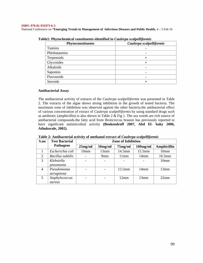
ISBN: 978-81-931973-6-3National Conference on “Emerging Trends in Management of Infectious Diseases and Public Health, 4 – 5 Feb 16
99
Table1: Phytochemical constituents identified in Caulerpa scalpelliformisPhytoconstituents Caulerpa scalpelliformis
Tannins -Phlobatannins -Terpenoids +Glycosides +Alkaloids -Saponins -Flavonoids -Steroids +
Antibacterial Assay
The antibacterial activity of extracts of the Caulerpa scalpelliformis was presented in Table2. The extracts of the algae shows strong inhibition in the growth of tested bacteria. Themaximum zone of inhibition was observed against the other bacteria.the antibacterial effectof various concentration of extract of Caulerpa scalpelliformis by using standard drugs suchas antibiotic (amphicillin) is also shown in Table 2 & Fig 1. The sea weeds are rich source ofantibacterial compounds.the fatty acid from Broticoccus braunii has previously reported tohave significant antimicrobial activity (Benkendroff 2007, Abd El- baky 2006,Athukorale, 2002).
Table 2: Antibacterial activity of methanol extract of Caulerpa scalpelliformisS.no Test Bacterial
PathogensZone of Inhibition
25mg/ml 50mg/ml 75mg/ml 100mg/ml Amphicillin1 Escherichia coli 10mm 13mm 14.5mm 15.5mm 10mm2 Bacillus subtilis - 9mm 11mm 14mm 16.5mm3 Klebsiella
pneumonia- - - - 10mm
4 Pseudomonasaeruginosa
- - 12.5mm 14mm 13mm
5 Staphylococcusaureus
- - 12mm 13mm 22mm
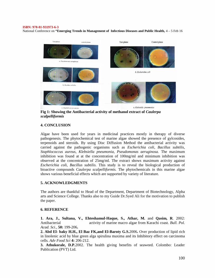
ISBN: 978-81-931973-6-3National Conference on “Emerging Trends in Management of Infectious Diseases and Public Health, 4 – 5 Feb 16
100
Fig 1: Showing the Antibacterial activity of methanol extract of Caulerpascalpelliformis
4. CONCLUSION
Algae have been used for years in medicinal practices mostly in therapy of diversepathogenesis. The phytochemical test of marine algae showed the presence of gylcosides,terpenoids and steroids. By using Disc Diffusion Method the antibacterial activity wascarried against the pathogenic organisms such as Escherichia coli, Bacillus subtilis,Staphlococcus aureus, Klebsiella pneumonia, Pseudomonas aeruginosa. The maximuminhibition was found at at the concentration of 100mg/ml and minimum inhibition wasobserved at the concentration of 25mg/ml. The extract shows maximum activity againstEscherichia coli, Bacillus subtilis. This study is to reveal the biological production ofbioactive compounds Caulerpa scalpelliformis. The phytochemicals in this marine algaeshows various beneficial effects which are supported by variety of literature.
5. ACKNOWLEDGMENTS
The authors are thankful to Head of the Department, Department of Biotechnology, Alphaarts and Science College. Thanks also to my Guide Dr.Syed Ali for the motivation to publishthe paper.
6. REFERENCE
1. Ara, J., Sultana, V., Ehteshamul-Haque, S., Athar, M. and Qasim, R. 2002:Antibacterial activity of marine macro algae from Karachi coast. Bull. Pol.Acad. Sci., 50: 199-206.2. Abd El- baky H.H., El Baz FK,and El-Baroty G.S.2006. Over production of lipid richin linolenic acid by blue green alga spirulina maxima and its Inhibitory effect on carcinomacells. Adv Food Sci 4: 206-212.3. Athukorale, D.P.2002. The health giving benefits of seaweed. Colombo: LeaderPublication (PVT) Ltd.
ISBN: 978-81-931973-6-3National Conference on “Emerging Trends in Management of Infectious Diseases and Public Health, 4 – 5 Feb 16
101
4. Benkendroff,K., Davis, A.R., Rogers, C.N., and Bremner, J.B.2007.Free fatty acids 1and 2 sterols in the benthic spawn of aquatic mollusks, and their associated antimicrobialthree properties. Journal of experimental marine biology and ecology, 316: 29-44.5. Baghavathy, S., Sumathi, P. and Jancy Sherene, B.I. 2011. Green algae Chlorococcumhumicola –a new source of bioactive compounds with antimicrobial activity. Asian PacificJournal Tropical Biomedicine, p.S1-S7.6. Fouladvand, M., Barazesh A., Farokhzad F., Malekizadeh H. and K.Sartavi. 2011.Evaluation of in vitro anti- Leishmanial activity of some brown, green and red algae fromthe Persian Gulf. European Review for Medical and Pharmacologicial Sciences, 15: 597-600.7. Ikekawa, N., Morisali, N. and Kyosuke, Tsudo.1968. Sterol compositions in some greenalgae and brown algae. Steroids, 12(1):41-48.8. Luesch, H., Yoshida,W.Y., Moore, R.E., Paul, V.J and Mooberry, S.L. 2000. Isolation,Structure determination and biological activity of Lyngbyabellin A from the marineCyanobacterium Lyngbya majuscule.Journal of Natural Products, 63: 611-615.9. Matsukawa, R., D ubinsky, Z., Kishimoto, E., Masuda, Y. and Takeuchi, T.1997. Acomparison of screening methods for antioxidant activity in seaweeds. Journal of AppliedPhycology, 9(1): 29-35.10. Peteros N.P and Uy M.M. 2010. Antioxidant and Cytotoxic activities andphytochemical screening of four philipine medicinal plants, Journal of medicinal plantsresearch, 4(5): 407-414.11. Rajasulochana, P., D hamotharan, R., Kirshnamoorthy, P. and Murugern, S. 2009.Antibacterial activity of the extracts of marine red and brown algae, Journal of AmericianScience, 5(3): 20-25.12. Rangari, V.D (2003), Pharmacognosy and phytochemistry part 2, career Publications339-341.13. Thillairajasekar .K., Duraipandian. V., Peumal. P. and Ignacimuthu .S. 2009Antimicrobial activity of Trichodesmium erythraceum (Microalgae) from south east coast ofTamil Nadu, India. International Journal of Integrated Biology, 5 (3): 167-170.14. Yuan, Y.V. 2008. Marine algal constituents. In: C. Barrow & F.Shahidi (Eds), Marinenutraceuticals and functional foods. Boca Raton, CRC, 259-296.15. Youngblood,W.W and Blumer, M. 1973.Alkanes and Alkenes in marine benthicalgae,marine biology, 21(3):163-172.
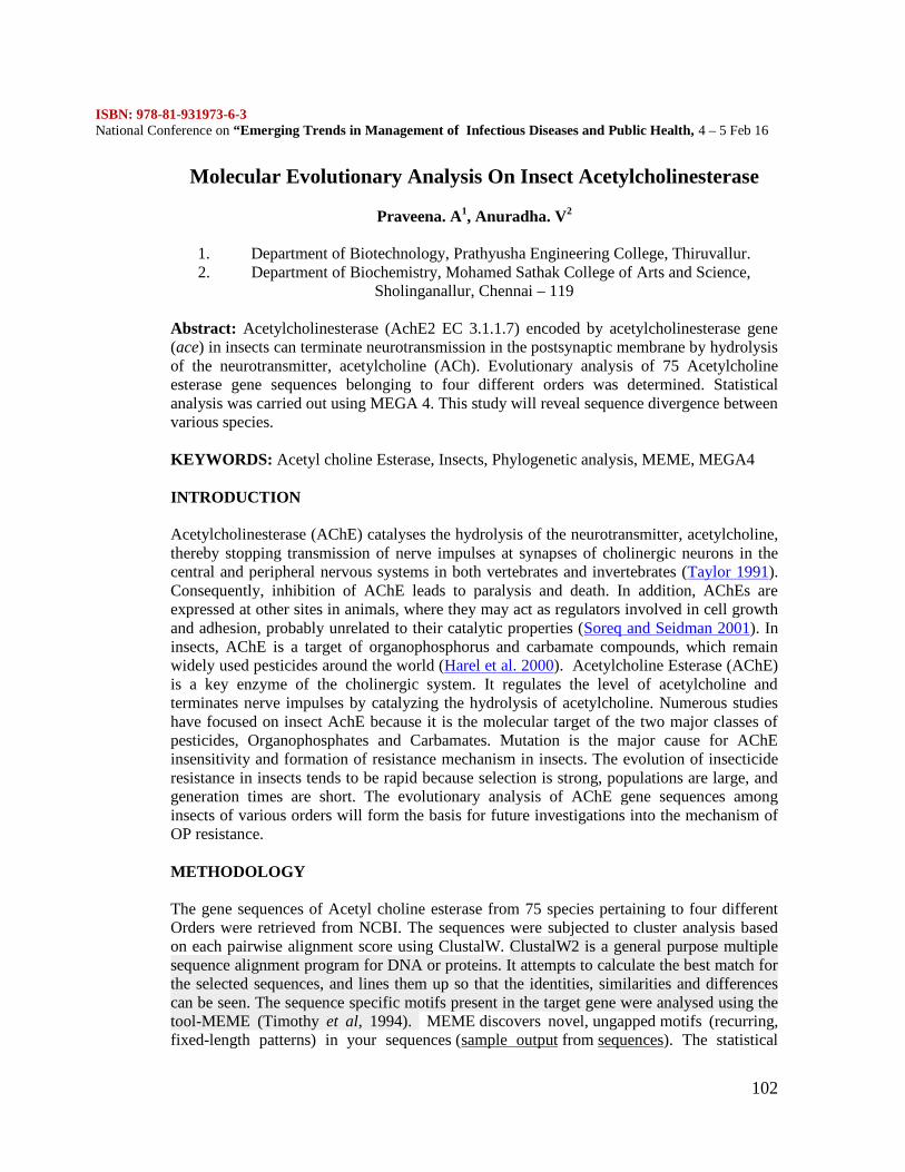
ISBN: 978-81-931973-6-3National Conference on “Emerging Trends in Management of Infectious Diseases and Public Health, 4 – 5 Feb 16
102
Molecular Evolutionary Analysis On Insect Acetylcholinesterase
Praveena. A1, Anuradha. V2
1. Department of Biotechnology, Prathyusha Engineering College, Thiruvallur.2. Department of Biochemistry, Mohamed Sathak College of Arts and Science,
Sholinganallur, Chennai – 119
Abstract: Acetylcholinesterase (AchE2 EC 3.1.1.7) encoded by acetylcholinesterase gene(ace) in insects can terminate neurotransmission in the postsynaptic membrane by hydrolysisof the neurotransmitter, acetylcholine (ACh). Evolutionary analysis of 75 Acetylcholineesterase gene sequences belonging to four different orders was determined. Statisticalanalysis was carried out using MEGA 4. This study will reveal sequence divergence betweenvarious species.
KEYWORDS: Acetyl choline Esterase, Insects, Phylogenetic analysis, MEME, MEGA4
INTRODUCTION
Acetylcholinesterase (AChE) catalyses the hydrolysis of the neurotransmitter, acetylcholine,thereby stopping transmission of nerve impulses at synapses of cholinergic neurons in thecentral and peripheral nervous systems in both vertebrates and invertebrates (Taylor 1991).Consequently, inhibition of AChE leads to paralysis and death. In addition, AChEs areexpressed at other sites in animals, where they may act as regulators involved in cell growthand adhesion, probably unrelated to their catalytic properties (Soreq and Seidman 2001). Ininsects, AChE is a target of organophosphorus and carbamate compounds, which remainwidely used pesticides around the world (Harel et al. 2000). Acetylcholine Esterase (AChE)is a key enzyme of the cholinergic system. It regulates the level of acetylcholine andterminates nerve impulses by catalyzing the hydrolysis of acetylcholine. Numerous studieshave focused on insect AchE because it is the molecular target of the two major classes ofpesticides, Organophosphates and Carbamates. Mutation is the major cause for AChEinsensitivity and formation of resistance mechanism in insects. The evolution of insecticideresistance in insects tends to be rapid because selection is strong, populations are large, andgeneration times are short. The evolutionary analysis of AChE gene sequences amonginsects of various orders will form the basis for future investigations into the mechanism ofOP resistance.
METHODOLOGY
The gene sequences of Acetyl choline esterase from 75 species pertaining to four differentOrders were retrieved from NCBI. The sequences were subjected to cluster analysis basedon each pairwise alignment score using ClustalW. ClustalW2 is a general purpose multiplesequence alignment program for DNA or proteins. It attempts to calculate the best match forthe selected sequences, and lines them up so that the identities, similarities and differencescan be seen. The sequence specific motifs present in the target gene were analysed using thetool-MEME (Timothy et al, 1994). MEME discovers novel, ungapped motifs (recurring,fixed-length patterns) in your sequences (sample output from sequences). The statistical
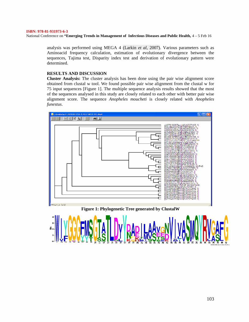
ISBN: 978-81-931973-6-3National Conference on “Emerging Trends in Management of Infectious Diseases and Public Health, 4 – 5 Feb 16
103
analysis was performed using MEGA 4 (Larkin et al, 2007). Various parameters such asAminoacid frequency calculation, estimation of evolutionary divergence between thesequences, Tajima test, Disparity index test and derivation of evolutionary pattern weredetermined.
RESULTS AND DISCUSSIONCluster Analysis: The cluster analysis has been done using the pair wise alignment scoreobtained from clustal w tool. We found possible pair wise alignment from the clustal w for75 input sequences [Figure 1]. The multiple sequence analysis results showed that the mostof the sequences analysed in this study are closely related to each other with better pair wisealignment score. The sequence Anopheles moucheti is closely related with Anophelesfunestus.
Figure 1: Phylogenetic Tree generated by ClustalW
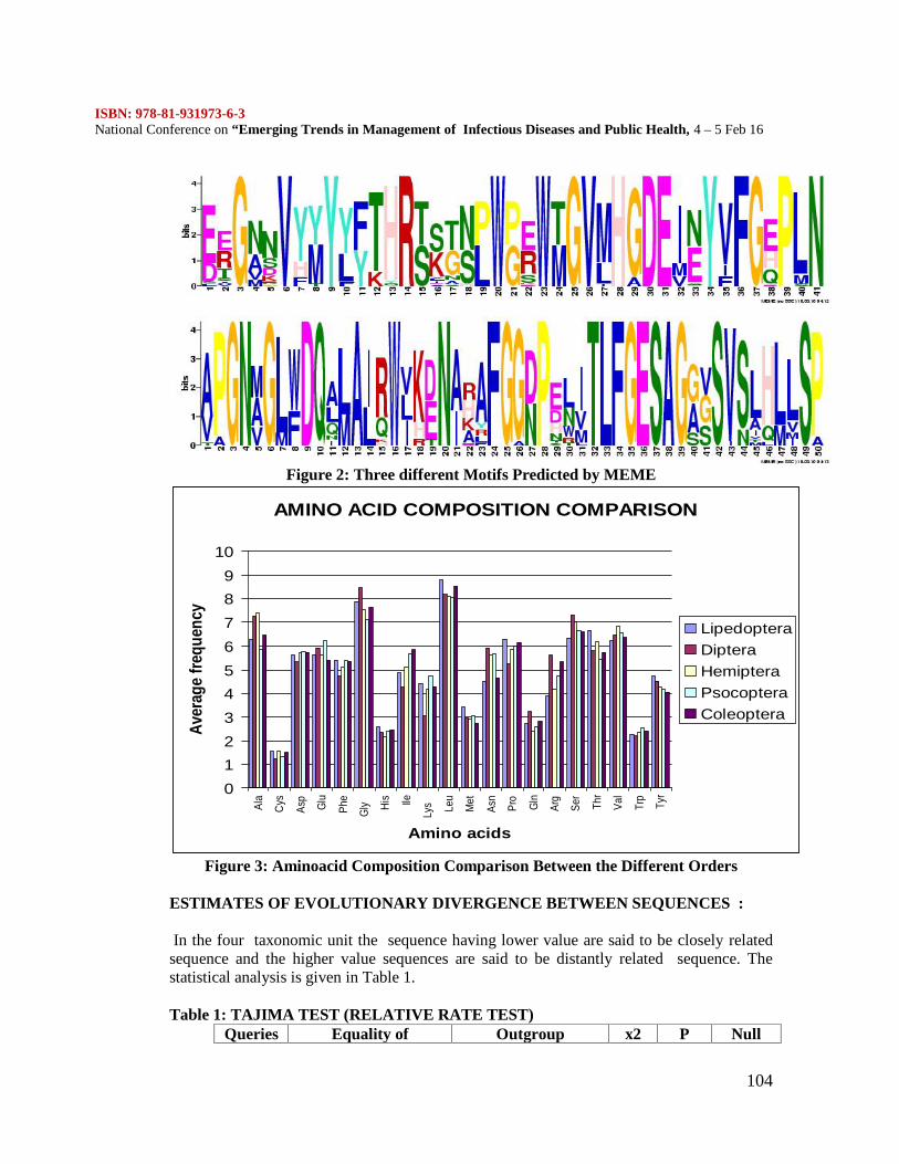
ISBN: 978-81-931973-6-3National Conference on “Emerging Trends in Management of Infectious Diseases and Public Health, 4 – 5 Feb 16
104
Figure 2: Three different Motifs Predicted by MEME
AMINO ACID COMPOSITION COMPARISON
0123456789
10
Ala
Cys
Asp
Glu
Phe
Gly Hi
s Ile
Lys Le
u
Met
Asn
Pro
Gln
Arg
Ser
Thr
Val
Trp
Tyr
Amino acids
Aver
age
frequ
ency
LipedopteraDipteraHemipteraPsocopteraColeoptera
Figure 3: Aminoacid Composition Comparison Between the Different Orders
ESTIMATES OF EVOLUTIONARY DIVERGENCE BETWEEN SEQUENCES :
In the four taxonomic unit the sequence having lower value are said to be closely relatedsequence and the higher value sequences are said to be distantly related sequence. Thestatistical analysis is given in Table 1.
Table 1: TAJIMA TEST (RELATIVE RATE TEST)Queries Equality of Outgroup x2 P Null
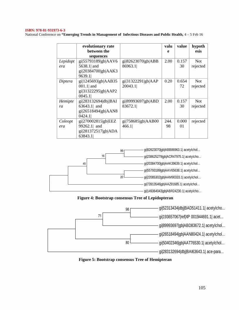
ISBN: 978-81-931973-6-3National Conference on “Emerging Trends in Management of Infectious Diseases and Public Health, 4 – 5 Feb 16
105
evolutionary ratebetween thesequences
value
value hypothesis
Lepidoptera
gi|55793189|gb|AAV65638.1| andgi|20384700|gb|AAK39639.1|
gi|82623070|gb|ABB86963.1|
2.00 0.15730
Notrejected
Diptera gi|1245693|gb|AAB35001.1| andgi|31322295|gb|AAP20045.1|
gi|31322291|gb|AAP20043.1|
0.20 0.65472
Notrejected
Hemiptera
gi|283132694|dbj|BAI63643.1| andgi|26518494|gb|AAN80424.1|
gi|89993697|gb|ABD83672.1|
2.00 0.15730
Notrejected
Coleoptera
gi|270002815|gb|EEZ99262.1| andgi|281372517|gb|ADA63843.1|
gi|758685|gb|AAB00466.1|
244.98
0.00001
rejected
gi|82623070|gb|ABB86963.1| acetylchol...
gi|238625279|gb|ACR47975.1| acetylcho...
gi|20384700|gb|AAK39639.1| acetylchol...
gi|55793189|gb|AAV65638.1| acetylchol...
gi|22085302|gb|AAM90333.1| acetylchol...
gi|73913546|gb|AAZ91685.1| acetylchol...
gi|149364043|gb|ABR24230.1| acetylcho...
99
20
16
45
Figure 4: Bootstrap consensus Tree of Lepidopteran
gi|52313434|dbj|BAD51411.1| acetylcho...
gi|193657067|ref|XP 001944691.1| acet...
gi|89993697|gb|ABD83672.1| acetylchol...
gi|26518494|gb|AAN80424.1| acetylchol...
gi|50402346|gb|AAT76530.1| acetylchol...
gi|283132694|dbj|BAI63643.1| ace-para...
7198
80
Figure 5: Bootstrap consensus Tree of Hemipteran
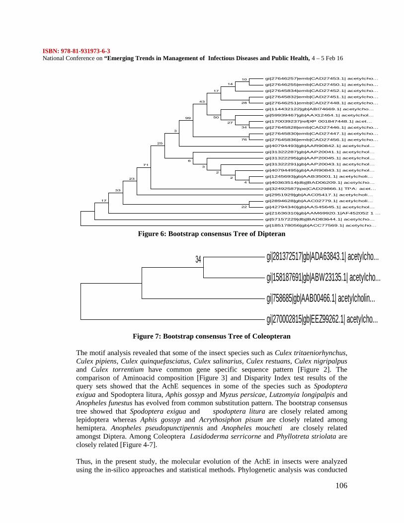
ISBN: 978-81-931973-6-3National Conference on “Emerging Trends in Management of Infectious Diseases and Public Health, 4 – 5 Feb 16
106
gi|27646257|emb|CAD27453.1| acetylcho...
gi|27646255|emb|CAD27450.1| acetylcho...
gi|27645834|emb|CAD27452.1| acetylcho...
gi|27645832|emb|CAD27451.1| acetylcho...
gi|27646251|emb|CAD27448.1| acetylcho...
gi|114432122|gb|ABI74669.1| acetylcho...
gi|59939467|gb|AAX12464.1| acetylchol...
gi|170039237|ref|XP 001847448.1| acet...
gi|27645828|emb|CAD27446.1| acetylcho...
gi|27645830|emb|CAD27447.1| acetylcho...
gi|27645836|emb|CAD27456.1| acetylcho...
gi|40794493|gb|AAR90842.1| acetylchol...
gi|31322287|gb|AAP20041.1| acetylchol...
gi|31322295|gb|AAP20045.1| acetylchol...
gi|31322291|gb|AAP20043.1| acetylchol...
gi|40794495|gb|AAR90843.1| acetylchol...
gi|1245693|gb|AAB35001.1| acetylcholi...
gi|40363514|dbj|BAD06209.1| acetylcho...
gi|32492587|tpe|CAD29866.1| TPA: acet...
gi|2951929|gb|AAC05417.1| acetylcholi...
gi|2894628|gb|AAC02779.1| acetylcholi...
gi|42794340|gb|AAS45645.1| acetylchol...
gi|21636310|gb|AAM69920.1|AF452052 1 ...
gi|57157229|dbj|BAD83644.1| acetylcho...
gi|185178056|gb|ACC77569.1| acetylcho...
4
76
3427
50
2
2
3
6
28
22
1014
17
43
99
3
25
71
23
33
17
Figure 6: Bootstrap consensus Tree of Dipteran
gi|281372517|gb|ADA63843.1| acetylcho...
gi|158187691|gb|ABW23135.1| acetylcho...
gi|758685|gb|AAB00466.1| acetylcholin...
gi|270002815|gb|EEZ99262.1| acetylcho...
34
Figure 7: Bootstrap consensus Tree of Coleopteran
The motif analysis revealed that some of the insect species such as Culex tritaeniorhynchus,Culex pipiens, Culex quinquefasciatus, Culex salinarius, Culex restuans, Culex nigripalpusand Culex torrentium have common gene specific sequence pattern [Figure 2]. Thecomparison of Aminoacid composition [Figure 3] and Disparity Index test results of thequery sets showed that the AchE sequences in some of the species such as Spodopteraexigua and Spodoptera litura, Aphis gossyp and Myzus persicae, Lutzomyia longipalpis andAnopheles funestus has evolved from common substitution pattern. The bootstrap consensustree showed that Spodoptera exigua and spodoptera litura are closely related amonglepidoptera whereas Aphis gossyp and Acrythosiphon pisum are closely related amonghemiptera. Anopheles pseudopunctipennis and Anopheles moucheti are closely relatedamongst Diptera. Among Coleoptera Lasidoderma serricorne and Phyllotreta striolata areclosely related [Figure 4-7].
Thus, in the present study, the molecular evolution of the AchE in insects were analyzedusing the in-silico approaches and statistical methods. Phylogenetic analysis was conducted
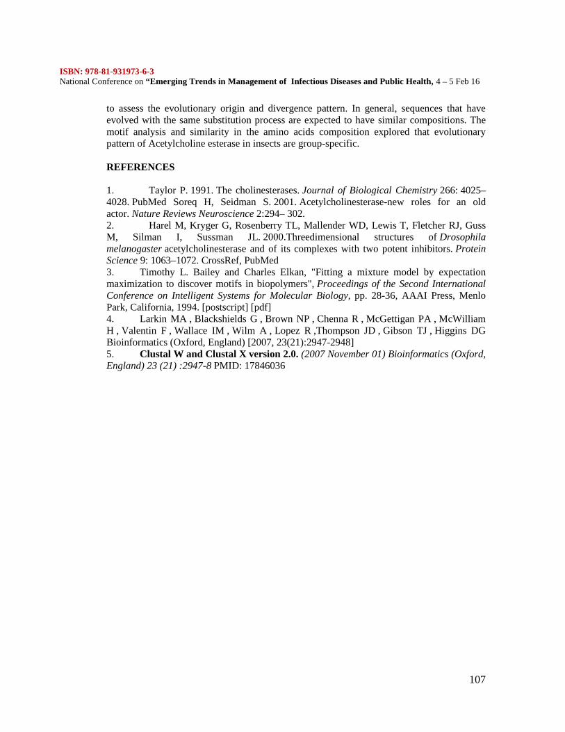
ISBN: 978-81-931973-6-3National Conference on “Emerging Trends in Management of Infectious Diseases and Public Health, 4 – 5 Feb 16
107
to assess the evolutionary origin and divergence pattern. In general, sequences that haveevolved with the same substitution process are expected to have similar compositions. Themotif analysis and similarity in the amino acids composition explored that evolutionarypattern of Acetylcholine esterase in insects are group-specific.
REFERENCES
1. Taylor P. 1991. The cholinesterases. Journal of Biological Chemistry 266: 4025–4028. PubMed Soreq H, Seidman S. 2001. Acetylcholinesterase-new roles for an oldactor. Nature Reviews Neuroscience 2:294– 302.2. Harel M, Kryger G, Rosenberry TL, Mallender WD, Lewis T, Fletcher RJ, GussM, Silman I, Sussman JL. 2000.Threedimensional structures of Drosophilamelanogaster acetylcholinesterase and of its complexes with two potent inhibitors. ProteinScience 9: 1063–1072. CrossRef, PubMed3. Timothy L. Bailey and Charles Elkan, "Fitting a mixture model by expectationmaximization to discover motifs in biopolymers", Proceedings of the Second InternationalConference on Intelligent Systems for Molecular Biology, pp. 28-36, AAAI Press, MenloPark, California, 1994. [postscript] [pdf]4. Larkin MA , Blackshields G , Brown NP , Chenna R , McGettigan PA , McWilliamH , Valentin F , Wallace IM , Wilm A , Lopez R ,Thompson JD , Gibson TJ , Higgins DGBioinformatics (Oxford, England) [2007, 23(21):2947-2948]5. Clustal W and Clustal X version 2.0. (2007 November 01) Bioinformatics (Oxford,England) 23 (21) :2947-8 PMID: 17846036
ISBN: 978-81-931973-6-3National Conference on “Emerging Trends in Management of Infectious Diseases and Public Health, 4 – 5 Feb 16
108
Preliminary study on isolation and characterization of dyedecolorizing bacteria from lime stone soils
Mahalakshmi R1, Balachandar C2, Antony Prabhu J3, Melchias G4
3,4 Department of Biotechnology, St. Joseph’s college, Tiruchchirappalli 6200021,2Department of Biotechnology, SRM Arts and Science College, Kattankulathur
6032034Corresponding author
ABSTRACT
Microorganisms from saline environments proved to be beneficial for commercial as well asresearch applications. Calcium carbonate rich lime stone fossils are one among thoseunfavourable conditions that supports extremely halophilic or moderately halophilicorganisms. Current investigation on ariylaur lime stone samples is a preliminary initiative tounearth microorganisms possessing beneficial applications. Four distinct isolates wereselected from pool of colonies and characterized for their ability to degrade followingcommercial dyes - reactive deep red, diazone green and reactive blue G. Results showed thatall these isolates are found to be decolorizing the dyes in effective manner and they can beconsidered as potential candidates to address the menace of dyes as environmental pollutant.
INTRODUCTION
Microorganisms are ubiquitous and are considered as ‘masters of the biosphere’. Everyprocess in the biosphere is touched by the seemingly endless capacity of microbes totransform the world around them. They underpin most geochemical cycles and many humanhealth conditions that were previously thought to be driven by inorganic processes andstress. (Reysenbach And Hamamura, 2008). Soil is considered a storehouse of microbialactivity (Tilak et al., 2005). Understanding the biodiversity of the soil is difficult because ofthe heterogeneity that exists on both local and geological scales (Horner-Devine et al., 2004;Fierer and Jackson, 2006; Kang and Mills, 2006). Texture, temperature, salinity, pH,contaminants and other characteristics allow a unique microbial community to establish aniche (Zwolinski, 2007). Extreme environments with variable salinity, temperatures,underground environments (ice and rocks), and deep sea vent (extremely high temperatureand pressure) contain specialized microbial communities, mostly known as Halophiles. Theyprovide important clues on the origin, evolution of life and geological time scales. They alsoprovide beneficial genomic, protein resources. Functional characterization ofmicroorganisms for specific functions such as salt tolerance, antibiotic production, orenzyme activity (hydrolysis, bioremediation) helps to identify microbes that have potentialapplications in medicine, agriculture or industry (Oren, 2002).
Ariyalur, youngest district in Tamil Nadu, is famous for limestone resources. According tothe previous archaeological study, about 100 million years ago, spontaneous transformationphenomenon of sea into land caused burial of aquatic organisms and subsequent formationof fossil prints of those creatures as soil layers of the site. These soil layers still hold thesame aquatic nature; hence researchers have opined that microorganisms of ancient timesmight still be existing along with the fossils underground (Manivel 2005; Seshachur 1932).
ISBN: 978-81-931973-6-3National Conference on “Emerging Trends in Management of Infectious Diseases and Public Health, 4 – 5 Feb 16
109
Thus analysis of microbial communities by traditional microbiological and recentlydeveloped metagenomic approach would offer evidence of the rare microbial communitiesand their evolutionary pathways. Additionally, beneficial genes and biochemical pathwayscan also be prospected from them.
In the study, soil samples collected 150 ft below ground off the Ariyalur site wereinvestigated for cultivable microbial diversity along with their possible beneficial genomicand protein contents. Cultivable microorganisms were isolated and subjected to preliminarycharacterization with routine microbiological techniques. Their functional property wasassessed for degrading hazardous chemicals in commercial dye solutions.
MATERIALS AND METHODS
To isolate the culturable bacteria from soil, a portion of the soil samples was overlaid withsterile distilled water in a sterile falcon tube and were left undisturbed for about a week. Thisprocedure allows the bacterial population to get concentrated in the overlaid water.
1) Isolation of single colonyTo separate mixed population of microorganisms, 100µl of the overlaid liquid from eachsample was serially diluted (10-1 to 10-10). An aliquot of 50µl was spread on Luria-Bertani(LB) agar (pH 7.5). Colonies obtained by were selected based morphology and werequadrant streaked on LB agar and single colonies were isolated and inoculated in an LBbroth (pH 7.2).2) Bacterial growth kineticsOne ml of the overlaying aqueous sample was inoculated into 50ml LB broth and incubatedat 37ºC in a shaker. The cultures were allowed to grow overnight. This served as theinoculum. An aliquot of 100µl inoculam was added to 30ml LB broth and kept in a shaker at37ºC. Sampling was done every 2 hours for 12 hours. Wet cell mass concentration andturbidity were measured.3) Morphological & Biochemical characterizationIsolates were then subjected to standard morphological characterization & biochemicalcharacterization. Gram’s staining, Capsular staining, Endospore staining, Flagellar staining,motility assay were performed to reveal the morphological organization of fossil isolates. 25Biochemical tests (name of tests) were done to characterize functional properties of isolates.4) Genome extraction and 16S AmplificationIn view of standardization and molecular characterization purposes, genome of isolatedcultures and soil microorganisms were extracted using Mo-Bio power soil DNA kit. 16SrRNA gene amplification for all the samples was standardized with repeated PCR using25pM each of universal primers 16F27 (5´-CCA GAG TTT GAT CMT GGC TCA G-3´)and 16R1525 (5´-TTC TGC AGT CTA GAA GGA GGT GWT CCA GCC-3´) (Pidiyar etal., 2004).Concentrations of other components are dNTPS (10mM each), Buffer (1X), Taqpolymerase (1U), sterile milliQ water (15.8 µl/ 25 µl) and finally 1 µl template DNA.5) Dye degradation studyThree dyes, diazone green, reactive deep red and reactive blue G were selected for study.Four falcon tubes were filled with 20ml of broth and 500µl of 0.1% of each dye was addedseparately. The tubes were then inoculated separately with 1ml of pure culture. The tubeswere incubated in a shaker (120 rpm at 37ºC) along with a positive control. The samples that
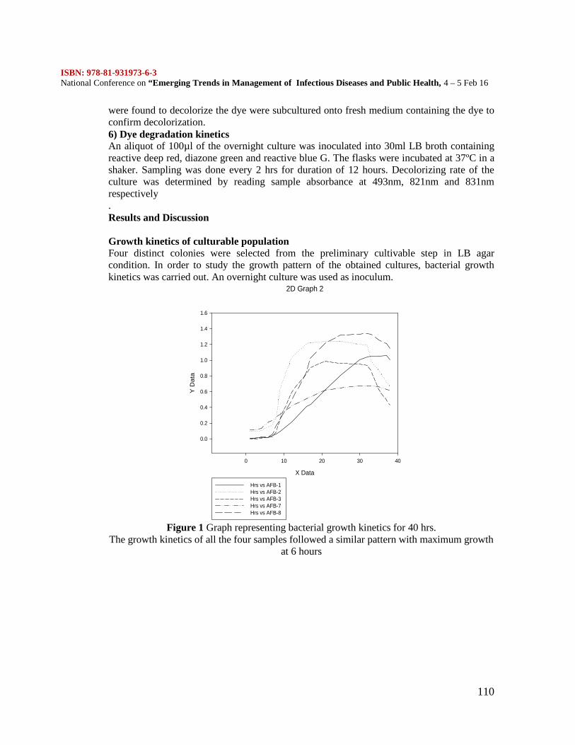
ISBN: 978-81-931973-6-3National Conference on “Emerging Trends in Management of Infectious Diseases and Public Health, 4 – 5 Feb 16
110
were found to decolorize the dye were subcultured onto fresh medium containing the dye toconfirm decolorization.6) Dye degradation kineticsAn aliquot of 100µl of the overnight culture was inoculated into 30ml LB broth containingreactive deep red, diazone green and reactive blue G. The flasks were incubated at 37ºC in ashaker. Sampling was done every 2 hrs for duration of 12 hours. Decolorizing rate of theculture was determined by reading sample absorbance at 493nm, 821nm and 831nmrespectively.Results and Discussion
Growth kinetics of culturable populationFour distinct colonies were selected from the preliminary cultivable step in LB agarcondition. In order to study the growth pattern of the obtained cultures, bacterial growthkinetics was carried out. An overnight culture was used as inoculum.
2D Graph 2
X Data
0 10 20 30 40
Y D
ata
0.0
0.2
0.4
0.6
0.8
1.0
1.2
1.4
1.6
Hrs vs AFB-1Hrs vs AFB-2Hrs vs AFB-3Hrs vs AFB-7Hrs vs AFB-8
Figure 1 Graph representing bacterial growth kinetics for 40 hrs.The growth kinetics of all the four samples followed a similar pattern with maximum growth
at 6 hours
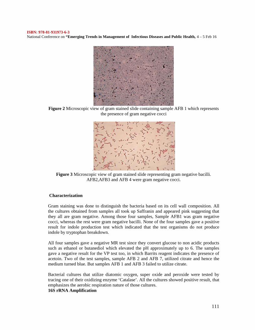
ISBN: 978-81-931973-6-3National Conference on “Emerging Trends in Management of Infectious Diseases and Public Health, 4 – 5 Feb 16
111
Figure 2 Microscopic view of gram stained slide containing sample AFB 1 which representsthe presence of gram negative cocci
.Figure 3 Microscopic view of gram stained slide representing gram negative bacilli.
AFB2,AFB3 and AFB 4 were gram negative cocci.
Characterization
Gram staining was done to distinguish the bacteria based on its cell wall composition. Allthe cultures obtained from samples all took up Saffranin and appeared pink suggesting thatthey all are gram negative. Among those four samples, Sample AFB1 was gram negativecocci, whereas the rest were gram negative bacilli. None of the four samples gave a positiveresult for indole production test which indicated that the test organisms do not produceindole by tryptophan breakdown.
All four samples gave a negative MR test since they convert glucose to non acidic productssuch as ethanol or butanediol which elevated the pH approximately up to 6. The samplesgave a negative result for the VP test too, in which Barrits reagent indicates the presence ofacetoin. Two of the test samples, sample AFB 2 and AFB 7, utilized citrate and hence themedium turned blue. But samples AFB 1 and AFB 3 failed to utilize citrate.
Bacterial cultures that utilize diatomic oxygen, super oxide and peroxide were tested bytracing one of their oxidizing enzyme ‘Catalase’. All the cultures showed positive result, thatemphasizes the aerobic respiration nature of those cultures.16S rRNA Amplification
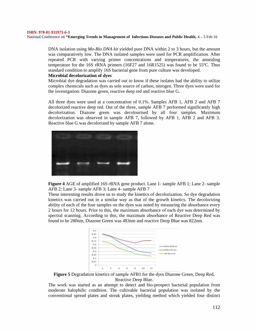
ISBN: 978-81-931973-6-3National Conference on “Emerging Trends in Management of Infectious Diseases and Public Health, 4 – 5 Feb 16
112
DNA isolation using Mo-Bio DNA kit yielded pure DNA within 2 to 3 hours, but the amountwas comparatively low. The DNA isolated samples were used for PCR amplification. Afterrepeated PCR with varying primer concentrations and temperatures, the annealingtemperature for the 16S rRNA primers (16F27 and 16R1525) was found to be 55ºC. Thusstandard condition to amplify 16S bacterial gene from pure culture was developed.Microbial decolorization of dyesMicrobial dye degradation was carried out to know if these isolates had the ability to utilizecomplex chemicals such as dyes as sole source of carbon, nitrogen. Three dyes were used forthe investigation: Diazone green, reactive deep red and reactive blue G.
All three dyes were used at a concentration of 0.1%. Samples AFB 1, AFB 2 and AFB 7decolorized reactive deep red. Out of the three, sample AFB 7 performed significantly highdecolorization. Diazone green was decolourised by all four samples. Maximumdecolorization was observed in sample AFB 7, followed by AFB 1, AFB 2 and AFB 3.Reactive blue G was decolorized by sample AFB 7 alone.
Figure 4 AGE of amplified 16S rRNA gene product. Lane 1- sample AFB 1; Lane 2- sampleAFB 2; Lane 3- sample AFB 3; Lane 4- sample AFB 7These interesting results drove us to study the kinetics of decolorization. So dye degradationkinetics was carried out in a similar way as that of the growth kinetics. The decolorizingability of each of the four samples on the dyes was noted by measuring the absorbance every2 hours for 12 hours. Prior to this, the maximum absorbance of each dye was determined byspectral scanning. According to this, the maximum absorbance of Reactive Deep Red wasfound to be 280nm, Diazone Green was 483nm and reactive Deep Blue was 822nm.
Figure 5 Degradation kinetics of sample AFB1 for the dyes Diazone Green, Deep Red,Reactive Deep Blue.
The work was started as an attempt to detect and bio-prospect bacterial population frommoderate halophilic condition. The cultivable bacterial population was isolated by theconventional spread plates and streak plates, yielding method which yielded four distinct
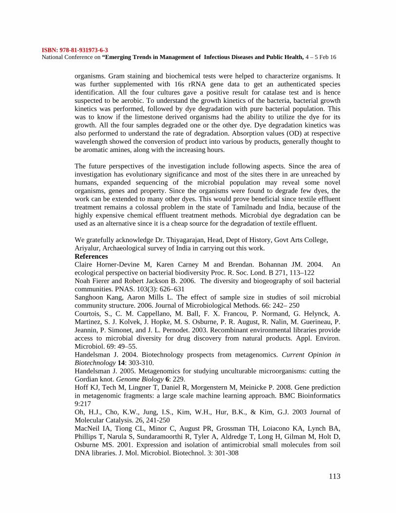
ISBN: 978-81-931973-6-3National Conference on “Emerging Trends in Management of Infectious Diseases and Public Health, 4 – 5 Feb 16
113
organisms. Gram staining and biochemical tests were helped to characterize organisms. Itwas further supplemented with 16s rRNA gene data to get an authenticated speciesidentification. All the four cultures gave a positive result for catalase test and is hencesuspected to be aerobic. To understand the growth kinetics of the bacteria, bacterial growthkinetics was performed, followed by dye degradation with pure bacterial population. Thiswas to know if the limestone derived organisms had the ability to utilize the dye for itsgrowth. All the four samples degraded one or the other dye. Dye degradation kinetics wasalso performed to understand the rate of degradation. Absorption values (OD) at respectivewavelength showed the conversion of product into various by products, generally thought tobe aromatic amines, along with the increasing hours.
The future perspectives of the investigation include following aspects. Since the area ofinvestigation has evolutionary significance and most of the sites there in are unreached byhumans, expanded sequencing of the microbial population may reveal some novelorganisms, genes and property. Since the organisms were found to degrade few dyes, thework can be extended to many other dyes. This would prove beneficial since textile effluenttreatment remains a colossal problem in the state of Tamilnadu and India, because of thehighly expensive chemical effluent treatment methods. Microbial dye degradation can beused as an alternative since it is a cheap source for the degradation of textile effluent.
We gratefully acknowledge Dr. Thiyagarajan, Head, Dept of History, Govt Arts College,Ariyalur, Archaeological survey of India in carrying out this work.ReferencesClaire Horner-Devine M, Karen Carney M and Brendan. Bohannan JM. 2004. Anecological perspective on bacterial biodiversity Proc. R. Soc. Lond. B 271, 113–122Noah Fierer and Robert Jackson B. 2006. The diversity and biogeography of soil bacterialcommunities. PNAS. 103(3): 626– 631Sanghoon Kang, Aaron Mills L. The effect of sample size in studies of soil microbialcommunity structure. 2006. Journal of Microbiological Methods. 66: 242– 250Courtois, S., C. M. Cappellano, M. Ball, F. X. Francou, P. Normand, G. Helynck, A.Martinez, S. J. Kolvek, J. Hopke, M. S. Osburne, P. R. August, R. Nalin, M. Guerineau, P.Jeannin, P. Simonet, and J. L. Pernodet. 2003. Recombinant environmental libraries provideaccess to microbial diversity for drug discovery from natural products. Appl. Environ.Microbiol. 69: 49–55.Handelsman J. 2004. Biotechnology prospects from metagenomics. Current Opinion inBiotechnology 14: 303-310.Handelsman J. 2005. Metagenomics for studying unculturable microorganisms: cutting theGordian knot. Genome Biology 6: 229.Hoff KJ, Tech M, Lingner T, Daniel R, Morgenstern M, Meinicke P. 2008. Gene predictionin metagenomic fragments: a large scale machine learning approach. BMC Bioinformatics9:217Oh, H.J., Cho, K.W., Jung, I.S., Kim, W.H., Hur, B.K., & Kim, G.J. 2003 Journal ofMolecular Catalysis. 26, 241-250MacNeil IA, Tiong CL, Minor C, August PR, Grossman TH, Loiacono KA, Lynch BA,Phillips T, Narula S, Sundaramoorthi R, Tyler A, Aldredge T, Long H, Gilman M, Holt D,Osburne MS. 2001. Expression and isolation of antimicrobial small molecules from soilDNA libraries. J. Mol. Microbiol. Biotechnol. 3: 301-308
ISBN: 978-81-931973-6-3National Conference on “Emerging Trends in Management of Infectious Diseases and Public Health, 4 – 5 Feb 16
114
Manivel, M., Sakthivel, R., Muthu, M.S.P., and Kamalakkannan,R 2005 EnvironmentalImpact Assessement Of Cretaceous Limestone Mining Area Of Dalmiapuram OfTiruchirappalli, India Using Remote Sensing And Gis Map India 2005 Geomatics 2005Oren A. 2002. Diversity of halophilic microorganisms: Environments, phylogeny,physiology, and applications. Journal of Industrial Microbiology & Biotechnology 28: 56-63Riesenfeld CS, Goodman RM, and Handelsman J. 2004. Uncultured soil bacteria are areservoir of new antibiotic resistance genes. Environ. Microbiol. 6: 981–989.Reysenbach A.L. and Hamamura. N. 2008. A geobiological perspective on metagenomics.Geobiology 6:337-340Seshachur B, Narayana Rao A, Ramasamy LS 1932 Further notes on Ariyalur fossils 1: 39-40Simu K, and Hagstrom A. 2004. Oligotrophic bacterioplankton with a novel single-cell lifestrategy. Appl. Environ. Microbiol. 70: 2445–2451.Stein JL, Marsh TL, Wu KY, Shizuya H, and DeLong EF. 1996. Characterization ofuncultivated prokaryotes: isolation and analysis of a 40- kilobase-pair genome fragmentfront a planktonic marine archaeon. J. Bacteriol. 178: 591–599.Tilak KVBR, Ranganayaki N, Pal KK, De R, Saxena AK, Nautiyal CS,Mittal S, TripathiAK, Johri BN. 2005. Diversity of plant growth and soil health supporting bacteria. Curr. Sci.89:136-150Venter, J. C., K. Remington, J. F. Heidelberg, A. L. Halpern, D. Rusch, J. A. Eisen, D. Wu,I. Paulsen, K. E. Nelson, W. Nelson, D. E. Fouts, S. Levy, A. H. Knap, M. W. Lomas, K.Nealson, O. White, J. Peterson, J. Hoffman, R. Parsons, H. Baden-Tillson, C. Pfannkoch, Y.H. Rogers, and H. O. Smith. 2004. Environmental genome shotgun sequencing of theSargasso Sea. Science 304:66–74.Vyankatesh Pidiyar J, Kamlesh Jangid, Milind Patole S and Yogesh Shouche S .2004.Studies on cultured and uncultured microbiota of wild culex quinquefasciatus mosquitomidgut based on 16s ribosomal rna gene analysis. Am. J. Trop. Med. Hyg., 70:597–603Zwolinski MD. 2007. DNA sequencing: strategies for soil microbiology. Soil Sci. Soc. Am.J. 71: 592-600.
ISBN: 978-81-931973-6-3National Conference on “Emerging Trends in Management of Infectious Diseases and Public Health, 4 – 5 Feb 16
115
EFFICIENT MANAGEMENT STRATEGIES OF COMBATTING FUNGALINFECTIONS IN COPRA (Cocos nucifera)Pandiyarajan G.1, Mary Swarnalatha S.2
1 II MSc Student, Department of Biotechnology, Alpha Arts and Science College, TamilNadu, India
2 Asst. Professor, Department of Biotechnology, Alpha Arts and Science College, TamilNadu, India
e- mail: [email protected] oil or copra oil is used in the country as a cooking fat, hair oil, body oil andindustrial oil. It is extracted from the kernel or meat of mature coconuts harvested fromthe coconut palm (Cocos nucifera) having maximum moisture content of six per cent.Refined coconut oil is mainly used in the manufacture of biscuits, chocolates and otherconfectionery items, ice cream, pharmaceutical products and costly paints. Coconutcopra is susceptible to fungal and insect attacks during storage. Aspergillus andPenicillium sp. infections are commonly reported in copra deterioration. Most farmerswho cultivate coconut palms stand at loss during harvest as fungi and insect infect thecopra. Hence management strategies are indispensible to eliminate fungal infections andensure high quality yield of coconut oil.KeywordsCocos nucifera, copra oil, Aspergillus and Penicillium sp., moistureINTRODUCTION
Coconut (Cocosnucifera) is a very important tree crop grown in many parts ofthe world. World production of coconuts has been steadily increasing during the lastseveral years due mainly to the increased planting of the palm in the form of bettermanaged large plantations in many of the major coconut-growing countries. More than90 per cent of area and production of coconut in India is from the four southern states ofKerala, Karnataka, Tamil Nadu and Andhra Pradesh.Dried coconut has umpty number of applications. Copra is the dried meat, or kernel, ofthe coconut. Coconut oil is extracted from it and has made copra an importantagricultural commodity for many coconut-producing countries. It also yields coconutcake which is mainly used as feed for livestock. Coconut cake is the residue left after theextraction of oil from copra which is mainly used as a cattle feed. Coconut cake contains4-5 per cent oil which is extracted by solvent extraction process. This oil is generallyused for industrial purpose and de-oiled cake is used to make mixed cattle feed. Thereare a few such units in the country especially in Kerala.In India, most farmers who cultivate coconut palms stand at loss during harvest as fungiand insect infect the copra. A number of pests including scale insects, bruchids, aphids,spider mites, mealybugs, palm weevils and some caterpillars are occasionally found oncoconut. Aspergillus and Penicillium sp. infections are commonly reported in copradeterioration.(Nathaneal, et al). The present study is aimed at studying storage problemsin copra and find good methods for high recovery of coconut oil production.MATERIALS AND METHODS1.Estimation of moisture content in coconut:The moisture content of coconut is important during drying to prevent the fungalinfection. The moisture content of the coconut was estimated by oven dry method. Ten
ISBN: 978-81-931973-6-3National Conference on “Emerging Trends in Management of Infectious Diseases and Public Health, 4 – 5 Feb 16
116
gram of the coconut sample was weighed and placed in the moisture box. The cover ofthe moisture box was removed and kept in a hot air oven previously heated to standardtemperature of 105oC. The moisture box was removed from oven exactly seven hours ofstandard temperature, 105oC and transferred to desiccators for cooling till it reaches theroom temperature. Then the final weight of sample was observed.Calculation
(Empty Wt + Sample Wt) – Final WtMoisture per cent = --------------------------------------------- × 100
Sample Wt
The fresh coconut was kept in room temperature for drying. The moisture content offresh coconut and 24 hrs interval up to 5 days was measured as mentioned above.
2. Identification of fungal infection in the coconut during Copra preparationThe fungal infection was observed during the drying process for copra. The fungus wereisolated and cultured in suitable media as mentioned below.2.a. Procedure for preparation of potato dextrose agar mediaThe potato dextrose agar media was prepared for culturing of fungus for further studies.ProcedureWeigh 3.9g of potato dextrose agar and mix 100ml of distilled water. After that add 2gmof agar-agar to dissolve shake nicely. The conical flask is to be capped (or) cottonplugged. Sterilize the conical flask in an autoclave at 121⁰C under 15 lbs. pressure for15 hour.ISOLATION OF FUNGAL CULTUREPROCEDURE:Label sterilized plate with name and dates liquidly the potato dextrose agar and pour intothe sterile plates using aseptic procedure. After pouring place on a plate tube and makesure that media is overly distributed in plate failure of cooling the media result incondensation of moisture on corner of plate which is considerable. These droplets willfail on the colonies spreading a detecting the entire isolation technique. Heats theinoculation loop to red hot, allow cooling and taking the fungal culture from infectedcoconut by using inoculation loop. Inoculate the fungal culture sample on potatodextrose agar in petriplate. Allowed for overnight culture in room temperature.ISOLATION OF FUNGUS
Identification of fungus from colony was done by the Lactophenol cotton blue method.PROCEDURE: Place a small drop of lactophenol cotton blue on a clean glassmicroscope slide before clean the laminar air low by using ethanol.Remove a smallportion of the yeast colony and placed it into the drop of lactophenol by usinginoculation loop and suspend the cells. Place a clean cover glass over the suspension andobserve microscopically.3. Effect of salt solution on the growth of fungus.Salt solution method in fresh Copra.Prepare the salt solution at 1%, 2% and 5%.1% SALT SOLUTION1g of salt was weighed and dissolved in 100ml of distilled water.Take a cotton scroll, itcan be soaked in the salt solution and the solution was applied in the fresh open
ISBN: 978-81-931973-6-3National Conference on “Emerging Trends in Management of Infectious Diseases and Public Health, 4 – 5 Feb 16
117
coconut.Leave it for 24hrs or few days for the fungal growth.Then finally go for fungalidentification.2% SALT SOLUTION2g of salt was weighed and dissolved in 100ml of distilled water.Take a cotton scroll, itcan be soaked in the salt solution and the solution was applied in the fresh opencoconut.Leave it for 24hrs or few days for the fungal growth.Then finally go for fungalidentification.5% SALT SOLUTION5g of salt was weighed and dissolved in 100ml of distilled water.Take a cotton scroll, itcan be soaked in the salt solution and the solution was applied in the fresh opencoconut.Leave it for 24hrs or few days for the fungal growth.Then finally go for fungalidentification.4.1 ACETIC ACID TREATMENTIn this method for avoid the fungal infection in fresh open coconut.Take a 250ml ofbeaker Prepare the acetic acid concentration at 5%, 7% and 10% by using micropipettes.4.2 SULFURIC ACID TREATMENTTake a 250ml of beaker Prepare the sulfuric acid concentration at 5%, 7% and 10% byusing micropipettes.4.3 ACETIC ACID WITH SULFURIC ACID TREATMENTPrepare the acetic acid 10% with sulfuric acid 10% and acetic acid 15% with sulfuricacid 5%.4.4 SPRAY TECHNIQUEUsing sprayer for spray in open coconut in above different concentration of acetic acidand sulfuric acid.4.5 DIPPING TECHNIQUEIn the method, a piece coconut dipped in above the different concentration of acetic acidand sulfuric acid.RESULTS AND DISCUSSIONMOISTUREIt was observed that the intial moisture content of freshly opened coconut is 40.91%.The high moisture content of fresh coconut and slow reduction in moisture content ofcopra to 16.56% both favors fungal growth during ordinary storage.
Days Percentage(%)
1 40.912 33.453 30.324 28.775 21.716 16.56
Effect of Treatment (Dipping method) on the fungal infectionThe effect of slat solution has less protection for the first day, but infection occurs from2nd day. The use of 15% Acetic acid and 5% Sulfuric acid solution have the best effecton fungal infection on dipping method.
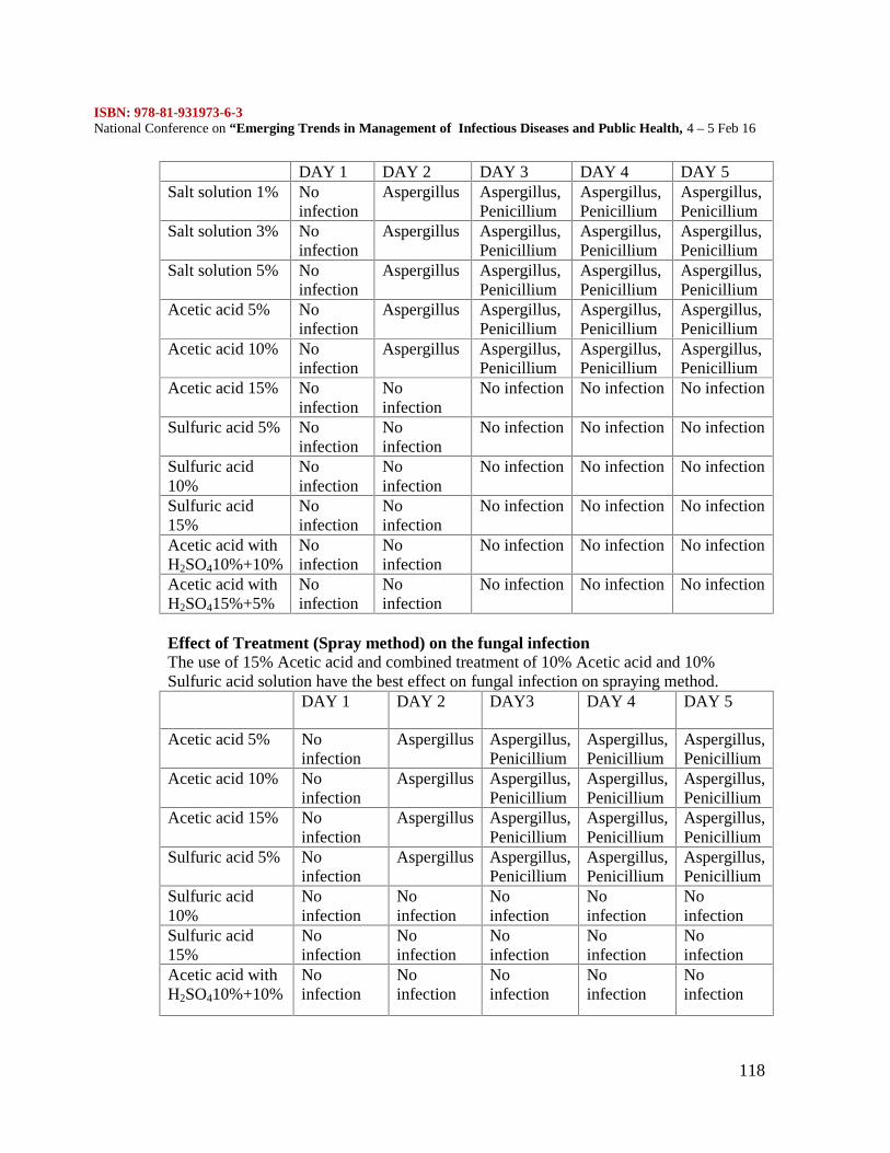
ISBN: 978-81-931973-6-3National Conference on “Emerging Trends in Management of Infectious Diseases and Public Health, 4 – 5 Feb 16
118
DAY 1 DAY 2 DAY 3 DAY 4 DAY 5Salt solution 1% No
infectionAspergillus Aspergillus,
PenicilliumAspergillus,Penicillium
Aspergillus,Penicillium
Salt solution 3% Noinfection
Aspergillus Aspergillus,Penicillium
Aspergillus,Penicillium
Aspergillus,Penicillium
Salt solution 5% Noinfection
Aspergillus Aspergillus,Penicillium
Aspergillus,Penicillium
Aspergillus,Penicillium
Acetic acid 5% Noinfection
Aspergillus Aspergillus,Penicillium
Aspergillus,Penicillium
Aspergillus,Penicillium
Acetic acid 10% Noinfection
Aspergillus Aspergillus,Penicillium
Aspergillus,Penicillium
Aspergillus,Penicillium
Acetic acid 15% Noinfection
Noinfection
No infection No infection No infection
Sulfuric acid 5% Noinfection
Noinfection
No infection No infection No infection
Sulfuric acid10%
Noinfection
Noinfection
No infection No infection No infection
Sulfuric acid15%
Noinfection
Noinfection
No infection No infection No infection
Acetic acid withH2SO410%+10%
Noinfection
Noinfection
No infection No infection No infection
Acetic acid withH2SO415%+5%
Noinfection
Noinfection
No infection No infection No infection
Effect of Treatment (Spray method) on the fungal infectionThe use of 15% Acetic acid and combined treatment of 10% Acetic acid and 10%Sulfuric acid solution have the best effect on fungal infection on spraying method.
DAY 1 DAY 2 DAY3 DAY 4 DAY 5
Acetic acid 5% Noinfection
Aspergillus Aspergillus,Penicillium
Aspergillus,Penicillium
Aspergillus,Penicillium
Acetic acid 10% Noinfection
Aspergillus Aspergillus,Penicillium
Aspergillus,Penicillium
Aspergillus,Penicillium
Acetic acid 15% Noinfection
Aspergillus Aspergillus,Penicillium
Aspergillus,Penicillium
Aspergillus,Penicillium
Sulfuric acid 5% Noinfection
Aspergillus Aspergillus,Penicillium
Aspergillus,Penicillium
Aspergillus,Penicillium
Sulfuric acid10%
Noinfection
Noinfection
Noinfection
Noinfection
Noinfection
Sulfuric acid15%
Noinfection
Noinfection
Noinfection
Noinfection
Noinfection
Acetic acid withH2SO410%+10%
Noinfection
Noinfection
Noinfection
Noinfection
Noinfection
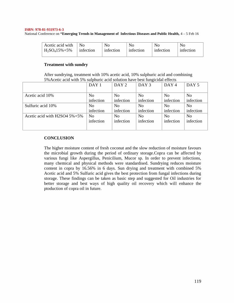
ISBN: 978-81-931973-6-3National Conference on “Emerging Trends in Management of Infectious Diseases and Public Health, 4 – 5 Feb 16
119
Acetic acid withH2SO415%+5%
Noinfection
Noinfection
Noinfection
Noinfection
Noinfection
Treatment with sundry
After sundrying, treatment with 10% acetic acid, 10% sulphuric acid and combining5%Acetic acid with 5% sulphuric acid solution have best fungicidal effects
DAY 1 DAY 2 DAY 3 DAY 4 DAY 5
Acetic acid 10% Noinfection
Noinfection
Noinfection
Noinfection
Noinfection
Sulfuric acid 10% Noinfection
Noinfection
Noinfection
Noinfection
Noinfection
Acetic acid with H2SO4 5%+5% Noinfection
Noinfection
Noinfection
Noinfection
Noinfection
CONCLUSION
The higher moisture content of fresh coconut and the slow reduction of moisture favoursthe microbial growth during the period of ordinary storage.Copra can be affected byvarious fungi like Aspergillus, Penicilium, Mucor sp. In order to prevent infections,many chemical and physical methods were standardised. Sundrying reduces moisturecontent in copra by 16.56% in 6 days. Sun drying and treatment with combined 5%Acetic acid and 5% Sulfuric acid gives the best protection from fungal infections duringstorage. These findings can be taken as basic step and suggested for Oil industries forbetter storage and best ways of high quality oil recovery which will enhance theptoduction of copra oil in future.
ISBN: 978-81-931973-6-3National Conference on “Emerging Trends in Management of Infectious Diseases and Public Health, 4 – 5 Feb 16
120
DETERMINATION OF FUNGAL QUALITY OF MARKETED HONEY SAMPLESS. Bhuvaneswari1., S. Deepa2.,HimaAishwarya3., N. Ashwin Karthick4 and N.K.UdayaPrakash4
1. Department of Plant Biology and Plant Biotechnology, LoganathaNarayanaswamyGovernment Arts College, Ponneri
2.Centre for Food Technology, Department of Biotechnology, Anna University, Chennai3. Department of Food Process Engineering, SRM University, Kattankulathur-603203,4. R and D, Marina Labs, 14, Kavya Gardens, N.T. Patel Road, Nerkundram, [email protected]
Honey is widely used as medicine and as a nutritional source. The anti-oxidant nature ofhoney favours its utility in nutraceuticals. Honey is also widely used as a medicine in IndianMedical System such as Siddha and Ayurveda. However, the quality of honey is a concern,as microorganisms are found to affect the health of the consumers as well as the quality andshelf life. Thus, the knowledge on the presence of microbes and their diversity is necessary.Though, the bacteria and the presence of yeasts are widely studied, the same related to thepresence of fungi is scarce. Hence the present study is conducted. Altogether, 33 honeysamples were procured from the markets of Chennai. The samples were serially diluted to10-2 and 1 ml of the diluted sample was plated with Potato Dextrose Agar and the plates weremaintained in triplicates. A total of 813 colonies belonging to 20 species were isolated.Aspergillus flavuswas the most dominant species isolated, followed by AspergillusnigerandPenicilliumoxalicum. The results showed that few of the samples were highlycontaminated and failed to comply with the industrial standards.
INTRODUCTION
Honey is used as medicine and as a nutritional source. Honey has long been used as a naturalsweetener in foods. The anti-oxidant nature of honey favours its utility in nutraceuticals.Honey is also widely used as a medicine in Indian Medical System such as Siddha andAyurveda which is now used for treatment of wounds, burns and even cancer due to its richantioxidant property (Layflurrie 2008, Brady et al 2008, Khalil et al 2011). Honey is a supersaturated solution of sugars composed mainly of monosaccharides, fructose and glucose,apart from other di- and trisacchardies, which gets eventually broken down intomonosaccharides by enzymes produced by bees (Martins et al. 2003). Honey is characterizedby low water activity, low protein content, natural acidity and high viscosity. Thecontamination of such highly used food or the product of pharmaceutical importance is amajor issue in the field of food safety. The quality of honey in terms of flavor, aroma or taintshould not be altered by the presence of objectionable or foreign matter or by the addition ofany food ingredient or removal of its constituent (CODEX STAN 12-1981 and EuropeanUnion Legislation DLgs 179/2004). These guidelines suggest that honey that is available forthe consumption or for addition in other food products as ingredients must lack pathogenicmicroorganisms or microorganisms that causes illness (Popa et al 2009). Thesemicroorganisms cause spoilage of honey by fermentation (Jimenez et al 1994).Microorganims inhabiting honey include bacteria, fungi and yeasts. They introduceantibiotics, enzymes, mycotoxins and other growth factors into honey (Goerzan 1991) thuscausing infections in consumers. The presence of such microorganisms also affect the
ISBN: 978-81-931973-6-3National Conference on “Emerging Trends in Management of Infectious Diseases and Public Health, 4 – 5 Feb 16
121
stability and thereby the shelf life of honey. Studies are extensively focused on Clostridiumbotulinum due to the risk of child botulinum, while industrial standards appreciate the testingof honey for pathogenic bacteria such as Salmonella and Staphylococcus. Yeasts and sporeforming bacteria are commonly found in honey (Farris et al 1986, Snowdon and Cliver 1996,Carvalho et al 2006, Rozanska 2011). Pertaining to fungi, reports are limited and theavailable literature has exposed several filamentous fungi especially of Aspergillus andPenicilliumgenera (Kacaniova et al 2009, Sinacori et al 2013). These potentially harmfulspecies though in dormant state at low water activity precluding their mycotoxin production,improper processing render them active toxin producers. Such infestation of toxins intohoney by fungi can be harmful to consumers. Thus, owing to the lack of data on fungalquality of honey, and to compare the difference in quality of processed and unprocessedhoney, the present study was instigated.MATERIALS AND METHODSCollection of Honey samples:Altogether 33 samples of honey were collected from the local markets of Chennai, India.Based on the manufacturing details, the samples are classified into branded and unbrandedhoney. Branded honey consist of 25 samples and unbranded honey consisting of 8. Theproduct name, manufacturer details, lot number and packaging details are presented in Table1.Table 1: Name of the Product, Manufacturer, Lot no and Packing details of honeysamples
No. Product name, Manufacturer, Lot no and Packing details.
1 Unbranded Local vendor
2 Amrutham Honey, Nature food products, Chennai, Tamil Nadu, Lot No: 04, Packed on: 07/2013
3 Agmark Kashmir Honey,NaturalShameethaBeeo, Viruthunagar, Tamil Nadu, Lot No:674, Packedon: 09/2013
4 Natural Honey J.J Agmark, Johnson Honey, Mumbai, Maharashtra, Lot No:38, Packed on:08/13
5 KodaikanalAgmark Honey, Sri Vignesh Cottage Industry, Chennai, Tamil Nadu, Lot No:5,Packed on:08/13
6 Unbranded Local vendor
7 Unbranded VSR Honey
8 Dabur Honey,Dabur India Ltd. Himachal Pradesh, Lot No: BDO 365, Packed on:07/13
9 SGS Honey Rex,SGS Cottage Industries, Chennai, Tamil Nadu
10 Unbranded Local Vendor
11 100% Natura Agmark Honey, New Indian Apiary Industry, Marthandam, Tamil Nadu, LotNo:380/2013, Packed on:09/2013
12 SwastikHoney,D.S.T.(Devi Swarna) Trade mark, Chennai, Tamil Nadu, (No other details)
13 Unbranded Local Vendor
14 100% Natural Marthandam Co-operative Agmark Honey, Co-operative Society Ltd, Marthandam,Tamil Nadu, Lot No:302/2013, Packed on:06/2013
15 AgmarkAnnai Honey, M/S Muruhan Co, Chennai, Tamil Nadu, Packed on:12/2012
16 Khadi Alocory Natural Kashmir Honey, Ashwin Khadi GramodyogSamitiModinagar, UttarPradesh, Packed on:09/2013
17 Devi’s Honey Taste, Devi Packaging, Chennai, Tamil Nadu. (No other details)
ISBN: 978-81-931973-6-3National Conference on “Emerging Trends in Management of Infectious Diseases and Public Health, 4 – 5 Feb 16
122
18 Jay Honey, J G S Cottage Industries, Chennai, Tamil Nadu, S.No:06, Packed on:06/2013
19 New gem Agmark Honey, Natural Honey Zone, Kuzhithurai, Tamil Nadu, Lot No:217/2013,Packed on:05/2013
20 Unbranded S.K Swastik Honey
21 Lion Honey, Lion Dates Imper Pvt. Ltd., Trichy, Tamil Nadu, Batch No:H105, Packed on:08/2013
22 Coorg Honey, The Coorg Honey & Wax Product Co-operation Marketing Society Ltd., Kodagu,Karnataka. Lot No:21,
23 Royal Agmark honey grade Natural Multi floral Honey, Royal Food Products, Chennai, TamilNadu,Packed on:09/2013
24 Marthandam Star Agmark Honey, Marthandam Star Honey Traders, Kanyakumari, Tamil Nadu, LotNo:172/ 2013, Packed on: 07/2013
25 Hills KodaikanalAgmarkHoney,SriBalamurugan Cottage Industries, Chennai, Tamil Nadu, LotNo:02, Packed on:06/2013
26 R.R Honey Agmark Honey, R.R Honey enterprises, Chennai, Tamil Nadu,Lot No:12, Packed on:10/2013
27 Reliance Healthy Life, Reliance Group of Companies, Mumbai, Maharastra, Lot No:Rl-045, Packedon:04/2014
28 Unbranded Khadi kraft
29 SKM Honey, SKM Siddha and ayurvedha Company(India) Ltd., Erode , Batch No:H0A13050, Mfgdt:10/2013
30 Unbranded Local Vendor
31 Sagar Forest Hill Nilgiri Honey, SagarNilgiri Oil Distillery, Ooty, Nilgiris, Tamil Nadu, Code 152
32 Heritage Agmark Honey, Prakruthi Health products, Banglore, Karnataka, Batch No:004,Packed on: 04/2013
33 HeavenaAgmark Honey, M/S Heavenly Fuel Pvt Ltd. (No other details)
Isolation and identification of Fungi:
The samples procured were stored in room temperature until processing. The samples wereprocessed within a week from the collection of samples. The microorganisms were isolatedby diluting with sterilized distilled water. The honey samples were serially diluted to achieve102 concentrations and 1 ml of the diluted sample was plated into petridishes containingPotato Dextrose Agar (PDA, HiMedia, Mumbai) supplemented with Streptomycin (0.06g/L)and maintained as triplicates at room temperature (28±2o C). The fungal colonies wereallowed to grow for 4-7 days and identified upto species level wherever possible based onthe morphological, macro and microscopical observations with reference to standardidentification manuals (Ellis, 1971, Onions et al, 1981, Subramaniam, 1971, Udayaprakash,2004).Presentation of data:
Percentage contribution = x 100
Frequencyoccurrence = x 100
RESULTS AND DISCUSSION
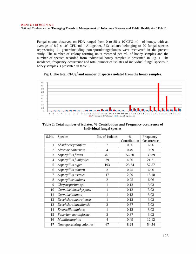
ISBN: 978-81-931973-6-3National Conference on “Emerging Trends in Management of Infectious Diseases and Public Health, 4 – 5 Feb 16
123
Fungal counts observed on PDA ranged from 0 to 88 x 102CFU ml-1 of honey, with anaverage of 8.2 x 10² CFU ml-1. Altogether, 813 isolates belonging to 20 fungal speciesrepresenting 11 generaincluding non-sporulatingcolonies were recovered in the presentstudy. The number of colony forming units recorded per ml. of honey samples and thenumber of species recorded from individual honey samples is presented in Fig. 1. Theincidence, frequency occurrence and total number of isolates of individual fungal species inhoney samples is presented in table 3.
Fig.1. The total CFUg-1and number of species isolated from the honey samples.
Table 2: Total number of isolates, % Contribution and Frequency occurrence ofIndividual fungal species
S.No. Species No. of Isolates %Contribution
FrequencyOccurrence
1 Absidiacorymbifera 7 0.86 6.06
2 Alternariaalternata 4 0.49 9.09
3 Aspergillus flavus 461 56.70 39.39
4 Aspergillus fumigatus 39 4.80 21.21
5 Aspergillus niger 193 23.74 57.57
6 Aspergillus tamarii 2 0.25 6.06
7 Aspergillus terreus 17 2.09 18.18
8 Aspergllusnidulans 2 0.25 6.06
9 Chrysosporium sp. 1 0.12 3.03
10 Curvulariabrachyspora 1 0.12 3.03
11 Curvularialunata 1 0.12 3.03
12 Drechsleraaustraliensis 1 0.12 3.03
13 Drechslerahawaiiensis 3 0.37 3.03
14 Emericillanidulans 1 0.12 3.03
15 Fusarium moniliforme 3 0.37 3.03
16 Moniliasitophila 4 0.49 12.12
17 Non-sporulating colonies 67 8.24 54.54
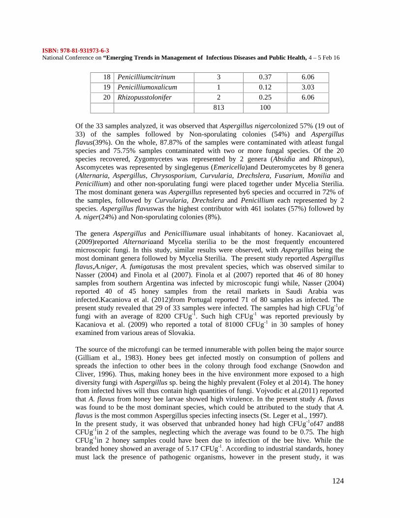
ISBN: 978-81-931973-6-3National Conference on “Emerging Trends in Management of Infectious Diseases and Public Health, 4 – 5 Feb 16
124
18 Penicilliumcitrinum 3 0.37 6.06
19 Penicilliumoxalicum 1 0.12 3.03
20 Rhizopusstolonifer 2 0.25 6.06
813 100
Of the 33 samples analyzed, it was observed that Aspergillus nigercolonized 57% (19 out of33) of the samples followed by Non-sporulating colonies (54%) and Aspergillusflavus(39%). On the whole, 87.87% of the samples were contaminated with atleast fungalspecies and 75.75% samples contaminated with two or more fungal species. Of the 20species recovered, Zygomycetes was represented by 2 genera (Absidia and Rhizopus),Ascomycetes was represented by singlegenus (Emericella)and Deuteromycetes by 8 genera(Alternaria, Aspergillus, Chrysosporium, Curvularia, Drechslera, Fusarium, Monilia andPenicillium) and other non-sporulating fungi were placed together under Mycelia Sterilia.The most dominant genera was Aspergillus represented by6 species and occurred in 72% ofthe samples, followed by Curvularia, Drechslera and Penicillium each represented by 2species. Aspergillus flavuswas the highest contributor with 461 isolates (57%) followed byA. niger(24%) and Non-sporulating colonies (8%).
The genera Aspergillus and Penicilliumare usual inhabitants of honey. Kacaniovaet al,(2009)reported Alternariaand Mycelia sterilia to be the most frequently encounteredmicroscopic fungi. In this study, similar results were observed, with Aspergillus being themost dominant genera followed by Mycelia Sterilia. The present study reported Aspergillusflavus,A.niger, A. fumigatusas the most prevalent species, which was observed similar toNasser (2004) and Finola et al (2007). Finola et al (2007) reported that 46 of 80 honeysamples from southern Argentina was infected by microscopic fungi while, Nasser (2004)reported 40 of 45 honey samples from the retail markets in Saudi Arabia wasinfected.Kacaniova et al. (2012)from Portugal reported 71 of 80 samples as infected. Thepresent study revealed that 29 of 33 samples were infected. The samples had high CFUg-1offungi with an average of 8200 CFUg-1. Such high CFUg-1 was reported previously byKacaniova et al. (2009) who reported a total of 81000 CFUg-1 in 30 samples of honeyexamined from various areas of Slovakia.
The source of the microfungi can be termed innumerable with pollen being the major source(Gilliam et al., 1983). Honey bees get infected mostly on consumption of pollens andspreads the infection to other bees in the colony through food exchange (Snowdon andCliver, 1996). Thus, making honey bees in the hive environment more exposed to a highdiversity fungi with Aspergillus sp. being the highly prevalent (Foley et al 2014). The honeyfrom infected hives will thus contain high quantities of fungi. Vojvodic et al.(2011) reportedthat A. flavus from honey bee larvae showed high virulence. In the present study A. flavuswas found to be the most dominant species, which could be attributed to the study that A.flavus is the most common Aspergillus species infecting insects (St. Leger et al., 1997).In the present study, it was observed that unbranded honey had high CFUg-1of47 and88CFUg-1in 2 of the samples, neglecting which the average was found to be 0.75. The highCFUg-1in 2 honey samples could have been due to infection of the bee hive. While thebranded honey showed an average of 5.17 CFUg-1. According to industrial standards, honeymust lack the presence of pathogenic organisms, however in the present study, it was
ISBN: 978-81-931973-6-3National Conference on “Emerging Trends in Management of Infectious Diseases and Public Health, 4 – 5 Feb 16
125
observed that pathogenic organisms such as A. flavus, A.fumigatuswere present in highnumber in few of the samples studied.
It was reported that all the processed honey samples from Nigeria was contaminated withfungi (Ayansola 2012) while the unprocessed honey from Nigeria was not reported withsignificant quantities of fungi. Popa et al. 200 reported microbial contamination in honeysamples during or after processing. However, samples collected from primary sources(honey from bee hives) showed absence of microbial contamination. The contamination inprocessed honey indicate improper or inadequate hygiene conditions during collecting,manipulating, processing or storing (Tchoumboue et al. 2007)
The present study also experienced similar results, with branded processed honey samplesshowing high contamination than unbranded honey samples. Also, it has been reported thatthe fungi produced Aflatoxin in all apiarian substrates (unprocessed honey, pollen, brodcomb, whole larvae and whole bees) in low levels except the unprocessed honey (Hilldrup etal. 1977).Wellford et al. (1978) also reported that though toxigenic strains of A. flavusNRRL 5862 and A. parasiticus NRRL 2999 colonized unprocessed honey samples,detectable amounts of aflatoxin production was not observed. Thus, the present study showsthat the processed or branded honey samples provided the habitat for abundance of fungalcolonies compared to unbranded honey. Also, it was observed that many of the samples hadabundance of pathogenic fungi and hence fail to comply with the industrial standards.
References:Ayansola, A.A. (2012). Fungal Isolates from the Honey Samples Collected from
Retail Outlets in Southwestern Nigeria. Journal of Biology and Life Science. 3 (1), 189-199Bardy, J., Slevin, N.J., Mais, K.L. and Molassiotis, A. (2008). A systematic review
of honey uses and its potential value within oncology care. J. Clin. Nurs., 17, 2604-2623.Bogdanov, S., Jurendic, T., Sieber, R., and Gallmann, P. (2008). Honey for nutrition
andCarvalho, M.C., Rocha, A., Estevinho, L., and Choupina, A.B. (2006).
Identification of honey yeast species based on RFLP analyses of the ITS region. Cienc.Tecnol. Alimentos., 5 (1), 11-17.
Ceausi, C., Togoe, L., Tudor, L., and Furnaris, F. (2009). The evaluation ofmicrobiological quality of some bee honey sorts. Bull. UASVM Animal. Scie. Bio., 66 (1),273e277
Codex Alimentarius Commission (2001). Revised Codex Standard for Honey.Codex STAN, 12e1981, Rev. 1 1987, Rev. 2.
Ellis, M.B. (1976). More Dematiaceous Hyphomycetes. CAB. InternationalMycological Institute, Kew, UK. 507 pp.
Farris, G.A., Fatichenti, F., Deina, P., and Agostini, F. (1986). Aerobic andanaerobic sporeforming bacteria in sardinian honey. Annalidellafacoltà diAgrariadell’Università di Sassari., 32, 173-179.
Finola, M.S., Lasagno, M.C., and Marioli, J.M. (2007). Microbiological andchemical characterization of honeys from central Argentina. Food Chem., 100(4), 1649–1653.
Gilliam, M., Taber, S., and Richardson, G.V. (1983). Hygienic behavior of honeybees in relation to chalkbrood disease. Apidologie, 14, 29–39.
ISBN: 978-81-931973-6-3National Conference on “Emerging Trends in Management of Infectious Diseases and Public Health, 4 – 5 Feb 16
126
Goerzen D.W. (1991). Microflora associated with the alfalfa leaf cuting bee,Egachilerotundata (Fab) (Hymenoptera:Megachilidae) in Saskatchewan, Canada.Apidologie., 22, 553-561.
health: A review. J. Am. Coll. Nutr., 27 (6), 677-689.Hilldrup J.A., Eadie T. and Liewellyn G.C. (1977): Fungal growth and aflatoxin
production on apiarian substrates. J.Ass. Off. Anal. Chem., 60, 96-99.Jiménez, M., Mateo, J. J., Huerta ,T. and Mateo, R. (1994). Influence of the storage
conditions on some Physicochemical and Mycological Parameters of Honey. J. Sci. FoodAgric., 64, 67-74.
Kacaniova, M., Knazovicka, V., Felsociova, S., and Rovna, K. (2012). Microscopicfungi recovered from honey and their toxinogenity, Journal of Environmental Science andHealth, Part A: Toxic/Hazardous Substances and Environmental Engineering, 47(11), 1659-1664
Kacaniova, M., Sudzina, M., Sudzinova, J., Fikselova,M., Cubon, J., and Hascik, P.(2007) Microbiological and physico-chemical quality of honey collected from differentSlovak habitats. Slov. J. Ani. Sci., 40(1), 38–43.
Khalil, M.I., Sulaiman, S.A., and Boukraa, L. (2010). Antioxidant Properties ofHoney and Its Role in Preventing Health Disorder. The Open Nutraceuticals Journal, 3, 6-16
Kirsten Foley, Fazio, G., Jensen, A. B., and Hughes, W. O.H. (2014). Thedistribution of Aspergillus spp. opportunistic parasites in hives and their pathogenicity tohoney bees. Veterinary Microbiology, 169, 203–210
Lay-Flurrie, K. (2008). Honey in wound care: effects, clinical application andpatient benefit. Br. J. Nurs., 17, 32-36.
Martins, H. A., Martins, M. L., and Bernardo, F. M. A. (2003). Bacillaceae spores,fungi and aflatoxins determination in honey. Rev. Portug. Cienc. Veter., 98, 85–88.
Nasser, L.A. (2004) Isolation and characterization of fungi contaminating packagedhoney commonly consumed in Saudi Arabia. Ass. Univ. Bull. Environ. Res., 7(1), 1–7.
Onions, A.H.S., Allsopp, D. and Eggins, H.O.W. (1981). Smith’s Introduction toIndustrial Mycology. 7th Ed. Edward Arnold, London.
Popa, M., Vica, M., Axinte, R., Glevitzky, M., and Varvara, S. (2009). Studyconcerning the honey qualities in transylvania region. Ann. Universe Apulensis SeriesOecon., 11(2), 1034–040
Rozanska, H. (2011). Microbiological quality of Polish honey. B. Vet. Pulawy., 55(3), 443-445.
Sinacori. M., Francesca. N., Alfonzo. A., Cruciata. M., Sannino. C., Settanni. L., andMoschetti G. (2013) Cultivable microorganisms associated with honeys of differentgeographical and botanical origin. Food Microbiol., 38, 284-294.
Snowdon, J. A. and Cliver, D. O. (1996). Microorganisms in honey. Review article.Int. J. Food Microb., 31, 1-26.
St. Leger, R.J., Joshi, L., and Roberts, D.W. (1997). Adaptation of proteases andcarbohydrases of saprophytic, phytopathogenic and entomopathogenic fungi to therequirements of their ecological niches. Microbiology, 143, 1983–1992.
Subramaniam, C.V., (1971). Hyphomycetes (An account of Indian species, exceptcarpospores)”, ICAR, New Delhi.
Tchoumboue, J., Awah-Ndukum, J., Fonteh, F.A., Dongock, N.D., Pinta, J., andMvondo, Z.A. (2007). Physico-chemical and microbiological characteristics of honey fromsudan-guinean zone of West Cameroon. Africa J. Biotech., 6, 908-913.
ISBN: 978-81-931973-6-3National Conference on “Emerging Trends in Management of Infectious Diseases and Public Health, 4 – 5 Feb 16
127
Udaya Prakash, N.K. (2004). Indoor molds-Isolation and Identification. Colourwings (M) Pvt. Ltd. Madras. pp- 99.
Vojvodic, S., Jensen, A.B., James, R.R., Boomsma, J.J., and Eilenberg, J. (2011).Temperature dependent virulence of obligate and facultative fungal pathogens of honeybeebrood. Vet. Microbiol., 149, 200–205.
Wellford T.E., Eadie T. and Liewellyn G.C. (1978): Evaluation the inhibitory actionof honey on fungal growth sporulation. In aflatoxin production. Z. LebensmUntersForsch.,28, 280-283.
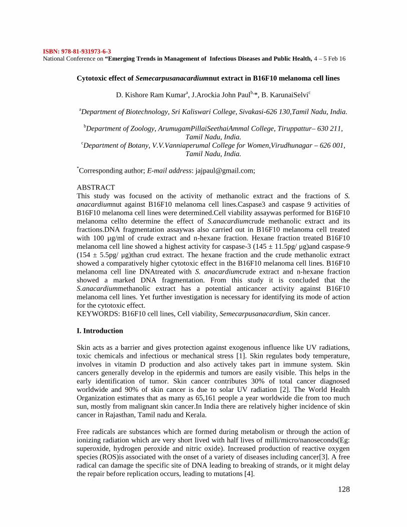
ISBN: 978-81-931973-6-3National Conference on “Emerging Trends in Management of Infectious Diseases and Public Health, 4 – 5 Feb 16
128
Cytotoxic effect of Semecarpusanacardiumnut extract in B16F10 melanoma cell lines
D. Kishore Ram Kumara, J.Arockia John Paulb,*, B. KarunaiSelvic
aDepartment of Biotechnology, Sri Kaliswari College, Sivakasi-626 130,Tamil Nadu, India.
bDepartment of Zoology, ArumugamPillaiSeethaiAmmal College, Tiruppattur– 630 211,Tamil Nadu, India.
cDepartment of Botany, V.V.Vanniaperumal College for Women,Virudhunagar – 626 001,Tamil Nadu, India.
*Corresponding author; E-mail address: [email protected];
ABSTRACTThis study was focused on the activity of methanolic extract and the fractions of S.anacardiumnut against B16F10 melanoma cell lines.Caspase3 and caspase 9 activities ofB16F10 melanoma cell lines were determined.Cell viability assaywas performed for B16F10melanoma cellto determine the effect of S.anacardiumcrude methanolic extract and itsfractions.DNA fragmentation assaywas also carried out in B16F10 melanoma cell treatedwith 100 µg/ml of crude extract and n-hexane fraction. Hexane fraction treated B16F10melanoma cell line showed a highest activity for caspase-3 (145 ± 11.5pg/ μg)and caspase-9(154 ± 5.5pg/ μg)than crud extract. The hexane fraction and the crude methanolic extractshowed a comparatively higher cytotoxic effect in the B16F10 melanoma cell lines. B16F10melanoma cell line DNAtreated with S. anacardiumcrude extract and n-hexane fractionshowed a marked DNA fragmentation. From this study it is concluded that theS.anacardiummethanolic extract has a potential anticancer activity against B16F10melanoma cell lines. Yet further investigation is necessary for identifying its mode of actionfor the cytotoxic effect.KEYWORDS: B16F10 cell lines, Cell viability, Semecarpusanacardium, Skin cancer.
I. Introduction
Skin acts as a barrier and gives protection against exogenous influence like UV radiations,toxic chemicals and infectious or mechanical stress [1]. Skin regulates body temperature,involves in vitamin D production and also actively takes part in immune system. Skincancers generally develop in the epidermis and tumors are easily visible. This helps in theearly identification of tumor. Skin cancer contributes 30% of total cancer diagnosedworldwide and 90% of skin cancer is due to solar UV radiation [2]. The World HealthOrganization estimates that as many as 65,161 people a year worldwide die from too muchsun, mostly from malignant skin cancer.In India there are relatively higher incidence of skincancer in Rajasthan, Tamil nadu and Kerala.
Free radicals are substances which are formed during metabolism or through the action ofionizing radiation which are very short lived with half lives of milli/micro/nanoseconds(Eg:superoxide, hydrogen peroxide and nitric oxide). Increased production of reactive oxygenspecies (ROS)is associated with the onset of a variety of diseases including cancer[3]. A freeradical can damage the specific site of DNA leading to breaking of strands, or it might delaythe repair before replication occurs, leading to mutations [4].
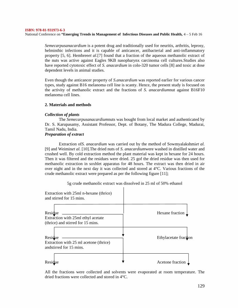
ISBN: 978-81-931973-6-3National Conference on “Emerging Trends in Management of Infectious Diseases and Public Health, 4 – 5 Feb 16
129
Semecarpusanacardium is a potent drug and traditionally used for neuritis, arthritis, leprosy,helminthic infections and it is capable of anticancer, antibacterial and anti-inflammatoryproperty [5, 6]. Hembreeet al.[7] found that a fraction of the aqueous methanolic extract ofthe nuts was active against Eagles 9KB nasopharynx carcinoma cell cultures.Studies alsohave reported cytotoxic effect of S. anacardium in colo-320 tumor cells [8] and toxic at dosedependent levels in animal studies.
Even though the anticancer property of S.anacardium was reported earlier for various cancertypes, study against B16 melanoma cell line is scanty. Hence, the present study is focused onthe activity of methanolic extract and the fractions of S. anacardiumnut against B16F10melanoma cell lines.
2. Materials and methods
Collection of plantsThe Semecarpusanacardiumnuts was bought from local market and authenticated by
Dr. S. Karupasamy, Assistant Professor, Dept. of Botany, The Madura College, Madurai,Tamil Nadu, India.Preparation of extract
Extraction ofS. anacardium was carried out by the method of Sowmyalakshmiet al.[9] and Weiminet al. [10].The dried nuts of S. anacardiumwere washed in distilled water andcrushed well. By cold extraction method the plant material was kept in hexane for 24 hours.Then it was filtered and the residues were dried. 25 gof the dried residue was then used formethanolic extraction in soxhlet apparatus for 48 hours. The extract was then dried in airover night and in the next day it was collected and stored at 4°C. Various fractions of thecrude methanolic extract were prepared as per the following figure [11];
5g crude methanolic extract was dissolved in 25 ml of 50% ethanol
Extraction with 25ml n-hexane (thrice)and stirred for 15 mins.
Residue Hexane fractionExtraction with 25ml ethyl acetate(thrice) and stirred for 15 mins.
Residue Ethylacetate fractionExtraction with 25 ml acetone (thrice)andstirred for 15 mins.
Residue Acetone fraction
All the fractions were collected and solvents were evaporated at room temperature. Thedried fractions were collected and stored in 4°C.
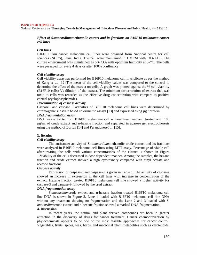
ISBN: 978-81-931973-6-3National Conference on “Emerging Trends in Management of Infectious Diseases and Public Health, 4 – 5 Feb 16
130
Effect of S.anacardiummethanolic extract and its fractions on B16F10 melanoma cancercell lines
Cell linesB16F10 Skin cancer melanoma cell lines were obtained from National centre for cellsciences (NCCS), Pune, India. The cell were maintained in DMEM with 10% FBS. Theculture environment was maintained as 5% CO2 with optimum humidity at 37°C. The cellswere passaged for every 4 days or after 100% confluency.
Cell viability assayCell viability assaywas performed for B16F10 melanoma cell in triplicate as per the methodof Kang et al. [12].The mean of the cell viability values was compared to the control todetermine the effect of the extract on cells. A graph was plotted against the % cell viability(B16F10 cells) Vs dilution of the extract. The minimum concentration of extract that wastoxic to cells was recorded as the effective drug concentration with compare to positivecontrol (cyclophasphamide).Determination of caspase activityCaspase3 and caspase 9 activities of B16F10 melanoma cell lines were determined bychromogenic substrate based colorimetric assays [13] and expressed as pg µg-1 protein.DNA fragmentation assayDNA was extractedfrom B16F10 melanoma cell without treatment and treated with 100µg/ml of crude extract and n-hexane fraction and separated in agarose gel electrophoresisusing the method of Burton [14] and Perandoneset al. [15].
3. ResultsCell viability assay
The anticancer activity of S. anacardiummethanolic crude extract and its fractionswere analyzed in B16F10 melanoma cell lines using MTT assay. Percentage of viable cellafter treating the cells with various concentrations of the extract is shown in Figure1.Viability of the cells decreased in dose dependent manner. Among the samples, the hexanefraction and crude extract showed a high cytotoxicity compared with ethyl acetate andacetone fractions.Caspase activity
Expression of caspase-3 and caspase-9 is given in Table 1. The activity of caspasesshowed an increase in expression in the cell lines with increase in concentration of theextract. Hexane fraction treated B16F10 melanoma cell line showed a higher activity forcaspase-3 and caspase-9 followed by the crud extract.DNA fragmentation assay
S.anacardiumcrude extract and n-hexane fraction treated B16F10 melanoma cellline DNA is shown in Figure 2. Lane 1 loaded with B16F10 melanoma cell line DNAwithout any treatment showing no fragmentation and the Lane 2 and 3 loaded with S.anacardiumcrude extract and n-hexane fraction showed a marked DNA fragmentation.4. Discussion
In recent years, the natural and plant derived compounds are been in greaterattraction in the discovery of drugs for cancer treatment. Cancer chemoprevention byphytochemicals appears to be one of the most feasible approaches for cancer control.Vegetables, fruits, spices, teas, herbs, and medicinal plant metabolites such as carotenoids,
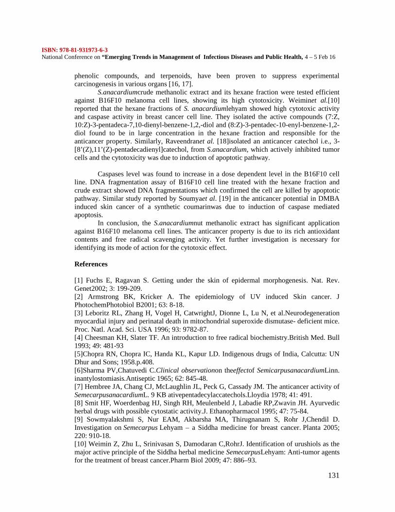
ISBN: 978-81-931973-6-3National Conference on “Emerging Trends in Management of Infectious Diseases and Public Health, 4 – 5 Feb 16
131
phenolic compounds, and terpenoids, have been proven to suppress experimentalcarcinogenesis in various organs [16, 17].
S.anacardiumcrude methanolic extract and its hexane fraction were tested efficientagainst B16F10 melanoma cell lines, showing its high cytotoxicity. Weiminet al.[10]reported that the hexane fractions of S. anacardiumlehyam showed high cytotoxic activityand caspase activity in breast cancer cell line. They isolated the active compounds (7:Z,10:Z)-3-pentadeca-7,10-dienyl-benzene-1,2,-diol and (8:Z)-3-pentadec-10-enyl-benzene-1,2-diol found to be in large concentration in the hexane fraction and responsible for theanticancer property. Similarly, Raveendranet al. [18]isolated an anticancer catechol i.e., 3-[8’(Z),11’(Z)-pentadecadienyl]catechol, from S.anacardium, which actively inhibited tumorcells and the cytotoxicity was due to induction of apoptotic pathway.
Caspases level was found to increase in a dose dependent level in the B16F10 cellline. DNA fragmentation assay of B16F10 cell line treated with the hexane fraction andcrude extract showed DNA fragmentations which confirmed the cell are killed by apoptoticpathway. Similar study reported by Soumyaet al. [19] in the anticancer potential in DMBAinduced skin cancer of a synthetic coumarinwas due to induction of caspase mediatedapoptosis.
In conclusion, the S.anacardiumnut methanolic extract has significant applicationagainst B16F10 melanoma cell lines. The anticancer property is due to its rich antioxidantcontents and free radical scavenging activity. Yet further investigation is necessary foridentifying its mode of action for the cytotoxic effect.
References
[1] Fuchs E, Ragavan S. Getting under the skin of epidermal morphogenesis. Nat. Rev.Genet2002; 3: 199-209.[2] Armstrong BK, Kricker A. The epidemiology of UV induced Skin cancer. JPhotochemPhotobiol B2001; 63: 8-18.[3] Leboritz RL, Zhang H, Vogel H, CatwrightJ, Dionne L, Lu N, et al.Neurodegenerationmyocardial injury and perinatal death in mitochondrial superoxide dismutase- deficient mice.Proc. Natl. Acad. Sci. USA 1996; 93: 9782-87.[4] Cheesman KH, Slater TF. An introduction to free radical biochemistry.British Med. Bull1993; 49: 481-93[5]Chopra RN, Chopra IC, Handa KL, Kapur LD. Indigenous drugs of India, Calcutta: UNDhur and Sons; 1958.p.408.[6]Sharma PV,Chatuvedi C.Clinical observationon theeffectof SemicarpusanacardiumLinn.inantylostomiasis.Antiseptic 1965; 62: 845-48.[7] Hembree JA, Chang CJ, McLaughlin JL, Peck G, Cassady JM. The anticancer activity ofSemecarpusanacardiumL. 9 KB ativepentadecylaccatechols.Lloydia 1978; 41: 491.[8] Smit HF, Woerdenbag HJ, Singh RH, Meulenbeld J, Labadie RP,Zwavin JH. Ayurvedicherbal drugs with possible cytostatic activity.J. Ethanopharmacol 1995; 47: 75-84.[9] Sowmyalakshmi S, Nur EAM, Akbarsha MA, Thirugnanam S, Rohr J,Chendil D.Investigation on Semecarpus Lehyam – a Siddha medicine for breast cancer. Planta 2005;220: 910-18.[10] Weimin Z, Zhu L, Srinivasan S, Damodaran C,RohrJ. Identification of urushiols as themajor active principle of the Siddha herbal medicine SemecarpusLehyam: Anti-tumor agentsfor the treatment of breast cancer.Pharm Biol 2009; 47: 886–93.
ISBN: 978-81-931973-6-3National Conference on “Emerging Trends in Management of Infectious Diseases and Public Health, 4 – 5 Feb 16
132
[11] Raj KS,Dhall A. Antiomicrobial and free radical scavenging ativity of extracts of someIndian medicinal plants. J. of Med. Plants Res 2010; 4: 2313-20.[12] Kang BK, Chon SK, Kim SH, Jeong SY, Kim MS, Cho SH, et al.Controlled release ofpaclitaxel from microemulsion containing PLGA and evaluation of anti-tumor activity invitro and in vivo. Int. J. of Pharmaceutics2004; 286: 147-56.[13] Das RK, HossainSkU, BhattacharyaS. Diphenylmethylselenocynate inhibits DMBA-croton oil induced two stae skin carcinogenesis by inducing apoptosis and inhibitingcutaneous cell proliferation. Cancer Lett 2005; 230: 90-101.[14] BurtonK. A study of conditions and mechanisms of diphenylamine reaction for theestimation of deoxyribonucleic acid.Biochemical J 1956; 62: 315-23.[15] Perandones CE, Illera VA, Peckham D, Stunz LL, Ashman RF. Regulation of apoptosisin vitro in mature murine spleen T-cells. J. Immunol1993; 151: 3521-29.[16] Wattenberg LW. Chemoprevention of cancer.Cancer Res 1985; 45: 1-8.[17] Borek C. Antioxidant health effects of aged garlic extract. J Nutr2001; 131: 1010-15.[18] Raveendran PKN, Melnick SJ, Wnuk SF, Rapp M, Escalon E, RamahandranC. Isolationand characterization of an anticancer catechol compound from Semecarpusanacardium. JofEthanopharmacol 2009; 122: 450-56.[19] Soumya SB, Paul S, Mandal SK, Banerjee A, Boujedaini N, Khuda-Bukhsh AR. Asynthetic coumarin (4-Methyl-7 hydroxycoumarin) has anti-cancer potentials againstDMBA-induced skin cancer in mice. European J. of Pharmacol 2009; 614: 128-36.
Table 2.Activity of caspase 3 and caspase9 in B16F10 melanoma cell lines aftertreatment.Values are mean ± SD (n=3).
Samples(μg/ml)
Caspase – 3(pg/ μg)
Caspase – 9(pg/μg)
Control 56 ± 4.2 65 ± 3.4
Hexane
10 68 ± 4.7 78 ± 4.6
100 127 ± 9.2 135 ± 4.9
500 145 ± 11.5 154 ± 5.5
Crude
100 72 ± 3.1 82 ± 1.9
500 99 ± 8.8 111 ± 7.1
ISBN: 978-81-931973-6-3National Conference on “Emerging Trends in Management of Infectious Diseases and Public Health, 4 – 5 Feb 16
133
Figure 2. Percentage of viable B16F10 melanoma cells treated with S.anacardiumnut crudemethanolic extract and its fractions. Error bars indicate standarddeviation.
Figure 3.DNA fragmentation assay.L1 –B16F10 melanoma cell DNA without treatment; L2 –B16F10 melanoma cell DNAtreated with 100µg/ml of crude extract; L3 –B16F10 melanoma cell DNA treated with100µg/ml of n-hexane fraction.
ISBN: 978-81-931973-6-3National Conference on “Emerging Trends in Management of Infectious Diseases and Public Health, 4 – 5 Feb 16
134
Herbo-mineral salts from Marine halophytes against human bacterial pathogens
V. Saravanan.*, M. Gnanadesigan**, S. Ravikumar***.,
* Department of Zoology, The MDT Hindu College,Tiunelveli-627 010, Tamilnadu.**Department of Biotechnology, National College, Tiruchirapalli, u-620001***School of Marine Sciences, Department of Marine Biology, Alagappa University,Thondi Campus, Thondi-623409, Tamil Nadu
ABSTRACT
Marine halophytes are also proven to have rich source of structurally diverse bioactivecompounds with valuable pharmaceutical potential. In the present study antibacterial activitywas carried out with herbo mineral salts of marine halophyte (Sesuvium portulacastrum,Salicornia brachiata, Suaeda maritima and Suaeda monoica) against antibiotic resistant(ABR) bacterial pathogens, Urinary tract infectious pathogens and human eye pathogens.Antibiotic sensitivity test was performed by agar well diffusion method. Of the selected saltextracts, the Sesuvium portulacastrum has potential antibacterial property. The results of thepresent study will open a new way for the development of alternative therapeutic sources forseveral infectious diseases.KEY WORDS: Marine Halophytes; Infectious Pathogens; Salts;1. INTRODUCTION:
Herbalism is the study and use of medicinal properties of plants and plant extracts. Many ofthe herbs and spices used by humans as medicinal compounds (Lai and Roy, 2004). The useof herbs to treat disease is almost universal among non-industrialized societies (Edgar et al.,2002; Ragunathan and Kitto, 2009; Saiman and Siegel, 2004)). In India, Ayurveda medicinehas used many herbs such as turmeric possibly as early a In Tamil Nadu, Tamils have theirown medicinal system now popularly called the Siddha medicinal system. The Siddhasystem is entirely in Tamil language. It contains roughly 300,000 verses covering diverseaspects of medicine such as anatomy, sex ("Kokokam" is the sexual treatise of parexcellence), herbal, mineral and metallic compositions to cure many diseases that arerelevant even to-day 1900 BC (Agarwal et al., 2007). Many infectious diseases are known tobe treated with herbal remedies through the history of mankind. Marine halophytes are alsoproven to have rich source of structurally diverse bioactive compounds with valuablepharmaceutical potential. Scientific reports suggested that, the herbo-minerals extracts fromterrestrial plants proved to have antibacterial, anti viral, anti fungal, antioxidant and immunemodulator properties (Agarwal et al., 2007). But the studies related to salt extraction fromthe marine halophytes are still lacking. Though mangrove plants are classified as saltextruders and salt intruders and hence the intruders are having a special adaptation ofaccumulating several therapeutically important salts at an optimum level are still unexplored.In view of this the present study was carried out to perform the salt extraction and mineralcomposition analysis with mangrove associates.2. Materials and Methods:2.1. Bacterial pathogensFour antibiotic resistant (ABR) human pathogens viz., Staphylococcus aureus, Pseudomonasaeruginosa, Streptococcus pneumoniae and Klebsiella pneumoniae were obtained from the
ISBN: 978-81-931973-6-3National Conference on “Emerging Trends in Management of Infectious Diseases and Public Health, 4 – 5 Feb 16
135
culture collections of Institute for Microbial Technology (IMTECH), Chandigarh, India. Allthe bacterial strains were suspended in nutrient broth and incubated at 37ºC for 48 hrs. Fiveurinary tract infectious pathogens viz., Pseudomonas aeruginosa, Enterobacter sp.Escherichia coli, Klebsiella pneumoniae and Proteus sp., were obtained from Jeyasekaranhospital, Nagercoil, Tamil Nadu, India. Five eye pathogens namely viz., Acinitobacter sp.,Proteus sp., Staphylococcus aureus, Escherichia coli and Streptococcus pyogenes wereobtained from the Aravind Eye Hospital, Madurai, Tamil Nadu, India2.2. Extraction of salts from halophytesThe green, succulent leaves of the four halophytes were collected by hand picking. Then thecleaned specimens were dried in a hot air oven at 100°C. When they were completely dried,they were burnt into ash by using muffle furnace (450°C). The ash was collected in a largemouthed big vessel. The salt was extracted from that ash by dissolving it in deionized water.Then the supernatant was heated and dried in hot air oven (150°C) to produce flaks.2.3. Antimicrobial sensitivity assay:2.3.1. Well diffusion method (Pandey et al., 2004)Antibiotic sensitivity test was performed by agar well diffusion method, which is designed todetermine the smallest amount of the bioactive secondary metabolites needs to inhibit thegrowth of the microorganism. The medium of choice is Mueller-Hinton agar with a pH of7.2 to 7.4, which is poured into plates to a uniform depth to 5 mm and allowed to solidify.Prior to use, the plates were transferred to an incubator at 37ºC for 10-20 minutes to dry themoisture that develops on the surface. Overnight growth of chosen bacterial broth culturewas swabbed on the surface of the agar media and further four wells were prepared by usingsterile well cutter. To each well, 70 µg, 80 µg, 90 µg and 100 µg mineral flake was addedand incubated in a thermostat incubator at 37ºC for 24 hours. After incubation, the zone ofinhibition around the well was calculated and expressed as zone of inhibition in millimeter indiameter.
3. ResultsThe antibacterial activity of the salts extracted from four salt marsh marine
halophytes viz., Sesuvium portulacastrum, Salicornia brachiata, Suaeda maritima andSuaeda monoica against antibiotic resistant (ABR) bacterial pathogens were carried out bythe present study and represented in Table 1. It revealed that, the salt extracted from the S.portulacastrum showed the maximum sensitivity (10.0 mm dia. and 11.0 mm dia.) againstStaphylococcus aureus and Streptococcus pneumoniae respectively at 100µg concentration.Likewise, the salt extracted from S. monoica showed the maximum sensitivity (10.0 mm dia.and 11.0 mm dia.) against Pseudomonas aeruginosa and Streptococcus pneumoniaerespectively at 100µg concentration. Whereas, the other two salts from S. brachiata and S.maritima did not show any activity against any of the antibiotic resistant human bacterialpathogens. The antibacterial activity of the salts against urinary tract infectious (UTI)pathogens were carried out by the present study and represented in Table 2. It reveals that,the salt extracted from the S. portulacastrum showed the maximum sensitivity (12.0 mmdia.) against Pseudomonas sp., Enterobacter sp., and Klebsiella sp. at 100 µg concentration.The salt from S. monoica showed the maximum sensitivity (10.0 mm dia. and 11.0 mm dia.)against Pseudomonas sp. and Escherichia coli at 100 µg concentration. The salt from S.brachiata showed maximum sensitivity (9.0 mm dia.) against the Klebsiella sp. at 100µgconcentration and the salt from S. maritima did not show any activity against UTI bacterialpathogensThe antibacterial activity of the salts extracted from salt marsh marine halophytesagainst human ophthalmic pathogens were carried out by the present study and represented
ISBN: 978-81-931973-6-3National Conference on “Emerging Trends in Management of Infectious Diseases and Public Health, 4 – 5 Feb 16
136
in Table 3. It reveals that, the salt extracted from S. portulacastrum showed maximumsensitivity (10.0 mm dia. and 13.0 mm dia.) against Streptococcus pyogens and Escherichiacoli respectively at 100 µg concentration. The salt extracted from S. monoica showedmaximum sensitivity (7.0 mm dia. and 9.0 mm dia.) against Acinitobacter sp., Proteus sp.and Streptococcus pyogens respectively at 100 µg concentration. The salts extracted from S.brachiata and S. maritima did not show any activity against ophthalmic pathogens.
4. Discussion
It was observed that, the salt extracted from the S. portulacastrum showedmaximum sensitivity against the tested microbial pathogens. Whereas, the other two saltsfrom S. brachiata and S. maritima did not show any activity against any of the bacterialpathogens. It was already reported that, if a whole plant is required to be given at a dose of10gm for a particular disease, the same plant’s uppu can have the same therapeutic value in adose of 50 to 100 mg (Sambasivapillai dictionary P.1170. Vol.I).The present study foundthat, for effective killing of the bacterial pathogens from different sources required 100µg ofsalt. Comparatively, the salt extracted from other terrestrial plants required moreconcentration to kill the bacterial pathogens whereas the salts extracted from marinehalophytic plants required very less (100 fold less) concentration to kill the bacterialpathogens. Earlier findings reported that, the salt extracted from erukku Calotropis gigantiaat a dose of 10-20 mg has been used for the treatment of spasmodic pains, bronchial asthma,toxic bites, the salt extracted from kuppaimeni Acalypha indica at a concentration of 50-100mg has been used for the treatment of vatha and kabha diseases, the salt extracted fromthuti Abutilon indicum at a dose of 50-100 mg has been used for the treatment of piles andulcers, the salt extracted from vaazhi Musa paradisiaca at a concentration of 50-100 mg hasbeen used for the treatment of diuretic, burning micturition, kidney stones, the salt extractedfrom murungai Moringa tinctoria at a dose of 50-100 mg has been used for the treatment ofdiuretic, breaks kidney and bladder stones, strengthens teeth, the salt extracted from naauruviAchyranthes aspera at a dose of 50-100 mg has been used for the treatment of diuretic, themineral extracted from thennai oolai leaves of Cocos nucifera at a dose of 50-100 mg hasbeen used for the treatment of diuretic, the salt extracted from arugu Cynodon dactylon at adose of 50-100 mg has been used for the treatment of skin diseases, the salt extracted frompirandai Cissus quadrangularis at a dose of 50-100 mg has been used for the treatment ofindigestion, piles and diarrhea, the salt extracted from ellu Sesamum indicum at a dose of 50-100 mg has been used for the treatment of diarrhea and pitha diseases, the salt extracted frompoovarasu Thespesia populnea at a dose of 50-100 mg has been used for the treatment ofintestinal worms, the neer mulli Hydrophila auriculata at a dose of 50-100 mg has been usedfor the treatment of anemia, drowsy, ascites and kidney stones, the salt extracted from panaiaan poo Borassus flabellifer (male flowers) at a dose of 50-100 mg has been used for thetreatment of diuretic, liver and spleen disorders, the salt extracted from pugaiilai Nicotianatabacum at a dose of 50-100 mg has been used for the treatment of ascites (Venkatappan andMalliga, 2004). It is concluded from the present study that, among the salts extracted fromsalt marsh marine herbals, the Sesuvium portulacastrum has potential antibacterial property.This will open a new way for the development of alternative therapeutic drug for thetreatment of several infectious and non- infectious diseases.
5. References:
ISBN: 978-81-931973-6-3National Conference on “Emerging Trends in Management of Infectious Diseases and Public Health, 4 – 5 Feb 16
137
Aggarwal, B. B., C. Sundaram, N. Malani and H. Ichikawa, 2007. Curcumin: the Indiansolid gold. Advanced Experimental Medical Biology, 595: 1–75.Edgar J. DaSilva, Elias Baydoun and Adnan Badran, 2002. Biotechnology and thedeveloping world". Electronic Journal of Biotechnology, 5 (1):12-18.Lai, P.K. and J. Roy, 2004. Antimicrobial and chemopreventive properties of herbs andspices. Curent Medical Chemistry, 11 (11): 1451–60.Pandey, B. P., P. Ghimire and V. P. Agarwal, 2004. Studies on the antibacterial activity ofactinomycetes isolated from the Khumbu region of Mt. Everest. A paper presented in theinternational conference on great Himalayas: Climate, Health, Ecology, Management andconservation, kathmandu, January 12-15, 2004. Organized by Kathmandu University and theAquatic Ecosystem health and Management Society, Canada.Raghunathan and Kitto, 2009. Drugs from the Indian Seas More expectations. CurrentScience, 97 (12): 1705-1706.Ravi Kumar, S., G. Ramanathan, M. Subhakaran and S. Jacob Inbaneson,2009.Antimicrobial compounds from marine halophytes for silkworm disease treatment.International Journal of Medical Science, 1(5): 184–191.Saiman, L. and J. Siegel, 2004. Infection control in Cystic fibrosis. Clinical microbiologyReviews, 17: 57-71.Sambasivam Pillai, T.V., 1938. A Tamil - English Dictionary of Medicine, Chemistry,Botany and Allied Sciences. Published by Department of Indian Medicine and Homeopathy,Chennai, Vol.I: p. 1170.Venkatappan, N. and K. Malliga, 2004. Uppu: a novel medicinal plant extract preparationin siddha medicine. In: Rajabudeen.E. (Ed.) Proceedings of State level seminar on medicinalplants in India-Prospects and Perspectives, December,15-16,2004, Department of Biology,Dr. Zakir Husain College, Illayangudi -630 702, Tamilnadu, India, pp: 24-27.
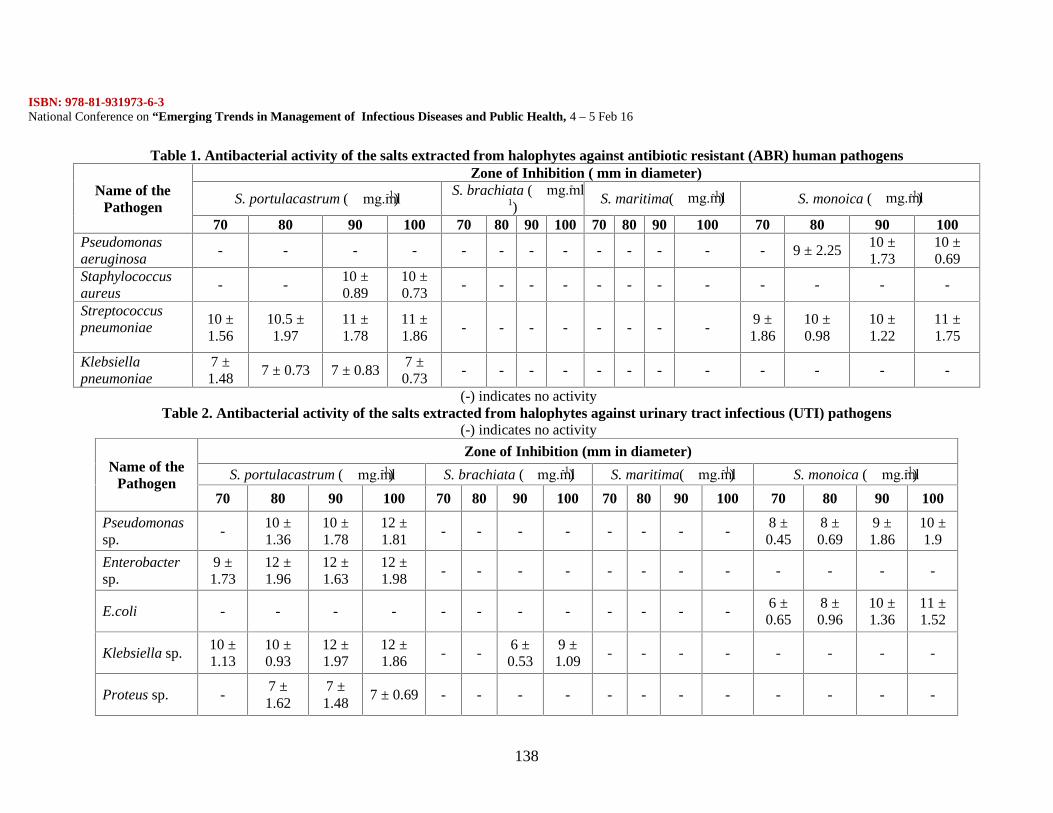
ISBN: 978-81-931973-6-3National Conference on “Emerging Trends in Management of Infectious Diseases and Public Health, 4 – 5 Feb 16
138
Table 1. Antibacterial activity of the salts extracted from halophytes against antibiotic resistant (ABR) human pathogens
Name of thePathogen
Zone of Inhibition ( mm in diameter)
S. portulacastrum (g.ml-1) S. brachiata (g.ml-1)
S. maritima(g.ml-1) S. monoica (g.ml-1)
70 80 90 100 70 80 90 100 70 80 90 100 70 80 90 100Pseudomonasaeruginosa
- - - - - - - - - - - - - 9 ± 2.2510 ±1.73
10 ±0.69
Staphylococcusaureus
- -10 ±0.89
10 ±0.73
- - - - - - - - - - - -
Streptococcuspneumoniae
10 ±1.56
10.5 ±1.97
11 ±1.78
11 ±1.86
- - - - - - - -9 ±1.86
10 ±0.98
10 ±1.22
11 ±1.75
Klebsiellapneumoniae
7 ±1.48
7 ± 0.73 7 ± 0.837 ±0.73
- - - - - - - - - - - -
(-) indicates no activityTable 2. Antibacterial activity of the salts extracted from halophytes against urinary tract infectious (UTI) pathogens
(-) indicates no activity
Name of thePathogen
Zone of Inhibition (mm in diameter)
S. portulacastrum (g.ml-1) S. brachiata (g.ml-1) S. maritima(g.ml-1) S. monoica (g.ml-1)70 80 90 100 70 80 90 100 70 80 90 100 70 80 90 100
Pseudomonassp.
-10 ±1.36
10 ±1.78
12 ±1.81
- - - - - - - -8 ±0.45
8 ±0.69
9 ±1.86
10 ±1.9
Enterobactersp.
9 ±1.73
12 ±1.96
12 ±1.63
12 ±1.98
- - - - - - - - - - - -
E.coli - - - - - - - - - - - -6 ±0.65
8 ±0.96
10 ±1.36
11 ±1.52
Klebsiella sp.10 ±1.13
10 ±0.93
12 ±1.97
12 ±1.86
- -6 ±0.53
9 ±1.09
- - - - - - - -
Proteus sp. -7 ±1.62
7 ±1.48
7 ± 0.69 - - - - - - - - - - - -
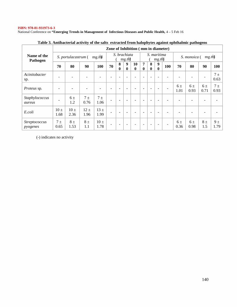
ISBN: 978-81-931973-6-3National Conference on “Emerging Trends in Management of Infectious Diseases and Public Health, 4 – 5 Feb 16
140
Table 3. Antibacterial activity of the salts extracted from halophytes against ophthalmic pathogens
Name of thePathogen
Zone of Inhibition ( mm in diameter)
S. portulacastrum (g.ml-1) S. brachiata(g.ml-1)
S. maritima(g.ml-1) S. monoica (g.ml-1)
70 80 90 100 70 80
90
100
70
80
90 100 70 80 90 100
Acinitobactersp.
- - - - - - - - - - - - - - -7 ±0.63
Proteus sp. - - - - - - - - - - - -6 ±1.01
6 ±0.93
6 ±0.71
7 ±0.93
Staphylococcusaureus
-6 ±1.2
7 ±0.76
7 ±1.06
- - - - - - - - - - - -
E.coli10 ±1.68
10 ±2.36
12 ±1.96
13 ±1.99
- - - - - - - - - - - -
Streptococcuspyogenes
7 ±0.65
8 ±1.53
8 ±1.1
10 ±1.78
- - - - - - - -6 ±0.36
6 ±0.98
8 ±1.5
9 ±1.79
(-) indicates no activity
ISBN: 978-81-931973-6-3National Conference on “Emerging Trends in Management of Infectious Diseases and Public Health, 4 – 5 Feb 16
141
Biosynthesis and characterization of Alkaline protease from Bacillus subtilisC Kiruba and K Manjula
Department of Biochemistry, Mohamed Sathak College of Arts and Science, sholinganallur,Chennai
Abstract:
In the present study, alkaline protease producing Bacillus subtilis was isolated and identified by variousbiochemical tests and by 16S rDNA sequencing. Agricultural waste was used as a solid substrate forenzyme production in solid state culture. The process parameters such as fermentation period (72 h), pH(9.0), moisture content (120%), inoculum (12%), carbon source (maltose), nitrogen source (casein) and in-organic ion (sodium di-hydrogen phosphate) were optimized for maximum enzyme production. The crudeenzyme was concentrated by ammonium sulphate precipitation and further purified by ion exchange andgel filtration chromatography. This enzyme was highly stable and active at 50 °C and at pH 8.0,respectively. The molecular weight of protease was estimated to be 42.5 KD by SDS-PAGE.
Introduction:
Microbial proteases are among the most important hydrolytic enzymes and have been studied extensivelysince the advent of enzymology. Microorganisms elaborate a large array of proteases, which areintracellular and/or extracellular. Intracellular proteases are important for various cellular and metabolicprocesses, such as sporulation and differentiation, protein turnover, maturation of enzymes and hormonesand maintenance of the cellular protein pool. Extracellular proteases are important for the hydrolysis ofproteins in cell-free environments and enable the cell to absorb and utilize hydrolytic products (Kalisz,1988). At the same time, these extracellular proteases have also been commercially exploited to assistprotein degradation in various industrial processes (Kumar and Takagi 1999; Outtrup and Boyce, 1990).Among bacteria, there are number of genera which produce proteases but Bacillus remains the organism ofchoice (Nomoto et al., 1984; Kumar and Takagi, 1999; Mehrotra et al., 1999). And, the proteasesproduced by Bacillus are mostly alkaline in nature and are highly stable against extreme temperature, pHand other conditions (Sharma et al., 1980; Yang et al., 2000). Proteases account for 40% of the totalenzyme sales worldwide and this trend is expected to increase in the near future. This has led to increasingattention towards the exploitation of potent microbial strains for the production of alkaline proteases froman industrial point of view (Ellaiah et al., 2002). Alkaline proteases are a physiologically andcommercially important group of enzymes used primarily as detergent additives. They play a specificcatalytic role in the hydrolysis of proteins. In 1994, the total market for industrial enzymes accounted forapproximately $400 million, of which enzymes worth $112 million were used for detergent purposes(Hodgson, 1994). In Japan, 1994 alkaline protease sales were estimated at 15000 million yen (equivalent to$116 million) (Horikoshi, 1996). There is expected to be an upward trend in the use of alkaline proteasesso that by the turn of the decade the total value for industrial enzymes is likely to reach $700 million ormore (Hodgson, 1994).
It is essential that these organisms be provided with optimal growth conditions to increase enzymeproduction. The culture conditions that promote protease production were found to be significantlydifferent from the culture conditions promoting cell growth (Moon and Parulekar, 1991). In the industrialproduction of alkaline proteases, technical media were usually employed that contained very highconcentrations (100–150 g dry weight/litre) of complex carbohydrates, proteins, and other mediacomponents (Aunstrup, 1980). With a view to develop an economically feasible technology, research
ISBN: 978-81-931973-6-3National Conference on “Emerging Trends in Management of Infectious Diseases and Public Health, 4 – 5 Feb 16
142
efforts are mainly focused on: (i) improvement in the yields of alkaline proteases; and (ii) optimization ofthe fermentation medium and production conditions.
As part of the search for a cheap alternative production system which utilizes simpler agro industrialwastes as substrates, solid state fermentation (SSF) has gained importance in the production of microbialenzymes. Production of biocatalyst using agro biotech substrates under SSF conditions provides severaladvantages in productivity, cost effectiveness in labour, time and medium components, use of simplemachinery, use of an inexpensive substrate, simple downstream processing, in addition to environmentaladvantages like less effluent production, waste minimization etc ( Lonsane et al., 1985; Pandey et al.,2000).
Materials and Methods:
Screening of protease producing strain: The soil samples were collected near to Nagercoil,Kanyakumari District, Tamilnadu for the screening of protease producing organism. The organisms weregrown in the minimal medium containing (g l-1): KH2PO4, 10; NaHPO4.12H2O, 3.0; MgSO4, 0.2; peptone,10; trisodium citrate, 3.0 and skim milk, 10, agar, 15. The media pH was adjusted to 7.0 with 1NHCl/NaOH for initial experiments. The sample was serially diluted and streaked on skimmed milk agarplates. The plates were incubated for 48 h at 37 °C and enzyme activity was observed. After incubation thecolonies formed were counted and respective colonies which differ in morphology and colour weresubcultured. Positive colonies those degraded skim milk were studied for further protease production. Theorganism was identified based on morphology and biochemical characteristics (Holt et al., 1994).Protease production in Submerged Fermentation
The organism was grown in a nutrient broth medium (g l-1) [peptic digest of animal tissue-5, beefextract-1.5, yeast extract-1.5 and sodium chloride-5]. It was incubated at 37 °C in an orbital shaker at 150rpm for 48 h. Five ml of culture medium was withdrawn for every 24 h of incubation and centrifuged at10,000 rpm for 10 min. The supernatant was assayed for protease activity. Briefly, one ml of crudeenzyme solution was added to one ml of casein substrate (1%, w/v), prepared in 50 mM Tris buffer (pH8.0) and incubated at 37 °C for 30 min. The reaction was terminated by adding 100 mM trichloroaceticacid and the content was filtered through Whatman No. 1 filter paper. The filtrate absorbance was read at280 nm using UV–visible spectrophotometer (Ultraviolet visible spectrophotometer) and the proteaseactivity was calculated using tyrosine standard curve. One unit of protease activity was defined as 1 µmolof tyrosine liberated ml-1 under the assay condition. The total protein content of the sample was determinedas described by Lowry et al. (1951).Solid state fermentation (SSF)A mass of 2.5 gram of wheat bran and agricultural waste (dried vegetable wastes) were taken in 100 mlErlenmeyer flask. To maintain the moisture level, 50 mM of Tris buffer (pH 8.0) was added. The contentof the flasks were mixed thoroughly and autoclaved at 121 °C for 20 min. After sterilization, 0.25 ml ofseed culture was inoculated on the substrates aseptically and incubated for 48 h at 37 °C. The substrate thatsupported to produce higher amount of proteolytic enzyme was used for further studies.Optimization of culture conditions for the production of proteasesVarious process parameters affecting enzyme production during SSF were optimized. The strategy was tooptimize each parameter independently of the others and subsequently optimal conditions were employedin all experiments. In a sequential order, the various process parameters were optimized for maximalenzyme production by using the agro-waste medium. The various process parameters tested wereincubation period (24, 48, 72 and 96 h, respectively), pH (5.0, 6.0, 7.0, 8.0, 9.0 and 10.0, respectively),moisture (80%, 100%, 120%, 140%, and 160%,) content, inoculum concentration (3%, 6%, 9%, 12%, 15%and 18% respectively), carbon sources (maltose, sucrose, starch, glucose, trehalose and xylose), nitrogensources (peptone, beef extract, yeast extract, casein, gelatin and urea) and inorganic salts (ammoniumsulphate, ferrous sulphate, ammonium chloride, di-sodium hydrogen phosphate, sodium di-hydrogenphosphate, sodium nitrate and calcium chloride).
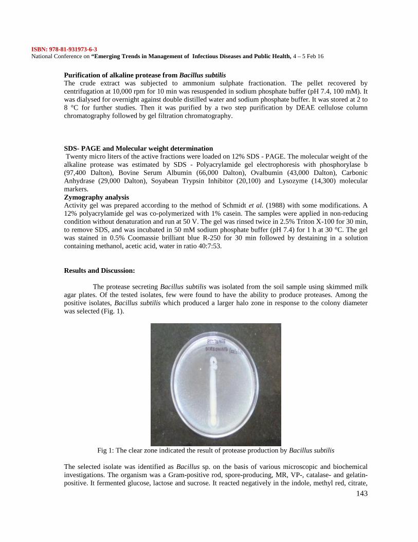
ISBN: 978-81-931973-6-3National Conference on “Emerging Trends in Management of Infectious Diseases and Public Health, 4 – 5 Feb 16
143
Purification of alkaline protease from Bacillus subtilisThe crude extract was subjected to ammonium sulphate fractionation. The pellet recovered bycentrifugation at 10,000 rpm for 10 min was resuspended in sodium phosphate buffer (pH 7.4, 100 mM). Itwas dialysed for overnight against double distilled water and sodium phosphate buffer. It was stored at 2 to8 °C for further studies. Then it was purified by a two step purification by DEAE cellulose columnchromatography followed by gel filtration chromatography.
SDS- PAGE and Molecular weight determinationTwenty micro liters of the active fractions were loaded on 12% SDS - PAGE. The molecular weight of the
alkaline protease was estimated by SDS - Polyacrylamide gel electrophoresis with phosphorylase b(97,400 Dalton), Bovine Serum Albumin (66,000 Dalton), Ovalbumin (43,000 Dalton), CarbonicAnhydrase (29,000 Dalton), Soyabean Trypsin Inhibitor (20,100) and Lysozyme (14,300) molecularmarkers.Zymography analysisActivity gel was prepared according to the method of Schmidt et al. (1988) with some modifications. A12% polyacrylamide gel was co-polymerized with 1% casein. The samples were applied in non-reducingcondition without denaturation and run at 50 V. The gel was rinsed twice in 2.5% Triton X-100 for 30 min,to remove SDS, and was incubated in 50 mM sodium phosphate buffer (pH 7.4) for 1 h at 30 °C. The gelwas stained in 0.5% Coomassie brilliant blue R-250 for 30 min followed by destaining in a solutioncontaining methanol, acetic acid, water in ratio 40:7:53.
Results and Discussion:
The protease secreting Bacillus subtilis was isolated from the soil sample using skimmed milkagar plates. Of the tested isolates, few were found to have the ability to produce proteases. Among thepositive isolates, Bacillus subtilis which produced a larger halo zone in response to the colony diameterwas selected (Fig. 1).
Fig 1: The clear zone indicated the result of protease production by Bacillus subtilis
The selected isolate was identified as Bacillus sp. on the basis of various microscopic and biochemicalinvestigations. The organism was a Gram-positive rod, spore-producing, MR, VP-, catalase- and gelatin-positive. It fermented glucose, lactose and sucrose. It reacted negatively in the indole, methyl red, citrate,
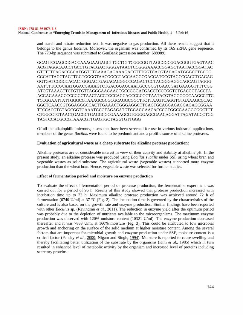
ISBN: 978-81-931973-6-3National Conference on “Emerging Trends in Management of Infectious Diseases and Public Health, 4 – 5 Feb 16
144
and starch and nitrate reduction test. It was negative to gas production. All these results suggest that itbelongs to the genus Bacillus. Moreover, the organism was confirmed by its 16S rRNA gene sequence.The 779-bp sequence was submitted to GenBank (accession number: 688989).
GCAGTCGAGCGGACCAAAGAAGAGCTTGCTCTTCGGCGGTTAGCGGCGGACGGGTGAGTAACACGTAGGCAACCTGCCTGTACGACTGGGATAACTCCGGGAAACCGGAGCTAATACCGGATACGTTTTTCAGACCGCATGGTCTGAAAGAGAAAGACCTTTGGTCACGTACAGATGGGCCTGCGGCGCATTAGCTAGTTGGTGGGGTAACGGCCTACCAAGGCGACGATGCGTAGCCGACCTGAGAGGGTGATCGGCCACACTGGGACTGAGACACGGCCCAGACTCCTACGGGAGGCAGCAGTAGGGAATCTTCCGCAATGGACGAAAGTCTGACGGAGCAACGCCGCGTGAACGATGAAGGTTTTCGGATCGTAAAGTTCTGTTGTTAGGGAAGAACCGCCGGGATGACCTCCCGGTCTGACGGTACCTAACGAGAAAGCCCCGGCTAACTACGTGCCAGCAGCCGCGGTAATACGTAGGGGGCAAGCGTTGTCCGGAATTATTGGGCGTAAAGCGCGCGCAGGCGGCTTCTTAAGTCAGGTGTGAAAGCCCACGGCTCAACCGTGGAGGGCCACTTGAAACTGGGAGGCTTGAGTGCAGGAGAGGAGAGCGGAATTCCACGTGTAGCGGTGAAATGCGTAGAGATGTGGAGGAACACCCGTGGCGAAGGCGGCTCTCTGGCCTGTAACTGACGCTGAGGCGCGAAAGCGTGGGGAGCGAACAGGATTAGATACCCTGGTAGTCCACGCCGTAAACGTTGAGTGCTAGGTGTTGGG
Of all the alkalophilic microorganisms that have been screened for use in various industrial applications,members of the genus Bacillus were found to be predominant and a prolific source of alkaline proteases.
Evaluation of agricultural waste as a cheap substrate for alkaline protease production:
Alkaline proteases are of considerable interest in view of their activity and stability at alkaline pH. In thepresent study, an alkaline protease was produced using Bacillus subtilis under SSF using wheat bran andvegetable wastes as solid substrate. The agricultural waste (vegetable wastes) supported more enzymeproduction than the wheat bran. Hence, vegetable waste was selected for further studies.
Effect of fermentation period and moisture on enzyme production
To evaluate the effect of fermentation period on protease production, the fermentation experiment wascarried out for a period of 96 h. Results of this study showed that protease production increased withincubation time up to 72 h. Maximum alkaline protease production was achieved around 72 h offermentation (6740 U/ml) at 37 °C (Fig. 2). The incubation time is governed by the characteristics of theculture and is also based on the growth rate and enzyme production. Similar findings have been reportedwith other Bacillus sp. (Ravindran et al., 2011). The reduction in enzyme yield after the optimum periodwas probably due to the depletion of nutrients available to the microorganisms. The maximum enzymeproduction was observed with 120% moisture content (10321 U/ml). The enzyme production decreasedthereafter and it was 7863 U/ml at 160% moisture (Fig. 3). This could be attributed to low microbialgrowth and anchoring on the surface of the solid medium at higher moisture content. Among the severalfactors that are important for microbial growth and enzyme production under SSF, moisture content is acritical factor (Pandey et al., 2000; Nigam and Singh, 1994). Moisture is reported to cause swelling andthereby facilitating better utilization of the substrate by the organisms (Kim et al., 1985) which in turnresulted in enhanced level of metabolic activity by the organism and increased level of proteins includingsecretory proteins.
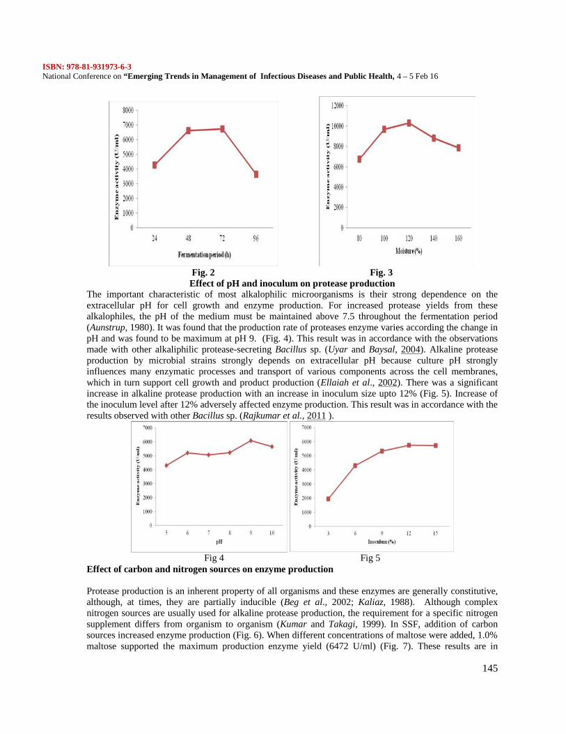
ISBN: 978-81-931973-6-3National Conference on “Emerging Trends in Management of Infectious Diseases and Public Health, 4 – 5 Feb 16
145
Fig. 2 Fig. 3Effect of pH and inoculum on protease production
The important characteristic of most alkalophilic microorganisms is their strong dependence on theextracellular pH for cell growth and enzyme production. For increased protease yields from thesealkalophiles, the pH of the medium must be maintained above 7.5 throughout the fermentation period(Aunstrup, 1980). It was found that the production rate of proteases enzyme varies according the change inpH and was found to be maximum at pH 9. (Fig. 4). This result was in accordance with the observationsmade with other alkaliphilic protease-secreting Bacillus sp. (Uyar and Baysal, 2004). Alkaline proteaseproduction by microbial strains strongly depends on extracellular pH because culture pH stronglyinfluences many enzymatic processes and transport of various components across the cell membranes,which in turn support cell growth and product production (Ellaiah et al., 2002). There was a significantincrease in alkaline protease production with an increase in inoculum size upto 12% (Fig. 5). Increase ofthe inoculum level after 12% adversely affected enzyme production. This result was in accordance with theresults observed with other Bacillus sp. (Rajkumar et al., 2011 ).
Fig 4 Fig 5Effect of carbon and nitrogen sources on enzyme production
Protease production is an inherent property of all organisms and these enzymes are generally constitutive,although, at times, they are partially inducible (Beg et al., 2002; Kaliaz, 1988). Although complexnitrogen sources are usually used for alkaline protease production, the requirement for a specific nitrogensupplement differs from organism to organism (Kumar and Takagi, 1999). In SSF, addition of carbonsources increased enzyme production (Fig. 6). When different concentrations of maltose were added, 1.0%maltose supported the maximum production enzyme yield (6472 U/ml) (Fig. 7). These results are in
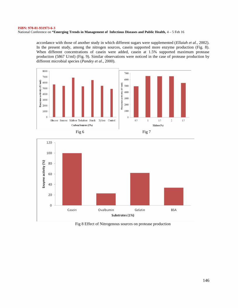
ISBN: 978-81-931973-6-3National Conference on “Emerging Trends in Management of Infectious Diseases and Public Health, 4 – 5 Feb 16
146
accordance with those of another study in which different sugars were supplemented (Ellaiah et al., 2002).In the present study, among the nitrogen sources, casein supported more enzyme production (Fig. 8).When different concentrations of casein were added, casein at 1.5% supported maximum proteaseproduction (5867 U/ml) (Fig. 9). Similar observations were noticed in the case of protease production bydifferent microbial species (Pandey et al., 2000).
Fig 6 Fig 7
Fig 8 Effect of Nitrogenous sources on protease production
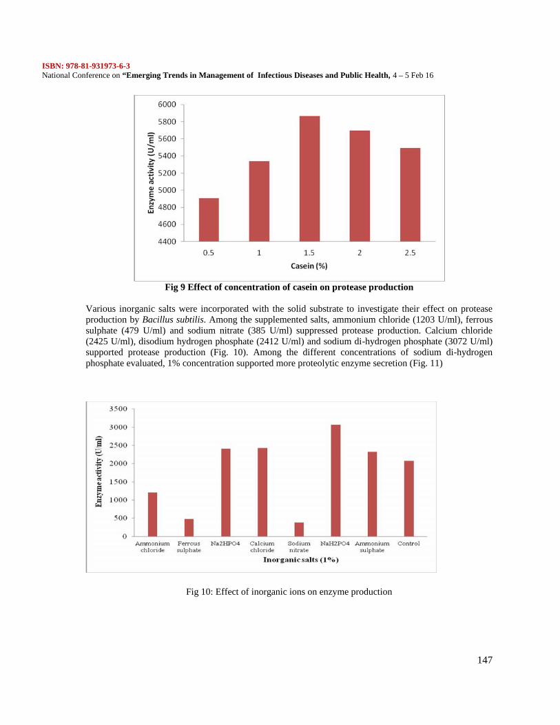
ISBN: 978-81-931973-6-3National Conference on “Emerging Trends in Management of Infectious Diseases and Public Health, 4 – 5 Feb 16
147
Fig 9 Effect of concentration of casein on protease production
Various inorganic salts were incorporated with the solid substrate to investigate their effect on proteaseproduction by Bacillus subtilis. Among the supplemented salts, ammonium chloride (1203 U/ml), ferroussulphate (479 U/ml) and sodium nitrate (385 U/ml) suppressed protease production. Calcium chloride(2425 U/ml), disodium hydrogen phosphate (2412 U/ml) and sodium di-hydrogen phosphate (3072 U/ml)supported protease production (Fig. 10). Among the different concentrations of sodium di-hydrogenphosphate evaluated, 1% concentration supported more proteolytic enzyme secretion (Fig. 11)
Fig 10: Effect of inorganic ions on enzyme production
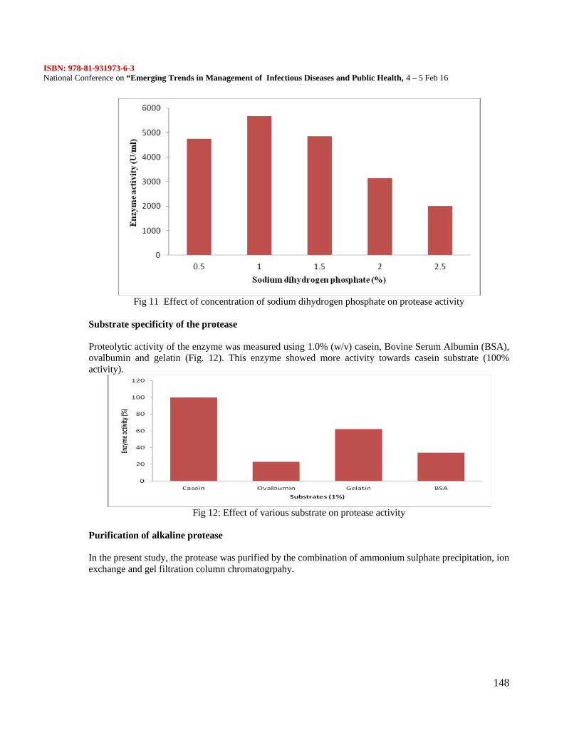
ISBN: 978-81-931973-6-3National Conference on “Emerging Trends in Management of Infectious Diseases and Public Health, 4 – 5 Feb 16
148
Fig 11 Effect of concentration of sodium dihydrogen phosphate on protease activity
Substrate specificity of the protease
Proteolytic activity of the enzyme was measured using 1.0% (w/v) casein, Bovine Serum Albumin (BSA),ovalbumin and gelatin (Fig. 12). This enzyme showed more activity towards casein substrate (100%activity).
Fig 12: Effect of various substrate on protease activity
Purification of alkaline protease
In the present study, the protease was purified by the combination of ammonium sulphate precipitation, ionexchange and gel filtration column chromatogrpahy.
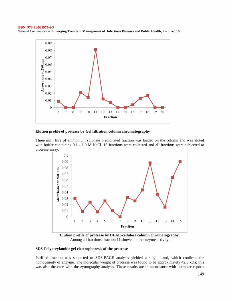
ISBN: 978-81-931973-6-3National Conference on “Emerging Trends in Management of Infectious Diseases and Public Health, 4 – 5 Feb 16
149
Elution profile of protease by Gel filtration column chromatography
Three milli litre of ammonium sulphate precipitated fraction was loaded on the column and was elutedwith buffer containing 0.1 - 1.0 M NaCl. 15 fractions were collected and all fractions were subjected toprotease assay.
Elution profile of protease by DEAE-cellulose column chromatography.Among all fractions, fraction 11 showed more enzyme activity.
SDS-Polyacrylamide gel electrophoresis of the protease
Purified fraction was subjected to SDS-PAGE analysis yielded a single band, which confirms thehomogeneity of enzyme. The molecular weight of protease was found to be approximately 42.5 kDa; thiswas also the case with the zymography analysis. These results are in accordance with literature reports
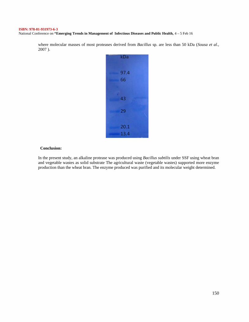
ISBN: 978-81-931973-6-3National Conference on “Emerging Trends in Management of Infectious Diseases and Public Health, 4 – 5 Feb 16
150
where molecular masses of most proteases derived from Bacillus sp. are less than 50 kDa (Sousa et al.,2007 ).
Conclusion:
In the present study, an alkaline protease was produced using Bacillus subtilis under SSF using wheat branand vegetable wastes as solid substrate The agricultural waste (vegetable wastes) supported more enzymeproduction than the wheat bran. The enzyme produced was purified and its molecular weight determined.
ISBN: 978-81-931973-6-3National Conference on “Emerging Trends in Management of Infectious Diseases and Public Health, 4 – 5 Feb 16
151
Modulatory effect of Polydatin on in vivo carbohydrate metabolism in streptozocin-induced type IIdiabetic rats.
R.Swaminathan1, M.Syed Ali*1 V.Anuradha, N.Yogananth
PG and Research Department of Biotechnology1, Mohamed Sathak College of Arts and Science,University of Madras, Sholiganallur, Chennai.PG and Research Department of Biochemistry1, Mohamed Sathak College of Arts and Science,University of Madras, Sholiganallur, Chennai
Abstract
The study was designed to investigate the antihyperglycemic effect of polydatin on streptozotocin-diabeticrats. Diabetes was induced in adult male albino rats of the wistar strain, weighing 170-210g, byadministration of streptozotocin (40mg/kg of body weight) intraperitoneally. Diabetic rats showed increasein plasma glucose and glycosylated hemoglobin and a decrease in plasma insulin and hemoglobin.Activities of gluconeogenic enzymes such as glucose 6-phosphatase, fructose 1, 6-bisphosphatase incresedand glucokinase, glucose 6-phosphatase dehydrogenase decreased in the liver of diabetic rats along withliver glycogen. Oral administration of Polydatin (150mg/kg of body weight) or glibenclamide (600 mg/kgof body weight) in saline, for 45 days, prevented the above changes and improved towards normal. Inagainst body weight loss of diabetic rats by polydatin was also observed. No significant effect wasobserved in normal rats treated with polydatin (150mg/kg of body weight). These results showed thatpolydatin has potantial antihyperglycemic activity at a dose of 150mg/kg of body weight in streptozotocininduced diabetic rats.Keywords: streptozotocin, polydatin, antihyperglcemia, glibenclamide, insulin
Introduction
The Health Equity and Accountability Act of 2012 will improve diabetes research, treatment and educationin minority populations, and address the unique circumstances faced by minority populations and women.This bill is critical to our nation’s fight to Stop Diabetes. Statistical projection about the diabetes affectsnearly 26 million people in the U.S. and another 79 million Americans have prediabetes, placing them athigher risk for developing type 2 diabetes. While 7.1 percent of non-Hispanic white adults have beendiagnosed with the disease, the statistics are much higher for minority populations in the U.S (Americandiabetes association. 2012). Type 2 diabetes mellitus, also known as noninsulin dependent diabetesmellitus, develops in middle or later life and affects 2–6% of adults in most Western societies (Bailey,2000). The pharmacological agents currently used for the treatment of type 2 diabetes includesulfonylurea, biguanide, thiazolidinedione and α-glycosidase inhibitors. These agents, however, haverestricted usage due to several undesirable side effects and fail to significantly alter the course of diabeticcomplications (Rang and Dale, 1991). The control of blood glucose in diabetic patients is achieved mainlyby the use of oral hypoglycemic/antihyperglycemic agents and insulin. However, all these treatments havelimited efficacy and have been reported to be associated with undesirable side effects (Harrower, 1994).Hyperglycemia, due to uncontrolled glucose regulation is considered as the causal link between diabetesand diabetic complications. A number of studies emphasize that alteration in glucose metabolism leads tohyperglycemia-induced cell damage by four key metabolic pathways. Protein kinase C (PKC) isoforms(Rolo and Palmeira 2006). An increase in the biosynthesis and or a decrease in the metabolism ofglycoproteins attributed to the deposition of these materials in the basal membrane of pancreatic cells. Inrecent times, many traditionally important medicinal plants have been tested for their efficacy againstimpaired glycoprotein levels in diabetes (Ramkumar et al. 2007). β- Cells are affected by manyimmunological and chemical agents leading to local inflammations producing IL-6 and glucocorticoids.IL-6/glucocorticoid stimulation produces an active transcriptional complex for Reg, a β-cell regenerating
ISBN: 978-81-931973-6-3National Conference on “Emerging Trends in Management of Infectious Diseases and Public Health, 4 – 5 Feb 16
152
factor gene, in which poly (ADP-ribose) synthetaseypolymerase (PARP) is involved. In the presence ofPARP inhibitors such as nicotinamide when PARP is not itself poly (ADP-ribosyl)-ated, thetranscriptional complex is stabilized and Reg gene transcription and subsequent Reg protein formationoccurs in β -cells. This protein acts as a growth factor on β -cells via Reg receptor. DNA replication in β -cells takes place and β-cell regeneration is accomplished. DNA damaging substances such as superoxide(O°2) and nitric oxide (NO°) are produced in inflammatory processes by cytotoxic agents such as STZ.When DNA is damaged, PARP senses the nicks and autopoly (ADP-ribosyl)-ates itself for DNA repair.Autopoly (ADP-ribosylation) of PARP inhibits the formation of Reg gene transcriptional complex andtranscription of this gene stops (Akiyama et al., 2001). Nowadays, clinical treatment of diabetes targetsboth insulin deficiency and resistance and more recently the prevention of pancreatic β cell functiondecline (Hansotia and Drucker, 2005)
Polydatin 3, 4’, 5-trihydroxystilbene-3-β-mono-D-glucoside is a monocrystalline compoundisolated from a traditional Chinese herbal medicine named polygonum cuspidatum sieb. It is a glucosideof reveratrol and has also been named piced. Both polydatin and reveratrol are the main effectivecompounds of polygonum cuspidatum. In plants, reveratrol can quickly be glycosylated into piced.Previous studies have shown that polydatin resist free radical, kill or inhibit tumor cells, antibacterialactivity (Shu, 2002; shu et al., 2004;Zhu and jin, 2005), exhibited anti-platelet aggregation, anti-oxidativeactivities (Zhang et al., 1995; Du et al., 2009; Kerem et al., 2006), antifatigue activity (Ding et al.,2009),reduce lipd oxidation (Pan et al., 2007), Polydatin attenuate the development of ventricular remodelinginduced by isoproterenol in mice and by pressure overload in rats, and it can decrease BP in high dose(Chang Xun Chen et al., 2010). Very little information is available on the absorption, distribution,metabolism or excretion of polydatin (Lin J.H et al., 2001). The genartion of preclinical pharmacokineticdata of polydatin requires the development of an analytical method in different biological matrices(Shouhong gao et al., 2006). Polydatin markedly lowers the serum levels of total cholesrerol, triglycerideand low-density lipoprotein cholesterol in hyperlipidamic rabbits and hamster in hepatic triglyceridesdecreased levels (Qin et al., 2008 and Qin et al., 2009).
Hence, the present study was conducted in order to find the therapeutic potential of polydatin as oralhypoglycemic agent and exhibits additional pharmacological effects on plasma glucose, insulin, totalhemoglobin, glycosylated hemoglobin, hepatic glycogen, and tissue (Liver and Kidney) carbohydratemetabolic enzymes in both normal and STZinduced diabetic rats. The effects produced by these treatmentsare compared with standard drug glipenclamide an anti-diabetic drug (Andrade-Cetto and Wiedefeld,2004).
Materials and MethodsAnimals
Female albino (9 week - old) rats of wistar strain with a body weight ranging from 180-210g, wereprocured from central animal house, Department of Experimental Medicine, Rajah Muthiah MedicalCollege and Hospital, Annamalai University, and were maintained in an air conditioned room (25±1 ºC)with a 12 h light/ 12 h dark cycle, feed and water were provide ad libitum to all the animals. The study wasapproved by the institutional Animal Ethics Committee of Rajah Muthiah Medical College and Hospital(Reg No. 160/1999/CPCSEA), Annamalai University, Annamalainagar.Chemical
Streptozotocin and Polydatin were purchased from Sigma-Aldrich (St, Louis, Missouri, USA). Allother chemicals used in this study were of analytical grade obtained from E.Merck or Himedia, India.Experimental induction of non insulin dependent diabetes mellitus
The animals were rendered diabetic by a single intraperitoneal injection of Streptozotocin(40mg/kg body weight) in freshly prepared citrate buffer (0.1 M, pH 4.5) after an overnight fast.Streptozotocin-injected animals were given 20% glucose solution for 24 h to prevent initial drug–inducedhypoglycemic mortality. Streptozotocin-injected animals exhibited hyperglycemia within a few days.
ISBN: 978-81-931973-6-3National Conference on “Emerging Trends in Management of Infectious Diseases and Public Health, 4 – 5 Feb 16
153
Diabetes in Streptozotocin rats was conformed by measuring the blood glucose (by glucose oxidasemethod) 96 h after injection with Streptozotocin. The animals with blood glucose above 230 mg/dl wereconsidered to be diabetic and used for the experiment.Experimental designThe animals were randomly divided into seven groups of six animals each as given below. 50,100,150mg/kg body weights of polydatin were dissolved in water. The administered post orally by intragastricintubations, once in a day in the morning for 45 days.Group I : Normal control rats receiving 0.1 citrate buffer (pH 4.5).Group II : Diabetic control rats.Group III : Normal control administered with polydatin (150mg/kg/body weight).Group IV : Diabetic rats administered with polydatin (50mg/kg/body weight).Group V : Diabetic rats administered with polydatin (100mg/kg/body weight).Group VI : Diabetic rats administered with polydatin (150mg/kg/body weight).Group VII : Diabetic rats administered with glibenclamide (600µg/kg/body weight).At the end of 45 days, the animals were deprived of food overnight and sacrificed by decapitation. Bloodwas collected in tubes containing potassium oxalate and sodium fluoride mixture for the estimation ofblood glucose. Plasma was separated for the estimation of insulin. Liver and kidney were immediatelydissected out, washed in ice-cold saline to remove the blood.
Biochemical Assays
Estimation of plasma glucose and hepatic glycogenFasting plasma glucose was estimated using glucose oxidase–peroxidase method (Trinder, 1969). Hepaticglycogen was estimated by the method (Morales et al., 1973).Determination of plasma insulinPlasma insulin level was assayed by enzyme-linked immunosorbent assay kit (ELISA) (BoehringerMannheim kit).Determination of total hemoglobin and glycosylated hemoglobinTotal hemoglobin was estimated by cyanmethaemoglobin method (Drabkin and Austin, 1932) andGlycosylated hemoglobin (HbA1C) was estimated by the method of Sudhakar Nayak and (Pattabiraman.,1981), as modified by (Bannon., 1982).
Carbohrdrate metabolic enzmesEstimation of hexokinase
Hepatic hexokinase activity was assayed by the method (Brandstrup et al., 1957). The reactionmixture a total volume of 5.3 ml cotained the following, 1 ml of glucose (5mM) solution, 0.5 ml of ATP(0.072 M) solution, 0.1 ml of megnesium chloride (0.05M) solution, 0.4 ml of potassium dihydrogenphosphate (0.0125 M), 0.4 ml of potassium chloride (0.1M), 0.4ml of sodium fluoride (0.5 M) and 2.5 mlof tris Hcl buffer (0.01 M, pH 8.0). the mixture was preincubated at 37°C for 5 min. the reaction wasinitiated by the addition of 2 ml of tissue homogenate. One millilitre of the reaction mixture wasimmediately transferred to the tubes containing 1 ml of 10% TCA that was considered as zerotime. Asecond aliquot was removed and deproteinised after 30 min of incubation at 37°C. The protein precipitatewas removed by centrifugetion and the residual glucose in the supernatant of tissue homogenate wasestimated by the method (Bolzan AD., 2003) as described previously.Estimation of glucose-6-phosphate dehydrogenase
Glucose-6-phosphate dehrdrogenase was assayed by the method (Ellis and kirkman., 1961). Theincubation mixture contained 1 ml of tris-Hcl buffer (0.05 M, pH7.5), 0.1 ml of magnesium chloride (0.1M), 0.1 ml of NADP + (0.1 M), 0.5 ml of phenazine methosulphate, 0.4 ml of the dye solution and therequisite amount of the enzyme extract. The mixture was allowed to stand for 10 min at romm temperatureto permit the oxidation of endogenous materials. The reaction was initiated by the addition of 0.5 ml of
ISBN: 978-81-931973-6-3National Conference on “Emerging Trends in Management of Infectious Diseases and Public Health, 4 – 5 Feb 16
154
glucose-6-phosphate. Absorbence of the sample was read at 640 nm against water blank at 1-min intervalsfor 3-5min. The enzyme was expressed in units by mutiplying the change in OD/min by the factor 6/17.6,the molar extinction coefficient of the reduced co-enzyme.Estimation of glucose-6-phosphate
Glucose-6-phosphate was assayed by the mothod (Koide and Oda., 1959). Incubation mixturecontained 0.7ml of citrate buffer (0.1 M, pH 6.5), 0.3 ml of substrate (0.01M), and 0.3 ml of tissuehomogenate. The reaction mixture was incubated at 37°C for 1 h. addition of 1 ml of 10% TCA to thereaction tubes terminated the reaction of the enzymes. The suspension was centrifuged and thephosphorous content of the supernatent the tissue homogenate was estimate by the method (Fiske andsubbarow., 1925). The supernatent was adjusted to known volume. To this, 1 ml of ammonium molybdatewas added followed by 0.4 ml of ANSA. After 20 min, the absorbance aws raed at 680 nm.
Estimation of fructose-1, 6-bisphosphataseFructose-1, 6-bisphosphatase activity was measured by the method (Gancedo and Ganced., 1971).
The assay mixture in a final volume of 2ml contained 1.2 ml of tris-Hcl buffer (0.1 M, pH 7.0), 0.1 mlsubstrate (0.05 M), 0.25 ml of magnesium chloride (0.1M), 0.1 ml of potassium chloride solution (0.1 M),0.25 ml of EDTA (0.001 M) solution, and 0.1 ml of enzyme homogenate. The incubaton was carried out at37°C for 5 min. The reaction terminated by the addition of 10% TCA. The suspension was centrifuged andthe supernatant was used for phosphorus determination by the method (Fiske and subbarow., 1925). Thesupernatant was made up to known volume. To this, 1ml of ammonium molybdate was added followed bythe 0.4ml of ANSA. After 20 min, the absorbance was read at 680nm. The activities of glucose-6-phosphate and fructose-1, 6-bisphosphatase were expressed as µmol of Pi liberated per minute. Totalprotein content of tissue homogenate was estimated by (Lowry et al., 1951).Determination of liver glycogen
Glycogen content was determined as described by (Morales et al., 1975). The alkali extract of thetissue was prepared by digesting 50 mg of fresh tissue with 3 ml of 30% potassium hydroxide solution inboiling water bath for 15min. The tubes were cooled and a drop of 1 M ammonium acetate was added toprecipitate glycogen and left it in freezer overnight for complete precipitation. Glycogen was collected bycentrifuging at 3,000 rpm for 20 min. The precipitate was dissolved with aid of heating and again theglycogen was reprecipitated with alcohol and 1M ammonium acetate and centrifuged. The final precipitatewas dissolved in saturated ammonium chloride solution by heating in a boiling water bath for 5min.Aliquots of glycogen solution solution were taken up for suitable dilution and 4ml of anthrone reagent wasadded by cooling the tubes in an ice bath. The tubes were shaken well, coverd with marble caps, andheated in boiling water bath for 20 min. After cooling, the absorance was read at 640 nm.Statistical analysis
The experimental results are expressed as the means ±SD were subjected to one way analysis ofvariance, using a computer software package (SPSS version 16.0, SPSS Inc, Cary, NC) and thecomparison of significant groups were performed using the Duncan Multible Range Test at P<0.05.Results
The effect of Polydatin on plasma glucose, bodyweight and insulin in diabetic rats in depicted inTable 1. The plasma glucose level elevated and body weight, insulin decreased significantly in diabeticrats. Treatment with polydatin at a dose of 50, 100 and 150 mg/kg/body weight lowered plasma glucoseand elevated body weight and insulin level significantly; 150 mg dose restored the plasma glucose level tonear normalcy than the other two doses.
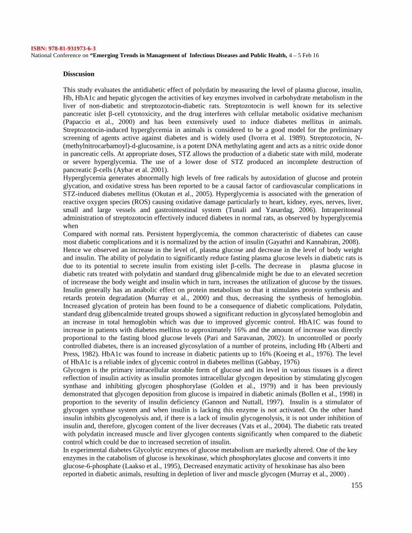
ISBN: 978-81-931973-6-3National Conference on “Emerging Trends in Management of Infectious Diseases and Public Health, 4 – 5 Feb 16
155
Disscusion
This study evaluates the antidiabetic effect of polydatin by measuring the level of plasma glucose, insulin,Hb, HbA1c and hepatic glycogen the activities of key enzymes involved in carbohydrate metabolism in theliver of non-diabetic and streptozotocin-diabetic rats. Streptozotocin is well known for its selectivepancreatic islet β-cell cytotoxicity, and the drug interferes with cellular metabolic oxidative mechanism(Papaccio et al., 2000) and has been extensively used to induce diabetes mellitus in animals.Streptozotocin-induced hyperglycemia in animals is considered to be a good model for the preliminaryscreening of agents active against diabetes and is widely used (Ivorra et al. 1989). Streptozotocin, N-(methylnitrocarbamoyl)-d-glucosamine, is a potent DNA methylating agent and acts as a nitric oxide donorin pancreatic cells. At appropriate doses, STZ allows the production of a diabetic state with mild, moderateor severe hyperglycemia. The use of a lower dose of STZ produced an incomplete destruction ofpancreatic β-cells (Aybar et al. 2001).Hyperglycemia generates abnormally high levels of free radicals by autoxidation of glucose and proteinglycation, and oxidative stress has been reported to be a causal factor of cardiovascular complications inSTZ-induced diabetes mellitus (Okutan et al., 2005). Hyperglycemia is associated with the generation ofreactive oxygen species (ROS) causing oxidative damage particularly to heart, kidney, eyes, nerves, liver,small and large vessels and gastrointestinal system (Tunali and Yanardag, 2006). Intraperitonealadministration of streptozotocin effectively induced diabetes in normal rats, as observed by hyperglycemiawhenCompared with normal rats. Persistent hyperglycemia, the common characteristic of diabetes can causemost diabetic complications and it is normalized by the action of insulin (Gayathri and Kannabiran, 2008).Hence we observed an increase in the level of, plasma glucose and decrease in the level of body weightand insulin. The ability of polydatin to significantly reduce fasting plasma glucose levels in diabetic rats isdue to its potential to secrete insulin from existing islet β-cells. The decrease in plasma glucose indiabetic rats treated with polydatin and standard drug glibencalmide might be due to an elevated secretionof incresease the body weight and insulin which in turn, increases the utilization of glucose by the tissues.Insulin generally has an anabolic effect on protein metabolism so that it stimulates protein synthesis andretards protein degradation (Murray et al., 2000) and thus, decreasing the synthesis of hemoglobin.Increased glycation of protein has been found to be a consequence of diabetic complications. Polydatin,standard drug glibencalmide treated groups showed a significant reduction in glycosylated hemoglobin andan increase in total hemoglobin which was due to improved glycemic control. HbA1C was found toincrease in patients with diabetes mellitus to approximately 16% and the amount of increase was directlyproportional to the fasting blood glucose levels (Pari and Saravanan, 2002). In uncontrolled or poorlycontrolled diabetes, there is an increased glycosylation of a number of proteins, including Hb (Alberti andPress, 1982). HbA1c was found to increase in diabetic patients up to 16% (Koeing et al., 1976). The levelof HbA1c is a reliable index of glycemic control in diabetes mellitus (Gabbay, 1976)Glycogen is the primary intracellular storable form of glucose and its level in various tissues is a directreflection of insulin activity as insulin promotes intracellular glycogen deposition by stimulating glycogensynthase and inhibiting glycogen phosphorylase (Golden et al., 1979) and it has been previouslydemonstrated that glycogen deposition from glucose is impaired in diabetic animals (Bollen et al., 1998) inproportion to the severity of insulin deficiency (Gannon and Nuttall, 1997). Insulin is a stimulator ofglycogen synthase system and when insulin is lacking this enzyme is not activated. On the other handinsulin inhibits glycogenolysis and, if there is a lack of insulin glycogenolysis, it is not under inhibition ofinsulin and, therefore, glycogen content of the liver decreases (Vats et al., 2004). The diabetic rats treatedwith polydatin increased muscle and liver glycogen contents significantly when compared to the diabeticcontrol which could be due to increased secretion of insulin.In experimental diabetes Glycolytic enzymes of glucose metabolism are markedly altered. One of the keyenzymes in the catabolism of glucose is hexokinase, which phosphorylates glucose and converts it intoglucose-6-phosphate (Laakso et al., 1995), Decreased enzymatic activity of hexokinase has also beenreported in diabetic animals, resulting in depletion of liver and muscle glycogen (Murray et al., 2000) .
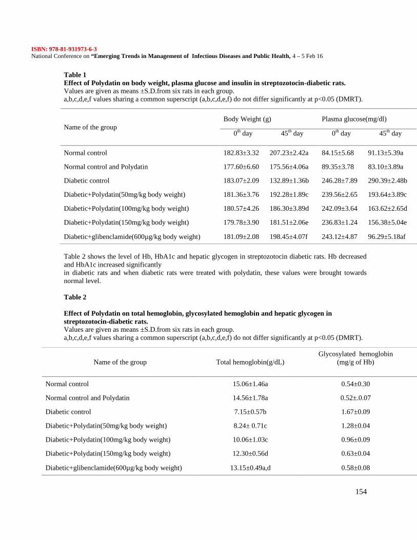
ISBN: 978-81-931973-6-3National Conference on “Emerging Trends in Management of Infectious Diseases and Public Health, 4 – 5 Feb 16
154
Table 1Effect of Polydatin on body weight, plasma glucose and insulin in streptozotocin-diabetic rats.Values are given as means ±S.D.from six rats in each group.a,b,c,d,e,f values sharing a common superscript (a,b,c,d,e,f) do not differ significantly at p<0.05 (DMRT).
Table 2 shows the level of Hb, HbA1c and hepatic glycogen in streptozotocin diabetic rats. Hb decreasedand HbA1c increased significantlyin diabetic rats and when diabetic rats were treated with polydatin, these values were brought towardsnormal level.
Table 2
Effect of Polydatin on total hemoglobin, glycosylated hemoglobin and hepatic glycogen instreptozotocin-diabetic rats.Values are given as means ±S.D.from six rats in each group.a,b,c,d,e,f values sharing a common superscript (a,b,c,d,e,f) do not differ significantly at p<0.05 (DMRT).
Name of the groupBody Weight (g) Plasma glucose(mg/dl)
Insulin(µU/mL)
0th day 45th day 0th day 45th day
Normal control 182.83±3.32 207.23±2.42a 84.15±5.68 91.13±5.39a 16.62±1.10a
Normal control and Polydatin 177.60±6.60 175.56±4.06a 89.35±3.78 83.10±3.89a 16.01±0.68a
Diabetic control 183.07±2.09 132.89±1.36b 246.28±7.89 290.39±2.48b 7.20±2.60b
Diabetic+Polydatin(50mg/kg body weight) 181.36±3.76 192.28±1.89c 239.56±2.65 193.64±3.89c 8.16±0.78c
Diabetic+Polydatin(100mg/kg body weight) 180.57±4.26 186.30±3.89d 242.09±3.64 163.62±2.65d 11.08±1.04d
Diabetic+Polydatin(150mg/kg body weight) 179.78±3.90 181.51±2.06e 236.83±1.24 156.38±5.04e 11.90±1.72d
Diabetic+glibenclamide(600µg/kg body weight) 181.09±2.08 198.45±4.07f 243.12±4.87 96.29±5.18af 15.20±1.55e
Name of the group Total hemoglobin(g/dL)Glycosylated hemoglobin
(mg/g of Hb)Hepatic glycogen(mg/100 g tissue)
Normal control 15.06±1.46a 0.54±0.30 54.06±3.09
Normal control and Polydatin 14.56±1.78a 0.52±.0.07 55.87±2.07
Diabetic control 7.15±0.57b 1.67±0.09 25.07±2.98
Diabetic+Polydatin(50mg/kg body weight) 8.24± 0.71c 1.28±0.04 33.02±3.05
Diabetic+Polydatin(100mg/kg body weight) 10.06±1.03c 0.96±0.09 41.43±3.12
Diabetic+Polydatin(150mg/kg body weight) 12.30±0.56d 0.63±0.04 47.17±1.97
Diabetic+glibenclamide(600µg/kg body weight) 13.15±0.49a,d 0.58±0.08 51.18±2.07
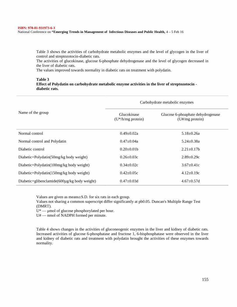
ISBN: 978-81-931973-6-3National Conference on “Emerging Trends in Management of Infectious Diseases and Public Health, 4 – 5 Feb 16
155
Table 3 shows the activities of carbohydrate metabolic enzymes and the level of glycogen in the liver ofcontrol and streptozotocin-diabetic rats.The activities of glucokinase, glucose 6-phosphate dehydrogenase and the level of glycogen decreased inthe liver of diabetic rats.The values improved towards normality in diabetic rats on treatment with polydatin.
Table 3Effect of Polydatin on carbohydrate metabolic enzyme activities in the liver of streptozotocin -diabetic rats.
Values are given as means±S.D. for six rats in each group.Values not sharing a common superscript differ significantly at pb0.05. Duncan's Multiple Range Test(DMRT).U* — μmol of glucose phosphorylated per hour.U# — nmol of NADPH formed per minute.
Table 4 shows changes in the activities of gluconeogenic enzymes in the liver and kidney of diabetic rats.Increased activities of glucose 6-phosphatase and fructose 1, 6-bisphosphatase were observed in the liverand kidney of diabetic rats and treatment with polydatin brought the activities of these enzymes towardsnormality.
Name of the group
Carbohydrate metabolic enzymes
Glucokinase(U*/h/mg protein)
Glucose 6-phoaphate dehydrogenase(U#/mg protein)
Normal control 0.49±0.02a 5.18±0.26a
Normal control and Polydatin 0.47±0.04a 5.24±0.38a
Diabetic control 0.20±0.01b 2.21±0.17b
Diabetic+Polydatin(50mg/kg body weight) 0.26±0.03c 2.89±0.29c
Diabetic+Polydatin(100mg/kg body weight) 0.34±0.02c 3.67±0.41c
Diabetic+Polydatin(150mg/kg body weight) 0.42±0.05c 4.12±0.19c
Diabetic+glibenclamide(600µg/kg body weight) 0.47±0.03d 4.67±0.57d
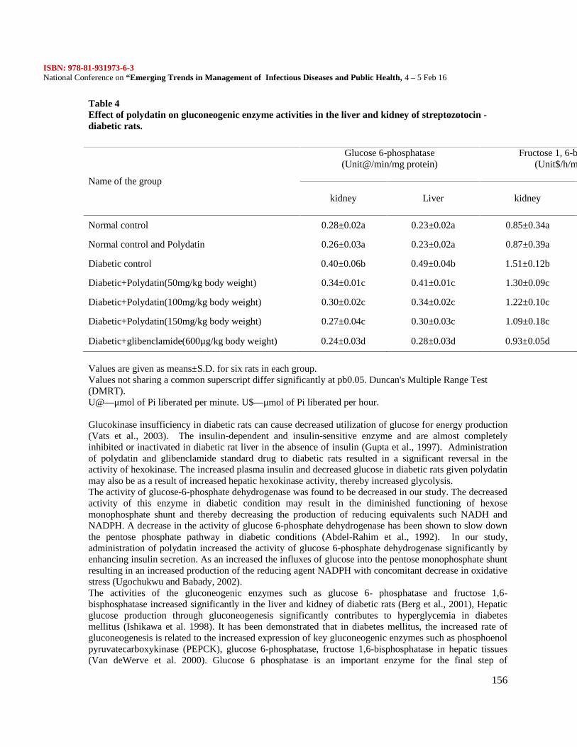
ISBN: 978-81-931973-6-3National Conference on “Emerging Trends in Management of Infectious Diseases and Public Health, 4 – 5 Feb 16
156
Table 4Effect of polydatin on gluconeogenic enzyme activities in the liver and kidney of streptozotocin -diabetic rats.
Values are given as means±S.D. for six rats in each group.Values not sharing a common superscript differ significantly at pb0.05. Duncan's Multiple Range Test(DMRT).U@—μmol of Pi liberated per minute. U$—μmol of Pi liberated per hour.
Glucokinase insufficiency in diabetic rats can cause decreased utilization of glucose for energy production(Vats et al., 2003). The insulin-dependent and insulin-sensitive enzyme and are almost completelyinhibited or inactivated in diabetic rat liver in the absence of insulin (Gupta et al., 1997). Administrationof polydatin and glibenclamide standard drug to diabetic rats resulted in a significant reversal in theactivity of hexokinase. The increased plasma insulin and decreased glucose in diabetic rats given polydatinmay also be as a result of increased hepatic hexokinase activity, thereby increased glycolysis.The activity of glucose-6-phosphate dehydrogenase was found to be decreased in our study. The decreasedactivity of this enzyme in diabetic condition may result in the diminished functioning of hexosemonophosphate shunt and thereby decreasing the production of reducing equivalents such NADH andNADPH. A decrease in the activity of glucose 6-phosphate dehydrogenase has been shown to slow downthe pentose phosphate pathway in diabetic conditions (Abdel-Rahim et al., 1992). In our study,administration of polydatin increased the activity of glucose 6-phosphate dehydrogenase significantly byenhancing insulin secretion. As an increased the influxes of glucose into the pentose monophosphate shuntresulting in an increased production of the reducing agent NADPH with concomitant decrease in oxidativestress (Ugochukwu and Babady, 2002).The activities of the gluconeogenic enzymes such as glucose 6- phosphatase and fructose 1,6-bisphosphatase increased significantly in the liver and kidney of diabetic rats (Berg et al., 2001), Hepaticglucose production through gluconeogenesis significantly contributes to hyperglycemia in diabetesmellitus (Ishikawa et al. 1998). It has been demonstrated that in diabetes mellitus, the increased rate ofgluconeogenesis is related to the increased expression of key gluconeogenic enzymes such as phosphoenolpyruvatecarboxykinase (PEPCK), glucose 6-phosphatase, fructose 1,6-bisphosphatase in hepatic tissues(Van deWerve et al. 2000). Glucose 6 phosphatase is an important enzyme for the final step of
Name of the group
Glucose 6-phosphatase(Unit@/min/mg protein)
Fructose 1, 6-bisphosphatase(Unit$/h/mg protein)
kidney Liver kidney Liver
Normal control 0.28±0.02a 0.23±0.02a 0.85±0.34a 0.45±0.02a
Normal control and Polydatin 0.26±0.03a 0.23±0.02a 0.87±0.39a 0.41±0.02a
Diabetic control 0.40±0.06b 0.49±0.04b 1.51±0.12b 0.79±0.04b
Diabetic+Polydatin(50mg/kg body weight) 0.34±0.01c 0.41±0.01c 1.30±0.09c 0.71±0.03c
Diabetic+Polydatin(100mg/kg body weight) 0.30±0.02c 0.34±0.02c 1.22±0.10c 0.61±0.01c
Diabetic+Polydatin(150mg/kg body weight) 0.27±0.04c 0.30±0.03c 1.09±0.18c 0.53±0.03c
Diabetic+glibenclamide(600µg/kg body weight) 0.24±0.03d 0.28±0.03d 0.93±0.05d 0.46±0.03d
ISBN: 978-81-931973-6-3National Conference on “Emerging Trends in Management of Infectious Diseases and Public Health, 4 – 5 Feb 16
157
gluconeogenesis or glucogenolysis in which it catalyzes the hydrolysis of glucose 6 phosphate to glucoseand phosphate. Glucose is transported out of the liver to increase blood glucose concentration. Normallyinsulin inhibits the hepatic glucose production by suppressing glucose 6 phosphatase and fructose 1, 6-bisphosphatase activity (Chen et al., 2000; Wiernsperger and Bailey, 1999). The hepatic glucose-6-phosphatase catalyses the terminal step of glucose production and it plays a key role in the maintenance ofblood glucose homeostasis. Increased activities of glucose-6-phosphatase in diabetic rats provide hydrogenwhich binds with NADP+ in the form of NADPH and enhances synthesis of fats from carbohydrates,lipogenesis (Bopanna et al., 1997). Fructose1-6-phosphatase is the important regulatory enzyme of thegluconeogenic pathway (Minnassian and Mitheux, 1994). The diabetic rats treated with polydatin broughtthese enzyme activities to near normal levels, which might be due to increased secretion of insulin. Thepossible mechanism by which polydatin bring about the normalization of enzyme activity may be bypotentiation of insulin release from β-cells of the islets of Langerhan’s which might enhance glucoseutilization.
ConclusionIn conclusion the investigation depicts that oral administration of polydatin has beneficial effect inreducing blood glucose, decreased Hb, increased level of HbA1c, hepatic glycogen and increased insulinlevels improving glycemic control as well as carbohydrate metabolism.
Reference1. American diabetes association. 20122. Bailey, Agardh CD, Bjorgell P, Nilson EP (2000). The effect of tolbutamide on lipoproteins, andlipoproteinlipase and hormone sensitive lipase. Diabetes Res Clin Pract 1999; 46:99-108.3. Rang and Dale, (1991), Diuretic, and antiinflammatory principle from Scoparia dulcis. Pharmazie56:657-660.4. Shu SY, Wang XY, Ling ZY, Lu ZY (2004) Effect of polydatin on phospholipase A2 in lungtissues in rats with endo toxic shock. Chin J Traumatol7 (4):239–43.5. Son.s. Lewis B.A (2002) Free radical scavenging and antioxidative activity of caffeic acid amideand ester analogues; structure activity relationships. J. Agric. Food Chem 30: 468-472.6. Sundaram, R.K., Bhaskar, A., Vijayalingam, S., Viswanatthan, M., Moha, R.,Shanmugasundaram,K.R. (1996)Antioxidant status and lipid peroxidation in type II diabetes mellitus with and withoutcomplications. Clinical Science 90, 255–260.7. The WHO Expert Committee on Diabetes Mellitus (1980) Technical Reports series 646. WorldHealth Organization, Geneva.8. Trinder P (1969) Determination of Glucose in Blood using glucose oxidase with an alternativeoxygen acceptor. Ann. Cli. Biochem 6: 24-25.9. Bourgeois. Wiggins, D., Hems, R., Gibbons, G.F (1995) VLDL output by hepatocytes from obeseZucker rats is resistant to the inhibitory effect of insulin. American Journal of Physiology 269, E208–E215.10. Bernal J, Mendiola JA, Ibanez E, Cifuentes A (2010) Advanced analysis of nutraceuticals. J pharmand Biomed Anal 55:758-74.11. Betteridge J (1997) Lipid disorder in diabetes mellitus. In: pickup, J.C., Williams, Text book ofDiabetes, second ed. Blakwell science 1-35.12. Blades, B., Garg,A (1995) Mechanisms of increase in plasma triacylglycerol concentration as aresult of high carbohydrate intakes in patients with noninsulin-dependent diabetes mellitus. AmericanJournal of Clinical Nutrition 62, 996-1002.13. Bopanna KN, Kannan J, Sushma G, Balaraman R, Rathod SP (1997) Antidiabetic andantihyperlipidemic effect of neem seed, kernel powder on alloxan diabetic rabbits. Ind J Pharmacol29:162-167.14. Papaccio et al., 2000, Lipid lowering and laque regression: new insights into prevention events ofplaque disruption and clinical in coronary disease. Circulation 81:1781.
ISBN: 978-81-931973-6-3National Conference on “Emerging Trends in Management of Infectious Diseases and Public Health, 4 – 5 Feb 16
158
15. Ivorra et al. 1989, Nutritional approaches to combat oxidative stress in Alzheimer’s disease. J NutrBiochem 13:444-61.16. Deliorman Orhan, D., Aslan, M., S¸ endo˘gdu, N., Ergun, F., Yes¸ ilada, E (2005)Evaluation of thehypoglycemic effect and antioxidant activity of three Viscum album subspecies (European mistletoe) instreptozotocin-diabetic rats. Journal of Ethnopharmacology 98, 95–102.17. Du, J., Sun, L.N., Xing, W.W., Huang, B.K., Jia, M., Wu, J.Z., Zhang, H., Qin, L.P (2009) Lipid-lowering effects of polydatin from Polygonum cuspidatum in hyperlipidemic hamsters. Phytomedicine 16,652–658.18. World Health Organization (2012),http://www.who.int/features/factfiles/diabetes/en/index.html.19. World Health Organization 2002, Launches the first global strategyontra-ditional and alternativemedicine. WHO Press release p.38:http://www.who.int/mediacentre/news/release/release38/en.20. Y.W.Bynes, R.Thrope (1999) Role of oxidative stress in diabetic complication, diabetes 481-9.21. Zhang PW, Yu CL, Wang YZ, Luo SF, Sun LS, Li RS (1995) Infuence of 3, 4', 5-trihydroxystibene- 3-beta -mono-D-glucoside on vascular endothelial epoprostenol and plateletaggregation. Zhongguo Yao Li Xue Bao 16(3):265–8.22. Zilversmit DB,Davis AK (1950) Micro determination of phospholipids by TCA precipitation. JLab Clin Med 35:155–9.23. Zlatkis A, ZakB, BoyleGJ (1953) A simple method for determination of serum cholesterol. Clin JMed 41:486–92.24. Bopanna et al., 1997 Micro determination of phospholipids by TCA precipitation. J Lab Clin Med35:155–9.25. Minnassian and Mitheux, 1994, Polydatin ameliorates experimental diabetes induced fibronectinthrough inhibiting the activation of NF-kB signaling pathway in rat glomerular mesangial cells, Mol andcellu Endocrine, 362; 183-193.
ISBN: 978-81-931973-6-3National Conference on “Emerging Trends in Management of Infectious Diseases and Public Health, 4 – 5 Feb 16
159
MOLECULAR DIAGNOSIS OF ISONIAZID DRUG RESISTANTMycobacterium tuberculosis IN HIV PATIENTS
Dr.B.Bharathi* and Kavitha.S
AbstractTuberculosis is an infectious disease caused by the bacillus Mycobacterium tuberculosis affecting nearlyone-third of the global population. Transmission of multidrug – resistant strains of Mycobacteriumtuberculosis (MDR-TB) presents a serious problem for TB control, particularly in the context of co-infection with the human immunodeficiency virus (HIV). MDR-TB has been well studied in outbreaks insettings of low endemicity in developed countries. However, the characteristics of MDR-TB in thecommunity with high endemicity such as India have not been well investigated. This study was designedto isolate and characterize inhA and katG gene from a clinical isolate of isoniazid drug resistant TBpatients co-infected with HIV. The blood sample obtained was first subjected to CD4 analysis using PartecFlow Cytometry instrument, RT-PCR analysis to confirm HIV positive. The serum albumin was separatedby 10% SDS – PAGE and then the suspected protein spot was sequenced using Nano LC/MS. The DNAwas isolated from the isoniazid resistant Mycobacterium tuberculosis culture, which was used for theamplification of the kat G and inhA genes using primers by PCR technique. The PCR product wassubjected to electrophoresis on 2% agarose gel and desired gene product was eluted by the gel cleanup kit.The kat G and inhA gene PCR purified products were analysed in the bioanalyser electropherogram todetect the molecular weight and was directly sequenced at Bioserve at Bangalore. The BLASTn andBLASTx search tool was used to compare the suspected DNA sequence for detecting the mutation toconfirm the drug resistance. Then the pure culture of Mycobacterium tuberculosis were then subjected tophylogenetic analysis using 16s rRNA primer by PCR technique. The 16s rRNA PCR purified productswas sequenced in the automated DNA sequencer. The MEGA search tool was used to compare anddifferentiate the 16s rRNA sequence from H37Rv wild strain and other Mycobacterium sp.
Introduction
Drug resistant strains of MTB are also increasing at an alarming rate which is a devastating threat to TBcontrol particularly in HIV patients. HIV/AIDS and TB are so closely connected that the term “co-epidemic” or “dual epidemic” is often used to describe their relationship. Drug-resistant tuberculosisposes a significant problem for treatment. The mechanisms of resistance to the front-line drug isoniazid(INH) are complex and can be mediated by katG, inhA and other unknown genes. Isoniazid (INH) is animportant first-line tuberculosis drug. INH is a pro-drug that requires activation by the M. tuberculosiscatalase-peroxidase enzyme (katG) to its active form (Zhang et al., 1992). Mutation of the katG gene,which leads to loss of or reduced catalase-peroxidase activity, is a major mechanism of INH resistance inM. tuberculosis (Zhang et al., 2000). Although various mutations in the katG gene have been reported inINH-resistant isolates, the most common mutation is the KatG Ser315Thr mutation, which is present inapproximately 50–90% of all INH-resistant isolates and is associated with relatively high-level resistanceto INH (Zhang et al., 2000). Mutations in inhA or its promoter region can cause INH resistance, withpromoter mutations being more frequent than mutations in the structural gene (Miesel et al., 1998).Mutations in inhA cause not only INH resistance, but also resistance to the structurally related second-linedrug ethionamide (ETH) (Banerjee et al., 1994). The TB bacteria are virulent enough that they can makeeven healthy adults and children sick, who in turn can spread it to others. If there are a lot of HIV-infectedpeople in the community, the vicious cycle results in more and more people with TB. In this study, weperformed a detailed characterization of a panel of primarily INH-resistant M. tuberculosis strains in termsof their mechanism of INH resistance in order to shed light on the frequency of such strains.
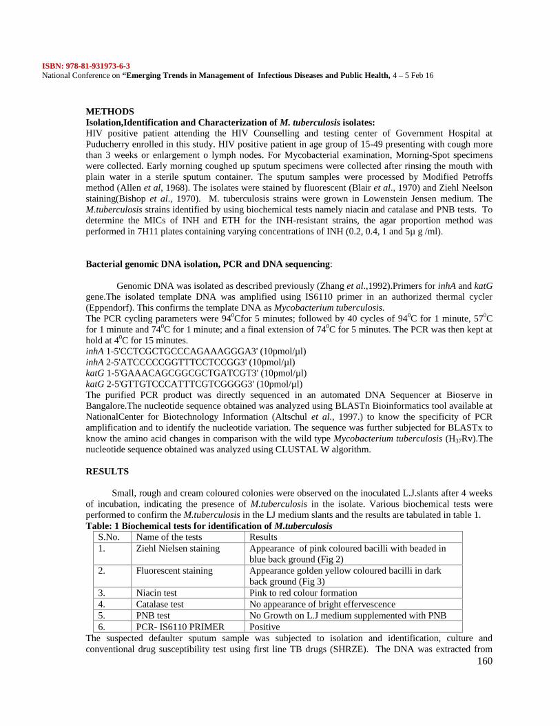
ISBN: 978-81-931973-6-3National Conference on “Emerging Trends in Management of Infectious Diseases and Public Health, 4 – 5 Feb 16
160
METHODSIsolation,Identification and Characterization of M. tuberculosis isolates:HIV positive patient attending the HIV Counselling and testing center of Government Hospital atPuducherry enrolled in this study. HIV positive patient in age group of 15-49 presenting with cough morethan 3 weeks or enlargement o lymph nodes. For Mycobacterial examination, Morning-Spot specimenswere collected. Early morning coughed up sputum specimens were collected after rinsing the mouth withplain water in a sterile sputum container. The sputum samples were processed by Modified Petroffsmethod (Allen et al, 1968). The isolates were stained by fluorescent (Blair et al., 1970) and Ziehl Neelsonstaining(Bishop et al., 1970). M. tuberculosis strains were grown in Lowenstein Jensen medium. TheM.tuberculosis strains identified by using biochemical tests namely niacin and catalase and PNB tests. Todetermine the MICs of INH and ETH for the INH-resistant strains, the agar proportion method wasperformed in 7H11 plates containing varying concentrations of INH (0.2, 0.4, 1 and 5µ g /ml).
Bacterial genomic DNA isolation, PCR and DNA sequencing:
Genomic DNA was isolated as described previously (Zhang et al.,1992).Primers for inhA and katGgene.The isolated template DNA was amplified using IS6110 primer in an authorized thermal cycler(Eppendorf). This confirms the template DNA as Mycobacterium tuberculosis.The PCR cycling parameters were 940Cfor 5 minutes; followed by 40 cycles of 940C for 1 minute, 570Cfor 1 minute and 740C for 1 minute; and a final extension of 740C for 5 minutes. The PCR was then kept athold at 40C for 15 minutes.inhA 1-5'CCTCGCTGCCCAGAAAGGGA3' (10pmol/µl)inhA 2-5'ATCCCCCGGTTTCCTCCGG3' (10pmol/µl)katG 1-5'GAAACAGCGGCGCTGATCGT3' (10pmol/µl)katG 2-5'GTTGTCCCATTTCGTCGGGG3' (10pmol/µl)The purified PCR product was directly sequenced in an automated DNA Sequencer at Bioserve inBangalore.The nucleotide sequence obtained was analyzed using BLASTn Bioinformatics tool available atNationalCenter for Biotechnology Information (Altschul et al., 1997.) to know the specificity of PCRamplification and to identify the nucleotide variation. The sequence was further subjected for BLASTx toknow the amino acid changes in comparison with the wild type Mycobacterium tuberculosis (H37Rv).Thenucleotide sequence obtained was analyzed using CLUSTAL W algorithm.
RESULTS
Small, rough and cream coloured colonies were observed on the inoculated L.J.slants after 4 weeksof incubation, indicating the presence of M.tuberculosis in the isolate. Various biochemical tests wereperformed to confirm the M.tuberculosis in the LJ medium slants and the results are tabulated in table 1.Table: 1 Biochemical tests for identification of M.tuberculosis
S.No. Name of the tests Results1. Ziehl Nielsen staining Appearance of pink coloured bacilli with beaded in
blue back ground (Fig 2)2. Fluorescent staining Appearance golden yellow coloured bacilli in dark
back ground (Fig 3)3. Niacin test Pink to red colour formation4. Catalase test No appearance of bright effervescence5. PNB test No Growth on L.J medium supplemented with PNB6. PCR- IS6110 PRIMER Positive
The suspected defaulter sputum sample was subjected to isolation and identification, culture andconventional drug susceptibility test using first line TB drugs (SHRZE). The DNA was extracted from
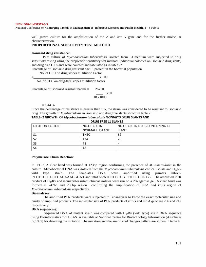
ISBN: 978-81-931973-6-3National Conference on “Emerging Trends in Management of Infectious Diseases and Public Health, 4 – 5 Feb 16
161
well grown culture for the amplification of inh A and kat G gene and for the further molecularcharacterization.PROPORTIONAL SENSITIVITY TEST METHOD
Isoniazid drug resistance:Pure culture of Mycobacterium tuberculosis isolated from LJ medium were subjected to drug
sensitivity testing using the proportion sensitivity test method. Individual colonies on Isoniazid drug slants,and drug free L.J slants were counted and tabulated as in table -2.Percentage of Isoniazid drug resistant bacilli present in the bacterial population
No. of CFU on drug slopes x Dilution Factor=___________________________________ x 100
No. of CFU on drug-free slopes x Dilution factor
Percentage of isoniazid resistant bacilli = 26x10____ x100
18 x1000
= 1.44 %Since the percentage of resistance is greater than 1%, the strain was considered to be resistant to Isoniaziddrug. The growth of M.tuberculosis in isoniazid and drug free slants shown in table 2.TABLE- 2 GROWTH OF Mycobacterium tuberculosis ISONIAZID DRUG SLANTS AND
DRUG FREE L.J SLANTSDILUTION FACTOR NO.OF CFU IN
NORMAL L.J SLANTNO.OF CFU IN DRUG CONTAINING L.JSLANT
S1 TNTC 62S2 124 26S3 78 -S4 18 -
Polymerase Chain Reaction:
In PCR, A clear band was formed at 123bp region confirming the presence of M. tuberculosis in theculture. Mycobacterial DNA was isolated from the Mycobacterium tuberculosis clinical isolate and H37Rvwild type strain. The templates DNA were amplified using primers inhA1-5'CCTCGCTGCCCAGAAAGGGA3' and inhA2-5'ATCCCCCGGTTTCCTCCG G3'. The amplified PCRproduct of H37Rv and isoniazid-resistant clinical isolates were run on a 2% agarose gel. A clear band wasformed at 247bp and 206bp region confirming the amplification of inhA and katG region ofMycobacterium tuberculosis respectively.Bioanalyzer:
The amplified PCR products were subjected to Bioanalyzer to know the exact molecular size andpurity of amplified products. The molecular size of PCR products of kat G and inh A gene are 206 and 247respectivelyDNA sequencing:
Sequenced DNA of mutant strain was compared with H37Rv (wild type) strain DNA sequenceusing Bioinformatics tool BLASTn available at National Centre for Biotechnology Information (Altschuletal,1997) for detecting the mutation. The mutation and the amino acid changes pattern are shown in table 4.
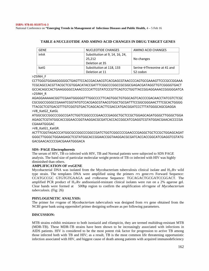
ISBN: 978-81-931973-6-3National Conference on “Emerging Trends in Management of Infectious Diseases and Public Health, 4 – 5 Feb 16
162
TABLE 4:NUCLEOTIDE AND AMINO ACID CHANGES IN DRUG TARGET GENES
GENE NUCLEOTIDE CHANGES AMINO ACID CHANGESinhA Substitution at 9, 14, 16, 24,
25,212Deletion at 35
No changes
katG Substitution at 118, 133Deletion at 11
Serine→Threonine at 41 and52 codon
>25INH_FCCTTGGGTGGAAGGGGGCTGAGTTCCACCGACAACGTCACGAGCGTAACCCCAGTGCGAAAGTTCCCGCCGGAAATCGCAGCCACGTTACGCTCGTGGACATACCGATTTCGGCCCGGCCGCGGCGAGACGATAGGTTGTCGGGGTGACTGCCACAGCCACTGAAGGGGCCAAACCCCCATTCGTATCCCGTTCAGTCCTGGTTACCGGAGGAAACCGGGGGATCA>25INH_RAGAGGAAAAACGGTTCGAATGGGGGTTTGGCCCCTTCAGTGGCTGTGGCAGTCACCCCGACAACCTATCGTCTCGCCGCGGCCGGGCCGAAATCGGTATGTCCACGAGCGTAACGTGGCTGCGATTTCCGGCGGGAACTTTCGCACTGGGGTTACGCTCGTGACGTTTGTCGGTGTGACTCAGCACACTTCGACCATGACGGATCCCTTTATGGGCAGCGAGGA>V8_KatG2_KatGLATGGCGCCGGCCCGGCCGATCTGGTCGGCCCCGAACCCGAGGCTGCTCCGCTGGAGCAGATGGGCTTGGGCTGGAAGAGCTCGTATGGCACCGGAACCGGTAAGGACGCGATCACCACCGGCATCGAGGTCGTATGGACGAACACCCCGACGAAATGGGAC>V8_KatG1_KatGR.ACTTTCGGTAGACCCATGGCGCCGGCCCGGCCGATCTGGTCGGCCCCGAACCCGAGGCTGCTCCGCTGGAGCAGATGGGCTTGGGCTGGAAGAGCTCGTATGGCACCGGAACCGGTAAGGACGCGATCACCACCGGCATCGAGGTCGTATGGACGAACACCCCGACGAAATGGGACA
SDS- PAGE ElectrophoresisThe serum of HIV, TB co infected with HIV, TB and Normal patients were subjected to SDS PAGEanalysis. The band size of particular molecular weight protein of TB co infected with HIV was highlydiminished than others.AMPLIFICATION OF rrsGENEMycobacterial DNA was isolated from the Mycobacterium tuberculosis clinical isolate and H37Rv wildtype strain. The templates DNA were amplified using the primers rrs gene:rrs Forward Sequence:CCATGCCGC GTGTGTGAAGA and rrsReverse Sequence: TGCAGACTGCGATCCGGACT. Theamplified PCR product of H37Rv andisoniazid-resistant clinical isolates were run on a 2% agarose gel.Clear bands were formed at 500bp region to confirm the amplification ofrrsgene of Mycobacteriumtuberculosis. (Fig: 26)
PHYLOGENETIC ANALYSIS:The primer for rrsgene of Mycobacterium tuberculosis was designed from rrs gene obtained from theNCBI gene bank using epperndorf primer designing software as per following parameters.
DISCUSSION:
MTB strains exhibit resistance to both isoniazid and rifampicin, they are termed multidrug-resistant MTB(MDR-TB). These MDR-TB strains have been shown to be increasingly associated with infections inAIDS patients. HIV is considered to be the most potent risk factor for progression to active TB amongthose infected both with TB and HIV; as a result, TB is the most common life threatening opportunisticinfection associated with HIV, and biggest cause of death among patients with acquired immunodeficiency
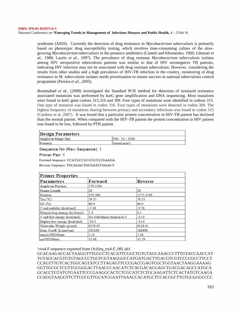
ISBN: 978-81-931973-6-3National Conference on “Emerging Trends in Management of Infectious Diseases and Public Health, 4 – 5 Feb 16
163
syndrome (AIDS). Currently the detection of drug resistance in Mycobacterium tuberculosis is primarilybased on phenotypic drug susceptibility testing, which involves time-consuming culture of the slow-growing Mycobacterium tuberculosis in the presence antibiotics (Canetti and Khomenko, 1969; Libonati etal., 1988; Laszlo et al., 1997). The prevalence of drug resistant Mycobacterium tuberculosis isolatesamong HIV seropositive tuberculosis patients was similar to that of HIV seronegative TB patients,indicating HIV infection may not be associated with drug resistant tuberculosis. However, considering theresults from other studies and a high prevalence of HIV-TB infection in the country, monitoring of drugresistance in M. tuberculosis isolates needs prioritization to ensure success in national tuberculosis controlprogramme (Pereira et al., 2005).
Bostanabad et al., (2008) investigated the Standard PCR method for detection of isoniazid resistanceassociated mutations was performed by katG gene amplification and DNA sequencing. Most mutationswere found in katG gene codons 315,316 and 309. Four types of mutations were identified in codons 315.One type of mutation was found in codon 316. Four types of mutations were detected in codon 309. Thehighest frequency of mutations sharing between primary and secondary infections was found in codon 315(Cardoso et al, 2007). It was found that a particular protein concentration in HIV-TB patient has declinedthan the normal patient. When compared with the HIV-TB patient the protein concentration in HIV patientwas found to be low, followed by PTB patient.
>rrs4-F sequence exported from OciSeq_rrs4-F_081.ab1GCACAAGACCACTAAGGTTTGGCCTCACATTCGGCTGTGTAGCAAACCCTTTGTACCAACCATTGTAGCACGTGTGTAGCCCTGGTCGTAAGGGCCATGATGACTTGACGTCGTCCCCGCCTTCCTCCAGTTTGTCACTGGCAGTATCCTTAGAGTTCCCGACCGAGTCGCTGGTAACTAAGGAAAAGGGTTGCGCTCGTTGCGGGACTTAACCCAACATCTCACGACACGAGCTGACGACAGCCATGCAGCACCTGTATGTGAATTCCCGAAGGCACTCTCGCATCTCTGCAAGATTCTCACTATGTCAAGACCAGGTAAGGTTCTTCGCGTTGCATCGAATTAAACCACATGCTCCACCGCTTGTGCGGGCCCC
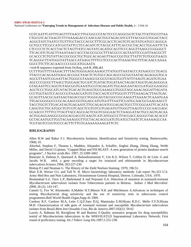
ISBN: 978-81-931973-6-3National Conference on “Emerging Trends in Management of Infectious Diseases and Public Health, 4 – 5 Feb 16
164
CGTCAATTCATTTGAGTTTTAACCTTGCGGCCGTACTCCCCAGGCGGTCTACTTATTGCGTTAACTGCGTCACTAAGTCTTTAAAAGACCCAACGACTGGTAGACATCGTTTACGGCGTGGACTACCAGGGTATCTAATCCTGTTTGCTACCCACGCTTTCGCACCTCAGTGTCAGTATGATGCCAGGGAGCTGCCTTCGCCATCGGTATTCCTCCAGATCTCTACGCATTTCACCGCTACACCTGGAATTCTACTCCCCTCTCACCTACTCTAGTTATCCAGTATCAGATGCAGTTCCCAGGTTAAGCCCGGGGCTTTCACATCTGACTTAAATAACCACCTACGCGCGCTTTACGCCCAGTAATTCCGATTAACGCTTGCACCCTCTGTATTACCGCGGCTGCTGGCACAGAGTTAGCCGGTGCTTATTCTGTGGGTAACGTCAGGGCTTATGGGTATTAACCATAAGCTTTTCCTCCCCACTTAAAGTGCTTTACAACCAAAAGGCCTTCTTCACAACCCCCGGCATGGAATA>rrs4-R sequence exported from OciSeq_rrs4-R_096.ab1CCTTTGGTTTTAAAACTTTTAGTGGGGAGGAAAGCTTATGGTTAATACCCATAAGCCCTGACGTTACCCACAGAATAAGCACCGGCTAACTCTGTGCCAGCAGCCGCGGTAATACAGAGGGTGCAAGCGTTAATCGGAATTACTGGGCGTAAAGCGCGCGTAGGTGGTTATTTAAGTCAGATGTGAAAGCCCCGGGCTTAACCTGGGAACTGCATCTGATACTGGATAACTAGAGTAGGTGAGAGGGGAGTAGAATTCCAGGTGTAGCGGTGAAATGCGTAGAGATCTGGAGGAATACCGATGGCGAAGGCAGCTCCCTGGCATCATACTGACACTGAGGTGCGAAAGCGTGGGTAGCAAACAGGATTAGATACCCTGGTAGTCCACGCCGTAAACGATGTCTACCAGTCGTTGGGTCTTTTAAAGACTTAGTGACGCAGTTAACGCAATAAGTAGACCGCCTGGGGAGTACGGCCGCAAGGTTAAAACTCAAATGAATTGACGGGGGCCCGCACAAGCGGTGGAGCATGTGGTTTAATTCGATGCAACGCGAAGAACCTTACCTGGTCTTGACATAGTGAGAATCTTGCAGAGATGCGAGAGTGCCTTCGGGAATTCACATACAGGTGCTGCATGGCTGTCGTCAGCTCGTGTCGTGAGATGTTGGGTTAAGTCCCGCAACGAGCGCAACCCTTTTCCTTAGTTACCAGCGACTCGGTCGGGAACTCTAAGGATACTGCCAGTGACAAACTGGAGGAAGGCGGGGACGACGTCAAGTCATCATGGCCCTTACGACCAGGGCTACACACGTGCTACAATGGTTGGTACAAAGGGTTGCTACACAGCGATGTGATGCTAATCTCAAAAAGCCAATCGTAGTCCGGTGCCCACTTCTTGTGAAAAAAAAGATTCTC
BIBLIOGRAPHY
Allen B.W and Baker F.J. Mycobacteria Isolation, Identification and Sensitivity testing. Butterworths.1968; 10.Altschul, Stephen F, Thomas L, Madden, Alejandro A, Schaffer, Jinghui Zhang, Zheng Zhang, WebbMiller, and David J.Lipman. “Gapped Blast and PSI-BLAST: A new generation of protein database searchprograms”, J Nucleic acids Res. 1997; 25:3389-3402Banerjee A, Dubnau E, Quemard A, Balasubramanian V, Um K.S, Wilson T, Collins D. de Lisle. G andJacobs W.R. inhA, a gene encoding a target for isoniazid and ethionamide in Mycobacteriumtuberculosis.Science.1994; 263:227-230.Bishop P.J and Neuman G. The History of the Ziehl Neelsen Staining. 1970; 196:51.Blair E.B, Weiser O.L and Tull N. H. Micro bacteriology laboratory methods- Lab report No.323 U.S.Army Med Res and Nutr Laboratory, Fitrzsimmoun General Hospital, Denver, Colorado, USA. 1970.Bostanabad S.Z, Titov L.P, Barhrmand A and Nojoumi S.A. Detection of mutation in isoniazid-resistantMycobacterium tuberculosis isolates from Tuberculosis patients in Belarus. Indian J Med Microbiol.2008; 26 (2): 143-147.Canetti G, Fox W, Khomenko A,Mahler H.T,Menon N.K and Mitchinson A.Advances in techniques oftesting Mycobacterial drug sensitivity and the use of sensitivity tests in tubercuosis controlprogrammes.Bull World Health Organ,Supp 41.1969.Cardoso R.F, Cardoso M.A, Leite C.Q.F,Sato D.Q, Mamizuka E.M,Hirata R.D.C, Mello F.F.D,HirataM.H. Characterization of ndh gene of isoniazid resistant and susceptible Mycobacterium tuberculosisisolates from Brazil.Mem Inst Oswaldo Cruz, Rio de Janeiro.2007;102(1): 59-61Lazzlo A, Rahman M, Raviglione M and Bustreo F.Quality assurance program for drug susceptibilitytestinf of Mycobacterium tuberculosis in the WHO/IUATLD Supranational Laboratory Network; Firstround of proficiency testing. Int J Tuberc Lung Dis.1997;1:231-238.
ISBN: 978-81-931973-6-3National Conference on “Emerging Trends in Management of Infectious Diseases and Public Health, 4 – 5 Feb 16
165
Libonati J.P, Stager C.E, Davis J.R and Siddiqi S.H. Direct anti microbial drug susceptibility testing ofMycobacterium tuberculosis by the radiometric method. Diagn Microbiol Infect Dis.1988; 10:41-48.Mani C, Selvakumar N, Kumar V, Narayanan S, Narayanan P.R. Comparison of DNA sequencing, PCR-SSCP and PhaB assays with indirect sensitivity testing for detection of rifampcin resistance inMycobacterium tuberculosis. Int J Tuber Lung Dis. 2003; 7:625-659.Miesel L, Weisbrod T.R, Marcinkeviciene J. A, Bittman R and Jacobs W. R. NADH dehydrogenasedefects confer isoniazid resistance and conditional lethality in Mycobacterium smegmatis. J Bacteriol.1998; 180: 2459-2467.Pereira M, Tripathy S, Inamdar V, Ramesh K, Bhavsar M, Date A, Iyyer R, Acchammachary A,Mehendale S and Risbud A. Drug resistance pattern of Mycobacterium tuberculosis in seropositive andseronegative HIV-TB patients in Pune, India.Indian J Med Res.2005; 121:235-239Zhang Y.B, Heym B, Allen D, Young and Cole S.T. The catalseperoxidase gene and isoniazid resistanceof Mycobacterium tuberculosis. Nature. 1992; 358: 501-593.Zhang Y, Telenti A. Genetics of drug resistance in Mycobacterium tuberculosis. Washington, ASM Press.2000.
ISBN: 978-81-931973-6-3National Conference on “Emerging Trends in Management of Infectious Diseases and Public Health, 4 – 5 Feb 16
166
PHYTOCHEMICAL SCREENING AND ANTICANCEROUS ACTIVITY OF RHIZOMEEXTRACT COLEUS FORSKHOLI ON HEP G2 CELL LINE
X.Asbin Mary1, Robert Lotha2
1 Assistant Professor, Department of Biotechnology, Alpha Arts and Science College, Chennai.2 Research Scholar, Departments of Biotechnology, Sastra University, Thanjavur.
ABSTRACTTo evaluate the presence of Photochemicals, and anticancerous activities of Coleus forskohlii. The plantmaterial was extracted by using acetone and aqueous solvents. The phyotochemical analysis of Coleusforskohlii reveals the presence of tannin, flavonoids, terpenoids, alkaloids, cardiac glycosides, coumarinand steroids. The different acetone extract of Coleus forskohlii were prepared 40mg/ml, 80mg/ml,120mg/ml, 160mg/ml, and Cyclophasphamide (Positive control) 180 µg/ml and their activity weredetermined by MTT Assay using Hep G2 Cell line (liver).Among these concentration the maximumanticancerous activity were observed at 120 mg/ml and it determined by using MTT Assay.KEYWORDS - Coleus forskohlii, Phytochemicals, MTT assay, Anticancerous activity.
INTRODUCTION
Herbs are widely exploited in the traditional medicine and their curative potentials are well documented(Dubey et al., 2004). About 61% of new drugs developed between 1981 and 2002 were based on naturalproducts and they have been very successful especially in the areas of infectious disease and cancer(Cragg, 2005). Drugs derived from natural sources play a significant role in the prevention and treatmentof human diseases. In many developing countries, traditional medicine is one of the primary health caresystems (Houghton, 1995).
Natural products of higher plants may give a new source of antimicrobial agents with possibly novelmechanisms of action (Runyoro et al., 2006; Shahidi et al., 2004). The effects of plant extracts onbacteria have been studied by a very large number of researchers in different parts of the world (Reddy,2001).Coleus forskohlii belonging to Lamiaceae family (Mint family) has been selected. The leaves of the greentype of country borage are often eaten raw with bread and butter. The chopped leaves are also used assubstitute for sage (Salvia officinalis L) in stuffing. Coleus forskohlii is used for seasoning meat dishes andin food products, while a decoction of its leaves is administered in cases of chronic cough and asthma(Kusumoto et al., 1995). It is considered to be an antispasmodic, stimulant and stomachic and is used forthe treatment of headache, fever, epilepsy and dyspepsia. It is used to treat conditions such as indigestion,diarrhea, nervous tension, insect bites, toothache, earache, rheumatism, whooping cough, and bronchitis(Warrier et al., 1995).Nearly 25,000 effective plant based formulations are used in folk medicine by rural communities in India(Ramakrishnappa et al., 2002). Both plant species and traditional knowledge are important to the herbalmedicine trade and the pharmaceutical industry, whereby plants provide raw materials and the traditionalknowledge prerequisite information (Tabuti et al., 2003). The present study aims to investigate thepresence of phytochemicals and anticancerous activity of Coleus forskohlii rhizome extract.
2. MATERIALS AND METHODS
Collection of Plant materialThe rhizome of Coleus forskohlii was collected from Chennai, Tamil Nadu. And it was washed thoroughlyin distilled water to remove to dirt and air dried.Preparation of plant powder
ISBN: 978-81-931973-6-3National Conference on “Emerging Trends in Management of Infectious Diseases and Public Health, 4 – 5 Feb 16
167
The air dried and powdered roots were extracted with solvent like acetone and aqueous extract using amortar and pestle. The extraction mixture was filtered through Whatman No.1 filter paper. The extract wasevaporated under reduced pressure at 50° C using a rota vapour apparatus. Finally 10 mg of residue wasdissolved in 10 ml of each solvent and these extract used for anticancerous activity.Phytochemical ScreeningPhytochemical screening for tannins, flavonoids, terpenoids, alkaloids, glycosides, cardiac glycosides,coumarins and steroids were carried out as described below.1. Test for tanninsAbout 0.5 g of plant rhizome extract was boiled in 20 ml of distilled water in a test tube and then filtered.0.1% FeCl3 was added to the filtrate. Appearance of brownish green or blue black coloration showed thepresence of tannins.2. Test for flavonoids5 ml of dilute ammonia solution were added to a portion of the extract followed by addition ofconcentrated H2SO4.Formation of yellow colour indicates the presence of flavonoids.3. Test for TerpenoidsTo 0.5 ml of the rhizome extract, 2 ml of chloroform was added and concentrated Sulphuric acid wasadded carefully. Formation of red brown colour at the interface indicates the presence of Terpenoids.4. Test for alkaloidsPlant extract (0.5 to 0.6 g) was mixed with about 8 ml of 1% HCl, warmed and filtered. 2 ml of filtratewere treated separately with Mayer’s reagent / Dragendorff’s reagent. Turbidity or precipitation wasobserved to indicate the presence of alkaloids.5. Test for GlycosidesTo 2ml of the rhizome extract, 3ml of chloroform and 10% ammonium solution was added. Formation ofpink colour indicates the presence of glycosides.6. Test for cardiac glycosidesTo 0.5 ml of the rhizome extract, 2 ml of glacial acetic acid and few drops of 5 % ferric chloride wereadded. This under layered with 1 ml of concentrated sulphuric acid. Formation of brown ring at interfaceindicates the presence of cardiac glycosides.7. Test for coumarinsMoistened plant rhizome extract (0.5 g) was taken in a small test tube and covered with filter papermoistened with 1 N NaOH. The test tube was placed for few minutes in boiling water. Then the filter paperwas removed and examined in UV light for yellow florescence to indicate the presence of coumarins.8. Test for SteriodsTo 0.5 ml of the rhizome extract, 2 ml of chloroform and 1 ml of Sulphuric acid were added. Formation ofreddish brown ring at interface indicates the presence of steroids.Anticancer activity from rhizome extracts of Coleus forskohliiCell line and cultureHep G2 cell line (Liver) was obtained from National centre for cell sciences Pune (NCCS). The cells were
maintained in Minimal Essential Media supplemented with 10% FBS (Foetal Bovine serum), penicillin(100 U/ml), and streptomycin (100 μg/ml) in a humidified atmosphere of 50 μg/ml CO2 at 37 °C.ReagentsMEM was purchased from Hi Media Laboratories, Foetal bovine serum (FBS) was purchased from Cistronlaboratories. Trypsin, methylthiazolyl diphenyl- tetrazolium bromide (MTT), and Dimethyl sulfoxide(DMSO) were purchased from (Sisco research laboratory chemicals Mumbai). All of other chemicals andreagents were obtained from Sigma Aldrich Mumbai.In vitro assay for Cytotoxicity activity (MTT assay)The Cytotoxicity of samples on Hep G2 cell line (Liver) was determined by the MTT assay (Mosmann etal., 1983). Cells (1 × 10⁵/well) were plated in 1ml of medium/well in 24-well plates (Costar Corning,Rochester,NY). After 24 hours incubation the cell reaches the confluence. Then, cells were incubated inthe presence of various concentrations of the samples in 0.1% DMSO for 24h at 37°C. After removal ofthe sample solution and washing with phosphate-buffered saline (pH 7.4), 200µl/well (5mg/ml) of 0.5% 3-
ISBN: 978-81-931973-6-3National Conference on “Emerging Trends in Management of Infectious Diseases and Public Health, 4 – 5 Feb 16
168
(4, 5-dimethyl-2-thiazolyl)-2, 5-diphenyl-tetrazolium bromide cells (MTT) phosphate -buffered salinesolution was added. After 4h incubation, 0.04M HCl/ isopropanol were added. Viable cells weredetermined by the absorbance at 570nm. Measurements were performed and the concentration required fora 50% inhibition of viability (IC50) was determined graphically. The absorbance at 570 nm was measuredwith a UV- Spectrophotometer using wells without sample containing cells as blanks. The effect of thesamples on the proliferation of Hep G2 cell line (Liver) was expressed as the % cell viability, using thefollowing formula:
% cell viability = A570 of treated cells / A570 of control cells × 100
3. RESULTS AND DISCUSSION
Preparation of plant materialThe Coleus forskohlii rhizome materials were collected and extracted with acetone and aqueous for the
analysis.Preparation of rhizome powderThe dried and powdered roots were extracted with acetone and aqueous extract.Phytochemical analysisThe phytochemical analysis revealed the presence of tannin, flavonoids, terpenoids, alkaloids, cardiacglycosides, coumarin and steroids (Table 1).
Table 1: Phytochemical analysis from rhizome extract of Coleus forskohliiPhytochemical
compositionRhizome extract
Acetone Aqueous
Tannins + -Flavonoids + -Terpenoids + +Alkaloids + -Glycosides + -Cardiac glycosides + +Coumarins + -Steroids + +
IN VITRO Assays (Cytotoxic studies)
MTT Assay
The IC50 value for Hep G2 cell line (Liver) (120 mg/ml) acetone rhizome extract was found to beeffective, the reduction percentage of MTT at 24Hrs also estimated for Hep G2 cell line (Liver). Whenincubated with the extract, it induced cytotoxicity in a significant manner which implicit the damage to themember integrity of the cell when contributed with control. The cytotoxicity was minimized in the extracttreated cells and near normal level was attained at various concentrations (40mg/ml, 80 mg/ml, 120mg/mland 160mg/ml) and maximum effect was found when treated at 120 mg/ml(Table 2, Fig 1). From theabove results, it was confirmed that Coleus forskohlii Acetone rhizome extract at 120 mg/ml seems to offersignificant protection and maintain the structural integrity of the hepatocellular membrane and this activeconcentration was followed for further studies.Tryphan Blue assay
Tryphan blue is one of the several stains recommended for use in dye exclusion procedure forviable cell counting. This assay is based on the principle that live cells do not take up blue, where as deadcells do and appear as blue under microscope - depicts the viability of cells by Tryphan Blue assay. The
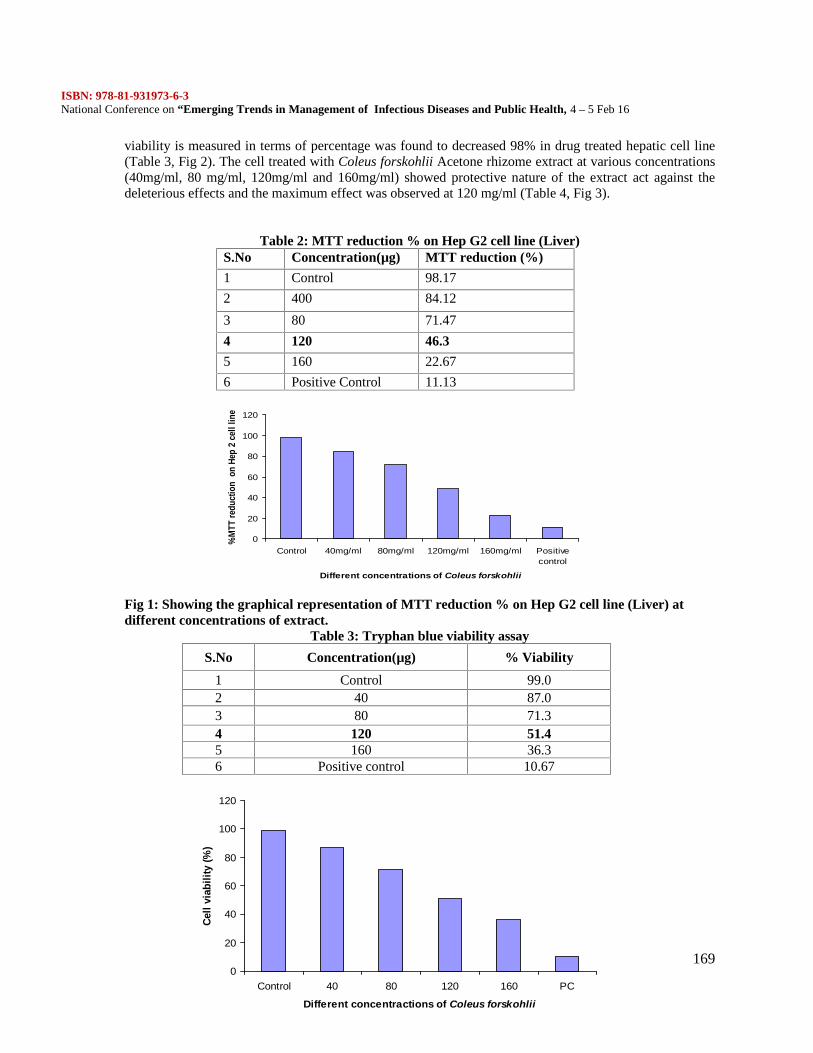
ISBN: 978-81-931973-6-3National Conference on “Emerging Trends in Management of Infectious Diseases and Public Health, 4 – 5 Feb 16
169
0
20
40
60
80
100
120
Control 40mg/ml 80mg/ml 120mg/ml 160mg/ml Positivecontrol
Different concentrations of Coleus forskohlii
%M
TT re
duct
ion
on
Hep
2 ce
ll lin
e
0
20
40
60
80
100
120
Control 40 80 120 160 PC
Different concentractions of Coleus forskohlii
Cell
viab
ility
(%)
viability is measured in terms of percentage was found to decreased 98% in drug treated hepatic cell line(Table 3, Fig 2). The cell treated with Coleus forskohlii Acetone rhizome extract at various concentrations(40mg/ml, 80 mg/ml, 120mg/ml and 160mg/ml) showed protective nature of the extract act against thedeleterious effects and the maximum effect was observed at 120 mg/ml (Table 4, Fig 3).
Table 2: MTT reduction % on Hep G2 cell line (Liver)S.No Concentration(µg) MTT reduction (%)
1 Control 98.17
2 400 84.12
3 80 71.47
4 120 46.3
5 160 22.67
6 Positive Control 11.13
Fig 1: Showing the graphical representation of MTT reduction % on Hep G2 cell line (Liver) atdifferent concentrations of extract.
Table 3: Tryphan blue viability assay
S.No Concentration(µg) % Viability
1 Control 99.02 40 87.03 80 71.34 120 51.45 160 36.36 Positive control 10.67
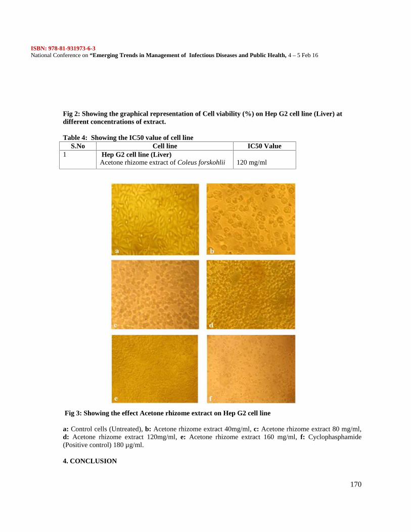
ISBN: 978-81-931973-6-3National Conference on “Emerging Trends in Management of Infectious Diseases and Public Health, 4 – 5 Feb 16
170
Fig 2: Showing the graphical representation of Cell viability (%) on Hep G2 cell line (Liver) atdifferent concentrations of extract.
Table 4: Showing the IC50 value of cell lineS.No Cell line IC50 Value
1 Hep G2 cell line (Liver)Acetone rhizome extract of Coleus forskohlii 120 mg/ml
Fig 3: Showing the effect Acetone rhizome extract on Hep G2 cell line
a: Control cells (Untreated), b: Acetone rhizome extract 40mg/ml, c: Acetone rhizome extract 80 mg/ml,d: Acetone rhizome extract 120mg/ml, e: Acetone rhizome extract 160 mg/ml, f: Cyclophasphamide(Positive control) 180 µg/ml.
4. CONCLUSION
ISBN: 978-81-931973-6-3National Conference on “Emerging Trends in Management of Infectious Diseases and Public Health, 4 – 5 Feb 16
171
In the present study the phytochemical analysis of Coleus forskohlii reveals, the presence of tannin,flavonoids, terpenoids, alkaloids, cardiac glycosides, coumarin and steroids. MTT Assay, using Hep G2Cell line (liver) at 120 mg/ml acetone rhizome extract was found to be effective, the reduction percentageof MTT and cytotoxicity were also determined.
6. ACKNOWLEDGMENTS
The authors are thankful to Head of the Department, Department of Biotechnology, Alpha arts and ScienceCollege. Thanks also to my Department Staffs, for their support.
7. REFERENCES\
1. Cragg GM, Newman DJ (2005). Biodiversity: a continuing source of novel drug leads. Pure ApplChem 77: 7–24.2. Dubey NK, Kumar R, Tripathi P (2004). Global promotion of herbal medicines: India’s opportunity.Current Science 86: 37‐41.3. Houghton PJ (1995). The role of plants in traditional medicine and current therapy. J Altern and ComplMed 1: 131‐143.4. Kusumoto IT, Nakabayashi T, Kida H (1995). Screening of various plant extracts used in ayurvedicmedicine for inhibitory effects on human immunodeficiency virus type 1 (HIV‐1) protease. PhytotherapyRes 9: 180‐184.5. Ramakrishnappa K. (2002) Impact of cultivation and gathering of medicinal plants on biodiversity:Case studies from India, In: Biodiversity and the Ecosystems Approach in Agriculture, Forestry andFisheries (Online), FAO.6. Reddy PS, Jamil K, Madhusudhan P (2001). Antibacterial activity of isolates from Piper longum andTaxus baccata. Pharmaceutical Biol 39: 236‐238.7. Runyoro D, Matee M, Olipa N, Joseph C, Mbwambo H (2006). Screening of Tanzanian medicinalplants for anti‐Candida activity. BMC Complement Altern Med 6(11).8. Shahidi BH (2004). Evaluation of antimicrobial properties of Iranian medicinal plants againstMicrococcus luteus, Serratia marcescens, Klebsiella pneumoniae and Bordetella bronchoseptica. Asian JPlant Sci 3: 82-86.9. Tabuti JRS, Lye KA, Dhillon SS. (2003) Traditional herbal drugs of Bulamogi, Uganda: Plants, useand administration. J. Ethnopharmacol, 88, 19-44.10. Warrier PK, Nambiar VP, Ramankutty (1995). Indian medicinal plants. 1 st Ed, Orient longmanlimited, Madras. 315.
ISBN: 978-81-931973-6-3National Conference on “Emerging Trends in Management of Infectious Diseases and Public Health, 4 – 5 Feb 16
172
STUDIES ON PHYSICO – CHEMICAL PROPERTIES, NODULATION PATTERN ANDRHIZOBIAL POPULATION OF BLACK GRAM CULTIVATED SOIL
N. Sivakumar.*1, K. Krishnapriya.2, P. Nirmala.3, A. Chandhru.4
Assistant Professor, Department of Microbiology, Mohamed Sathak arts and Science College,Sholinganallure, Tamil Nadu, India1,3,4 [email protected]*1
Assistant Professor in Microbiology, Marudupandiyar College, Thanjavur, India2.
ABSTRACT
Black gram (Vigna mungo L.) is one of the important pulse crops gaining importance all over the world inrecent years. It is rich in proteins and contains amino acids higher quantities than any other cereals andpulses. Black gram is an annual food legume. Black gram seeds are boiled and eaten whole or aftersplitting into dhal. The dried seeds contain approximately 9.7 % water, 23.4 % protein, 1 % fat, 57.3 %carbohydrate, 3.8 % fibre and 4.8 % ash. It is very nutritious and is recommended for diabetics. Thenutrient status of soil is more essential for root nodulation. The black gram soil samples were collectedfrom Nagapattinam and Cuddalore belonged to 3 textural group viz., Red loam, Clay loam and Block soils.The soil samples were analysed for its pH, Electrical conductivity, soil organic carbon availabe N, P and Kcontent (Tab-1). The nodulation pattern and its rhizobial population on the rhizosphere soil of blackgram plants were also studied. The nodulation pattern was ranged from 5.00 to 21.00 plants (Tab-2).Native rhizobial populations (Tab-3) were also estimated for the 30 locations from the abovementioned Districts of saline areas in Tamil Nadu. Among the 30 locations , 13 locations were recorded
more than 3×10³ g-1.
Key words: Physico-chemical, nodulation, rhizobial population, Black gram, Saline areas.
INTRODUCTION
Black gram is a member of the Vigna Asiatic crop group. It is an annual pulse grown mostly as a followcrop in rotation with cereals. Similar to the pulses, black gram, being a legume, it enriches the soil nitrogencontent and has relatively a short (90-120 days) duration. It is widely cultivated in India, Pakistan andother Asian countries. It is part of diet for millions of people in these countries and a cheap source ofprotein with 17-34% of protein in seeds (Gour, 1993). Black gram is very responsive to in organicnutrition. Some bacterial strains are also considered to be very effective in augmenting the growth andyield of black gram. Black gram is one of the important pulse crops in India. It has been reported thatblack gram has been cultivated in India since ancient times. It disbelieved that black gram is a native ofIndia and Central Asia and grown in these regions since prehistoric times. It is widely cultivatedthroughout the Asia, including India, Pakistan, Bangladesh, Sri Lanka, Thailand, Laos, Cambodia,Vietnam, Indonesia, Malaysia, south China, and Formosa. In Africa and U.S.A. it is probably recent. Blackgram is a protein rich staple food. It contains about 25 percent protein, which is almost three times that ofcereals. It supplies protein requirement of vegetarian population of the country. Black gram is a proteinrich staple food. It contains about 25 per cent protein, which is almost three times that of cereals. Itsupplies protein requirement of vegetarian population of the country. It is consumed in the form of splitpulse as well as whole pulse, which is an essential supplement of cereal based diet. (Sureshkumar et al.,2011 and Kanchana et al., 2013). The moong dal Khichdi is recommended to the ill or aged person as it iseasily digestible and considered as complete diet. Roti with Moong dal and Moong dal chawal is animportant ingredient in the average Indian diet.
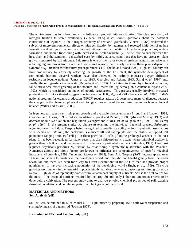
ISBN: 978-81-931973-6-3National Conference on “Emerging Trends in Management of Infectious Diseases and Public Health, 4 – 5 Feb 16
173
The environment has long been known to influence symbiotic nitrogen fixation. The clear sensitivity ofnitrogen fixation to water availability (Vincent 1965) raises serious questions about the potentialcontribution of legumes to the nitrogen economy of semiarid grasslands. Vincent (1965) reviewed thesubject of micro-environmental effects on nitrogen fixation by legumes and reported inhibition of noduleformation and nitrogen fixation by combined nitrogen and stimulation of bacterial populations, noduleformation, and nodule functioning with increased soil water availability. The delicate balance between thehost plant and the symbiont is disturbed even by mildly adverse conditions that have no effect on plantgrowth supported by soil nitrogen. Salt stress is one of the major types of environmental stress adverselyaffecting legume production in arid and semi- arid regions, particularly because these plants depend onsymbiotic N2 fixation for their nitrogen requirements (El- sheikh and Wood 1995). High soil salinity canlimit productivity by adversely affecting the growth of the host plant, the symbiotic development ofroot-nodule bacteria. Several workers have also observed that salinity increases oxygen diffusionresistance in legume nodules (James et al, 1993; Georgiev and Atkins, 1993; Serraj et al, 1994) and,finally, the nitrogen-fixation capacity (Delgado et al., 1993). In addition to these physiological responses,saline stress accelerates greening of the nodules and lowers the leg hemo-globin content (Delgado et al.1993), which is considered an index of nodule senescence. This process usually involves increasedproduction of toxic-activated oxygen species such as H202, 02, and OH (Becana et al., 1986). India'snational program for organic production (NPOP) requires atleast a 2 years pose many challenges, becausethe changes in the chemical, physical and biological properties of the soil take time to reach an ecologicalbalance (Willer and Yussefi, 2005).
In legumes, salt stress can limit plant growth and available photosynthates (Brignoli and Lauteri, 1991;Georgiev and Atkins, 1993), reduce nodulation (Sprent and Zahran, 1988; Qifu and Murray, 1993) anddecrease nodule N2 fixation and respiration (Georgiev and Atkins, 1993; Delgado et al, 1993, 1994; Serrajet al, 1994). In the present study, we choose to examine the individual bacterial species, Rhizobiumleguminosarum by trifolii. Despite being recognized primariliy its ability to form symbiotic associationswith species of Trifolium, the bacterium is a succesfull soil saprophyte with the ability to support soilpopulation ranging from 10 8 cell g-1 in rhizosphere to 10 cells g-1 in the prolonged absence of the hostplant. It has been recognized for many years that plant rhizosphere is a zone where microbial activity isgreater than in bulk soil and that legume rhizospheres are particularly active (Bottomley, 1992). Like mostlegumes, soyabeans performs N2 fixation by establishing a symbiotic relationship with the Rhizobia.Numerous abiotic and biotic factors are known to influence the competitiveness of specific rhizobialinoculants. (Bottomley, 1992; Turco and Sadowsky, 1995). Semi Arid Tropics (SAT) regions spread over11.6 million square kilometers in the developing world, and they did not benefit greatly from the greenrevolution and there is a need for “Grey to Green Revolution” in the SAT to feed and provide propernourishment to the ever increasing population of the developing world (Singh, et al., 1999). The cropgrowing environment in the semiarid tropics is highly variable due to erratic spacing and timing of seasonrainfall. High yields of top quality crops require an abundant supply of nutrients. Soil is the best source forthe most of the essential nutrients required by the crop. So soil analysis became important criteria to bedone before cultivation. The present study aims to analyze physico-chemical properties of soil, existingrhizobial population and nodulation pattern of black gram cultivated soil.
MATERIALS AND METHODSSoil Analysis (pH)
Soil pH was determined in Elico Model LT-10T pH meter by preparing 1:2:5 soil: water suspension andstirring by means of a glass rod (Jackson 1973).
Estimation of Electrical Conductivity (EC)
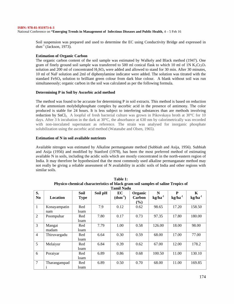
ISBN: 978-81-931973-6-3National Conference on “Emerging Trends in Management of Infectious Diseases and Public Health, 4 – 5 Feb 16
174
Soil suspension was prepared and used to determine the EC using Conductivity Bridge and expressed indsm-1 (Jackson, 1973).
Estimation of Organic CarbonThe organic carbon content of the soil sample was estimated by Walkely and Black method (1947). Onegram of finely ground soil sample was transferred to 500 ml conical flask to which 10 ml of 1N K2Cr2O7
solution and 200 ml of concentrated H2SO4 were added and allowed to stand for 30 min. After 30 minutes,10 ml of NaF solution and 2ml of diphenylamine indicator were added. The solution was titrated with thestandard FeSO4 solution to brilliant green colour from dark blue colour. A blank without soil was runsimultaneously; organic carbon in the soil was calculated as per the following formula.
Determining P in Soil by Ascorbic acid method
The method was found to be accurate for determining P in soil extracts. This method is based on reductionof the ammonium molybdiphosphate complex by ascorbic acid in the presence of antimony. The colorproduced is stable for 24 hours. It is less subject to interfering substances than are methods involvingreduction by SnCl2. A loopful of fresh bacterial culture was grown in Pikovskaya broth at 30°C for 10days. After 3 h incubation in the dark at 30°C, the absorbance at 630 nm by calorimetrically was recordedwith non-inoculated supernatant as reference. The strain was analysed for inorganic phosphatesolubilization using the ascorbic acid method (Watanabe and Olsen, 1965).
Estimation of N in soil available nutrients
Available nitrogen was estimated by Alkaline permanganate method (Subbiah and Asija, 1956). Subbiahand Asija (1956) and modified by Stanford (1978), has been the most preferred method of estimatingavailable N in soils, including the acidic soils which are mostly concentrated in the north-eastern region ofIndia. It may therefore be hypothesized that the most commonly used alkaline permanganate method maynot really be giving a reliable assessment of N availability in acidic soils of India and other regions withsimilar soils.
Table 1:Physico-chemical characteristics of black gram soil samples of saline Tropics of
Tamil NaduS.No Location
SoilType
Soil pH EC(dsm-1)
OrganicCarbon
(%)
Nkg/ha-1
Pkg/ha-1
Kkg/ha-1
1 Konayampatinnam
Redloam
7.9 0.12 0.62 98.65 17.20 158.50
2 Poompuhar Redloam
7.80 0.17 0.73 97.35 17.80 180.00
3 Mangaimadam
Redloam
7.79 1.00 0.58 126.00 18.00 98.00
4 Thiruvargadu Redloam
6.64 0.30 0.59 68.00 17.00 77.00
5 Melaiyur Redloam
6.84 0.39 0.62 67.00 12.00 178.2
6 Poraiyar Redloam
6.89 0.86 0.68 100.50 11.00 130.10
7 Tharangampadi
Redloam
6.89 0.50 0.70 68.00 11.00 169.85
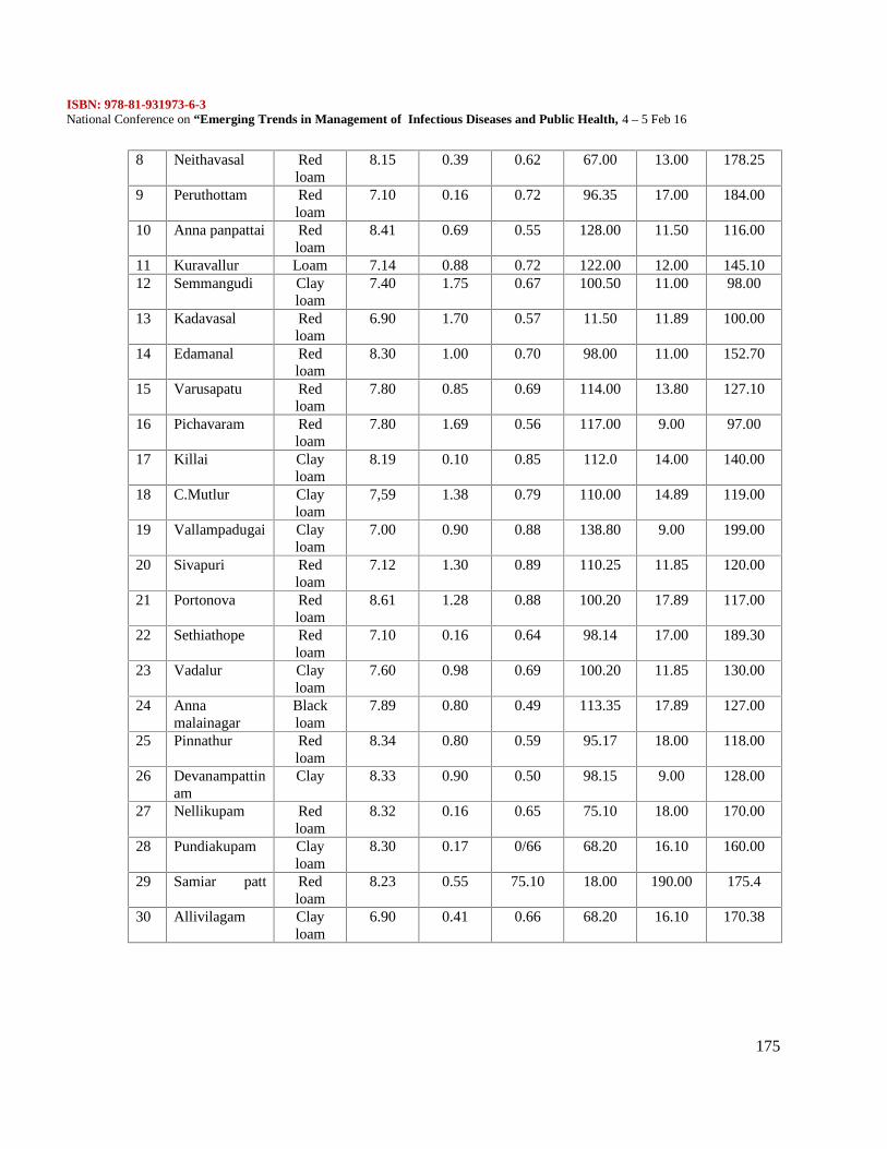
ISBN: 978-81-931973-6-3National Conference on “Emerging Trends in Management of Infectious Diseases and Public Health, 4 – 5 Feb 16
175
8 Neithavasal Redloam
8.15 0.39 0.62 67.00 13.00 178.25
9 Peruthottam Redloam
7.10 0.16 0.72 96.35 17.00 184.00
10 Anna panpattai Redloam
8.41 0.69 0.55 128.00 11.50 116.00
11 Kuravallur Loam 7.14 0.88 0.72 122.00 12.00 145.1012 Semmangudi Clay
loam7.40 1.75 0.67 100.50 11.00 98.00
13 Kadavasal Redloam
6.90 1.70 0.57 11.50 11.89 100.00
14 Edamanal Redloam
8.30 1.00 0.70 98.00 11.00 152.70
15 Varusapatu Redloam
7.80 0.85 0.69 114.00 13.80 127.10
16 Pichavaram Redloam
7.80 1.69 0.56 117.00 9.00 97.00
17 Killai Clayloam
8.19 0.10 0.85 112.0 14.00 140.00
18 C.Mutlur Clayloam
7,59 1.38 0.79 110.00 14.89 119.00
19 Vallampadugai Clayloam
7.00 0.90 0.88 138.80 9.00 199.00
20 Sivapuri Redloam
7.12 1.30 0.89 110.25 11.85 120.00
21 Portonova Redloam
8.61 1.28 0.88 100.20 17.89 117.00
22 Sethiathope Redloam
7.10 0.16 0.64 98.14 17.00 189.30
23 Vadalur Clayloam
7.60 0.98 0.69 100.20 11.85 130.00
24 Annamalainagar
Blackloam
7.89 0.80 0.49 113.35 17.89 127.00
25 Pinnathur Redloam
8.34 0.80 0.59 95.17 18.00 118.00
26 Devanampattinam
Clay 8.33 0.90 0.50 98.15 9.00 128.00
27 Nellikupam Redloam
8.32 0.16 0.65 75.10 18.00 170.00
28 Pundiakupam Clayloam
8.30 0.17 0/66 68.20 16.10 160.00
29 Samiar patt Redloam
8.23 0.55 75.10 18.00 190.00 175.4
30 Allivilagam Clayloam
6.90 0.41 0.66 68.20 16.10 170.38
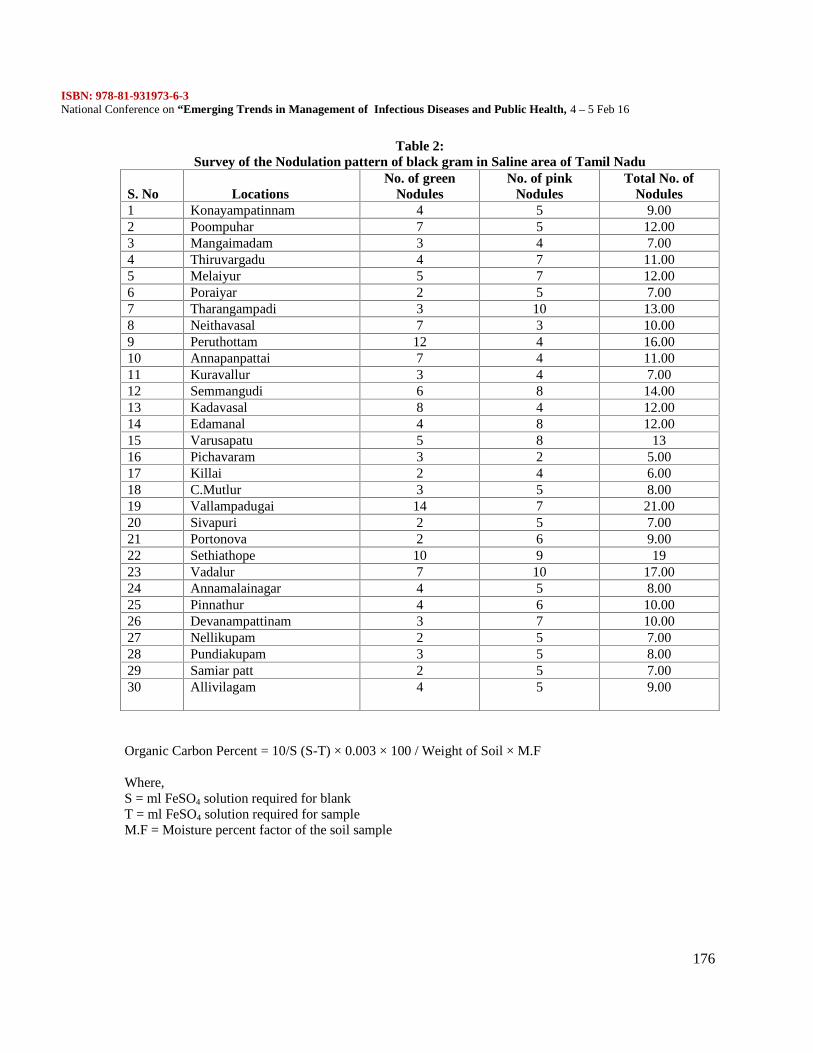
ISBN: 978-81-931973-6-3National Conference on “Emerging Trends in Management of Infectious Diseases and Public Health, 4 – 5 Feb 16
176
Table 2:Survey of the Nodulation pattern of black gram in Saline area of Tamil Nadu
S. No LocationsNo. of green
NodulesNo. of pink
NodulesTotal No. of
Nodules1 Konayampatinnam 4 5 9.002 Poompuhar 7 5 12.003 Mangaimadam 3 4 7.004 Thiruvargadu 4 7 11.005 Melaiyur 5 7 12.006 Poraiyar 2 5 7.007 Tharangampadi 3 10 13.008 Neithavasal 7 3 10.009 Peruthottam 12 4 16.0010 Annapanpattai 7 4 11.0011 Kuravallur 3 4 7.0012 Semmangudi 6 8 14.0013 Kadavasal 8 4 12.0014 Edamanal 4 8 12.0015 Varusapatu 5 8 1316 Pichavaram 3 2 5.0017 Killai 2 4 6.0018 C.Mutlur 3 5 8.0019 Vallampadugai 14 7 21.0020 Sivapuri 2 5 7.0021 Portonova 2 6 9.0022 Sethiathope 10 9 1923 Vadalur 7 10 17.0024 Annamalainagar 4 5 8.0025 Pinnathur 4 6 10.0026 Devanampattinam 3 7 10.0027 Nellikupam 2 5 7.0028 Pundiakupam 3 5 8.0029 Samiar patt 2 5 7.0030 Allivilagam 4 5 9.00
Organic Carbon Percent = 10/S (S-T) × 0.003 × 100 / Weight of Soil × M.F
Where,S = ml FeSO4 solution required for blankT = ml FeSO4 solution required for sampleM.F = Moisture percent factor of the soil sample
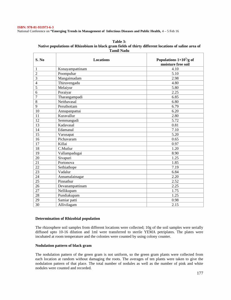
ISBN: 978-81-931973-6-3National Conference on “Emerging Trends in Management of Infectious Diseases and Public Health, 4 – 5 Feb 16
177
Table 3:Native populations of Rhizobium in black gram fields of thirty different locations of saline area of
Tamil Nadu
S. No Locations Populations 1×10³/g ofmoisture free soil
1 Konayampattinam 4.102 Poompuhar 5.103 Mangaimadam 2.984 Thiruvengadu 4.805 Melaiyur 5.806 Poraiyar 2.257 Tharangampadi 6.858 Neithavasal 6.809 Peruthottam 6.7910 Annapanpattai 6.2011 Kuravallur 2.8012 Semmangudi 5.7213 Kadavasal 0.8114 Edamanal 7.1015 Varusapat 5.2016 Pichavaram 0.6517 Killai 0.9718 C.Mutlur 1.2019 Vallampadugai 8.9020 Sivapuri 1.2521 Portonova 1.8522 Sethiathope 7.1923 Vadalur 6.8424 Annamalainagar 2.2025 Pinnathur 2.5226 Devanampattinam 2.2527 Nellikupam 1.7528 Pundiakupam 1.2529 Samiar patti 0.9830 Allivilagam 2.15
Determination of Rhizobial population
The rhizosphere soil samples from different locations were collected; 10g of the soil samples were seriallydiffused upto 10-16 dilution and 1ml were transferred to sterile YEMA petriplates. The plates wereincubated at room temperature and the colonies were counted by using colony counter.
Nodulation pattern of black gram
The nodulation pattern of the green gram is not uniform, so the green gram plants were collected fromeach location at random without damaging the roots. The averages of ten plants were taken to give thenodulation pattern of that place. The total number of nodules as well as the number of pink and whitenodules were counted and recorded.
ISBN: 978-81-931973-6-3National Conference on “Emerging Trends in Management of Infectious Diseases and Public Health, 4 – 5 Feb 16
178
RESULTS AND DISCUSSION
In the present study of soil samples were collected from 30 different saline area locations of 2 districts ofTamil nadu, namely Nagapattinam, Cuddalore and they were subjected to physio-chemical analysis. In ourfindings most of the soils from these regions were belonged to 3 textural groups, namely Red loam, Clayloam and Black soils. Soil pH ranged from 6.65-7.85, EC ranged from 0.10-1.75 dsm-1. The nutrientcontent of the soil is most important for root nodulation and nitrogen fixation. In our study, soil organiccarbon contents ranged from 67.00-138.00 kg ha-1, phosphorous and potassium contents ranged from9.00-19.00, 77-200.00 kg ha-1 EC respectively and concluded that organic carbon N, P and K status ofsaline areas were low. This was proved by earlier workers that the wide variation in rhizobial cell countsand nodule mass in green gram are relapsed to variation in physio- chemical characteristics of the soil.The organic matter content, salinity, texture and pH were found to affect mycorrhizal development in soils(Saif et al., 1975).
The nodulation pattern of black gram and native rhizobial populations of the above 30 locations were alsostudied. Among the 30 locations, 4 locations were recorded less than 1×10³ g-1, 13 locations were recorded1×103 to 3×10³ g-1 and 13 locations recorded more than 3×10³ g-1 of rhizobial populations from therhizosphere of green gram plants. The number of nodules / plant was also recorded. The total number ofnodules ranged from 5.00 to 21.00.plant-1. Vallampadugai location recorded highest number of nodules of21.00 Plant-1. The lowest nodules number was recorded in Pichavaram i.e. 5.00 Plant-1 in CuddaloreDistrict.
REFERENCE
Becana M, Aparicio-Tejo P, Irigoyen JJ, Sanchez-Diaz M (1986). Some enzymes of hydrogen peroxidemetabolism in leaves and root nodules of Medicago sativa. Plant Physiol. 82: 1169-1171.
Bottomley, P.J. 1992. Ecology of Bradyrhizobium and Rhizobium. In: Stacey, G., Burris, R.H. & Evarts,H.J. editors, Biological Nitrogen fixation, New York: Chapman and Hall. pp. 293-348.
ISBN 0-41202421-7. Cultivations as oilseed crop in China. J. Agric. Sci., 28: 281-283.
Brignoli E, Lauteri M. 1991. Effects of salinity on stomatal conductance, photosynthetic capacity, andcarbon isotope discrimination of salt-tolerant (Gossypium hirsutum L.) and salt-sensitive (Phaseolusvulgaris L.) C3 non-halophytes. Plant Physiology 95, 628-35.
Delgado MJ, Garrido JM, Ligero F, Lluch C. 1993. Nitrogen fixation and carbon metabolism by nodulesand bacteroids of pea plants under sodium chloride stress. Physiologia Plantarum 89, 824-9.
Delgado MJ, Ligero F, Lluch C. 1994. Effect of salt stress on growth and nitrogen fixation by pea, Faba-bean, common bean and soybean plants. Soil Biology and Biochemistry 26, 371-6.
Elsheikh, E.A.E. and Wood, M. (1995) “Nodulation and N2 fixation by soybean inoculated with salt-tolerant rhizobia or salt-sensitive Bradyrhizobia in saline soil”, Soil Biol. Biochem., 27, 657-661.
Georgiev GI, Atkins CA. 1993. Effects of salinity on N2 fixation, nitrogen metabolism and export anddiffusive conductance of cowpea root nodules. Symbiosis 15, 239-55.
ISBN: 978-81-931973-6-3National Conference on “Emerging Trends in Management of Infectious Diseases and Public Health, 4 – 5 Feb 16
179
Gour, Y.D., 1993. Microbiology, physiology and agronomy of nitrogen fixation: Legume-Rhizobiumsymbiosis. Proc. Indian Nat. Sci. Acad., B(59): 333-358.
Jackson, M.L. 1973. Soil chemical analysis. Prentice-Hall of Indian Pvt. Ltd., New Delhi: India
James EK, Sprent JI, Hay GT, Minchin FR. 1993. The effect of irradiance on the recovery of soybeannodules from sodium chloride induced senescence. Journal of Experimental Botany AA, 997-1005
Kanchana, D., M. Jayanthi, G. Usharani, P. Saranraj and D. Sujitha. 2013. Prevelence of Azotobacter sp. inChilli (Capsicum annuum L.) Rhizosphere Soil of Cuddalore District, Tamil Nadu, India. InternationalJournal of Microbiological Research, 4(3): 296 - 299.
Quifu MA, Murray AF. 1993. Effects of SO2 and salinity on nitrogenase activity, nitrogen concentrationand growth of young soybean plants. Environmental and Experimental Botany 33, 529-37.
Saif. S.R., Sheikh, M.A. and Khan, A.G. 1975. Ecology of endogone. I. Relationship of Endogone SporePopulations with physical soil factors. Islambad. J. Sci., 2: 1-5.
Serraj R, Roy G, Drevon JJ. 1994. Salt stress induces a decrease in the oxygen uptake of soybean nodulesand their permeability to oxygen diffusion. Physiologia Plantarum 91, 161-8.
Singh, H.P., Sharma, K.L., and Venkateswarlu, B., 1999. Fertilizer use in rainfed areas. Fert. News 44: 27-30 & 33-38.
Sprent JI, Zahran HH. 1988. Infection, development and functioning of nodules under drought and salinity.In: Beck DP, Materon LA, eds. Nitrogen fixation by legumes in Mediterranean agriculture. Dordrecht:Martinus Nijhoff, 145-51.
Stanford G. 1978. Evaluation of ammonium release by alkaline permanganate extraction as an index ofsoil nitrogen availability. Soil Science, 126: 244–253.
Subbiah, B.V. and C.V. Asija. 1956. A rapid, procedure for estimation of available nitrogen in soil. Curr.Sci., 25: 259-260.
Suresh Kumar, R., P. Ganesh, K. Tharmaraj and P. Saranraj. 2011. Growth and development of black gram(Vigna mungo. L) under foliar application of Panchagavya as organic source of nutrient. Current Botany, 2(3): 9 – 11.
Turco, R.F. and Sadowsky, M.J. 1995. The microflora of Micromediation, In: Bioremediation:Science and Applications, Soil Science Special Publication No. 43 eds Skipper, H.D. and Turco, R.F. PP.87-102.Vincent, J.M. 1965. Environmental factors in the fixation of nitrogen by the legume In: Soil nitrogen. Eds.W.V. Bartholomas and F.E. Clark. Pp. 384-435. Am.Soc. Agronomy Inc. Publ. Madison, USA.Walkey A. and Black, 1.4.1947. Chronic acid titration method for determination of soil organicmatter. Soil. Sci., 63:251
Watanabe, I. and Olsen, S.R. 1965. Test of an ascorbic acid method for determining P in H2O andNaHCO3 extracts from the soil. Soil. Sci. Soc. Am. Proc., 29: 678-679
Willer H, Yussefi, M. 2005. The world of organic agriculture statistics and emerging trends 2005.International federation of organic agriculture movements (IFOAM), Bonn, Germany.
ISBN: 978-81-931973-6-3National Conference on “Emerging Trends in Management of Infectious Diseases and Public Health, 4 – 5 Feb 16
180
Evaluation of phytochemical screening and in vitro bioactivity of leaf and stem extracts ofSolanum xanthocarpum (Solanaceae)
Selvi. R1 and Yogananth. N2*
1Department of Microbiology, Idaya College of Arts and Science, Thiruvannamalai - 606 7052PG and Research Department of Biotechnology, Mohamed Sathak College of Arts and Science,Sholinganallur, Chennai – 119.*corresponding author’s email; [email protected].
AbstractThe present study was carried out for phytochemical screening of principle bioactive compounds
and antibacterial activity in Solanum xanthocarpum Schrad and Wendl. Phytochemical analysis revealedthe presence of Alkaloids, Flavonoids, Phenols, Phlobatannins, Saponins, Steroid, Tannins andTriterpenoides. The ethanol and acetone extracts were subjected for antibacterial activity against ninebacterial strains using agar well diffusion method. Ethanol extracts of Leaf inhibited Bacillus sp, E.faecalis, K. pneumoniae, Micrococcus sp, P. mirabilis and S. epidermidis. Acetone extracts of stempossessed antimicrobial activity against Bacillus sp, K. pneumonia, Micrococcus sp, P. mirabilis,P.aeruginosa, S. aureus and S. epidermidis. In stem extracts, the ethanol extracts exhibit anti bacterialactivity against Bacillus sp, E. coli, K. pneumonia, Micrococcus sp and P. mirabilis. The acetone extractsof stem gave antibacterial activity against Bacillus sp, E. faecalis, E. coli, K. pneumonia, Micrococcus sp,P. mirabilis and S. epidermidis. K. pneumoniae showed significant sensitivity to both leaf and stemextracts. The results suggested that ethanol and acetone extracts of leaf and stem extracts were highlypotent against K. pneumoniae and P. mirabilis can be used in treatment of nosocomial infections such aspneumonia, urinary tract infections (UTIs) and bacteremia. Extensive animal studies may be requiredbefore investigating the role of Solanum xanthocarpum for treating RTI and UTIKeywords: Solanum xanthocarpum, Phytochemical analysis, antibacterial activity, agar well diffusionmethod.
Introduction
Solanum xanthocarpum (Solanaceae) smoke is inhaled through mouth to led through mouth totreat toothache has profound use in Ayurveda and folkore medicine. It is supposed that the plant hassolasonine and solasomargine (Yoshida et al., 2006) sapogenins (Khanna et al., 1976) and solasodine(Oudhia, and Kadu Pani, 2007) which are responsible for medicinal effect. The whole plant extracts of S.xanthocarpum have a larvicidal defect (Rajkumar, and Jebanesan, 2005) hypoglycemic activity (Kar et al.,2006) bronchitis and antitussive response (Govindan et al., 1999). Dried or fresh fruits are kept in fire andsmoke is inhaled through mouth to led through mouth to treat toothache. The present investigation is aimto focus the phytochemicals of Solanum xanthocarpum leaf and stem and its antibacterial action againstGram-positive and Gram-negative bacteria.
Materials and methods
Leaves and Stems of S. xanthocarpum were collected from the Medicinal plant garden, Idaya College ofArts and Science, Tiruvannamalai, Tamil nadu, India.The plants were dried and broken into small pieces under sterile conditions, and 20 g of each plant partwere extracted with 100 ml of ethanol and acetone solvent (Merck, Darmstadt) for 24 h by using Soxhletequipment (Bradshaw, 1992).Phytochemical tests of each extracts were carried out to establish the presence of Alkaloids, Flavonoids,Phenols, Phlobatannins, Saponins, Steroid, Tannins and Triterpenoides using standard procedure (Jigna etal., 2006).
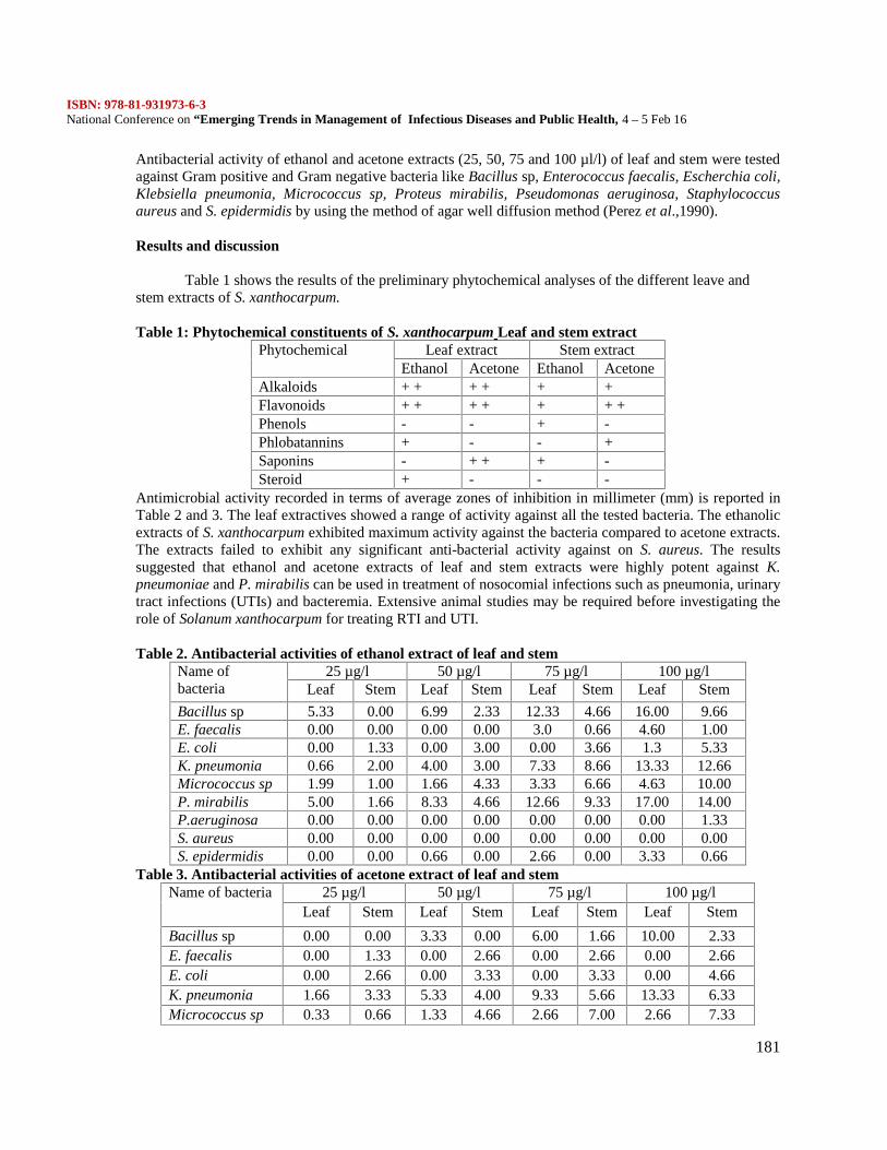
ISBN: 978-81-931973-6-3National Conference on “Emerging Trends in Management of Infectious Diseases and Public Health, 4 – 5 Feb 16
181
Antibacterial activity of ethanol and acetone extracts (25, 50, 75 and 100 µl/l) of leaf and stem were testedagainst Gram positive and Gram negative bacteria like Bacillus sp, Enterococcus faecalis, Escherchia coli,Klebsiella pneumonia, Micrococcus sp, Proteus mirabilis, Pseudomonas aeruginosa, Staphylococcusaureus and S. epidermidis by using the method of agar well diffusion method (Perez et al.,1990).
Results and discussion
Table 1 shows the results of the preliminary phytochemical analyses of the different leave andstem extracts of S. xanthocarpum.
Table 1: Phytochemical constituents of S. xanthocarpum Leaf and stem extractPhytochemical Leaf extract Stem extract
Ethanol Acetone Ethanol AcetoneAlkaloids + + + + + +Flavonoids + + + + + + +Phenols - - + -Phlobatannins + - - +Saponins - + + + -Steroid + - - -
Antimicrobial activity recorded in terms of average zones of inhibition in millimeter (mm) is reported inTable 2 and 3. The leaf extractives showed a range of activity against all the tested bacteria. The ethanolicextracts of S. xanthocarpum exhibited maximum activity against the bacteria compared to acetone extracts.The extracts failed to exhibit any significant anti-bacterial activity against on S. aureus. The resultssuggested that ethanol and acetone extracts of leaf and stem extracts were highly potent against K.pneumoniae and P. mirabilis can be used in treatment of nosocomial infections such as pneumonia, urinarytract infections (UTIs) and bacteremia. Extensive animal studies may be required before investigating therole of Solanum xanthocarpum for treating RTI and UTI.
Table 2. Antibacterial activities of ethanol extract of leaf and stemName ofbacteria
25 µg/l 50 µg/l 75 µg/l 100 µg/lLeaf Stem Leaf Stem Leaf Stem Leaf Stem
Bacillus sp 5.33 0.00 6.99 2.33 12.33 4.66 16.00 9.66E. faecalis 0.00 0.00 0.00 0.00 3.0 0.66 4.60 1.00E. coli 0.00 1.33 0.00 3.00 0.00 3.66 1.3 5.33K. pneumonia 0.66 2.00 4.00 3.00 7.33 8.66 13.33 12.66Micrococcus sp 1.99 1.00 1.66 4.33 3.33 6.66 4.63 10.00P. mirabilis 5.00 1.66 8.33 4.66 12.66 9.33 17.00 14.00P.aeruginosa 0.00 0.00 0.00 0.00 0.00 0.00 0.00 1.33S. aureus 0.00 0.00 0.00 0.00 0.00 0.00 0.00 0.00S. epidermidis 0.00 0.00 0.66 0.00 2.66 0.00 3.33 0.66
Table 3. Antibacterial activities of acetone extract of leaf and stemName of bacteria 25 µg/l 50 µg/l 75 µg/l 100 µg/l
Leaf Stem Leaf Stem Leaf Stem Leaf Stem
Bacillus sp 0.00 0.00 3.33 0.00 6.00 1.66 10.00 2.33E. faecalis 0.00 1.33 0.00 2.66 0.00 2.66 0.00 2.66E. coli 0.00 2.66 0.00 3.33 0.00 3.33 0.00 4.66K. pneumonia 1.66 3.33 5.33 4.00 9.33 5.66 13.33 6.33Micrococcus sp 0.33 0.66 1.33 4.66 2.66 7.00 2.66 7.33
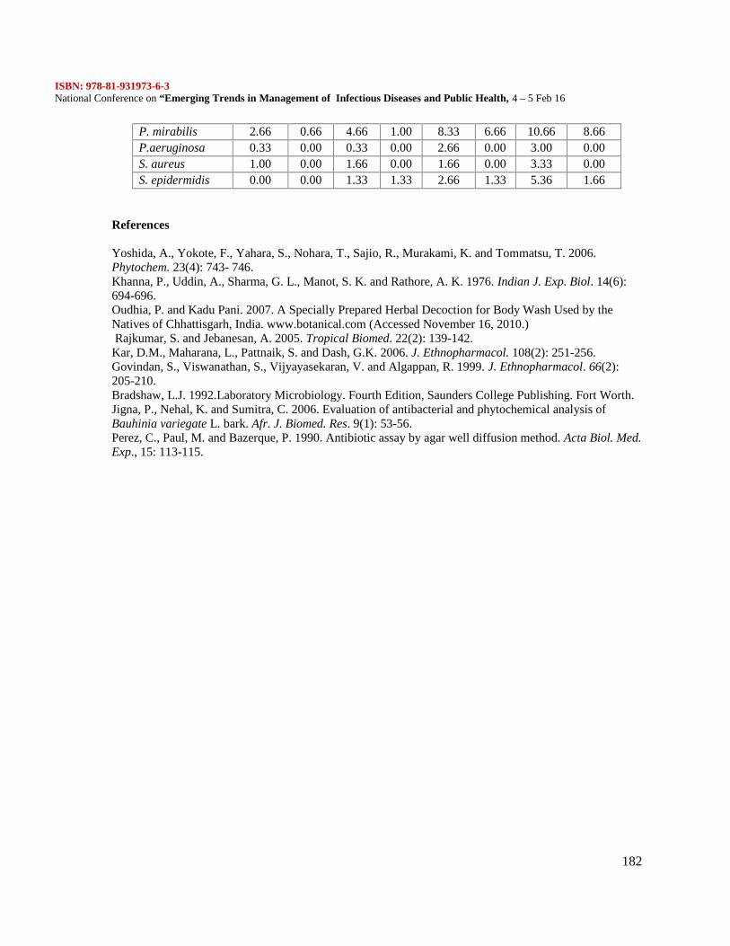
ISBN: 978-81-931973-6-3National Conference on “Emerging Trends in Management of Infectious Diseases and Public Health, 4 – 5 Feb 16
182
P. mirabilis 2.66 0.66 4.66 1.00 8.33 6.66 10.66 8.66P.aeruginosa 0.33 0.00 0.33 0.00 2.66 0.00 3.00 0.00S. aureus 1.00 0.00 1.66 0.00 1.66 0.00 3.33 0.00S. epidermidis 0.00 0.00 1.33 1.33 2.66 1.33 5.36 1.66
References
Yoshida, A., Yokote, F., Yahara, S., Nohara, T., Sajio, R., Murakami, K. and Tommatsu, T. 2006.Phytochem. 23(4): 743- 746.Khanna, P., Uddin, A., Sharma, G. L., Manot, S. K. and Rathore, A. K. 1976. Indian J. Exp. Biol. 14(6):694-696.Oudhia, P. and Kadu Pani. 2007. A Specially Prepared Herbal Decoction for Body Wash Used by theNatives of Chhattisgarh, India. www.botanical.com (Accessed November 16, 2010.)Rajkumar, S. and Jebanesan, A. 2005. Tropical Biomed. 22(2): 139-142.
Kar, D.M., Maharana, L., Pattnaik, S. and Dash, G.K. 2006. J. Ethnopharmacol. 108(2): 251-256.Govindan, S., Viswanathan, S., Vijyayasekaran, V. and Algappan, R. 1999. J. Ethnopharmacol. 66(2):205-210.Bradshaw, L.J. 1992.Laboratory Microbiology. Fourth Edition, Saunders College Publishing. Fort Worth.Jigna, P., Nehal, K. and Sumitra, C. 2006. Evaluation of antibacterial and phytochemical analysis ofBauhinia variegate L. bark. Afr. J. Biomed. Res. 9(1): 53-56.Perez, C., Paul, M. and Bazerque, P. 1990. Antibiotic assay by agar well diffusion method. Acta Biol. Med.Exp., 15: 113-115.
ISBN: 978-81-931973-6-3National Conference on “Emerging Trends in Management of Infectious Diseases and Public Health, 4 – 5 Feb 16
183
Phytochemical Screening and Anti-inflammatory activity of Citrus Limetta peel. Oil extract
Shenbagam.R*1, Dr.Sheela Sasikumar*2, Dr.Thendral Hepisba*3
1Department of Biotechnology, Alpha Arts and Science College. Chennai-600056, Tamilnadu2Chief Scientific Officer, Frontier Lifeline Pvt. Ltd, Tamilnadu3Department of Biotechnology , Ethiraj College for Women.Chennai-600008, Tamilnadu
ABSTRACT
Medicinal plants offer alternative remedies with incredible opportunities. It can be used to prevent andcure several diseases & disorders. Citrus limetta (Rutaceae) fruit is commonly called as sweet lime amedium sized shrubs or tree which is distributed in all over the world. It has vast medicinal properties. Theobjective of this study is to analysis the phyotoconstituents and to evaluate the anti inflammatory activityof essential oil extracted from citrus limetta peel. The medicinal plant has more chemical componentswhich gives definite physiological actions to human bodies . Significantly high amount of secondarymetabolites like steroids, cardio glycoside, phenol, terpenoids and saponins are present in peel oil extract.Inflammation is the trait of several human diseases and disorders like Atherosclerosis and Rheumatoidarthritis. Inhibitions of protein denaturation of extracted oil showed a better ability compared to standardAspirin. The oil extract exhibited a significant activity of human RBC membrane stabilization. Thus thecurrent studies expose that peel oil extract of citrus limetta fruit has better anti- inflammatory activity.Key words: Phytoconstituents, Medicinal plant, Inflammation, Anti-inflammation, Protein Denature,Membrane Stabilization, Citrus limetta.
1. Introduction
The traditional drug gives an alternate source of remedies with tremendous opportunities. It canbe used to prevent and cure several diseases & disorders. Modern medicine shows undesirable side effectsto humans (Henry David). Even today majority of people depend on herbal medicinal practice because itprovides safe and affordable primary healthcare to the people. Phyto chemical compounds are naturallyavailable in all plants and it plays a major role in metabolic activity of humans. The major phyto chemicalcompounds includes, Steroids are capable in decreasing the inflammation and reduce the immune activityof immune system which is responsible for inflammation and also have anti-bacterial activity, Cardiacglycoside is regulating endothelial tissue factor expression in culture and many phyotoconstituents arepresent in all medicinal plants each has a different properties and functions. Inflammation is first line ofdefence mechanism to protect from body’s tissue injury, microbial agents, and toxic chemicals, and otherallergens (Shodhganga). In olden days people considered inflammation is a part of healing process butnowadays, inflammation is recognized as a complex disorder and a major response of immune system toinfection and tissue damage (Harley I. et al., 2006). Nowadays the medicinal plants are containing antiinflammatory constituent to cure many inflammatory diseases and disorders. The plant Citrus limetta ,Rutaceae family was used as a medicine to treat many diseases. The citrus fruit juice used to treat jaundice,cold and cough and gastrointestinal problems and also the peel oil has antiseptic properties (Riyaz ahmadet al., 2010) The present work is to determine the anti inflammatory effects of citrus limetta oil extractedfrom peel.
2. Materials and Methods2.1 Collection and Preparation of extract
The fresh Citrus limetta fruits (figure 1) were collected around Poonamallee area. The peels wereremoved and cut into small pieces and washed twice using distilled water. Then added an equal volume ofdistilled water and it was boiled using steam distillation unit. The extract contains both water and oilmixture. To separate the oil and water added 20 ml of diethyl ether and was separated using separating
ISBN: 978-81-931973-6-3National Conference on “Emerging Trends in Management of Infectious Diseases and Public Health, 4 – 5 Feb 16
184
funnel ( Zarina, Z. and Tan S. Y. 2013) . After separation the extract was evaporate at 340c in water bathto evaporate the diethyl ether.2.2 Preliminary and Quantitative determination of phyto constituents:
The extracted oil of citrus limetta was subjected to preliminary phtochemical screening includessteroids, cardio glycoside, phenol, tannins terpinoids , saponins and the total phenol and flavonoid contentwas determined in a quantitative way (Sriparna KunduSen et al. 2012) .2.3 Anti inflammatory activity:2.3.1 Inhibition of Protein denaturation methodThe inhibition of protein denaturation activity of extract was determined properties (Riyaz ahmad et al.,2010) The decreased settling of denatured protein in a tube indicates the inhibition potent of extract. Theinhibition of denatured protein was calculated by the formula,(%) inhibition = [100- (test OD - product control OD) / test control OD]*1002.3.2 Human Red Blood Cell membrane stabilization
The membrane stabilization activity of extracted oil was identified towards the tonicity inducedmembrane lysis and it was evaluated (Kavita S. Mundhe et al.2009). It was calculated by the formula,
(%) stabilization = [(control-test)/control]*1002.3.3 Hemolysis method
Hemolysis method is used to determine the membrane stabilization properties of extracted oil.RBC was subjected to pre and post treatment using hypotonic solutions.
3. Results and Discussions
The preliminary phyto constituent’s screenings confirms the presence of steroids, cardio glycoside, phenol,terpenoids and saponnins. A very low amount of tannin was screened in the extracted oil. The results areshown in table 1. The appreciable amount of phenol and flavonoid content present in extracted oil which isdetermined by quantitative estimations of phyto constituents. The results are shown in figure 2.
The oil extract of Citrus limetta showed high anti inflammatory activity. The inhibition concentration ofprotein denaturation is shown in table 2. The IC50 value of 100µg/ml showed a higher inhibition activitycompare to standard.
The Human Red Blood Cell membrane stabilization and Hemolysis method showed a better antiinflammatory activity. The results are shown in table 3 and figure 3. The above observations reveal that theCitrus limetta oil extracted from peel has a high anti inflammatory activity due to the secondarymetabolites and various bioactive components present in the extracted Citrus limetta oil have ability tostabilize the membranes of human Red Blood cells.
4. Conclusion
The present study has been proven that the oil extract of Citrus limetta has responsible for antiinflammatory activity. Therefore, the further study is to investigate the anti-atherosclerosis activity andbioactive components present in the oil through GCMS.
5. Acknowledgement
The authors are thankful to the Head of the Department, Department of Biotechnology, Alpha Arts andScience College and Frontier Life Line Pvt Ltd., and also thank full to all my co-guides and my friendswho giving me a great support to do the research work successfully.
6. Reference
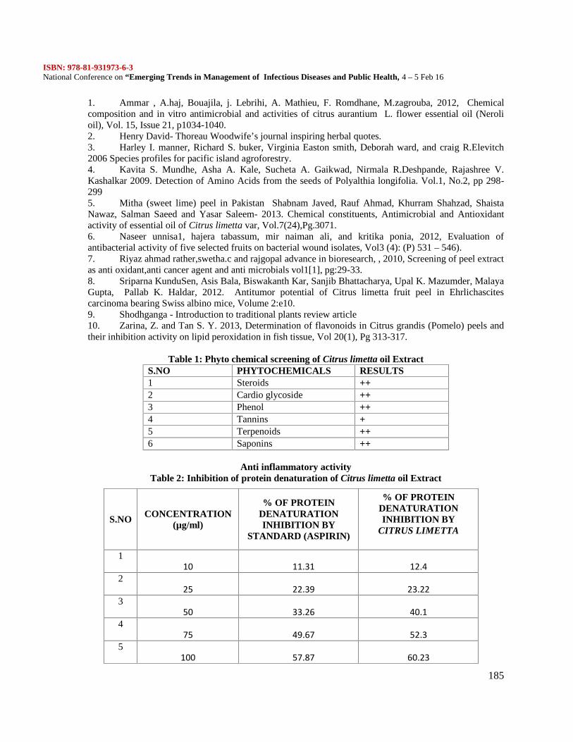
ISBN: 978-81-931973-6-3National Conference on “Emerging Trends in Management of Infectious Diseases and Public Health, 4 – 5 Feb 16
185
1. Ammar , A.haj, Bouajila, j. Lebrihi, A. Mathieu, F. Romdhane, M.zagrouba, 2012, Chemicalcomposition and in vitro antimicrobial and activities of citrus aurantium L. flower essential oil (Nerolioil), Vol. 15, Issue 21, p1034-1040.2. Henry David- Thoreau Woodwife’s journal inspiring herbal quotes.3. Harley I. manner, Richard S. buker, Virginia Easton smith, Deborah ward, and craig R.Elevitch2006 Species profiles for pacific island agroforestry.4. Kavita S. Mundhe, Asha A. Kale, Sucheta A. Gaikwad, Nirmala R.Deshpande, Rajashree V.Kashalkar 2009. Detection of Amino Acids from the seeds of Polyalthia longifolia. Vol.1, No.2, pp 298-2995. Mitha (sweet lime) peel in Pakistan Shabnam Javed, Rauf Ahmad, Khurram Shahzad, ShaistaNawaz, Salman Saeed and Yasar Saleem- 2013. Chemical constituents, Antimicrobial and Antioxidantactivity of essential oil of Citrus limetta var, Vol.7(24),Pg.3071.6. Naseer unnisa1, hajera tabassum, mir naiman ali, and kritika ponia, 2012, Evaluation ofantibacterial activity of five selected fruits on bacterial wound isolates, Vol3 (4): (P) 531 – 546).7. Riyaz ahmad rather,swetha.c and rajgopal advance in bioresearch, , 2010, Screening of peel extractas anti oxidant,anti cancer agent and anti microbials vol1[1], pg:29-33.8. Sriparna KunduSen, Asis Bala, Biswakanth Kar, Sanjib Bhattacharya, Upal K. Mazumder, MalayaGupta, Pallab K. Haldar, 2012. Antitumor potential of Citrus limetta fruit peel in Ehrlichascitescarcinoma bearing Swiss albino mice, Volume 2:e10.9. Shodhganga - Introduction to traditional plants review article10. Zarina, Z. and Tan S. Y. 2013, Determination of flavonoids in Citrus grandis (Pomelo) peels andtheir inhibition activity on lipid peroxidation in fish tissue, Vol 20(1), Pg 313-317.
Table 1: Phyto chemical screening of Citrus limetta oil ExtractS.NO PHYTOCHEMICALS RESULTS1 Steroids ++2 Cardio glycoside ++3 Phenol ++4 Tannins +5 Terpenoids ++6 Saponins ++
Anti inflammatory activityTable 2: Inhibition of protein denaturation of Citrus limetta oil Extract
S.NO CONCENTRATION(µg/ml)
% OF PROTEINDENATURATIONINHIBITION BY
STANDARD (ASPIRIN)
% OF PROTEINDENATURATIONINHIBITION BY
CITRUS LIMETTA
110 11.31 12.4
225 22.39 23.22
350 33.26 40.1
475 49.67 52.3
5100 57.87 60.23
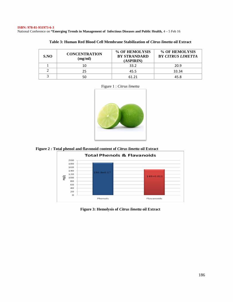
ISBN: 978-81-931973-6-3National Conference on “Emerging Trends in Management of Infectious Diseases and Public Health, 4 – 5 Feb 16
186
Table 3: Human Red Blood Cell Membrane Stabilization of Citrus limetta oil Extract
Figure 1 : Citrus limetta
Figure 2 : Total phenol and flavonoid content of Citrus limetta oil Extract
Figure 3: Hemolysis of Citrus limetta oil Extract
S.NO CONCENTRATION(mg/ml)
% OF HEMOLYSISBY STRANDARD
(ASPIRIN)
% OF HEMOLYSISBY CITRUS LIMETTA
1 10 33.2 20.92 25 45.5 33.343 50 61.21 45.8
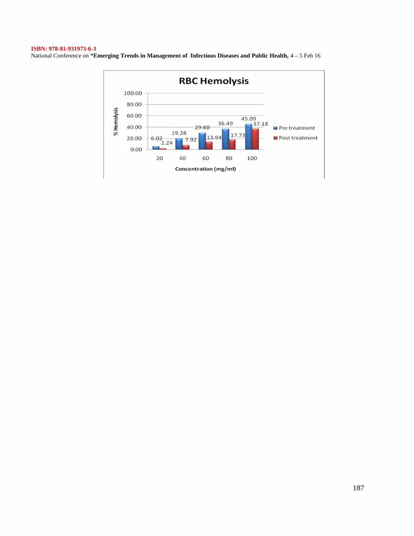
ISBN: 978-81-931973-6-3National Conference on “Emerging Trends in Management of Infectious Diseases and Public Health, 4 – 5 Feb 16
187
ISBN: 978-81-931973-6-3National Conference on “Emerging Trends in Management of Infectious Diseases and Public Health, 4 – 5 Feb 16
188
Galactose mediated synthesis of silver nanoparticles (AgNPs) and its antimicrobial activity
S.Krishnakumar*, R. Divya, N.R. Kanchana Devi, G. Keerthana and A. Ancy JudiFaculty of Bio and Chemical Engineering, Department of Biomedical Engineering, Sathyabama
University, Chennai 600 119.E.mail: [email protected]; [email protected];
mobile: 9444728401ABSTRACT
Nanotechnology has drawn significant attention in recent years due to their unique and exceptionalapplications. The sustainable chemical methods of silver nanoparticles (Ag-NPs) synthesis have significantinterest in the current scenario due to high demand. Silver nanoparticles were synthesized in solutions ofgalactose in DMSO and Milli-Q water at high temperature (80oC) under continuous stirring. Galactoseacted as a reducing agents and stabilizing agents simultaneously for the synthesis of silver nanoparticles.Silver nitrate (AgNO3) was used as the metal precursor for the preparation of Ag-NPs over the reactiontime. The Plasmon resonance kinetics and their activation energy were determined by UV – visiblespectroscopy. The Milli-Q water solution of galactose was better reductive activity than the DMSOsolution of galactose. The synthesized silver nanoparticles were subjected to characterized antimicrobialactivity against selected bacterial and fungal pathogens. The Milli-Q water solution of galactose mediatedsilver nanoparticles were exhibited better antimicrobial activity against tested bacterial pathogens. Furthercharacterization studies are under progress to pinpoint the size, morphology and actual constituentsresponsible for the antibacterial activity.[Key words: galactose, silver nanoparticles, antimicrobial activity, pathogens]
INTRODUCTION
Nanotechnology is an emerging and important field of modern nanoscience dealing with design, synthesis,and manipulation of particles ranging from 1-100 nm. Nanoparticles (NPs) have wide range of applicationsin health care, cosmetics, food and feed, environmental health, biomedical sciences, chemical industries,drug-gene delivery, energy science, catalysis applications. (Colvin et al 1994; Wang et al 1991; Schmid etal 1991). Nanobiotechnology is an important area of research for the synthesis of NPs with differentchemical compositions, sizes and morphologies (Hoffman et al 1992). Several methods have been devisedto prepare metallic nanoparticles. For instance, the most important current method is crystallization ofnanoparticles in micro-emulsions using a variety of chemicals as precursors and surfactants as stabilizingagents (Leon et al 2013). Nowadays, there is a growing need to develop eco-friendly processes, which donot use toxic chemicals in the synthesis protocols. Silver NPs were prepared using water as anenvironmentally friendly solvent and polysaccharides as capping/reducing agents. For instance, synthesisof starch-silver NPs was carried out with starch (capping agent) and β-D-glucose (reducing agent) in agently heated system (Raveendran et al 2003).
Although biosynthesis of Ag-NPs are considered to be more eco- friendly (Krishnakumar et al 2015),chemical or physical method of synthesis of preparation of silver particles are normally used because theyare readily available and can be used to synthesize in large amounts in relatively less time. Chemicalmethods may be divided into those using in non-deleterious solvent and naturally occurring reducing agentsuch as polysaccharides or plants extract or microorganism such as bacteria and fungi as a reducing agents( Dokuchaev et al., 1997). Chemical reduction is the most frequently applied method for the preparation ofAgNPs as stable, colloidal dispersions in water or organic solvents. Commonly used reductants areborohydride, citrate, ascorbate elemental hydrogen and polysccharides. The reduction of silver ions (Ag+)in aqueous solution generally yields colloidal silver with particle diameters of several nanometers (Mergaet al., 2007). In this research, present results on the synthesis of silver nanoparticles using galactosedissolved in DMSO and Milli-Q water. Galactose acts as reducing agent and stabilizing agent in silver
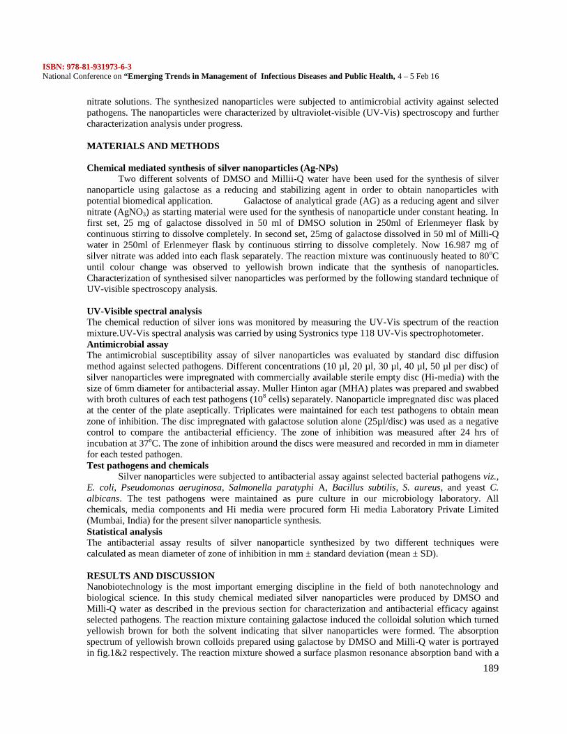
ISBN: 978-81-931973-6-3National Conference on “Emerging Trends in Management of Infectious Diseases and Public Health, 4 – 5 Feb 16
189
nitrate solutions. The synthesized nanoparticles were subjected to antimicrobial activity against selectedpathogens. The nanoparticles were characterized by ultraviolet-visible (UV-Vis) spectroscopy and furthercharacterization analysis under progress.
MATERIALS AND METHODS
Chemical mediated synthesis of silver nanoparticles (Ag-NPs)Two different solvents of DMSO and Millii-Q water have been used for the synthesis of silver
nanoparticle using galactose as a reducing and stabilizing agent in order to obtain nanoparticles withpotential biomedical application. Galactose of analytical grade (AG) as a reducing agent and silvernitrate (AgNO3) as starting material were used for the synthesis of nanoparticle under constant heating. Infirst set, 25 mg of galactose dissolved in 50 ml of DMSO solution in 250ml of Erlenmeyer flask bycontinuous stirring to dissolve completely. In second set, 25mg of galactose dissolved in 50 ml of Milli-Qwater in 250ml of Erlenmeyer flask by continuous stirring to dissolve completely. Now 16.987 mg ofsilver nitrate was added into each flask separately. The reaction mixture was continuously heated to 80oCuntil colour change was observed to yellowish brown indicate that the synthesis of nanoparticles.Characterization of synthesised silver nanoparticles was performed by the following standard technique ofUV-visible spectroscopy analysis.
UV-Visible spectral analysisThe chemical reduction of silver ions was monitored by measuring the UV-Vis spectrum of the reactionmixture.UV-Vis spectral analysis was carried by using Systronics type 118 UV-Vis spectrophotometer.Antimicrobial assayThe antimicrobial susceptibility assay of silver nanoparticles was evaluated by standard disc diffusionmethod against selected pathogens. Different concentrations (10 µl, 20 µl, 30 µl, 40 µl, 50 µl per disc) ofsilver nanoparticles were impregnated with commercially available sterile empty disc (Hi-media) with thesize of 6mm diameter for antibacterial assay. Muller Hinton agar (MHA) plates was prepared and swabbedwith broth cultures of each test pathogens (108 cells) separately. Nanoparticle impregnated disc was placedat the center of the plate aseptically. Triplicates were maintained for each test pathogens to obtain meanzone of inhibition. The disc impregnated with galactose solution alone (25µl/disc) was used as a negativecontrol to compare the antibacterial efficiency. The zone of inhibition was measured after 24 hrs ofincubation at 37oC. The zone of inhibition around the discs were measured and recorded in mm in diameterfor each tested pathogen.Test pathogens and chemicals
Silver nanoparticles were subjected to antibacterial assay against selected bacterial pathogens viz.,E. coli, Pseudomonas aeruginosa, Salmonella paratyphi A, Bacillus subtilis, S. aureus, and yeast C.albicans. The test pathogens were maintained as pure culture in our microbiology laboratory. Allchemicals, media components and Hi media were procured form Hi media Laboratory Private Limited(Mumbai, India) for the present silver nanoparticle synthesis.Statistical analysisThe antibacterial assay results of silver nanoparticle synthesized by two different techniques werecalculated as mean diameter of zone of inhibition in mm ± standard deviation (mean ± SD).
RESULTS AND DISCUSSIONNanobiotechnology is the most important emerging discipline in the field of both nanotechnology andbiological science. In this study chemical mediated silver nanoparticles were produced by DMSO andMilli-Q water as described in the previous section for characterization and antibacterial efficacy againstselected pathogens. The reaction mixture containing galactose induced the colloidal solution which turnedyellowish brown for both the solvent indicating that silver nanoparticles were formed. The absorptionspectrum of yellowish brown colloids prepared using galactose by DMSO and Milli-Q water is portrayedin fig.1&2 respectively. The reaction mixture showed a surface plasmon resonance absorption band with a
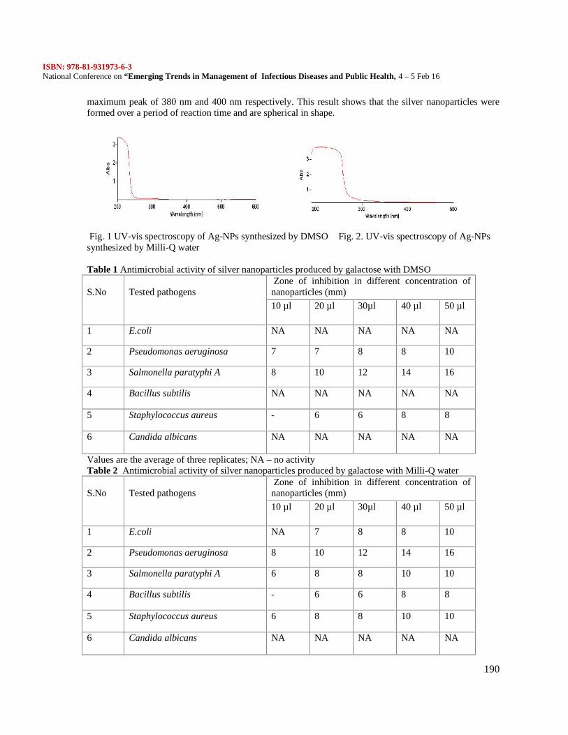
ISBN: 978-81-931973-6-3National Conference on “Emerging Trends in Management of Infectious Diseases and Public Health, 4 – 5 Feb 16
190
maximum peak of 380 nm and 400 nm respectively. This result shows that the silver nanoparticles wereformed over a period of reaction time and are spherical in shape.
Fig. 1 UV-vis spectroscopy of Ag-NPs synthesized by DMSO Fig. 2. UV-vis spectroscopy of Ag-NPssynthesized by Milli-Q water
Table 1 Antimicrobial activity of silver nanoparticles produced by galactose with DMSO
S.No Tested pathogensZone of inhibition in different concentration of
nanoparticles (mm)
10 µl 20 µl 30µl 40 µl 50 µl
1 E.coli NA NA NA NA NA
2 Pseudomonas aeruginosa 7 7 8 8 10
3 Salmonella paratyphi A 8 10 12 14 16
4 Bacillus subtilis NA NA NA NA NA
5 Staphylococcus aureus - 6 6 8 8
6 Candida albicans NA NA NA NA NA
Values are the average of three replicates; NA – no activityTable 2 Antimicrobial activity of silver nanoparticles produced by galactose with Milli-Q water
S.No Tested pathogensZone of inhibition in different concentration of
nanoparticles (mm)
10 µl 20 µl 30µl 40 µl 50 µl
1 E.coli NA 7 8 8 10
2 Pseudomonas aeruginosa 8 10 12 14 16
3 Salmonella paratyphi A 6 8 8 10 10
4 Bacillus subtilis - 6 6 8 8
5 Staphylococcus aureus 6 8 8 10 10
6 Candida albicans NA NA NA NA NA
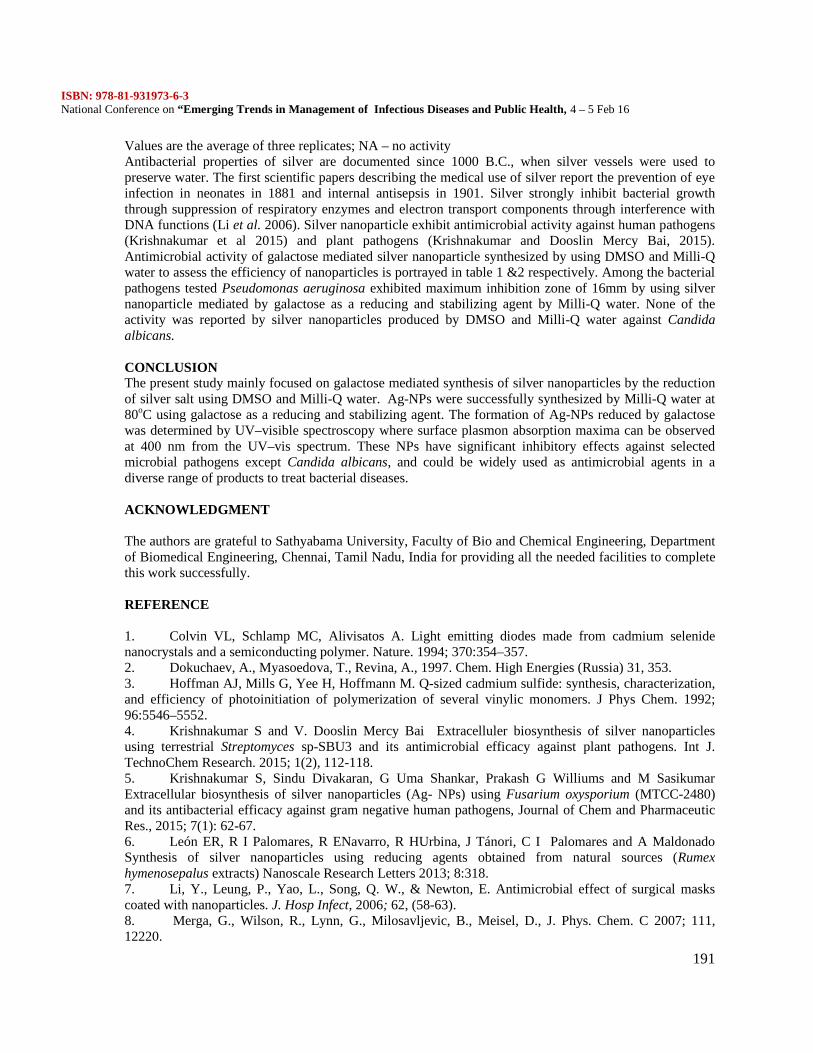
ISBN: 978-81-931973-6-3National Conference on “Emerging Trends in Management of Infectious Diseases and Public Health, 4 – 5 Feb 16
191
Values are the average of three replicates; NA – no activityAntibacterial properties of silver are documented since 1000 B.C., when silver vessels were used topreserve water. The first scientific papers describing the medical use of silver report the prevention of eyeinfection in neonates in 1881 and internal antisepsis in 1901. Silver strongly inhibit bacterial growththrough suppression of respiratory enzymes and electron transport components through interference withDNA functions (Li et al. 2006). Silver nanoparticle exhibit antimicrobial activity against human pathogens(Krishnakumar et al 2015) and plant pathogens (Krishnakumar and Dooslin Mercy Bai, 2015).Antimicrobial activity of galactose mediated silver nanoparticle synthesized by using DMSO and Milli-Qwater to assess the efficiency of nanoparticles is portrayed in table 1 &2 respectively. Among the bacterialpathogens tested Pseudomonas aeruginosa exhibited maximum inhibition zone of 16mm by using silvernanoparticle mediated by galactose as a reducing and stabilizing agent by Milli-Q water. None of theactivity was reported by silver nanoparticles produced by DMSO and Milli-Q water against Candidaalbicans.
CONCLUSIONThe present study mainly focused on galactose mediated synthesis of silver nanoparticles by the reductionof silver salt using DMSO and Milli-Q water. Ag-NPs were successfully synthesized by Milli-Q water at80oC using galactose as a reducing and stabilizing agent. The formation of Ag-NPs reduced by galactosewas determined by UV–visible spectroscopy where surface plasmon absorption maxima can be observedat 400 nm from the UV–vis spectrum. These NPs have significant inhibitory effects against selectedmicrobial pathogens except Candida albicans, and could be widely used as antimicrobial agents in adiverse range of products to treat bacterial diseases.
ACKNOWLEDGMENT
The authors are grateful to Sathyabama University, Faculty of Bio and Chemical Engineering, Departmentof Biomedical Engineering, Chennai, Tamil Nadu, India for providing all the needed facilities to completethis work successfully.
REFERENCE
1. Colvin VL, Schlamp MC, Alivisatos A. Light emitting diodes made from cadmium selenidenanocrystals and a semiconducting polymer. Nature. 1994; 370:354–357.2. Dokuchaev, A., Myasoedova, T., Revina, A., 1997. Chem. High Energies (Russia) 31, 353.3. Hoffman AJ, Mills G, Yee H, Hoffmann M. Q-sized cadmium sulfide: synthesis, characterization,and efficiency of photoinitiation of polymerization of several vinylic monomers. J Phys Chem. 1992;96:5546–5552.4. Krishnakumar S and V. Dooslin Mercy Bai Extracelluler biosynthesis of silver nanoparticlesusing terrestrial Streptomyces sp-SBU3 and its antimicrobial efficacy against plant pathogens. Int J.TechnoChem Research. 2015; 1(2), 112-118.5. Krishnakumar S, Sindu Divakaran, G Uma Shankar, Prakash G Williums and M SasikumarExtracellular biosynthesis of silver nanoparticles (Ag- NPs) using Fusarium oxysporium (MTCC-2480)and its antibacterial efficacy against gram negative human pathogens, Journal of Chem and PharmaceuticRes., 2015; 7(1): 62-67.6. León ER, R I Palomares, R ENavarro, R HUrbina, J Tánori, C I Palomares and A MaldonadoSynthesis of silver nanoparticles using reducing agents obtained from natural sources (Rumexhymenosepalus extracts) Nanoscale Research Letters 2013; 8:318.7. Li, Y., Leung, P., Yao, L., Song, Q. W., & Newton, E. Antimicrobial effect of surgical maskscoated with nanoparticles. J. Hosp Infect, 2006; 62, (58-63).8. Merga, G., Wilson, R., Lynn, G., Milosavljevic, B., Meisel, D., J. Phys. Chem. C 2007; 111,12220.
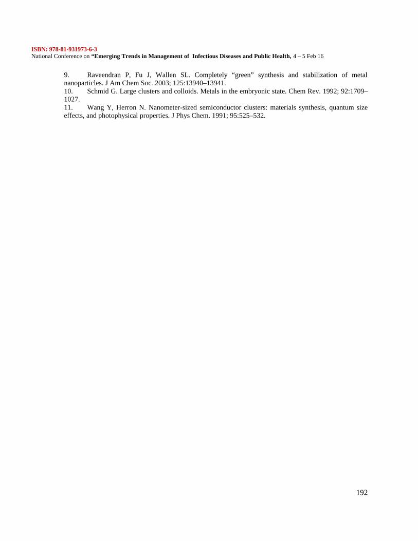
ISBN: 978-81-931973-6-3National Conference on “Emerging Trends in Management of Infectious Diseases and Public Health, 4 – 5 Feb 16
192
9. Raveendran P, Fu J, Wallen SL. Completely “green” synthesis and stabilization of metalnanoparticles. J Am Chem Soc. 2003; 125:13940–13941.10. Schmid G. Large clusters and colloids. Metals in the embryonic state. Chem Rev. 1992; 92:1709–1027.11. Wang Y, Herron N. Nanometer-sized semiconductor clusters: materials synthesis, quantum sizeeffects, and photophysical properties. J Phys Chem. 1991; 95:525–532.
ISBN: 978-81-931973-6-3National Conference on “Emerging Trends in Management of Infectious Diseases and Public Health, 4 – 5 Feb 16
193
Antimicrobial Activity Of Bougainvillea spectabilis And Identification Of Its Active Compounds ByGC- MS
Valli. S.1 and Jaganathan. S2
PG & Research Department of Microbiology,Mohamed Sathak College of Arts andscience,Sholinganallur,Chennai-119
INTRODUCTION
Microbial infections can cause severe health effects and can even lead to death among the residingpopulation, especially in the developing regions of the world. Selection of an effective antimicrobial agentfor a microbial infection requires knowledge of the potential microbial pathogen, an understanding of thepathophysiology of the infectious process and an understanding of the pharmacology and pharmacokinetics of the intended therapeutic agents (1). Urinary tract infection represents one of the most commondiseases encountered in medical practice today and occurring from the neonate to the geriatric age group(2,3) Enteric fever (typhoid) caused by Salmonella typhi is a global bacterial infection with an annualinfection rate of 21.6 million and 10% fatality rate (4). In developing countries, typhoid is more severe dueto poor hygiene, indiscriminate use of antibiotics and a rapid rise in multidrug resistance. Resistance to thefirst line drugs chloramphenicol, ciprofloxacinand amoxycillin has been reported.(5).
The continual emergence of antibiotic-resistant microorganisms has prompted researchersworld over to search for new antimicrobial agents that are more effective against the resistant microbialpathogens .With the increase in resistance to antityphoid drugs, medicinal plants have gained popularityamong both urban and rural dwellers in the treatment of the ailment. In the scope of searching newantimicrobial agents, antimicrobials derived from plant materials are often regarded as natural and safecompared to industrial chemicals
Ornamental plants offer aesthetic value and ecological balance in ecosystems. Most floweringornamental plants however are utilized more for their beauty as they impart different colourless to theirsurroundings. As their flower blooms, they serve this purpose and when they wilt, they fall off as trash.These flowers could be still used as pharmaceuticals or nutraceuticals. Bougainvillea spectabilis belongingto the family Nyctaginaceae is a very common ornamental plant grown almost all over the world intropical and sub-tropical gardens. It grows as a shrub or thorny, woody vine reaching upwards of 12 meterstall and 7 meters wide. Recent research has shown B. spectabilis to possess some potentially usefulantiviral and antibacterial compounds (6,7) as well as anti-diabetic properties (8).Phytochemical characterization of plant material is important as it relates to the therapeutic actions.Phytochemical evaluation of a herbal drug is an essential criteria, before proceeding for itspharmacological and toxicological studies (9). Gas chromatography separates the components of a mixtureand mass spectroscopy characterizes each of the components individually. By combining the twotechniques, an analytical chemist can both qualitatively and quantitatively evaluate a solution containing anumber of chemicals.(10)There had been a limited screening of phytochemicals, antioxidant and antimicrobial from flowers ofornamental plants Keeping in view the above idea and considering the vast potentiality of Bougainvilleaspectabilis as sources for antimicrobial drugs, a systematic investigation was undertaken to evaluate theantibacterial and antifungal activity from its flower extracts against clinical pathogens ,screen forphytochemicals and separate its active constituents by GC MS analysis.
MATERIALS AND METHODS:
Sample Collection and Processing :Blood, urine and pus samples were collected in sterile containers as per standard procedures and
processed for the isolation of bacterial pathogen. Skin scrapings were collected and processed for theisolation of dermatophytes. The bacterial pure culture obtained was further subjected to Gram staining,motility and various biochemical tests and streaked on selective media for the identification of each isolate
ISBN: 978-81-931973-6-3National Conference on “Emerging Trends in Management of Infectious Diseases and Public Health, 4 – 5 Feb 16
194
The fungal culture isolated was further subjected to KOH mount and Lacto Phenol Cotton Blue Stainingmethod
ANTIBIOTIC SEITIVITY TEST BY KIRBY BAUER DISC DIFFUSION METHODThe sensitivity pattern of isolates to standard antibiotics was tested by the Kirby Bauer(11) disc diffusionmethod as follows Four or five colonies of the test organisms were transferred to Muller Hinton broth Theturbidity was adjusted to McFarland standard (0.5) A sterile cotton swab was immersed in the bacterialsuspension and excess fluid was compressed against the wall of the test tube. The surface of the sterileMueller Hinton Agar plates was swabbed by passing the swab three times over the entire surface. Theplates were allowed to dry for ten minutes. The discs containing antimicrobial agents were placed in such away that it adheres perfectly to the surface of the agar by gentle pressing. The plates were incubated at37oC for 24hours. After incubation the size of the zones were measured and the results were interpretedand compared with the ATCC control strains by standard zone size interpretative chart as per CLSI.
ANTIMICROBIAL ACTIVITY OF FLORAL EXTRACTS OF Bougainvilla SpectablisCollection of B. spectabilis flowersB. spectabilis flowers were collected from surrounding areas of Besant nagar, Chennai and their identity
was verified and authenticated from Prof. P. Jayaraman, Director Plant Anatomy Research Centre,Tambaram.Extraction & DryingThe extracts of the flower samples were prepared in a direct method by soaking 25 g of flower samples in75 ml of different solvents (chloroform, ethyl acetate and ethanol) for 48 h. At the end of each respectiveextraction, the extracts were filtered using Whatma1 filter paper. The filtrate was concentrated underreduced pressure in vacuum at 40°C for 25 min using a rotary evaporator (Super fit-ROTAVAP, India).The percentage yield of extracts was calculated.using the formula:-
Percent yield = Weight of the crude extract obtained in gram x 100Total weight of plant powder in gram
SCREENING OF THE EXTRACT FOR ANTIBACTERIAL ACTIVITYPREPARATION AND STANDARDISATION OF INOCULUMThe invitro susceptibility of any bacteria to potential drug or standard drug will vary depending on numberof organism in the given inoculums increases.. Four to five colonies from pure growth of each testorganism were transferred to5ml of MHB. The broth was incubated at 35-37ºc for 18 hours. The turbidityof the culture was compared to 0.5 Mcfarland Nephelometer standard to get 150X10 CFU/ml. Thestandardized inoculums suspension was inoculated within 15-20 minutes (12). For fungi, Sabauroudsdextrose broth(SDB) was used.4.AGAR WELL DIFFUSION METHODScreening of antibacterial activity was performed by well diffusion technique (13). The Muller HintonAgar plates were prepared and then seeded with 0.1ml of standardized inoculums of each test organism.The inoculum was spread evenly over plate with sterile glass spreader or sterile cotton swab. The seededplates were allowed to dry for 10 minutes. A standard cork borer of 8mm was used to cut uniform wells onthe surface of the MHA and 5µl(3.9mg), 10µl(7.8mg), 15µl(11.7mg), 20µl(15.7mg) of flower extractdispensed into well separately and each plate was labeled with the test organism.The dissolution of the organic extract was aided by 1% DMSO, which did not effect the growth ofmicroorganisms, in accordance with the control experiments. The plates were then incubated at 37ºC for24 hours. The antibacterial activity was assayed by measuring the diameter of the inhibition zone formedaround the well (NCCLS, 2000). The experiments were performed in duplicate and repeated thriceMINIMUM INHIBITORY CONCENTRATION (MIC) OF THE FLORAL EXTRACTSThe Minimum Inhibitory Concentration(MIC) of the flower extract was determined by tube dilutiontechniques Muller Hinton Broth(Merck) according to NCCLS(2000). The concentration ranged from
ISBN: 978-81-931973-6-3National Conference on “Emerging Trends in Management of Infectious Diseases and Public Health, 4 – 5 Feb 16
195
3.9mg to 195mg. 1ml of nutrient broth was taken in each of sterile test tubes and the ethanolic extract ofthe flowers were added to all the tubes serially. 500µl of bacterial suspension that was previously grown innutrient broth were added to all tubes and incubated at 37ºC for 24 hours. The lowest concentration thatexhibited no visible growth were recorded as the MIC of the extract.PHYTOCHEMICAL ANALYSIS OF FLOWER EXTRACT OF Bougainvillea spectabilisSpecific qualitative tests were performed for the presence and absence of phytochemicals viz, alkaloids,tannins, flavanoids, saponins, glycosided etc., in flower extract to identify the constituents using standardprocedures.(14)GAS CHROMATOGRAPHY – MASS SPECTRUM ANALYSIS (GC-MS)
GC-MS analysis of this extract was performed using GC SHIMADZU QP2010 system and gaschromatograph interfaced to a Mass Spectrometer (GC-MS) equipped with Elite-1 fused silica capillarycolumn (Length : 30.0 m, Diameter : 0.25 mm, Film thickness : 0.25 is Composed of 100% Dimethyl polysiloxane). An electron ionization energy system with ionization energy of 70eV was used. Helium gas(99.999%) was used as the carrier gas at a constant flow rate of 1.51ml/min and an injection volume of 2ìlwas employed (split ratio: 20). Injector temperature 200°C; Ion-source temperature 200°C. The oventemperature was programmed from 70°C (isothermal for 2 min.), with an increase of 300°C for 10 min.Mass spectra were taken at 70eV; a scan interval of 0.5 seconds with scan range of 40 – 1000 m/z. TotalGC running time was 35 min. The relative percentage amount of each component was calculated bycomparing its average peak area to the total areasIdentification of componentsInterpretation of mass spectrum GC-MS was conducted using the database of National Institute Standardand Technique (NIST08s), WILEY8 and FAME having more patterns.. The active components of theextract were identified. By comparing the spectrum of the unknown component with the spectrum of theknown components stored in the NIST08s, WILEY8 and FAME library
RESULTS
Samples of blood urine and pus were processed for the isolation of pathogen. Based on the morphologicaland biochemical characteristics the isolates from blood was identified as Salmonella typhi. Pseudomonasaeruginosa and Staphylococcus aureus were isolated from pus E.coli and Enterococcus faecalis.from urinesample. Trichophyton rubrum and Epidermophyton flocossum were the dermatophytes isolated from skinscrapingsANTIBIOTIC SUSCEPTIBILITY:-The resistance / susceptibility pattern of the clinical isolates to various antibiotics have been assessed bydisc diffusion method. Of all the 5 isolates it was found that. E.coli isolated from urine sample exhibitedmultidrug resistance .It was sensitive to Amikacin, Nitrothrantoin, Imipenem, Co- trimoxazole and wasresistant to Amoxylin, Genofloxacin, Amoxyclav, Cefotaxime, Ciprofloxacin, Ampicillin, Ceftriaxone,Astreonam, Ceftazidime, Gentamycin, Cefepime, Cefaloxin and Nalidixic acid.ANTIMICROBIAL ACTIVITY OF FLORAL EXTRACTS OF B. spectabilis
Ethanol, chloroform and ethyl acetate extracts of the plant were prepared, filtered andevaporated as per standard methods Maximum percentage yield was observed in ethanolic extract offlowers 5% for 25g of fresh flowers used for extraction, followed by ethyl acetate (2.2%) and chloroform(1.2%).
The various solvent extracts of the plant B. spectabilis were observed for antibacterial activityagainst the pathogens isolated from clinical samples. Their antibacterial potency was assessed by thepresence or absence of inhibition of zones and zone diameters (mm).(Table1)ANTIFUNGAL ACTIVITY OF SOLVENT EXTRACTS OF B. spectabilis:-The antifungal activities of the various solvent extracts of flowers of B. spectabilis were tested by welldiffusion method .Trichophyton rubrum exhibited maximum zone of inhibition 24mm at a concentration of20mg of ethanolic extract of flowers of B. spectabilis and Epidermophyton exhibited 23mm zone ofinhibition.(Table2)
ISBN: 978-81-931973-6-3National Conference on “Emerging Trends in Management of Infectious Diseases and Public Health, 4 – 5 Feb 16
196
MINIMUM INHIBITORY CONCENTRATION OF SOLVENT EXTRACTS OF B. spectabilisFLOWERS FOR BACTERIAL ISOLATES:-The minimum inhibitory concentration of various solvent extracts of Bougainvillea spectabilis flowers wasmeasured by broth dilution method. The MIC value of ethanolic and ethyl acetate solvent extract for E.coli at lowest concentration 11.7mg/ml. The minimum inhibitory concentration of ethanolic flower extractwas 19.5mg, chloroform extract 15.6mg and ethyl acetate extract 30mg for Salmonella typhiPHYTOCHEMICAL ANALYSIS
The major phytochemicals found were saponins, carbohydrates, phlobotannins, flavanoids,trepenoids and alkaloids. All extracts showed the absence of sterols, quinines, glycosides and tannins.Ethyl acetate extract yielded maximum phytochemicalsGC-MS ANALYSIS:-GC-MS analysis was carried out on ethanolic extract of B. spectabilis flowers and four different
compounds were identified. The chromatogram revealed four peaks in the retention time range 10.63minutes to 14.5 minutes. The largest peak at13.18 min was identified as 2(3H)Furanone-dihydro5-tetradecyl with 25%. The second less prominent peak at 13.57 minutes corresponds to the compoundDasycarpidan-1-methanol, acetate (ester) with 16%. The third prominent peak at 10.63% minute with 12%area was due to the presence of Ergoline-8-carboxylic acid, 10-methoxy-6-methyl-,methyl ester. Thefourth prominent peak at 11.9 minute corresponds to the compound Ascorbic acid 2,6-dihexadecanoatewith peak area 5% (Table3).
DISCUSSION:
There has been an increasing incidence of multiple resistances in human pathogenic microorganisms inrecent years, largely due to indiscriminate use of commercial antimicrobial drugs commonly employed inthe treatment of infectious diseases. This medical challenge has made man to depend on his immediateenvironment taking advantage of nature’s provisions for life and survival the present study was undertakento screen for antimicrobial activity of floral extracts of Bougainvillea spectabilis, and identify the bioactivecomponents responsible for antimicrobial activity by GC-MS analysis.. The present study demonstrated E. coli and E. faecalis be the predominant aetiological agent as thecausative agent of UTI. These findings are similar to studies conducted by(15). E. coli exhibited a high rateof resistance to many antibiotics used in the present study. This pattern of resistance also has been reportedby(16). S.aureus and P. aeruginosa were identified as aetiological agent from wound infections.Similarresults were reported by(17). , Salmonella typhi was found to be the aetiological agents of blood samplescollected from enteric fever patients. Anthropophilic dermatophytes are the main agents of Tinea cruristhroughout the world in particular, Trichophyton rubrum and Epidermophyton flocossum. In the presentstudy Trichophyton rubrum was the predominant against isolated followed by E. flocossum, thusconfirming the results of earlier studies by( 18)There is a global interest in non-synthetic natural drugs derived from plant sources, because of low cost,non-toxic nature and availability. The research for medicinal plants having dictinguishable medicinalproperties, among which one is Bougainvillea spectabilis. It is an ornamental flowering plant, belongingto th family Nyctaginaceae. It is used in herbal combination for the treatment of diabetes. The incidence ofscreening B. spectabilis flowers for pharmacological evaluation was very less and hence the present studywas aimed to investigate the antimicrobial activity of different solvent extracts of Bougainvillea spectabilisflowers and identify the bioactive components.Different solvent extracts of B. spectabilis were prepared by Soxhlet extraction method and evaluated for
antimicrobial activity by disc diffusion method. The percentage yield of various solvents was calculatedand it was found to be less. Ethanolic extract yield was only 5% per 25g of flowers the antibacterialactivity of 3 solvent extracts of flowers of Bougainvilla spectabilis were studied against Gram positive andGram negative pathogens. Ethanolic extract showed inhibitory effect on all pathogens isolated fromclinical samples. Escherichia coli and Enterococcus the aetiological agent of U.T.I. were highly
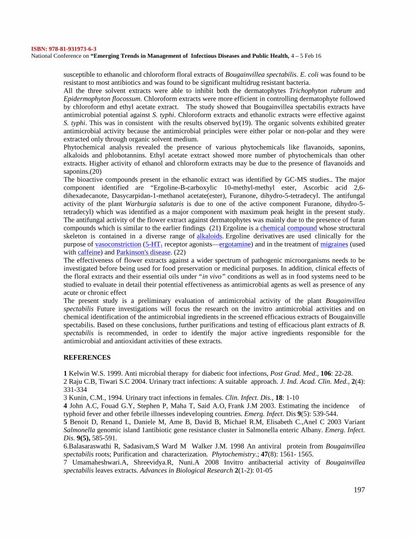
ISBN: 978-81-931973-6-3National Conference on “Emerging Trends in Management of Infectious Diseases and Public Health, 4 – 5 Feb 16
197
susceptible to ethanolic and chloroform floral extracts of Bougainvillea spectabilis. E. coli was found to beresistant to most antibiotics and was found to be significant multidrug resistant bacteria.All the three solvent extracts were able to inhibit both the dermatophytes Trichophyton rubrum andEpidermophyton flocossum. Chloroform extracts were more efficient in controlling dermatophyte followedby chloroform and ethyl acetate extract. The study showed that Bougainvillea spectabilis extracts haveantimicrobial potential against S. typhi. Chloroform extracts and ethanolic extracts were effective againstS. typhi. This was in consistent with the results observed by(19). The organic solvents exhibited greaterantimicrobial activity because the antimicrobial principles were either polar or non-polar and they wereextracted only through organic solvent medium.Phytochemical analysis revealed the presence of various phytochemicals like flavanoids, saponins,alkaloids and phlobotannins. Ethyl acetate extract showed more number of phytochemicals than otherextracts. Higher activity of ethanol and chloroform extracts may be due to the presence of flavanoids andsaponins.(20)The bioactive compounds present in the ethanolic extract was identified by GC-MS studies.. The majorcomponent identified are “Ergoline-B-carboxylic 10-methyl-methyl ester, Ascorbic acid 2,6-dihexadecanote, Dasycarpidan-1-methanol acetate(ester), Furanone, dihydro-5-tetradecyl. The antifungalactivity of the plant Warburgia salutaris is due to one of the active component Furanone, dihydro-5-tetradecyl) which was identified as a major component with maximum peak height in the present study.The antifungal activity of the flower extract against dermatophytes was mainly due to the presence of furancompounds which is similar to the earlier findings (21) Ergoline is a chemical compound whose structuralskeleton is contained in a diverse range of alkaloids. Ergoline derivatives are used clinically for thepurpose of vasoconstriction (5-HT1 receptor agonists—ergotamine) and in the treatment of migraines (usedwith caffeine) and Parkinson's disease. (22)The effectiveness of flower extracts against a wider spectrum of pathogenic microorganisms needs to beinvestigated before being used for food preservation or medicinal purposes. In addition, clinical effects ofthe floral extracts and their essential oils under “in vivo” conditions as well as in food systems need to bestudied to evaluate in detail their potential effectiveness as antimicrobial agents as well as presence of anyacute or chronic effectThe present study is a preliminary evaluation of antimicrobial activity of the plant Bougainvilleaspectabilis Future investigations will focus the research on the invitro antimicrobial activities and onchemical identification of the antimicrobial ingredients in the screened efficacious extracts of Bougainvillespectabilis. Based on these conclusions, further purifications and testing of efficacious plant extracts of B.spectabilis is recommended, in order to identify the major active ingredients responsible for theantimicrobial and antioxidant activities of these extracts.
REFERENCES
1 Kelwin W.S. 1999. Anti microbial therapy for diabetic foot infections, Post Grad. Med., 106: 22-28.2 Raju C.B, Tiwari S.C 2004. Urinary tract infections: A suitable approach. J. Ind. Acad. Clin. Med., 2(4):331-3343 Kunin, C.M., 1994. Urinary tract infections in females. Clin. Infect. Dis., 18: 1-104 John A.C, Fouad G.Y, Stephen P, Maha T, Said A.O, Frank J.M 2003. Estimating the incidence oftyphoid fever and other febrile illnesses indeveloping countries. Emerg. Infect. Dis 9(5): 539-544.5 Benoit D, Renand L, Daniele M, Ame B, David B, Michael R.M, Elisabeth C.,Anel C 2003 VariantSalmonella genomic island 1antibiotic gene resistance cluster in Salmonella enteric Albany. Emerg. Infect.Dis. 9(5), 585-591.6.Balasaraswathi R, Sadasivam,S Ward M Walker J.M. 1998 An antiviral protein from Bougainvilleaspectabilis roots; Purification and characterization. Phytochemistry.; 47(8): 1561- 1565.7 Umamaheshwari.A, Shreevidya.R, Nuni.A 2008 Invitro antibacterial activity of Bougainvilleaspectabilis leaves extracts. Advances in Biological Research 2(1-2): 01-05
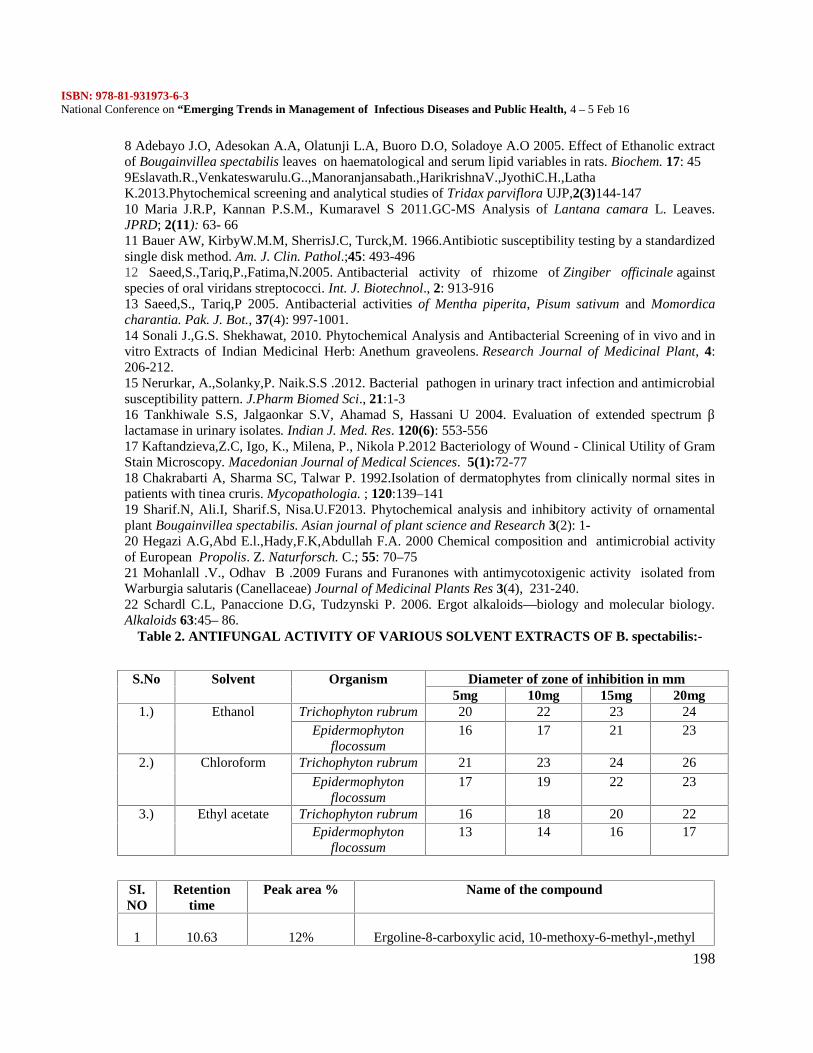
ISBN: 978-81-931973-6-3National Conference on “Emerging Trends in Management of Infectious Diseases and Public Health, 4 – 5 Feb 16
198
8 Adebayo J.O, Adesokan A.A, Olatunji L.A, Buoro D.O, Soladoye A.O 2005. Effect of Ethanolic extractof Bougainvillea spectabilis leaves on haematological and serum lipid variables in rats. Biochem. 17: 459Eslavath.R.,Venkateswarulu.G..,Manoranjansabath.,HarikrishnaV.,JyothiC.H.,LathaK.2013.Phytochemical screening and analytical studies of Tridax parviflora UJP,2(3)144-14710 Maria J.R.P, Kannan P.S.M., Kumaravel S 2011.GC-MS Analysis of Lantana camara L. Leaves.JPRD; 2(11): 63- 6611 Bauer AW, KirbyW.M.M, SherrisJ.C, Turck,M. 1966.Antibiotic susceptibility testing by a standardizedsingle disk method. Am. J. Clin. Pathol.;45: 493-49612 Saeed,S.,Tariq,P.,Fatima,N.2005. Antibacterial activity of rhizome of Zingiber officinale againstspecies of oral viridans streptococci. Int. J. Biotechnol., 2: 913-91613 Saeed,S., Tariq,P 2005. Antibacterial activities of Mentha piperita, Pisum sativum and Momordicacharantia. Pak. J. Bot., 37(4): 997-1001.14 Sonali J.,G.S. Shekhawat, 2010. Phytochemical Analysis and Antibacterial Screening of in vivo and invitro Extracts of Indian Medicinal Herb: Anethum graveolens. Research Journal of Medicinal Plant, 4:206-212.15 Nerurkar, A.,Solanky,P. Naik.S.S .2012. Bacterial pathogen in urinary tract infection and antimicrobialsusceptibility pattern. J.Pharm Biomed Sci., 21:1-316 Tankhiwale S.S, Jalgaonkar S.V, Ahamad S, Hassani U 2004. Evaluation of extended spectrum βlactamase in urinary isolates. Indian J. Med. Res. 120(6): 553-55617 Kaftandzieva,Z.C, Igo, K., Milena, P., Nikola P.2012 Bacteriology of Wound - Clinical Utility of GramStain Microscopy. Macedonian Journal of Medical Sciences. 5(1):72-7718 Chakrabarti A, Sharma SC, Talwar P. 1992.Isolation of dermatophytes from clinically normal sites inpatients with tinea cruris. Mycopathologia. ; 120:139–14119 Sharif.N, Ali.I, Sharif.S, Nisa.U.F2013. Phytochemical analysis and inhibitory activity of ornamentalplant Bougainvillea spectabilis. Asian journal of plant science and Research 3(2): 1-20 Hegazi A.G,Abd E.l.,Hady,F.K,Abdullah F.A. 2000 Chemical composition and antimicrobial activityof European Propolis. Z. Naturforsch. C.; 55: 70–7521 Mohanlall .V., Odhav B .2009 Furans and Furanones with antimycotoxigenic activity isolated fromWarburgia salutaris (Canellaceae) Journal of Medicinal Plants Res 3(4), 231-240.22 Schardl C.L, Panaccione D.G, Tudzynski P. 2006. Ergot alkaloids—biology and molecular biology.Alkaloids 63:45– 86.
Table 2. ANTIFUNGAL ACTIVITY OF VARIOUS SOLVENT EXTRACTS OF B. spectabilis:-
S.No Solvent Organism Diameter of zone of inhibition in mm5mg 10mg 15mg 20mg
1.) Ethanol Trichophyton rubrum 20 22 23 24Epidermophyton
flocossum16 17 21 23
2.) Chloroform Trichophyton rubrum 21 23 24 26Epidermophyton
flocossum17 19 22 23
3.) Ethyl acetate Trichophyton rubrum 16 18 20 22Epidermophyton
flocossum13 14 16 17
SI.NO
Retentiontime
Peak area % Name of the compound
1 10.63 12% Ergoline-8-carboxylic acid, 10-methoxy-6-methyl-,methyl
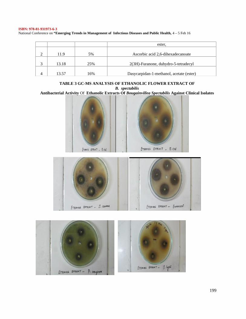
ISBN: 978-81-931973-6-3National Conference on “Emerging Trends in Management of Infectious Diseases and Public Health, 4 – 5 Feb 16
199
TABLE 3 GC-MS ANALYSIS OF ETHANOLIC FLOWER EXTRACT OFB. spectabilis
Antibacterial Activity Of Ethanolic Extracts Of Bougainvillea Spectabilis Against Clinical Isolates
ester,
2 11.9 5% Ascorbic acid 2,6-dihexadecanoate
3 13.18 25% 2(3H)-Furanone, duhydro-5-tetradecyl
4 13.57 16% Dasycarpidan-1-methanol, acetate (ester)
ISBN: 978-81-931973-6-3National Conference on “Emerging Trends in Management of Infectious Diseases and Public Health, 4 – 5 Feb 16
200
Effect of Monocrotophos and Azadiractin on germination of Pisum sativumand Zea mays
Praveena. A1, Anuradha. V2
1. Department of Biotechnology, Prathyusha Engineering College, Thiruvallur.2. Department of Biochemistry, Mohamed Sathak College of Arts and Science, Sholinganallur,
Chennai - 119ABSTRACT: The use of pesticides has greatly improved agricultural yield through inhibition of diseasecaused by many soil pests. Also the pesticide residues in the soil affect the crop seed germination andgrowth. The extensive use of insecticide in agricultural and their potential carcinogenicity strongly suggestthe need to extend the cytotoxic evaluations. The active ingredients of the pesticides bind to the cell cycleenzymes and inhibit the mitotic cell division of the plants. The objective of the present study focuses onthe effect of Monocrotophos and Azadirachtin on green peas (Pisum sativum) and maize (Zea mays) seedgermination and on root tip mitotic index. The difference between Botanical pesticide and Chemicalpesticides on the growth of plant was also assessed. The molecular interaction studies were also done usingbioinformatics tools.Keywords: Monocrotophos, Azadiractin, Seed germination, Mitotic index, Molecular interaction.INTRODUCTION
In developed agricultural system inorganic fertilizers are applied to the soil to supply theessential nutrients required for plants. The use of pesticides has greatly improved agriculturalyield through inhibition of disease causing and by acting against pest in the field and duringstorage of agricultural products (Taylor et.al., 1997). The killing insect “Pest,” insecticidesobviously have the potential to harm non-target insects such as beneficial natural predators andpollinators. Less obviously, weed-killers can also be harmful to beneficial insects (Hassan, et al.1988). It is estimated that often than 0.1 percent of an applied pesticide reaches the target pest,leaving 99.9 percent an unintended pollutant in the environment, including in soil, air and water,or on nearby vegetation (Pimentel, 1995).
Hence, the study on the effect of insecticides on plant seedling growth and germinationalong with root tip analysis is an essential tool to derive the best insecticide which will provideboth disease resistance and improves the crop yield.MATERIALS AND METHODS
Different concentration of Monocrotophos and Azadirachtin 100,200 and 400ppm is takento study their effect on germination and growth of Green pea and Maize seeds. The test Green peaand Maize seeds are sterilized (to avoid fungal infestation) with 0.1% Mercuric chloride solutionfor 5minutes, followed by several washing under running tap water to remove the residues. Threereplicates, each containing 10seeds of Green pea and Maize are arranged in circles on a twolayered Whatman No.1 filter paper placed in 9 cm diameter Petri dishes and to this 3ml ofinsecticide at different concentrations is added. Thereafter, 2ml of insecticide is added onalternate days till the end of the experiment. Distilled water is used as control. The germination ofseeds is recorded at 3, 5, 7 and 9 days and the seedling length was also measured.
In this experiment, Green pea and Maize seeds were placed in a 9cm diameter Petridishes containing the double layered filter paper. Different concentrations ranging from 100, 200and 400 ppm of the insecticide solution are applied in respective Petri plates and distilled water
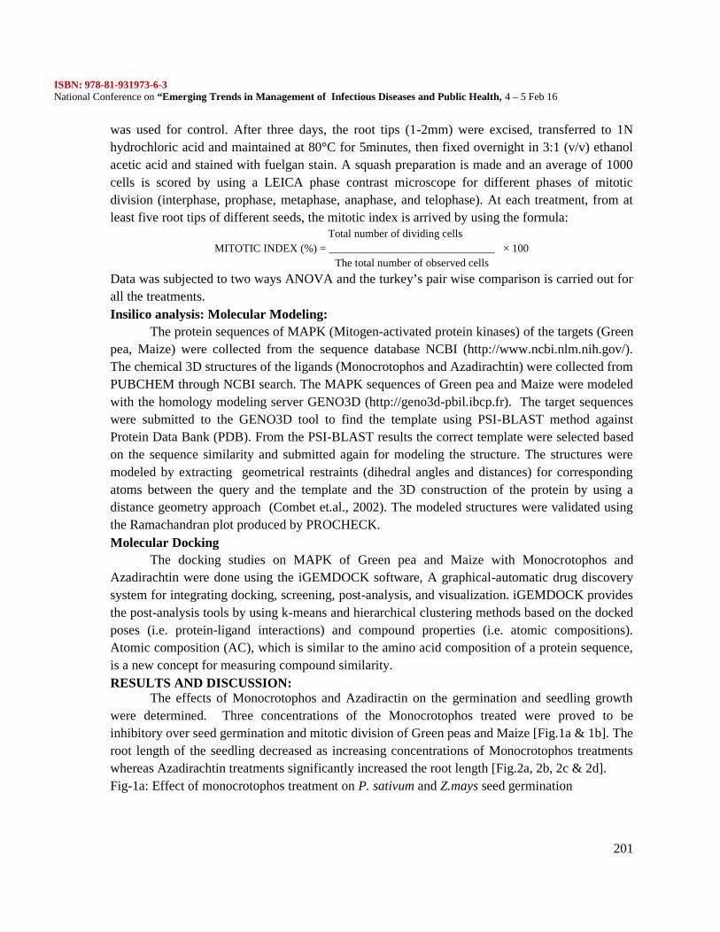
ISBN: 978-81-931973-6-3National Conference on “Emerging Trends in Management of Infectious Diseases and Public Health, 4 – 5 Feb 16
201
was used for control. After three days, the root tips (1-2mm) were excised, transferred to 1Nhydrochloric acid and maintained at 80°C for 5minutes, then fixed overnight in 3:1 (v/v) ethanolacetic acid and stained with fuelgan stain. A squash preparation is made and an average of 1000cells is scored by using a LEICA phase contrast microscope for different phases of mitoticdivision (interphase, prophase, metaphase, anaphase, and telophase). At each treatment, from atleast five root tips of different seeds, the mitotic index is arrived by using the formula:
Total number of dividing cellsMITOTIC INDEX (%) = ______________________________ × 100
The total number of observed cells
Data was subjected to two ways ANOVA and the turkey’s pair wise comparison is carried out forall the treatments.Insilico analysis: Molecular Modeling:
The protein sequences of MAPK (Mitogen-activated protein kinases) of the targets (Greenpea, Maize) were collected from the sequence database NCBI (http://www.ncbi.nlm.nih.gov/).The chemical 3D structures of the ligands (Monocrotophos and Azadirachtin) were collected fromPUBCHEM through NCBI search. The MAPK sequences of Green pea and Maize were modeledwith the homology modeling server GENO3D (http://geno3d-pbil.ibcp.fr). The target sequenceswere submitted to the GENO3D tool to find the template using PSI-BLAST method againstProtein Data Bank (PDB). From the PSI-BLAST results the correct template were selected basedon the sequence similarity and submitted again for modeling the structure. The structures weremodeled by extracting geometrical restraints (dihedral angles and distances) for correspondingatoms between the query and the template and the 3D construction of the protein by using adistance geometry approach (Combet et.al., 2002). The modeled structures were validated usingthe Ramachandran plot produced by PROCHECK.Molecular Docking
The docking studies on MAPK of Green pea and Maize with Monocrotophos andAzadirachtin were done using the iGEMDOCK software, A graphical-automatic drug discoverysystem for integrating docking, screening, post-analysis, and visualization. iGEMDOCK providesthe post-analysis tools by using k-means and hierarchical clustering methods based on the dockedposes (i.e. protein-ligand interactions) and compound properties (i.e. atomic compositions).Atomic composition (AC), which is similar to the amino acid composition of a protein sequence,is a new concept for measuring compound similarity.RESULTS AND DISCUSSION:
The effects of Monocrotophos and Azadiractin on the germination and seedling growthwere determined. Three concentrations of the Monocrotophos treated were proved to beinhibitory over seed germination and mitotic division of Green peas and Maize [Fig.1a & 1b]. Theroot length of the seedling decreased as increasing concentrations of Monocrotophos treatmentswhereas Azadirachtin treatments significantly increased the root length [Fig.2a, 2b, 2c & 2d].Fig-1a: Effect of monocrotophos treatment on P. sativum and Z.mays seed germination
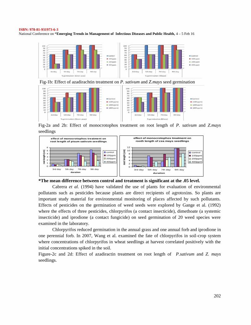
ISBN: 978-81-931973-6-3National Conference on “Emerging Trends in Management of Infectious Diseases and Public Health, 4 – 5 Feb 16
202
Fig-1b: Effect of azadirachtin treatment on P. sativum and Z.mays seed germination
Fig-2a and 2b: Effect of monocrotophos treatment on root length of P. sativum and Z.maysseedlings
effect of monocrotophos treatment onroot length of pisum sativum seedlings
0
12
3
45
6
3rd day 5th day 7th day 9th dayduration
root
leng
ht (c
m)
control
100ppm
200ppm
400ppm
effect of monocrotophos treatment onrooth length of zea mays seedlings
0246
81012
3rd day 5th day 7th day 9th day
duration
root
leng
ht (c
m)
control100ppm200ppm400ppm
*The mean difference between control and treatment is significant at the .05 level.Cabrera et al. (1994) have validated the use of plants for evaluation of environmental
pollutants such as pesticides because plants are direct recipients of agrotoxins. So plants areimportant study material for environmental monitoring of places affected by such pollutants.Effects of pesticides on the germination of weed seeds were explored by Gange et al. (1992)where the effects of three pesticides, chlorpyrifos (a contact insecticide), dimethoate (a systemicinsecticide) and iprodione (a contact fungicide) on seed germination of 20 weed species wereexamined in the laboratory.
Chlorpyrifos reduced germination in the annual grass and one annual forb and iprodione inone perennial forb. In 2007, Wang et al. examined the fate of chlorpyrifos in soil-crop systemwhere concentrations of chlorpyrifos in wheat seedlings at harvest correlated positively with theinitial concentrations spiked in the soil.Figure-2c and 2d: Effect of azadiractin treatment on root length of P.sativum and Z. maysseedlings.
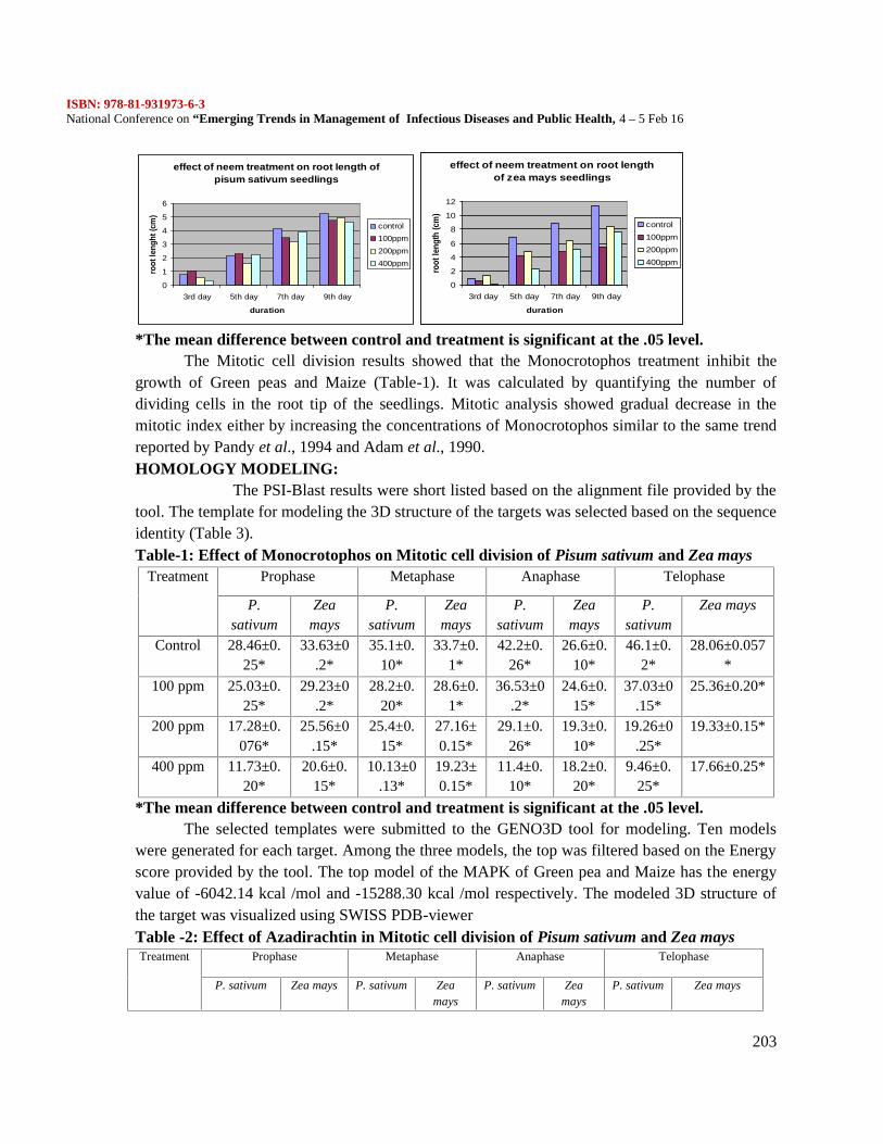
ISBN: 978-81-931973-6-3National Conference on “Emerging Trends in Management of Infectious Diseases and Public Health, 4 – 5 Feb 16
203
effect of neem treatment on root length ofpisum sativum seedlings
0
1
2
3
4
5
6
3rd day 5th day 7th day 9th day
duration
root
leng
ht (c
m)
control100ppm200ppm400ppm
effect of neem treatment on root lengthof zea mays seedlings
0
2
4
6
8
10
12
3rd day 5th day 7th day 9th day
duration
root
leng
th (c
m)
control100ppm200ppm400ppm
*The mean difference between control and treatment is significant at the .05 level.The Mitotic cell division results showed that the Monocrotophos treatment inhibit the
growth of Green peas and Maize (Table-1). It was calculated by quantifying the number ofdividing cells in the root tip of the seedlings. Mitotic analysis showed gradual decrease in themitotic index either by increasing the concentrations of Monocrotophos similar to the same trendreported by Pandy et al., 1994 and Adam et al., 1990.HOMOLOGY MODELING:
The PSI-Blast results were short listed based on the alignment file provided by thetool. The template for modeling the 3D structure of the targets was selected based on the sequenceidentity (Table 3).Table-1: Effect of Monocrotophos on Mitotic cell division of Pisum sativum and Zea mays
Treatment Prophase Metaphase Anaphase Telophase
P.sativum
Zeamays
P.sativum
Zeamays
P.sativum
Zeamays
P.sativum
Zea mays
Control 28.46±0.25*
33.63±0.2*
35.1±0.10*
33.7±0.1*
42.2±0.26*
26.6±0.10*
46.1±0.2*
28.06±0.057*
100 ppm 25.03±0.25*
29.23±0.2*
28.2±0.20*
28.6±0.1*
36.53±0.2*
24.6±0.15*
37.03±0.15*
25.36±0.20*
200 ppm 17.28±0.076*
25.56±0.15*
25.4±0.15*
27.16±0.15*
29.1±0.26*
19.3±0.10*
19.26±0.25*
19.33±0.15*
400 ppm 11.73±0.20*
20.6±0.15*
10.13±0.13*
19.23±0.15*
11.4±0.10*
18.2±0.20*
9.46±0.25*
17.66±0.25*
*The mean difference between control and treatment is significant at the .05 level.The selected templates were submitted to the GENO3D tool for modeling. Ten models
were generated for each target. Among the three models, the top was filtered based on the Energyscore provided by the tool. The top model of the MAPK of Green pea and Maize has the energyvalue of -6042.14 kcal /mol and -15288.30 kcal /mol respectively. The modeled 3D structure ofthe target was visualized using SWISS PDB-viewerTable -2: Effect of Azadirachtin in Mitotic cell division of Pisum sativum and Zea maysTreatment Prophase Metaphase Anaphase Telophase
P. sativum Zea mays P. sativum Zeamays
P. sativum Zeamays
P. sativum Zea mays

ISBN: 978-81-931973-6-3National Conference on “Emerging Trends in Management of Infectious Diseases and Public Health, 4 – 5 Feb 16
204
Control 28.46±0.25 33.63±0.2 35.1±0.1 33.7±0.1 42.2±0.26*
26.6±0.1 46.1±0.20 28.06±0.057
100 ppm 28.60±0.1 33.2±0.15 35±0.1 33.36±0.15
41.4±0.11*
26.36±0.20
45.76±0.11
27.83±0.057
200 ppm 25.4±0.1* 29.3±0.17*
28.7±0.20*
28.6±0.1*
36.53±0.20*
24.1±0.1*
37.16±0.15*
24.56±0.30*
400 ppm 18.3±0.057* 27.4±0.1* 23.4±0.20*
28.03±0.15*
27.56±0.15*
2106±0.1*
19.66±0.15*
20.60±0.20*
*The mean difference between control and treatment is significant at the .05 level.. Table 3: Template used for modeling the 3D structure of the targets.
Table 4: Ramachandran plot of the top model produced by PROCHECKTarget % of residues present in
Core Allowed DisallowedMapk of Pisum sativum 65.6 31.2 0.8
Mapk of zea mays 74.6 22.5 1.0MOLECULAR DOCKING: The iGEMDOCK results showed that the intermolecular complexformed between the MAPK of Pisum sativum, zea mays and Monocrotophos, Azadirachtin arestable with lower energy values (Table 6). Since molecules in nature have a tendency to be foundin their low energy form, the final configuration of intermolecular complex should also be of lowenergy. The interaction between the target and the ligand is due to van der waals and hydrogenbond interaction.
The post dock analysis explored the amino acids involved in the intermolecular complexformation. The results showed that Monocrotophos and Azadirachtin probably binds to the samesite of MAPK of Pisum sativum, where the amino acids, “ARG”, “GLU”, “SER”, “ASP” arepresent. In the case of zea mays, the binding pocket amino acids are ARG, PRO, GLU, LYS andILE (Table 6, Figure-4, 5).Table 5: Interaction profile of Monocrotophos, Azadirachtin with the MAPK of Pisumsativum, zea mays
Intermolecularcomplex
Total energyvalue
VDW H bond Binding pocket residues
MAPK of Pisumsativum –
Monocrotophos
-91.65 -71.52 -20.13 ARG97, ARG121, GLU125, SER124,SER126, ASP127
Target Name of the obtainedsignificant template
% of sequenceidentity betweentarget and template
E-Value PDB ID ofthe template
Mapk of Pisumsativum
Putative uncharacterizedprotein of Oryza sativasubsp. indica
75 7e-59 A2X0W6
Mapk of zeamays
Mitogen-activated proteinkinase of Dunaliella salina
71 8e-67 A2TDK2
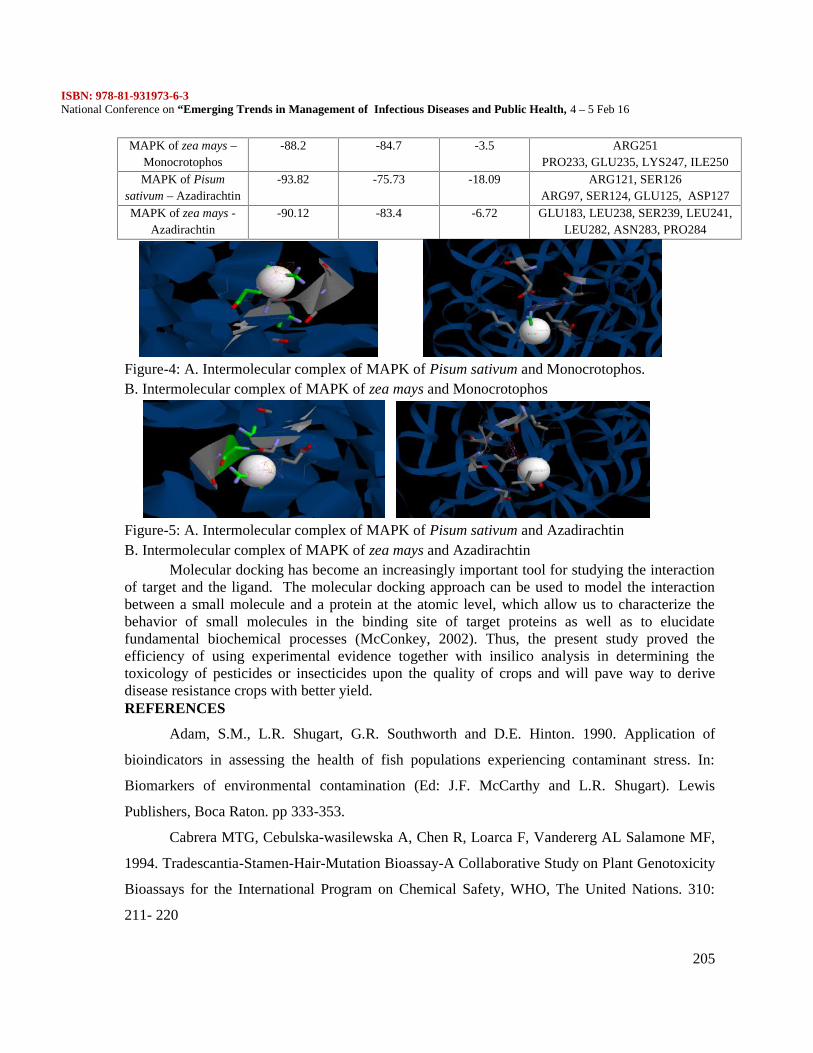
ISBN: 978-81-931973-6-3National Conference on “Emerging Trends in Management of Infectious Diseases and Public Health, 4 – 5 Feb 16
205
MAPK of zea mays –Monocrotophos
-88.2 -84.7 -3.5 ARG251PRO233, GLU235, LYS247, ILE250
MAPK of Pisumsativum – Azadirachtin
-93.82 -75.73 -18.09 ARG121, SER126ARG97, SER124, GLU125, ASP127
MAPK of zea mays -Azadirachtin
-90.12 -83.4 -6.72 GLU183, LEU238, SER239, LEU241,LEU282, ASN283, PRO284
Figure-4: A. Intermolecular complex of MAPK of Pisum sativum and Monocrotophos.B. Intermolecular complex of MAPK of zea mays and Monocrotophos
Figure-5: A. Intermolecular complex of MAPK of Pisum sativum and AzadirachtinB. Intermolecular complex of MAPK of zea mays and Azadirachtin
Molecular docking has become an increasingly important tool for studying the interactionof target and the ligand. The molecular docking approach can be used to model the interactionbetween a small molecule and a protein at the atomic level, which allow us to characterize thebehavior of small molecules in the binding site of target proteins as well as to elucidatefundamental biochemical processes (McConkey, 2002). Thus, the present study proved theefficiency of using experimental evidence together with insilico analysis in determining thetoxicology of pesticides or insecticides upon the quality of crops and will pave way to derivedisease resistance crops with better yield.REFERENCES
Adam, S.M., L.R. Shugart, G.R. Southworth and D.E. Hinton. 1990. Application of
bioindicators in assessing the health of fish populations experiencing contaminant stress. In:
Biomarkers of environmental contamination (Ed: J.F. McCarthy and L.R. Shugart). Lewis
Publishers, Boca Raton. pp 333-353.
Cabrera MTG, Cebulska-wasilewska A, Chen R, Loarca F, Vandererg AL Salamone MF,
1994. Tradescantia-Stamen-Hair-Mutation Bioassay-A Collaborative Study on Plant Genotoxicity
Bioassays for the International Program on Chemical Safety, WHO, The United Nations. 310:
211- 220
ISBN: 978-81-931973-6-3National Conference on “Emerging Trends in Management of Infectious Diseases and Public Health, 4 – 5 Feb 16
206
Combet C., Blanchet C., Geourjon C. and Deleage G. 2000, NPS@: Network Protein
Sequence analysis.
Gange AC, Brown VK, Farmer LM, 1992.Effects of Pesticides on the Germination of
Weed Seeds:Implications for Manipulative Experiments. Journal of Applied Ecology,29(2):303-
310.
Hassan SA, Bigler F, Bogenschütz H, Boller E, Brun J, Chiverton P, Edwards P, Mansour
F, Naton E, Oomen PA, Overmeer PJ, Polgar L, Rieckmann W, Samsøe-Petersen L, Stäubli A,
Sterk G, Tavares K, Tuset JJ, Viggiani G, Vivas AG. 1988, Results of the fourth joint pesticide
testing programme carried out by the IOBC/WPRS-Working Group “Pesticides and Beneficial
Organsisms”. J Appl. Entomol 105: 321-329.
McConkey BJ, Sobolev V, Edelman M. 2002, The performance of current methods in
ligand-protein docking. Current Science; 83:845–855
Nwachukwu E. O. and Umechuruba C. I. 2001. Antifungal activities of some leave
extracts on seed-borne fungi of African yam bean seeds. Journal of Applied and Natural Sciences
1:29-31
Pandey N. D., Mathur K. K., Pandey S. and Tripathi R. A. 1986. Effects of some plant
extracts against pulse beetle, Callosobruchus chinensis Linnaeus. Indian J. of Entomology 48(1):
85-90.
Pimentel D., Harvey C., Resosudarmo P., Sinclair K., Kurz D., McNair M., Crist S.,
Sphpritz, L.,Fitton, L., Saffouri, R. and Blair, R., 1995, ‘Environmental and economic costs of
soil erosion and conservation benefits’,Science 267,1117–1123.
Taylor D, Green N, Stout G. 1997. Biological Science. 3rd edition Cambridge University
Press, Australia.Trends Biochem.Sci.,25, 147–150.
Wang L, Jiang X, Yan D, Wu J, Bian Y, Wang F, 2007. Behavior and fate of chlorpyrifos
introduced into soil-crop systems by irrigation. Chemosphere, 66:391-396.
ISBN: 978-81-931973-6-3National Conference on “Emerging Trends in Management of Infectious Diseases and Public Health, 4 – 5 Feb 16
207
Publish Books with ISBN