Embed Size (px)
Citation preview

AUTISMSCIENCEDIGEST:THEJOURNALOFAUTISMONEISSUE03REPRINTEDWITHPERMISSION www.autismone.org8
Dr. richarD DethisProfessorofPharmacologyatNortheasternUniversity,where he has maintained a research laboratory since 1976. Currently his lab is focused on understanding the relationship between antioxidant status and methylation reactions, including its role in autism and other neurological disorders,suchasADHD,schizophrenia,andAlzheimer’sdisease.Dr.MostafaWalyreceivedhisPhDfromNortheasternUniversityin2003andiscurrentlyanAssistantProfessorofNutritionatSultanQaboosUniversityinOman.Dr.ChristinaMuratorereceivedherPhDfromNortheasternUniversityin2010andiscurrentlyapostdoctoralfellowatHarvardUniversity.NathanielHodgsonandMalavTrivediarecurrentlydoctoralstudentsatNortheastern.
MalavTrivedi
NathanielHodgson
ChristinaMuratore,PhD
AUTISMSCIENCEDIGEST:THEJOURNALOFAUTISMONEISSUE03REPRINTEDWITHPERMISSION www.autismone.org
MostafaWaly,PhD
www.autismone.org REPRINTEDWITHPERMISSIONAUTISMSCIENCEDIGEST:THEJOURNALOFAUTISMONEISSUE03 9
IntroductIonWhere to begin in trying to understand autism? Autism is so complex and so variable from child to child. The brain, too, is complex. Is it possible to comprehend the events that cause certain children to stray from the path of normal neurodevelopment? What is“neurodevelopment”anyway?Thereareso many autism theories: it’s the gut, it’s the immune system, it’s the mitochondria, it’s the environment, it’s the genes, it’s vaccination. But wait! We’ve learned so much in the past decade. What is all this new science telling us about the cause(s) of autism? Let’s take a step back and try to assemble the pieces of the puzzle that is autism.
First of all, let’s be clear that this is not a contest among different theories, with winners and losers. Every legitimate scientific and clinical (and parental) observation is correct and is a potentially important piece of the puzzle. In other words, it’s A and B and C,
not A or B or C. That said, the most useful perspective is the one that encompasses the largest number of observations and leaves the fewest unaccounted for. It is also obvious that we will learn more from observations made directly on individual autistic children than from some population-based statistical construct far removed from the actual clinical disorder. This is especially true because the high level of genetic variability among humans tends to wash out factors not present inthemajorityofthepopulation.Inreality,thehuman population is a collection of minority populationswhenitcomestogenetics;although autism rates are increasing, the autisticpopulationisstilljustoneofthoseminorities. Finally, among observations, interventions that improve autism have special distinction and value, both for the benefit they provide and for the insights they convey about the disorder.
Remembering that autism is a develop-
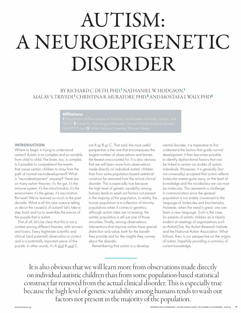
Autism: A neuroepigenetic
disorder
It is also obvious that we will learn more from observations made directly on individual autistic children than from some population-based statistical
construct far removed from the actual clinical disorder. This is especially true because the high level of genetic variability among humans tends to wash out
factors not present in the majority of the population.
mental disorder, it is imperative to first understand the factors that guide normal development. It then becomes possible to identify dysfunctional factors that can be linked to autism via studies of autistic individuals.Moreover,itisgenerally(butnot universally) accepted that autism reflects molecular events gone awry, so the level of knowledge and the vocabulary we use must be molecular. This represents a challenge in communication since the general population is not widely conversant in the language of molecules and biochemistry. However, when the need is great, one can learn a new language. Such is the case for parents of autistic children as is clearly evident at meetings of organizations such as AutismOne, the Autism Research Institute, andtheNationalAutismAssociation.Whatfollows, then, is our perspective on the origins of autism, hopefully providing a summary of current knowledge.
By RIChaRd C. dETh, Phd,1 NaThaNIEl W. hOdgSON,1 Malav S. TRIvEdI,1 ChRISTINa R. MuRaTORE, Phd,2 aNd MOSTafa I. Waly, Phd3
Affiliations: 1DepartmentofPharmaceuticalSciences,NortheasternUniversity,Boston,MA02115;2CenterforNeurologicalDiseases,HarvardMedicalSchool,Boston,MA02115;and3SultanQaboosUniversity,Muscat,Oman
AUTISMSCIENCEDIGEST:THEJOURNALOFAUTISMONEISSUE03REPRINTEDWITHPERMISSION www.autismone.org10
EpIgEnEtIc fActors control dEvElopmEntHuman development begins with the uniting of sperm and egg, triggering an ongoing series of rapid cell divisions. The new cells gradually diverge in their gene expression and their properties, giving rise to various stem cell lineages and ultimately to different tissues and organs. Throughout this truly amazingprocessofdevelopment,theDNAsequence of each cell is essentially identical, whether assessed in the fertilized egg or in the liver or brain of the fully developed adult. Differences between cell types reflect alternative patterns of gene expression, a process known as transcription, which give risetodifferentpatternsofmessengerRNA(mRNA),codingforadifferentpatternofproteins in each cell type, a process known as translation. For example, the casein protein is an important component of milk, and its gene is only available for transcription (mRNAformation)inmilk-producingcellsofthe female breast, even though every cell has the casein gene. In contrast, genes for proteins that support the most fundamental metabolic activities in all cells (such as the enzymes involved in glucose metabolism) are actively transcribed in all cells.
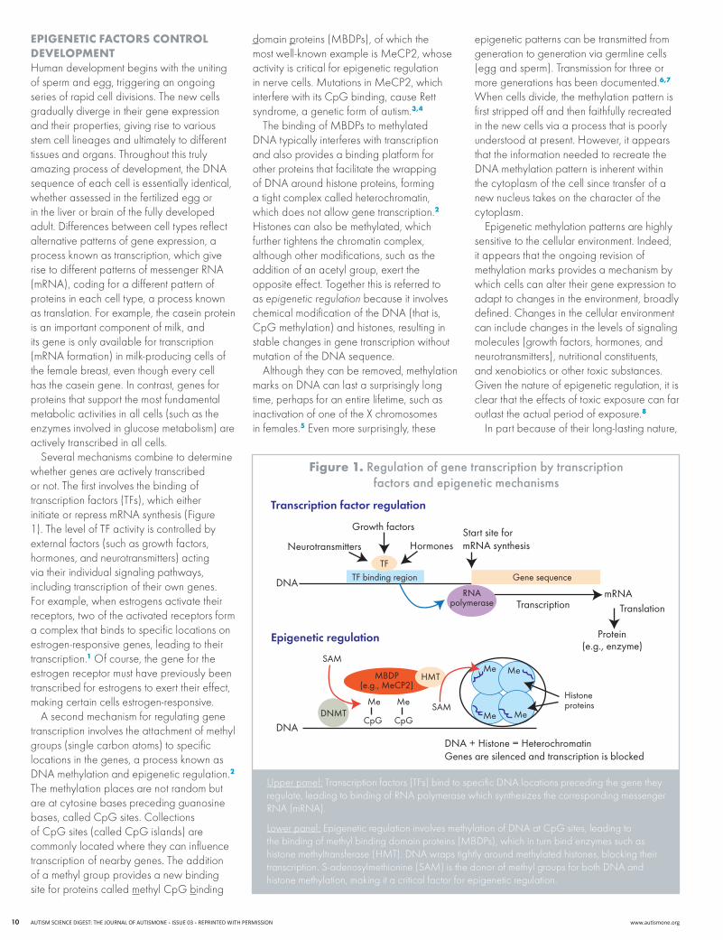
Several mechanisms combine to determine whether genes are actively transcribed or not. The first involves the binding of transcription factors (TFs), which either initiateorrepressmRNAsynthesis(Figure1). The level of TF activity is controlled by external factors (such as growth factors, hormones, and neurotransmitters) acting via their individual signaling pathways, including transcription of their own genes. For example, when estrogens activate their receptors, two of the activated receptors form a complex that binds to specific locations on estrogen-responsive genes, leading to their transcription.1 Of course, the gene for the estrogen receptor must have previously been transcribed for estrogens to exert their effect, making certain cells estrogen-responsive.
A second mechanism for regulating gene transcription involves the attachment of methyl groups (single carbon atoms) to specific locations in the genes, a process known as DNAmethylationandepigeneticregulation.2 The methylation places are not random but are at cytosine bases preceding guanosine bases, called CpG sites. Collections of CpG sites (called CpG islands) are commonly located where they can influence transcription of nearby genes. The addition of a methyl group provides a new binding site for proteins called methyl CpG binding
domain proteins(MBDPs),ofwhichthemostwell-knownexampleisMeCP2,whoseactivity is critical for epigenetic regulation innervecells.MutationsinMeCP2,whichinterfere with its CpG binding, cause Rett syndrome, a genetic form of autism.3,4
ThebindingofMBDPstomethylatedDNAtypicallyinterfereswithtranscriptionand also provides a binding platform for other proteins that facilitate the wrapping ofDNAaroundhistoneproteins,forminga tight complex called heterochromatin, which does not allow gene transcription.2 Histones can also be methylated, which further tightens the chromatin complex, although other modifications, such as the addition of an acetyl group, exert the opposite effect. Together this is referred to as epigenetic regulation because it involves chemicalmodificationoftheDNA(thatis,CpG methylation) and histones, resulting in stable changes in gene transcription without mutationoftheDNAsequence.
Although they can be removed, methylation marksonDNAcanlastasurprisinglylongtime, perhaps for an entire lifetime, such as inactivation of one of the X chromosomes in females.5 Even more surprisingly, these
epigenetic patterns can be transmitted from generation to generation via germline cells (egg and sperm). Transmission for three or more generations has been documented.6,7 When cells divide, the methylation pattern is first stripped off and then faithfully recreated in the new cells via a process that is poorly understood at present. However, it appears that the information needed to recreate the DNAmethylationpatternisinherentwithinthe cytoplasm of the cell since transfer of a new nucleus takes on the character of the cytoplasm.
Epigenetic methylation patterns are highly sensitive to the cellular environment. Indeed, it appears that the ongoing revision of methylation marks provides a mechanism by which cells can alter their gene expression to adapt to changes in the environment, broadly defined. Changes in the cellular environment can include changes in the levels of signaling molecules (growth factors, hormones, and neurotransmitters), nutritional constituents, and xenobiotics or other toxic substances. Given the nature of epigenetic regulation, it is clear that the effects of toxic exposure can far outlast the actual period of exposure.8
In part because of their long-lasting nature,
Transcription factor regulation
Epigenetic regulation
DNA + Histone = HeterochromatinGenes are silenced and transcription is blocked
mRNA
Protein(e.g., enzyme)
DNA
DNA
Transcription Translation
Start site formRNA synthesis
Growth factors
Neurotransmitters Hormones
TF binding region Gene sequence
RNApolymerase
Histoneproteins
HMT
SAM
SAMDNMT
MBDP(e.g., MeCP2)
Me Me
CpG CpG
Me Me
MeMe
TF
Upper panel:Transcriptionfactors(TFs)bindtospecificDNAlocationsprecedingthegenetheyregulate,leadingtobindingofRNApolymerasewhichsynthesizesthecorrespondingmessengerRNA(mRNA).
Lower panel:EpigeneticregulationinvolvesmethylationofDNAatCpGsites,leadingtothebindingofmethylbindingdomainproteins(MBDPs),whichinturnbindenzymessuchashistonemethyltransferase(HMT).DNAwrapstightlyaroundmethylatedhistones,blockingtheirtranscription.S-adenosylmethionine(SAM)isthedonorofmethylgroupsforbothDNAandhistone methylation, making it a critical factor for epigenetic regulation.
figure 1. regulation of gene transcription by transcription factors and epigenetic mechanisms
www.autismone.org REPRINTEDWITHPERMISSIONAUTISMSCIENCEDIGEST:THEJOURNALOFAUTISMONEISSUE03 11
epigenetic mechanisms are central to normal development.InherentwithinourDNAisaprogrammed sequence of changes in the factors that regulate gene expression during development. Disruption of methylation by xenobiotics and other toxic substances can therefore cause developmental disorders via their epigenetic influence.
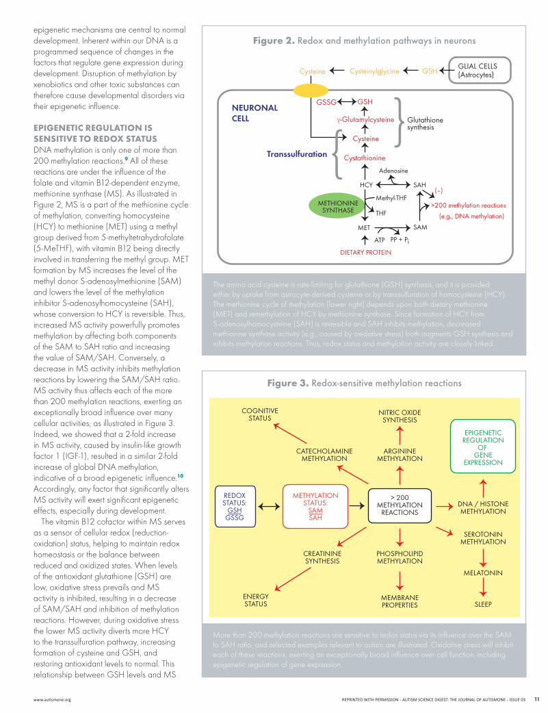
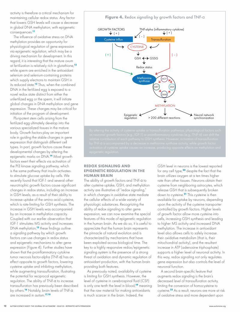
EpIgEnEtIc rEgulAtIon Is sEnsItIvE to rEdox stAtusDNAmethylationisonlyoneofmorethan200 methylation reactions.9 All of these reactions are under the influence of the folate and vitamin B12-dependent enzyme, methioninesynthase(MS).AsillustratedinFigure2,MSisapartofthemethioninecycleof methylation, converting homocysteine (HCY)tomethionine(MET)usingamethylgroup derived from 5-methyltetrahydrofolate (5-MeTHF),withvitaminB12beingdirectlyinvolvedintransferringthemethylgroup.METformationbyMSincreasesthelevelofthemethyldonorS-adenosylmethionine(SAM)and lowers the level of the methylation inhibitor S-adenosylhomocysteine (SAH), whoseconversiontoHCYisreversible.Thus,increasedMSactivitypowerfullypromotesmethylation by affecting both components oftheSAMtoSAHratioandincreasingthevalueofSAM/SAH.Conversely,adecreaseinMSactivityinhibitsmethylationreactionsbyloweringtheSAM/SAHratio.MSactivitythusaffectseachofthemorethan 200 methylation reactions, exerting an exceptionally broad influence over many cellular activities, as illustrated in Figure 3. Indeed, we showed that a 2-fold increase inMSactivity,causedbyinsulin-likegrowthfactor 1 (IGF-1), resulted in a similar 2-fold increaseofglobalDNAmethylation,indicative of a broad epigenetic influence.10 Accordingly, any factor that significantly alters MSactivitywillexertsignificantepigeneticeffects, especially during development.
ThevitaminB12cofactorwithinMSservesas a sensor of cellular redox (reduction-oxidation) status, helping to maintain redox homeostasis or the balance between reduced and oxidized states. When levels of the antioxidant glutathione (GSH) are low,oxidativestressprevailsandMSactivity is inhibited, resulting in a decrease ofSAM/SAHandinhibitionofmethylationreactions. However, during oxidative stress thelowerMSactivitydivertsmoreHCYto the transsulfuration pathway, increasing formation of cysteine and GSH, and restoring antioxidant levels to normal. This relationshipbetweenGSHlevelsandMS
figure 2. redox and methylation pathways in neurons
Transsulfuration
NEURONALCELL
GLIAL CELLS(Astrocytes)
Glutathionesynthesis
GSSG GSH
γ-Glutamylcysteine
Cysteine
Cystathionine
(–)
>200 methylation reactions
(e.g., DNA methylation)
DIETARY PROTEIN
Adenosine
METHIONINESYNTHASE
SAH
SAM
THF
MET
Methyl-THF
HCY
ATP PP + Pi
Cysteine Cysteinylglycine GSH
The amino acid cysteine is rate-limiting for glutathione (GSH) synthesis, and it is provided eitherbyuptakefromastrocyte-derivedcysteineorbytranssulfurationofhomocysteine(HCY).The methionine cycle of methylation (lower right) depends upon both dietary methionine (MET)andremethylationofHCYbymethioninesynthase.SinceformationofHCYfromS-adenosylhomocysteine (SAH) is reversible and SAH inhibits methylation, decreased methionine synthase activity (e.g., caused by oxidative stress) both augments GSH synthesis and inhibits methylation reactions. Thus, redox status and methylation activity are closely linked.
Morethan200methylationreactionsaresensitivetoredoxstatusviaitsinfluenceovertheSAMto SAH ratio, and selected examples relevant to autism are illustrated. Oxidative stress will inhibit each of these reactions, exerting an exceptionally broad influence over cell function, including epigenetic regulation of gene expression.
COGNITIVESTATUS
NITRIC OXIDESYNTHESIS
ARGININEMETHYLATION
CATECHOLAMINEMETHYLATION
CREATININESYNTHESIS
ENERGYSTATUS
MEMBRANEPROPERTIES
PHOSPHOLIPIDMETHYLATION
DNA / HISTONEMETHYLATION
SEROTONINMETHYLATION
MELATONIN
SLEEP
EPIGENETICREGULATION
OF GENE
EXPRESSION
REDOXSTATUS:
GSHGSSG
METHYLATIONSTATUS:
SAMSAH
> 200METHYLATION
REACTIONS
figure 3. redox-sensitive methylation reactions
AUTISMSCIENCEDIGEST:THEJOURNALOFAUTISMONEISSUE03REPRINTEDWITHPERMISSION www.autismone.org12
activity is therefore a critical mechanism for maintaining cellular redox status. Any factor that lowers GSH levels will cause a decrease inglobalDNAmethylation,withepigeneticconsequences.11
TheinfluenceofoxidativestressonDNAmethylation provides an opportunity for physiological regulation of gene expression via epigenetic regulation, which may be a driving mechanism for development. In this regard, it is interesting that the mature ovum at fertilization is relatively rich in glutathione,12 while sperm are enriched in the antioxidant selenium and selenium-containing proteins which supply electrons to maintain GSH in its reduced state.13 Thus, when the combined DNAinthefertilizedeggisexposedtoanovel redox state distinct from either the unfertilized egg or the sperm, it will initiate globalchangesinDNAmethylationandgeneexpression. These changes may be critical for initiation of the program of development.
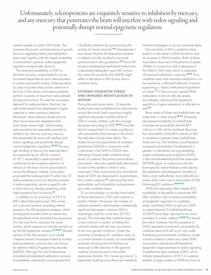
Pluripotent stem cells arising from the fertilized egg ultimately develop into the various specialized tissues in the mature body. Growth factors play an important role in guiding the stable changes in gene expression that distinguish different cell types. In part, growth factors cause these developmental changes by altering the epigeneticmarksonDNA.14Mostgrowthfactors exert their effects via activation of the PI3 kinase signaling pathway, which is the same pathway that insulin activates to stimulate glucose uptake by cells. We recently found that IGF-1 and several other neurotrophic growth factors cause significant changes in redox status, including an increase in GSH levels, as a result of their ability to increase uptake of the amino acid cysteine, which is rate-limiting for GSH synthesis. The increase in GSH levels was accompanied by an increase in methylation capacity. Coupled with our earlier observation that IGF-1stimulatesMSactivityandincreasesDNAmethylation,15 these findings outline a signaling pathway by which growth factors can use changes in redox status and epigenetic mechanisms to alter gene expression (Figure 4). Further studies have shown that the proinflammatory cytokine tumornecrosisfactor-alpha(TNF-α) has an effect opposite to growth factors, lowering cysteine uptake and inhibiting methylation, while augmenting transsulfuration, illustrating the potential for reciprocal epigenetic regulation.TheabilityofTNF-α to increase transsulfuration has previously been described by others.16Notably,brainlevelsofTNF-α are increased in autism.17,18
rEdox sIgnAlIng And EpIgEnEtIc rEgulAtIon In thE humAn brAInTheabilityofgrowthfactorsandTNF-α to alter cysteine uptake, GSH, and methylation activityareillustrativeof“redoxsignaling,”in which changes in oxidative state mediate the cellular effects of a wide variety of physiologic substances. Recognizing the ability of redox signaling to alter gene expression, we can now examine the special features of this mode of epigenetic regulation in the human brain. As we do so, it is useful to appreciate that the human brain represents the pinnacle of natural evolution and is characterized by mechanisms that have been exploited across biological time. The key to a highly responsive redox/epigenetic signaling system is the presence of a strong threat of oxidation and dynamic regulation of antioxidant production, with the human brain providing both features.
As previously noted, availability of cysteine is limiting for GSH synthesis. However, the level of cysteine in cerebrospinal fluid (CSF) is only one tenth the level in blood,19 meaning that the raw material for making antioxidants is much scarcer in the brain. Indeed, the
GSH level in neurons is the lowest reported for any cell type,20 despite the fact that the brain utilizes oxygen at a ten times higher ratethanothertissues.Neuronsobtaintheircysteine from neighboring astrocytes, which release GSH that is subsequently broken down to cysteine.21 This cysteine is then available for uptake by neurons, depending upon the activity of the cysteine transporter controlled by growth factors. Higher levels of growth factor allow more cysteine into cells, increasing GSH synthesis and leading tohigherMSactivityandincreasedDNAmethylation. The increase in antioxidant level also allows cells to safely increase their oxidative metabolism (that is, their mitochondrial activity), and the resultant increase in ATP (adenosine triphosphate) supports a higher level of neuronal activity. In this way, redox signaling not only regulates gene expression but also controls the level of neuronal function.
A second brain-specific feature that augments redox signaling is the brain’s decreased level of transsulfuration activity, limiting the conversion of homocysteine to cysteine.22 As a result, neurons are more at risk of oxidative stress and more dependent upon
REDOX SIGNALING
METHYLATION
> 200 different reactionsNeural networksynchronization
Epigeneticregulation
Redox status
GSH GSSG
HCY
SAH
SAM
MET
Methioninesynthase
( – )
( + )( + )( – )
( + )
CYSTEINE
TNF-alpha (inflammatory cytokine)GROWTH FACTORS
TranssulfurationCysteine influx
By altering the activity of cysteine uptake or transsulfuration pathways, physiological factors such asneuronalgrowthfactors(e.g.,IGF-1)orproinflammatorycytokines(e.g.,TNF-α) can shift the relative contribution of each pathway to GSH synthesis. However, activation of transsulfuration byTNF-α is accompanied by a decrease in methionine synthase activity, while growth factor activation of cysteine uptake causes an increase, producing opposite effects on methylation and epigenetic regulation.
figure 4. redox signaling by growth factors and tNF-α
www.autismone.org REPRINTEDWITHPERMISSIONAUTISMSCIENCEDIGEST:THEJOURNALOFAUTISMONEISSUE03 13
it facilitates attention by synchronizing the activity of neural networks.28 Subsequently it was shown that the D4 dopamine receptor is indeed critically involved in neuronal synchronization during attention.29 Since D4 receptor-mediated phospholipid methylation isabsolutelydependentuponMSactivity,this raises the possibility that ADHD might reflectadecreaseinMSactivityduetooxidative stress.
systEmIc oxIdAtIvE strEss And ImpAIrEd mEthylAtIon In AutIsmDuring the past seven years, 12 separate studies have been published from laboratories around the world, each reporting a highly significant decrease in plasma level of GSH in autistic children, with the average decrease amounting to 35%.30-42 Since the blood compartment is in ready equilibrium with extracellular fluid (except in the brain), this indicates a body-wide deficit. The studies found increased levels of oxidized glutathione(GSSG)inconjunctionwitha decrease in the GSH to GSSG ratio, indicative of body-wide oxidative stress. Levels of cysteine, the primary extracellular antioxidant, were also significantly decreased in each of the studies in which it was measured. Other work shows that intracellular levels of GSH are decreased in lymphoblasts fromautisticsubjects,43 indicating that both extracellular and intracellular compartments are under oxidative stress.
To our knowledge, no studies have failed to find a decrease in GSH and cysteine in autisticchildren.Moreover,thenumberofsubjectsneededtodemonstrateastatisticallysignificant decrease in plasma GSH is surprisingly small (no more than 20-30/group). This indicates that oxidative stress is very common in autism, a finding that contrasts starkly with the rare occurrence of de novo genetic mutations. Given the unanimity of evidence for oxidative stress as a common feature of autism, it is remarkable and even shocking that this finding has received so little attention in the general medical community and in the public awarenessdomain.This“activeignorance”isespecially troubling since there are metabolic
treatment strategies to correct oxidative stress.ThesensitivityofMStooxidativestress
leadstoadecreaseinSAMformationandan increase in SAH formation. Both of these have been observed in the plasma of autistic childreninconjunctionwithadecreaseintheSAMtoSAHratio,whichisindicativeof impaired methylation capacity.44,45 Thus, oxidative stress and impaired methylation can be viewed as confirmed hallmarks of autism, supportinga“redox/methylationhypothesisofautism.”11Inthisscenario,globalDNAmethylation in blood cells decreases accordingly, indicating that epigenetic regulation of gene expression is affected as well.42
Brain levels of folate and methylfolate are lower than in other tissues.46,47Moreover,decreased availability of methylfolate increases the probability that the B12 cofactorinMSwillbeoxidized.BecausethevulnerabilityoftheB12cofactorinMSto oxidation is greater when methylfolate levelsarelow,MSinhibition(andthereforeincreased vulnerability of methylation) is greater in individuals carrying disabling singlenucleotidepolymorphisms(SNPs)in the methylenetetrahydrofolate reductase (MTHFR)gene.Inconjunctionwiththebrain-specific redox features noted above, the metabolic and therapeutic benefits of folinic acid, methylfolate, and methyl-B12 in autismreflecttheirroleinreactivationofMSfollowing B12 oxidation.30,44,48
SNPsthatadverselyaffectvitaminB12availabilityortheabilitytoreactivateMSafter oxidation also increase vulnerability of epigenetic regulation to oxidative stress,andtheseSNPsaswellasaSNPin catecholamine-O-methyltransferase (COMT)havebeenreportedtobemoreprevalentinautisticsubjects.45,49–51 It should be noted, however, that the presence of SNPsrepresentsapotential vulnerability for oxidative stress that will occur only under threatening environmental conditions. Under normalconditions,theseverysameSNPsmay endow individuals with beneficial epigenetic responsiveness to redox signaling. Cystathionine beta-synthase (CBS), which initiatestranssulfurationofHCYtocysteine,exhibitsalargenumberofSNPsbuthasnot
cysteine uptake to sustain GSH levels. This increases the power and importance of growth factors in regulating redox and methylation status and, together with the limited availability of extracellular cysteine, makes epigenetic regulation exceptionally dynamic.
The restricted availability of GSH in the brain is partly compensated for by an increased dependence upon selenoproteins to sustain antioxidant activity. Selenoproteins (a class of proteins that contain selenium in the form of the amino acid selenocysteine) perform a variety of important redox-related biological functions. To meet the increased demand for selenoproteins, the brain (as well as the testes) has developed a higher capacity to retain precious selenium. Moreover,whenseleniumlevelsarelow,other tissues become depleted while brain levels remain high. Unfortunately, selenoproteins are exquisitely sensitive to inhibition by mercury, and any mercury that penetrates the brain will interfere with redox signaling and potentially disrupt normal epigenetic regulation.23,24 The very tight binding of mercury by selenoproteins (as measured by an affinity constant of 1045), especially to selenoprotein P, contributes to the long-term retention of mercury in the brain and its accumulation across the lifespan. Indeed, it has been proposed that selenoprotein P, which has 10 selenocysteines and is not directly involved in redox regulation, serves a specific role to bind mercury, thereby protecting other selenoproteins from its toxicity.25
InadditiontoitsconversionofHCYtoMET(describedpreviously),MScarriesout a second reaction, providing methyl groups to the D4 dopamine receptor, which subsequently transfers them to membrane phospholipids when stimulated by dopamine. Our lab was first to discover this novel activity, which appears to only be carried out by the D4 dopamine receptor.15,26,27 Genetic variants of the D4 receptor (such as the 7-repeat variant) have been linked to novelty-seeking behavior and are also risk factors for attention-deficit/hyperactivity disorder (ADHD). Although the role of dopamine-stimulated phospholipid methylation remains incompletely understood, we proposed that
unfortunately, selenoproteins are exquisitely sensitive to inhibition by mercury, and any mercury that penetrates the brain will interfere with redox signaling and
potentially disrupt normal epigenetic regulation.

AUTISMSCIENCEDIGEST:THEJOURNALOFAUTISMONEISSUE03REPRINTEDWITHPERMISSION www.autismone.org14
beenstudiedasextensivelyasMTHFR.Onestudy found that the risk of having a child with autismwashigherifthemothercarriedSNPsineitherMTHFRorCBSanddidnottakeprenatal vitamins.52
ExplorIng thE rEdox/mEthylAtIon hypothEsIsIn collaboration with colleagues at the Sultan QaboosUniversitySchoolofMedicineinOman, we recently evaluated the frequency of autism in Oman.53 Our study, which involved 30 autistic and 30 control children (with 15 males and 15 females in each group), also assessed the children’s nutritional status, serum levels of redox and methylation-related sulfur metabolites, and vitamin B12 and folic acid levels. In contrast to autistic children in the US, malnutrition was common in Omani children and associated with highly significant deficits in B12 and folic acid levels (Figure 5). Similar to the US, we observed a significantly lower level of GSH in autistic children (48% of controls for combined groups), with the extent of the decrease being greater in male versus femalesubjects,comparedwithsame-sexcontrols. We speculate that autism in Omani children may result mainly from an antioxidant deficit associated with a nutritional deficiency inthecofactorsforMS,whereasautisminthe US appears to result from environmental
toxin exposure in a genetically vulnerable population. The occurrence of lower GSH levels arising from different etiological factors is very strong evidence for the fundamental importance of impaired antioxidant capacity in autism.
The fact that mitochondrial dysfunction is common in autism can be directly related to the presence of oxidative stress and to an increase of GSSG. When the level of GSSG increases, it can react with cysteine residues in proteins, leaving half of the GSSG attached to the sulfhydryl (SH) group on the protein, a process called glutathionylation.54 The other half of GSSG is released as GSH, providing more antioxidant. A classic example of how glutathionylation works involves complex I in the mitochondrial electron transport chain. When GSSG builds up during oxidative stress, complex I is increasingly glutathionylated, causing a decrease in the flow of electrons toward oxygen reduction and ATP generation, leading to mitochondrial dysfunction.55 However, this is actually a useful survival mechanismforcellsunderoxidativestress;shutting down mitochondrial function lowers the amount of reactive oxygen species (ROS) production, thereby decreasing the strain on already low levels of antioxidant. Thus, mitochondrial dysfunction can occur
secondary to oxidative stress and the accumulation of oxidized glutathione, which is a metabolic feature of autism.
Recently, thanks to research grants from theAutismResearchInstitute,theNationalAutismAssociation,andSafeMinds,wehad the opportunity to evaluate the level of MSmRNAinpostmortemhumancortexinautisticsubjectsandage-andsex-matchedcontrols. As illustrated in Figure 6, we found a remarkable pattern of progressive decreaseinmRNAlevelsacrossthelifespaninthecontrolsubjects,amountingtomorethan a 400-fold decrease. The decrease occurredintwophases.HighinitialmRNAlevels rapidly decreased up until the end of the teens and decreased more gradually thereafter. This biphasic pattern appears to correspond to the period of rapid linear growth lasting through puberty, followed by the relatively static state of adulthood. DespitethedramaticdecreaseinmRNAlevels,levelsofMSproteinremainedrelatively static, suggesting the possibility that the rate of new protein synthesis from mRNAdecreaseswithageandthattheMSproteins are lasting longer as we get older. This is in keeping with the general concept of gradually decreasing metabolic activity with advancing age, but at this time it is unclearwhetherMShasaspecialroleincoordinating the decrease.
In comparison, we found an abnormal age-dependent pattern in autism. The high initialphaseofmRNAwasessentiallyabsent,withmRNAlevelsin4-to10-year-oldautistic
Autism in Oman appears to be largely a consequence of malnutrition and nutritional factors, including significantly lower levels of vitamin B12 and folic acid, unlike autism in the US. Thus, different causes converge on methionine synthase, indicating its central role in autism.
figure 5. Serum levels of folic acid and vitamin B12 in autistic subjects from Oman
Anage-dependentdecreaseinMSmRNAofmorethan400-foldoccurs across the lifespan. An initial stage of rapid decrease lasts until the end of adolescence, followed by a slower decline thereafter.
figure 6. Levels of MS mrNa in postmortem human cortex decrease with age
Serum Folate Levels
Serum B12 Levels
age (years)
Male Control
Male Autistic
female Control
female Autistic
Male Control
Male Autistic
female Control
female Autistic
Seru
m F
olat
eng
/ m
lSe
rum
B12
pg /
ml
** p < 0.001
** p < 0.0001
** **
****
6
5
4
3
2
1
0
600
500
400
300
200
100
0
300
200
100
00 10 20 30 40 50 60 70 80 90 100
MS
cob
mrN
a (
arbi
trar
y un
its) t ½ fast = 3.4 years
t ½ slow = 29.4 years
r2 = .91
www.autismone.org REPRINTEDWITHPERMISSIONAUTISMSCIENCEDIGEST:THEJOURNALOFAUTISMONEISSUE03 15
subjectsbeingsimilartolevelsin30-year-oldcontrolsubjects(Figure7).Onaverage,themRNAlevelsintheautisticsampleswere reduced by about 50% at each ageversuspairedcontrolsubjects.Thesedata provide clear evidence that in autism MSmRNAstatusisalteredinthebrain.Although additional study is required to fully understand the functional significance of the decrease, at this point it seems reasonable to conclude that the normal age-dependent decrease in metabolic activity of the developing cortex is accelerated in autism. Progressive changes in redox status are likely to underlie the normal age-dependent decrease, although this remains to be proven, and the oxidative stress that is characteristic of autism may accelerate this progression, with untoward epigenetic consequences. Essentially, this perspective postulates a redox-driven“epigeneticclock”mechanismof development in which both intrinsic genetic factors (in the cases of Rett and Angelman syndromes) and extrinsic factors (such as redox-active xenobiotic substances) can disrupt the mechanism and contribute to neurodevelopmental disorders such as autism.
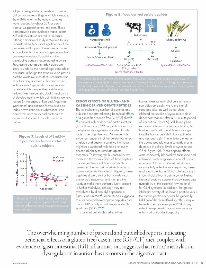
rEdox EffEcts of glutEn- And cAsEIn-dErIvEd opIAtE pEptIdEsThe overwhelming number of parental and published reports indicating beneficial effects of a gluten-free/casein-free (GF/CF) diet,56–
59 coupled with evidence of gastrointestinal (GI) inflammation,59–61 suggests that redox/methylation dysregulation in autism has its rootsinthedigestivetract.Moreover,thisevidence suggests that the deleterious effects of gluten and casein in sensitive individuals might be associated with their previously described ability to stimulate opiate receptors. To investigate this possibility, we examined the redox effects of three peptides that are relatively stable end products of gluten and beta-casein of either human or bovine origin. As illustrated in Figure 8, these peptides share a similar but non-identical amino acid sequence, and their proline residues make them comparatively resistant to further hydrolysis, although they are hydrolyzed by dipeptidyl peptidase-4 (DPP-IVorCD26).62 Recent studies suggest a role for casein-derived opiate peptides and lowDPP-IVactivityinsuddeninfantdeathsyndrome (SIDS).63,64
In cultured cell studies using either
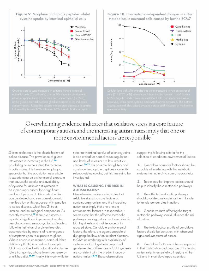
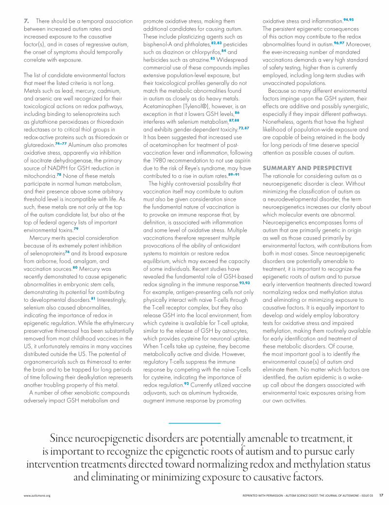
human intestinal epithelial cells or human neuroblastoma cells, we found that all three peptides, as well as morphine, inhibited the uptake of cysteine in a dose-dependent manner after a 30-minute period of incubation (Figure 9). While morphine was clearly the most powerful inhibitor, the bovine (cow’s milk) peptide was stronger than the human peptide in both epithelial and neuronal cells. The inhibitory effect of the bovine peptide was also evident as a decrease in cellular levels of cysteine and GSH (Figure 10). These peptide effects were completely blocked by naltrexone and naloxone, confirming involvement of opiate receptors. Although cultured cell studies may not fully reflect in vivo responses, these results indicate that a GF/CF diet may exert its beneficial effect in autism by facilitating intestinal cysteine uptake, thereby increasing availability of this essential raw material for GSH synthesis. In addition, the greater inhibitory activity of the bovine peptide versus the human peptide supports the generally held belief that breastfeeding offers unique benefits to early development65 that may reflect the epigenetic consequences of an enhanced antioxidant capacity.
MessengerRNA(mRNA)levelsarelowerinautisticsubjectsversusage-matchedcontrols,with the difference being most pronounced at younger ages.
figure 7. Levels of MS mrNa in postmortem human cortex of
autistic subjects
FOOD-DERIVED OPIATE PEPTIDES
Human breast milk Wheat, barley, rye
Beta-Casein
-L - V - Y - P - F - P - G - P - I - X
Gliadins
α-gliadin-7
Tyr-Pro-Gin-Pro-Gin-Pro-PheTyr-Pro-Phe-Val-Glu-Pro-Ile
Tyr-Pro-Phe-Pro-Gly-Pro-Ile
β-Casein (40% of milk protein)
Human β-Casomorphin-7
Bovine β-Casomorphin-7(0.4 g/L milk)
A1(His at 67)
A2(Pro at 67)
59 66-67
The proteins beta-casein from milk and gliadin from wheat, barley, and rye are broken down in the intestine to peptides that have opiate activity. The seven amino acid-containing peptides from human milk, cow’s milk (bovine), and wheat have similar but not identical sequences, including two or three proline residues (Pro) that increase their stability.
figure 8. Food-derived opiate peptides
The overwhelming number of parental and published reports indicating beneficial effects of a gluten-free/casein-free (gf/Cf) diet, coupled with
evidence of gastrointestinal (gI) inflammation, suggests that redox/methylation dysregulation in autism has its roots in the digestive tract.
MS
cob
mrN
a (
arbi
trar
y un
its)
400
300
200
100
0
age (years)0 10 20 30 40
ControlAutism
AUTISMSCIENCEDIGEST:THEJOURNALOFAUTISMONEISSUE03REPRINTEDWITHPERMISSION www.autismone.org16
Gluten intolerance is the classic feature of celiac disease. The prevalence of gluten intolerance is increasing in the US,66 paralleling, to some extent, the increase in autism rates. It is therefore tempting to speculate that the population as a whole is experiencing an environmental exposure that causes the uptake and availability of cysteine for antioxidant synthesis to be increasingly critical for a significant number of persons. In this context, autism can be viewed as a neurodevelopmental manifestation of this exposure, with parallels to celiac disease, which has GI tract, immune, and neurological components. As recently reviewed,67 there are numerous reports of significant improvement in other neurological and neuropsychiatric disorders following institution of a gluten-free diet, accompanied by reports of re-emergence of symptoms upon re-exposure to gluten. Where casein is concerned, cerebral folate deficiency (CFD) is a pertinent example. CFD is associated with autoantibodies to the folate transporter, whose levels decrease on a milk-free diet.68,69 Finally, it is worthwhile to
note that intestinal uptake of selenocysteine is also critical for normal redox regulation, and levels of selenium are low in autistic children.70,71 It is possible that gluten- and casein-derived opiate peptides may inhibit selenocysteine uptake, but this has yet to be investigated.
WhAt Is cAusIng thE rIsE In AutIsm rAtEs?Overwhelming evidence indicates that oxidative stress is a core feature of contemporary autism, and the increasing autism rates imply that one or more environmental factors are responsible. It seems clear that the affected metabolic pathways causing autism are those affecting GSH synthesis and maintenance of its reduced state. Candidate environmental factors, therefore, are agents capable of disrupting the flow of antioxidant electrons to GSH or interfering with availability of cysteine for GSH synthesis. Reports of gender-related differences in GSH synthesis are consistent with the predominance of autistic males.72,73 These observations
suggest the following criteria for the selection of candidate environmental factors:
1. Candidate causative factors should be capable of interfering with the metabolic systems that maintain a normal redox status.
2. Treatments that improve autism should help to identify these metabolic pathways.
3. The affected metabolic pathways should provide a rationale for the 4:1 male to female gender bias in autism.
4. Genetic variants affecting the target metabolic pathway should influence the risk of autism.
5. The toxicological profile of candidate factors should be consistent with observed signs and symptoms of autism.
6. Candidate factors must be widespread in their distribution and capable of increasing autism rates in essentially all regions of the US and in most developed countries.
Cysteine uptake was measured in cultured human intestinal epithelial cells (Caco2 cells) after a 30-minute incubation with either morphine, human or bovine β-casomorphin-7(BCM7),or the gliadin-derived peptide gliadinomorphin at the indicated concentrations.Morphinecausedthegreatestdecreaseinuptake,followedbybovineBCM7,humanBCM7,andgliadinomorphin.
figure 9. Morphine and opiate peptides inhibit cysteine uptake by intestinal epithelial cells
Cellular levels of sulfur metabolites were measured in human neuronal cells(SH-SY5Ycells)followinga4-hourincubationwith1µMbovineBCM7.Significantdecreasesincysteine,GSH,andmethioninewereobserved, while homocysteine and cystathionine increased. This pattern is consistent with decreased cysteine uptake and inhibition of methionine synthase activity.
figure 10. concentration-dependent changes in sulfur metabolites in neuronal cells caused by bovine BcM7
Overwhelming evidence indicates that oxidative stress is a core feature of contemporary autism, and the increasing autism rates imply that one or
more environmental factors are responsible.
Morphine
Bovine BCM7
human BCM7
gliadinomorphin
cys
tein
e U
ptak
e(n
m o
l/m
g of
pro
tein
)
6
4
2
0
concentrations (M)0 -10 -8 -6 -4
Cysteine
Methionine
homocysteine
gSh
Cystathionine
nm o
l/m
g of
pro
tein
concentration (M)0 -10 -9 -8 -7 -6
100
75
50
25
0
www.autismone.org REPRINTEDWITHPERMISSIONAUTISMSCIENCEDIGEST:THEJOURNALOFAUTISMONEISSUE03 17
7. There should be a temporal association between increased autism rates and increased exposure to the causative factor(s), and in cases of regressive autism, the onset of symptoms should temporally correlate with exposure.
The list of candidate environmental factors that meet the listed criteria is not long. Metalssuchaslead,mercury,cadmium,and arsenic are well recognized for their toxicological actions on redox pathways, including binding to selenoproteins such as glutathione peroxidases or thioredoxin reductases or to critical thiol groups in redox-active proteins such as thioredoxin or glutaredoxin.74–77 Aluminum also promotes oxidative stress, apparently via inhibition of isocitrate dehydrogenase, the primary sourceofNADPHforGSHreductioninmitochondria.78Noneofthesemetalsparticipate in normal human metabolism, and their presence above some arbitrary threshold level is incompatible with life. As such, these metals are not only at the top of the autism candidate list, but also at the top of federal agency lists of important environmental toxins.79
Mercurymeritsspecialconsiderationbecause of its extremely potent inhibition of selenoproteins74 and its broad exposure from airborne, food, amalgam, and vaccination sources.80Mercurywasrecently demonstrated to cause epigenetic abnormalities in embryonic stem cells, demonstrating its potential for contributing to developmental disorders.81 Interestingly, selenium also caused abnormalities, indicating the importance of redox in epigenetic regulation. While the ethylmercury preservative thimerosal has been substantially removed from most childhood vaccines in the US, it unfortunately remains in many vaccines distributed outside the US. The potential of organomercurials such as thimerosal to enter the brain and to be trapped for long periods of time following their dealkylation represents another troubling property of this metal.
A number of other xenobiotic compounds adversely impact GSH metabolism and
promote oxidative stress, making them additional candidates for causing autism. These include plasticizing agents such as bisphenol-A and phthalates,82,83 pesticides such as diazinon or chlorpyrifos,84 and herbicides such as atrazine.85 Widespread commercial use of these compounds implies extensive population-level exposure, but their toxicological profiles generally do not match the metabolic abnormalities found in autism as closely as do heavy metals. Acetaminophen (Tylenol®), however, is an exception in that it lowers GSH levels,86 interferes with selenium metabolism,87,88 and exhibits gender-dependent toxicity.72,87 It has been suggested that increased use of acetaminophen for treatment of post-vaccination fever and inflammation, following the 1980 recommendation to not use aspirin due to the risk of Reye’s syndrome, may have contributed to a rise in autism rates.89–91
The highly controversial possibility that vaccination itself may contribute to autism must also be given consideration since the fundamental nature of vaccination is to provoke an immune response that, by definition, is associated with inflammation andsomelevelofoxidativestress.Multiplevaccinations therefore represent multiple provocations of the ability of antioxidant systems to maintain or restore redox equilibrium, which may exceed the capacity of some individuals. Recent studies have revealed the fundamental role of GSH-based redox signaling in the immune response.92,93
For example, antigen-presenting cells not only physically interact with naive T-cells through the T-cell receptor complex, but they also release GSH into the local environment, from which cysteine is available for T-cell uptake, similar to the release of GSH by astrocytes, which provides cysteine for neuronal uptake. When T-cells take up cysteine, they become metabolically active and divide. However, regulatory T-cells suppress the immune response by competing with the naive T-cells for cysteine, indicating the importance of redox regulation.92 Currently utilized vaccine adjuvants,suchasaluminumhydroxide,augment immune response by promoting
oxidative stress and inflammation.94,95 The persistent epigenetic consequences of this action may contribute to the redox abnormalities found in autism.96,97Moreover,the ever-increasing number of mandated vaccinations demands a very high standard of safety testing, higher than is currently employed, including long-term studies with unvaccinated populations.
Because so many different environmental factors impinge upon the GSH system, their effects are additive and possibly synergistic, especially if they impair different pathways. Nonetheless,agentsthathavethehighestlikelihood of population-wide exposure and are capable of being retained in the body for long periods of time deserve special attention as possible causes of autism.
summAry And pErspEctIvEThe rationale for considering autism as a neuroepigenetic disorder is clear. Without minimizing the classification of autism as a neurodevelopmental disorder, the term neuroepigenetics increases our clarity about which molecular events are abnormal. Neuroepigeneticsencompassesformsofautism that are primarily genetic in origin as well as those caused primarily by environmental factors, with contributions from both in most cases. Since neuroepigenetic disorders are potentially amenable to treatment, it is important to recognize the epigenetic roots of autism and to pursue early intervention treatments directed toward normalizing redox and methylation status and eliminating or minimizing exposure to causative factors. It is equally important to develop and widely employ laboratory tests for oxidative stress and impaired methylation, making them routinely available for early identification and treatment of these metabolic disorders. Of course, the most important goal is to identify the environmental cause(s) of autism and eliminatethem.Nomatterwhichfactorsareidentified, the autism epidemic is a wake-up call about the dangers associated with environmental toxic exposures arising from our own activities.
Since neuroepigenetic disorders are potentially amenable to treatment, it is important to recognize the epigenetic roots of autism and to pursue early
intervention treatments directed toward normalizing redox and methylation status and eliminating or minimizing exposure to causative factors.
AUTISMSCIENCEDIGEST:THEJOURNALOFAUTISMONEISSUE03REPRINTEDWITHPERMISSION www.autismone.org18
1. Dietz SC, Carroll JS. Interrogating the genome to understand oestrogen-receptor-mediated transcription. Expert Rev Mol Med.2008;10:e10.
2. Feinberg AP. Epigenetics at the epicenter of modern medicine. JAMA.2008;299(11):1345-1350.
3.LasalleJM,YasuiDH.EvolvingroleofMeCP2inRettsyndrome and autism. Epigenomics.2009;1(1):119-130.
4. GonzalesML,LaSalleJM.TheroleofMeCP2inbraindevelopment and neurodevelopmental disorders. Curr Psychiatry Rep. 2010;12(2):127-134.
5. Wutz A. Gene silencing in X-chromosome inactivation: advances in understanding facultative heterochromatin formation. Nat Rev Genet. 2011;12(8):542-553.
6.AnwayMD,CuppAS,UzumcuM,SkinnerMK.Epigenetictransgenerational actions of endocrine disruptors and male fertility. Science.2005;308(5727):1466-1469.
7.SkinnerMK,Guerrero-BosagnaC.Environmentalsignals and transgenerational epigenetics. Epigenomics. 2009;1(1):111-117.
8. Perera F, Herbstman J. Prenatal environmental exposures, epigenetics, and disease. Reprod Toxicol.2011;31(3):363-373.
9. Petrossian TC, Clarke SG. Uncovering the human methyltransferasome. Mol Cell Proteomics. 2011;10(1):M110.000976.
10. WalyM,OlteanuH,BanerjeeR,ChoiSW,MasonJB, Parker BS, et al. Activation of methionine synthase by insulin-like growth factor-1 and dopamine: a target for neurodevelopmental toxins and thimerosal. Mol Psychiatry. 2004;9(4):358-370.
11. DethR,MuratoreC,BenzecryJ,Power-CharnitskyV-A,WalyM.Howenvironmentalandgeneticfactorscombine to cause autism: a redox/methylation hypothesis. Neurotoxicology.2008;29(1):190-201.
12. Luberda Z. The role of glutathione in mammalian gametes. Reprod Biol.2005;5(1):5-17.
13. Boitani C, Puglisi R. Selenium, a key element in spermatogenesis and male fertility. Adv Exp Med Biol. 2008;636:65-73.
14.RothTL,SweattJD.EpigeneticmarkingoftheBDNFgene by early-life adverse experiences. Horm Behav. 2011;59(3):315-320.
15. WalyM,OlteanuH,BanerjeeR,ChoiSW,MasonJB, Parker BS, et al. Activation of methionine synthase by insulin-like growth factor-1 and dopamine: a target for neurodevelopmental toxins and thimerosal. Mol Psychiatry. 2004;9(4):358-370.
16. ZouC-G,BanerjeeR.Tumornecrosisfactor-alpha-induced targeted proteolysis of cystathionine beta-synthase modulates redox homeostasis. J Biol Chem. 2003;278(19):16802-16808.
17. ChezMG,DowlingT,PatelPB,KhannaP,KominskyM.Elevation of tumor necrosis factor-alpha in cerebrospinal fluid of autistic children. Pediatr Neurol. 2007;36(6):361-365.
18. LiX,ChauhanA,SheikhAM,PatilS,ChauhanV,LiXM,etal. Elevated immune response in the brain of autistic patients. J Neuroimmunol.2009;207(1-2):111-116.
19. Castagna A, Le Grazie C, Accordini A, Guilidori P, Cavalli G, Bottiglieri T, et al. Cerebrospinal fluid S-adenosylmethionine(SAMe)andglutathioneconcentrationsinHIVinfection:effectofparenteraltreatmentwithSAMe.Neurology.1995;45(9):1678-1683.
20. SunX,ShihAY,JohannssenHC,ErbH,LiP,MurphyTH. Two-photon imaging of glutathione levels in intact brain indicates enhanced redox buffering in developing neurons and cells at the cerebrospinal fluid and blood-brain interface. J Biol Chem.2006;281(25):17420-17431.
21. Dringen R, Hirrlinger J. Glutathione pathways in the brain. Biol Chem.2003;384(4):505-516.
22. TallanHH,MooreS,SteinWH.L-cystathionineinhumanbrain. J Biol Chem.1958;230(2):707-716.
23. RalstonNVC,RalstonCR,BlackwellJL3rd,RaymondLJ. Dietary and tissue selenium in relation to methylmercury toxicity. Neurotoxicology.2008;29(5):802-811.
24. RalstonNVC,RaymondLJ.Dietaryselenium’sprotective effects against methylmercury toxicity. Toxicology. 2010. Available at: http://www.ncbi.nlm.nih.gov/pubmed/20561558. Accessed September 29, 2010.
25. SuzukiKT,SasakuraC,YonedaS.Bindingsitesforthe(Hg-Se) complex on selenoprotein P. Biochim Biophys Acta. 1998;1429(1):102-112.
26. SharmaA,KramerML,WickPF,LiuD,ChariS,ShimS, et al. D4 dopamine receptor-mediated phospholipid methylation and its implications for mental illnesses such as schizophrenia. Mol Psychiatry. 1999;4(3):235-246.
27. ZhaoR,ChenY,TanW,WalyM,SharmaA,StoverP, et al. Relationship between dopamine-stimulated phospholipid methylation and the single-carbon folate pathway. J Neurochem.2001;78(4):788-796.
28. KuznetsovaAY,DethRC.Amodelformodulationof neuronal synchronization by D4 dopamine receptor-mediated phospholipid methylation. J Comput Neurosci. 2008;24(3):314-329.
29. DemiralpT,HerrmannCS,ErdalME,ErgenogluT,KeskinYH,ErgenM,etal.DRD4andDAT1polymorphismsmodulate human gamma band responses. Cereb Cortex. 2007;17(5):1007-1019.
30. JamesSJ,MelnykS,FuchsG,ReidT,JerniganS,PavlivO, et al. Efficacy of methylcobalamin and folinic acid treatment on glutathione redox status in children with autism. Am J Clin Nutr.2009;89(1):425-430.
31. AdamsJB,AudhyaT,McDonough-MeansS,RubinRA,QuigD,GeisE,etal.Nutritionalandmetabolicstatusof children with autism vs. neurotypical children, and the association with autism severity. Nutr Metab (Lond). 2011;8(1):34.
32. AdamsJB,BaralM,GeisE,MitchellJ,IngramJ,Hensley A, et al. The severity of autism is associated with toxic metal body burden and red blood cell glutathione levels. J Toxicol. 2009;2009:532640.
33. PasturalE,RitchieS,LuY,JinW,KavianpourA,KhineSu-MyatK,etal.Novelplasmaphospholipidbiomarkersof autism: mitochondrial dysfunction as a putative causative mechanism. Prostaglandins Leukot Essent Fatty Acids. 2009;81(4):253-264.
34. Al-GadaniY,El-AnsaryA,AttasO,Al-AyadhiL.Metabolicbiomarkersrelatedtooxidativestressandantioxidant status in Saudi autistic children. Clin Biochem. 2009;42(10-11):1032-1040.
35. Paşca SP, Dronca E, Kaucsár T, Craciun EC, Endreffy E, Ferencz BK, et al. One carbon metabolism disturbances andtheC677TMTHFRgenepolymorphisminchildrenwith autism spectrum disorders. J Cell Mol Med. 2009;13(10):4229-4238.
36. Geier DA, Kern JK, Garver CR, Adams JB, Audhya T,NatafR,etal.Biomarkersofenvironmentaltoxicityandsusceptibility in autism. J Neurol Sci.2009;280(1-2):101-108.
37. Geier DA, Kern JK, Garver CR, Adams JB, Audhya T,GeierMR.Aprospectivestudyoftranssulfurationbiomarkers in autistic disorders. Neurochem Res. 2009;34(2):386-393.
38. JamesSJ,JillJamesS,MelnykS,HubanksA,Rose S, Gaylor DW. Abnormal transmethylation/transsulfurationmetabolismandDNAhypomethylationamong parents of children with autism. J Autism Dev Disord. 2008;38(10):1966-1975.
39. GeierDA,GeierMR.Acaseseriesofchildrenwithapparent mercury toxic encephalopathies manifesting with clinical symptoms of regressive autistic disorders. J Toxicol Environ Health Part A.2007;70(10):837-851.
40. JamesSJ,MelnykS,JerniganS,ClevesMA,HalstedCH,WongDH,etal.Metabolicendophenotypeandrelated genotypes are associated with oxidative stress in children with autism. Am J Med Genet B Neuropsychiatr Genet. 2006;141B(8):947-956.
41. JamesSJ,CutlerP,MelnykS,JerniganS,JanakL,GaylorDW,etal.Metabolicbiomarkersofincreasedoxidative stress and impaired methylation capacity in children with autism. Am J Clin Nutr.2004;80(6):1611-1617.
42. MelnykS,FuchsGJ,SchulzE,LopezM,KahlerSG,FussellJJ,etal.Metabolicimbalanceassociatedwithmethylation dysregulation and oxidative damage in children with autism. J Autism Dev Disord. 2011. Available at: http://www.ncbi.nlm.nih.gov/pubmed/21519954. Accessed July 27, 2011.
43. JamesSJ,RoseS,MelnykS,JerniganS,BlossomS,Pavliv O, et al. Cellular and mitochondrial glutathione redox imbalance in lymphoblastoid cells derived from children with autism. FASEB J.2009;23(8):2374-2383.
44. JamesSJ,CutlerP,MelnykS,JerniganS,JanakL,GaylorDW,etal.Metabolicbiomarkersofincreasedoxidative stress and impaired methylation capacity in children with autism. Am J Clin Nutr.2004;80(6):1611-1617.
45. JamesSJ,MelnykS,JerniganS,ClevesMA,HalstedCH,WongDH,etal.Metabolicendophenotypeandrelatedgenotypes are associated with oxidative stress in children with autism. Am J Med Genet B Neuropsychiatr Genet. 2006;141B(8):947-956.
46. Elmore CL, Wu X, Leclerc D, Watson ED, Bottiglieri T, KrupenkoNI,etal.Metabolicderangementofmethionineand folate metabolism in mice deficient in methionine synthase reductase. Mol Genet Metab.2007;91(1):85-97.
47. LamarreSG,MolloyAM,ReinkeSN,SykesBD,BrosnanME,BrosnanJT.Formatecandifferentiatebetweenhyperhomocysteinemia due to impaired remethylation and impaired transsulfuration. Am J Physiol Endocrinol Metab. 2011. Available at: http://www.ncbi.nlm.nih.gov/pubmed/21934042. Accessed September 29, 2011.
48. MorettiP,SahooT,HylandK,BottiglieriT,PetersS, del Gaudio D, et al. Cerebral folate deficiency with developmental delay, autism, and response to folinic acid. Neurology.2005;64(6):1088-1090.
49. LiuX,SolehdinF,CohenIL,GonzalezMG,JenkinsEC,LewisME,etal.Population-andfamily-basedstudiesassociatetheMTHFRgenewithidiopathicautisminsimplexfamilies. J Autism Dev Disord.2011;41(7):938-944.
50. Boris,M,Goldblatt,A,Galanko,J,James,SJ.AssociationofMTHFRgenevariantswithautism.Journal of American Physicians and Surgeons.2004;9:106-8.
51. Paşca SP, Dronca E, Kaucsár T, Craciun EC, Endreffy E, Ferencz BK, et al. One carbon metabolism disturbances and theC677TMTHFRgenepolymorphisminchildrenwithautismspectrum disorders. J Cell Mol Med. 2009;13(10):4229-4238.
52. Schmidt RJ, Hansen RL, Hartiala J, Allayee H, Schmidt LC, Tancredi DJ, et al. Prenatal vitamins, one-carbon metabolism gene variants, and risk for autism. Epidemiology. 2011;22(4):476-485.
53. Al-FarsiYM,Al-SharbatiMM,Al-FarsiOA,Al-ShafaeeMS,BrooksDR,WalyMI.Briefreport:prevalenceofautisticspectrum disorders in the Sultanate of Oman. J Autism Dev Disord.2011;41(6):821-825.
54. Dalle-DonneI,RossiR,ColomboG,GiustariniD,MilzaniA. Protein S-glutathionylation: a regulatory device from bacteria to humans. Trends Biochem Sci.2009;34(2):85-96.
55. BeerSM,TaylorER,BrownSE,DahmCC,CostaNJ,RunswickMJ,etal.Glutaredoxin2catalyzesthereversible oxidation and glutathionylation of mitochondrial membrane thiol proteins: implications for mitochondrial redox regulation and antioxidant defense. J Biol Chem. 2004;279(46):47939-47951.
56. WhiteleyP,HaracoposD,KnivsbergA-M,ReicheltKL, Parlar S, Jacobsen J, et al. The ScanBrit randomised, controlled, single-blind study of a gluten- and casein-free dietary intervention for children with autism spectrum disorders. Nutr Neurosci. 2010;13(2):87-100.
57. HsuC-L,LinC-Y,ChenC-L,WangC-M,WongM-K.Theeffects of a gluten and casein-free diet in children with autism: a case report. Chang Gung Med J.2009;32(4):459-465.
58. Genuis SJ, Bouchard TP. Celiac disease presenting as autism. J Child Neurol. 2010;25(1):114-119.
59. Ashwood P, Anthony A, Pellicer AA, Torrente F, Walker-Smith JA, Wakefield AJ. Intestinal lymphocyte populations in children with regressive autism: evidence for extensive mucosal immunopathology. J Clin Immunol. 2003;23(6):504-517.
60. Buie T, Campbell DB, Fuchs GJ 3rd, Furuta GT, Levy J, VandewaterJ,etal.Evaluation,diagnosis,andtreatmentofgastrointestinal disorders in individuals with ASDs: a consensus report. Pediatrics.2010;125Suppl1:S1-18.
61. WakefieldAJ,MurchSH,AnthonyA,LinnellJ,CassonDM,MalikM,etal.Ileal-lymphoid-nodularhyperplasia,non-specific colitis, and pervasive developmental disorder in children. Lancet.1998;351(9103):637-641.
62. TiruppathiC,MiyamotoY,GanapathyV,LeibachFH.GeneticevidenceforroleofDPPIVinintestinalhydrolysisandassimilation of prolyl peptides. Am J Physiol.1993;265(1Pt1):G81-89.
63.WasilewskaJ,KaczmarskiM,KostyraE,IwanM.Cow’s-milk-induced infant apnoea with increased serum content of bovine β -casomorphin-5. J Pediatr Gastroenterol Nutr. 2011;52(6):772-775.
64. Wasilewska J, Sienkiewicz-Szłapka E, Kuźbida E, JarmolowskaB,KaczmarskiM,KostyraE.TheexogenousopioidpeptidesandDPPIVserumactivityininfantswithapnoea expressed as apparent life threatening events (ALTE). Neuropeptides.2011;45(3):189-195.
rEfErENCES

www.autismone.org REPRINTEDWITHPERMISSIONAUTISMSCIENCEDIGEST:THEJOURNALOFAUTISMONEISSUE03 19
65. Goldman AS. The immune system in human milk and the developing infant. Breastfeed Med.2007;2(4):195-204.
66. Rubio-Tapia A, Kyle RA, Kaplan EL, Johnson DR, Page W, Erdtmann F, et al. Increased prevalence and mortality in undiagnosed celiac disease. Gastroenterology. 2009;137(1):88-93.
67. JacksonJR,EatonWW,CascellaNG,FasanoA,KellyDL.Neurologicandpsychiatricmanifestationsofceliacdisease and gluten sensitivity. Psychiatr Q. 2011. Available at: http://www.ncbi.nlm.nih.gov/pubmed/21877216. Accessed September 30, 2011.
68. RamaekersVT,RothenbergSP,SequeiraJM,OpladenT,BlauN,QuadrosEV,etal.Autoantibodiestofolatereceptorsin the cerebral folate deficiency syndrome. N Engl J Med. 2005;352(19):1985-1991.
69.RamaekersVT,SequeiraJM,BlauN,QuadrosEV.Amilk-free diet downregulates folate receptor autoimmunity in cerebral folate deficiency syndrome. Dev Med Child Neurol. 2008;50(5):346-352.
70. PriyaMDL,GeethaA.Leveloftraceelements(copper,zinc, magnesium and selenium) and toxic elements (lead and mercury) in the hair and nail of children with autism. Biol Trace Elem Res. 2010;Jul13.[Epubaheadofprint].
71.JoryJ,McGinnisW.Red-celltracemineralsinchildrenwith autism. Am J Biochem & Biotech.2008;4:104-8.
72.MasubuchiY,NakayamaJ,WatanabeY.Sexdifferencein susceptibility to acetaminophen hepatotoxicity is reversed by buthionine sulfoximine. Toxicology.2011;287(1-3):54-60.
73.PrudovaA,AlbinM,BaumanZ,LinA,VitvitskyV,BanerjeeR.Testosteroneregulationofhomocysteinemetabolism modulates redox status in human prostate cancer cells. Antioxid Redox Signal.2007;9(11):1875-1881.
74. CarvalhoCML,ChewE-H,HashemySI,LuJ,Holmgren A. Inhibition of the human thioredoxin system. A molecular mechanism of mercury toxicity. J Biol Chem. 2008;283(18):11913-11923.
75. CarvalhoCML,LuJ,ZhangX,ArnérESJ,HolmgrenA. Effects of selenite and chelating agents on mammalian thioredoxin reductase inhibited by mercury: implications for treatment of mercury poisoning. FASEB J.2011;25(1):370-381.
76. Whanger PD. Selenium and the brain: a review. Nutr Neurosci.2001;4(2):81-97.
77. MessarahM,KlibetF,BoumendjelA,AbdennourC,BouzernaN,BoulakoudMS,etal.Hepatoprotectiveroleand antioxidant capacity of selenium on arsenic-induced liverinjuryinrats.Exp Toxicol Pathol. 2010. Available at: http://www.ncbi.nlm.nih.gov/pubmed/20851583. Accessed October 3, 2011.
78.MurakamiK,YoshinoM.AluminumdecreasestheglutathioneregenerationbytheinhibitionofNADP-isocitrate dehydrogenase in mitochondria. J Cell Biochem. 2004;93(6):1267-1271.
79. Agency for Toxic Substances & Disease Registry. 2007 CERCLA Priority List of Hazardous Substances. 2007. Available at: http://www.atsdr.cdc.gov/cercla/07list.html. Accessed October 3, 2011.
80.MahaffeyKR.Mercuryexposure:medicalandpublichealth issues. Trans Am Clin Climatol Assoc.2005;116:127-153;discussion153-154.
81.AraiY,OhganeJ,YagiS,ItoR,IwasakiY,SaitoK,et al. Epigenetic assessment of environmental chemicals detected in maternal peripheral and cord blood samples. J Reprod Dev.2011;57(4):507-517.
82.JainS,KumarCHM,SuranagiUD,MedirattaPK.ProtectiveeffectofN-acetylcysteineonbisphenolA-induced cognitive dysfunction and oxidative stress in rats. Food Chem Toxicol.2011;49(6):1404-1409.
83.PereiraC,RaoCV.Combinedandindividualadministration of diethyl phthalate and polychlorinated biphenyls and its toxicity in female Wistar rats. Environ Toxicol Pharmacol. 2006;21(1):93-102.
84.GiordanoG,AfsharinejadZ,GuizzettiM,VitaloneA,Kavanagh TJ, Costa LG. Organophosphorus insecticides chlorpyrifos and diazinon and oxidative stress in neuronal cells in a genetic model of glutathione deficiency. Toxicol Appl Pharmacol.2007;219(2-3):181-189.
85.SinghM,SandhirR,KiranR.Effectsonantioxidantstatus of liver following atrazine exposure and its attenuation by vitamin E. Exp Toxicol Pathol. 2011;63(3):269-276.
86.BeckMJ,McLellanC,LightleRL,PhilbertMA,HarrisC.Spatial glutathione and cysteine distribution and chemical modulation in the early organogenesis-stage rat conceptus in utero. Toxicol Sci. 2001;62(1):92-102.
87. MattowJ,DemuthI,HaeselbarthG,JungblutPR,KloseJ.Selenium-bindingprotein2,themajorhepatictargetforacetaminophen, shows sex differences in protein abundance. Electrophoresis.2006;27(8):1683-1691.
88. HoivikDJ,ManautouJE,TveitA,MankowskiDC,Khairallah EA, Cohen SD. Evidence suggesting the 58-kDa acetaminophen binding protein is a preferential target for acetaminophen electrophile. Fundam Appl Toxicol. 1996;32(1):79-86.
89. Torres AR. Is fever suppression involved in the etiology of autism and neurodevelopmental disorders? BMC Pediatr. 2003;3:9.
90. Schultz ST, Klonoff-Cohen HS, Wingard DL, Akshoomoff NA,MaceraCA,JiM.Acetaminophen(paracetamol)use,measles-mumps-rubella vaccination, and autistic disorder: the results of a parent survey. Autism.2008;12(3):293-307.
91. Good P. Did acetaminophen provoke the autism epidemic? Altern Med Rev.2009;14(4):364-372.
92. YanZ,GargSK,BanerjeeR.RegulatoryTcellsinterferewith glutathione metabolism in dendritic cells and T cells. J Biol Chem.2010;285(53):41525-41532.
93. GargSK,YanZ,VitvitskyV,BanerjeeR.Differentialdependence on cysteine from transsulfuration versus transport during T cell activation. Antioxid Redox Signal. 2011;15(1):39-47.
94. MarrackP,McKeeAS,MunksMW.Towardsanunderstandingoftheadjuvantactionofaluminium.Nat Rev Immunol.2009;9(4):287-293.
95. Lerner A. Aluminum is a potential environmental factor for Crohn’s disease induction: extended hypothesis. Ann N Y Acad Sci. 2007;1107:329-345.
96.TomljenovicL,ShawCA.Doaluminumvaccineadjuvantscontribute to the rising prevalence of autism? J Inorg Biochem.2011;InPress.
97. TomljenovicL,ShawCA.Aluminumvaccineadjuvants:are they safe? Curr Med Chem.2011;18(17):2630-2637.