Embed Size (px)
Citation preview
Nasal Septal Deviation and Nasofacial Skeletal
Form: A Cross-Sectional CBCT Study of a 7-18
Year-Old Cohort
A thesis submitted to the faculty of the Graduate School
of the University of Minnesota
By
Matthew Joseph Goergen
In partial fulfillment of the requirements
for the degree of Master of Science
Adviser: Thorsten Grünheid, DDS, Dr med dent, PhD
August 2016

Copyright 2016 Matthew Joseph Goergen

i
Acknowledgements
I would like to thank all those who helped me in completing this project.
Specifically, I would like to thank Nathan Holton for his expertise on the subject and
guidance throughout the project. Also, thank you to Thorsten Grünheid for serving as my
thesis adviser and for the invaluable insights and advice while reviewing the manuscript.
Thank you to Gary Anderson and Kim Mansky for serving on my thesis committee and
offering support and useful feedback. Thank you also to Angela Bukstein for help with
data collection and being available to assist when needed. I would finally like to thank
the entire University of Minnesota Department of Orthodontics for allowing me to pursue
a career in orthodontics and for fostering such a strong research program.
ii
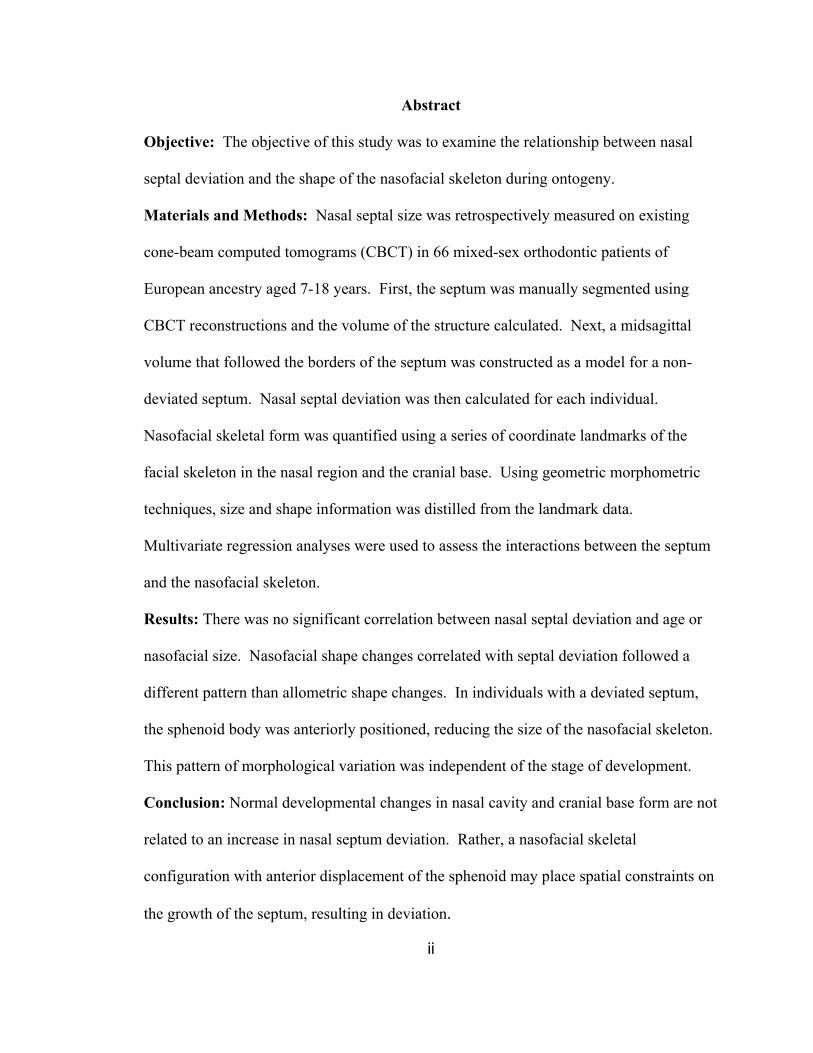
Abstract Objective: The objective of this study was to examine the relationship between nasal
septal deviation and the shape of the nasofacial skeleton during ontogeny.
Materials and Methods: Nasal septal size was retrospectively measured on existing
cone-beam computed tomograms (CBCT) in 66 mixed-sex orthodontic patients of
European ancestry aged 7-18 years. First, the septum was manually segmented using
CBCT reconstructions and the volume of the structure calculated. Next, a midsagittal
volume that followed the borders of the septum was constructed as a model for a non-
deviated septum. Nasal septal deviation was then calculated for each individual.
Nasofacial skeletal form was quantified using a series of coordinate landmarks of the
facial skeleton in the nasal region and the cranial base. Using geometric morphometric
techniques, size and shape information was distilled from the landmark data.
Multivariate regression analyses were used to assess the interactions between the septum
and the nasofacial skeleton.
Results: There was no significant correlation between nasal septal deviation and age or
nasofacial size. Nasofacial shape changes correlated with septal deviation followed a
different pattern than allometric shape changes. In individuals with a deviated septum,
the sphenoid body was anteriorly positioned, reducing the size of the nasofacial skeleton.
This pattern of morphological variation was independent of the stage of development.
Conclusion: Normal developmental changes in nasal cavity and cranial base form are not
related to an increase in nasal septum deviation. Rather, a nasofacial skeletal
configuration with anterior displacement of the sphenoid may place spatial constraints on
the growth of the septum, resulting in deviation.
iii
Table of Contents
List of Tables iv
List of Figures v
Introduction 1
Literature Review 4
Aims and Hypotheses 13
Subjects and Methods 14
Results 23
Discussion 32
Conclusions 37
References 38
iv
List of Tables
Table 1. Study sample demographics. 15
Table 2. Reduced major axis (RMA) regression parameters. 29
v
List of Figures
Figure 1. Nasal septum image segmentation. 16
Figure 2. Examples of three-dimensional reconstructions of 17
a nasal septum and a non-deviated septal model in situ.
Figure 3. Landmarks used to quantify the size and shape of the nasal 19
region in a midsagittal section of a cone beam computed
tomography image.
Figure 4. Box plot of septal deviation values for each age group. 24
Figure 5. Examples of coronal cone beam computed tomography images 24
of subjects with nasal septal deviation.
Figure 6. Bar graph of mean values for measured nasal septal volume and 25
modeled non-deviated volume.
Figure 7. Scatter plot of septal deviation on centroid size. 26
Figure 8. Scatter plot of log transformed measured nasal septal and 28
modeled non-deviated volumes on log transformed
centroid size (excluding pronasale).
Figure 9. Allometric shape variation. 30
Figure 10. Shape variation correlated with nasal septal deviation. 31
1
Introduction
The nasal septum is a midline structure that divides the nasal cavity into bilateral
nasal passages. It is composed of the septal cartilage, the perpendicular plate of the
ethmoid, the vomer, and the crests of the maxillary and palatine bones (Kim et al., 2010).
The nasal septum has been described as a key facial growth center that has a
morphogenetic influence on facial skeletal development (Scott, 1953). During
development, the nasal septum often becomes deviated indicating a complex interaction
between the septum and surrounding skeleton. Nasal septal deviation is defined as the
displacement of the bony or cartilaginous septum to one or both sides. Numerous
etiological factors for nasal septal deviation have been described. For instance, septal
deviation can occur due to a failure of development at any embryological stage, from
either genetic or environmental causes (Pirsig, 1992). During normal development,
septal deviation can result at an early stage from prolonged intrauterine pressures and
transnatal pressures on the fetus (Gray, 1978). Septal deviations can also be caused by
genetic influences, mechanical injuries, and, rarely, by congenital malformations,
infections, or neoplasia. Therefore, septal deviation can occur in utero, during delivery,
and throughout the entire life (Pirsig, 1992).
It has been suggested that humans, when compared to other mammals, may be
predisposed to nasal septal deviation (Gray, 1978; Takahashi, 1987). Across mammals,
there is an inverse relationship between the size of the facial skeleton and nasal septal
deviation (Gray, 1978; Takahashi, 1987). Humans, having comparatively short faces to
other mammals, have a high incidence of nasal septal deviation, whereas this condition is
virtually non-existent in long-snouted animals. This pattern may suggest that nasal septal
2
deviation results from discordant growth between the septum and surrounding facial
skeleton. Indeed, studies indicate smaller facial dimensions in individuals with deviated
septa (Freng et al., 1988). Smaller nasal cavities were present in adults with deviated
nasal septal cartilage in comparison to individuals without septal deviation, suggesting
that an undersized skeletal frame in the sagittal plane may have led to the buckled non-
fitting septal cartilage (Freng et al., 1988). This notion is supported by animal studies in
which anteroposterior facial growth was experimentally reduced via fixation of the
circummaxillary sutures. The spatial constraint altered nasal septal and facial skeletal
relationships resulting in an increase nasal septal deviation (Rönning and Kantomaa,
1985; Holton et al., 2011). Collectively, these studies suggest that the surrounding
skeletal architecture of the nasal cavity may impose constraints on nasal septal growth
resulting in deviation.
In contrast to the above, there is also evidence that nasal septal deviation may be
the result of increased septal growth, rather than resulting from an undersized
surrounding facial skeleton (Vetter et al., 1984; Van Loosen et al., 1996; Holton et al.,
2012). Looking at population variation in septal size and magnitude of septal deviation,
individuals of European descent have both increased prevalence of septal deviation and
larger nasal septa when compared to individuals of African descent (Holton et al., 2012).
Studies on the postnatal growth of the nasal septum also show that the nasal septal
cartilage and perpendicular plate of the ethmoid continue to grow into adulthood
following the cessation of skeletal growth (Vetter et al., 1984; Van Loosen et al., 1996).
Therefore, it is possible that the continued growth of the nasal septum into adulthood is at
least partly responsible for septal deviation in humans.
3
Although discordance between the nasal septum and facial skeleton has been
shown, the ontogeny of septal deviation and the interaction between the nasal septum and
surrounding nasofacial skeleton during ontogeny is not well understood. “Ontogeny”
refers to growth and development. Part of the difficulty in determining the influence of
the nasal septum on nasofacial form in human samples is the paucity of data regarding
the interaction between the nasal septum and facial form during human ontogeny. This
data has been difficult to obtain historically due to the lack of practical, low-cost, non-
invasive methods for analyzing the human nasal septum in vivo. Cone-beam computed
tomography (CBCT) imaging offers a non-invasive, three-dimensional (3-D), high-
resolution method of analyzing the nasal septum and facial form in growing individuals
from a normal population. A thorough understanding of this relationship is particularly
important given that nasal septal deviation, especially in more severe cases, can result in
a higher incidence of mouth breathing, which may be associated with aberrant patterns of
facial growth and the development of certain types of malocclusion (Freng et al., 1988).
Therefore, the goal of the present study was to assess the developmental patterns of nasal
septal deviation and the morphological relationship between the nasal septum and
surrounding nasofacial skeleton in a cross-sectional human sample using data derived
from CBCT scans.
4
Literature Review
The Nasal Septum as a Facial Growth Center
The nasal septum has been suggested to be a key growth center of the facial
skeleton (Scott, 1953). However, the precise role of the nasal septum on facial growth is
not well understood and various models have been proposed. The nasal septal traction
model describes the nasal septum as having a morphogenetic influence on surrounding
skeletal structures (Scott, 1953). This growth model maintains that the nasal septum has
intrinsic growth potential and therefore serves as an endochondral growth plate that
drives anteroposterior and vertical craniofacial growth (Scott, 1953; Copray, 1986;
Wealthall and Herring, 2006). As the cartilaginous component of the nasal septum
expands, it exerts a force on the surrounding skeletal tissues inducing growth at key facial
growth sites, the craniofacial sutures (Latham, 1970; Siegel et al., 1990; Wealthall and
Herring, 2006; Al Dayeh and Herring, 2014).
The nasal septal traction model is supported by a number of experimental studies
using animal models. For instance, Copray (1986) investigated the intrinsic capacity for
growth of the nasal septum using a rat model ex vivo. The nasal septal cartilage was
excised and cultured for 10 days and the growth of the septum was analyzed. Overall, the
nasal septal cartilage increased considerably in size while the shape was preserved. The
center of the septum and the area adjacent to the septo-ethmoidal junction were the areas
of greatest cellular proliferation and the greatest increase in size was found in the anterior
posterior direction. These results suggest a prominent role for the nasal septum on
midfacial growth of the rat (Copray, 1986).
5
Following from the work of Copray (1986), Wealthall and Herring (2006)
investigated whether growth of the nasal septum in mice elongates the facial skeleton in
the same way that the epiphyseal growth plates of the long bones and synchondroses of
the cranial base elongate the long bones and neurocranium of mammals. The authors
examined endochondral ossification at the caudal end of the cartilaginous nasal septum in
mice from postnatal days 0-15 compared to known cranial growth sites, the
synchondroses. It was found that the septum contributes to enlarging the facial skeleton
by displacing facial bones, primarily by septal interstitial growth and also, to a lesser
degree, endochondral ossification along the perpendicular plate of the ethmoid (Wealthall
and Herring, 2006). The same group also measured the mechanical properties of the
nasal septum to determine if the septum is mechanically able to play an active role in
midfacial growth (Al Dayeh and Herring, 2014). It was hypothesized that if the nasal
septum is a growth center, then its growth pressure should be enough to separate the
facial sutures and stiff enough to withstand recoil pressure of the sutures (Al Dayeh and
Herring, 2014). Experiments in pigs showed that the force produced by septal expansion
corresponded to latent mechanical separation of the facial sutures, which suggests that the
growth of the nasal septum is capable of placing pressure on surrounding structures in
order to drive sutural opening and facial growth.
In contrast to the nasal septal traction model, which emphasizes the
morphogenetic influence of cranial cartilages on skeletal growth, others have suggested
that the nasal cartilage plays a minimal role in facial growth (Moss et al., 1968; Moss and
Salentijn, 1969, Stenström and Thilander, 1970). The functional matrix hypothesis, for
example, describes the nasal septum not as driving facial growth itself, but rather skeletal
6
and cartilage growth occurring in response to the functional need for respiration and
secondary to growth of the soft tissues (Moss et al., 1968; Moss and Salentijn, 1969).
Essentially, the functional matrix model argues that the septum does little more than
contribute to projection of the external nose and elevation of the nasal bridge and does
not play an important role in the anterior growth of the facial skeleton (Moss et al., 1968;
Moss and Salentijn, 1969, Stenström and Thilander, 1970). Additional evidence for this
model is seen in patients with congenital craniofacial anomalies that affect the nasal
septum. For instance, patients with holoprosencephaly and cyclopia with
arrhinencephaly, conditions where the nasal septum is absent, have normal midfacial
growth except for a lack of external nose projection and nasal bridge elevation during
craniofacial development (Moss et al., 1968; Moss and Salentijn, 1969).
Evidence from Animal Experiments
Septal excision experiments have demonstrated that the nasal septal cartilage is an
intrinsic growth center for facial growth in several animal species. Experimental studies
in rabbits, for example, have shown that surgical extirpation of all or part of the nasal
septum decreases the anteroposterior growth pattern of the midface (Wexler and Sarnat,
1961; Sarnat and Wexler, 1966, 1967; Rhys-Evans and Brain, 1981). Wexler and Sarnat
(1961) and Sarnat and Wexler (1966) showed this by resecting the cartilaginous nasal
septum in young growing rabbits and comparing them to rabbits that were used as
unoperated and sham-operated controls. Postmortem analysis of the experimental
animals revealed that the snout was shorter and smaller with a severe relative mandibular
prognathism, the nasal and premaxillary bones were smaller, and the nasal cavity and
7
piriform aperture were smaller than in the control animals. In addition, the incisors were
in malocclusion, malshaped, and overerupted (Wexler and Sarnat, 1961; Sarnat and
Wexler, 1966). When these experiments were repeated on adult rabbits, however, there
was very little difference in facial form between experimental animals and controls
(Sarnat and Wexler, 1967). These results support the nasal septum’s importance during
growth in rabbits.
Holton et al. (2011) used a novel approach of experimentally induced synostosis
in the craniofacial skeleton rather than altering the nasal septum itself. Using a pig
model, the length of the facial skeleton was experimentally reduced via rigid plate
fixation of the frontonaso-maxillary and zygomaticomaxillary sutures (Holton et al.,
2011). Despite sutural restriction, the nasal septum grew to normal length as measured
by vomer length, and the reduction in facial length led to compensatory lengthening of
the premaxilla (Holton et al., 2011). This experiment supports the nasal septal traction
model and is indicative of integration between nasal septal and premaxillary growth.
Some nasal septal extirpation experiments, however, did not find significant
changes in facial growth following extirpation. Surgical resections of the nasal septum
on guinea pigs, for example, showed minimal effect on anterior growth of the facial
skeleton (Stenström and Thilander, 1970). Experiments focusing on shorter-face
mammals, such as the work done on chimpanzees by Siegel and Sadler (1981), also
showed that septal resection had minimal effect on facial growth when compared to
controls. In addition, experiments using short-snouted animals, such as domestic cats by
Freng (1981) and ferrets by Cupero et al. (2001), contradict the findings from
experiments on long-snouted animals. For instance, surgical extirpation of the entire
8
cartilaginous nasal septum and vomer of growing domestic cats showed no difference in
sagittal mid-facial development among the three groups when compared to sham-
operated and unoperated control cats (Freng, 1981). Similarly, following partial resection
of the vomer or nasal septal cartilage in ferrets, there was no change in anteroposterior
facial length compared to controls (Cupero et al., 2001). These findings suggest
taxonomic variation in the role of nasal septal traction in facial growth and that the nasal
septum may play less of a role in the anterior growth of the face in shorter-face taxa.
Role of the Nasal Septum in Human Facial Growth
While there is considerable experimental evidence indicating that the nasal
septum can have a significant morphogenetic influence on the growth of the facial
skeleton, much of this work is derived from long-snouted animal models. As such, it is
unclear to what degree the findings are applicable to growth dynamics in shorter-faced
humans. One method previously used to analyze the contribution of the nasal septum to
anterior facial growth in the human skeleton was to evaluate individuals with congential
labiomaxillary clefts or facial injury (e.g. Delaire and Precious, 1986; Mooney et al.,
1989; Siegel et al., 1991; Hall and Precious, 2013). Based on experiments using path
analysis to examine the anatomical relationships between the cleft premaxilla and several
other midfacial structures in both cleft and normal fetal samples, it has been concluded
that the septopremaxillary segment is important in anterior facial growth and the nasal
septal traction model is more explanatory than the functional matrix model in both
normal and cleft samples (Mooney et al., 1989; Siegel et al., 1991).
9
Surgical repair of unilateral or bilateral facial clefts also provides insight into the
role of the nasal septum as pacemaker for midfacial growth and shows that the maxillary
labial frenum is an important constituent of the septopremaxillary traction system. The
frenum houses the septopremaxillary ligament, which extends from the nasal septum to
the mucosal part of the lip (Hall and Precious, 2013). Forces generated by the nasolabial
muscles are transmitted to this structure, and in turn to the anterior surfaces of the maxilla
during facial growth. In individuals with complete unilateral cleft lip, the perioral and
nasolabial muscles on the side of the cleft are underdeveloped, retracted, and laterally
displaced, while the nasolabial muscles on the non-cleft side insert into the cartilaginous
septum and anterior nasal spine. This results in displacement and abnormal development
of the premaxillary region (Hall and Precious, 2013).
There are only few reports in the literature about when the nasal septal cartilage
stops growing. For instance, Van Loosen et al. (1996) quantified growth rates by
creating growth curves using a specially designed algorithm for surface area
measurements from a sample of post-mortem human specimens from birth to 62 years of
age. These measurements showed that the growth rate of the nasal septum is highest in
the newborn until two years of life, and then slows down continuously until a plateau is
reached at an age of approximately 36 years. The size of the cartilaginous septum was
found to increase in the sagittal dimension during the first two years of life. After that,
the total area of the cartilaginous part of the septum remains constant due to a balance
between new formation of cartilage and an equal amount of cartilage transformed into
bone by endochondral ossification. Based on these findings, it has been concluded that
10
the cartilaginous septum reaches adult size at the age of two years, and any subsequent
growth is caused by expansion of the perpendicular plate (Van Loosen et al., 1996).
Nasal Septal Deviation
The study of the role of the nasal septum on the growth of the facial skeleton in
humans is complicated by the presence of septal deviation, a condition that is virtually
non-existent in long-snouted animals. This may suggest that the role of the nasal septum
in human facial growth may differ from the patterns identified in most animal models.
While the nasal septum may be a key growth center in long-snouted animals, its influence
may be limited in humans.
Nasal septum deviation occurs when the septum is displaced away from the
midline, for which numerous etiological factors have been described. Some of these
factors are related to intrauterine pressures. For instance, Gray (1978) examined 2,380
infants at birth and found that 4% had anterior cartilage deformity, giving credence to his
theory of transmitted pressures during pregnancy or childbirth. Podoshin et al. (1991)
investigated 4,090 neonates with no evidence of birth trauma as the cause of congenital
nasal deformities for nasal septal deviations and proposed that the majority of
dislocations originated during intrauterine life. Other etiological factors include
discordant growth between the septum and surrounding facial skeleton. For instance,
Freng et al. (1988), in a study examining cephalometric morphology in adults with
deviated septal cartilage, found that smaller nasal cavities were present in adults with
deviated nasal septal cartilage. This suggests that an undersized skeletal frame in the
anteroposterior dimension may have led to the deviated septal cartilage and that nasal
11
septal deviation may result from growth restriction of the nasal septum due to space
constraints from the surrounding facial skeleton.
On the other hand, there is also evidence that nasal septal deviation may be the
result of increased septal growth. Kim et al. (2012) found that reduced ossification of the
sphenoidal process of the septal cartilage led to greater overall septal length and
increased nasal septal deviation. Similarly, individuals with larger nasal septa were
found to have increased deviation (Holton et al., 2012). The notion of increased septal
growth is further supported by the finding that the nasal septum continues to grow into
adulthood following the cessation of skeletal growth (Vetter et al., 1984; Van Loosen et
al., 1996).
Accurately determining the prevalence of nasal septal deviation is complicated by
differences regarding the definition of nasal septal deviation and measuring techniques
used (Vig, 1998). This leads to a significant variation of the reported range of nasal
septal deviation in newborns across studies. For instance, Gray (1978) found that 42% of
septa of Caucasian infants were straight, 27% deviated, and 31% kinked, whereas Kent et
al. (1988) reported an incidence of nasal septum deformity in only 2.9% of 1000
consecutive neonates. Similarly, Podoshin et al. (1991) found an incidence of 0.93% of
anterior nasal septal cartilaginous dislocation in newborns investigated for nasal septal
deviations while Šubarić and Mladina (2002) reported a prevalence of 28% children aged
2-6 to 41.8% in young adults aged 19-22. It appears that there is a gradually increasing
prevalence of deformities involving the posterior (bony) parts of the septum with age.
This age-related increase in septal deformity is consistent with studies on the prevalence
of septal deformity worldwide (Šubarić and Mladina, 2002).
12
More recently and using newer technology, such as computed tomography (CT)
and magnetic resonance imaging (MRI), Reitzen et al. (2011) measured the tortuosity of
the septum at four points along the septum in order to overcome the shortcomings of
previous studies in which deviation was evaluated only at one specific point along the
septum. Tortuosity was measured in 81 patients from age 2 months to 80 years by
dividing “actual” length of the septum by the “ideal” length that was represented by a
straight line from the superior to the inferior aspect of the septum. The results show that
nasal septum deviation is more common in older children and adults when using
tortuosity as a measure of deviation (Reitzen et al., 2011). Similarly, Mladina et al.
(2008) found the incidence of septal deviation to be as high as 89.2% using anterior
rhinoscopy in adult patients seeking medical care for nasal complaints.
Clinical Significance of Nasal Septal Deviation
There are several clinical implications to nasal septal deviation. The space
between the septum and lateral walls of the nasal cavity regulates nasal airflow and
respiration. In infants, open nasal passages are required to feed properly. Severe and
bilateral deviation in infants can result in poor feeding and/or choking from food in the
respiratory tract and sudden infant death syndrome (Kawalski and Spiewak, 1998). In
adults, deviation can lead to mouth breathing, nasal crusting, epistaxis, and sinusitis
depending on the severity and location of the deviation (Aziz et al., 2014). Dental
findings in patients with nasal obstruction as a consequence of septal deviation have been
reported as Class II malocclusion with increased anterior facial height, retrognathic
mandible with increased overjet and constricted maxilla (D’Ascanio et al., 2010).
13
Aims and Hypotheses
The present work aimed at examining the magnitude of nasal septal deviation and
patterns of covariation between the nasal septum and nasofacial skeleton during ontogeny
in a cross-sectional human sample using data derived from CBCT scans. First, the
magnitude of ontogenetic variation in nasal septal deviation was assessed using a
measured midsagittal nasal septum volume and a model for a non-deviated septum in
each subject. The modeled non-deviated volume served as a measure of the minimum
amount of space available in the midline nasal. It was hypothesized that, if the
magnitude of nasal septal deviation increases with age, the discrepancy between
measured septal volume and modeled non-deviated volume will increase with age. Next,
the allometric relationship between measured nasal septal volume and modeled non-
deviated volume was examined. “Allometry” refers to changes in shape with
development. If the magnitude of nasal septal deviation increases during growth and
development, then measured nasal septal volume should scale with greater positive
allometry compared to modeled non-deviated septal volume. Finally, the interaction
between the septum and surrounding nasofacial skeleton during ontogeny was assessed.
If the pattern of covariation between nasal septal deviation and the shape of the nasofacial
skeleton corresponds to changes reflective of normal growth and development in the
nasofacial region, this would indicate that the magnitude of septal deviation is associated
with ontogenetic changes in the shape of the nasofacial skeleton. Alternatively, if septal
deviation is not associated with shape changes in nasofacial region that occur during
normal growth and development, this would suggest that the magnitude of septal
deviation varies independent of ontogeny.
14
Subjects and Methods
Subject selection
The research protocol including the use of existing CBCT scans was approved by
the Institutional Review Board at University of Minnesota (Study Number 1410M54305).
A total of 66 patients (34 male, 32 female) who presented for orthodontic treatment at the
University of Minnesota were included in this retrospective cohort study. The patients
ranged from 7-18 years in age and were selected using the following inclusion criteria: 1)
CBCT scan prior to the start of orthodontic treatment, and 2) being seen only for the
treatment of skeletal or dental malocclusion. Patients were excluded if they had
craniofacial anomalies (e.g., cleft lip/palate), syndromes, or were undergoing surgical or
simultaneous craniofacial treatments. All patients were grouped by age in two-year
intervals (e.g., 7-8, 9-10, etc.) to achieve a minimum of n=10 individuals within each age
group, while maintaining approximately equal numbers of males and females. Study
sample demographics are shown in Table 1.
All CBCT scans were full field-of-view (17x23 cm) and were taken using an
iCAT Next Generation (Imaging Sciences International, Hatfield, PA, USA) at 120 kV
and 18.54 mAs with a pulsed scan time of 8.9 s. The scan data were reconstructed with a
voxel size of 0.3 mm3.
15
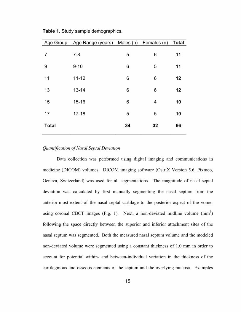
Table 1. Study sample demographics.
Age Group Age Range (years) Males (n) Females (n) Total 7 7-8 5 6 11 9 9-10 6 5 11 11 11-12 6 6 12 13 13-14 6 6 12 15 15-16 6 4 10 17 17-18 5 5 10 Total 34 32 66
Quantification of Nasal Septal Deviation
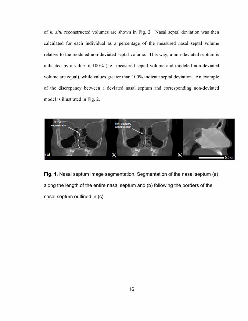
Data collection was performed using digital imaging and communications in
medicine (DICOM) volumes. DICOM imaging software (OsiriX Version 5.6, Pixmeo,
Geneva, Switzerland) was used for all segmentations. The magnitude of nasal septal
deviation was calculated by first manually segmenting the nasal septum from the
anterior-most extent of the nasal septal cartilage to the posterior aspect of the vomer
using coronal CBCT images (Fig. 1). Next, a non-deviated midline volume (mm3)
following the space directly between the superior and inferior attachment sites of the
nasal septum was segmented. Both the measured nasal septum volume and the modeled
non-deviated volume were segmented using a constant thickness of 1.0 mm in order to
account for potential within- and between-individual variation in the thickness of the
cartilaginous and osseous elements of the septum and the overlying mucosa. Examples
16
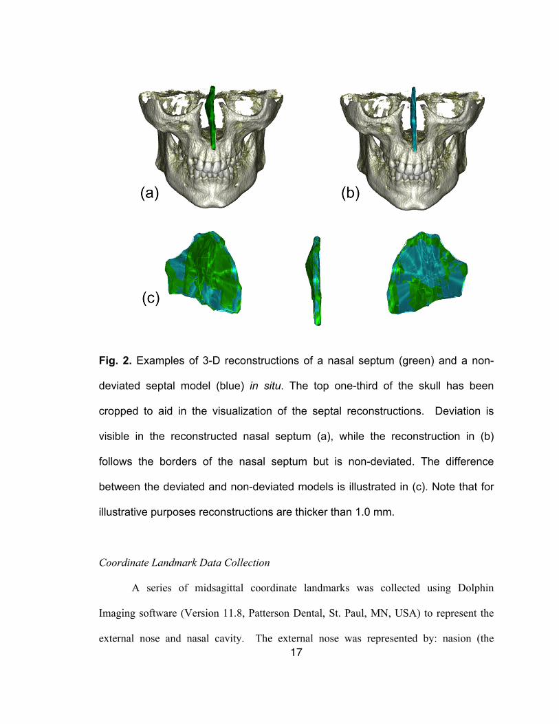
of in situ reconstructed volumes are shown in Fig. 2. Nasal septal deviation was then
calculated for each individual as a percentage of the measured nasal septal volume
relative to the modeled non-deviated septal volume. This way, a non-deviated septum is
indicated by a value of 100% (i.e., measured septal volume and modeled non-deviated
volume are equal), while values greater than 100% indicate septal deviation. An example
of the discrepancy between a deviated nasal septum and corresponding non-deviated
model is illustrated in Fig. 2.
Fig. 1. Nasal septum image segmentation. Segmentation of the nasal septum (a)
along the length of the entire nasal septum and (b) following the borders of the
nasal septum outlined in (c).
17
Fig. 2. Examples of 3-D reconstructions of a nasal septum (green) and a non-
deviated septal model (blue) in situ. The top one-third of the skull has been
cropped to aid in the visualization of the septal reconstructions. Deviation is
visible in the reconstructed nasal septum (a), while the reconstruction in (b)
follows the borders of the nasal septum but is non-deviated. The difference
between the deviated and non-deviated models is illustrated in (c). Note that for
illustrative purposes reconstructions are thicker than 1.0 mm.
Coordinate Landmark Data Collection
A series of midsagittal coordinate landmarks was collected using Dolphin
Imaging software (Version 11.8, Patterson Dental, St. Paul, MN, USA) to represent the
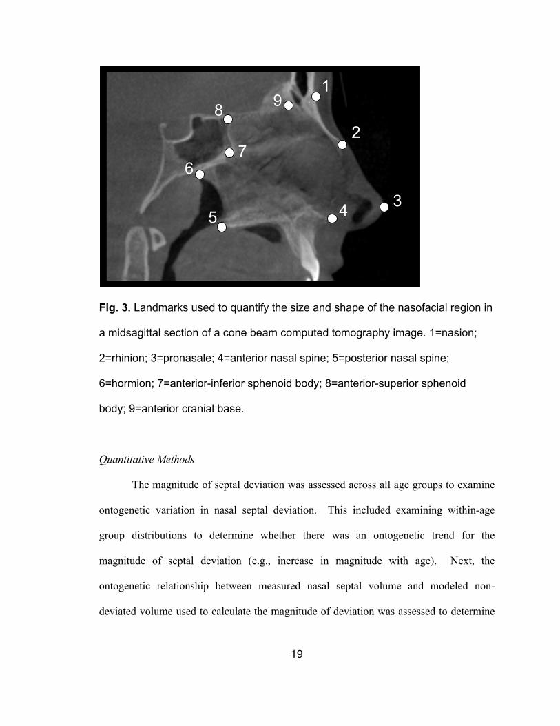
external nose and nasal cavity. The external nose was represented by: nasion (the
18
articulation between the frontal and nasal bones in the midsagittal plane); rhinion (the
anterior tip of the nasal bones); pronasale (a soft-tissue landmark located on the anterior-
most point on the external nose); and ANS (the tip of the anterior nasal spine). The nasal
cavity was represented by: PNS (posterior-most landmark on the hard palate/nasal floor);
hormion (posterior-most aspect of the vomer in the midline); the anterior-inferior
articulation between the sphenoid body and the nasal septum (i.e., inferior aspect of the
sphenoethmoidal synchondrosis); the anterior-superior articulation between the sphenoid
body and the nasal septum (i.e., the superior aspect of the sphenoethmoidal
synchondrosis); and the anterior-most aspect of the anterior cranial base. The landmarks
are shown in Fig. 3.
19
Fig. 3. Landmarks used to quantify the size and shape of the nasofacial region in
a midsagittal section of a cone beam computed tomography image. 1=nasion;
2=rhinion; 3=pronasale; 4=anterior nasal spine; 5=posterior nasal spine;
6=hormion; 7=anterior-inferior sphenoid body; 8=anterior-superior sphenoid
body; 9=anterior cranial base.
Quantitative Methods
The magnitude of septal deviation was assessed across all age groups to examine
ontogenetic variation in nasal septal deviation. This included examining within-age
group distributions to determine whether there was an ontogenetic trend for the
magnitude of septal deviation (e.g., increase in magnitude with age). Next, the
ontogenetic relationship between measured nasal septal volume and modeled non-
deviated volume used to calculate the magnitude of deviation was assessed to determine
1
2
3 4 5
6 7
8 9
20
whether measured nasal septal volume was consistently larger than modeled non-deviated
volume across age groups.
Given that chronological age is only a general proxy measure for growth and
development, it was also examined whether there was a correlation between septal
deviation and nasofacial size. To test this, the nasofacial size was measured as the
centroid size of the coordinate landmarks used to represent the nasal region as detailed
above (Fig. 3). Centroid size is a composite size measure that is calculated as the sum of
the squared distances between each landmark in a configuration and a centroid landmark
(i.e., the mean x and y coordinates for all landmarks in the configuration). For this
comparison, pronasale, an external landmark located at the anterior aspect of the nasal
septal cartilage, was excluded from the nasal cavity centroid size measure.
All measurements were made by a single operator and repeated after a washout
period of 5 weeks for 13 randomly chosen subjects to assess intra-examiner reliability.
Statistical Analyses
Intra-examiner reliability was tested by calculating the percentage difference
between the two observations and intra-class correlations coefficients. With regard to
coordinate landmark data, the Euclidean distance between homologous landmarks (i.e.,
the milimetric distance between a landmark in the first observation and second
observations) was calculated to assess the absolute landmark distance between the two
observations.
Differences between measured septal volume and modeled non-deviated volume
were tested for statistical significance using Wilcoxon signed-rank tests. The relationship
21
between septal deviation and nasofacial size was examined using reduced major axis
(RMA) regression. The cube root of measured nasal septal and modeled non-deviated
values were log-transformed and regressed against log-transformed nasofacial centroid
size. The variation in the regression slopes was determined to assess whether measured
septal volume and the modeled non-deviated volume exhibited differences in slope
values relative to nasofacial centroid size. Analysis of covariance (ANCOVA) was used
to compare least-squares (LS) regression slopes. Additionally, the allometric relationship
between measured nasal septal volume and modeled non-deviated volume was assessed
using RMA regression of log-transformed variables. Specifically, it was tested whether
measured septal volume scaled with positive allometry relative to modeled non-deviated
volume (indicating an ontogenetic increase in septal deviation), or whether the two
variables scaled isometrically (indicating that the magnitude of septal deviation is
maintained through ontogeny).
Finally, an assessment was made whether there was a morphological relationship
between nasal septal deviation and the shape of the nasofacial region (i.e., external nose
and internal nasal cavity) during growth and development. For this, the individual
nasofacial region landmark configurations were superimposed using Procrustes analysis.
This superimposition method rotates, translates, and scales all landmark configurations,
leaving only residual shape information. Shape variation was visualized using wireframe
models and thin plate splines. All geometric morphometric analyses were conducted
using MorphoJ (Klingenberg, The University of Manchester, Manchester, UK).
Multivariate regression was used to examine the correlation between the Procrustes
scaled landmark configurations (dependent variables) and nasofacial centroid size
22
calculated for all landmarks (independent variable) in order to examine the allometric
component of shape variation in the sample (i.e., the component of shape that varies with
ontogeny). Thereafter, a multivariate regression analysis was performed to assess the
relationship between the Procrustes scaled landmark configurations (dependent variables)
and the magnitude of nasal septal deviation (independent variable) to determine whether
there was a significant correlation between septal deviation and the shape of the
nasofacial region, and if the pattern of correlated shape variation mirrored ontogenetic
changes in the nasal region. All statistical analyses were performed using SPSS (IBM
Corporation, Armonk, NY, USA) with P-values of less than 0.05 considered statistically
significant.
23
Results
Intra-examiner Reliability
With regard to measured septal volume and modeled non-deviated volume values,
there was, on average, a 2.2% and 3.1% difference between the first and second
observation, with intra-class correlation coefficients of r=0.99 (P<0.001) indicating a
high degree of intra-examiner reliability. With regard to coordinate landmark
acquisition, the Euclidean distance values between landmarks at the two observations
ranged from 0.91 mm to 1.96 mm, indicating a similarly high degree of intra-examiner
reliability.
Ontogenetic Variation in Nasal Septal Deviation
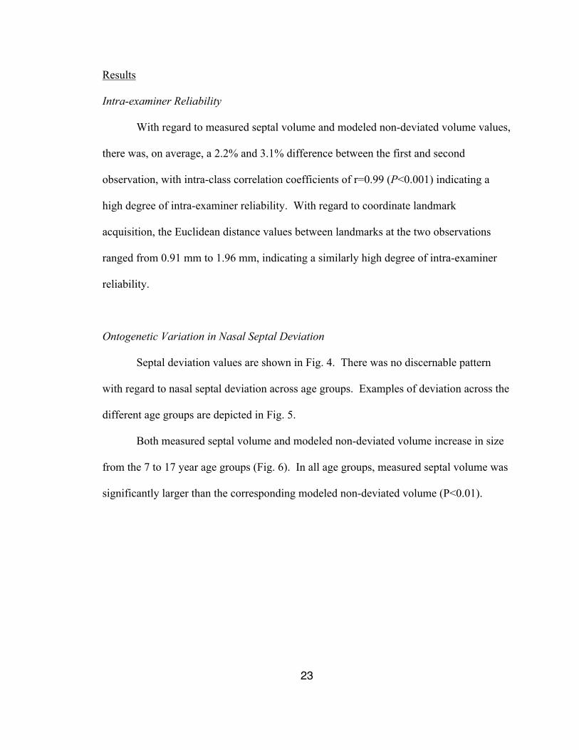
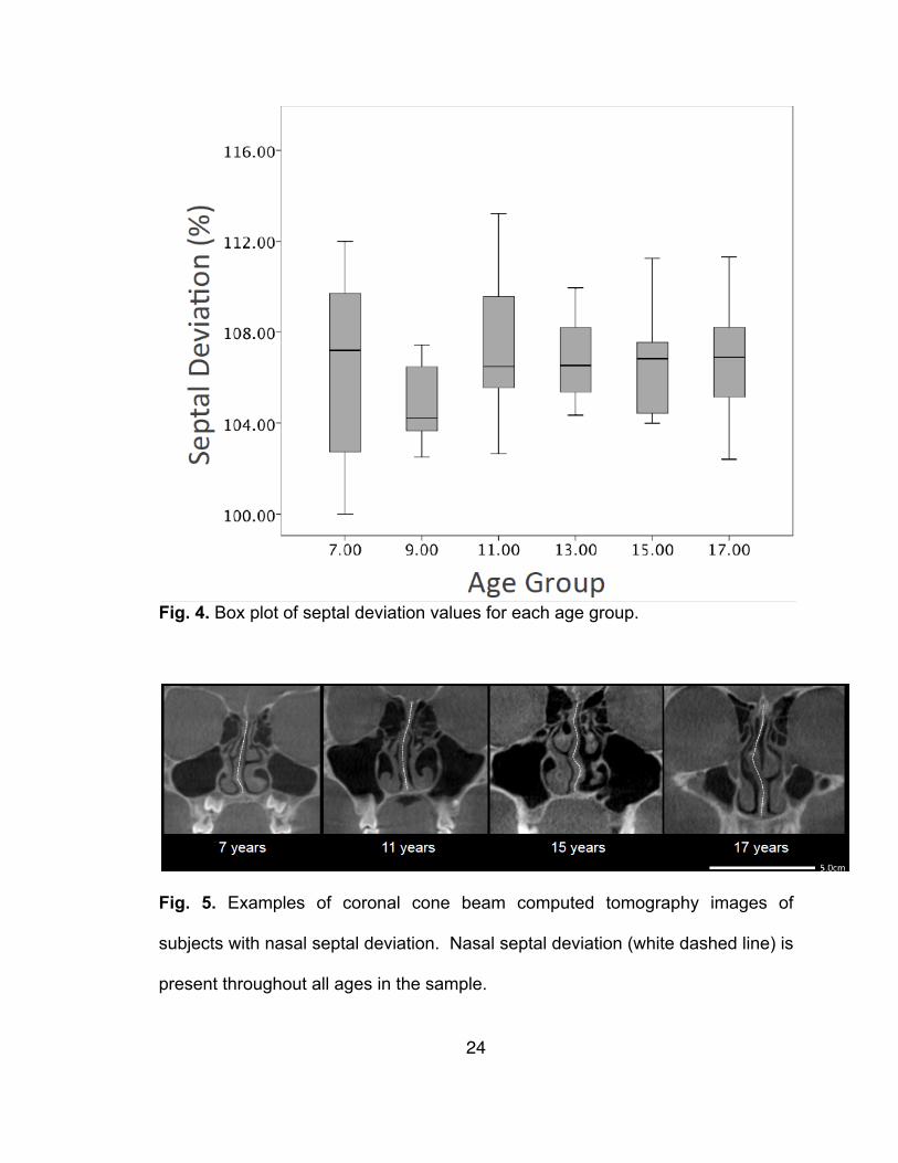
Septal deviation values are shown in Fig. 4. There was no discernable pattern
with regard to nasal septal deviation across age groups. Examples of deviation across the
different age groups are depicted in Fig. 5.
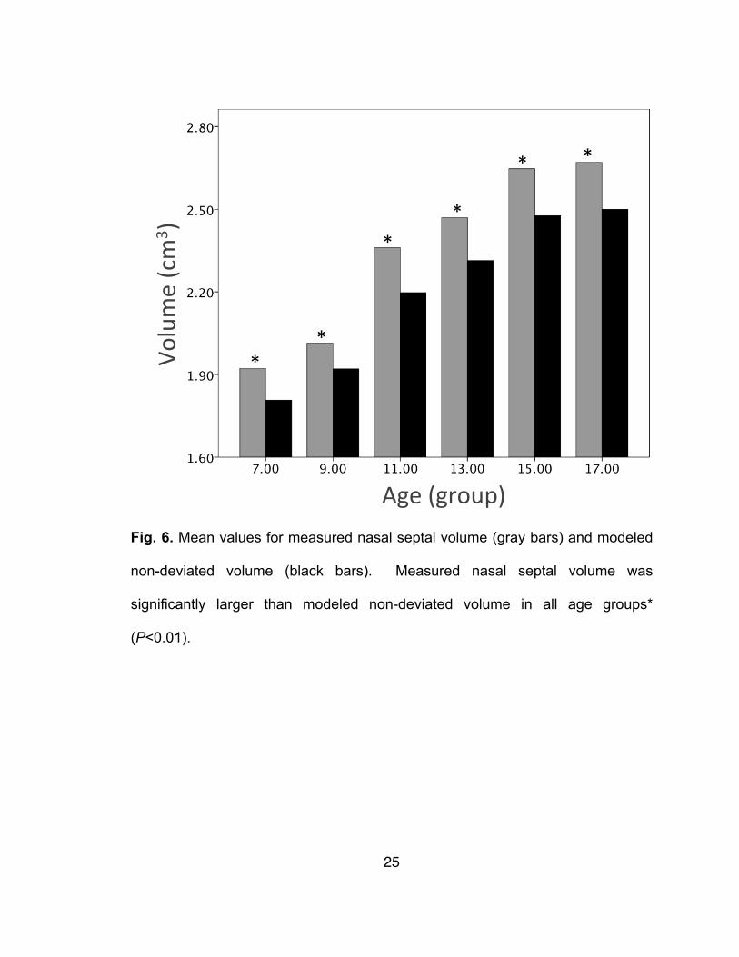
Both measured septal volume and modeled non-deviated volume increase in size
from the 7 to 17 year age groups (Fig. 6). In all age groups, measured septal volume was
significantly larger than the corresponding modeled non-deviated volume (P<0.01).
24
Fig. 4. Box plot of septal deviation values for each age group.
Fig. 5. Examples of coronal cone beam computed tomography images of
subjects with nasal septal deviation. Nasal septal deviation (white dashed line) is
present throughout all ages in the sample.
25
Fig. 6. Mean values for measured nasal septal volume (gray bars) and modeled
non-deviated volume (black bars). Measured nasal septal volume was
significantly larger than modeled non-deviated volume in all age groups*
(P<0.01).
Age$(group)$
Volume$(cm
3 )$
*$*$
*$*$
*$ *$
26
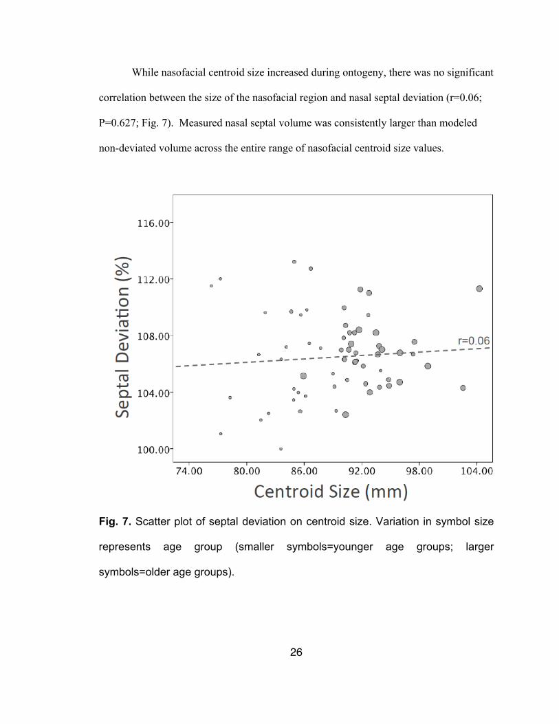
While nasofacial centroid size increased during ontogeny, there was no significant
correlation between the size of the nasofacial region and nasal septal deviation (r=0.06;
P=0.627; Fig. 7). Measured nasal septal volume was consistently larger than modeled
non-deviated volume across the entire range of nasofacial centroid size values.
Fig. 7. Scatter plot of septal deviation on centroid size. Variation in symbol size
represents age group (smaller symbols=younger age groups; larger
symbols=older age groups).
27
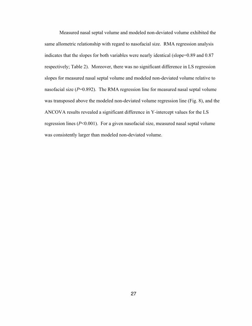
Measured nasal septal volume and modeled non-deviated volume exhibited the
same allometric relationship with regard to nasofacial size. RMA regression analysis
indicates that the slopes for both variables were nearly identical (slope=0.89 and 0.87
respectively; Table 2). Moreover, there was no significant difference in LS regression
slopes for measured nasal septal volume and modeled non-deviated volume relative to
nasofacial size (P=0.892). The RMA regression line for measured nasal septal volume
was transposed above the modeled non-deviated volume regression line (Fig. 8), and the
ANCOVA results revealed a significant difference in Y-intercept values for the LS
regression lines (P<0.001). For a given nasofacial size, measured nasal septal volume
was consistently larger than modeled non-deviated volume.
28
Fig. 8. Scatter plot of log transformed measured nasal septal and modeled non-
deviated volumes on log transformed centroid size (excluding pronasale).
Parallel RMA regression lines are shown.
ln#Septal#Volume#
ln#Modeled#Volume#
ln#Centroid#Size#
ln#Volum
e#
29
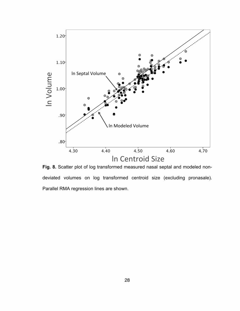
Table 2. Reduced major axis (RMA) regression parameters. ln Measured Volume ln Modeled Volume RMA Slope 0.89 0.87 RMS 95% CI 0.77-1.00 0.77-0.97 RMA Y-intercept -2.96 -2.84 RMA R2 0.75 0.79
Septal Deviation and Nasal Shape
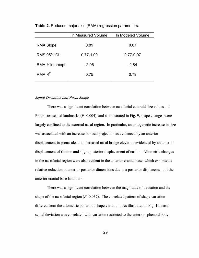
There was a significant correlation between nasofacial centroid size values and
Procrustes scaled landmarks (P=0.004), and as illustrated in Fig. 9, shape changes were
largely confined to the external nasal region. In particular, an ontogenetic increase in size
was associated with an increase in nasal projection as evidenced by an anterior
displacement in pronasale, and increased nasal bridge elevation evidenced by an anterior
displacement of rhinion and slight posterior displacement of nasion. Allometric changes
in the nasofacial region were also evident in the anterior cranial base, which exhibited a
relative reduction in anterior-posterior dimensions due to a posterior displacement of the
anterior cranial base landmark.
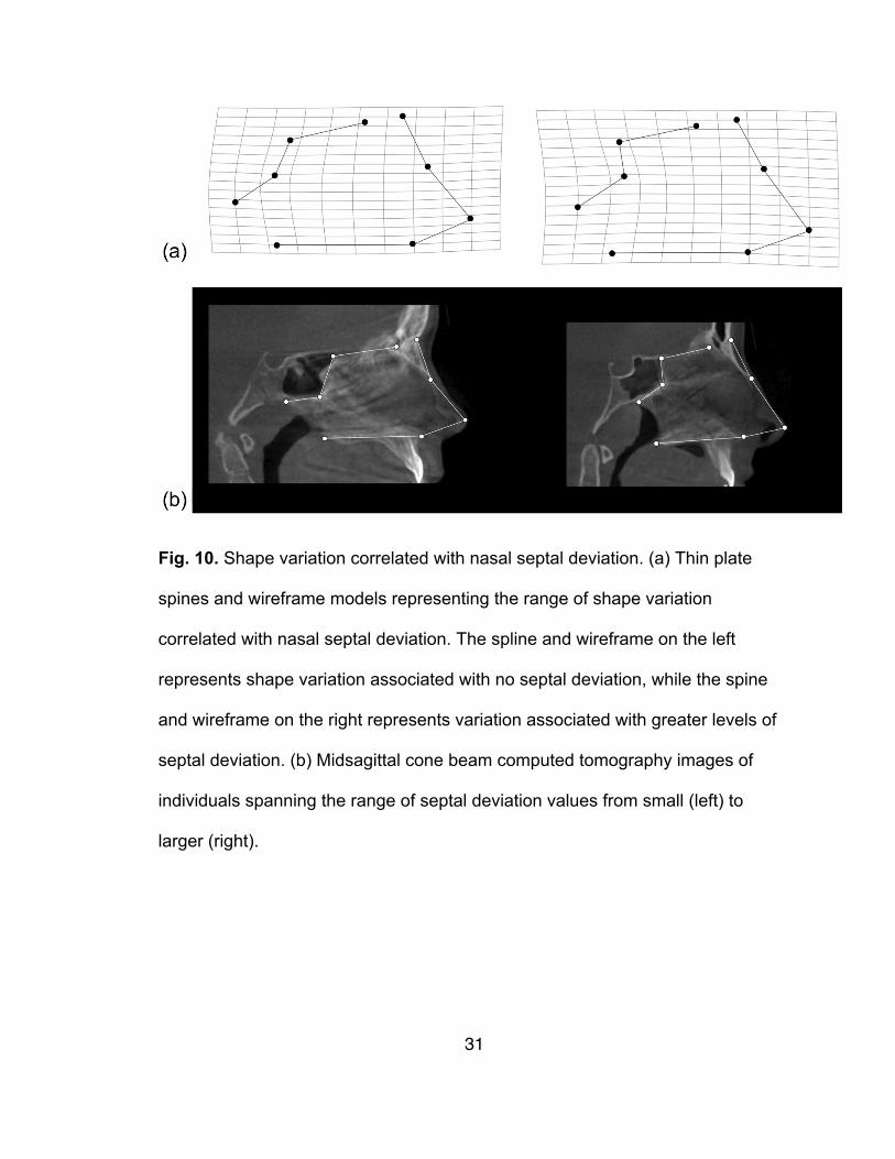
There was a significant correlation between the magnitude of deviation and the
shape of the nasofacial region (P=0.037). The correlated pattern of shape variation
differed from the allometric pattern of shape variation. As illustrated in Fig. 10, nasal
septal deviation was correlated with variation restricted to the anterior sphenoid body.
30
Fig. 9. Allometric shape variation. (a) Thin plate spines and wireframe models
representing the range of shape variation correlated with centroid size. The
spline and wireframe model on the left represents shape associated with smaller
centroid values (i.e., younger individuals), while the spline and wireframe model
on the right represents shape associated with larger centroid values (i.e., older
individuals). (b) Midsagittal cone beam computed tomography images of
individuals spanning the range of centroid size values from small (left) to larger
(right).
31
Fig. 10. Shape variation correlated with nasal septal deviation. (a) Thin plate
spines and wireframe models representing the range of shape variation
correlated with nasal septal deviation. The spline and wireframe on the left
represents shape variation associated with no septal deviation, while the spine
and wireframe on the right represents variation associated with greater levels of
septal deviation. (b) Midsagittal cone beam computed tomography images of
individuals spanning the range of septal deviation values from small (left) to
larger (right).
32
Discussion
The nasal septum has been described as a key growth center in the facial skeleton
and discordance in growth between the septum and the surrounding facial skeleton is
thought to result in septal deviation (e.g., Scott, 1953; Gray, 1978; Takahashi, 1987).
Currently, we lack a good understanding about the ontogeny of septal deviation, both
with regard to changes in deviation and developmental interaction between the septum
and surrounding nasofacial region. In an attempt to add to the current body of knowledge
on the ontogeny of septal deviation, we used CBCT data of individuals ranging from 7 to
18 years to study ontogenetic variation in the magnitude of nasal septal deviation, and its
correlation to nasofacial size and shape during development. In the sample studied, there
was no evidence of an ontogenetic increase in nasal septal deviation with regard to age
(as an approximation of growth) or nasofacial centroid size (as a more direct measure of
growth). As the nasofacial region increased in size, the magnitude of nasal septal
deviation was maintained across the entire size range, indicating a relatively isometric
relationship during ontogeny.
In addition, the findings suggest that the magnitude of nasal septal deviation is
established early in ontogeny by 7-8 years, and is maintained through 17-18 years of age.
This resembles the findings of Yildirim and Okur (2003), who found that 16.5% of
subjects aged 4-6 years presented with septal deviation. This frequency increased to
38.7% in their 7-12 year age group and was maintained through later ages. In contrast,
other studies report ontogenetic increases in deviation later in development. For instance,
Šubarić and Mladina (2002) found an increase in deviation from their 7-14 to their 15-18
year age groups. These findings suggest that there is variation in the frequency of septal
33
deviation across studies. The most likely explanation for this observed variation is
variation in the study population. For instance, most study samples consist of patients
who were receiving medical intervention for nasal obstruction problems or other head
and neck conditions (e.g., Mladina et al., 2008; Reitzen et al., 2011). In contrast, the
present study sample consisted of orthodontic patients without obvious airway
restrictions. Patients with nasal obstruction may have greater magnitudes of deviation
than the orthodontic patients who were not undergoing surgical or simultaneous
craniofacial treatments and were being seen only for the treatment of skeletal or dental
malocclusion. Therefore, the present sample is potentially more representative of the
population as a whole rather than a pathological subset.
Another possible explanation for the variation in septal deviation reported across
studies is the lack of consistency in how deviation is measured. Nasal septal deviation
has been measured in several different ways in previous studies, such as acoustic
rhinometry, rhinomanometry, and nasal spectral sound analysis (Aziz et al., 2014).
Moreover, deviation has often been measured at only one specific point along the septum
or qualitatively using a yes/no categorical scale from photographs or two-dimensional (2-
D) radiographs (e.g., Gray, 1978; Hafezi et al., 2010; Kim et al., 2011; Akbay et al.,
2013). This inconsistency in measurement methods across studies likely contributes to
the variation in results.
The present study utilized a quantitative 3-D approach that characterized the
morphology of the entire nasal septum. Using this approach, rather than relying on
qualitative or 2-D approaches to measuring septal deviation as in previous studies,
allowed us to more accurately characterize the magnitude of deviation and gave us finer
34
resolution for assessing morphological variation in the nasal septum, and the relationship
between the nasal septum and the surrounding skeletal anatomy. The method has been
shown to be accurate at measuring septal deviation in previous studies that have assessed
the relationships between septal deviation, facial skeletal form, and facial asymmetries
(Holton et al., 2012; Hartman et al., 2016).
The use of CBCT data allowed quantification of the magnitude of deviation along
the entire septum rather than evaluation of the frequency of nasal septal deviation using a
yes/no scale. Thus, deviation was measured as a continuous rather than a categorical
variable. If the septum exhibited deviation in one region, that individual would be
considered deviated when using a categorical scale, as done in previous studies.
However, quantifying deviation across the entire septum may help explain why we do not
see ontogenetic changes in septal deviation in this study. As a result, we cannot assess
ontogenetic changes in the frequency of deviation, but we can conclude that the
magnitude of deviation is maintained in our sample.
The developmental interaction between the nasal septum and the surrounding
nasofacial skeleton was studied using multivariate regression analyses. These analyses
were performed to examine correlations between nasofacial region shape variation and
normal growth and development, and correlations between nasofacial region shape
variation and nasal septal deviation. The results indicate that nasofacial region shape
changes correlated with septal deviation are different than shape changes correlated with
normal growth. As the magnitude of nasal septal deviation increased, the inferior aspect
of the sphenoid body tended to be positioned more anteriorly. A relatively more anterior
position of the sphenoid body may restrict space in the nasal cavity leading to septal
35
deviation. This assumption is corroborated by a number of studies that have shown that
reduced midfacial dimensions are associated with deviation (Gray, 1978; Takahashi,
1987; Freng et al., 1988; Rönning and Kantomaa, 1985; Holton et al., 2011). Given the
developmental relationship between the nasal septum and sphenoid that begins during
early chondrocranial development through fusion of the spheno-ethmoid synchondrosis
around age 6 (Scott, 1958), these results suggest that the differences in the position of the
sphenoid body are established early in development. Thus, while patterns of covariation
between nasal septal deviation and nasofacial form are non-allometric and are established
by at least 7 years of age, they are potentially established much earlier in ontogeny.
The finding that the magnitude of nasal septal deviation is established by at least 7
years of age and maintained throughout ontogeny contradicts the findings of previous
studies in which deviation was found to increase during ontogeny (e.g., Šubarić and
Mladina, 2002). Moreover, nasal septal deviation was not associated with normal shape
changes in the nasofacial region during growth and development, suggesting that normal
developmental changes in nasal cavity and cranial base form are not related to an increase
in nasal septum deviation. Finally, this study supports previous research that has found
septal deviation to be the result of an undersized facial skeletal frame (e.g., Freng et al.,
1988). Specifically, a nasofacial skeletal configuration with a relatively anterior position
of the sphenoid may place spatial constraints on the growth of the septum, resulting in
deviation. While these findings cannot establish a causal relationship between nasal
septal deviation and nasofacial size and shape, they do support the nasal traction model of
midfacial growth, which emphasizes the morphogenetic influence of the nasal septum on
growth of the facial skeleton (Scott, 1953; Latham, 1970; Copray, 1986; Siegel et al.,
36
1990; Wealthall and Herring, 2006; Al Dayeh and Herring, 2014). These findings are in
contrast to the functional matrix theory, which suggests that the nasal cartilage plays a
minimal role in facial growth (Moss et al., 1968; Moss and Saletijn, 1969, Stenström and
Thilander, 1970). Future works should assess the longitudinal interaction between nasal
septal deviation and other components of the facial skeleton to help gain a better
understanding of the role of the nasal septum on facial growth.
37
Conclusions
1. The magnitude of nasal septal deviation and the position of the sphenoid are
established by approximately 7 years of age and then maintained throughout
ontogeny.
2. Normal developmental changes in nasal cavity and cranial base form do not result
in an increase in nasal septum deviation.
3. A facial skeletal configuration with an anteriorly positioned sphenoid may place
spatial constraints on the growth of the septum, resulting in deviation.
38
References
1. Akbay E, Cokkeser Y, Yilmaz O, Cevik C (2013). The relationship between posterior septum deviation and depth of maxillopalatal arch. Auris Nasus Larynx 40:286–290.
2. Al Dayeh AA, Herring SW (2014). Compressive and tensile mechanical
properties of the porcine nasal septum. J Biomech 47:154–161.
3. Aziz T, Biron VL, Ansari K, Flores-Mir C (2014). Measurement tools for the diagnosis of nasal septal deviation: a systematic review. J Otolaryngol Head Neck Surg 43:11.
4. Copray JC (1986). Growth of the nasal septal cartilage of the rat in vitro. J Anat
144:99–111.
5. Cupero TM, Middleton CE, Silva AB (2001). Effects of functional septoplasty on the facial growth of ferrets. Arch Otolaryngol Head Neck Surg 127:1367–1369.
6. D’Ascanio L, Lancione C, Pompa G, Rebuffini E, Mansi N, Manzini M (2010).
Craniofacial growth in children with nasal septum deviation: a cephalometric comparative study. Int J Pediatr Otorhinolaryngol 74: 1180–1183.
7. Delaire J, Precious, D (1986). Influence of the nasal septum on maxillonasal
growth in patients with congenital labiomaxillary cleft. Cleft Palate J 23:270–277.
8. Freng A (1981). Mid-facial sagittal growth following resection of the nasal
septum-vomer: a roentgencephalometric study in the domestic cat. Acta Otolaryngol 92:363–370.
9. Freng A, Kvam E, Kramer J (1988). Facial skeletal dimensions in patients with
nasal septal deviation. Scand J Plast Reconstr Surg Hand Surg 22:77–81.
10. Gray LP (1978). Deviated nasal septum. Incidence and etiology. Ann Otol Rhinol Laryngol Suppl 87:3–20.
11. Hafezi F, Naghibzadeh B, Nouhi A, Yavari P (2010). Asymmetric facial growth
and deviated nose: a new concept. Ann Plast Surg 64:47–51.
12. Hall BK, Precious DS (2013). Cleft lip, nose, and palate: the nasal septum as the pacemaker for midfacial growth. Oral Surg Oral Med Oral Pathol Oral Radiol 115:442–447.
39
13. Hartman C, Holton N, Miller S, Yokley T, Marshall S, Srinivasan S, Southard T (2016). Nasal septal deviation and facial skeletal asymmetries. Anat Rec (Hoboken) 299:295–306.
14. Holton NE, Franciscus RG, Marshall SD, Southard TE, Nieves MA (2011). Nasal
septal and premaxillary developmental integration: implications for facial reduction in Homo. Anat Rec (Hoboken) 294:68–78.
15. Holton NE, Yokley TR, Figueroa A (2012). Nasal septal and craniofacial form in
European- and African-derived populations. J Anat 221:263–274.
16. Kawalski H, Spiewak P (1998). How septum deformations in newborns occur. Int J Pediatr Otorhinolaryngol 44:23–30.
17. Kent SE, Reid AP, Nairn ER, Brain DJ (1988). Neonatal septal deviations. J R
Soc Med 81:132–135.
18. Kim J, Cho JH, Kim SW, Kim BV, Lee DC, Kim SW (2010). Anatomical variation of the nasal septum: Correlation among septal components. Clin Anat 23:945–949.
19. Kim J, Kim SW, Kim SW, Cho JH, Park YL (2012). Role of the sphenoidal
process of the septal cartilage in the development of septal deviation. Otolaryngol Head Neck Surg 146:151–155.
20. Kim YM, Rha KS, Weissman JD, Hwang PH, Most SP (2011). Correlation of
asymmetric facial growth with deviated nasal septum. Laryngoscope 121:1144–1148.
21. Latham, RA (1970). Maxillary development and growth: the septo-premaxillary
ligament J Anat 107 (Pt 3):471–478
22. Mladina R, Čujić E, Šubarić M, Vuković K. Nasal septal deformities in ear, nose and throat patients: an international study. Am J Otolaryngol 29:75–82.
23. Mooney MP, Siegel MI, Kimes KR, Todhunter J (1989). A test of two midfacial
growth models using path analysis of normal human fetal material. Cleft Palate J 26:93–99.
24. Moss ML, Bromberg BE, Song IC, Eisenman G (1968). The passive role of nasal
septal cartilage in mid-facial growth. Plast Reconstr Surg 41:536–542.
25. Moss ML, Salentijn L (1969). The primary role of functional matrices in facial growth. Am J Orthod 55:566–577.
40
26. Pirsig W (1992). Growth of the deviated septum and its influence on midfacial development. Facial Plast Surg 8:224–232.
27. Podoshin L, Gertner R, Fradis M, Berger A (1991). Incidence and treatment of
deviation of nasal septum in newborns. Ear Nose Throat J 70:485–487.
28. Reitzen SD, Chung W, Shah AR (2011). Nasal septal deviation in the pediatric and adult populations. Ear Nose Throat J 90:112–115.
29. Rhys Evans PF, Brain DJ (1981). The effects of nasal surgery on the growth of
the rabbit snout. Rhinology 19:101–105
30. Rönning O, Kantomaa T (1985). Experimental septal deviation in the rat. Eur J Orthod 7:248–254.
31. Sarnat BG, Wexler MR (1966). Growth of the face and jaws after resection of the
septal cartilage in the rabbit. Am J Anat 118:755–767.
32. Sarnat BG, Wexler MR (1967). The snout after resection of nasal septum in adult rabbits. Arch Otolaryngol 86:463–466.
33. Scott JH (1953). The cartilage of the nasal septum. Brit Dent J 95:37–44.
34. Scott JH (1958). The cranial base. Am J Phys Antropol 16:319–48.
35. Siegel MI, Sadler D (1981). Nasal septum resection and craniofacial growth in a
chimpanzee animal model: implications for cleft palate surgery. Plast Reconstr Surg 68:849–853.
36. Siegel MI, Mooney MP, Eichberg JW, Gest J, Lee RD (1990). Septomaxillary
ligament resection and midfacial growth in a chimpanzee model. J Craniofac Surg 1:182–186.
37. Siegel MI, Mooney MP, Kimes KR, Todhunter J (1991). Developmental
correlates of midfacial components in a normal and cleft lip and palate human fetal sample. Cleft Palate Craniofac J 28:408–412.
38. Stenström SJ, Thilander BL (1970). Effects of nasal septal cartilage resections on
young guinea pigs. Plast Reconstr Surg 45:160–170.
39. Šubarić M, Mladina R (2002). Nasal setum deformities in children and adolescents: a cross sectional study of children from Zagreb, Croatia. Int J Pediatr Otorhinolaryngol 63: 41–48.
41
40. Takahashi R (1987). The formation of the nasal septum and the etiology of septal deformity. The concept of evolutionary paradox. Acta Otolaryngol Suppl 443:1–160.
41. Van Loosen J, Van Zanten GA, Howard CV, Verwoerd-Verhoef HL, Van Velzen
D, Verwoerd CD (1996). Growth characteristics of the human nasal septum. Rhinology 34:78–82.
42. Vetter U, Pirsig W, Heinze E (1984). Postnatal growth of the human septal
cartilage. Preliminary report. Acta Otolaryngol 97:131–136.
43. Vig KW (1998). Nasal obstruction and facial growth: the strength of evidence for clinical assumptions. Am J Orthod Dentofacial Orthop 113:603–611.
44. Wealthhall RJ, Herring SW (2006). Endochondral ossification of the mouse nasal
septum. Anat Rec A Discov Mol Cell Evol Biol 288:1163–1172.
45. Wexler MR, Sarnat BG (1961). Rabbit snout growth. Effect of injury to septovomeral region. Arch Otolaryngol 74:305–313.
46. Yildirim I, Okur E (2003). The prevalence of nasal septal deviation in children
from Kahramanmaras, Turkey. Int J Pediatr Otorhinolaryngol 67:1203–1206.


![Nasal Septal Schwannoma – A Rare ause of Unilateral Nasal ... · Schwannomas of the nasal septum is excep-tionally rare[11,12]. A case of Schwannoma of nasal septum was first described](https://img.pdfslide.us/doc/110x75/5e82705b149bda43a714c9c2/nasal-septal-schwannoma-a-a-rare-ause-of-unilateral-nasal-schwannomas-of-the.jpg)