Embed Size (px)
Citation preview


NASAL CAVITY
Wedge shaped spaces; 5 cm in height, 5-7 cm in length
Large inferior base- 1-5cm
Narrow superior apex- 1-2 mm
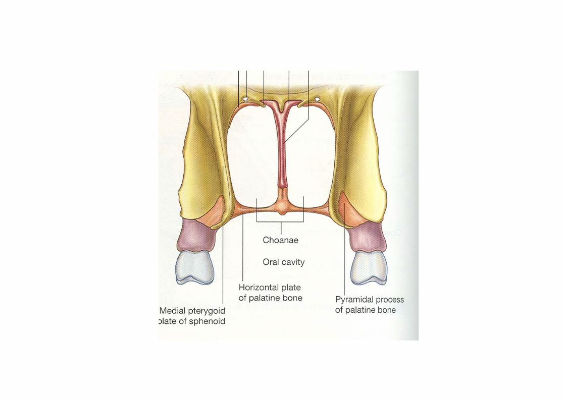
Anterior aperture- External nares- 1.5-2 cm ; 0.5-1 cm (flexible)posterior nasal apertures (choanae)– 2.5 by 1.3 cm (rigid)Separated from : each other- nasal septum
oral cavity-hard palatecranial cavity-parts of frontal, ethmoid,
sphenoid bonesLateral to nasal cavity- orbit
each half- roof , floor
medial wall, lateral wall
three regions- vestibule
respiratory region
olfactory region
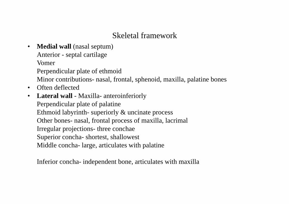
Skeletal framework• Medial wall (nasal septum)
Anterior - septal cartilageVomerPerpendicular plate of ethmoidMinor contributions- nasal, frontal, sphenoid, maxilla, palatine bones
• Often deflected• Lateral wall - Maxilla- anteroinferiorly
Perpendicular plate of palatineEthmoid labyrinth- superiorly & uncinate processOther bones- nasal, frontal process of maxilla, lacrimal Irregular projections- three conchaeSuperior concha- shortest, shallowestMiddle concha- large, articulates with palatine
Inferior concha- independent bone, articulates with maxilla

Skeletal framework-contd.• Floor:Smooth, concave, wider than roofPalatine process of maxillaHorizontal plate of palatine (hard palate)Soft tissue• Roof: narrow, highest in the center
Cribriform plate of ethmoidAnteriorly- nasal spine of frontal, nasal bones, septal
cartilage, major alar cartilagePosteriorly: sphenoid, ala of vomer, palatine, medial
pterygoid plateRoof is perforated by openings in the cribriform plate and a separate foramen for anterior ethmoidal Ns & Vs.


Lateral wall• Corresponding meatuses• Sphenoethmoidal recess• Superior meatus- posterior ethmoidal cells• Middle meatus- continues atrium of middle
meatus, Features- bulla ethmoidalis, hiatus semilunaris, ethmoidal infundibulumOpenings of sinuses- anterior and middle ethmoidal, frontal, maxillary
• Inferior meatus- nasolacrimal duct


Paranasal Sinuses
•Allow equilibration of air and movement of mucous
•All open in to nasal cavity
•Rudimentary/ absent at birth; Variable size, usually asymmetrical
•FRONTAL: posterior to supercilliary arches, 3 cms above nasion
junction of medial 1/3 and lateral 2/3 of supra orbital margin
3.2x2.6x1.8 cm
opening- ethmoidal infundibulum
arterial supply- supraorbital & anterior ethmoidal
nerve supply- supraorbital nerve

ETHMOIDAL: 3-18 in number
anterior (infundibular)-11; middle (bullar)- 3
posterior – 1-7
arterial supply- sphenopalatine & anterior & posterior ethmoidal
nerve supply- anterior & posterior ethmoidal
orbital branch of pterygopalatine ganglion
SPHENOIDAL: Posterior to upper part of nasal cavityabove- optic chiasma & pitutiary glandsides- ICA, cavernous sinus2x 1.8x2.2 cmopening- spheno ethmoidal recessarterial & nerve supply- posterior ethmoidal A & nerve
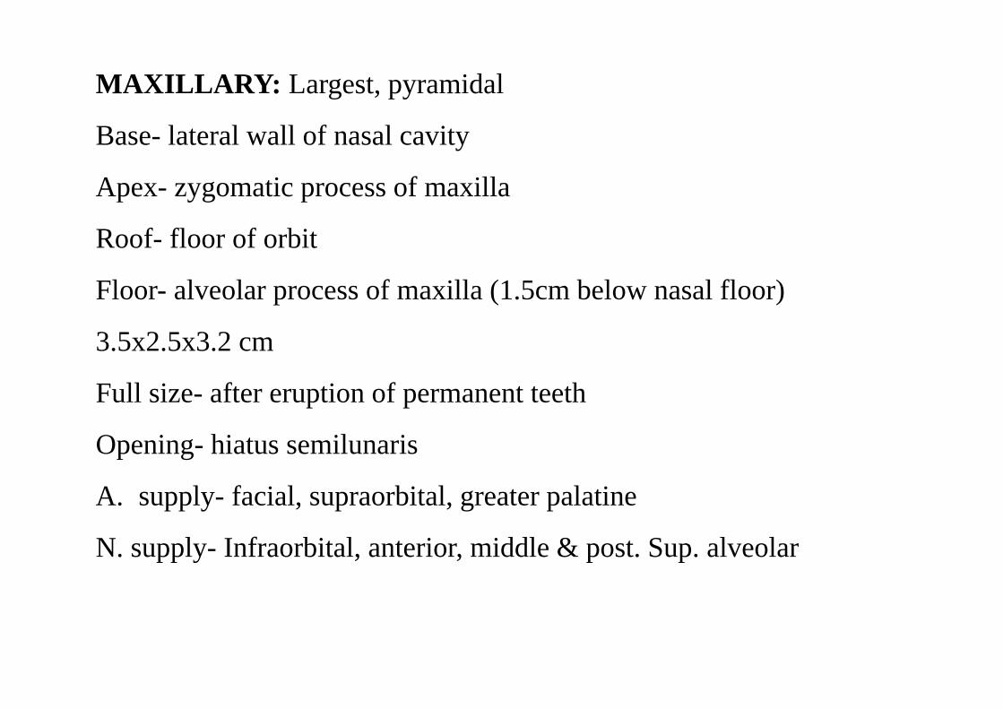
MAXILLARY: Largest, pyramidal
Base- lateral wall of nasal cavity
Apex- zygomatic process of maxilla
Roof- floor of orbit
Floor- alveolar process of maxilla (1.5cm below nasal floor)
3.5x2.5x3.2 cm
Full size- after eruption of permanent teeth
Opening- hiatus semilunaris
A. supply- facial, supraorbital, greater palatine
N. supply- Infraorbital, anterior, middle & post. Sup. alveolar

Clinical anatomy• Sinusitis• Spread of infection• Drainage• Diagnosis- touch, transillumination, radiography• Nasal cavity: Rhinitis
Impaction of foreign bodyEpistaxis ( Little’s area)Deflected nasal septumCleft palate


![Iris Transillumination Defect Spectrum in Pigment ...€¦ · means to detect and record iris transillumination (Figure 1) [3]. Using near infrared iris transillumination imaging](https://img.pdfslide.us/doc/110x75/5f274a006abd3133f941d958/iris-transillumination-defect-spectrum-in-pigment-means-to-detect-and-record.jpg)