Embed Size (px)
Citation preview
Protocol for the Examination of Specimens from Patients with Carcinomas of the Nasal Cavity and Paranasal Sinuses
Protocol applies to all invasive carcinomas of the nasal cavity and paranasal sinuses. Mucosal malignant melanoma is included. Lymphomas, neuroectodermal neoplasms, and sarcomas are not included.
Based on AJCC/UICC TNM, 7th edition Protocol web posting date: October 2009 Procedures • Biopsy • Resection Authors Diane L. Carlson, MD, FCAP* Department of Pathology, Memorial Sloan-Kettering Cancer Center, New York, NY Leon Barnes, MD Department of Pathology, University of Pittsburgh School of Medicine, Pittsburgh, PA John Chan, MD, FCAP Department of Pathology, Queen Elizabeth Hospital, Hong Kong
Gary Ellis, DDS ARUP Laboratories, Salt Lake City, UT Louis B. Harrison, MD Department of Radiation Oncology, Beth Israel Medical Center, St. Luke’s and Roosevelt
Hospitals, New York, NY Jennifer Leigh Hunt, MD, FCAP Department of Pathology, Massachusetts General Hospital, Boston, MA Mary S. Richardson, MD, DDS
Department of Pathology, Medical University of South Carolina, Charleston, SC Jatin Shah, MD, FACS Head and Neck Service, Department of Surgery, Memorial Sloan Kettering Cancer
Center, New York, NY
Lester D. R. Thompson, MD, FCAP Department of Pathology, Southern California Permanente Medical Group, Woodland Hills, CA
Richard Zarbo, MD, DMD, FCAP Department of Pathology, Henry Ford Health Systems, Detroit, MI Bruce M. Wenig, MD, FCAP†
Department of Pathology and Laboratory Medicine, Beth Israel Medical Center, St. Luke’s and Roosevelt Hospitals, New York, NY
For the Members of the Cancer Committee, College of American Pathologists *denotes primary author. † denotes senior author. All other contributing authors are listed alphabetically.
Previous contributors: Ben Z. Pilch, MD; Elizabeth Gillies, MD; John R. Houck Jr, MD; Kyung-Whan Min, MD; David Novis, MD; Jatin Shah, MD; Richard J. Zarbo, MD, DMD; Bruce M Wenig, MD
Head and Neck • Nasal Cavity, Paranasal Sinuses NasalCavityParanasalSinus 3.0.0.0
2
© 2009 College of American Pathologists (CAP). All rights reserved. The College does not permit reproduction of any substantial portion of these protocols without its written authorization. The College hereby authorizes use of these protocols by physicians and other health care providers in reporting on surgical specimens, in teaching, and in carrying out medical research for nonprofit purposes. This authorization does not extend to reproduction or other use of any substantial portion of these protocols for commercial purposes without the written consent of the College.
The CAP also authorizes physicians and other health care practitioners to make modified versions of the Protocols solely for their individual use in reporting on surgical specimens for individual patients, teaching, and carrying out medical research for non-profit purposes.
The CAP further authorizes the following uses by physicians and other health care practitioners, in reporting on surgical specimens for individual patients, in teaching, and in carrying out medical research for non-profit purposes: (1) Dictation from the original or modified protocols for the purposes of creating a text-based patient record on paper, or in a word processing document; (2) Copying from the original or modified protocols into a text-based patient record on paper, or in a word processing document; (3) The use of a computerized system for items (1) and (2), provided that the Protocol data is stored intact as a single text-based document, and is not stored as multiple discrete data fields.
Other than uses (1), (2), and (3) above, the CAP does not authorize any use of the Protocols in electronic medical records systems, pathology informatics systems, cancer registry computer systems, computerized databases, mappings between coding works, or any computerized system without a written license from CAP. Applications for such a license should be addressed to the SNOMED Terminology Solutions division of the CAP.
Any public dissemination of the original or modified Protocols is prohibited without a written license from the CAP.
The College of American Pathologists offers these protocols to assist pathologists in providing clinically useful and relevant information when reporting results of surgical specimen examinations of surgical specimens. The College regards the reporting elements in the “Surgical Pathology Cancer Case Summary (Checklist)” portion of the protocols as essential elements of the pathology report. However, the manner in which these elements are reported is at the discretion of each specific pathologist, taking into account clinician preferences, institutional policies, and individual practice.
The College developed these protocols as an educational tool to assist pathologists in the useful reporting of relevant information. It did not issue the protocols for use in litigation, reimbursement, or other contexts. Nevertheless, the College recognizes that the protocols might be used by hospitals, attorneys, payers, and others. Indeed, effective January 1, 2004, the Commission on Cancer of the American College of Surgeons mandated the use of the checklist elements of the protocols as part of its Cancer Program Standards for Approved Cancer Programs. Therefore, it becomes even more important for pathologists to familiarize themselves with these documents. At the same time, the College cautions that use of the protocols other than for their intended educational purpose may involve additional considerations that are beyond the scope of this document.
The inclusion of a product name or service in a CAP publication should not be construed as an endorsement of such product or service, nor is failure to include the name of a product or service to be construed as disapproval.
Head and Neck • Nasal Cavity, Paranasal Sinuses NasalCavityParanasalSinus 3.0.0.0
3
CAP Nasal Cavity, Paranasal Sinuses Protocol Revision History
Version Code The definition of the version code can be found at www.cap.org/cancerprotocols.
Version: NasalCavityParanasalSinus 3.0.0.0 Summary of Changes No changes have been made since the October 2009 release.
CAP Approved Head and Neck • Nasal Cavity, Paranasal Sinuses NasalCavityParanasalSinus 3.0.0.0
* Data elements with asterisks are not required. However, these elements may be clinically important but are not yet validated or regularly used in patient management.
3
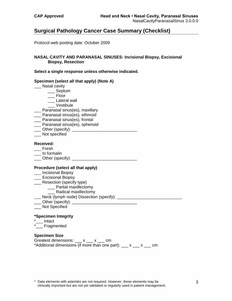
Surgical Pathology Cancer Case Summary (Checklist)
Protocol web posting date: October 2009 NASAL CAVITY AND PARANASAL SINUSES: Incisional Biopsy, Excisional
Biopsy, Resection Select a single response unless otherwise indicated. Specimen (select all that apply) (Note A) ___ Nasal cavity ___ Septum ___ Floor ___ Lateral wall ___ Vestibule ___ Paranasal sinus(es), maxillary ___ Paranasal sinus(es), ethmoid ___ Paranasal sinus(es), frontal ___ Paranasal sinus(es), sphenoid ___ Other (specify): ____________________________ ___ Not specified Received: ___ Fresh ___ In formalin ___ Other (specify): ____________________________ Procedure (select all that apply) ___ Incisional Biopsy ___ Excisional Biopsy ___ Resection (specify type) ___ Partial maxillectomy ___ Radical maxillectomy
___ Neck (lymph node) Dissection (specify): ____________________________
___ Other (specify): ____________________________ ___ Not Specified *Specimen Integrity *___ Intact *___ Fragmented Specimen Size Greatest dimensions: ___ x ___ x ___ cm *Additional dimensions (if more than one part): ___ x ___ x ___ cm
CAP Approved Head and Neck • Nasal Cavity, Paranasal Sinuses NasalCavityParanasalSinus 3.0.0.0
* Data elements with asterisks are not required. However, these elements may be clinically important but are not yet validated or regularly used in patient management.
4
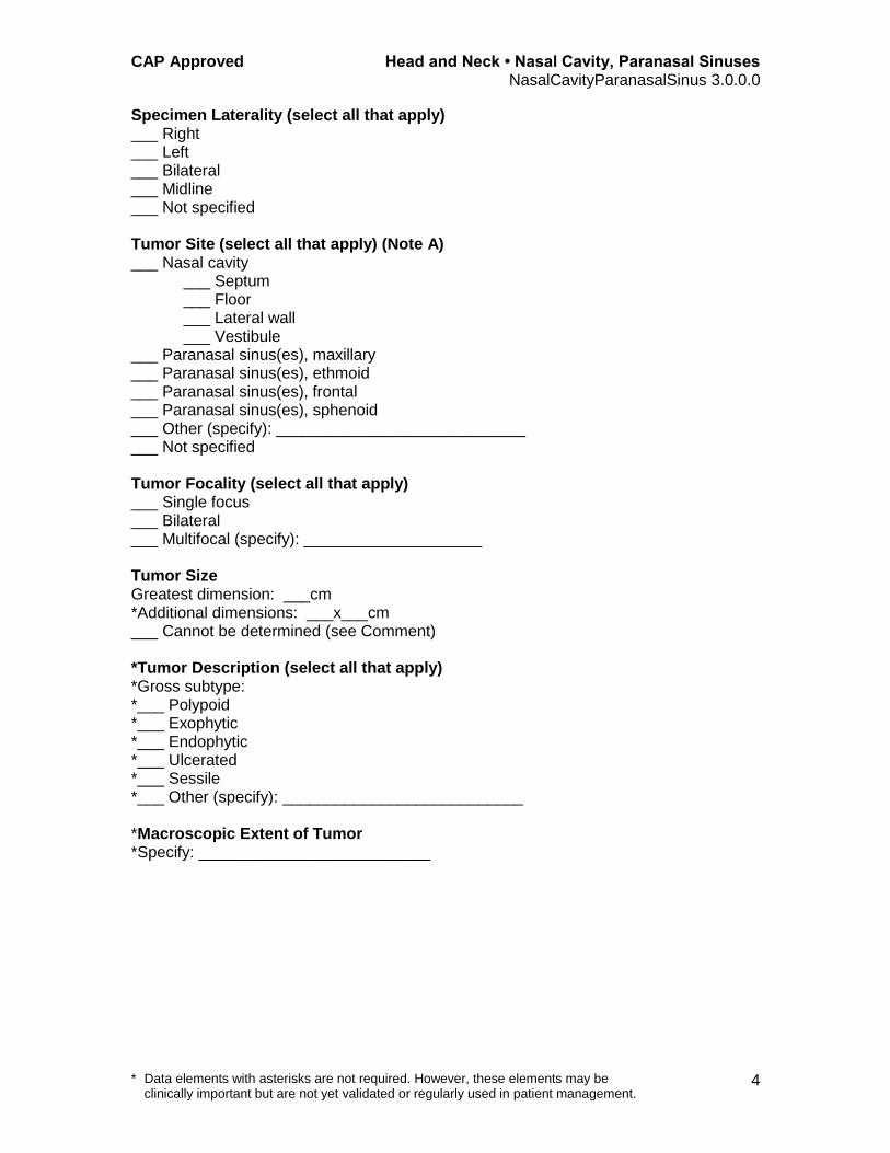
Specimen Laterality (select all that apply) ___ Right ___ Left ___ Bilateral ___ Midline ___ Not specified Tumor Site (select all that apply) (Note A) ___ Nasal cavity ___ Septum ___ Floor ___ Lateral wall ___ Vestibule ___ Paranasal sinus(es), maxillary ___ Paranasal sinus(es), ethmoid ___ Paranasal sinus(es), frontal ___ Paranasal sinus(es), sphenoid ___ Other (specify): ____________________________ ___ Not specified Tumor Focality (select all that apply) ___ Single focus ___ Bilateral ___ Multifocal (specify): ____________________ Tumor Size Greatest dimension: ___cm *Additional dimensions: ___x___cm ___ Cannot be determined (see Comment) *Tumor Description (select all that apply) *Gross subtype: *___ Polypoid *___ Exophytic *___ Endophytic *___ Ulcerated *___ Sessile *___ Other (specify): ___________________________ *Macroscopic Extent of Tumor *Specify: __________________________
CAP Approved Head and Neck • Nasal Cavity, Paranasal Sinuses NasalCavityParanasalSinus 3.0.0.0
* Data elements with asterisks are not required. However, these elements may be clinically important but are not yet validated or regularly used in patient management.
5
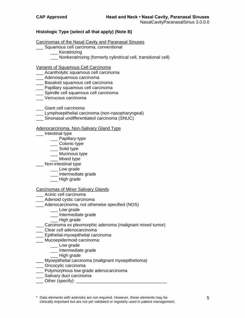
Histologic Type (select all that apply) (Note B) Carcinomas of the Nasal Cavity and Paranasal Sinuses ___ Squamous cell carcinoma, conventional ___ Keratinizing ___ Nonkeratinizing (formerly cylindrical cell, transitional cell) Variants of Squamous Cell Carcinoma ___ Acantholytic squamous cell carcinoma ___ Adenosquamous carcinoma ___ Basaloid squamous cell carcinoma ___ Papillary squamous cell carcinoma ___ Spindle cell squamous cell carcinoma ___ Verrucous carcinoma ___ Giant cell carcinoma
___ Lymphoepithelial carcinoma (non-nasopharyngeal) ___ Sinonasal undifferentiated carcinoma (SNUC) Adenocarcinoma, Non-Salivary Gland Type ___ Intestinal type ___ Papillary-type ___ Colonic-type ___ Solid type ___ Mucinous type ___ Mixed type ___ Non-intestinal type ___ Low grade ___ Intermediate grade ___ High grade Carcinomas of Minor Salivary Glands ___ Acinic cell carcinoma ___ Adenoid cystic carcinoma ___ Adenocarcinoma, not otherwise specified (NOS) ___ Low grade ___ Intermediate grade ___ High grade ___ Carcinoma ex pleomorphic adenoma (malignant mixed tumor) ___ Clear cell adenocarcinoma ___ Epithelial-myoepithelial carcinoma ___ Mucoepidermoid carcinoma: ___ Low grade ___ Intermediate grade ___ High grade ___ Myoepithelial carcinoma (malignant myoepithelioma) ___ Oncocytic carcinoma ___ Polymorphous low-grade adenocarcinoma ___ Salivary duct carcinoma ___ Other (specify): _____________________________________

CAP Approved Head and Neck • Nasal Cavity, Paranasal Sinuses NasalCavityParanasalSinus 3.0.0.0
* Data elements with asterisks are not required. However, these elements may be clinically important but are not yet validated or regularly used in patient management.
6
Neuroendocrine Carcinoma ___ Typical carcinoid tumor (well differentiated neuroendocrine carcinoma) ___ Atypical carcinoid tumor (moderately differentiated neuroendocrine carcinoma) ___ Small cell carcinoma (poorly differentiated neuroendocrine carcinoma) ___ Combined (or composite) small cell carcinoma, neuroendocrine type ___ Mucosal malignant melanoma ___ Other (specify): _____________________________________ ___ Carcinoma, type cannot be determined Histologic Grade (Note C) ___ Not applicable ___ GX: Cannot be assessed ___ G1: Well differentiated ___ G2: Moderately differentiated ___ G3: Poorly differentiated ___ Other (specify): _____________________________________ *Microscopic Tumor Extension *Specify: ____________________________ Margins (select all that apply) (Notes D and E) ___ Cannot be assessed ___ Margins uninvolved by invasive carcinoma Distance from closest margin: ___ mm or ___ cm Specify margin(s), per orientation, if possible: _______________ ___ Margins involved by invasive carcinoma Specify margin(s), per orientation, if possible: _______________ ___ Margins uninvolved by carcinoma in situ (includes moderate and severe dysplasia#)
(Note D) Distance from closest margin: ___ mm or ___ cm Specify margin(s), per orientation, if possible: _______________ ___ Margins involved by carcinoma in situ (includes moderate and severe dysplasia#)
(Note D) Specify margin(s), per orientation, if possible: _______________ ___ Not applicable # Applicable only to squamous cell carcinoma and histologic variants.
*Treatment Effect (applicable to carcinomas treated with neoadjuvant therapy) *___ Not identified *___ Present (specify): ____________________ *___ Indeterminate Lymph-Vascular Invasion ___ Not Identified ___ Present ___ Indeterminate
CAP Approved Head and Neck • Nasal Cavity, Paranasal Sinuses NasalCavityParanasalSinus 3.0.0.0
* Data elements with asterisks are not required. However, these elements may be clinically important but are not yet validated or regularly used in patient management.
7
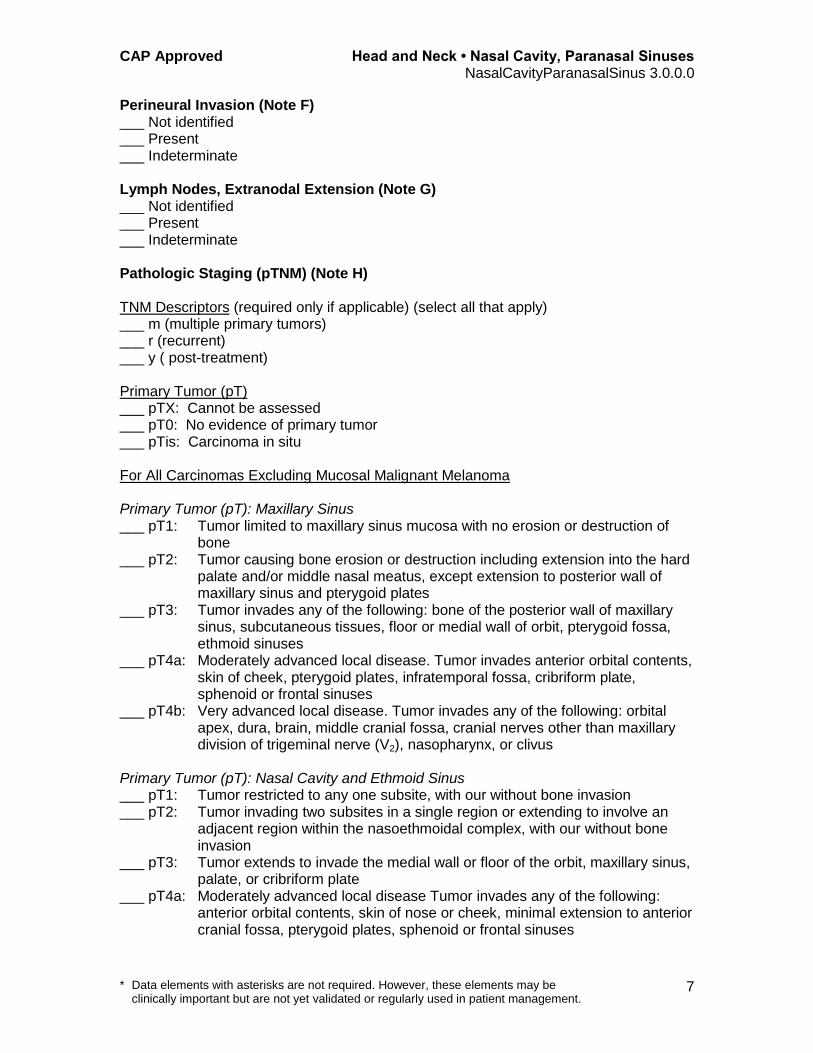
Perineural Invasion (Note F) ___ Not identified ___ Present ___ Indeterminate Lymph Nodes, Extranodal Extension (Note G) ___ Not identified ___ Present ___ Indeterminate Pathologic Staging (pTNM) (Note H) TNM Descriptors (required only if applicable) (select all that apply) ___ m (multiple primary tumors) ___ r (recurrent) ___ y ( post-treatment) Primary Tumor (pT) ___ pTX: Cannot be assessed ___ pT0: No evidence of primary tumor ___ pTis: Carcinoma in situ For All Carcinomas Excluding Mucosal Malignant Melanoma Primary Tumor (pT): Maxillary Sinus ___ pT1: Tumor limited to maxillary sinus mucosa with no erosion or destruction of
bone ___ pT2: Tumor causing bone erosion or destruction including extension into the hard
palate and/or middle nasal meatus, except extension to posterior wall of maxillary sinus and pterygoid plates
___ pT3: Tumor invades any of the following: bone of the posterior wall of maxillary sinus, subcutaneous tissues, floor or medial wall of orbit, pterygoid fossa, ethmoid sinuses
___ pT4a: Moderately advanced local disease. Tumor invades anterior orbital contents, skin of cheek, pterygoid plates, infratemporal fossa, cribriform plate, sphenoid or frontal sinuses
___ pT4b: Very advanced local disease. Tumor invades any of the following: orbital apex, dura, brain, middle cranial fossa, cranial nerves other than maxillary division of trigeminal nerve (V2), nasopharynx, or clivus
Primary Tumor (pT): Nasal Cavity and Ethmoid Sinus ___ pT1: Tumor restricted to any one subsite, with our without bone invasion ___ pT2: Tumor invading two subsites in a single region or extending to involve an
adjacent region within the nasoethmoidal complex, with our without bone invasion
___ pT3: Tumor extends to invade the medial wall or floor of the orbit, maxillary sinus, palate, or cribriform plate
___ pT4a: Moderately advanced local disease Tumor invades any of the following: anterior orbital contents, skin of nose or cheek, minimal extension to anterior cranial fossa, pterygoid plates, sphenoid or frontal sinuses
CAP Approved Head and Neck • Nasal Cavity, Paranasal Sinuses NasalCavityParanasalSinus 3.0.0.0
* Data elements with asterisks are not required. However, these elements may be clinically important but are not yet validated or regularly used in patient management.
8
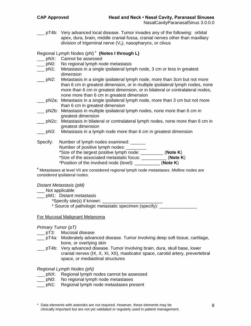
___ pT4b: Very advanced local disease. Tumor invades any of the following: orbital apex, dura, brain, middle cranial fossa, cranial nerves other than maxillary division of trigeminal nerve (V2), nasopharynx, or clivus
Regional Lymph Nodes (pN) # (Notes I through L)
___ pNX: Cannot be assessed ___ pN0: No regional lymph node metastasis ___ pN1: Metastasis in a single ipsilateral lymph node, 3 cm or less in greatest
dimension ___ pN2: Metastasis in a single ipsilateral lymph node, more than 3cm but not more
than 6 cm in greatest dimension, or in multiple ipsilateral lymph nodes, none more than 6 cm in greatest dimension, or in bilateral or contralateral nodes, none more than 6 cm in greatest dimension
___ pN2a: Metastasis in a single ipsilateral lymph node, more than 3 cm but not more than 6 cm in greatest dimension
___ pN2b: Metastasis in multiple ipsilateral lymph nodes, none more than 6 cm in greatest dimension
___ pN2c: Metastasis in bilateral or contralateral lymph nodes, none more than 6 cm in greatest dimension
___ pN3: Metastasis in a lymph node more than 6 cm in greatest dimension Specify: Number of lymph nodes examined: ______ Number of positive lymph nodes: ________ *Size of the largest positive lymph node: _________ (Note K) *Size of the associated metastatic focus: __________ (Note K) *Position of the involved node (level): __________ (Note K) # Metastases at level VII are considered regional lymph node metastases. Midline nodes are
considered ipsilateral nodes.
Distant Metastasis (pM) ___ Not applicable ___ pM1: Distant metastasis *Specify site(s) if known: ________________________ * Source of pathologic metastatic specimen (specify): _______________
For Mucosal Malignant Melanoma Primary Tumor (pT) ___ pT3: Mucosal disease ___ pT4a: Moderately advanced disease. Tumor involving deep soft tissue, cartilage,
bone, or overlying skin ___ pT4b: Very advanced disease. Tumor involving brain, dura, skull base, lower
cranial nerves (IX, X, XI, XII), masticator space, carotid artery, prevertebral space, or mediastinal structures
Regional Lymph Nodes (pN) ___ pNX: Regional lymph nodes cannot be assessed ___ pN0: No regional lymph node metastases ___ pN1: Regional lymph node metastases present
CAP Approved Head and Neck • Nasal Cavity, Paranasal Sinuses NasalCavityParanasalSinus 3.0.0.0
* Data elements with asterisks are not required. However, these elements may be clinically important but are not yet validated or regularly used in patient management.
9
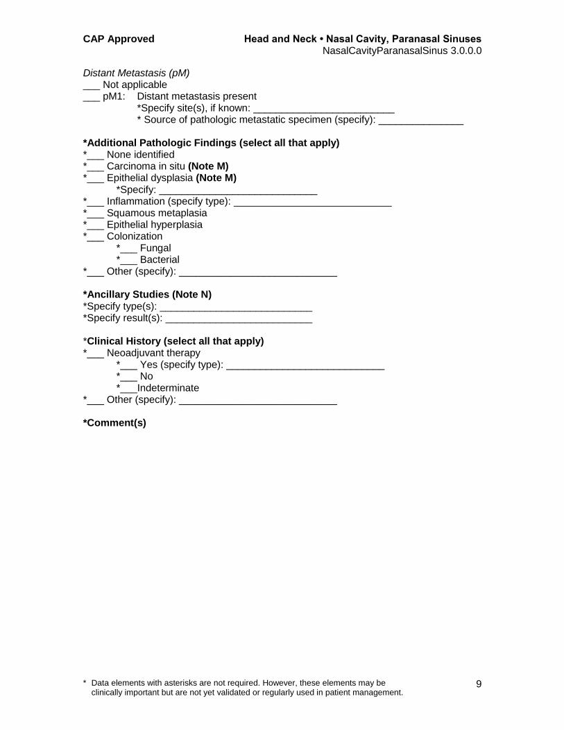
Distant Metastasis (pM) ___ Not applicable ___ pM1: Distant metastasis present *Specify site(s), if known: _________________________
* Source of pathologic metastatic specimen (specify): _______________ *Additional Pathologic Findings (select all that apply) *___ None identified *___ Carcinoma in situ (Note M) *___ Epithelial dysplasia (Note M) *Specify: ____________________________ *___ Inflammation (specify type): ____________________________ *___ Squamous metaplasia *___ Epithelial hyperplasia *___ Colonization *___ Fungal *___ Bacterial *___ Other (specify): ____________________________ *Ancillary Studies (Note N) *Specify type(s): ___________________________ *Specify result(s): __________________________ *Clinical History (select all that apply) *___ Neoadjuvant therapy *___ Yes (specify type): ____________________________ *___ No *___Indeterminate *___ Other (specify): ____________________________ *Comment(s)
Background Documentation Head and Neck • Nasal Cavity, Paranasal Sinuses NasalCavityParanasalSinus 3.0.0.0
10
Explanatory Notes
Scope of Guidelines The reporting of oral cancer including the lip is facilitated by the provision of a checklist illustrating the features required for comprehensive patient care. However, there are many cases in which the individual practicalities of applying such a checklist may not be straightforward. Common examples include finding the prescribed number of lymph nodes, trying to determine the levels of the radical neck dissection, and determining if isolated tumor cells in a lymph node represent metastatic disease. Checklists have evolved to include clinical, radiographic, morphologic, immunohistochemical, and molecular results in an effort to guide clinical management. Adjuvant and neoadjuvant therapy can significantly alter histologic findings, making accurate classification an increasingly complex and demanding task. This checklist tries to remain simple while still incorporating important pathologic features as proposed by the American Joint Committee on Cancer (AJCC) cancer staging manual, the World Health Organization classification of tumours, the TNM classification, the American College of Surgeons Commission on Cancer, and the International Union on Cancer (UICC). This checklist is to be used as a guide and resource, an adjunct to diagnosing and managing cancers of the oral cavity in a standardized manner. It should not be used as a substitute for dissection or grossing techniques and does not give histologic parameters to reach the diagnosis. Subjectivity is always a factor, and elements listed are not meant to be arbitrary but are meant to provide uniformity of reporting across all the disciplines that use the information. It is a foundation of practical information that will help to meet the requirements of daily practice to benefit both clinicians and patients alike.
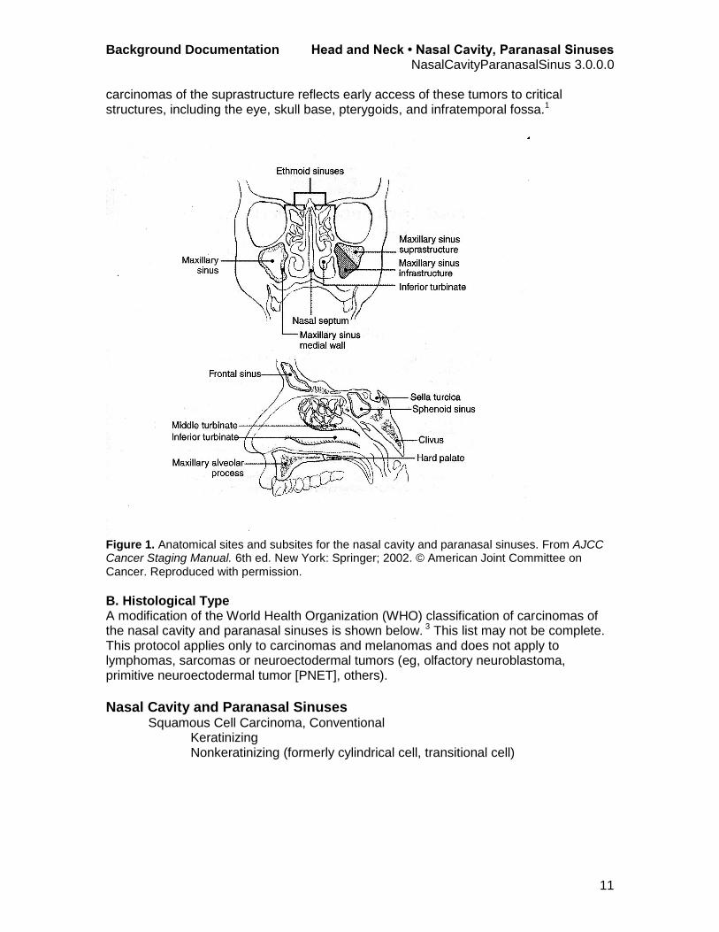
A. Anatomical Sites and Subsites for the Nasal Cavity and Paranasal Sinuses (Figure 1) The nasal cavity is divided in the midline to right and left halves by the septum; each half opens on the face via the nares or nostrils and communicates behind with the nasopharynx through the posterior nasal apertures or the choanae. The nasal cavity is divided into 4 subsites including the septum, floor, lateral wall, and vestibule. The paranasal sinuses represent a grouping of 4 paired sinuses including the maxillary sinuses, ethmoid sinuses, frontal sinuses, and sphenoid sinuses. The nasoethmoidal complex is divided into 2 sites including the nasal cavity and the ethmoid sinuses. Cancers of the maxillary sinuses are the most common sinonasal malignancies followed by cancers of the ethmoid sinuses, which are much less common.1 Cancers of the frontal and sphenoid sinuses are rare. When considering the nasal cavity and paranasal sinuses, 60% of malignant neoplasms originate from the maxillary sinus, 20% to 30% from the nasal cavity, 10% to 15% from the ethmoid sinus and 1% from the sphenoid and frontal sinuses. 2 When only considering the paranasal sinuses, 77% of malignant neoplasms originate from the maxillary sinus, 22% from the ethmoid sinus, and 1% from the sphenoid and frontal sinuses.2 The location as well as the extent of the mucosal lesion in the maxillary sinus has prognostic importance. Ohngren's line, connecting the medial canthus of the eye to the angle of the mandible, divides the maxillary sinus into an anterioinferior portion (infrastructure) and superioposterior portion (suprastructure) structures. Carcinomas of the infrastructure are associated with a good prognosis; carcinomas of the suprastructure are associated with a poor prognosis. The poorer prognosis with
Background Documentation Head and Neck • Nasal Cavity, Paranasal Sinuses NasalCavityParanasalSinus 3.0.0.0
11
carcinomas of the suprastructure reflects early access of these tumors to critical structures, including the eye, skull base, pterygoids, and infratemporal fossa.1
Figure 1. Anatomical sites and subsites for the nasal cavity and paranasal sinuses. From AJCC Cancer Staging Manual. 6th ed. New York: Springer; 2002. © American Joint Committee on Cancer. Reproduced with permission.
B. Histological Type A modification of the World Health Organization (WHO) classification of carcinomas of the nasal cavity and paranasal sinuses is shown below. 3 This list may not be complete. This protocol applies only to carcinomas and melanomas and does not apply to lymphomas, sarcomas or neuroectodermal tumors (eg, olfactory neuroblastoma, primitive neuroectodermal tumor [PNET], others).
Nasal Cavity and Paranasal Sinuses Squamous Cell Carcinoma, Conventional Keratinizing Nonkeratinizing (formerly cylindrical cell, transitional cell)
Background Documentation Head and Neck • Nasal Cavity, Paranasal Sinuses NasalCavityParanasalSinus 3.0.0.0
12
Variants of Squamous Cell Carcinoma (in alphabetical order)
Acantholytic squamous cell carcinoma Adenosquamous carcinoma Basaloid squamous cell carcinoma Papillary squamous cell carcinoma Spindle cell squamous cell carcinoma Verrucous carcinoma
Giant cell carcinoma# Lymphoepithelial carcinoma (non-nasopharyngeal) Sinonasal undifferentiated carcinoma (SNUC) Adenocarcinoma, Non-Salivary Gland Type Intestinal-type Non-intestinal type Carcinomas of Minor Salivary Glands Acinic cell carcinoma Adenoid cystic carcinoma Adenocarcinoma, not otherwise specified (NOS) Carcinoma ex pleomorphic adenoma (malignant mixed tumor) Clear cell adenocarcinoma Mucoepidermoid carcinoma: Epithelial-myoepithelial carcinoma Myoepithelial carcinoma (malignant myoepithelioma) Oncocytic carcinoma Polymorphous low-grade adenocarcinoma Salivary duct carcinoma Other Neuroendocrine Carcinoma Typical carcinoid tumor (well differentiated neuroendocrine carcinoma) Atypical carcinoid tumor (moderately differentiated neuroendocrine carcinoma) Small cell (undifferentiated) carcinoma (poorly differentiated neuroendocrine
carcinoma) Combined (or composite) small cell carcinoma, neuroendocrine type##
Mucosal Malignant Melanoma # Not included in WHO classification. ## Represents a carcinoma showing combined features of small cell neuroendocrine carcinoma associated with a squamous or adenocarcinomatous component.4 C. Histologic Grade
Background Documentation Head and Neck • Nasal Cavity, Paranasal Sinuses NasalCavityParanasalSinus 3.0.0.0
13
For histologic types of carcinomas that are amenable to grading, 3 histologic grades are suggested, as shown below. When a tumor manifests more than 1 grade of differentiation, the surgical report must designate both the highest and the most prevalent tumor grades.5,6 Grade X Cannot be assessed Grade 1 Well differentiated Grade 2 Moderately differentiated Grade 3 Poorly differentiated This grading system does not apply to all salivary gland tumors. The histologic (microscopic) grading of salivary gland carcinomas has been shown to be an independent predictor of behavior and plays a role in optimizing therapy.7-11 Further, there is often a positive correlation between histologic grade and clinical stage. For the majority of salivary gland carcinomas there is only a single histologic grade and classification alone determines the histologic grade (eg, acinic cell carcinoma is a histologically low-grade carcinoma; salivary duct carcinoma is a histologically high-grade carcinoma). With some exceptions, histologic grading is predicated on cytomorphologic features. In this histologic grading scheme, 3 histologic grades are suggested, as follows: Grade 1 Well differentiated = Low-grade Grade 2 Moderately differentiated = Intermediate-grade Grade 3 Poorly differentiated = High-grade Grade X Cannot be assessed When a tumor manifests more than 1 grade of differentiation, the surgical report must designate both the highest and the most prevalent tumor grades. In some carcinomas, histologic grading may be based on growth pattern, such as in adenoid cystic carcinoma, for which a histologic high-grade variant has been recognized based on the percentage of solid growth.7 Those adenoid cystic carcinomas showing 30% or greater of solid growth pattern are considered to be histologically high-grade carcinomas.7,9,12 The histologic grading of mucoepidermoid carcinoma includes a combination of growth pattern characteristics (eg, cystic, solid, neurotropism) and cytomorphologic findings (eg, anaplasia, mitoses, necrosis).13-15
D. Surgical Margins Reporting of surgical margins should include information regarding the distance of invasive carcinoma, carcinoma in situ or high grade dysplasia (moderate to severe) from the surgical margin. Closeness of the above, microscopically less than 5 mm, from the surgical border should be noted in the report. Presence of the above lesions found within 5 mm of the surgical border carry a significant risk for subsequent local recurrence.16-18 Reporting of surgical margins for carcinomas of the minor salivary glands should follow those used for squamous cell carcinoma of oral cavity. There is no category of carcinoma in situ relative to carcinomas of salivary glands (major, minor). Unlike the oral cavity and larynx, intraepithelial dysplasias including nonkeratinizing and keratinizing dysplasias as well as carcinoma in situ of the nasal cavity and paranasal sinuses are uncommon, especially as an isolated clinical and/or histopathologic lesion. In the sinonasal tract, when carcinoma in situ is identified, it usually is seen in
Background Documentation Head and Neck • Nasal Cavity, Paranasal Sinuses NasalCavityParanasalSinus 3.0.0.0
14
association with an invasive carcinoma. In this setting, the same criteria detailed in the oral cavity and laryngeal protocols apply (see Protocol for the Examination of Specimens from Patients with Carcinomas of the Lip and Oral Cavity and Protocol for the Examination of Specimens from Patients with Carcinomas of the Larynx).
E. Orientation of Specimen Complex specimens should be examined and oriented with the assistance of attending surgeons. Direct communication between the surgeon and pathologist is a critical component in specimen orientation and proper sectioning. Whenever possible, the tissue examination request form should include a drawing of the resected specimen showing the extent of the tumor and its relation to the anatomic structures of the region. The lines and extent of the resection can be depicted on preprinted adhesive labels and attached to the surgical pathology request forms. F. Perineural Invasion The presence of perineural invasion (neurotropism) is an important predictor of poor prognosis in head and neck cancer of virtually all sites.19 The presence of perineural invasion (neurotropism) in the primary cancer is associated with poor local disease control and regional control, as well as being associated with metastasis to regional lymph nodes.19 Further, perineural invasion is associated with decrease in disease-specific survival and overall survival.19 There is conflicting data relative to an association between the presence of perineural invasion and the development of distant metastasis with some studies showing an increased association with distant metastasis but other studies not showing any correlation with distant metastasis.19 The relationship between perineural invasion and prognosis is independent of nerve diameter.20 Although perineural invasion of small unnamed nerves may not produce clinical symptoms, the reporting of perineural invasion includes nerves of all sizes including small peripheral nerves (ie, less than 1 mm in diameter). Aside from the impact on prognosis, the presence of perineural invasion also guides therapy. Concurrent adjuvant chemoradiation therapy has been shown to improve outcomes in patients with perineural invasion (as well as in patients with extranodal extension and bone invasion).21,22 Given the significance relative to prognosis and treatment, perineural invasion is a required data element in the reporting of head and neck cancers. G. Extranodal Extension The status of cervical lymph nodes is the single most important prognostic factor in aerodigestive cancer. All macroscopically negative or equivocal lymph nodes should be submitted in toto. Grossly positive nodes may be partially submitted for microscopic documentation of metastasis. Reporting of lymph nodes containing metastasis should include whether there is presence or absence of extranodal extension (EE). This finding consists of extension of metastatic tumor, present within the confines of the lymph node, through the lymph node capsule into the surrounding connective tissue, with or without associated stromal reaction. If macroscopic examination suggests EE, this tissue should be submitted for microscopic confirmation. EE is a predictor of regional relapse and a criterion for postoperative radiotherapy.23-26 H. TNM and Stage Groupings The protocol recommends the TNM staging system of the American Joint Committee on Cancer (AJCC) and the International Union Against Cancer (UICC) for nasal cavity and paranasal sinus cancer.1,27 Of note in the 7th edition of the AJCC staging of head and
Background Documentation Head and Neck • Nasal Cavity, Paranasal Sinuses NasalCavityParanasalSinus 3.0.0.0
15
neck cancers1 is the division of T4 lesions into T4a (moderately advanced local disease) and T4b (very advanced local disease), leading to the stratification of stage IV into stage IVA (moderately advanced local/regional disease), stage IVB (very advanced local/regional disease), and stage IVC (distant metastatic disease). The 7th edition of the AJCC staging of head and neck cancers includes mucosal malignant melanomas.1 Approximately two-thirds of mucosal malignant melanomas arise in the sinonasal tract, one-quarter are found in the oral cavity and the remainder occur only sporadically in other mucosal sites of the head and neck.1 Even small cancers behave aggressively with high rates of recurrence and death.1 To reflect this aggressive behavior, primary cancers limited to the mucosa are considered T3 lesions. Advanced mucosal melanomas are classified as T4a and T4b. The anatomic extent criteria to define moderately advanced (T4a) and very advanced (T4b) disease are given below. The AJCC staging for mucosal malignant melanomas does not provide for the histologic definition of a T3 lesion; as the majority of mucosal malignant melanomas are invasive at presentation, mucosal based melanomas (T3 lesions) include those lesions that involve either the epithelium and/or lamina propria of the involved site. Rare examples of in situ mucosal melanomas occur but in situ mucosal melanomas are excluded from staging, as they are extremely rare.1 For All Carcinomas Excluding Mucosal Malignant Melanoma Primary Tumor: Maxillary Sinus TX Cannot be assessed T0 No evidence of primary tumor Tis Carcinoma in situ T1 Tumor limited to the maxillary sinus mucosa with no erosion or destruction of
bone T2 Tumor causing bone erosion or destruction including extension into the hard
palate and/or middle nasal meatus, except extension to posterior wall of maxillary sinus and pterygoid plates
T3 Tumor invades any of the following: bone of the posterior wall of maxillary sinus, subcutaneous tissues, floor or medial wall of orbit, pterygoid fossa, ethmoid sinuses
T4a Tumor invades anterior orbital contents, skin of cheek, pterygoid plates, infratemporal fossa, cribriform plate, sphenoid or frontal sinuses
T4b Tumor invades any of the following: orbital apex, dura, brain, middle cranial fossa, cranial nerves other than maxillary division of trigeminal nerve (V2), nasopharynx, or clivus
Primary Tumor: Nasal Cavity and Ethmoid Sinus TX Cannot be assessed T0 No evidence of primary tumor Tis Carcinoma in situ T1 Tumor restricted to any one subsite, with or without bone invasion T2 Tumor invading two subsites in a single region or extending to involve an
adjacent region within the nasoethmoidal complex, with our without bone invasion
T3 Tumor extends to invade the medial wall or floor of the orbit, maxillary sinus, palate, or cribriform plate
Background Documentation Head and Neck • Nasal Cavity, Paranasal Sinuses NasalCavityParanasalSinus 3.0.0.0
16
T4a Tumor invades any of the following: anterior orbital contents, skin of nose or cheek, minimal extension to anterior cranial fossa, pterygoid plates, sphenoid or frontal sinuses
T4b Tumor invades any of the following: orbital apex, dura, brain, middle cranial fossa, cranial nerves other than maxillary division of trigeminal nerve (V2), nasopharynx, or clivus
Regional Lymph Nodes# NX Cannot be assessed N0 No regional lymph node metastasis N1 Metastasis in a single ipsilateral lymph node, 3 cm or less in greatest dimension N2 Metastasis in a single ipsilateral lymph node, more than 3 cm but not more than
6 cm in greatest dimension, or in multiple ipsilateral lymph nodes, none more than 6 cm in greatest dimension, or in bilateral or contralateral nodes, none more than 6 cm in greatest dimension
N2a Metastasis in a single ipsilateral lymph node, more than 3 cm but not more than 6 cm in greatest dimension
N2b Metastasis in multiple ipsilateral lymph nodes, none more than 6 cm in greatest dimension
N2c Metastasis in bilateral or contralateral lymph nodes, none more than 6 cm in greatest dimension
N3 Metastasis in a lymph node more than 6 cm in greatest dimension # Metastases at level VII are considered regional lymph node metastases. Midline nodes are considered ipsilateral nodes. Distant Metastasis M0 No distant metastasis M1 Distant metastasis For Mucosal Malignant Melanoma Primary Tumor T3 Mucosal disease T4a Moderately advanced disease. Tumor involving deep soft tissue, cartilage, bone,
or overlying skin T4b Very advanced disease. Tumor involving brain, dura, skull base, lower cranial
nerves (IX, X, XI, XII), masticator space, carotid artery, prevertebral space, or mediastinal structures
Regional Lymph Nodes NX Regional lymph nodes cannot be assessed N0 No regional lymph node metastases N1 Regional lymph node metastases present Distant Metastasis M0 No distant metastasis M1 Distant metastasis present By AJCC/UICC convention, the designation “T” refers to a primary tumor that has not been previously treated. The symbol “p” refers to the pathologic classification of the
Background Documentation Head and Neck • Nasal Cavity, Paranasal Sinuses NasalCavityParanasalSinus 3.0.0.0
17
TNM, as opposed to the clinical classification, and is based on gross and microscopic examination. pT entails a resection of the primary tumor or biopsy adequate to evaluate the highest pT category, pN entails removal of nodes adequate to validate lymph node metastasis, and pM implies microscopic examination of distant lesions. Clinical classification (cTNM) is usually carried out by the referring physician before treatment during initial evaluation of the patient or when pathologic classification is not possible. Pathologic staging is usually performed after surgical resection of the primary tumor. Pathologic staging depends on pathologic documentation of the anatomic extent of disease, whether or not the primary tumor has been completely removed. If a biopsied tumor is not resected for any reason (eg, when technically unfeasible) and if the highest T and N categories or the M1 category of the tumor can be confirmed microscopically, the criteria for pathologic classification and staging have been satisfied without total removal of the primary cancer. Stage Groupings – For All Cancers Except Mucosal Malignant Melanoma Stage 0 Tis N0 M0 Stage I T1 N0 M0 Stage II T2 N0 M0 Stage III T1 N1 M0 T2 N1 M0 T3 N0,N1 M0 Stage IVA T1,T2,T3 N2 M0 T4a N0,N1,N2 M0 Stage IVB T4b Any N M0 Any T N3 M0 Stage IVC Any T Any N M1
Stage Groupings – For Mucosal Malignant Melanoma Stage III T3 N0 M0 Stage IVA T4a N0 M0 T3-T4a N1 M0 Stage IVB T4b Any N M0 Stage IVC Any T Any N M1 TNM Descriptors For identification of special cases of TNM or pTNM classifications, the “m” suffix and “y” and “r” prefixes are used. Although they do not affect the stage grouping, they indicate cases needing separate analysis. The “m” suffix indicates the presence of multiple primary tumors in a single site and is recorded in parentheses: pT(m)NM. The “y” prefix indicates those cases in which classification is performed during or following initial multimodality therapy (ie, neoadjuvant chemotherapy, radiation therapy, or both chemotherapy and radiation therapy). The cTNM or pTNM category is identified by a “y” prefix. The ycTNM or ypTNM categorizes the extent of tumor actually present at the time of that examination. The “y” categorization is not an estimate of tumor prior to multimodality therapy (ie, before initiation of neoadjuvant therapy).
Background Documentation Head and Neck • Nasal Cavity, Paranasal Sinuses NasalCavityParanasalSinus 3.0.0.0
18
The “r” prefix indicates a recurrent tumor when staged after a documented disease-free interval, and is identified by the “r” prefix: rTNM. Additional Descriptors Residual Tumor (R) Tumor remaining in a patient after therapy with curative intent (eg, surgical resection for cure) is categorized by a system known as R classification, shown below. RX Presence of residual tumor cannot be assessed R0 No residual tumor R1 Microscopic residual tumor R2 Macroscopic residual tumor For the surgeon, the R classification may be useful to indicate the known or assumed status of the completeness of a surgical excision. For the pathologist, the R classification is relevant to the status of the margins of a surgical resection specimen. That is, tumor involving the resection margin on pathologic examination may be assumed to correspond to residual tumor in the patient and may be classified as macroscopic or microscopic according to the findings at the specimen margin(s). I. Classification of Neck Dissection 1. Radical neck dissection 2. Modified radical neck dissection, internal jugular vein and/or sternocleidomastoid
muscle spared 3. Selective neck dissection (SND), as specified by the surgeon a. Supraomohyoid neck dissection b. Posterolateral neck dissection c. Lateral neck dissection d. Central compartment neck dissection 4. Selective neck dissection (SND), as specified by the surgeon -“SND” with levels and
sublevels designated (Figure 2).28-30 5. Extended radical neck dissection, as specified by the surgeon J. Regional Lymph Nodes (pN0): Isolated Tumor Cells Isolated tumor cells (ITCs) are single cells or small clusters of cells not more than 0.2 mm in greatest dimension. Lymph nodes or distant sites with ITCs found by either histologic examination, immunohistochemistry, or non-morphologic techniques (eg, flow cytometry, DNA analysis, polymerase chain reaction [PCR] amplification of a specific tumor marker) should be classified as N0 or M0, respectively. Specific denotation of the assigned N category is suggested as follows for cases in which ITCs are the only evidence of possible metastatic disease.27,31,32
pN0 No regional lymph node metastasis histologically, no examination for
isolated tumor cells (ITCs) pN0(i-) No regional lymph node metastasis histologically, negative morphologic
(any morphologic technique, including hematoxylin-eosin and immunohistochemistry) findings for ITCs
Background Documentation Head and Neck • Nasal Cavity, Paranasal Sinuses NasalCavityParanasalSinus 3.0.0.0
19
pN0(i+) No regional lymph node metastasis histologically, positive morphologic (any morphologic technique, including hematoxylin-eosin and immunohistochemistry) findings for ITCs
pN0(mol-) No regional lymph node metastasis histologically, negative non-morphologic (molecular) findings for ITCs
pN0(mol+) No regional lymph node metastasis histologically, positive non-morphologic (molecular) findings for ITCs
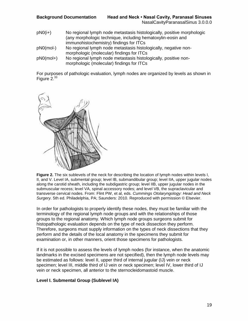
For purposes of pathologic evaluation, lymph nodes are organized by levels as shown in Figure 2.33
Figure 2. The six sublevels of the neck for describing the location of lymph nodes within levels I, II, and V. Level IA, submental group; level IB, submandibular group; level IIA, upper jugular nodes along the carotid sheath, including the subdigastric group; level IIB, upper jugular nodes in the submuscular recess; level VA, spinal accessory nodes; and level VB, the supraclavicular and transverse cervical nodes. From: Flint PW, et al, eds. Cummings Otolaryngology: Head and Neck Surgery. 5th ed. Philadelphia, PA; Saunders: 2010. Reproduced with permission © Elsevier.
In order for pathologists to properly identify these nodes, they must be familiar with the terminology of the regional lymph node groups and with the relationships of those groups to the regional anatomy. Which lymph node groups surgeons submit for histopathologic evaluation depends on the type of neck dissection they perform. Therefore, surgeons must supply information on the types of neck dissections that they perform and the details of the local anatomy in the specimens they submit for examination or, in other manners, orient those specimens for pathologists.
If it is not possible to assess the levels of lymph nodes (for instance, when the anatomic landmarks in the excised specimens are not specified), then the lymph node levels may be estimated as follows: level II, upper third of internal jugular (IJ) vein or neck specimen; level III, middle third of IJ vein or neck specimen; level IV, lower third of IJ vein or neck specimen, all anterior to the sternocleidomastoid muscle. Level I. Submental Group (Sublevel IA)
Background Documentation Head and Neck • Nasal Cavity, Paranasal Sinuses NasalCavityParanasalSinus 3.0.0.0
20
Lymph nodes within the triangular boundary of the anterior belly of the digastric muscles and the hyoid bone. Level I. Submandibular Group (Sublevel IB) Lymph nodes within the boundaries of the anterior and posterior bellies of the digastric muscle and the body of the mandible. The submandibular gland is included in the specimen when the lymph nodes within this triangle are removed. Level II. Upper Jugular Group (Sublevels IIA and IIB) Lymph nodes located around the upper third of the internal jugular vein and adjacent spinal accessory nerve extending from the level of the carotid bifurcation (surgical landmark) or hyoid bone (clinical landmark) to the skull base. The posterior boundary is the posterior border of the sternocleidomastoid muscle, and the anterior boundary is the lateral border of the sternohyoid muscle. Level III. Middle Jugular Group Lymph nodes located around the middle third of the internal jugular vein extending from the carotid bifurcation superiorly to the omohyoid muscle (surgical landmark), or cricothyroid notch (clinical landmark) inferiorly. The posterior boundary is the posterior border of the sternocleidomastoid muscle, and the anterior boundary is the lateral border of the sternohyoid muscle. Level IV. Lower Jugular Group Lymph nodes located around the lower third of the internal jugular vein extending from the omohyoid muscle superiorly to the clavicle inferiorly. The posterior boundary is the posterior border of the sternocleidomastoid muscle, and the anterior boundary is the lateral border of the sternohyoid muscle. Level V. Posterior Triangle Group (Sublevels VA and VB) This group comprises predominantly the lymph nodes located along the lower half of the spinal accessory nerve and the transverse cervical artery. The supraclavicular nodes are also included in this group. The posterior boundary of the posterior triangle is the anterior border of the trapezius muscle, the anterior boundary of the posterior triangle is the posterior border of the sternocleidomastoid muscle, and the inferior boundary of the posterior triangle is the clavicle. Level VI. Anterior (Central) Compartment Lymph nodes in this compartment include the pre- and paratracheal nodes, precricoid (Delphian) node, and the perithyroidal nodes, including the lymph nodes along the recurrent laryngeal nerve. The superior boundary is the hyoid bone, the inferior boundary is the suprasternal notch, the lateral boundaries are the common carotid arteries and the posterior boundary by the prevertebral fascia. Level VII. Superior Mediastinal Lymph Nodes Metastases at level VII are considered regional lymph node metastases; all other mediastinal lymph node metastases are considered distant metastases. Lymph node groups removed from areas not included in the above levels, eg, scalene, suboccipital, and retropharyngeal, should be identified and reported from all levels separately. Midline nodes are considered ipsilateral nodes.
Background Documentation Head and Neck • Nasal Cavity, Paranasal Sinuses NasalCavityParanasalSinus 3.0.0.0
21
K. Lymph Nodes Lymph Node Number Histological examination of a selective neck dissection specimen will ordinarily include 6 or more lymph nodes. Histological examination of a radical or modified radical neck dissection specimen will ordinarily include 10 or more lymph nodes in the untreated neck. Measurement of Tumor Metastasis The cross-sectional diameter of the largest metastasis in a lymph node containing metastatic tumor is measured in the gross specimen at the time of macroscopic examination or, if necessary, on the histologic slide at the time of microscopic examination. There is conflicting data in the literature on the significance of the size of the largest metastatic lymph node on the risk of regional recurrence and a predictor of poor overall survival.19 While the diameter of the largest positive lymph node may potentially serve as a predictor of outcome, it may not represent an independent predictor of outcome when other pathologic factors are considered.19 L. Special Procedures for Lymph Nodes
At the current time, no additional special techniques should be used other than routine histology for the assessment of nodal metastases. Immunohistochemistry and PCR to detect isolated tumor cells are considered investigational techniques at this time. M. Dysplasia of the Upper Aerodigestive Tract (UADT) Epithelial dysplasias of the nasal cavity and paranasal sinuses as a precursor lesion for sinonasal carcinomas are less common and less well defined as compared to epithelial dysplasias of the oral cavity and the larynx.34 Further, unlike dysplastic lesions of the oral cavity and/or the larynx, precursor lesions of the nasal cavity and paranasal sinuses are generally asymptomatic and therefore are not biopsied. Instead, they are identified more often in association with another lesion, such as an invasive carcinoma. N. Ancillary Studies At the current time, no additional special techniques are required other than routine histology for the assessment of nasal cavity and paranasal sinus carcinomas. Immunohistochemistry and in situ hybridization (ISH) to detect the presence of viruses (eg, human papillomavirus, Epstein-Barr virus) are considered investigational techniques at this time.
References 1. Patel S, Shah JP. Part II: Head and neck sites. In: Edge SB, Byrd DR, Carducci
MA, Compton CA, eds. AJCC Cancer Staging Manual. 7th ed. New York, NY: Springer; 2009.
2. Barnes L, Tse LLY, Hunt JL, Brandwein-Gensler M, Curtin HC, Boffetta P. Tumours of the nasal cavity and paranasal sinuses: Introduction. In: Barnes L, Eveson J, Reichart P, Sidransky D, eds World Health Organization Classification of Tumours: Pathology and Genetics of Head and Neck Tumours. Lyon, France: IARC Press; 2005:12-14.
3. Barnes L. WHO histological classification of tumours of the nasal cavity and paranasal sinuses. In: Barnes EL, Eveson JW, Reichart P, Sidransky D, eds. World
Background Documentation Head and Neck • Nasal Cavity, Paranasal Sinuses NasalCavityParanasalSinus 3.0.0.0
22
Health Organization Classification of Tumours: Pathology and Genetics of Head and Neck Tumours. Lyon, France: IARC Press; 2005: 10.
4. Barnes L. Neuroendocrine tumours. In: Barnes L, Eveson JW, Reichart P et al, eds. World Health Organization Classification of Tumours: Pathology and Genetics of Head and Neck Tumours. Lyon, France: IARC Press; 2005.
5. Crissman JD, Sakr WA. Squamous neoplasia of the upper aerodigestive tract. Intraepithelial and invasive squamous cell carcinoma. In: Pilch BZ, ed. Head and
Neck Surgical Pathology. Philadelphia, PA: Lippincott Williams & Wilkins; 2001:42-43.
6. Mills SE, Gaffey MJ, Frierson HF, Jr. Tumors of the upper aerodigestive tract and ear. In: Atlas of Tumor Pathology. 3rd Series. Fascicle 26. Washington, DC: Armed Forces Institute of Pathology; 2000: 16.
7. Ellis GL, Auclair PL. Salivary gland tumors: General considerations. In: Silverberg SG, ed. Tumors of the Salivary Glands: AFIP Atlas of Tumor Pathology. Series 4. Fascicle 9. Armed Forces Institute of Pathology. Washington, DC. 2008:27-48.
8. Spiro RH, Huvos AG, Strong EW. Adenocarcinoma of salivary gland origin. Clinicopathologic study of 204 patients. Am J Surg. 1982;144:423-431.
9. Szanto PA, Luna MA, Tortoledo ME, White RA. Histologic grading of adenoid cystic carcinoma of the salivary glands. Cancer. 1984;84:1062-1069.
10. Spiro RH, Thaler HT, Hicks WF, Kher UA, Huvos AG, Strong EW. The importance of clinical staging in minor salivary gland carcinoma. Am J Surg. 1991;162:330-336.
11. Kane WJ, McCaffrey TV, Olsen KD, Lewis JE. Primary parotid malignancies: a clinical and pathologic review. Arch Otolaryngol Head Neck Surg. 1991;117:307-315.
12. Greiner TC, Robinson RA, Maves MD. Adenoid cystic carcinoma: a clinicopathologic study with flow cytometric analysis. Am J Clin Pathol. 1989;92:711-720.
13. Auclair PL, Goode RK, Ellis GL. Mucoepidermoid carcinoma of intraoral salivary glands: evaluation and application of grading criteria in 143 cases. Cancer. 1992;69:2021-30.
14. Goode RK, Auclair PL, Ellis GL. Mucoepidermoid carcinoma of the major salivary glands: clinical and histopathologic analysis of 234 cases with evaluation of grading criteria. Cancer. 1998;82:1217-24.
15. Brandwein MS, Ivanov K, Wallace DI, et al. Mucoepidermoid carcinoma: a clinicopathologic study of 80 patients with special reference to histological grading. Am J Surg Pathol. 2001;25:835-45.
16. Bradley PJ et al. Status of primary tumour surgical margins in squamous head and neck cancer: prognostic implications. Curr Opin Otolaryngol Lead Neck Surg.
2007;15:74-81. 17. Laramore GE, Scott CB, al-Sarraf M, et al. Adjuvant chemotherapy for resectable squamous cell carcinomas of the head and neck: report on Intergroup
Study 0034. Int J Radiat Oncol Biol Phys. 1992;23:705-713. 18. Zelefsky MJ, Harrison LB, Fass DE, Armstrong JG, Shah JP, Strong EW. Postoperative radiation therapy for squamous cell carcinomas of the oral cavity and
oropharynx: impact of therapy on patients with positive surgical margins. Int J Radiat Oncol Biol Phys. 1993;25:17-21.
19. Smith BD, Haffty BG. Prognostic factors in patients with head and neck cancer. In: Harrison LB, Sessions RB, Hong WK, eds. Head and Neck Cancer: A Multidisciplinary Approach. 3rd ed. Philadelphia, PA: Lippincott Williams & Wilkins; 2009: 51-75.
Background Documentation Head and Neck • Nasal Cavity, Paranasal Sinuses NasalCavityParanasalSinus 3.0.0.0
23
20. Fagan JJ, Collins B, Barnes L, et al. Perineural invasion in squamous cell carcinoma of the head and neck. Arch Otolaryngol Head Neck Surg. 1998;124:637-640.
21. Cooper JS, Pajak TF, Forastiere AA, et al. Postoperative concurrent radiotherapy and chemotherapy for high-risk squamous-cell carcinoma of the head and neck. N Engl J Med. 2004;350:1937-1944.
22. Bernier J, Domenge C, Ozsahin M, et al. Postoperative irradiation with or without concomitant chemotherapy for locally advanced head and neck cancer. N Engl J Med. 2004;350:1945-1952,
23. Woolgar J, Triantafyllou A. Neck dissections: a practical guide for the reporting histopathologist. Curr Diag Pathol. 2007;13:499-511.
24. Leemans CR, Tiwari R, Nauta JJ, van der Waal I, Snow GB. Regional lymph node involvement and its significance in the development of distant metastases in head and neck carcinoma. Cancer. 1993;71:452-456.
25. Johnson JT, Barnes EL, Meyers EN, et al. The extracapsular spread of tumors in cervical node metastases. Head Neck Surg. 1981;107:725-729
26. Woolgar JA, Rogers SN, Lowe D, Brown JS, Vaughn ED. Cervical lymph node metastasis in oral cancer: the importance of even microscopic extracapsular spread. Oral Oncol. 2003;39:130-137.
27. Sobin LH, Gospodarowicz MK, Wittekind CH, eds UICC TNM Classification of Malignant Tumors. 7th ed. New York, NY: Wiley-Liss; in press.
28. Robbins KT et al. Neck dissection classification update. Arch Otolaryngol Head Neck Surg. 2002;128:751-758.
29. Robbins TK et al. Consensus statement on the classification and terminology of neck dissection. Arch Otolaryngol Head Neck Surg. 2008;134:536-538
30. Robbins T, Medina JE, Wolfe GT, Levine PA, Sessions RB, Pruet CW. Standardizing neck dissection terminology: official report of the academy’s committee for head and neck surgery and oncology. Arch Otolaryngol Head Neck Surg. 1991;117:601-605.
31. Wittekind C, Greene FL, Henson DE, Hutter RVP, Sobin LH, eds. TNM Supplement: A Commentary on Uniform Use. 3rd ed. New York: Wiley-Liss; 2003
32. Singletary SE, Greene FL, Sobin LH. Classification of isolated tumor cells: clarification of the 6th edition of the American Joint Committee on Cancer Staging Manual. Cancer. 2003;90(12):2740-2741.
33. Flint PW, et al, eds. Cummings Otolaryngology: Head and Neck Surgery. 4th ed. Philadelphia, PA: Saunders; 2010.
34. Pilch BZ, Bouquot J, Thompson LDR. Squamous cell carcinoma. In: Barnes L, Eveson J, Reichart P, Sidransky D, eds. World Health Organization Classification of Tumours: Pathology and Genetics of Head and Neck Tumours. Lyon, France: IARC Press; 2005.