Embed Size (px)
Citation preview

Nanomolar dose of bisphenol A rapidly modulates spinogenesis in adulthippocampal neurons
Nobuaki Tanabe a,1, Hinako Yoshino a,1, Tetsuya Kimoto a,c, Yasushi Hojo a,c,d, Mari Ogiue-Ikeda a,b,Yasuyuki Shimohigashi e, Suguru Kawato a,b,c,d,⇑a Department of Biophysics and Life Sciences, Graduate School of Arts and Sciences, The University of Tokyo, Komaba 3-8-1, Meguro, Tokyo 153-8902, Japanb National MEXT Project in Special Coordinate Funds for Promoting Science and Technology, The University of Tokyo, Japanc Core Research for Evolutional Science and Technology Project of Japan Science and Technology Agency, The University of Tokyo, Japand Bioinformatics Project, Japan Science and Technology Agency, The University of Tokyo, Japane Department of Chemistry, Faculty and Graduate School of Sciences, Kyushu University, Hakozaki 6-10-1, Higashi, Fukuoka 812, Japan
a r t i c l e i n f o
Article history:Received 6 July 2011Received in revised form 23 November 2011Accepted 9 January 2012Available online 16 January 2012
Keywords:Bisphenol AEndocrine disrupterERRcHippocampusSpineEstradiol
a b s t r a c t
We demonstrated the rapid effects of 10 nM bisphenol A (BPA) on the spinogenesis of adult rat hippo-campal slices. The density of spines was analyzed by imaging Lucifer Yellow-injected CA1 neurons inslices. Not only the total spine density but also the head diameter distribution of spine was quantitativelyanalyzed. Spinogenesis was significantly enhanced by BPA within 2 h. In particular, the density of middle-head spine (with head diameter of 0.4–0.5 lm) was significantly increased.
Hydroxytamoxifen, an antagonist of both estrogen-related receptor gamma (ERRc) and estrogen recep-tors (ERa/ERb), blocked the BPA-induced enhancement of the spine density. However, ICI 182,780, anantagonist of ERa/ERb, did not suppress the BPA effects. Therefore, ERRc is deduced to be a high affinityreceptor of BPA, responsible for modulation of spinogenesis. The BPA-induced enhancement of spinogen-esis was also suppressed by MAP kinase inhibitor, PD98059, and the blocker of NMDA receptors, MK-801.Washout of BPA for additional 2 h after 2 h BPA treatment abolished the BPA-induced enhancement ofspinogenesis, suggesting that the BPA effect was reversible. ERRc was localized at synapses as well as cellbodies of principal neurons. ERRc at synapses may contribute to the observed rapid effect. The level ofBPA in the hippocampal slices was determined by mass-spectrometric analysis.
� 2012 Elsevier Ireland Ltd. All rights reserved.
1. Introduction
Low dose exposure to bisphenol A (BPA) may induce hormone-like effects on wildlife and humans. BPA is a widely used syntheticmaterial included in polycarbonate resin used in water pipesealant, dental prostheses, compact discs and baby bottles. Toxiceffects of high dose BPA (mg/kg weight/day) have been investi-gated in relation to the development and functions of thereproduction systems (Fisher et al., 1999; Al-Hiyasat et al., 2002;
Grote et al., 2004; Halldin et al., 2005). However, the low doseexposure to BPA (lg/kg/day or nanomolar doses) shows ratherweak toxic effects on reproductive or endocrine functions in theperipheral tissues, probably due to the efficient detoxification ofBPA by the liver. On the other hand, low dose exposure to BPAmay significantly affect the brain function, because the detoxifica-tion of BPA in the brain is probably very weak, due to the extremelylow expression of drug-metabolizing enzymes in the brain (Miksysand Tyndale, 2002; Kishimoto et al., 2004; Chinta et al., 2005).
The low dose exposure to BPA during fetal/neonatal stages hasbeen extensively investigated. For example, fetal or neonatal expo-sure to BPA inhibits sexual differentiation of nonreproductivebehaviors of adult animals, including maze learning behavior (Carret al., 2003; Kubo et al., 2003; Fujimoto et al., 2006), at doses as lowas 1/1000 of those required for the stimulation of uterine growth(Ashby, 2001). On the other hand, inadequate information is avail-able for the low dose exposure to BPA in the adult stage, exceptsome pioneer works in vivo (MacLusky et al., 2005; Leranth et al.,2008; Hajszan and Leranth, 2010).
The high affinity functional receptor for BPA has not beenidentified yet. Although ERa is one candidate of BPA receptor, the
0303-7207/$ - see front matter � 2012 Elsevier Ireland Ltd. All rights reserved.doi:10.1016/j.mce.2012.01.008
Abbreviations: ACSF, artificial cerebrospinal fluid; AMPA, a-amino-3-hydroxy-5-methyl-4-isoxazolepropionic acid; BPA, bisphenol A; CNQX, 6-cyano-7-nitroqui-noxaline-2,3-dione; DG, dentate gyrus; E2, 17b-estradiol; ER, estrogen receptor;ERRc, estrogen-related receptor gamma; ICI, ICI182,780; MR, median raphe; MSDB,medial septum/diagonal band of Broca; NMDA, N-methyl-D-aspartate; OH-Tam, 4-hydroxytamoxifen; OVX, ovariectomized; PSD, postsynaptic density; PY, pyramidalneurons; SUM, supramammillary area.⇑ Corresponding author at: Department of Biophysics and Life Sciences, Graduate
School of Arts and Sciences, The University of Tokyo, Komaba 3-8-1, Meguro, Tokyo153-8902, Japan. Tel./fax: +81 3 5454 6517.
E-mail addresses: [email protected], [email protected](S. Kawato).
1 These authors contributed equally.
Molecular and Cellular Endocrinology 351 (2012) 317–325
Contents lists available at SciVerse ScienceDirect
Molecular and Cellular Endocrinology
journal homepage: www.elsevier .com/locate /mce
affinity of ERa for BPA is very low, in the order of 1/100–1/1000 ofthat for 17b-estradiol (E2) (Kuiper et al., 1997; Morohoshi et al.,2005). On the other hand, estrogen-related receptor gamma (ERRc)is a high affinity binding protein for BPA (Takayanagi et al., 2006).However, ERRc has not been recognized as a BPA receptor, becauseERRc shows constitutive transcriptional activity even without anyligand (Coward et al., 2001).
We here performed the investigation on the rapid modulationby nanomolar doses of BPA on the density and morphology of den-dritic spines in the adult hippocampal slices, including investiga-tions of BPA receptors. In order to observe the direct effects ofBPA on hippocampal neurons, we used isolated ‘acute’ hippocam-pal slices which do not have projections of cholinergic or seroto-nergic neurons from outside of the hippocampus.
2. Materials and methods
2.1. Chemicals
BPA, 6-cyano-7-nitroquinoxaline-2,3-dione (CNQX), cyclohexi-mide, nicardipine, N-methyl-D-aspartate (NMDA), PD98059, ICI182,780 (ICI), MK-801 and Lucifer Yellow were purchased fromSigma (USA). 4-hydroxy-tamoxifen (OH-Tam) was from Calbio-chem (Germany). Other chemicals used were of highest puritycommercially available. Polyclonal anti-ERRc antibody (against C-terminal of ligand binding site) was prepared by Dr. Shimohigashiat Kyushu Univ. (Tokunaga et al., 2006).
2.2. Animals
Adult male Wistar rats (12 weeks old, 340–360 g) were pur-chased from Saitama Experimental Animal Supply (Saitama, Ja-pan). The experimental procedure of this research was approvedby the Committee for Animal Research of the University of Tokyo.
2.3. Preparation of ‘acute’ hippocampal slices
Rats were deeply anesthetized with ethyl ether and decapi-tated. The brains from adult rats were removed and placed at4 �C in artificial cerebrospinal fluid (ACSF) consisted of (mM):124 NaCl, 5.0 KCl, 1.25 NaH2PO4, 2.0 MgSO4, 2.0 CaCl2, 22 NaHCO3,10 glucose and was equilibrated with 95% O2/5% CO2. The hippo-campus was dissected and 300 lm-thick transverse dorsal ‘fresh’slices to the long axis were prepared with a vibratome (DosakaEM, Kyoto, Japan). ‘Acute’ slices were prepared from these ‘freshlyprepared’ slices by 2 h recovery incubation at 25 �C in ACSF.
2.4. Imaging and analysis of spinogenesis
Experimental details are described in elsewhere (Mukai et al.,2007).
2.4.1. Current injection of Lucifer Yellow‘Acute’ slices were further incubated with BPA in the presence
or absence of other drugs such as OH-Tam or PD98059. Drug-trea-ted slices were then prefixed with 4% paraformaldehyde in PBS(0.1 M phosphate buffer and 0.14 M NaCl, pH 7.3) at 4 �C for 2–4 h.
Neurons within slices were visualized by an injection of LuciferYellow under a Nikon E600FN microscope (Japan) equipped with aC2400–79H infrared camera (Hamamatsu Photonics, Japan) andwith a 40� water immersion lens (Nikon). A glass electrode wasfilled with 5% Lucifer Yellow, which was then injected for 15 minusing Axopatch 200B (Axon Instruments, USA). Approximately fiveneurons within a 100–200 lm depth from the surface of a slice
were injected (Duan et al., 2002). After injection, slices were fixedagain with 4% paraformaldehyde at 4 �C overnight.
2.4.2. Confocal laser microscopy and morphological analysisThe imaging was performed from sequential z-series scans with
LSM5 PASCAL confocal microscope (Zeiss, Germany). For analysisof spines, three-dimensional images were constructed fromapproximately 40 sequential z-series sections of neurons scannedevery 0.45 lm with a 63� water immersion lens, NA 1.2 (Zeiss).For Lucifer Yellow, the excitation and emission wavelengths were488 nm and 515 nm, respectively. The applied zoom factor (3.0)yielded 23 pixels per 1 lm. The z-axis resolution was approxi-mately 0.71 lm. The confocal lateral resolution was approximately0.26 lm. Confocal images were then deconvoluted using AUTODE-BLUR software (AutoQuant, USA). The density of spine as well asthe head diameter was analyzed with Spiso-3D (automated soft-ware mathematically calculating geometrical parameters ofspines) developed by Bioinformatics Project of Kawato’s group(Mukai et al., 2011). Results obtained by Spiso-3D are almost iden-tical to those by Neurolucida (manual-based analysis software)(MicroBrightField, USA) within assessment difference of 2%, andSpiso-3D considerably reduces human errors and experimental la-bor of manual software. We analyzed the secondary dendrites inthe stratum radiatum, lying between 100 and 250 lm from thesoma. The spine density was calculated from the number of spineson dendrites having a total length of 50–80 lm. In total, we inves-tigated 3–4 rats, 6–8 slices, 12–16 neurons, 24–32 dendrites and1200–2000 spines. Spine shapes were classified into threecategories as follows. (1) A small-head spine whose head diameteris 0.2–0.4 lm. (2) A middle-head spine whose head diameteris 0.4–0.5 lm. (3) A large-head spine whose head diameter is0.5–1.0 lm. These three categories were useful to distinguish dif-ferent responses upon inhibitor application. Because the majorityof spines (>95%) had a distinct head and neck, and stubby typeand filopodium type spines did not contribute much to overallchanges, we analyzed spines having a distinct head.
All protrusions from the dendrites were treated as ‘spines’,although with confocal microscopy, it was not possible to deter-mine whether they formed synapses, or whether some of themwere filopodia protrusions which did not form synapses (Sorraand Harris, 2000). While counting the spines in the reconstructedimages, the position and verification of spines were aided by rota-tion of three-dimensional reconstructions and by observation ofthe images in consecutive single planes.
2.5. Immunohistochemical staining of hippocampal slices
Immunohistochemical staining of hippocampal slices was per-formed as described in elsewhere (Kimoto et al., 2001; Kawatoet al., 2002) and Supplementary material. Staining of ERRc wasperformed using the avidin–biotin peroxidase complex technique.After application of anti-ERRc antibody (1/250), the slices wereincubated for 24 h at 4 �C, in the presence of 0.5% Triton X-100and 3% skim milk with gentle shaking.
For preabsorption of anti-ERRc antibody with antigen, excessamount of antigen was preincubated with anti-ERRc antibody for15 h at 4 �C. Characterization of anti-ERRc antibody is describedin Supplementary material with Fig. S1.
2.6. Preparation of synaptic, cytoplasmic and nuclear fractions
Fractionation of the homogenates obtained from hippocampalslices was performed by a combination of centrifugations at 4 �C(Cohen et al., 1977). Detailed procedures to obtain the raft fraction,nuclear fraction, postsynaptic density (PSD) fraction, low densitymembrane fraction (presynaptic membrane-enriched fraction),
318 N. Tanabe et al. / Molecular and Cellular Endocrinology 351 (2012) 317–325
high density membrane fraction (microsome and postsynapticmembrane-enriched fraction), and the cytoplasmic fraction are de-scribed in elsewhere (Mukai et al., 2007) and Supplementarymaterial.
2.7. Western immunoblot analysis
The protein blots were probed with anti-ERRc antibody (dilutedto 1/3000) for 12 h at 4 �C, and incubated with horseradish perox-idase-conjugated goat anti-rabbit IgG. Detailed procedures are de-scribed in Supplementary material.
2.8. Mass-spectrometric assay of BPA
Experimental details are described in elsewhere (Hojo et al.,2009) and Supplementary material. The extraction of steroids fromhippocampal slices was performed by hexane: ethylacetate = 2:3mixtures. The steroid extracts were applied to a C18 Amprep solidphase column (Amersham Biosciences, Piscataway, NJ). The BPAfraction was separated from eluted steroids using a normal phaseHPLC system (Jasco, Tokyo, Japan) with a silica gel column. Therecoveries of BPA through the steps above were approx 40%. To in-crease the ionization efficiency, BPA was derivatized to BPA-dipi-colinoyl-ester (Hojo et al., 2009). The LC–MS/MS system, whichconsisted of a reverse phase LC coupled with an API 5000 triple-stage quadrupole mass spectrometer (Applied Biosystems, FosterCity, CA), was operated with electron spray ionization. The LC chro-matographic separation for BPA derivatives was performed on Ca-denza CD-C18 column (Imtakt, Kyoto, Japan). The MS/MS processmonitored the m/z transition from 439.3 to 239.8 Deuterium la-beled BPA derivative (2, 20, 6, 60-d4-BPA-dipicolinoyl-ester) wasused for internal standards in order to measure the recovery ofBPA as well as to calibrate the retention time. After derivatization,purification and MS/MS detection, the recovery for BPA was deter-mined to be approximately 70%. The limit of quantification for BPAwas 5 pg per 0.1 g of hippocampal tissue (Table S1). The linearitywas observed between 5 pg and 1000 pg. For more detailed proce-dures, see Supplementary material.
2.9. Statistical analysis
In spine analysis, data are expressed as mean ± SEM. The signif-icance of drug effect was examined via statistical analysis usingTukey–Kramer post hoc multiple comparison test when one wayANOVA tests yielded P < 0.05.
3. Results
3.1. Rapid effect of BPA on spinogenesis
We analyzed the effect of BPA on the modulation of the densityand head diameter of spines in the CA1 region. To do this, singlespine imaging was performed for Lucifer Yellow-injected neuronsin hippocampal slices from adult male rats. We analyzed secondarybranches of the apical dendrites located 100–200 lm distant fromthe pyramidal cell body around the middle of the stratum radiatumof CA1 region.
3.1.1. Total spine density analysisFollowing a 2 h treatment with BPA, dendrites had significantly
more spines than control (Fig. 1–1). Time dependency was exam-ined by treating slices for 0, 0.5, 1, 1.5 and 2 h with 10 nM BPA.The enhancing effect on the total spine density was approximatelyproportional to the incubation time, showing 0.94 (0 h), 0.95(0.5 h), 1.21 (1 h), 1.21 (1.5 h) and 1.58 spines/lm (2 h) (Figs. 1
and 2). Dose dependency was also examined after 2 h incubation.Dose dependency showed that the enhancing effect was most sig-nificant at 10 nM BPA (1.55 spines/lm) as compared with 1 nM(0.95 spines/lm), 100 nM (1.40 spines/lm) and 10 lM BPA(0.92 spines/lm) (Figs. 1 and 2). Because a 2 h treatment with10 nM BPA was most effective in spinogenesis, this incubation con-dition was used in the following investigations unless specified.
To investigate signaling pathway of BPA-induced increase in the totalspine density, several antagonists and blockers were applied (Fig. 2).
Application of ICI, an antagonist of ERa/ERb, did not suppressthe increase in the spine density by 10 nM BPA (1.56 spines/lm).On the other hand, application of OH-Tam, an antagonist of bothERRc and ERa/ERb, completely suppressed the enhancement ofspine density by BPA (0.94 spines/lm).
Application of MK-801, an antagonist of NMDA receptor,completely suppressed the enhancement of spine density byBPA (0.96 spines/lm). Application of CNQX, an antagonist ofAMPA receptor, partially-prevented the enhancement by BPA(1.24 spines/lm). Application of nicardipine, an antagonist of L-typevoltage-dependent Ca2+ channel, did not inhibit the enhancement ofspine density induced by 10 nM BPA (1.48 spines/lm). When Ca2+
free ACSF was used, BPA-induced spinogenesis was completely sup-pressed (0.80 spines/lm). Application of PD98059, an inhibitor ofErk MAP kinase, completely prevented the BPA-induced spinogene-sis (0.84 spines/lm). Washout of BPA for additional 2 h after the 2 hBPA treatment abolished the BPA-induced enhancement of spino-genesis, implying that the BPA effect was reversible. It should benoted that inhibitors (OH-Tam, ICI, PD98059) alone did not signifi-cantly affect the total spine density within experimental error, indi-cating that the observed inhibitory effects are not due to simpleblocker’s poison effects (Fig. S2 in Supplementary material).
3.1.2. Spine head diameter analysisThe morphological changes in the spine head diameter induced
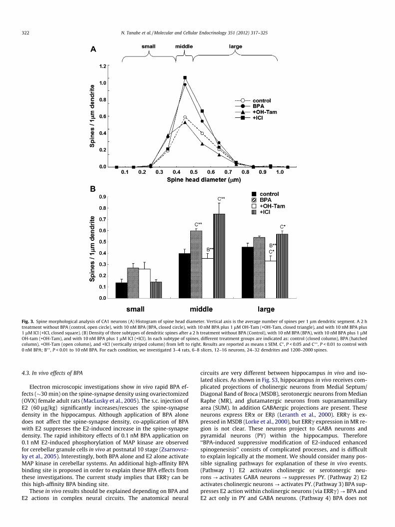
by a 2 h treatment with 10 nM BPA were assessed (Fig. 3). Themajority of spines (>95%) had a distinct head, therefore we statis-tically analyzed these spines having distinct heads. We classifiedthe spines into three categories depending on their head diameter,i.e. small-head spines (0.2–0.4 lm), middle-head spines (0.4–0.5 lm), and large-head spines (0.5–1.0 lm). The categorizationinto three subclasses enabled to distinguish different responsesin spine subpopulations upon application of BPA, receptor antago-nist or kinase inhibitor. Small-, middle-, and large-head spines aredifferent in the density of AMPA receptors (Shinohara et al., 2008),therefore these three types of spines may have different physiolog-ical functions. The density of AMPA receptors in the spine posi-tively correlates with the postsynaptic density (PSD) area size.
We performed a statistical analysis using three subclasses(Fig. 3B). In control slices (0 nM BPA), the spine density was0.14 spines/lm for small-head spines, 0.40 spines/lm for middle-head spines, and 0.45 spines/lm for large-head spines. Upon treat-ment with BPA, the density of middle-head spines increased signif-icantly to 0.60 spines/lm, while the density of small-head andlarge-head spines was not significantly altered (Fig. 3B). The co-application of OH-Tam with BPA considerably suppressed BPA-in-duced increase in the density of middle-head spines to 0.35 spines/lm (Fig. 3B). No significant change in BPA-induced increase in thedensity of three subclasses of spines was induced by co-applicationwith ICI (Fig. 3B).
3.2. Staining of ERRc
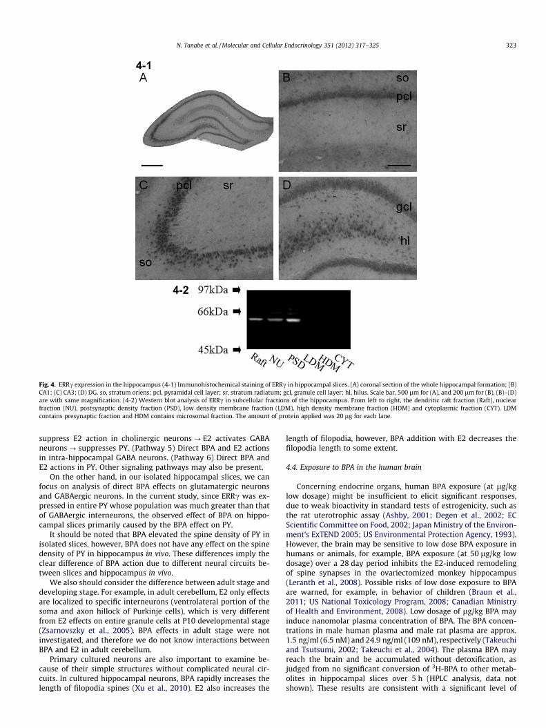
Hippocampal distribution of ERRc was investigated by immu-nostaining of hippocampal slices with anti-ERRc antibody. Astrong immunostaining of pyramidal and granule neurons was ob-served in CA1–CA3 and DG (Fig. 4–1). No staining was observed
N. Tanabe et al. / Molecular and Cellular Endocrinology 351 (2012) 317–325 319
when anti-ERRc antibody preabsorbed with the excess amount ofantigen peptide was used as a primary antibody. Subcellularexpression of ERRc was analyzed by Western blot. A clear singleband with M.W. of approx. 55 kDa was observed in PSD fraction,dendritic raft fraction and nuclear fraction of hippocampal neurons(Fig. 4–2).
3.3. Concentration of BPA in the hippocampus
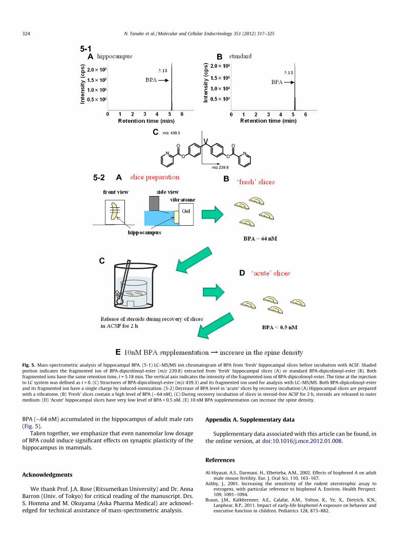
Because BPA is widely present in environments, the endogenousconcentration of BPA was carefully determined for adult male rathippocampus with mass-spectrometric investigations using achromatogram analysis of the fragmented ions (Fig. 5–1). Afterselection of mother ions, fragmentation and detection were per-formed via MS/MS procedures. Chromatographic profiles for thefragmented ions of BPA-dipicolinoyl-ester having m/z = 239.8showed a clear peak with the retention time of 5.18 min whichwas the same as that of the standard BPA derivative (Fig. 5–1).The average concentration of BPA in ‘fresh’ hippocampal slices
(before incubation with ACSF) was evaluated to be 14.6 ± 1.8 ng/gwet weight (i.e., 64 ± 8 nM) from 4 animals. In contrast, the BPAconcentration in ‘acute’ slices, used for spinogenesis experiments,was less than 0.5 nM, due to significant release of BPA to ACSF dur-ing 2 h recovery incubation (Fig. 5–2). These results imply that theelevation of BPA from <0.5 nM to 10 nM occurred upon applicationof 10 nM BPA in acute slices in spinogenesis experiments.
We need to subtract blank values of BPA (0.027 ± 0.006 ng/mL)that was measured in blank samples which were prepared along-side hippocampal samples through the whole extraction, fraction-ation and purification procedures. These subtraction proceduresare essential, because BPA was observed even in pure water (seeSupplementary material).
4. Discussion
Current investigations of the rapid effects of BPA on spinogene-sis in isolated hippocampal slices lead to the finding of ERRc-med-iated BPA action at synapses of pyramidal neurons.
Fig. 1. Effect of BPA on spines in adult hippocampal CA1 neurons (1-1) Spine images along the secondary dendrites in the stratum radiatum. Maximal intensity projectionsonto XY plane from z-series confocal micrographs (Max XY), spine images analyzed by Spiso-3D (S) and 3 dimensional model illustration (Model). (A) Control spines withoutdrug-treatments (control, 0 nM BPA), (B) with 10 nM BPA (BPA), and (C) with 10 nM BPA and 1 lM OH-Tam (OH-Tam), and (D) with 10 nM BPA and 1 lM ICI (ICI). Bar = 5 lm.(1-2) (A) Time-dependence of BPA effect. The total spine density after 0, 0.5, 1, 1.5 and 2 h treatments with 10 nM BPA. Vertical axis is the total number of spines per 1 lmdendritic segment. Results are reported as means ± SEM. Statistical significance, ⁄⁄P < 0.01 vs. control (0 h). (B) Dose-dependence of BPA effect on total spine density. Verticalaxis is the total spine density after a 2 h treatment with 0 nM, 1 nM, 10 nM, 100 nM and 10 lM of BPA. Statistical significance, ⁄⁄P < 0.01 vs. control (0 nM BPA). For eachcondition, we investigated 3–4 rats, 6–8 slices, 12–16 neurons, 24–32 dendrites and 1200–2000 spines.
320 N. Tanabe et al. / Molecular and Cellular Endocrinology 351 (2012) 317–325

4.1. Comparison of BPA and E2 effects on spinogenesis in slices
The elevation of BPA level to 10 nM (from less than 0.5 nM inacute slices) induced a rapid increase in the density of spines.The enhancing effect on spinogenesis was not greater at higherconcentrations of BPA (100 nM) than those at lower concentra-tions of BPA (10 nM) (Fig. 1). BPA predominantly increased thedensity of middle-head spines by approx. 1.5-fold (Fig. 3). Thereason is not clear for this selective increase, since BPA wouldaffect all spines. We cannot eliminate the possibility that BPAmay increase small-head spines then enlarge them to middle-head spines. Although receptors are different, some similarityis observed between BPA and E2 about modulation effects onspinogenesis in adult slices. The increase of middle-head spinesalso occurs upon 1 nM E2 treatments for 2 h in CA1 of hippo-campus (Mukai et al., 2007). The E2-induced spinogenesis is alsodriven by Erk MAP kinase pathway (Mukai et al., 2007). Blockingof NMDA receptors by MK-801 abolishes the enhancing effectsby E2 and BPA, suggesting that both E2 and BPA signaling needa basal level of Ca2+ which is kept by Ca2+ influx into spines viaNMDA receptors due to spontaneous spiking (Ishii et al., 2007;Ogiue-Ikeda et al., 2008). As an another example, in organotypichippocampal slice cultures, the pretreatment for 24 h with E2 orBPA at 10 nM exacerbates the CA3 neuronal damage caused byglutamate, due to enhanced spinogenesis by E2 or BPA (Satoet al., 2002).
4.2. ERRc is a high affinity receptor for BPA
Although ERa had been assumed to be a receptor of BPA in ear-lier studies, the binding affinity of BPA to ERa is much lower(approx. 1/100–1/2000) than that of E2 (Kuiper et al., 1997;Morohoshi et al., 2005). Therefore, nanomolar BPA probably cannotinduce significant effects on spinogenesis through ERa. On theother hand, BPA tightly binds to ERRc (Takayanagi et al., 2006).To identify the receptor responsible for the BPA-induced modula-tion of spinogenesis, we used OH-Tam, an antagonist of ERRc/ERa/ERb (Fitts et al., 2011), and ICI, an antagonist of ERa/ERb.OH-Tam completely suppressed the enhancement of spinogenesis
by BPA, however, ICI did not suppress the BPA-induced enhance-ment of spinogenesis. Therefore, we conclude that ERRc is a highaffinity functional receptor for BPA. Note that E2 does not bind toERRc (Takayanagi et al., 2006).
BPA rapidly activates the transcription factor, cAMP-responsiveelement binding protein (CREB) in pancreatic b-cells (Quesadaet al., 2002). Phosphorylated CREB is rapidly (�5 min) increasedafter application of 1 nM of BPA. The increase in phosphorylatedCREB is not inhibited by ICI, implying that ERa/ERb is not involvedin these processes.
Until the current study, ERRc has not been proven as a func-tional BPA receptor, although ERRc is shown as a tight binding siteof BPA. By means of the luciferase reporter gene assay, ERRc showsconstitutive transcriptional activity even without any ligand,including BPA. OH-Tam inhibits this ERRc transcriptional activityby binding strongly to ERRc with IC50 of 10.9 nM (Coward et al.,2001). BPA also binds strongly to ERRc with IC50 of 13 nM(Takayanagi et al., 2006). BPA antagonizes the deactivation activityof OH-Tam, resulting in the recovery of the transcriptional activityof ERRc. In other words, OH-Tam is an inverse agonist. BPA alone,however, cannot modulate the high basal transcriptional activity ofERRc (Takayanagi et al., 2006; Okada et al., 2008), preventingassignment of ERRc as BPA receptor. Fortunately in the currentstudy, we could demonstrate the function of ERRc as an inducerof spinogenesis upon binding of BPA. It is, however, also possibleto explain that an endogenous inverse agonist of ERRc (if the in-verse agonist exists) inhibits ERRc in the absence of BPA, and thatBPA application reverses this inhibition. This explanation assumesthat synaptic ERRc is also a constitutively active receptor protein,which is the case for nuclear ERRc.
Although ERRc is highly expressed in adult rat brain (Eudy et al.,1998; Heard et al., 2000; Lorke et al., 2000), localization of ERRc inthe hippocampus had not been fully demonstrated. We showedthat ERRc is significantly expressed in pyramidal neurons andgranule cells in the hippocampus as judged from immunostainingand Western blot analysis (Fig. 4). Since ERRc may phosphorylateCREB, nanomolar BPA could also alter gene expression. Furtherinvestigations are important to fully clarify the ERRc-driving sig-naling in modulation of synaptic plasticity.
Fig. 2. Total spine density analysis for treatments with BPA, blockers of receptors and inhibitor of kinase. A 2 h treatment in ACSF without BPA (0 nM BPA), with 10 nM BPA(10 nM BPA), with 10 nM BPA and 1 lM OH-Tam (+OH-Tam), with 10 nM BPA and 1 lM ICI (+ICI), with 10 nM BPA and 50 lM 29 PD98059 (+ PD98059), with 10 nM BPA and20 lM MK-801 (+MK-801), with 10 nM BPA and 20 lM CNQX (+CNQX), with 10 nM BPA and 1 lM nicardipine (+NIC), a 2 h treatment with 10 nM BPA in ACSF containing noCa2+ (Ca2+ free), and 2 h washout with ACSF after 2 h treatment with 10 nM BPA (Washout). Note that the spine density did not change over 2–4 h incubations in ACFS.Therefore, the controls for the washout condition (incubated for 4 h) were not shown. Vertical axis shows the total number of spines per 1 lm dendritic segment. Results aremeans ± SEM. C⁄, P < 0.05 and C⁄⁄, P < 0.01 vs. control (0 nM BPA); B⁄⁄, P < 0.01 vs. 10 nM BPA. For each condition, we investigated 3–4 rats, 6–8 slices, 12–16 neurons, 24–32dendrites and 1200–2000 spines.
N. Tanabe et al. / Molecular and Cellular Endocrinology 351 (2012) 317–325 321
4.3. In vivo effects of BPA
Electron microscopic investigations show in vivo rapid BPA ef-fects (�30 min) on the spine-synapse density using ovariectomized(OVX) female adult rats (MacLusky et al., 2005). The s.c. injection ofE2 (60 lg/kg) significantly increases/rescues the spine-synapsedensity in the hippocampus. Although application of BPA alonedoes not affect the spine-synapse density, co-application of BPAwith E2 suppresses the E2-induced increase in the spine-synapsedensity. The rapid inhibitory effects of 0.1 nM BPA application on0.1 nM E2-induced phosphorylation of MAP kinase are observedfor cerebellar granule cells in vivo at postnatal 10 stage (Zsarnovsz-ky et al., 2005). Interestingly, both BPA alone and E2 alone activateMAP kinase in cerebellar systems. An additional high-affinity BPAbinding site is proposed in order to explain these BPA effects fromthese investigations. The current study implies that ERRc can bethis high-affinity BPA binding site.
These in vivo results should be explained depending on BPA andE2 actions in complex neural circuits. The anatomical neural
circuits are very different between hippocampus in vivo and iso-lated slices. As shown in Fig. S3, hippocampus in vivo receives com-plicated projections of cholinergic neurons from Medial Septum/Diagonal Band of Broca (MSDB), serotonergic neurons from MedianRaphe (MR), and glutamatergic neurons from supramammillaryarea (SUM). In addition GABAergic projections are present. Theseneurons express ERa or ERb (Leranth et al., 2000). ERRc is ex-pressed in MSDB (Lorke et al., 2000), but ERRc expression in MR re-gion is not clear. These neurons project to GABA neurons andpyramidal neurons (PY) within the hippocampus. Therefore‘‘BPA-induced suppressive modification of E2-induced enhancedspinogenesisis’’ consists of complicated processes, and is difficultto explain logically at the moment. We should consider many pos-sible signaling pathways for explanation of these in vivo events.(Pathway 1) E2 activates cholinergic or serotonergic neu-rons ? activates GABA neurons ? suppresses PY. (Pathway 2) E2activates cholinergic neurons ? activates PY. (Pathway 3) BPA sup-presses E2 action within cholinergic neurons (via ERRc) ? BPA andE2 act only in PY and GABA neurons. (Pathway 4) BPA does not
Fig. 3. Spine morphological analysis of CA1 neurons (A) Histogram of spine head diameter. Vertical axis is the average number of spines per 1 lm dendritic segment. A 2 htreatment without BPA (control, open circle), with 10 nM BPA (BPA, closed circle), with 10 nM BPA plus 1 lM OH-Tam (+OH-Tam, closed triangle), and with 10 nM BPA plus1 lM ICI (+ICI, closed square). (B) Density of three subtypes of dendritic spines after a 2 h treatment without BPA (Control), with 10 nM BPA (BPA), with 10 nM BPA plus 1 lMOH-tam (+OH-Tam), and with 10 nM BPA plus 1 lM ICI (+ICI). In each subtype of spines, different treatment groups are indicated as: control (closed column), BPA (hatchedcolumn), +OH-Tam (open column), and +ICI (vertically striped column) from left to right. Results are reported as means ± SEM. C⁄, P < 0.05 and C⁄⁄, P < 0.01 to control with0 nM BPA; B⁄⁄, P < 0.01 to 10 nM BPA. For each condition, we investigated 3–4 rats, 6–8 slices, 12–16 neurons, 24–32 dendrites and 1200–2000 spines.
322 N. Tanabe et al. / Molecular and Cellular Endocrinology 351 (2012) 317–325
suppress E2 action in cholinergic neurons ? E2 activates GABAneurons ? suppresses PY. (Pathway 5) Direct BPA and E2 actionsin intra-hippocampal GABA neurons. (Pathway 6) Direct BPA andE2 actions in PY. Other signaling pathways may also be present.
On the other hand, in our isolated hippocampal slices, we canfocus on analysis of direct BPA effects on glutamatergic neuronsand GABAergic neurons. In the current study, since ERRc was ex-pressed in entire PY whose population was much greater than thatof GABAergic interneurons, the observed effect of BPA on hippo-campal slices primarily caused by the BPA effect on PY.
It should be noted that BPA elevated the spine density of PY inisolated slices, however, BPA does not have any effect on the spinedensity of PY in hippocampus in vivo. These differences imply theclear difference of BPA action due to different neural circuits be-tween slices and hippocampus in vivo.
We also should consider the difference between adult stage anddeveloping stage. For example, in adult cerebellum, E2 only effectsare localized to specific interneurons (ventrolateral portion of thesoma and axon hillock of Purkinje cells), which is very differentfrom E2 effects on entire granule cells at P10 developmental stage(Zsarnovszky et al., 2005). BPA effects in adult stage were notinvestigated, and therefore we do not know interactions betweenBPA and E2 in adult cerebellum.
Primary cultured neurons are also important to examine be-cause of their simple structures without complicated neural cir-cuits. In cultured hippocampal neurons, BPA rapidly increases thelength of filopodia spines (Xu et al., 2010). E2 also increases the
length of filopodia, however, BPA addition with E2 decreases thefilopodia length to some extent.
4.4. Exposure to BPA in the human brain
Concerning endocrine organs, human BPA exposure (at lg/kglow dosage) might be insufficient to elicit significant responses,due to weak bioactivity in standard tests of estrogenicity, such asthe rat uterotrophic assay (Ashby, 2001; Degen et al., 2002; ECScientific Committee on Food, 2002; Japan Ministry of the Environ-ment’s ExTEND 2005; US Environmental Protection Agency, 1993).However, the brain may be sensitive to low dose BPA exposure inhumans or animals, for example, BPA exposure (at 50 lg/kg lowdosage) over a 28 day period inhibits the E2-induced remodelingof spine synapses in the ovariectomized monkey hippocampus(Leranth et al., 2008). Possible risks of low dose exposure to BPAare warned, for example, in behavior of children (Braun et al.,2011; US National Toxicology Program, 2008; Canadian Ministryof Health and Environment, 2008). Low dosage of lg/kg BPA mayinduce nanomolar plasma concentration of BPA. The BPA concen-trations in male human plasma and male rat plasma are approx.1.5 ng/ml (6.5 nM) and 24.9 ng/ml (109 nM), respectively (Takeuchiand Tsutsumi, 2002; Takeuchi et al., 2004). The plasma BPA mayreach the brain and be accumulated without detoxification, asjudged from no significant conversion of 3H-BPA to other metab-olites in hippocampal slices over 5 h (HPLC analysis, data notshown). These results are consistent with a significant level of
Fig. 4. ERRc expression in the hippocampus (4-1) Immunohistochemical staining of ERRc in hippocampal slices. (A) coronal section of the whole hippocampal formation; (B)CA1; (C) CA3; (D) DG. so, stratum oriens; pcl, pyramidal cell layer; sr, stratum radiatum; gcl, granule cell layer; hl, hilus. Scale bar, 500 lm for (A), and 200 lm for (B). (B)–(D)are with same magnification. (4-2) Western blot analysis of ERRc in subcellular fractions of the hippocampus. From left to right, the dendritic raft fraction (Raft), nuclearfraction (NU), postsynaptic density fraction (PSD), low density membrane fraction (LDM), high density membrane fraction (HDM) and cytoplasmic fraction (CYT). LDMcontains presynaptic fraction and HDM contains microsomal fraction. The amount of protein applied was 20 lg for each lane.
N. Tanabe et al. / Molecular and Cellular Endocrinology 351 (2012) 317–325 323
BPA (�64 nM) accumulated in the hippocampus of adult male rats(Fig. 5).
Taken together, we emphasize that even nanomolar low dosageof BPA could induce significant effects on synaptic plasticity of thehippocampus in mammals.
Acknowledgments
We thank Prof. J.A. Rose (Ritsumeikan University) and Dr. AnnaBarron (Univ. of Tokyo) for critical reading of the manuscript. Drs.S. Homma and M. Okuyama (Aska Pharma Medical) are acknowl-edged for technical assistance of mass-spectrometric analysis.
Appendix A. Supplementary data
Supplementary data associated with this article can be found, inthe online version, at doi:10.1016/j.mce.2012.01.008.
References
Al-Hiyasat, A.S., Darmani, H., Elbetieha, A.M., 2002. Effects of bisphenol A on adultmale mouse fertility. Eur. J. Oral Sci. 110, 163–167.
Ashby, J., 2001. Increasing the sensitivity of the rodent uterotrophic assay toestrogens, with particular reference to bisphenol A. Environ. Health Perspect.109, 1091–1094.
Braun, J.M., Kalkbrenner, A.E., Calafat, A.M., Yolton, K., Ye, X., Dietrich, K.N.,Lanphear, B.P., 2011. Impact of early-life bisphenol A exposure on behavior andexecutive function in children. Pediatrics 128, 873–882.
Fig. 5. Mass-spectrometric analysis of hippocampal BPA. (5-1) LC–MS/MS ion chromatogram of BPA from ‘fresh’ hippocampal slices before incubation with ACSF. Shadedportion indicates the fragmented ion of BPA-dipicolinoyl-ester (m/z 239.8) extracted from ‘fresh’ hippocampal slices (A) or standard BPA-dipicolinoyl-ester (B). Bothfragmented ions have the same retention time, t = 5.18 min. The vertical axis indicates the intensity of the fragmented ions of BPA-dipicolinoyl-ester. The time at the injectionto LC system was defined as t = 0. (C) Structures of BPA-dipicolinoyl-ester (m/z 439.3) and its fragmented ion used for analysis with LC–MS/MS. Both BPA-dipicolinoyl-esterand its fragmented ion have a single charge by induced-inonization. (5-2) Decrease of BPA level in ‘acute’ slices by recovery incubation (A) Hippocampal slices are preparedwith a vibratome. (B) ‘Fresh’ slices contain a high level of BPA (�64 nM). (C) During recovery incubation of slices in steroid-free ACSF for 2 h, steroids are released to outermedium. (D) ‘Acute’ hippocampal slices have very low level of BPA < 0.5 nM. (E) 10 nM BPA supplementation can increase the spine density.
324 N. Tanabe et al. / Molecular and Cellular Endocrinology 351 (2012) 317–325

Canadian Ministry of Health and Environment, 2008. Health Risk Assessment ofBisphenol A from Food Packaging Applications. Available from: <http://www.hc-sc.gc.ca/fn-an/securit/packag-emball/bpa/bpa_hra-ers-eng.php>.
Carr, R., Bertasi, F., Betancourt, A., Bowers, S., Gandy, B.S., Ryan, P., Willard, S., 2003.Effect of neonatal rat bisphenol A exposure on performance in the Morris watermaze. J. Toxicol. Environ. Health A 66, 2077–2088.
Chinta, S.J., Kommaddi, R.P., Turman, C.M., Strobel, H.W., Ravindranath, V., 2005.Constitutive expression and localization of cytochrome P-450 1A1 in rat andhuman brain: presence of a splice variant form in human brain. J. Neurochem.93, 724–736.
Cohen, R.S., Blomberg, F., Berzins, K., Siekevitz, P., 1977. The structure ofpostsynaptic densities isolated from dog cerebral cortex. I. Overallmorphology and protein composition. J. Cell Biol. 74, 181–203.
Coward, P., Lee, D., Hull, M.V., Lehmann, J.M., 2001. 4-Hydroxytamoxifen binds toand deactivates the estrogen-related receptor gamma. Proc. Natl. Acad. Sci. USA98, 8880–8884.
Degen, G.H., Janning, P., Wittsiepe, J., Upmeier, A., Bolt, H.M., 2002. Integration ofmechanistic data in the toxicological evaluation of endocrine modulators.Toxicol. Lett. 127, 225–237.
Duan, H., Wearne, S.L., Morrison, J.H., Hof, P.R., 2002. Quantitative analysis of thedendritic morphology of corticocortical projection neurons in the macaquemonkey association cortex. Neuroscience 114, 349–359.
EC Scientific Committee on Food, 2002. Opinion of the scientific committee on foodon bisphenol A. Brussels:European Commission Health & Consumer ProtectionDirectorate- General. Available from: <http://ec.europa.eu/food/fs/sc/scf/out128_en.pdf>.
Eudy, J.D., Yao, S., Weston, M.D., Ma-Edmonds, M., Talmadge, C.B., Cheng, J.J.,Kimberling, W.J., Sumegi, J., 1998. Isolation of a gene encoding a novel memberof the nuclear receptor superfamily from the critical region of Usher syndrometype IIa at 1q41. Genomics 50, 382–384.
Fisher, J.S., Turner, K.J., Brown, D., Sharpe, R.M., 1999. Effect of neonatal exposure toestrogenic compounds on development of the excurrent ducts of the rat testisthrough puberty to adulthood. Environ. Health Perspect. 107, 397–405.
Fitts, J.M., Klein, R.M., Powers, C.A., 2011. Tamoxifen regulation of bone growth andendocrine function in the ovariectomized rat: discrimination of responsesinvolving estrogen receptor alpha/estrogen receptor beta, G protein-coupledestrogen receptor, or estrogen-related receptor gamma using fulvestrant (ICI182780). J. Pharmacol. Exp. Ther. 338, 246–254.
Fujimoto, T., Kubo, K., Aou, S., 2006. Prenatal exposure to bisphenol A impairs sexualdifferentiation of exploratory behavior and increases depression-like behaviorin rats. Brain Res. 1068, 49–55.
Grote, K., Stahlschmidt, B., Talsness, C.E., Gericke, C., Appel, K.E., Chahoud, I., 2004.Effects of organotin compounds on pubertal male rats. Toxicology 202, 145–158.
Hajszan, T., Leranth, C., 2010. Bisphenol A interferes with synaptic remodeling.Front Neuroendocrinol. 31, 519–530.
Halldin, K., Axelsson, J., Brunstrom, B., 2005. Effects of endocrine modulators onsexual differentiation and reproductive function in male Japanese quail. BrainRes. Bull. 65, 211–218.
Heard, D.J., Norby, P.L., Holloway, J., Vissing, H., 2000. Human ERRgamma, a thirdmember of the estrogen receptor-related receptor (ERR) subfamily of orphannuclear receptors: tissue-specific isoforms are expressed during developmentand in the adult. Mol. Endocrinol. 14, 382–392.
Hojo, Y., Higo, S., Ishii, H., Ooishi, Y., Mukai, H., Murakami, G., Kominami, T., Kimoto,T., Honma, S., Poirier, D., Kawato, S., 2009. Comparison between hippocampus-synthesized and circulation-derived sex steroids in the hippocampus.Endocrinology 150, 5106–5112.
Ishii, H., Tsurugizawa, T., Ogiue-Ikeda, M., Asashima, M., Mukai, H., Murakami, G.,Hojo, Y., Kimoto, T., Kawato, S., 2007. Local production of sex hormones andtheir modulation of hippocampal synaptic plasticity. Neuroscientist 13, 323–334.
Japan Ministry of the Environment’s ExTEND, 2005. Summary of Public Opinionsand MOE’s Views on Perspectives on Endocrine Disrupting Effects ofSubstances. Available from: <http://www.env.go.jp/en/chemi/ed.html>.
Kawato, S., Hojo, Y., Kimoto, T., 2002. Histological and metabolism analysis of P450expression in the brain. Methods Enzymol. 357, 241–249.
Kimoto, T., Tsurugizawa, T., Ohta, Y., Makino, J., Tamura, H., Hojo, Y., Takata, N.,Kawato, S., 2001. Neurosteroid synthesis by cytochrome p450-containingsystems localized in the rat brain hippocampal neurons: N-methyl-D-aspartate and calcium-dependent synthesis. Endocrinology 142, 3578–3589.
Kishimoto, W., Hiroi, T., Shiraishi, M., Osada, M., Imaoka, S., Kominami, S., Igarashi,T., Funae, Y., 2004. Cytochrome P450 2D catalyze steroid 21-hydroxylation inthe brain. Endocrinology 145, 699–705.
Kubo, K., Arai, O., Omura, M., Watanabe, R., Ogata, R., Aou, S., 2003. Low dose effectsof bisphenol A on sexual differentiation of the brain and behavior in rats.Neurosci. Res. 45, 345–356.
Kuiper, G.G., Carlsson, B., Grandien, K., Enmark, E., Haggblad, J., Nilsson, S.,Gustafsson, J.A., 1997. Comparison of the ligand binding specificity andtranscript tissue distribution of estrogen receptors alpha and beta.Endocrinology 138, 863–870.
Leranth, C., Shanabrough, M., Horvath, T.L., 2000. Hormonal regulation ofhippocampal spine synapse density involves subcortical mediation.Neuroscience 101, 349–356.
Leranth, C., Hajszan, T., Szigeti-Buck, K., Bober, J., MacLusky, N.J., 2008. Bisphenol Aprevents the synaptogenic response to estradiol in hippocampus and prefrontalcortex of ovariectomized nonhuman primates. Proc. Natl. Acad. Sci. USA 105,14187–14191.
Lorke, D.E., Susens, U., Borgmeyer, U., Hermans-Borgmeyer, I., 2000. Differentialexpression of the estrogen receptor-related receptor gamma in the mousebrain. Brain Res. Mol. Brain Res. 77, 277–280.
MacLusky, N.J., Hajszan, T., Leranth, C., 2005. The environmental estrogen bisphenolA inhibits estradiol-induced hippocampal synaptogenesis. Environ. HealthPerspect. 113, 675–679.
Miksys, S.L., Tyndale, R.F., 2002. Drug-metabolizing cytochrome P450s in the brain.J. Psychiatry Neurosci. 27, 406–415.
Morohoshi, K., Yamamoto, H., Kamata, R., Shiraishi, F., Koda, T., Morita, M., 2005.Estrogenic activity of 37 components of commercial sunscreen lotionsevaluated by in vitro assays. Toxicol. In Vitro 19, 457–469.
Mukai, H., Tsurugizawa, T., Murakami, G., Kominami, S., Ishii, H., Ogiue-Ikeda, M.,Takata, N., Tanabe, N., Furukawa, A., Hojo, Y., Ooishi, Y., Morrison, J.H., Janssen,W.G., Rose, J.A., Chambon, P., Kato, S., Izumi, S., Yamazaki, T., Kimoto, T., Kawato,S., 2007. Rapid modulation of long-term depression and spinogenesis viasynaptic estrogen receptors in hippocampal principal neurons. J. Neurochem.100, 950–967.
Mukai, H., Hatanaka, Y., Mitsuhashi, K., Hojo, Y., Komatsuzaki, Y., Sato, R., Murakami,G., Kimoto, T., Kawato, S., 2011. Automated analysis of spines from confocallaser microscopy images: application to the discrimination of androgen andestrogen effects on spinogenesis. Cereb. Cortex 21, 2704–2711.
Ogiue-Ikeda, M., Tanabe, N., Mukai, H., Hojo, Y., Murakami, G., Tsurugizawa, T.,Takata, N., Kimoto, T., Kawato, S., 2008. Rapid modulation of synaptic plasticityby estrogens as well as endocrine disrupters in hippocampal neurons. Brain Res.Rev. 57, 363–375.
Okada, H., Tokunaga, T., Liu, X., Takayanagi, S., Matsushima, A., Shimohigashi, Y.,2008. Direct evidence revealing structural elements essential for the highbinding ability of bisphenol A to human estrogen-related receptor-gamma.Environ. Health Perspect. 116, 32–38.
Quesada, I., Fuentes, E., Viso-Leon, M.C., Soria, B., Ripoll, C., Nadal, A., 2002. Lowdoses of the endocrine disruptor bisphenol-A and the native hormone 17beta-estradiol rapidly activate transcription factor CREB. FASEB J. 16, 1671–1673.
Sato, K., Matsuki, N., Ohno, Y., Nakazawa, K., 2002. Effects of 17beta-estradiol andxenoestrogens on the neuronal survival in an organotypic hippocampal culture.Neuroendocrinology 76, 223–234.
Shinohara, Y., Hirase, H., Watanabe, M., Itakura, M., Takahashi, M., Shigemoto, R.,2008. Left-right asymmetry of the hippocampal synapses with differentialsubunit allocation of glutamate receptors. Proc. Natl. Acad. Sci. USA 105, 19498–19503.
Sorra, K.E., Harris, K.M., 2000. Overview on the structure, composition, function,development, and plasticity of hippocampal dendritic spines. Hippocampus 10,501–511.
Takayanagi, S., Tokunaga, T., Liu, X., Okada, H., Matsushima, A., Shimohigashi, Y.,2006. Endocrine disruptor bisphenol A strongly binds to human estrogen-related receptor gamma (ERRgamma) with high constitutive activity. Toxicol.Lett. 167, 95–105.
Takeuchi, T., Tsutsumi, O., 2002. Serum bisphenol A concentrations showed genderdifferences, possibly linked to androgen levels. Biochem. Biophys. Res.Commun. 291, 76–78.
Takeuchi, T., Tsutsumi, O., Nakamura, N., Ikezuki, Y., Takai, Y., Yano, T., Taketani, Y.,2004. Gender difference in serum bisphenol A levels may be caused by liverUDP-glucuronosyltransferase activity in rats. Biochem. Biophys. Res. Commun.325, 549–554.
Tokunaga, T., Liu, X., Okada, H., Matsushima, A., Nose, T., Shimohigashi, M.,Shimohigashi, Y., 2006. Conformation change of a-helix peptide for sensing ofdeactivation of nuclear receptor: immunoassay using polyclonal antibodyspecific for the C-terminal a-Helix 12 of estrogen-related receptor c (ERRc).Peptide Sci. 176.
U.S. EPA, 1993, Bisphenol A, CASRN 80-05-7. Washington, DC:Integrated RiskInformation System, U.S. Environmental Protection Agency. Available from:<http://www.epa.gov/iris/subst/0356.htm>.
US National Toxicology Program, 2008. NTP-CERHR Monograph on the PotentialHuman Reproductive and Developmental Effects of Bisphenol A. Available from:<http://ntp.niehs.nih.gov/ntp/ohat/bisphenol/bisphenol.pdf>.
Xu, X., Ye, Y., Li, T., Chen, L., Tian, D., Luo, Q., Lu, M., 2010. Bisphenol-A rapidlypromotes dynamic changes in hippocampal dendritic morphology throughestrogen receptor-mediated pathway by concomitant phosphorylation ofNMDA receptor subunit NR2B. Toxicol. Appl. Pharmacol. 249, 188–196.
Zsarnovszky, A., Le, H.H., Wang, H.S., Belcher, S.M., 2005. Ontogeny of rapidestrogen-mediated extracellular signal-regulated kinase signaling in the ratcerebellar cortex: potent nongenomic agonist and endocrine disrupting activityof the xenoestrogen bisphenol A. Endocrinology 146, 5388–5396.
N. Tanabe et al. / Molecular and Cellular Endocrinology 351 (2012) 317–325 325

1
Supplementary material
Methods
Chemical synthesis of ERR C-terminal peptides
The C-terminal peptide fragment (residues 419-435) of ERR
(GKVPMHKLFLEMLEAKV) was synthesized (0.1 mmol scale) on an
automated peptide synthesizer ABI 433A (Applied Biosystems Inc., Foster
City, CA, USA) using Rink Amide MBHA resin (Novabiochem, La Jolla, CA,
USA) with Fmoc synthetic strategy. The synthetic peptide was liberated from
the resin by Reagent K at 25˚C for 2 h. After evaporation, the residue was
solidified with diethyl ether. Purification was carried out first by a gel
filtration column (1.0 x 75 cm) of Sephadex G-15 (Amersham Bioscience,
Piscataway, NJ, USA) eluted with 30% acetic acid. For further purification,
reversed-phase high performance liquid chromatography (RP-HPLC) was
performed on a preparative column (25 x 250 mm, Cica-Merck LiChrospher
RP-18 (e), 5 mm) with a linear gradient of 0.1% trifluoroacetic acid and 80%
acetonitrile and the fraction containing a pure peptide was pooled and
lyophilized to obtain the final peptide sample.
The purity was verified by analytical RP-HPLC (4 x 250 mm,
Cica-Merck LiChrospher 100 RP-18, 5 mm). Mass spectra were measured on
a mass spectrometer VoyagerTM
DE-PRO (PreSeptive Biosystems Inc.,
Framingham, MA, USA) by matrix-assisted laser desorption ionization
time-of-flight (MALDI-TOF) to confirm the chemical structure of the peptide
synthesized. To prepare an antibody against ERR C-terminal peptide, the
thiol-containing amino acid Cys was attached to the N-terminus.
Preparation of polyclonal antibody
Synthesized Cys-attached ERR C-terminal peptide was chemically
conjugated to a carrier protein keyhole limpet hemocyanin (KLH:
Sigma-Aldrich). The cross-linking coupling between peptide and protein
was performed with the heterobifunctional reagent
m-maleimidobenzoyl-N-hydroxysuccinimide ester (MBS) (Pierce, Rockford,
IL, USA), which contains N-hydroxysuccinimide ester and maleimide groups
that allow covalent conjugation between the KLH amine and the PDF Cys-thiol
groups. A polyclonal antibody was prepared by immunizing New Zealand
2
white rabbits with the KLH-conjugated ERR C-terminal peptide. After the
final boost, blood was collected from the ear artery of rabbits for serum
preparations. The serum was treated with KLH (0.5 mg/ml) overnight at
4˚C, and then the mixture was centrifuged (14,000 g) for 5 min. Purification
by immunoprecipitation was repeated several times, and the supernatant
collected was purified by affinity chromatography on a column packed with
the gel of ERR C-terminal peptide-linked agarose (SulfoLink® Coupling Gel,
Pierce).
Characterization of polyclonal antibody
ERR C-terminal peptide was conjugated to a carrier protein bovine
thyroglobulin G (bThG). The cross-linking coupling was carried out by
MBS, as for KLH-conjugated ERR C-terminal peptide. To examine the
anti-ERR C-terminal peptide (merely denoted as anti-ERR pAb
hereafter) by ELISA, the bThG-conjugated antigen peptide solutions were
incubated for 2 h at 25˚C to immobilize them in the 96-well ELISA plate.
HRP-conjugated anti-rabbit IgG (Jackson ImmunoResearch, West Grove,
PA, USA) was used for the coloring at 405 nm on a microplate reader
(Immuno-mini NJ-2300, Intermed, Tokyo). Prepared anti-ERR pAb was
found to recognize very well the antigen peptide per se. However, this
antibody did not react with the peptides derived from the C-terminal
moieties of ER and ER. Similar results were obtained also for ERR,
ER, and ER ligand-binding domain (LBD) proteins. Anti-ERR pAb
reacted with ERR, but not with ER and ER.
For the specific detection of ERR protein by anti-ERR pAb, the western
blotting assay was also performed. For this identification, we tested
GST-fused ERR-LBD together with GST-ER-LBD and GST-ER-LBD.
After electrophoresis, gels were electro-blotted onto Hybond-P (GE
Healthcare; Chicago, IL, USA), and the blot was incubated for 2 h in the
blocking solution (3% BSA-PBS with 0.1% Tween 20). After consecutive
washings with PBST, the blot was further incubated for overnight in the
presence of anti-ERR pAb. Visualization of proteins was performed by
chemi-luminescence (GE Healthcare, Waukesha, WI, USA) using
anti-mouse IgG horseradish peroxidase-conjugated secondary antibody.
Again, anti-ERR pAb reacted with ERR, but not with ER and ER (see
Fig. S1).
3
Figures
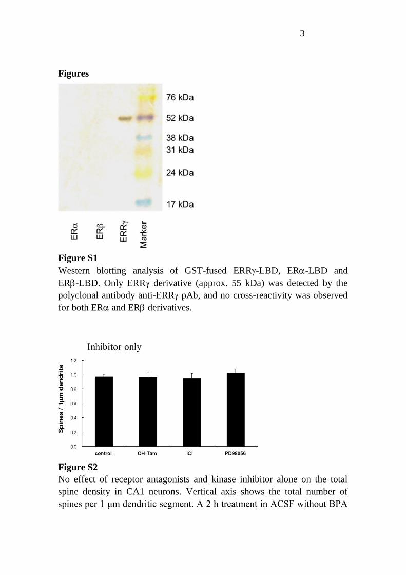
Figure S1
Western blotting analysis of GST-fused ERR-LBD, ER-LBD and
ER-LBD. Only ERR derivative (approx. 55 kDa) was detected by the
polyclonal antibody anti-ERR pAb, and no cross-reactivity was observed
for both ER and ER derivatives.
Figure S2
No effect of receptor antagonists and kinase inhibitor alone on the total
spine density in CA1 neurons. Vertical axis shows the total number of
spines per 1 μm dendritic segment. A 2 h treatment in ACSF without BPA

4
(0 nM BPA), with 1 μM OH-Tam (OH-Tam), with 1 μM ICI (ICI) and
with 50 μM PD98059 (PD98059).
Figure S3
Neural circuits in vivo including the hippocampus. The hippocampus
receives complex projections, including cholinergic neurons from medial
septum and diagonal band of Broca (MSDB), serotonergic neurons from
median raphe (MR), and glutamatergic neurons from supramammillary
neucleus (SUM). GABAergic neurons are also included. ERα/ERβ is
expressed in these neurons. Cholinergic neurons in MSDB express ERRγ.
Isolated hippocampal slice has only local neural circuit.
5
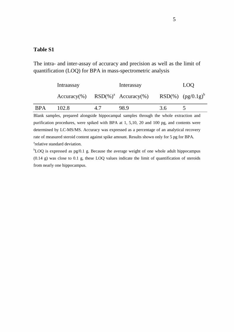
Table S1
The intra- and inter-assay of accuracy and precision as well as the limit of
quantification (LOQ) for BPA in mass-spectrometric analysis
Intraassay Interassay LOQ
Accuracy(%) RSD(%)a Accuracy(%) RSD(%) (pg/0.1g)
b
BPA 102.8 4.7 98.9 3.6 5
Blank samples, prepared alongside hippocampal samples through the whole extraction and
purification procedures, were spiked with BPA at 1, 5,10, 20 and 100 pg, and contents were
determined by LC-MS/MS. Accuracy was expressed as a percentage of an analytical recovery
rate of measured steroid content against spike amount. Results shown only for 5 pg for BPA.
arelative standard deviation.
bLOQ is expressed as pg/0.1 g. Because the average weight of one whole adult hippocampus
(0.14 g) was close to 0.1 g, these LOQ values indicate the limit of quantification of steroids
from nearly one hippocampus.