Embed Size (px)
Citation preview
N-Acyl-Homoserine Lactone Primes Plants for Cell WallReinforcement and Induces Resistance to BacterialPathogens via the Salicylic Acid/Oxylipin PathwayC W OPEN
Sebastian T. Schenk,a Casandra Hernández-Reyes,a Birgit Samans,b Elke Stein,a Christina Neumann,a
Marek Schikora,c Michael Reichelt,d Axel Mithöfer,e Annette Becker,f Karl-Heinz Kogel,a and Adam Schikoraa,1
a Institute of Phytopathology and Applied Zoology, IFZ, Justus Liebig University Giessen, 35392 Giessen, GermanybDepartment of Plant Breeding, IFZ, Justus Liebig University Giessen, 35392 Giessen, GermanycDepartment of Sensor Data and Information Fusion, Fraunhofer FKIE, 53343 Wachtberg, GermanydDepartment of Biochemistry, Max Planck Institute for Chemical Ecology, 07745 Jena, GermanyeDepartment of Bioorganic Chemistry, Plant Defense, Max Planck Institute for Chemical Ecology, 07745 Jena, Germanyf Plant Developmental Biology Group, Institute of Botany, Justus Liebig University Giessen, 35392 Giessen, Germany
ORCID ID: 0000-0002-0869-6423 (A.S.)
The ability of plants to monitor their surroundings, for instance the perception of bacteria, is of crucial importance. Theperception of microorganism-derived molecules and their effector proteins is the best understood of these monitoringprocesses. In addition, plants perceive bacterial quorum sensing (QS) molecules used for cell-to-cell communicationbetween bacteria. Here, we propose a mechanism for how N-acyl-homoserine lactones (AHLs), a group of QS molecules,influence host defense and fortify resistance in Arabidopsis thaliana against bacterial pathogens. N-3-oxo-tetradecanoyl-L-homoserine lactone (oxo-C14-HSL) primed plants for enhanced callose deposition, accumulation of phenolic compounds,and lignification of cell walls. Moreover, increased levels of oxylipins and salicylic acid favored closure of stomata in responseto Pseudomonas syringae infection. The AHL-induced resistance seems to differ from the systemic acquired and the inducedsystemic resistances, providing new insight into inter-kingdom communication. Consistent with the observation that short-chain AHLs, unlike oxo-C14-HSL, promote plant growth, treatments with C6-HSL, oxo-C10-HSL, or oxo-C14-HSL resulted indifferent transcriptional profiles in Arabidopsis. Understanding the priming induced by bacterial QS molecules augments ourknowledge of plant reactions to bacteria and suggests strategies for using beneficial bacteria in plant protection.
INTRODUCTION
In plants, contact with microorganisms can induce local defensemechanisms based on the detection of either microbe-associatedmolecular patterns (MAMPs), which result in MAMP-triggeredimmunity (MTI), or effector proteins, resulting in effector-triggeredimmunity. In addition, plant tissues distal to sites of infection candevelop systemic resistance. The two best understood mecha-nisms of systemic resistance are induced systemic resistance(ISR) and systemic acquired resistance (SAR), the former ofwhich is a response to beneficial, soil-born microbes, whereasthe later is associated with pathogen attack. Both require thepresence of Nonexpressor of PR1 (NPR1) and the action ofspecific phytohormone signal cascades. SAR and ISR havebeen studied in depth and exhaustively reviewed (Van Weeset al., 2008; Dempsey and Klessig, 2012; Fu and Dong, 2013;
Shah and Zeier, 2013). ISR requires components from the eth-ylene (ET) and jasmonic acid (JA) signaling cascades. In Arabi-dopsis thaliana, the myb72 transcription factor mutant isimpaired in ISR (Van der Ent et al., 2008), and the interactionbetween MYB72 and the ethylene-responsive EIN3-like tran-scription factor3 suggests that crosstalk exists between the JAand ET pathways. SAR, typically induced after infection witha pathogen, is dependent on the accumulation of salicylic acid(SA) at the infection site and on either or both MTI or effector-triggered immunity (Fu and Dong, 2013). Several genetic de-terminants of SAR have been identified, including SA bindingproteins, methyl transferases, lipid transfer proteins, and tran-scription factors (for review, see Dempsey and Klessig, 2012;Shah and Zeier, 2013). Nevertheless, the molecular basis of thesystemic signaling is not completely understood, since thebeneficial bacteria Pseudomonas fluorescens strain 89B61 andBacillus pumilus strain T4 induce resistance in a JA- and ET-independent or NPR1-independent manner, respectively (Ryuet al., 2003).The multitude of low molecular weight metabolites and mobile
proteins involved in establishing SAR is striking. Methyl salicylate(MeSA), dehydroabietinal, azelaic acid, a glycerol-3-phosphate–derived factor, pipecolic acid, as well as the putative lipid transferproteins DIR1 and DIR1-like are proposed to be involved in theinduction of systemic resistance (Park et al., 2007; Jung et al.,2009; Chanda et al., 2011; Chaturvedi et al., 2012; Návarová et al.,
1 Address correspondence to [email protected] author responsible for distribution of materials integral to the findingspresented in this article in accordance with the policy described in theInstructions for Authors (www.plantcell.org) is: Adam Schikora ([email protected]).C Some figures in this article are displayed in color online but in black andwhite in the print edition.W Online version contains Web-only data.OPENArticles can be viewed online without a subscription.www.plantcell.org/cgi/doi/10.1105/tpc.114.126763
The Plant Cell, Vol. 26: 2708–2723, June 2014, www.plantcell.org ã 2014 American Society of Plant Biologists. All rights reserved.
2012; Champigny et al., 2013). The current model predicts that allsignal cascades induced by these molecules require NPR1(Dempsey and Klessig, 2012; Shah and Zeier, 2013). In addition,oxylipins other than JA and JA derivatives could play a role in plantdefense mechanisms. For example, colneleic and colnelenic acidsaccumulate in potato (Solanum tuberosum) leaves infected withfungi or viruses (Weber et al., 1999), and together with other oxy-lipins are thought to act similarly to methyl jasmonate (Blée, 2002).Synthesis of active phytooxylipins is initiated either by lip-ooxygenases (LOX) that introduce an oxygen atom at the C9 orC13 position on the lipid chain (Andreou and Feussner, 2009) or bythe nonenzymatic generation of structurally similar phytoprostanes(Sattler et al., 2006). A genome-wide analysis of Arabidopsis re-vealed that more than 150 genes responded to the application of12-oxo-phytodienoic acid (cis-OPDA), though not to JA or methyljasmonate (Taki et al., 2005). cis-OPDA–regulated genes encodedproteins involved in the stress response and signal transduction.Interestingly, expression of the majority of those genes dependedon the TGACG motif binding transcription factors TGA2, TGA5,and TGA6 (Mueller et al., 2008), similar to genes regulated by SAand some pathogens.
Priming is yet another process related to defense mecha-nisms in plants. Primed plants respond in a stronger and/orfaster manner to an infection than do nonprimed plants. Thisphenomenon, called “sensitization,” has been used in agricul-ture since the early 1930s. Many potent “resistance inducers,”including low concentrations of SA, benzothiadiazole, andb-aminobutyric acid (BABA), have been intensively studied inthe last years (Conrath et al., 2002). Azelaic acid was shown toaccumulate in local and systemic tissue upon SAR, priming theplant for enhanced SA production and conferring resistance toPseudomonas syringae (Jung et al., 2009). Recently, two dis-coveries have shed light on the molecular basis of priming. Thefirst was the accumulation of the inactive form of mitogen-activated protein kinases that could be activated upon sec-ondary stimulation (Beckers et al., 2009); the second was histonemethylation (H3K4me3 and H3K4me2) and acetylation (H3K9,H4K5, H4K8, and H4K12) in the promoter regions of the de-fense-associated WRKY6, WRKY26, and WRKY53 transcriptionfactors (Jaskiewicz et al., 2011). Whether the expression or ac-tion of mitogen-activated protein kinases and chromatin modi-fication are connected remains unclear. The primed state canalso be inherited. Transgenerational priming was observed inprogeny of plants exposed to P. syringae, BABA, or herbivores(Luna et al., 2012; Rasmann et al., 2012; Slaughter et al., 2012).Interestingly, while this phenomenon has an epigenetic basis,the priming induced by P. syringae or BABA depended on SA,whereas herbivore-induced transgenerational priming requiredCOI1 and, thus, JA perception. Together, these findings suggestthe importance of epigenetic mechanisms in induced resistance.
Recently, several reports have suggested that bacterial quo-rum sensing molecules also induce a primed state. Currently,N-acyl-homoserine lactones (AHLs) are the best-studied groupof quorum sensing molecules having this activity. Many Gram-negative bacteria produce AHLs in order to monitor their pop-ulation density. In plants, AHL application induces changes ingene expression, alters protein profiles, and modifies root de-velopment (Mathesius et al., 2003; Ortíz-Castro et al., 2008; von
Rad et al., 2008; Schikora et al., 2011; Bai et al., 2012; Schenket al., 2012). The first indication that AHLs play a role in plantimmunity originated from a study of a Serratia liquefaciens–to-mato (Solanum lycopersicum) interaction, in which the wild-typeS. liquefaciens strain MG1, but not the corresponding AHL-negative mutant, induced resistance against the fungal patho-gen Alternaria alternata (Schuhegger et al., 2006). Likewise,resistance against P. syringae induced by Ensifer meliloti(Sinorhizobium meliloti ) in Arabidopsis plants depended on AHLaccumulation (Zarkani et al., 2013). Usually bacteria producehigh concentrations of quorum sensing molecules (e.g., AHLs)when growing in biofilms or mature colonies (above OD600 of0.5). The production of AHLs on root tissues by bacteria growingin biofilms has been shown (Zarkani et al., 2013), and the pro-duction of AHLs in biofilms on leaves is likely to occur, but has notbeen demonstrated yet. In addition, the application of commercialAHLs induced resistance against bacterial and fungal pathogens inArabidopsis. Our previous study suggested that this effect de-pends on a stronger and prolonged activation of MPK6 (Schikoraet al., 2011). Moreover, plant responses to different AHL moleculesappear to be AHL specific. Proteome analysis revealed ;150 dif-ferentially accumulated proteins in response to the application ofeither commercial oxo-C12-HSL or oxo-C16:1-HSL isolated fromcultured S. meliloti (Mathesius et al., 2003).In this article, we substantiate previous studies indicating that
the plant responds to different AHLs at the transcriptional level.We also demonstrate that, after exposure to oxo-C14-HSL,a secondary challenge augments phenolic, lignin, and callosedepositions in the cell wall. Furthermore, accumulation of oxy-lipins in distal tissues promoted stomata closure, thus enhanc-ing plant resistance to bacterial infection. These data show howboth local and systemic phenomena contribute to the enhancedresistance observed in AHL-primed plants.
RESULTS
N-Acyl-Homoserine Lactones Induce AHL-Specific Responses
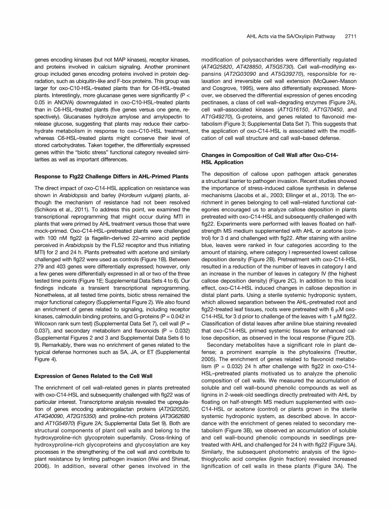
Independent studies suggested that plants react to different AHLmolecules in a specific manner (Mathesius et al., 2003; Ortíz-Castroet al., 2008; Schenk et al., 2012). To gain insight into plant re-sponses, we performed a transcriptome analysis of Arabidopsisseedlings treated with three different AHLs know to have differenteffects. N-hexanoyl-L-homoserine lactone (C6-HSL) was selectedbecause of its plant growth promoting effect (Ortíz-Castro et al.,2008; von Rad et al., 2008; Schikora et al., 2011; Liu et al., 2012;Schenk et al., 2012); N-3-oxo-decanoyl-L-homoserine lactone (oxo-C10-HSL) influences root morphology and root hair formation(Ortíz-Castro et al., 2008); and N-3-oxo-tetradecanoyl-L-homoserinelactone (oxo-C14-HSL) induces plant resistance (Schikora et al.,2011) (Figure 1A). Two-week old Arabidopsis seedlings weretransferred into six-well plates and floated on half-strength Mura-shige and Skoog (MS) medium supplemented with either 6 mMAHLor acetone (control), the solvent for AHLs. After 3 d, whole seedlingswere harvested (Figure 1B). We interrogated the Arabidopsis(4x44k) Gene Expression Microarray from Agilent Technology to
AHL Acts via the SA/Oxylipin Pathway 2709
identify differentially expressed genes showing a log2 fold change of1 and a moderate P value of 0.05 (Smyth, 2004). Within thesethresholds, we found 954 differentially regulated genes (Figure 1C;Supplemental Data Sets 1 to 4). Of the 55 genes that responded toall three AHLs, 45% (25 genes) were related to stress responsesand signaling (Figure 1D; Supplemental Data Set 1). Furthermore,
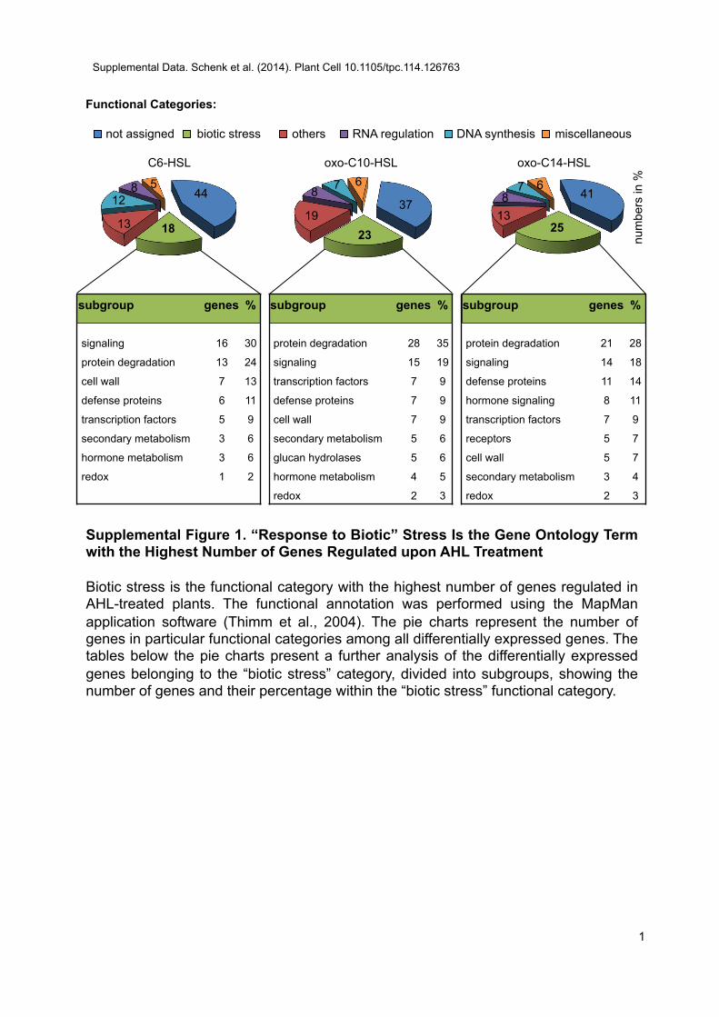
214, 226, and 177 genes (total of 617 genes) responded only tothe application of C6-HSL, oxo-C10-HSL, or oxo-C14-HSL, re-spectively, indicating that the different AHLs regulated a specific setof host genes. Among all of the genes responsive to a specific AHLmolecule, 18 to 25% were assigned to the functional category“biotic stress” (Supplemental Figure 1), especially signaling-related
Figure 1. Arabidopsis Responds to Different AHLs with Diverse Transcriptional Reprogramming.
(A) AHLs were prepared as 60 mM stocks in acetone and used at a working concentration of 6 mM in half-strength MS medium for (pre)treatments.(B) Experimental setup to analyze the transcriptional responses to treatments with 6 mM C6-HSL, oxo-C10-HSL, and oxo-C14-HSL as well as theresponse of oxo-C14-HSL–pretreated plants to secondary challenge with flg22. Two-week-old Arabidopsis seedlings were treated with three differenthomoserine lactones (AHL-priming). After 3 d, the oxo-C14-HSL–pretreated seedlings were challenged with 100 nM flg22 to trigger the defense response.(C) Number of genes differentially expressed upon AHL (pre)treatment and the flg22 challenge. Differentially expressed genes were selected by a log2
fold change of 1 and a moderate P value of 0.05.(D) Venn diagram of genes up- and downregulated after treatment with the three different AHLs. Only 55 genes were regulated by all AHLs, and ;200genes were regulated by each AHL molecule.(E) Venn diagram of genes differentially expressed in response to 100 nM flg22 in oxo-C14-HSL–pretreated seedlings after 0, 2, and 24 h. The responseis very dynamic, as shown by the small overlap in differentially expressed genes at different time points after the secondary challenge.
2710 The Plant Cell
genes encoding kinases (but not MAP kinases), receptor kinases,and proteins involved in calcium signaling. Another prominentgroup included genes encoding proteins involved in protein deg-radation, such as ubiquitin-like and F-box proteins. This group waslarger for oxo-C10-HSL–treated plants than for C6-HSL–treatedplants. Interestingly, more glucanase genes were significantly (P <0.05 in ANOVA) downregulated in oxo-C10-HSL–treated plantsthan in C6-HSL–treated plants (five genes versus one gene, re-spectively). Glucanases hydrolyze amylose and amylopectin torelease glucose, suggesting that plants may reduce their carbo-hydrate metabolism in response to oxo-C10-HSL treatment,whereas C6-HSL–treated plants might conserve their level ofstored carbohydrates. Taken together, the differentially expressedgenes within the “biotic stress” functional category revealed simi-larities as well as important differences.
Response to Flg22 Challenge Differs in AHL-Primed Plants
The direct impact of oxo-C14-HSL application on resistance wasshown in Arabidopsis and barley (Hordeum vulgare) plants, al-though the mechanism of resistance had not been resolved(Schikora et al., 2011). To address this point, we examined thetranscriptional reprogramming that might occur during MTI inplants that were primed by AHL treatment versus those that weremock-primed. Oxo-C14-HSL–pretreated plants were challengedwith 100 nM flg22 (a flagellin-derived 22–amino acid peptideperceived in Arabidopsis by the FLS2 receptor and thus initiatingMTI) for 2 and 24 h. Plants pretreated with acetone and similarlychallenged with flg22 were used as controls (Figure 1B). Between279 and 403 genes were differentially expressed; however, onlya few genes were differentially expressed in all or two of the threetested time points (Figure 1E; Supplemental Data Sets 4 to 6). Ourfindings indicate a transient transcriptional reprogramming.Nonetheless, at all tested time points, biotic stress remained themajor functional category (Supplemental Figure 2). We also foundan enrichment of genes related to signaling, including receptorkinases, calmodulin binding proteins, and G-proteins (P = 0.042 inWilcoxon rank sum test) (Supplemental Data Set 7), cell wall (P =0.037), and secondary metabolism and flavonoids (P = 0.032)(Supplemental Figures 2 and 3 and Supplemental Data Sets 6 to9). Remarkably, there was no enrichment of genes related to thetypical defense hormones such as SA, JA, or ET (SupplementalFigure 4).
Expression of Genes Related to the Cell Wall
The enrichment of cell wall–related genes in plants pretreatedwith oxo-C14-HSL and subsequently challenged with flg22 was ofparticular interest. Transcriptome analysis revealed the upregula-tion of genes encoding arabinogalactan proteins (AT2G20520,AT4G40090, AT2G15350) and proline-rich proteins (AT3G62680and AT1G54970) (Figure 2A; Supplemental Data Set 9). Both arestructural components of plant cell walls and belong to thehydroxyproline-rich glycoprotein superfamily. Cross-linking ofhydroxyproline-rich glycoproteins and glycosylation are keyprocesses in the strengthening of the cell wall and contribute toplant resistance by limiting pathogen invasion (Wei and Shirsat,2006). In addition, several other genes involved in the
modification of polysaccharides were differentially regulated(AT4G25820, AT428850, AT5G5730). Cell wall–modifying ex-pansins (AT2G03090 and AT5G39270), responsible for re-laxation and irreversible cell wall extension (McQueen-Masonand Cosgrove, 1995), were also differentially expressed. More-over, we observed the differential expression of genes encodingpectinases, a class of cell wall–degrading enzymes (Figure 2A),cell wall–associated kinases (AT1G16150, AT1G70450, andAT1G49270), G-proteins, and genes related to flavonoid me-tabolism (Figure 3; Supplemental Data Set 7). This suggests thatthe application of oxo-C14-HSL is associated with the modifi-cation of cell wall structure and call wall–based defense.
Changes in Composition of Cell Wall after Oxo-C14-HSL Application
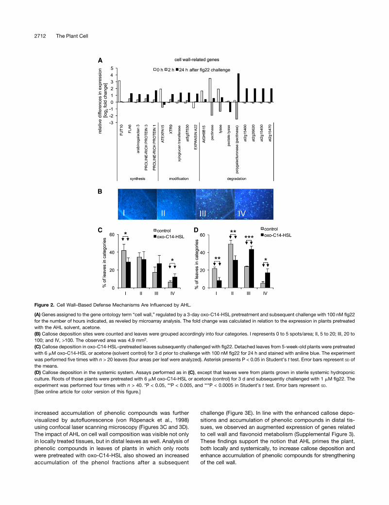
The deposition of callose upon pathogen attack generatesa structural barrier to pathogen invasion. Recent studies showedthe importance of stress-induced callose synthesis in defensemechanisms (Jacobs et al., 2003; Ellinger et al., 2013). The en-richment in genes belonging to cell wall–related functional cat-egories encouraged us to analyze callose deposition in plantspretreated with oxo-C14-HSL and subsequently challenged withflg22. Experiments were performed with leaves floated on half-strength MS medium supplemented with AHL or acetone (con-trol) for 3 d and challenged with flg22. After staining with anilineblue, leaves were ranked in four categories according to theamount of staining, where category I represented lowest callosedeposition density (Figure 2B). Pretreatment with oxo-C14-HSLresulted in a reduction of the number of leaves in category I andan increase in the number of leaves in category IV (the highestcallose deposition density) (Figure 2C). In addition to this localeffect, oxo-C14-HSL induced changes in callose deposition indistal plant parts. Using a sterile systemic hydroponic system,which allowed separation between the AHL-pretreated root andflg22-treated leaf tissues, roots were pretreated with 6 mM oxo-C14-HSL for 3 d prior to challenge of the leaves with 1 mM flg22.Classification of distal leaves after aniline blue staining revealedthat oxo-C14-HSL primed systemic tissues for enhanced cal-lose deposition, as observed in the local response (Figure 2D).Secondary metabolites have a significant role in plant de-
fense; a prominent example is the phytoalexins (Treutter,2005). The enrichment of genes related to flavonoid metabo-lism (P = 0.032) 24 h after challenge with flg22 in oxo-C14-HSL–pretreated plants motivated us to analyze the phenoliccomposition of cell walls. We measured the accumulation ofsoluble and cell wall–bound phenolic compounds as well aslignins in 2-week-old seedlings directly pretreated with AHL byfloating on half-strength MS medium supplemented with oxo-C14-HSL or acetone (control) or plants grown in the sterilesystemic hydroponic system, as described above. In accor-dance with the enrichment of genes related to secondary me-tabolism (Figure 3B), we observed an accumulation of solubleand cell wall–bound phenolic compounds in seedlings pre-treated with AHL and challenged for 24 h with flg22 (Figure 3A).Similarly, the subsequent photometric analysis of the ligno-thioglycolic acid complex (lignin fraction) revealed increasedlignification of cell walls in these plants (Figure 3A). The
AHL Acts via the SA/Oxylipin Pathway 2711
increased accumulation of phenolic compounds was furthervisualized by autofluorescence (von Röpenack et al., 1998)using confocal laser scanning microscopy (Figures 3C and 3D).The impact of AHL on cell wall composition was visible not onlyin locally treated tissues, but in distal leaves as well. Analysis ofphenolic compounds in leaves of plants in which only rootswere pretreated with oxo-C14-HSL also showed an increasedaccumulation of the phenol fractions after a subsequent
challenge (Figure 3E). In line with the enhanced callose depo-sitions and accumulation of phenolic compounds in distal tis-sues, we observed an augmented expression of genes relatedto cell wall and flavonoid metabolism (Supplemental Figure 3).These findings support the notion that AHL primes the plant,both locally and systemically, to increase callose deposition andenhance accumulation of phenolic compounds for strengtheningof the cell wall.
Figure 2. Cell Wall–Based Defense Mechanisms Are Influenced by AHL.
(A) Genes assigned to the gene ontology term “cell wall,” regulated by a 3-day oxo-C14-HSL pretreatment and subsequent challenge with 100 nM flg22for the number of hours indicated, as reveled by microarray analysis. The fold change was calculated in relation to the expression in plants pretreatedwith the AHL solvent, acetone.(B) Callose deposition sites were counted and leaves were grouped accordingly into four categories. I represents 0 to 5 spots/area; II, 5 to 20; III, 20 to100; and IV, >100. The observed area was 4.9 mm2.(C) Callose deposition in oxo-C14-HSL–pretreated leaves subsequently challenged with flg22. Detached leaves from 5-week-old plants were pretreatedwith 6 mM oxo-C14-HSL or acetone (solvent control) for 3 d prior to challenge with 100 nM flg22 for 24 h and stained with aniline blue. The experimentwas performed five times with n > 20 leaves (four areas per leaf were analyzed). Asterisk presents P < 0.05 in Student’s t test. Error bars represent SD ofthe means.(D) Callose deposition in the systemic system. Assays performed as in (C), except that leaves were from plants grown in sterile systemic hydroponicculture. Roots of those plants were pretreated with 6 mM oxo-C14-HSL or acetone (control) for 3 d and subsequently challenged with 1 mM flg22. Theexperiment was performed four times with n > 40. *P < 0.05, **P < 0.005, and ***P < 0.0005 in Student’s t test. Error bars represent SD.[See online article for color version of this figure.]
2712 The Plant Cell
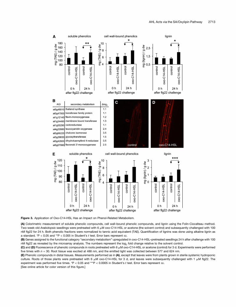
Figure 3. Application of Oxo-C14-HSL Has an Impact on Phenol-Related Metabolism.
(A) Colorimetric measurement of soluble phenolic compounds, cell wall–bound phenolic compounds, and lignin using the Folin-Ciocalteau method.Two-week-old Arabidopsis seedlings were pretreated with 6 mM oxo-C14-HSL or acetone (the solvent control) and subsequently challenged with 100nM flg22 for 24 h. Both phenolic fractions were normalized to tannic acid equivalent (TAE). Quantification of lignins was done using alkaline lignin asa standard. *P < 0.05 and **P < 0.005 in Student’s t test. Error bars represent SD.(B) Genes assigned to the functional category “secondary metabolism” upregulated in oxo-C14-HSL–pretreated seedlings 24 h after challenge with 100nM flg22 as revealed by the microarray analysis. The numbers represent the log2 fold change relative to the solvent control.(C) and (D) Fluorescence of phenolic compounds in roots pretreated with 6 mM oxo-C14-HSL or acetone (control) for 3 d. Experiments were performedfive times with n = 30. Root tissue was excited at 488 nm, and the emitted light was collected between 577 and 624 nm.(E) Phenolic compounds in distal tissues. Measurements performed as in (A), except that leaves were from plants grown in sterile systemic hydroponicculture. Roots of those plants were pretreated with 6 mM oxo-C14-HSL for 3 d, and leaves were subsequently challenged with 1 mM flg22. Theexperiment was performed five times. *P < 0.05 and ***P < 0.0005 in Student’s t test. Error bars represent SD.[See online article for color version of this figure.]
AHL Acts via the SA/Oxylipin Pathway 2713
Oxo-C14-HSL–Induced Resistance Requires NPR1 and IsCOI1 Independent
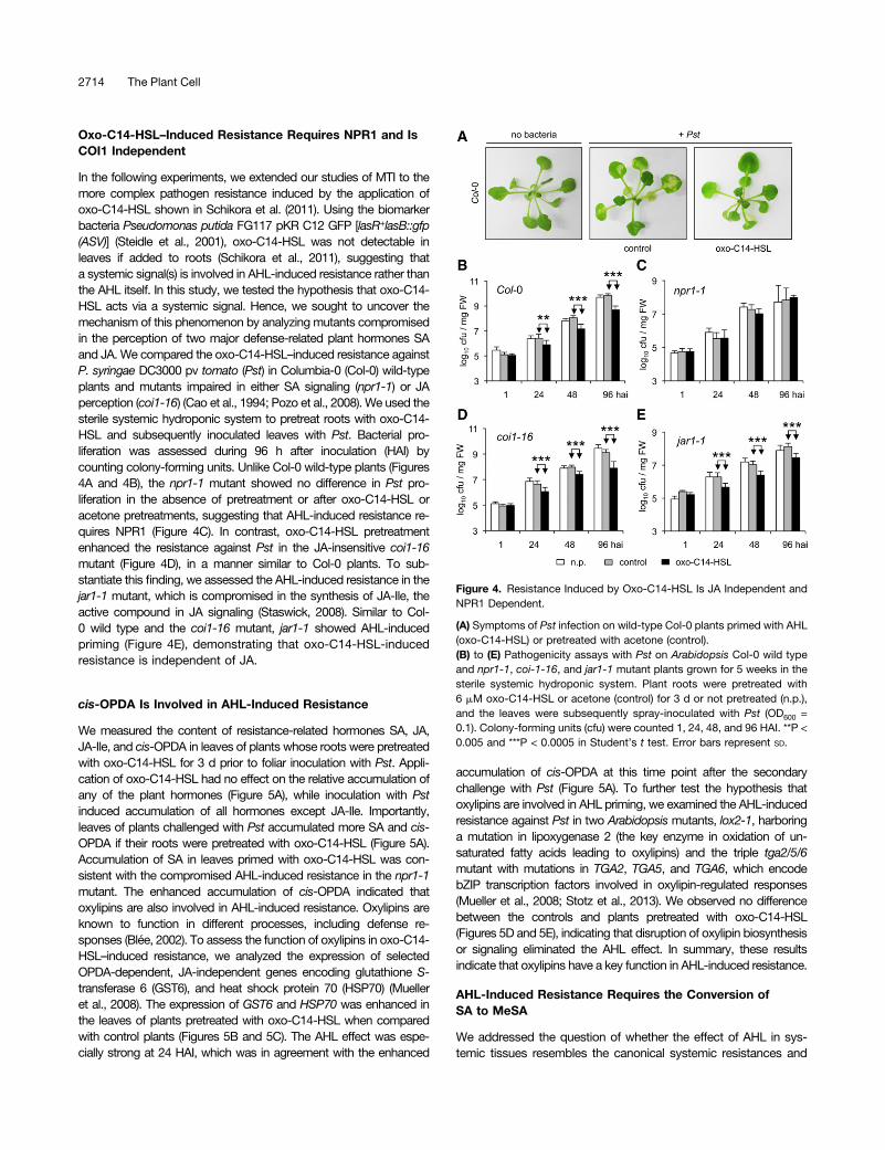
In the following experiments, we extended our studies of MTI to themore complex pathogen resistance induced by the application ofoxo-C14-HSL shown in Schikora et al. (2011). Using the biomarkerbacteria Pseudomonas putida FG117 pKR C12 GFP [lasR+lasB::gfp(ASV)] (Steidle et al., 2001), oxo-C14-HSL was not detectable inleaves if added to roots (Schikora et al., 2011), suggesting thata systemic signal(s) is involved in AHL-induced resistance rather thanthe AHL itself. In this study, we tested the hypothesis that oxo-C14-HSL acts via a systemic signal. Hence, we sought to uncover themechanism of this phenomenon by analyzing mutants compromisedin the perception of two major defense-related plant hormones SAand JA. We compared the oxo-C14-HSL–induced resistance againstP. syringae DC3000 pv tomato (Pst) in Columbia-0 (Col-0) wild-typeplants and mutants impaired in either SA signaling (npr1-1) or JAperception (coi1-16) (Cao et al., 1994; Pozo et al., 2008). We used thesterile systemic hydroponic system to pretreat roots with oxo-C14-HSL and subsequently inoculated leaves with Pst. Bacterial pro-liferation was assessed during 96 h after inoculation (HAI) bycounting colony-forming units. Unlike Col-0 wild-type plants (Figures4A and 4B), the npr1-1 mutant showed no difference in Pst pro-liferation in the absence of pretreatment or after oxo-C14-HSL oracetone pretreatments, suggesting that AHL-induced resistance re-quires NPR1 (Figure 4C). In contrast, oxo-C14-HSL pretreatmentenhanced the resistance against Pst in the JA-insensitive coi1-16mutant (Figure 4D), in a manner similar to Col-0 plants. To sub-stantiate this finding, we assessed the AHL-induced resistance in thejar1-1 mutant, which is compromised in the synthesis of JA-Ile, theactive compound in JA signaling (Staswick, 2008). Similar to Col-0 wild type and the coi1-16 mutant, jar1-1 showed AHL-inducedpriming (Figure 4E), demonstrating that oxo-C14-HSL-inducedresistance is independent of JA.
cis-OPDA Is Involved in AHL-Induced Resistance
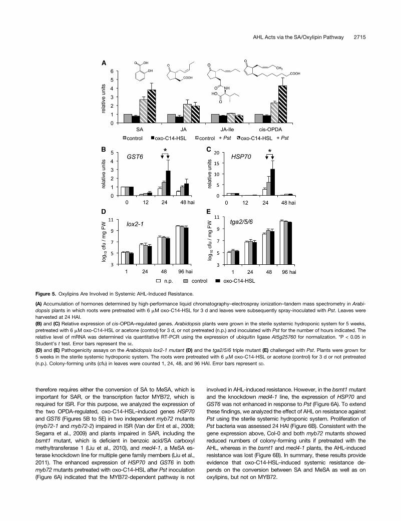
We measured the content of resistance-related hormones SA, JA,JA-Ile, and cis-OPDA in leaves of plants whose roots were pretreatedwith oxo-C14-HSL for 3 d prior to foliar inoculation with Pst. Appli-cation of oxo-C14-HSL had no effect on the relative accumulation ofany of the plant hormones (Figure 5A), while inoculation with Pstinduced accumulation of all hormones except JA-Ile. Importantly,leaves of plants challenged with Pst accumulated more SA and cis-OPDA if their roots were pretreated with oxo-C14-HSL (Figure 5A).Accumulation of SA in leaves primed with oxo-C14-HSL was con-sistent with the compromised AHL-induced resistance in the npr1-1mutant. The enhanced accumulation of cis-OPDA indicated thatoxylipins are also involved in AHL-induced resistance. Oxylipins areknown to function in different processes, including defense re-sponses (Blée, 2002). To assess the function of oxylipins in oxo-C14-HSL–induced resistance, we analyzed the expression of selectedOPDA-dependent, JA-independent genes encoding glutathione S-transferase 6 (GST6), and heat shock protein 70 (HSP70) (Muelleret al., 2008). The expression of GST6 and HSP70 was enhanced inthe leaves of plants pretreated with oxo-C14-HSL when comparedwith control plants (Figures 5B and 5C). The AHL effect was espe-cially strong at 24 HAI, which was in agreement with the enhanced
accumulation of cis-OPDA at this time point after the secondarychallenge with Pst (Figure 5A). To further test the hypothesis thatoxylipins are involved in AHL priming, we examined the AHL-inducedresistance against Pst in two Arabidopsis mutants, lox2-1, harboringa mutation in lipoxygenase 2 (the key enzyme in oxidation of un-saturated fatty acids leading to oxylipins) and the triple tga2/5/6mutant with mutations in TGA2, TGA5, and TGA6, which encodebZIP transcription factors involved in oxylipin-regulated responses(Mueller et al., 2008; Stotz et al., 2013). We observed no differencebetween the controls and plants pretreated with oxo-C14-HSL(Figures 5D and 5E), indicating that disruption of oxylipin biosynthesisor signaling eliminated the AHL effect. In summary, these resultsindicate that oxylipins have a key function in AHL-induced resistance.
AHL-Induced Resistance Requires the Conversion ofSA to MeSA
We addressed the question of whether the effect of AHL in sys-temic tissues resembles the canonical systemic resistances and
Figure 4. Resistance Induced by Oxo-C14-HSL Is JA Independent andNPR1 Dependent.
(A) Symptoms of Pst infection on wild-type Col-0 plants primed with AHL(oxo-C14-HSL) or pretreated with acetone (control).(B) to (E) Pathogenicity assays with Pst on Arabidopsis Col-0 wild typeand npr1-1, coi-1-16, and jar1-1 mutant plants grown for 5 weeks in thesterile systemic hydroponic system. Plant roots were pretreated with6 mM oxo-C14-HSL or acetone (control) for 3 d or not pretreated (n.p.),and the leaves were subsequently spray-inoculated with Pst (OD600 =0.1). Colony-forming units (cfu) were counted 1, 24, 48, and 96 HAI. **P <0.005 and ***P < 0.0005 in Student’s t test. Error bars represent SD.
2714 The Plant Cell
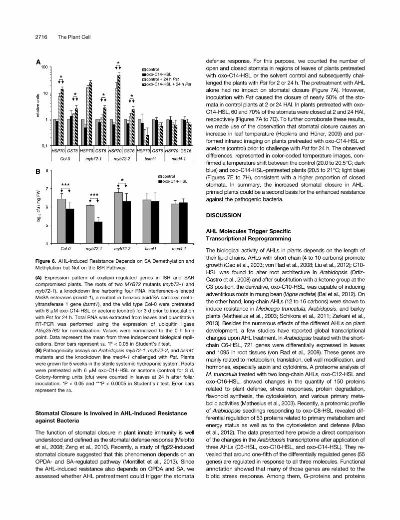
therefore requires either the conversion of SA to MeSA, which isimportant for SAR, or the transcription factor MYB72, which isrequired for ISR. For this purpose, we analyzed the expression ofthe two OPDA-regulated, oxo-C14-HSL–induced genes HSP70and GST6 (Figures 5B to 5E) in two independent myb72 mutants(myb72-1 and myb72-2) impaired in ISR (Van der Ent et al., 2008;Segarra et al., 2009) and plants impaired in SAR, including thebsmt1 mutant, which is deficient in benzoic acid/SA carboxylmethyltransferase 1 (Liu et al., 2010), and med4-1, a MeSA es-terase knockdown line for multiple gene family members (Liu et al.,2011). The enhanced expression of HSP70 and GST6 in bothmyb72mutants pretreated with oxo-C14-HSL after Pst inoculation(Figure 6A) indicated that the MYB72-dependent pathway is not
involved in AHL-induced resistance. However, in the bsmt1mutantand the knockdown med4-1 line, the expression of HSP70 andGST6 was not enhanced in response to Pst (Figure 6A). To extendthese findings, we analyzed the effect of AHL on resistance againstPst using the sterile systemic hydroponic system. Proliferation ofPst bacteria was assessed 24 HAI (Figure 6B). Consistent with thegene expression above, Col-0 and both myb72 mutants showedreduced numbers of colony-forming units if pretreated with theAHL, whereas in the bsmt1 and med4-1 plants, the AHL-inducedresistance was lost (Figure 6B). In summary, these results provideevidence that oxo-C14-HSL–induced systemic resistance de-pends on the conversion between SA and MeSA as well as onoxylipins, but not on MYB72.
Figure 5. Oxylipins Are Involved in Systemic AHL-Induced Resistance.
(A) Accumulation of hormones determined by high-performance liquid chromatography–electrospray ionization–tandem mass spectrometry in Arabi-dopsis plants in which roots were pretreated with 6 mM oxo-C14-HSL for 3 d and leaves were subsequently spray-inoculated with Pst. Leaves wereharvested at 24 HAI.(B) and (C) Relative expression of cis-OPDA–regulated genes. Arabidopsis plants were grown in the sterile systemic hydroponic system for 5 weeks,pretreated with 6 mM oxo-C14-HSL or acetone (control) for 3 d, or not pretreated (n.p.) and inoculated with Pst for the number of hours indicated. Therelative level of mRNA was determined via quantitative RT-PCR using the expression of ubiquitin ligase At5g25760 for normalization. *P < 0.05 inStudent’s t test. Error bars represent the SE.(D) and (E) Pathogenicity assays on the Arabidopsis lox2-1 mutant (D) and the tga2/5/6 triple mutant (E) challenged with Pst. Plants were grown for5 weeks in the sterile systemic hydroponic system. The roots were pretreated with 6 mM oxo-C14-HSL or acetone (control) for 3 d or not pretreated(n.p.). Colony-forming units (cfu) in leaves were counted 1, 24, 48, and 96 HAI. Error bars represent SD.
AHL Acts via the SA/Oxylipin Pathway 2715
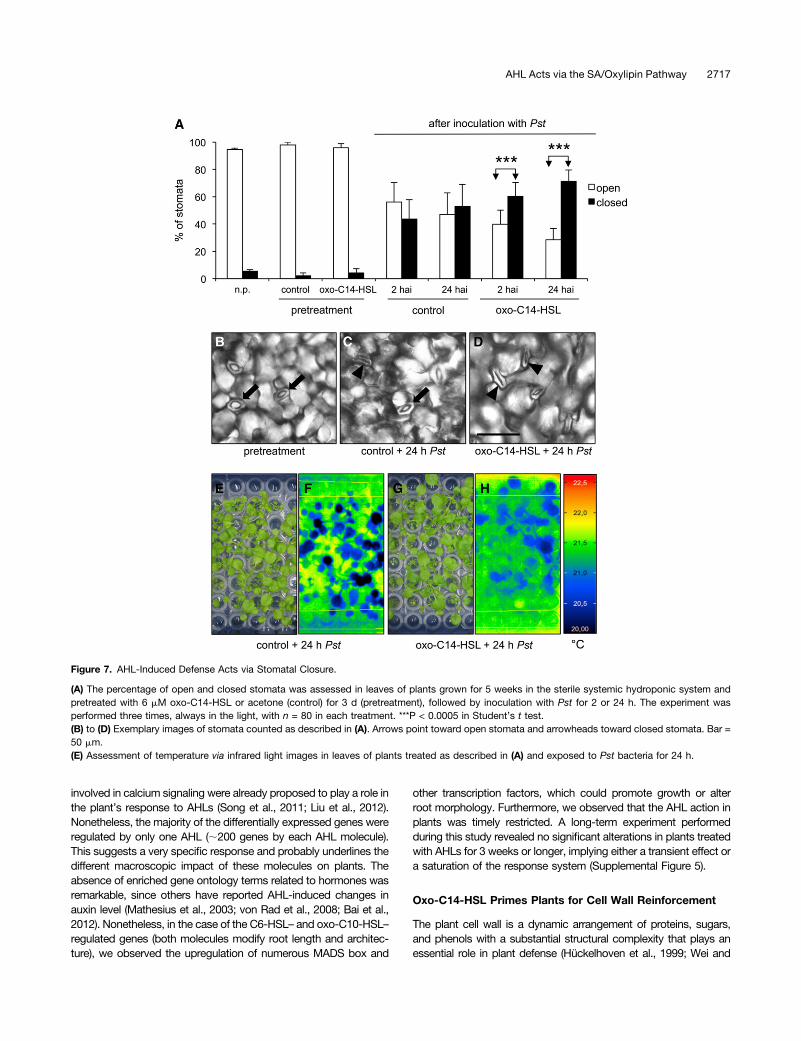
Stomatal Closure Is Involved in AHL-Induced Resistanceagainst Bacteria
The function of stomatal closure in plant innate immunity is wellunderstood and defined as the stomatal defense response (Melottoet al., 2008; Zeng et al., 2010). Recently, a study of flg22-inducedstomatal closure suggested that this phenomenon depends on anOPDA- and SA-regulated pathway (Montillet et al., 2013). Sincethe AHL-induced resistance also depends on OPDA and SA, weassessed whether AHL pretreatment could trigger the stomata
defense response. For this purpose, we counted the number ofopen and closed stomata in regions of leaves of plants pretreatedwith oxo-C14-HSL or the solvent control and subsequently chal-lenged the plants with Pst for 2 or 24 h. The pretreatment with AHLalone had no impact on stomatal closure (Figure 7A). However,inoculation with Pst caused the closure of nearly 50% of the sto-mata in control plants at 2 or 24 HAI. In plants pretreated with oxo-C14-HSL, 60 and 70% of the stomata were closed at 2 and 24 HAI,respectively (Figures 7A to 7D). To further corroborate these results,we made use of the observation that stomatal closure causes anincrease in leaf temperature (Hopkins and Hüner, 2009) and per-formed infrared imaging on plants pretreated with oxo-C14-HSL oracetone (control) prior to challenge with Pst for 24 h. The observeddifferences, represented in color-coded temperature images, con-firmed a temperature shift between the control (20.0 to 20.5°C; darkblue) and oxo-C14-HSL–pretreated plants (20.5 to 21°C; light blue)(Figures 7E to 7H), consistent with a higher proportion of closedstomata. In summary, the increased stomatal closure in AHL-primed plants could be a second basis for the enhanced resistanceagainst the pathogenic bacteria.
DISCUSSION
AHL Molecules Trigger SpecificTranscriptional Reprogramming
The biological activity of AHLs in plants depends on the length oftheir lipid chains. AHLs with short chain (4 to 10 carbons) promotegrowth (Gao et al., 2003; von Rad et al., 2008; Liu et al., 2012); C10-HSL was found to alter root architecture in Arabidopsis (Ortíz-Castro et al., 2008) and after substitution with a ketone group at theC3 position, the derivative, oxo-C10-HSL, was capable of inducingadventitious roots in mung bean (Vigna radiata) (Bai et al., 2012). Onthe other hand, long-chain AHLs (12 to 16 carbons) were shown toinduce resistance in Medicago truncatula, Arabidopsis, and barleyplants (Mathesius et al., 2003; Schikora et al., 2011; Zarkani et al.,2013). Besides the numerous effects of the different AHLs on plantdevelopment, a few studies have reported global transcriptionalchanges upon AHL treatment. In Arabidopsis treated with the short-chain C6-HSL, 721 genes were differentially expressed in leavesand 1095 in root tissues (von Rad et al., 2008). These genes aremainly related to metabolism, translation, cell wall modification, andhormones, especially auxin and cytokinins. A proteome analysis ofM. truncatula treated with two long-chain AHLs, oxo-C12-HSL andoxo-C16-HSL, showed changes in the quantity of 150 proteinsrelated to plant defense, stress responses, protein degradation,flavonoid synthesis, the cytoskeleton, and various primary meta-bolic activities (Mathesius et al., 2003). Recently, a proteomic profileof Arabidopsis seedlings responding to oxo-C8-HSL revealed dif-ferential regulation of 53 proteins related to primary metabolism andenergy status as well as to the cytoskeleton and defense (Miaoet al., 2012). The data presented here provide a direct comparisonof the changes in the Arabidopsis transcriptome after application ofthree AHLs (C6-HSL, oxo-C10-HSL, and oxo-C14-HSL). They re-vealed that around one-fifth of the differentially regulated genes (55genes) are regulated in response to all three molecules. Functionalannotation showed that many of those genes are related to thebiotic stress response. Among them, G-proteins and proteins
Figure 6. AHL-Induced Resistance Depends on SA Demethylation andMethylation but Not on the ISR Pathway.
(A) Expression pattern of oxylipin-regulated genes in ISR and SARcompromised plants. The roots of two MYB72 mutants (myb72-1 andmyb72-1), a knockdown line harboring four RNA interference–silencedMeSA esterases (med4-1), a mutant in benzoic acid/SA carboxyl meth-yltransferase 1 gene (bsmt1), and the wild type Col-0 were pretreatedwith 6 mM oxo-C14-HSL or acetone (control) for 3 d prior to inoculationwith Pst for 24 h. Total RNA was extracted from leaves and quantitativeRT-PCR was performed using the expression of ubiquitin ligaseAt5g25760 for normalization. Values were normalized to the 0 h timepoint. Data represent the mean from three independent biological repli-cations. Error bars represent SE. *P < 0.05 in Student’s t test.(B) Pathogenicity assays on Arabidopsis myb72-1, myb72-2, and bsmt1mutants and the knockdown line med4-1 challenged with Pst. Plantswere grown for 5 weeks in the sterile systemic hydroponic system. Rootswere pretreated with 6 mM oxo-C14-HSL or acetone (control) for 3 d.Colony-forming units (cfu) were counted in leaves at 24 h after foliarinoculation. *P < 0.05 and ***P < 0.0005 in Student’s t test. Error barsrepresent the SD.
2716 The Plant Cell
involved in calcium signaling were already proposed to play a role inthe plant’s response to AHLs (Song et al., 2011; Liu et al., 2012).Nonetheless, the majority of the differentially expressed genes wereregulated by only one AHL (;200 genes by each AHL molecule).This suggests a very specific response and probably underlines thedifferent macroscopic impact of these molecules on plants. Theabsence of enriched gene ontology terms related to hormones wasremarkable, since others have reported AHL-induced changes inauxin level (Mathesius et al., 2003; von Rad et al., 2008; Bai et al.,2012). Nonetheless, in the case of the C6-HSL– and oxo-C10-HSL–regulated genes (both molecules modify root length and architec-ture), we observed the upregulation of numerous MADS box and
other transcription factors, which could promote growth or alterroot morphology. Furthermore, we observed that the AHL action inplants was timely restricted. A long-term experiment performedduring this study revealed no significant alterations in plants treatedwith AHLs for 3 weeks or longer, implying either a transient effect ora saturation of the response system (Supplemental Figure 5).
Oxo-C14-HSL Primes Plants for Cell Wall Reinforcement
The plant cell wall is a dynamic arrangement of proteins, sugars,and phenols with a substantial structural complexity that plays anessential role in plant defense (Hückelhoven et al., 1999; Wei and
Figure 7. AHL-Induced Defense Acts via Stomatal Closure.
(A) The percentage of open and closed stomata was assessed in leaves of plants grown for 5 weeks in the sterile systemic hydroponic system andpretreated with 6 mM oxo-C14-HSL or acetone (control) for 3 d (pretreatment), followed by inoculation with Pst for 2 or 24 h. The experiment wasperformed three times, always in the light, with n = 80 in each treatment. ***P < 0.0005 in Student’s t test.(B) to (D) Exemplary images of stomata counted as described in (A). Arrows point toward open stomata and arrowheads toward closed stomata. Bar =50 mm.(E) Assessment of temperature via infrared light images in leaves of plants treated as described in (A) and exposed to Pst bacteria for 24 h.
AHL Acts via the SA/Oxylipin Pathway 2717
Shirsat, 2006; Hückelhoven, 2007; Deepak et al., 2010). Previously,we observed enhanced formation of papillae in barley plants pre-treated with oxo-C14-HSL and challenged with the pathogenicfungus Blumeria graminis f sp hordei (Schikora et al., 2011). Theformation of papilla results from the accumulation and reactiveoxygen species–induced cross-linkage of glycoproteins and ondeposition of callose and phenolic compounds at the formation site(Hückelhoven, 2007; Deepak et al., 2010). In this study, we ob-served an enrichment of genes in the cell wall and glycoproteinsfunctional categories in oxo-C14-HSL–pretreated plants, particularlyafter challenge with flg22. Furthermore, we observed an increase ofcallose deposition, phenolic compounds, and lignins in flg22-challenged, AHL-primed plants. Together, these results suggestthat AHL application primed plants for cell wall reinforcement, whichcould explain their enhanced resistance to bacterial pathogens.
Systemic Resistance in AHL-Primed Plants
In contrast to C6-HSL, long-chain oxo-C14-HSL is not trans-ported within Arabidopsis plants (Schikora et al., 2011). A recentuptake and transport study of radioactive C8-HSL and C10-HSLshowed that the intermediate-length lipid chain AHLs are taken upand transported into the lower shoot zone, although not into theupper leaf tissues of barley plants (Sieper et al., 2013). Therefore,a likely explanation of oxo-C14-HSL–induced resistance in distaltissue is a systemic induced resistance. The two best understoodmechanisms of systemic resistance, ISR and SAR, depend on JA/ET and SA, respectively. However, JA- and SA-independent in-duction of resistance has also been reported (Ryu et al., 2003).Here, we speculated whether JA/ET and/or SA are involved in theestablishment of AHL-induced resistance. On one hand, severallines of evidence indicate JA independence, including the inducedresistance in coi1-16 and jar1-1 mutants and the expression of JA-responsiveMYC2 and VSP2 genes, which was not affected by AHLapplication (Supplemental Figure 6). Similarly, the accumulation ofJA or its bioactive derivative JA-Ile was not influenced by AHLpretreatment. Moreover, the enhanced expression of HSP70 andGST6 in bothmyb72mutants after pretreatment with oxo-C14-HSLas well as the lack of enhanced expression of the MYB72 or ET-responsive genes in AHL-primed wild-type plants (SupplementalFigures 6 and 7) indicate that JA and the ISR mechanisms are notinvolved in oxo-C14-HLS–induced resistance. On the other hand,the loss of the AHL effect in the npr1-1 mutant, the effect of oxo-C14-HSL on SA accumulation after inoculation with Pst, and theexpression of PR1 after oxo-C14-HSL pretreatment (SupplementalFigure 6) suggest that SA might be involved in AHL-induced re-sistance. However, the derivative MeSA and not SA per se waspostulated to be the mobile signal (Vernooij et al., 1994; Park et al.,2007). Therefore, we monitored the expression of oxo-C14-HSL–induced genes in plants impaired in MeSA accumulation (bsmt1)(Liu et al., 2010) and its conversion back to SA (med4-1) (Liu et al.,2011). The oxo-C14-HSL–induced gene expression is lost in bothmutants, suggesting the importance of MeSA and the conver-sion between MeSA and SA. Usually, SAR is associated with theexpression of Pathogenesis Related (PR) genes. In addition, thePAL gene encoding phenylalanine-ammonia lyase is highly ex-pressed in benzothiadiazole-primed plants after a subsequentchallenge (Kohler et al., 2002). Nonetheless, neither PAL nor
other SA-regulated genes (PR2 and PR5) (Supplemental Figures6 and 7) were differentially regulated in plants pretreated withAHL and inoculated with Pst, suggesting that the AHL-inducedresistance could differ from the typical SA-dependent response.Recently, oxylipins (JA precursors) were postulated to be in-volved in stress responses (Stintzi et al., 2001; Blée, 2002; Takiet al., 2005). Besides an accumulation of SA, we observed anincreased accumulation of the oxylipin cis-OPDA. OPDA hasbeen shown to induce a set of genes that are COI1 independent(Stintzi et al., 2001; Mueller et al., 2008) and require the bZIPtranscription factors TGA2, TGA5, and TGA6 (Stotz et al., 2013).Similarly, AHL-induced resistance required LOX2 lipoxygenaseand TGA2/5/6 transcription factors and was independent ofCOI1. Moreover, the enhanced expression of OPDA-regulatedgenes (GST6 and HSP70) (Mueller et al., 2008) in AHL-primedplants supports the notion that oxo-C14-HSL–induced re-sistance depends on OPDA. The three cytosolic lipoxygenases(LOX2, LOX3, and LOX4) involved in OPDA biosynthesis werenot differentially expressed, suggesting that LOX activity issufficient for the AHL effect (Supplemental Figure 8).A recently published study suggested that OPDA and SA
are involved in flg22-induced stomatal closure (Montillet et al.,2013). Pathogen-induced stomatal closure plays a central func-tion in innate immunity, since it controls one of the major bac-terial entry routes into the plant (Melotto et al., 2008; Zeng et al.,2010). Abscisic acid (ABA) usually regulates stomatal closure;however, flg22-induced stomatal closure appears to be in-dependent of ABA (Montillet et al., 2013). A similar situation wasobserved in plants treated with oxo-C14-HSL, which show en-hanced stomatal closure after Pst inoculation, while the ex-pression of genes regulated by ABA (RD22, RD29, and RAB18)was not affected (Supplemental Figure 6). Based on these andthe above observations, we postulate that the effect of AHL onstomata depends on the oxylipin-SA pathway. Moreover, thehigher percentage of closed stomata in AHL-primed plantssuggests that the JA-independent AHL priming possibly pro-tects the plant from the bacterial toxin coronatine, which in-duces reopening of stomata and depends on JA perception(Melotto et al., 2006). Interestingly, mycorrhizal roots typicallyallow high colonization densities of selected plant growth–pro-moting rhizobia strains, a phenomenon commonly referred to as“the mycorrhizosphere effect.” It was recently suggested thatthese mycorrhizosphere bacteria could contribute to mycor-rhiza-induced resistance (Cameron et al., 2013). In this context,it is tempting to speculate that high densities of mycor-rhizophere bacteria can give rise to the production of resistance-inducing long-chain AHLs.Overall, our results and those of others suggest that plants
respond to the presence of different AHL molecules in a specificway. The resistance inducing effect of the long-chain AHLs isbased on reinforcement of the cell wall with callose, phenoliccompounds, and lignins as well as enhanced stomatal closure,and the latter effect is driven by the increased concentration ofcis-OPDA and SA. Taken together, we present here insight intothe role of quorum-sensing molecules in the interaction betweenplants and bacteria and propose a mechanistic explanation ofhow those molecules influence plant resistance against bacterialpathogens.
2718 The Plant Cell
METHODS
Plant Growth
Arabidopsis thaliana Col-0 (N60000) and Arabidopsis mutant plants weregrown in three different systems. For microarrays and local-induced ex-periments, seeds were surface-disinfected and grown on half-strength MSplates for 2 weeks. Thereafter, seedlings were transferred to six-well plateswith 5 mL half-strength MS per well for the AHL (pre)treatment and thesubsequent challengewith 100 nM flg22.Whole seedlingswere harvested forextractions of total RNA or phenolic compounds. For all systemic analyses,Arabidopsis plants were grown in a sterile systemic hydroponic system,which allows the separation of roots and shoots. Surface-disinfected seeds(3 min with 50% ethanol/0.5% Tritron X-100 mix and briefly rinsed with 95%ethanol) were germinated and grown for 5weeks at 22°Cwith 150mmol/m2/slight in 8/16-h day/night photoperiod. Seeds were directly planted ontoperforated 96-well plates filled with half-strength MS medium supplementedwith 0.5% agar. Plates were placed on 200 mL liquid half-strength MS insterile boxes. Medium was exchanged every third week. For the callosedeposition experiment, Arabidopsis plants were grown on soil under short-day conditions (8/16-h day/night photoperiod) at 21°C for 5 weeks and usedto obtain detached leaves or plants were grown as for systemic analyses.
AHL Treatment
Plants were pretreated with AHLs: C6-HSL, oxo-C10-HSL, or oxo-C14-HSL(Sigma-Aldrich) for 3 d. AHLs were dissolved in acetone as 60 mM stocksand used at a concentration of 6 mM or lower if indicated (SupplementalFigure 9). All experiments were performed with the solvent (acetone) asa control at an end dilution of 1:10,000. Additionally, in pathogenicity andquantitative RT-PCR tests, a nonpretreated control was included.
Microarrays
Total RNA was extracted with the RNeasy plant mini kit 74903 (Qiagen) andpurified with the RNeasy kit 74104 (Qiagen). RNA quality checks wereperformed using the Agilent 2100 Bioanalyzer. The microarray experimentwas performed using the Arabidopsis (4x44k) Gene Expression Microarrayfrom Agilent Technology with the Low Input Quick Amp Labeling Kit One-Color and the Gene Expression Hybridization Kit from Agilent. The labelingand hybridization procedures were performed according to the One-ColorMicroarray-Based Gene Expression Analysis Low Input Quick Amp LabelingProtocol from Agilent Technology. The image files of the scanned micro-arrays were analyzed using Agilent’s Feature Extraction software version10.7.3.1. To assess the quality of the slides, diagnostic plots were generated.For each spot, the median signal and background intensities were obtained.Systematic biases within the data were standardized using quantile nor-malization. To select differentially expressed genes between differenttreatments, the fold change and the moderate t-statistic implemented in thelimma package of Bioconductor were calculated. Differentially expressedgenes were selected based on a moderate P value of 0.05 and a log2 foldchange of 1. The R environment software (http://www.r-project.org/) wasused for data analysis. GeneOntology termsanalysiswasperformedwith thehelp of MapMan (Thimm et al., 2004). The microarray experiment consistedof three independent biological replications and was validated usingquantitative RT-PCR of 16 selected genes. The data are stored in GeneExpression Omnibus (accession number: GSE52979) and comply withMIAME (minimal information about a microarray experiment) guidelines.
Callose Deposition
Detached leaves from 5-week-old soil-grown Arabidopsis plants werefloated in half-strength MSmedia supplemented with 6 mMoxo-C14-HSLor acetone for 3 d. After pretreatment, leaves were challenged with 100 nM
flg22, briefly vacuum infiltrated, and incubated on wet filter paper for24 h. The leaves were destained in 1:3 acetic acid/ethanol for 24 h andthereafter stained with 0.01% aniline blue in 150 mM K2POH4, pH 9.5.Callose deposition assays in distal tissues were performed on leaves ofplants grown for 5 weeks in the sterile systemic hydroponic system, whereroots were pretreated with oxo-C14-HSL for 3 d and leaves subsequentlychallenged with 1 mM flg22 for 24 h.
Phenolic Compound Extraction
Two-week-old Arabidopsis seedlings were pretreated with 6 mM oxo-C14-HSL or acetone in half-strength MS liquid media for 3 d and sub-sequently challenged with 100 nM flg22. For distal tissues, phenoliccompounds were measured in leaves of plants grown for 5 weeks in thesterile systemic hydroponic system, in which roots were pretreated withoxo-C14-HSL for 3 d and leaves subsequently challenged with 1 mM flg22for 24 h. Samples were taken 0 and 24 h after challenge, freeze-dried, andcrushed with metal beads in a TissueLyser. All phenolic compounds weremeasured via a colorimetric assay based on the Folin-Ciocalteau method(Eynck et al., 2009) with some modifications. The soluble phenoliccompounds were extracted twice with 80% aqueous methanol for 1 h.The phenolic compounds were measured in the merged supernatants.The cell wall–bound phenolic compounds in the remaining pellet werewashed with subsequently: 80% aqueous methanol, distilled water, andacetone. Pellets were dried and alkaline hydrolysis was performed with1MNaOH at 80°C for 1 hwith continuous shaking. After 12 h incubation atroom temperature, the solution was acidified with 86% H3PO4 and in-cubated for 30 min with ethyl acetate. The samples were centrifuged andthe supernatant collected. After a second incubation with ethyl acetate,the supernatants were merged and dried. Residual pellet was re-suspended in 80%methanol. All phenolic compounds were measured viaa colorimetric assay based on the Folin-Ciocalteau method.
Lignin Extraction and Quantification
Cell walls after extraction of bound phenolic compounds were washedsequentially with 80%methanol, distilled water, and acetone, followed bydrying. The pellets were dissolved in 2 M HCl and thioglycolic acid, in-cubated for 4 h at 95°C with continuous shaking, and centrifuged for 5 minat 13,000g and 4°C. The supernatant was discarded. The remainingpellets were washed twice with water and incubated in 0.5 M NaOH for12 h. After centrifugation, the supernatant was collected and 0.5 M NaOHwas readded to the residues for repeated incubations. Pooled super-natants were acidified with 32% HCl and precipitated as lignothioglycolicacid complex. Samples were incubated for 4 h at 4°C and centrifuged, andthe lignin pellets were resuspended in 0.5MNaOH. The lignin content wasmeasured at 340 nm. Quantification was done according to a calibrationcurve obtained for alkaline lignin.
Hormone Measurements
Arabidopsis Col-0 plants grown in the sterile systemic hydroponic system for5 weeks were inoculated with Pseudomonas syringae DC3000 pv tomato(OD600 = 0.1) for 0 and 24 h and frozen in liquid N2. Frozen tissue (250mg) wasused for hormone extraction and quantification via liquid chromatography–tandem mass spectrometry according to Nakamura et al. (2013) with somemodifications. Finely ground leafmaterial was extractedwith 1mLofmethanolcontaining 40ngof 9,10-D2-9,10-dihydrojasmonic acid, 40 ngD4-salicylic acid(Sigma-Aldrich), 40 ng D6-abscisic acid (Santa Cruz Biotechnology), and 8 ngof jasmonic acid-13C6-isoleucine conjugate as internal standards. Jasmonicacid-13C6-isoleucine conjugatewas synthesized as described by Kramell et al.(1988) using 13C6-Ile (Sigma-Aldrich). The homogenate was mixed for 30 minand centrifuged at 14,000 rpm for 20 min at 4°C. The supernatant was col-lected. The homogenate was reextracted with 500 mL methanol, mixed, and
AHL Acts via the SA/Oxylipin Pathway 2719
centrifuged, and supernatants were pooled. The combined extracts wasevaporated in speed-vac at 30°C and redissolved in 500 mL methanol.Chromatography was performed on an Agilent 1200 HPLC system (AgilentTechnologies). Separation was achieved on a Zorbax Eclipse XDB-C18column (50 3 4.6 mm, 1.8 mm; Agilent). Formic acid (0.05%) in water andacetonitrile were employed as mobile phases A and B, respectively. Theelution profile was: 0 to 0.5 min, 5% B; 0.5 to 9.5 min, 5 to 42%B; 9.5 to 9.51min 42 to 100% B; 9.51 to 12 min 100% B, and 12.1 to 15 min 5% B. Themobile phase flow rate was 1.1 mL/min. The column temperature wasmaintained at 25°C. An API 5000 tandem mass spectrometer (Applied Bio-systems) equipped with a Turbospray ion source was operated in negativeionization mode. The instrument parameters were optimized by infusion ex-periments with pure standards, where available. The ion spray voltage wasmaintained at 24500 eV. The turbo gas temperature was set at 700°C.Nebulizing gas was set at 60 p.s.i., curtain gas at 25 p.s.i., heating gas at 60 p.s.i., and collision gas at 7 p.s.i. Multiple reaction monitoring was used tomonitor analyte parent ion→ product ion:m/z 136.9→ 93.0 (collision energy[CE]222 V; declustering potential [DP]235 V) for salicylic acid;m/z 140.9→97.0 (CE222 V; DP235 V) for D4-salicylic acid;m/z 209.1→ 59.0 (CE224 V;DP235 V) for jasmonic acid;m/z 290.1→ 165.1 (CE224 V; DP245 V) for 12-oxo-phytodienoic acid;m/z 213.1→ 56.0 (CE224 V; DP235 V) for 9,10-D2-9,10-dihydrojasmonic acid;m/z 322.2→ 130.1 (CE230V;DP250V) for JA-Ileconjugate;m/z 328.2→ 136.1 (CE230 V; DP250 V) for jasmonic acid-13C6-isoleucine conjugate. Both Q1 and Q3 quadrupoles were maintained at unitresolution. Analyst 1.5 software (Applied Biosystems) was used for dataacquisition and processing. Linearity in ionization efficiencies was verified byanalyzing dilution series of standardmixtures. Phytohormoneswere quantifiedrelative to the signal of their corresponding internal standard. For quantificationof 12-oxo-phytodienoic acid, cis-OPDA, 9,10-D2-9,10-dihydrojasmonic acidwas used as the internal standard, applying an experimentally determinedresponse factor of 1.0.
Pathogenicity Tests
Plants grown in the sterile systemic hydroponics system were sprayinoculated with Pst grown overnight in King’s B medium supplementedwith rifampicin (50 mg/mL), washed in 10 mMMgSO4, and adjusted to anoptical density (OD600) = 0.1 in 10 mMMgSO4 with 0.02% Silwet 77. After1, 24, 48, and 96 h, 100 mg of leaves was harvested and homogenized.Serial dilutions of the homogenate were plated onto King’s Bmedia plateswith selective antibiotics (rifampicin and kanamycin 50 mg/mL). Colony-forming units were counted after 2 d. Each of three independent biologicalexperiments was performed using four technical repetitions.
Quantitative RT-PCR
Total RNA extraction was performed using TRIzol (Invitrogen) according tothe manufacturer’s instructions. RNA concentration was measured pho-tometrically using NanoDrop (Thermo Fisher Scientific). Two micrograms oftotal RNA from each sample was treatedwith DNase-I (Quanta BioScience).cDNA was synthesized using the qScript cDNA Synthesis Kit (QuantaBioScience). The efficiency of the reverse transcription was verified withsemiquantitative amplification of the actin2 transcript. Quantitative RT-PCRwas performed using the Applied Biosystems 7500 real-time PCR systemusing primers listed in Supplemental Table 1. Values were normalized to thehousekeeping geneAt5g25760 (ubiquitin ligase) and to the 0 HAI time point.
Stomata Closure
ArabidopsisCol-0 plants grown in the sterile systemic hydroponic systemfor 5 weeks were pretreated with 6 mM oxo-C14-HSL or acetone (control)and inoculated with Pst bacteria. Stamata were observed 2 or 24 h afterinoculation. Counting of closed or open stomata was performed after atleast 6 h of light.
Accession Numbers
The accession numbers for the genes discussed in this article are as follows:PR1 (At2g14610),Pdf1.2 (At5g44420),GST6 (At2g47730),HSP70 (At3g12580),VSP2 (At5g24770), MYC2 (At1g32640), PR2 (At3g57260), PR5 (At1g75040),RD29 (At5g52300), RD22 (At5g25610), RAB18 (At5g66400), MYB72(At1g56160), PAL1 (At2g37040), LOX2 (At3g45140), LOX3 (At1g17420), LOX4(At1g72520), ETR1 (At1g66340), ERF5 (At5g47230), PR3 (At3g12500), XTR9(At4g25820), XTH26 (At4g28850), EXP22 (At5g39270), flavonoid 39-mono-oxygenase (At5g07990), flavin-monooxygenase (At1g12140), UDP-Glycosyl-transferase (At3g29630), actin2 (At3g18780), and ubiquitin ligase (At5g25760).The microarray data are stored in the Gene Expression Omnibus under ac-cession number GSE52979.
Supplemental Data
The following materials are available in the online version of this article.
Supplemental Figure 1. “Response to Biotic Stress” Is the GeneOntology Term with the Highest Number of Genes Regulated uponAHL Treatment.
Supplemental Figure 2. Response to a Secondary Challenge inOxo-C14-HSL–Pretreated Plants Varies from the Response in Non-Pretreated Plants.
Supplemental Figure 3. Expression of Cell Wall–Related Genes,Which Were Locally Regulated in Oxo-C14-HSL–Primed Plants afterFlg22 Challenge, in Systemic Tissues of Oxo-C14-HSL–PretreatedPlants Challenged with Pst.
Supplemental Figure 4. Hormones-Associated Gene Ontology TermsAre Not Enriched within the Differentially Expressed Gene Groups.
Supplemental Figure 5. Long-Term Observation of the Influence ofAHLs on Plants Development and Defense System.
Supplemental Figure 6. Pretreatment with Oxo-C14-HSL Has NoImpact on the Expression of JA-, SA-, ET-, and ABA-Regulated Genes.
Supplemental Figure 7. Neither the MYB72 nor the PAL1 Gene WasInduced upon AHL Priming.
Supplemental Figure 8. Expression of LOX Genes.
Supplemental Figure 9. Effect of Different Concentrations of Oxo-C14-HSL on Induced Resistance.
Supplemental Table 1. List of Primers Used in Quantitative RT-PCR.
Supplemental Data Set 1. Genes Regulated by the Three AHLs.
Supplemental Data Set 2. Genes Differentially Expressed after 3-dTreatment with C6-HSL.
Supplemental Data Set 3. Genes Differentially Expressed after 3-dTreatment with Oxo-C10-HSL.
Supplemental Data Set 4. Genes Differentially Expressed after 3-dTreatment with Oxo-C14-HSL.
Supplemental Data Set 5. Genes Differentially Expressed after 3-dPretreatment with Oxo-C14-HSL and Additional Challenge with 100mM Flg22 for 2 h.
Supplemental Data Set 6. Genes Differentially Expressed after 3-dPretreatment with Oxo-C14-HSL and Additional Challenge with 100mM Flg22 for 24 h.
Supplemental Data Set 7. Differentially Regulated Genes Related toSignaling in Oxo-C14-HSL–Pretreated Plants after Challenge withFlg22.
Supplemental Data Set 8. Differentially Regulated PR Genes in Oxo-C14-HSL–Pretreated Plants after Challenge with Flg22.
2720 The Plant Cell
Supplemental Data Set 9. Differentially Regulated Genes Related tothe Cell Wall in Oxo-C14-HSL–Pretreated Plants after Challenge withFlg22.
ACKNOWLEDGMENTS
We thank Susanne Berger for sharing the tga2/5/6 mutant, CornéPieterse for the myb72-1 and myb72-2 mutants, and Daniel Klessig forthe med4-1 line and bsmt1 mutant seeds. We also thank Andrea Weisertfor her skillful help with quantitative RT-PCR and the long-term HSLexperiment. The work of S.T.S., K.-H.K., and A.S. was supported byBundesanstalt für Landwirtschaft und Ernährung Grant2811NA033.C.H.-R. was supported by the CONACYT fellowship from MexicanMinistry for Science.
AUTHOR CONTRIBUTIONS
S.T.S., C.H.-R., B.S., A.B., and A.S. designed the experiments. S.T.S., C.H.-R.,E.S., C.N., M.S., and M.R. performed the experiments. S.T.S., C.H.-R., B.S.,M.S., A.M., A.B., and A.S. analyzed the experiments. S.T.S., C.H.-R., B.S.,M.R., A.M., A.B., K.-H.K., and A.S. wrote the article.
Received April 17, 2014; revised May 27, 2014; accepted May 31, 2014;published June 24, 2014.
REFERENCES
Andreou, A., and Feussner, I. (2009). Lipoxygenases - Structure andreaction mechanism. Phytochemistry 70: 1504–1510.
Bai, X., Todd, C.D., Desikan, R., Yang, Y., and Hu, X. (2012). N-3-oxo-decanoyl-L-homoserine-lactone activates auxin-induced adventitiousroot formation via hydrogen peroxide- and nitric oxide-dependent cyclicGMP signaling in mung bean. Plant Physiol. 158: 725–736.
Beckers, G.J., Jaskiewicz, M., Liu, Y., Underwood, W.R., He, S.Y.,Zhang, S., and Conrath, U. (2009). Mitogen-activated proteinkinases 3 and 6 are required for full priming of stress responses inArabidopsis thaliana. Plant Cell 21: 944–953.
Blée, E. (2002). Impact of phyto-oxylipins in plant defense. TrendsPlant Sci. 7: 315–322.
Cameron, D.D., Neal, A.L., van Wees, S.C., and Ton, J. (2013).Mycorrhiza-induced resistance: more than the sum of its parts?Trends Plant Sci. 18: 539–545.
Cao, H., Bowling, S.A., Gordon, A.S., and Dong, X. (1994).Characterization of an Arabidopsis mutant that is nonresponsive toinducers of systemic acquired resistance. Plant Cell 6: 1583–1592.
Champigny, M.J., Isaacs, M., Carella, P., Faubert, J., Fobert, P.R.,and Cameron, R.K. (2013). Long distance movement of DIR1 andinvestigation of the role of DIR1-like during systemic acquiredresistance in Arabidopsis. Front. Plant Sci. 4: 230.
Chanda, B., Xia, Y., Mandal, M.K., Yu, K., Sekine, K.T., Gao, Q.M.,Selote, D., Hu, Y., Stromberg, A., Navarre, D., Kachroo, A., andKachroo, P. (2011). Glycerol-3-phosphate is a critical mobileinducer of systemic immunity in plants. Nat. Genet. 43: 421–427.
Chaturvedi, R., Venables, B., Petros, R.A., Nalam, V., Li, M., Wang,X., Takemoto, L.J., and Shah, J. (2012). An abietane diterpenoid isa potent activator of systemic acquired resistance. Plant J. 71:161–172.
Conrath, U., Pieterse, C.M., and Mauch-Mani, B. (2002). Priming inplant-pathogen interactions. Trends Plant Sci. 7: 210–216.
Deepak, S., Shailasree, S., Kini, R.K., Muck, A., Mithofer, A., andShetty, S.H. (2010). Hydroxyproline-rich glycoproteins and plantdefence. J. Phytopathol. 158: 585–593.
Dempsey, D.A., and Klessig, D.F. (2012). SOS - too many signals forsystemic acquired resistance? Trends Plant Sci. 17: 538–545.
Ellinger, D., Naumann, M., Falter, C., Zwikowics, C., Jamrow, T.,Manisseri, C., Somerville, S.C., and Voigt, C.A. (2013). Elevatedearly callose deposition results in complete penetration resistanceto powdery mildew in Arabidopsis. Plant Physiol. 161: 1433–1444.
Eynck, C., Koopmann, B., Karlovsky, P., and von Tiedemann, A.(2009). Internal resistance in winter oilseed rape inhibits systemicspread of the vascular pathogen Verticillium longisporum. Phytopathology99: 802–811.
Fu, Z.Q., and Dong, X. (2013). Systemic acquired resistance: turning localinfection into global defense. Annu. Rev. Plant Biol. 64: 839–863.
Gao, M., Teplitski, M., Robinson, J.B., and Bauer, W.D. (2003).Production of substances by Medicago truncatula that affectbacterial quorum sensing. Mol. Plant Microbe Interact. 16: 827–834.
Hopkins, W.G., and Hüner, N.P.A. (2009). Introduction to PlantPhysiology, 4th ed. (New York: John Wiley & Sons).
Hückelhoven, R. (2007). Cell wall-associated mechanisms of diseaseresistance and susceptibility. Annu. Rev. Phytopathol. 45: 101–127.
Hückelhoven, R., Fodor, J., Preis, C., and Kogel, K.H. (1999).Hypersensitive cell death and papilla formation in barley attackedby the powdery mildew fungus are associated with hydrogenperoxide but not with salicylic acid accumulation. Plant Physiol.119: 1251–1260.
Jacobs, A.K., Lipka, V., Burton, R.A., Panstruga, R., Strizhov, N.,Schulze-Lefert, P., and Fincher, G.B. (2003). An Arabidopsiscallose synthase, GSL5, is required for wound and papillary calloseformation. Plant Cell 15: 2503–2513.
Jaskiewicz, M., Conrath, U., and Peterhänsel, C. (2011). Chromatinmodification acts as a memory for systemic acquired resistance inthe plant stress response. EMBO Rep. 12: 50–55.
Jung, H.W., Tschaplinski, T.J., Wang, L., Glazebrook, J., andGreenberg, J.T. (2009). Priming in systemic plant immunity.Science 324: 89–91.
Kohler, A., Schwindling, S., and Conrath, U. (2002). Benzothiadiazole-induced priming for potentiated responses to pathogen infection,wounding, and infiltration of water into leaves requires the NPR1/NIM1gene in Arabidopsis. Plant Physiol. 128: 1046–1056.
Kramell, R., Schmidt, J., Schneider, G., Sembdner, G., andSchreiber, K. (1988). Synthesis of N-(jasmonoyl)amino acid conjugates.Tetrahedron 44: 5791–5807.
Liu, F., Bian, Z., Jia, Z., Zhao, Q., and Song, S. (2012). The GCR1 andGPA1 participate in promotion of Arabidopsis primary root elongationinduced by N-acyl-homoserine lactones, the bacterial quorum-sensingsignals. Mol. Plant Microbe Interact. 25: 677–683.
Liu, P.P., von Dahl, C.C., and Klessig, D.F. (2011). The extent towhich methyl salicylate is required for signaling systemic acquiredresistance is dependent on exposure to light after infection. PlantPhysiol. 157: 2216–2226.
Liu, P.P., Yang, Y., Pichersky, E., and Klessig, D.F. (2010). Alteringexpression of benzoic acid/salicylic acid carboxyl methyltransferase1 compromises systemic acquired resistance and PAMP-triggeredimmunity in arabidopsis. Mol. Plant Microbe Interact. 23: 82–90.
Luna, E., Bruce, T.J., Roberts, M.R., Flors, V., and Ton, J. (2012).Next-generation systemic acquired resistance. Plant Physiol. 158:844–853.
Mathesius, U., Mulders, S., Gao, M., Teplitski, M., Caetano-Anolles, G., Rolfe, B.G., and Bauer, W.D. (2003). Extensive andspecific responses of a eukaryote to bacterial quorum-sensingsignals. Proc. Natl. Acad. Sci. USA 100: 1444–1449.
AHL Acts via the SA/Oxylipin Pathway 2721
McQueen-Mason, S.J., and Cosgrove, D.J. (1995). Expansin modeof action on cell walls. Analysis of wall hydrolysis, stress relaxation,and binding. Plant Physiol. 107: 87–100.
Melotto, M., Underwood, W., and He, S.Y. (2008). Role of stomata inplant innate immunity and foliar bacterial diseases. Annu. Rev.Phytopathol. 46: 101–122.
Melotto, M., Underwood, W., Koczan, J., Nomura, K., and He, S.Y.(2006). Plant stomata function in innate immunity against bacterialinvasion. Cell 126: 969–980.
Miao, C., Liu, F., Zhao, Q., Jia, Z., and Song, S. (2012). A proteomicanalysis of Arabidopsis thaliana seedling responses to 3-oxo-octanoyl-homoserine lactone, a bacterial quorum-sensing signal.Biochem. Biophys. Res. Commun. 427: 293–298.
Montillet, J.L., et al. (2013). An abscisic acid-independent oxylipinpathway controls stomatal closure and immune defense in Arabidopsis.PLoS Biol. 11: e1001513.
Mueller, S., Hilbert, B., Dueckershoff, K., Roitsch, T., Krischke, M.,Mueller, M.J., and Berger, S. (2008). General detoxification andstress responses are mediated by oxidized lipids through TGA transcriptionfactors in Arabidopsis. Plant Cell 20: 768–785.
Nakamura, Y., Reichelt, M., Mayer, V.E., and Mithöfer, A. (2013).Jasmonates trigger prey-induced formation of ‘outer stomach’ incarnivorous sundew plants. Proc. Biol. Sci. 280: 20130228.
Návarová, H., Bernsdorff, F., Döring, A.C., and Zeier, J. (2012).Pipecolic acid, an endogenous mediator of defense amplificationand priming, is a critical regulator of inducible plant immunity. PlantCell 24: 5123–5141.
Ortíz-Castro, R., Martínez-Trujillo, M., and López-Bucio, J. (2008).N-acyl-L-homoserine lactones: a class of bacterial quorum-sensingsignals alter post-embryonic root development in Arabidopsisthaliana. Plant Cell Environ. 31: 1497–1509.
Park, S.W., Kaimoyo, E., Kumar, D., Mosher, S., and Klessig, D.F.(2007). Methyl salicylate is a critical mobile signal for plant systemicacquired resistance. Science 318: 113–116.
Pozo, M.J., Van Der Ent, S., Van Loon, L.C., and Pieterse, C.M.(2008). Transcription factor MYC2 is involved in priming for enhanceddefense during rhizobacteria-induced systemic resistance in Arabidopsisthaliana. New Phytol. 180: 511–523.
Rasmann, S., De Vos, M., Casteel, C.L., Tian, D., Halitschke, R.,Sun, J.Y., Agrawal, A.A., Felton, G.W., and Jander, G. (2012).Herbivory in the previous generation primes plants for enhancedinsect resistance. Plant Physiol. 158: 854–863.
Ryu, C.-M., Hu, C.-H., Reddy, M.S., and Kloepper, J.W. (2003).Different signaling pathways of induced resistance by rhizobacteriain Arabidopsis thaliana against two pathovars of Pseudomonas syringae.New Phytol. 160: 413–420.
Sattler, S.E., Mène-Saffrané, L., Farmer, E.E., Krischke, M.,Mueller, M.J., and DellaPenna, D. (2006). Nonenzymatic lipidperoxidation reprograms gene expression and activates defensemarkers in Arabidopsis tocopherol-deficient mutants. Plant Cell 18:3706–3720.
Schenk, S.T., Stein, E., Kogel, K.H., and Schikora, A. (2012).Arabidopsis growth and defense are modulated by bacterial quorumsensing molecules. Plant Signal. Behav. 7: 178–181.
Schikora, A., Schenk, S.T., Stein, E., Molitor, A., Zuccaro, A., andKogel, K.H. (2011). N-acyl-homoserine lactone confers resistancetoward biotrophic and hemibiotrophic pathogens via altered activationof AtMPK6. Plant Physiol. 157: 1407–1418.
Schuhegger, R., Ihring, A., Gantner, S., Bahnweg, G., Knappe, C.,Vogg, G., Hutzler, P., Schmid, M., Van Breusegem, F., Eberl, L.,Hartmann, A., and Langebartels, C. (2006). Induction of systemicresistance in tomato by N-acyl-L-homoserine lactone-producingrhizosphere bacteria. Plant Cell Environ. 29: 909–918.
Segarra, G., Van der Ent, S., Trillas, I., and Pieterse, C.M. (2009).MYB72, a node of convergence in induced systemic resistancetriggered by a fungal and a bacterial beneficial microbe. Plant Biol(Stuttg) 11: 90–96.
Shah, J., and Zeier, J. (2013). Long-distance communication andsignal amplification in systemic acquired resistance. Front. PlantSci. 4: 30.
Sieper, T., Forczek, S., Matucha, M., Krämer, P., Hartmann, A., andSchröder, P. (2013). N-acyl-homoserine lactone uptake andsystemic transport in barley rest upon active parts of the plant. NewPhytol. 201: 545–555.
Slaughter, A., Daniel, X., Flors, V., Luna, E., Hohn, B., and Mauch-Mani, B. (2012). Descendants of primed Arabidopsis plants exhibitresistance to biotic stress. Plant Physiol. 158: 835–843.
Smyth, G.K. (2004). Linear models and empirical bayes methods forassessing differential expression in microarray experiments. Stat.Appl. Genet. Mol. Biol. 3: e3.
Song, S., Jia, Z., Xu, J., Zhang, Z., and Bian, Z. (2011). N-butyryl-homoserine lactone, a bacterial quorum-sensing signaling molecule,induces intracellular calcium elevation in Arabidopsis root cells.Biochem. Biophys. Res. Commun. 414: 355–360.
Staswick, P.E. (2008). JAZing up jasmonate signaling. Trends PlantSci. 13: 66–71.
Steidle, A., Sigl, K., Schuhegger, R., Ihring, A., Schmid, M., Gantner, S.,Stoffels, M., Riedel, K., Givskov, M., Hartmann, A., Langebartels, C.,and Eberl, L. (2001). Visualization of N-acylhomoserine lactone-mediated cell-cell communication between bacteria colonizing thetomato rhizosphere. Appl. Environ. Microbiol. 67: 5761–5770.
Stintzi, A., Weber, H., Reymond, P., Browse, J., and Farmer, E.E.(2001). Plant defense in the absence of jasmonic acid: the role ofcyclopentenones. Proc. Natl. Acad. Sci. USA 98: 12837–12842.
Stotz, H.U., Mueller, S., Zoeller, M., Mueller, M.J., and Berger, S.(2013). TGA transcription factors and jasmonate-independent COI1signalling regulate specific plant responses to reactive oxylipins. J.Exp. Bot. 64: 963–975.
Taki, N., et al. (2005). 12-oxo-phytodienoic acid triggers expressionof a distinct set of genes and plays a role in wound-induced geneexpression in Arabidopsis. Plant Physiol. 139: 1268–1283.
Thimm, O., Bläsing, O., Gibon, Y., Nagel, A., Meyer, S., Krüger, P., Selbig,J., Müller, L.A., Rhee, S.Y., and Stitt, M. (2004). MAPMAN: a user-driventool to display genomics data sets onto diagrams of metabolic pathwaysand other biological processes. Plant J. 37: 914–939.
Treutter, D. (2005). Significance of flavonoids in plant resistance andenhancement of their biosynthesis. Plant Biol (Stuttg) 7: 581–591.
Van der Ent, S., Verhagen, B.W., Van Doorn, R., Bakker, D.,Verlaan, M.G., Pel, M.J., Joosten, R.G., Proveniers, M.C., VanLoon, L.C., Ton, J., and Pieterse, C.M. (2008). MYB72 is requiredin early signaling steps of rhizobacteria-induced systemic resistance inArabidopsis. Plant Physiol. 146: 1293–1304.
Van Wees, S.C., Van der Ent, S., and Pieterse, C.M. (2008). Plantimmune responses triggered by beneficial microbes. Curr. Opin.Plant Biol. 11: 443–448.
Vernooij, B., Friedrich, L., Morse, A., Reist, R., Kolditz-Jawhar, R., Ward,E., Uknes, S., Kessmann, H., and Ryals, J. (1994). Salicylic-acid is notthe translocated signal responsible for inducing systemic acquired-resistance but is required in signal transduction. Plant Cell 6: 959–965.
von Rad, U., Klein, I., Dobrev, P.I., Kottova, J., Zazimalova, E.,Fekete, A., Hartmann, A., Schmitt-Kopplin, P., and Durner, J.(2008). Response of Arabidopsis thaliana to N-hexanoyl-DL-homoserine-lactone, a bacterial quorum sensing molecule produced in therhizosphere. Planta 229: 73–85.
von Röpenack, E., Parr, A., and Schulze-Lefert, P. (1998). Structuralanalyses and dynamics of soluble and cell wall-bound phenolics in
2722 The Plant Cell

a broad spectrum resistance to the powdery mildew fungus inbarley. J. Biol. Chem. 273: 9013–9022.
Weber, H., Chételat, A., Caldelari, D., and Farmer, E.E. (1999).Divinyl ether fatty acid synthesis in late blight-diseased potato leaves.Plant Cell 11: 485–494.
Wei, G., and Shirsat, A.H. (2006). Extensin over-expression inArabidopsis limits pathogen invasiveness. Mol. Plant Pathol. 7:579–592.
Zarkani, A.A., Stein, E., Röhrich, C.R., Schikora, M., Evguenieva-Hackenberg, E., Degenkolb, T., Vilcinskas, A., Klug, G., Kogel,K.H., and Schikora, A. (2013). Homoserine lactones influencethe reaction of plants to rhizobia. Int. J. Mol. Sci. 14: 17122–17146.
Zeng, W., Melotto, M., and He, S.Y. (2010). Plant stomata: a checkpointof host immunity and pathogen virulence. Curr. Opin. Biotechnol. 21:599–603.
AHL Acts via the SA/Oxylipin Pathway 2723
not assigned biotic stress RNA regulation others
subgroup genes %
signaling 16 30
protein degradation 13 24
cell wall 7 13
defense proteins 6 11
transcription factors 5 9
secondary metabolism 3 6
hormone metabolism 3 6
redox 1 2
subgroup genes %
protein degradation 28 35
signaling 15 19
transcription factors 7 9
defense proteins 7 9
cell wall 7 9
secondary metabolism 5 6
glucan hydrolases 5 6
hormone metabolism 4 5
redox 2 3
subgroup genes %
protein degradation 21 28
signaling 14 18
defense proteins 11 14
hormone signaling 8 11
transcription factors 7 9
receptors 5 7
cell wall 5 7
secondary metabolism 3 4
redox 2 3
37
23 19
8 7 6 44
18 13
12 8 5
41
25 13 8
7 6
C6-HSL
DNA synthesis miscellaneous
oxo-C10-HSL oxo-C14-HSL
Functional Categories:
num
bers
in %
Supplemental Figure 1. “Response to Biotic” Stress Is the Gene Ontology Term with the Highest Number of Genes Regulated upon AHL Treatment Biotic stress is the functional category with the highest number of genes regulated in AHL-treated plants. The functional annotation was performed using the MapMan application software (Thimm et al., 2004). The pie charts represent the number of genes in particular functional categories among all differentially expressed genes. The tables below the pie charts present a further analysis of the differentially expressed genes belonging to the “biotic stress” category, divided into subgroups, showing the number of genes and their percentage within the “biotic stress” functional category.
Supplemental Data. Schenk et al. (2014). Plant Cell 10.1105/tpc.114.126763
1
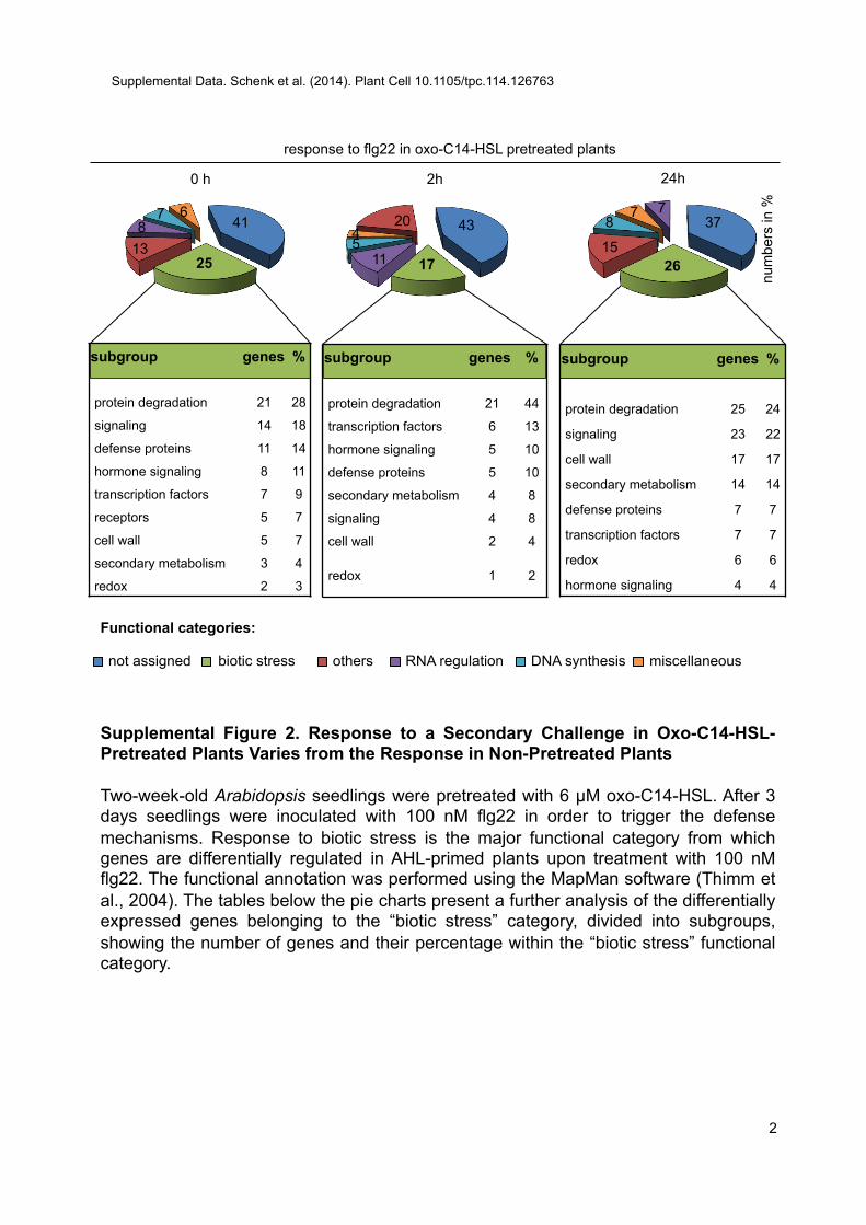
response to flg22 in oxo-C14-HSL pretreated plants
2h 24h
subgroup genes %
protein degradation 25 24
signaling 23 22
cell wall 17 17
secondary metabolism 14 14
defense proteins 7 7
transcription factors 7 7
redox 6 6
hormone signaling 4 4
subgroup genes %
protein degradation 21 44
transcription factors 6 13
hormone signaling 5 10
defense proteins 5 10
secondary metabolism 4 8
signaling 4 8
cell wall 2 4
redox 1 2
43
17 11 5 4
20 37
26 15
8 7 7
num
bers
in %
Functional categories:
subgroup genes %
protein degradation 21 28
signaling 14 18
defense proteins 11 14
hormone signaling 8 11
transcription factors 7 9
receptors 5 7
cell wall 5 7
secondary metabolism 3 4
redox 2 3
41
25 13 8
7 6
0 h
Supplemental Figure 2. Response to a Secondary Challenge in Oxo-C14-HSL-Pretreated Plants Varies from the Response in Non-Pretreated Plants Two-week-old Arabidopsis seedlings were pretreated with 6 µM oxo-C14-HSL. After 3 days seedlings were inoculated with 100 nM flg22 in order to trigger the defense mechanisms. Response to biotic stress is the major functional category from which genes are differentially regulated in AHL-primed plants upon treatment with 100 nM flg22. The functional annotation was performed using the MapMan software (Thimm et al., 2004). The tables below the pie charts present a further analysis of the differentially expressed genes belonging to the “biotic stress” category, divided into subgroups, showing the number of genes and their percentage within the “biotic stress” functional category.
not assigned biotic stress RNA regulation others DNA synthesis miscellaneous
Supplemental Data. Schenk et al. (2014). Plant Cell 10.1105/tpc.114.126763
2
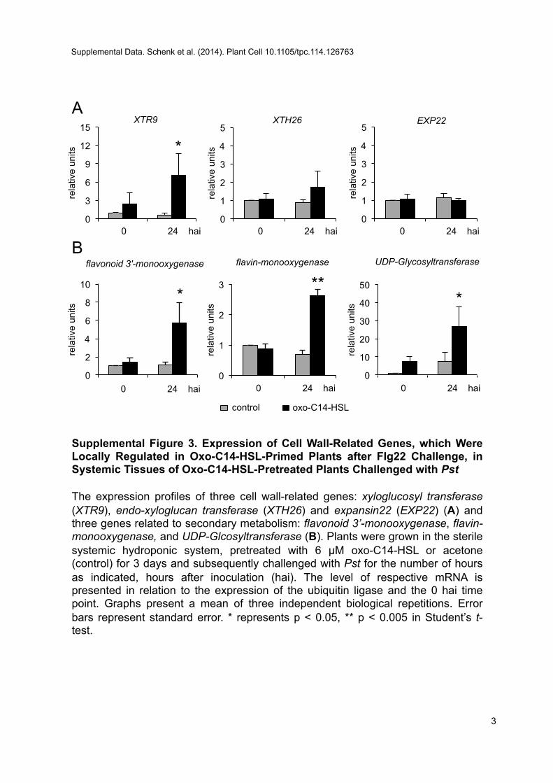
0
2
4
6
8
10
0hai 24hai
rela
tive
units
flavonoid 3'-monooxygenase
0
1
2
3
0hai 24hai
rela
tive
units
flavin-monooxygenase
0
3
6
9
12
15
0hai 24hai
rela
tive
units
XTR9
0
10
20
30
40
50
0hai 24hai
rela
tive
units
UDP-Glycosyltransferase
0
1
2
3
4
5
0hai 24hai
rela
tive
units
XTH26
0
1
2
3
4
5
0hai 24hai
rela
tive
units
EXP22
Supplemental Figure 3. Expression of Cell Wall-Related Genes, which Were Locally Regulated in Oxo-C14-HSL-Primed Plants after Flg22 Challenge, in Systemic Tissues of Oxo-C14-HSL-Pretreated Plants Challenged with Pst The expression profiles of three cell wall-related genes: xyloglucosyl transferase (XTR9), endo-xyloglucan transferase (XTH26) and expansin22 (EXP22) (A) and three genes related to secondary metabolism: flavonoid 3’-monooxygenase, flavin-monooxygenase, and UDP-Glcosyltransferase (B). Plants were grown in the sterile systemic hydroponic system, pretreated with 6 µM oxo-C14-HSL or acetone (control) for 3 days and subsequently challenged with Pst for the number of hours as indicated, hours after inoculation (hai). The level of respective mRNA is presented in relation to the expression of the ubiquitin ligase and the 0 hai time point. Graphs present a mean of three independent biological repetitions. Error bars represent standard error. * represents p < 0.05, ** p < 0.005 in Student’s t-test.
* ** *
*
A
B
control oxo-C14-HSL
0 24 hai 0 24 hai 0 24 hai
0 24 hai 0 24 hai 0 24 hai
Supplemental Data. Schenk et al. (2014). Plant Cell 10.1105/tpc.114.126763
3
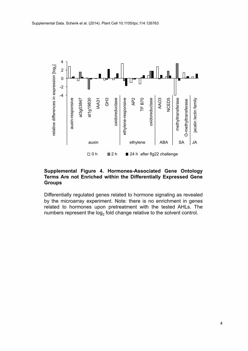
-4
-2
0
2
4
auxi
n-re
spon
sive
at3g
0384
7
at1g
1983
0
IAA
31
GH
3
oxid
ored
ucta
se
ethy
lene
-res
pons
ive
AP
2
TF B
70
oxid
ored
ucta
se
AA
O3
NC
ED
5
met
hyltr
ansf
eras
e
O-m
ethy
ltran
sfer
ase
jaca
lin le
ctin
fam
ily
auxin ethylene ABA SA JA
rela
tive
diffe
renc
es in
exp
ress
ion
[log 2
]
0h 2h 24h
Supplemental Figure 4. Hormones-Associated Gene Ontology Terms Are not Enriched within the Differentially Expressed Gene Groups Differentially regulated genes related to hormone signaling as revealed by the microarray experiment. Note: there is no enrichment in genes related to hormones upon pretreatment with the tested AHLs. The numbers represent the log2 fold change relative to the solvent control.
0 h 2 h 24 h after flg22 challenge
Supplemental Data. Schenk et al. (2014). Plant Cell 10.1105/tpc.114.126763
4
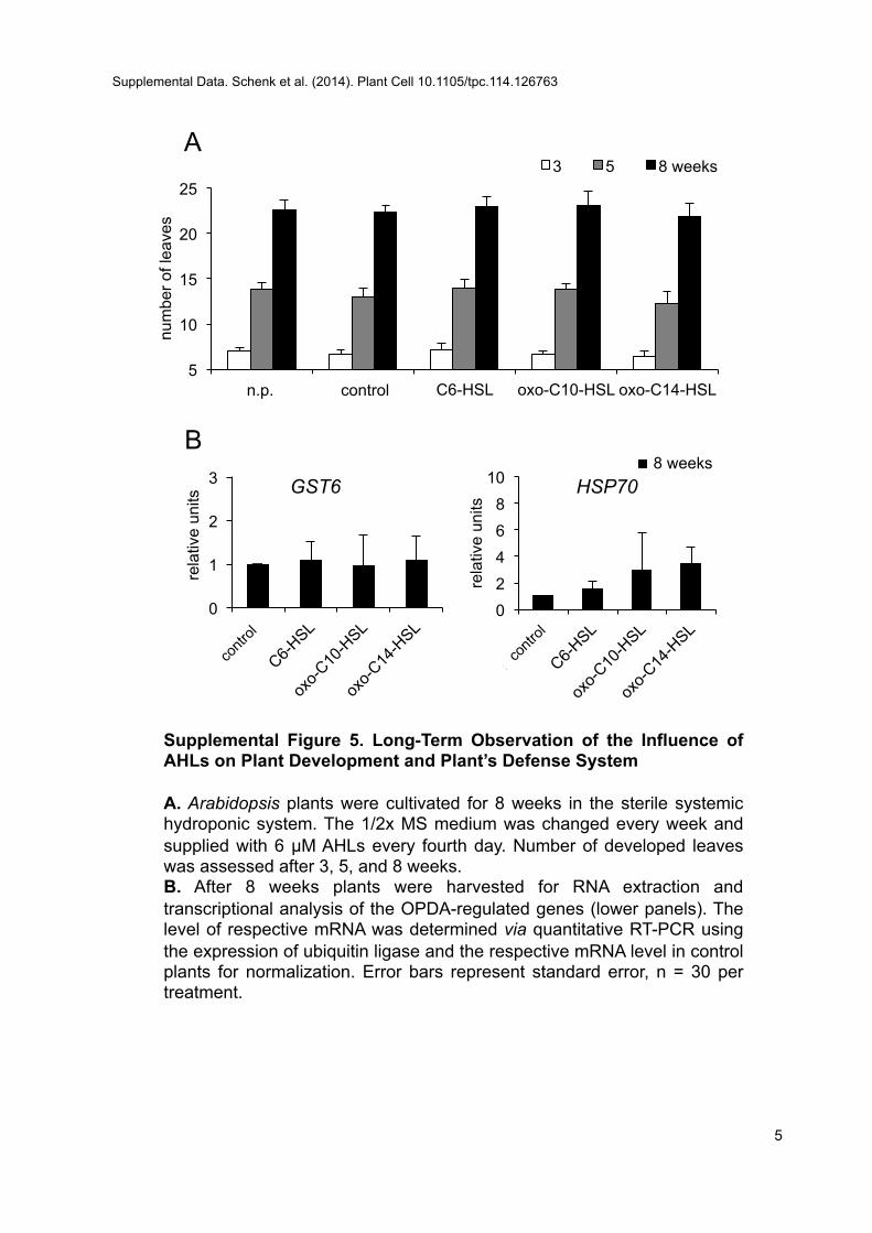
5
10
15
20
25
control acetone C6-HSL oxo-C10-HSL oxo-C14-HSL
num
ber o
f lea
ves
3 5 8 weeks
0 2 4 6 8
10 re
lativ
e un
its HSP70
0
1
2
3
rela
tive
units
GST6 8 weeks
n.p. control
Supplemental Figure 5. Long-Term Observation of the Influence of AHLs on Plant Development and Plant’s Defense System A. Arabidopsis plants were cultivated for 8 weeks in the sterile systemic hydroponic system. The 1/2x MS medium was changed every week and supplied with 6 µM AHLs every fourth day. Number of developed leaves was assessed after 3, 5, and 8 weeks. B. After 8 weeks plants were harvested for RNA extraction and transcriptional analysis of the OPDA-regulated genes (lower panels). The level of respective mRNA was determined via quantitative RT-PCR using the expression of ubiquitin ligase and the respective mRNA level in control plants for normalization. Error bars represent standard error, n = 30 per treatment.
A
B
Supplemental Data. Schenk et al. (2014). Plant Cell 10.1105/tpc.114.126763
5
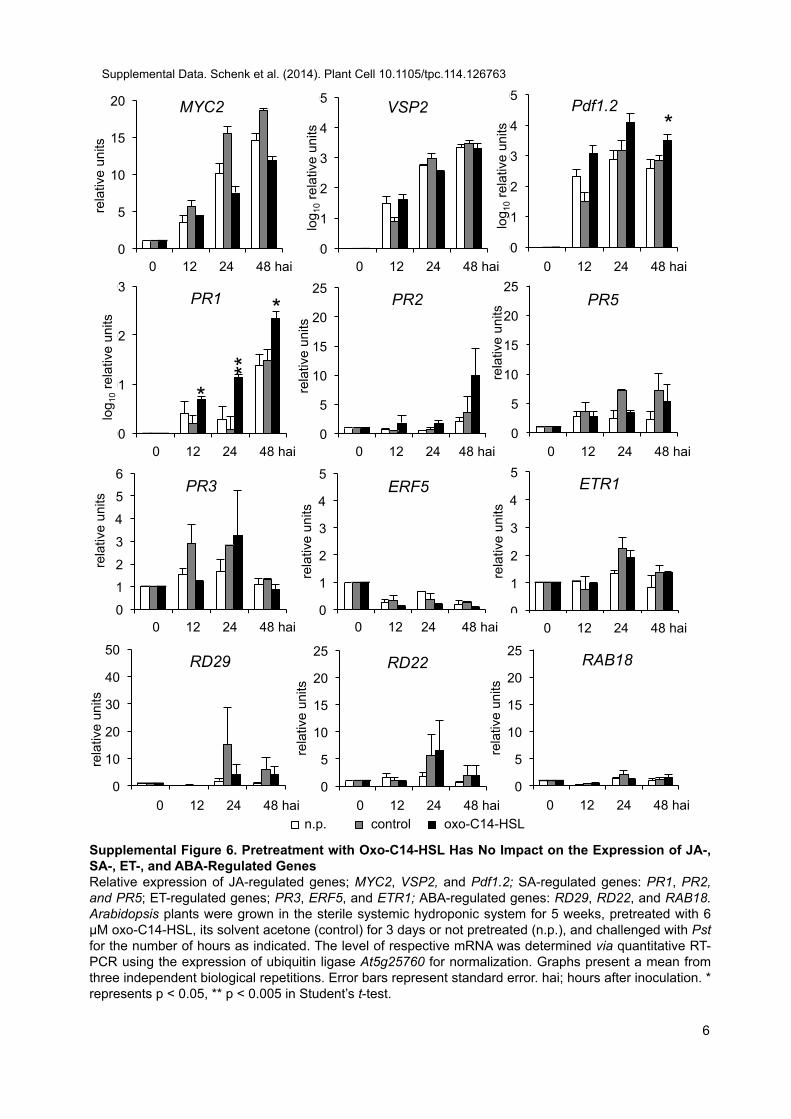
0
5
10
15
20
0hai 12hai 24hai 48hai
rela
tive
units
MYC2
0
5
10
15
20
25
0 12 24 48
rela
tive
units
PR5
1,E+00
1,E+01
1,E+02
1,E+03
1,E+04
1,E+05
0hai 12hai 24hai 48hai
Pdf1.2
log 1
0 rel
ativ
e un
its
1,E+00
1,E+01
1,E+02
1,E+03
1,E+04
1,E+05
0hai 12hai 24hai 48hai
VSP2
0
5
10
15
20
25
0 12 24 48
rela
tive
units
PR2
Supplemental Figure 6. Pretreatment with Oxo-C14-HSL Has No Impact on the Expression of JA-, SA-, ET-, and ABA-Regulated Genes Relative expression of JA-regulated genes; MYC2, VSP2, and Pdf1.2; SA-regulated genes: PR1, PR2, and PR5; ET-regulated genes; PR3, ERF5, and ETR1; ABA-regulated genes: RD29, RD22, and RAB18. Arabidopsis plants were grown in the sterile systemic hydroponic system for 5 weeks, pretreated with 6 µM oxo-C14-HSL, its solvent acetone (control) for 3 days or not pretreated (n.p.), and challenged with Pst for the number of hours as indicated. The level of respective mRNA was determined via quantitative RT-PCR using the expression of ubiquitin ligase At5g25760 for normalization. Graphs present a mean from three independent biological repetitions. Error bars represent standard error. hai; hours after inoculation. * represents p < 0.05, ** p < 0.005 in Student’s t-test.
0
1
2
3
4
5
0 12 24 48
rela
tive
units
ERF5
0
1
2
3
4
5
6
0 12 24 48
rela
tive
units
PR3
1,E+00
1,E+01
1,E+02
1,E+03
0hai 12hai 24hai 48hai
PR1
log 1
0 rel
ativ
e un
its
*
*
**
*
0 12 24 48 hai 0 12 24 48 hai 0 12 24 48 hai
log 1
0 rel
ativ
e un
its
0 12 24 48 hai 0
1
2
3
4
5
0 12 24 48
rela
tive
units
ETR1
0 12 24 48 hai 0 12 24 48 hai 0 12 24 48 hai
n.p. control oxo-C14-HSL
0
10
20
30
40
50
0hai 12hai 24hai 48hai
rela
tive
units
RD29
0
5
10
15
20
25
0 12 24 48
rela
tive
units
RAB18
0
5
10
15
20
25
0 12 24 48
rela
tive
units
RD22
0 12 24 48 hai 0 12 24 48 hai 0 12 24 48 hai
0 12 24 48 hai 0 12 24 48 hai
Supplemental Data. Schenk et al. (2014). Plant Cell 10.1105/tpc.114.126763
6
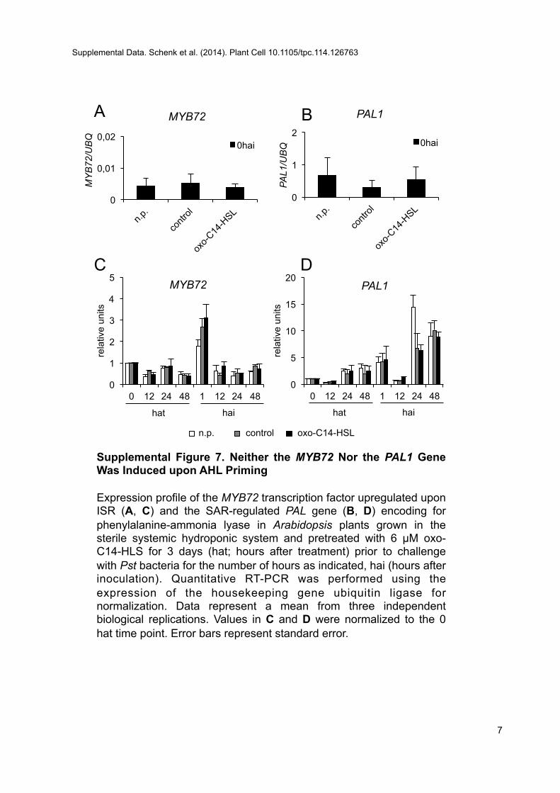
A B
0
0,01
0,02
MY
B72
/UB
Q
MYB72
0hai
C D
0
1
2
PAL1
/UB
Q
PAL1
0hai
Supplemental Figure 7. Neither the MYB72 Nor the PAL1 Gene Was Induced upon AHL Priming Expression profile of the MYB72 transcription factor upregulated upon ISR (A, C) and the SAR-regulated PAL gene (B, D) encoding for phenylalanine-ammonia lyase in Arabidopsis plants grown in the sterile systemic hydroponic system and pretreated with 6 µM oxo-C14-HLS for 3 days (hat; hours after treatment) prior to challenge with Pst bacteria for the number of hours as indicated, hai (hours after inoculation). Quantitative RT-PCR was performed using the expression of the housekeeping gene ubiquitin ligase for normalization. Data represent a mean from three independent biological replications. Values in C and D were normalized to the 0 hat time point. Error bars represent standard error.
0
1
2
3
4
5
0 12 24 48 1 12 24 48
rela
tive
units
MYB72
0
5
10
15
20
0 12 24 48 1 12 24 48
rela
tive
units
PAL1
n.p. control oxo-C14-HSL
hat hai hat hai
Supplemental Data. Schenk et al. (2014). Plant Cell 10.1105/tpc.114.126763
7
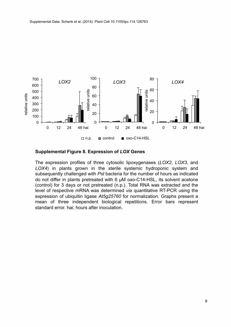
0
20
40
60
80
0 12 24 48
rela
tive
units
LOX4
0
20
40
60
80
100
0 12 24 48
rela
tive
units
LOX3
0 100 200 300 400 500 600 700
0hai 12hai 24hai 48hai
rela
tive
units
LOX2
Supplemental Figure 8. Expression of LOX Genes The expression profiles of three cytosolic lipoxygenases (LOX2, LOX3, and LOX4) in plants grown in the sterile systemic hydroponic system and subsequently challenged with Pst bacteria for the number of hours as indicated do not differ in plants pretreated with 6 µM oxo-C14-HSL, its solvent acetone (control) for 3 days or not pretreated (n.p.). Total RNA was extracted and the level of respective mRNA was determined via quantitative RT-PCR using the expression of ubiquitin ligase At5g25760 for normalization. Graphs present a mean of three independent biological repetitions. Error bars represent standard error. hai; hours after inoculation.
0 12 24 48 hai
n.p. control oxo-C14-HSL
0 12 24 48 hai 0 12 24 48 hai
Supplemental Data. Schenk et al. (2014). Plant Cell 10.1105/tpc.114.126763
8
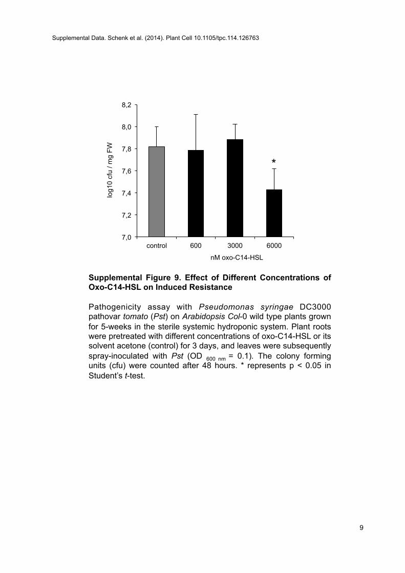
Supplemental Figure 9. Effect of Different Concentrations of Oxo-C14-HSL on Induced Resistance Pathogenicity assay with Pseudomonas syringae DC3000 pathovar tomato (Pst) on Arabidopsis Col-0 wild type plants grown for 5-weeks in the sterile systemic hydroponic system. Plant roots were pretreated with different concentrations of oxo-C14-HSL or its solvent acetone (control) for 3 days, and leaves were subsequently spray-inoculated with Pst (OD 600 nm = 0.1). The colony forming units (cfu) were counted after 48 hours. * represents p < 0.05 in Student’s t-test.
7,0
7,2
7,4
7,6
7,8
8,0
8,2
control 600 3000 6000
log1
0 cf
u / m
g FW
nM oxo-C14-HSL
*
Supplemental Data. Schenk et al. (2014). Plant Cell 10.1105/tpc.114.126763
9
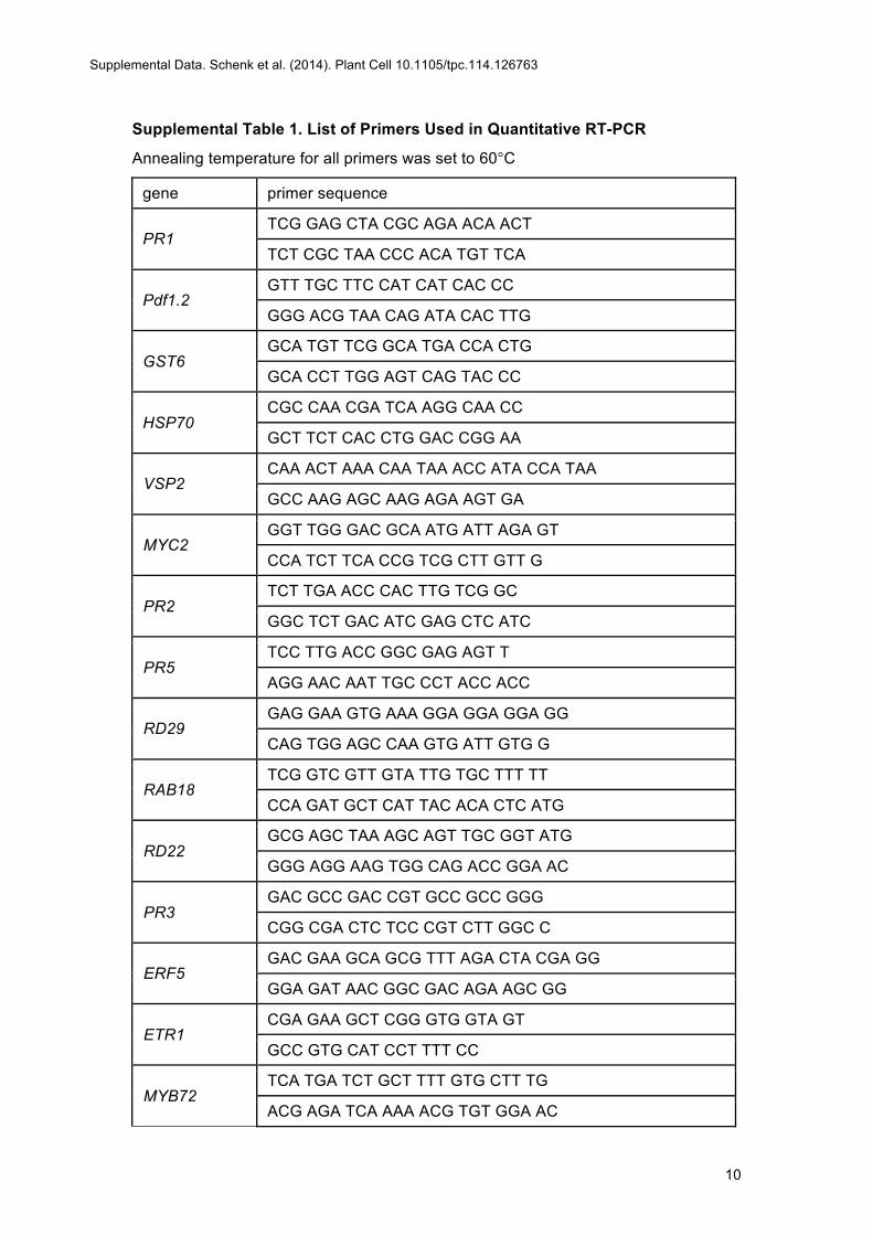
10
Supplemental Table 1. List of Primers Used in Quantitative RT-PCR
Annealing temperature for all primers was set to 60°C
gene primer sequence
PR1 TCG GAG CTA CGC AGA ACA ACT
TCT CGC TAA CCC ACA TGT TCA
Pdf1.2 GTT TGC TTC CAT CAT CAC CC
GGG ACG TAA CAG ATA CAC TTG
GST6 GCA TGT TCG GCA TGA CCA CTG
GCA CCT TGG AGT CAG TAC CC
HSP70 CGC CAA CGA TCA AGG CAA CC
GCT TCT CAC CTG GAC CGG AA
VSP2 CAA ACT AAA CAA TAA ACC ATA CCA TAA
GCC AAG AGC AAG AGA AGT GA
MYC2 GGT TGG GAC GCA ATG ATT AGA GT
CCA TCT TCA CCG TCG CTT GTT G
PR2 TCT TGA ACC CAC TTG TCG GC
GGC TCT GAC ATC GAG CTC ATC
PR5 TCC TTG ACC GGC GAG AGT T
AGG AAC AAT TGC CCT ACC ACC
RD29 GAG GAA GTG AAA GGA GGA GGA GG
CAG TGG AGC CAA GTG ATT GTG G
RAB18 TCG GTC GTT GTA TTG TGC TTT TT
CCA GAT GCT CAT TAC ACA CTC ATG
RD22 GCG AGC TAA AGC AGT TGC GGT ATG
GGG AGG AAG TGG CAG ACC GGA AC
PR3 GAC GCC GAC CGT GCC GCC GGG
CGG CGA CTC TCC CGT CTT GGC C
ERF5 GAC GAA GCA GCG TTT AGA CTA CGA GG
GGA GAT AAC GGC GAC AGA AGC GG
ETR1 CGA GAA GCT CGG GTG GTA GT
GCC GTG CAT CCT TTT CC
MYB72 TCA TGA TCT GCT TTT GTG CTT TG
ACG AGA TCA AAA ACG TGT GGA AC
Supplemental Data. Schenk et al. (2014). Plant Cell 10.1105/tpc.114.126763
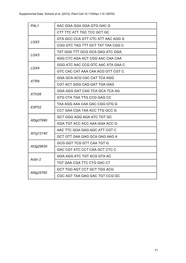
11
PAL1 AAC GGA GGA GGA GTG GAC G
CTT TTC ATT TGC TCC GCT GC
LOX2 GTA GCC CCA GTT CTC ATT AAC AGG G
CGG GTC TAG TTT GCT TAT TAA CGG C
LOX3 TAT GGA TTT GCG GCA GAG ATC GGA
AGG CTC AGA ACT CGG AAC CAA CAA
LOX4 GGG ATC AAC CCG GTC AAC ATA GAA C
GTC CAC CAT AAA CAA ACG GTT CGT C
XTR9 GGA GCA ACG CAC CAT TCA AGG
CGT ACT GGG CAG GAT TGA GAG
XTH26 GGA AGG GAT CAG TCA GCA TCA AG
GTG CTA TGA TTG CCG GAG CC
EXP22 TAA AGG AAA CAA GAC CGG GTG G
CCT GAA CGA TAA ACC TTG GCC G
At5g07990 GCT GGG AGG AGA ATC TGT GC
GGA TGT ACC ACC AAA GGA ACC G
At1g12140 AAC TTC GGA GAG AGC ATT CGT C
GCT GTT GAA GAG GCA GAG AAG A
At3g29630 GCG GGT TCG GTT CAA TGT G
GAC CGT ATC CCT CAA GCT CTC C
Actin 2 GGA AGG ATC TGT ACG GTA AC
TGT GAA CGA TTC CTG GAC CT
At5g25760 GCT TGG AGT CCT GCT TGG ACG
CGC AGT TAA GAG GAC TGT CCG GC
Supplemental Data. Schenk et al. (2014). Plant Cell 10.1105/tpc.114.126763
DOI 10.1105/tpc.114.126763; originally published online June 24, 2014; 2014;26;2708-2723Plant Cell
Schikora, Michael Reichelt, Axel Mithöfer, Annette Becker, Karl-Heinz Kogel and Adam SchikoraSebastian T. Schenk, Casandra Hernández-Reyes, Birgit Samans, Elke Stein, Christina Neumann, Marek
Bacterial Pathogens via the Salicylic Acid/Oxylipin Pathway-Acyl-Homoserine Lactone Primes Plants for Cell Wall Reinforcement and Induces Resistance toN
This information is current as of August 4, 2014
Supplemental Data http://www.plantcell.org/content/suppl/2014/06/16/tpc.114.126763.DC1.html
References http://www.plantcell.org/content/26/6/2708.full.html#ref-list-1
This article cites 67 articles, 29 of which can be accessed free at:
Permissions https://www.copyright.com/ccc/openurl.do?sid=pd_hw1532298X&issn=1532298X&WT.mc_id=pd_hw1532298X
eTOCs http://www.plantcell.org/cgi/alerts/ctmain
Sign up for eTOCs at:
CiteTrack Alerts http://www.plantcell.org/cgi/alerts/ctmain
Sign up for CiteTrack Alerts at:
Subscription Information http://www.aspb.org/publications/subscriptions.cfm
is available at:Plant Physiology and The Plant CellSubscription Information for
ADVANCING THE SCIENCE OF PLANT BIOLOGY © American Society of Plant Biologists